involvement of sirt7 in resumption of rdna …1institut jacques monod, umr 7592 cnrs/universités...
TRANSCRIPT
489Research Article
IntroductionThe nucleolus is the nuclear compartment in which ribosomebiogenesis takes place, i.e. ribosomal gene (rDNA) transcription,precursor ribosomal RNA (rRNA) processing and assembly ofmature rRNAs with ribosomal proteins (Boisvert et al., 2007;Carmo-Fonseca et al., 2000; Hadjiolov, 1985). rDNA transcriptionis subject to extensive modulation during cell growth and/or cellcycle. In mammalian cells the correlation between cell cycle andrRNA synthesis is well established. Up- or downregulation of rRNAsynthesis, which can be reversed in appropriate cases, correlateswith situations such as nutrient starvation, hormone or drugtreatment, aging, tumor and viral infections (Grummt, 2003; Jacoband Ghosh, 1999; Ruggero and Pandolfi, 2003). It is now becomingclear that several signaling pathways are involved in the regulationof rDNA transcription (Kim et al., 2003; Russell and Zomerdijk,2005; Schmelzle and Hall, 2000; Stefanovsky et al., 2001).
A crucial event in nucleolar activity is the resumption of rDNAtranscription at the exit from mitosis. In higher eukaryotic cells,rDNA transcription is repressed from late prophase to telophase(Prescott and Bender, 1962; Roussel et al., 1996), and consequentlynucleoli are no longer maintained. Even if recent analyses performedin living cells showed that some subunits of RNA polymerase I(Pol I) are absent at nucleolar organizer regions (NORs) frommetaphase to anaphase (Chen et al., 2005; Leung et al., 2004), theRNA Pol I machinery, as observed for RNA Pol I (Gilbert et al.,1995; Scheer and Rose, 1984), the promoter selectivity factor (SL1),the upstream binding factor (UBF) (Jordan et al., 1996; Roussel etal., 1996) and the transcription termination factor (TTF-1) (Sirri etal., 1999), remains mainly associated in an inactive state at NORs,i.e. in chromosomal sites where rDNAs are clustered.Phosphorylation of components of the RNA Pol I machinery by the
cyclin-dependent kinase (CDK) 1-cyclin B pathway is responsiblefor the repression of rDNA transcription during mitosis, anddephosphorylation of these components resumes rDNA transcriptionat the exit from mitosis (Heix et al., 1998; Kuhn et al., 1998; Sirriet al., 2000). However, other players most probably participate inrestoring rDNA transcription in late telophase.
Mammalian sirtuins (SIRT1-7), homologs of the yeast Sir2, haverecently been proposed to be involved in the control of criticalmetabolic pathways as well as apoptosis, stress responses, DNArepair, cell cycle, genomic stability and gene expression. Sirtuins,also designated class III histone deacetylases, are proteindeacetylases/ADP ribosyltransferases (Blander and Guarente, 2004).These enzymes are highly conserved from prokaryotes toeukaryotes. They all share a conserved NAD-dependent catalyticcore domain, and exhibit variable N-terminal and C-terminalextensions that contribute to their unique subcellular localizationand may also regulate their catalytic activity. The subcellulardistribution, substrate specificity and cellular function of sirtuinsare quite diverse (for reviews, see Denu, 2005; Guarente, 2000;North and Verdin, 2004). SIRT2 is a predominantly cytoplasmicprotein (Dryden et al., 2003; Michishita et al., 2005; North et al.,2003), SIRT3-5 are mitochondrial (Michishita et al., 2005) andSIRT1, -6 and -7 are localized in the nucleus. SIRT1, the mostclosely related to yeast Sir2 and the best characterized sirtuin,possesses a large number of substrates, including p53, Ku70, NF-κB and forkhead transcription factors, that regulate cellular oxidativeand genotoxic stresses (Brunet et al., 2004; Cohen et al., 2004; Luoet al., 2001; Motta et al., 2004; Vaziri et al., 2001; Yeung et al.,2004). SIRT6 is involved in important functions in preserving cellsfrom genomic instability and progeroid phenotype (Mostoslavskyet al., 2006). Moreover, SIRT6 is the only sirtuin to exhibit a robust
Sirtuins, also designated class III histone deacetylases, areimplicated in the regulation of cell division, apoptosis, DNAdamage repair, genomic silencing and longevity. The nucleolarSirtuin7 (SIRT7) was reported to be involved in the regulationof ribosomal gene (rDNA) transcription, but there are no dataconcerning the regulation of SIRT7 during the cell cycle. Herewe have analyzed the behavior of endogenous SIRT7 duringmitosis, while rDNA transcription is repressed. SIRT7 remainsassociated with nucleolar organizer regions, as does the RNApolymerase I machinery. SIRT7 directly interacts with therDNA transcription factor UBF. Moreover, SIRT7 isphosphorylated via the CDK1-cyclin B pathway during mitosisand dephosphorylated by a phosphatase sensitive to okadaic
acid at the exit from mitosis before onset of rDNA transcription.Interestingly, dephosphorylation events induce a conformationalmodification of the carboxy-terminal region of SIRT7 beforethe release of mitotic repression of rDNA transcription. AsSIRT7 activity is required to resume rDNA transcription intelophase, we propose that this conformational modificationregulates onset of rDNA transcription.
Supplementary material available online athttp://jcs.biologists.org/cgi/content/full/122/4/489/DC1
Key words: Sirtuins, Nucleolus, Cell cycle, NOR, CDK1
Summary
Involvement of SIRT7 in resumption of rDNAtranscription at the exit from mitosisAlice Grob1, Pascal Roussel1, Jane E. Wright2, Brian McStay2, Danièle Hernandez-Verdun1 andValentina Sirri1,*1Institut Jacques Monod, UMR 7592 CNRS/Universités Paris 6 et 7, 2 Place Jussieu, 75251 Paris Cedex 05, France2Biomedical Research Center, Ninewells Hospital and Medical School, University of Dundee, Dundee DD1 9SY, Scotland, UK*Author for correspondence (e-mail: [email protected])
Accepted 14 October 2008Journal of Cell Science 122, 489-498 Published by The Company of Biologists 2009doi:10.1242/jcs.042382
Jour
nal o
f Cel
l Sci
ence
490
auto-ADP-ribosyltransferase activity (Liszt et al., 2005). SIRT7 isthe only sirtuin localized in nucleoli (Ford et al., 2006; Michishitaet al., 2005). It was shown to exhibit no deacetylase or ADP-ribosyltransferase activity when tested on acetylated histones andvarious acetylated components of the RNA Pol I machinery (Fordet al., 2006). Concerning the nucleolar function of SIRT7, Ford etal. (Ford et al., 2006) proposed that SIRT7 could be a positiveregulator of rDNA transcription via its association with RNA PolI. Its overexpression enhances rDNA transcription, whereas itsinhibition reduces rDNA transcription. Interestingly, expression ofSIRT7 is positively correlated with cell growth: SIRT7 is abundantin metabolically active tissues such as liver, spleen and testes (Fordet al., 2006; Michishita et al., 2005). To date, there are no dataconcerning the cell cycle regulation of SIRT7 and its fate duringmitosis when rDNA transcription is repressed.
In the present study, we investigated the behavior of endogenousSIRT7 during mitosis and the involvement of SIRT7 at the onsetof rDNA transcription as cells exit mitosis. By confocal microscopyanalyses we showed that SIRT7 remains at NORs during mitosis,and consequently, as previously established for several RNA Pol Isubunits, UBF, SL1 and TTF-1, SIRT7 is not released from rDNAsduring mitotic repression of rDNA transcription. Glutathione S-transferase (GST) pull-down experiments showed that SIRT7directly interacts with the rDNA transcription factor UBF. Moreover,we demonstrate that SIRT7 is phosphorylated during mitosis andthat its phosphorylation is governed by the CDK1-cyclin B pathway.Interestingly, mitotic phosphorylation of SIRT7 decreases thereactivity of antibodies directed against its C-terminal region fromprophase to anaphase. Conversely, inhibition of the CDK1-cyclinB pathway in late mitosis and consequently dephosphorylation ofSIRT7 via an okadaic acid sensitive phosphatase increase thereactivity of antibodies to its C-terminal region. Moreover, inhibitionof SIRT7 by sirtinol in late mitosis demonstrates that SIRT7 activityis required for onset of rDNA transcription at the exit from mitosis.We propose a model in which SIRT7 remains at NORs duringmitosis in a phosphorylated inactive form and undergoes adephosphorylation-dependent conformational rearrangement intelophase that precedes the resumption of rDNA transcription.
ResultsEndogenous SIRT7 localizes in rDNA transcription sites duringinterphase and remains associated with NORs during mitosisvia interaction with UBFTo begin assessing the biological function of SIRT7, weinvestigated its cellular localization by immunofluorescenceconfocal microscopy. We first analyzed in interphase HeLa cellsthe localization of endogenous SIRT7 together with that of UBFor with the rDNA transcription sites revealed by bromouridine 5�-triphosphate (BrUTP) incorporation. The superimposition oflabelings revealed co-localization of SIRT7 and UBF (Fig. 1Aa-d) at the level of the rDNA transcription sites (Fig. 1Ae-h).Knockdown of SIRT7 by siRNAs eliminated the nucleolar signal(Fig. 1Bc) demonstrating the specificity of the anti-SIRT7-C-terminal antibodies raised against a peptide located in the C-terminal region (amino acids 384-400) of SIRT7. SIRT7 is theonly sirtuin to present an arginine-rich domain (amino acids 1-74) that is involved in its nucleolar localization (supplementarymaterial Fig. S1). In contrast to endogenous SIRT7, the exogenousC-terminal GFP-tagged SIRT7 (GFP-SIRT7) showed a morediffuse nucleolar localization (supplementary material Fig. S2Aa-d). Moreover, in HeLa cells treated with actinomycin D (AMD),
which inhibits rDNA transcription and consequently inducesnucleolar reorganization (Hadjiolov, 1985), the GFP-SIRT7 fusionwas largely excluded from the caps containing UBF(supplementary material Fig. S2Ae-h), whereas the endogenousSIRT7 remained co-localized with UBF in these caps(supplementary material Fig. S2Ai-l).
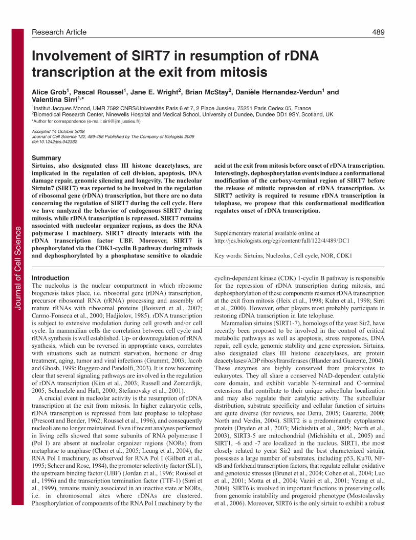
It was previously reported that GFP-SIRT7 remains associatedwith condensed chromosomes in mitotic living human cells (Fordet al., 2006; Michishita et al., 2005). The association of GFP-SIRT7with metaphase chromosomes appears diffuse rather than NOR-specific. Because in interphase cells endogenous and exogenousSIRT7 did not exhibit the same localization, we verified thelocation of endogenous SIRT7 during mitosis. Using anti-SIRT7-C-terminal antibodies for immunofluorescence confocalmicroscopy, we compared endogenous SIRT7 to UBF known toremain associated with NORs throughout mitosis (Roussel et al.,1996). In mitotic cells, SIRT7 detected as dots during prophase,metaphase, anaphase and telophase co-localized with UBF (Fig.1Ca-d,e-h). Therefore in contrast to GFP-SIRT7, endogenousSIRT7 remains associated with NORs during mitosis, as docomponents of the RNA Pol I machinery. Interestingly, thecomparison between the UBF and SIRT7 signals showed that theintensity of the UBF signal was similar whatever the mitotic stage,whereas that of SIRT7 was weak in early mitosis (Fig. 1Ca-d) andhigh in late mitosis (Fig. 1Ce-h). The yellow signals in merge imagesrepresent co-localization of equivalent red and green labelings(compare Fig. 1Cd,h). The quantification of UBF and SIRT7 signalsindicated that the SIRT7 signal was about almost 25% that of UBFduring early stages of mitosis (Fig. 1D). The SIRT7 signal increasedduring telophase and became comparable to that of UBF at latetelophase. Thus, endogenous SIRT7 remains associated with theRNA Pol I machinery during mitosis, yet its labeling signalincreases at the exit from mitosis.
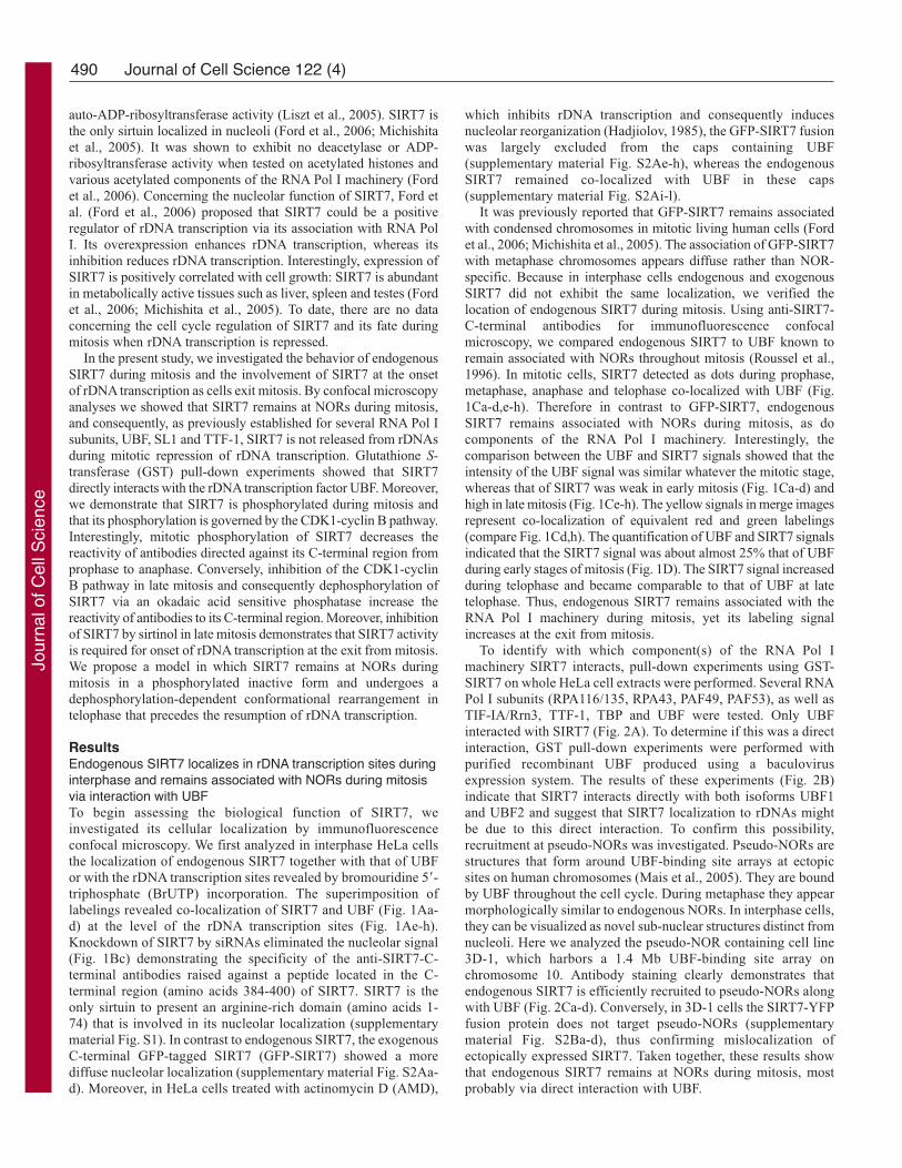
To identify with which component(s) of the RNA Pol Imachinery SIRT7 interacts, pull-down experiments using GST-SIRT7 on whole HeLa cell extracts were performed. Several RNAPol I subunits (RPA116/135, RPA43, PAF49, PAF53), as well asTIF-IA/Rrn3, TTF-1, TBP and UBF were tested. Only UBFinteracted with SIRT7 (Fig. 2A). To determine if this was a directinteraction, GST pull-down experiments were performed withpurified recombinant UBF produced using a baculovirusexpression system. The results of these experiments (Fig. 2B)indicate that SIRT7 interacts directly with both isoforms UBF1and UBF2 and suggest that SIRT7 localization to rDNAs mightbe due to this direct interaction. To confirm this possibility,recruitment at pseudo-NORs was investigated. Pseudo-NORs arestructures that form around UBF-binding site arrays at ectopicsites on human chromosomes (Mais et al., 2005). They are boundby UBF throughout the cell cycle. During metaphase they appearmorphologically similar to endogenous NORs. In interphase cells,they can be visualized as novel sub-nuclear structures distinct fromnucleoli. Here we analyzed the pseudo-NOR containing cell line3D-1, which harbors a 1.4 Mb UBF-binding site array onchromosome 10. Antibody staining clearly demonstrates thatendogenous SIRT7 is efficiently recruited to pseudo-NORs alongwith UBF (Fig. 2Ca-d). Conversely, in 3D-1 cells the SIRT7-YFPfusion protein does not target pseudo-NORs (supplementarymaterial Fig. S2Ba-d), thus confirming mislocalization ofectopically expressed SIRT7. Taken together, these results showthat endogenous SIRT7 remains at NORs during mitosis, mostprobably via direct interaction with UBF.
Journal of Cell Science 122 (4)
Jour
nal o
f Cel
l Sci
ence
491SIRT7 during mitosis
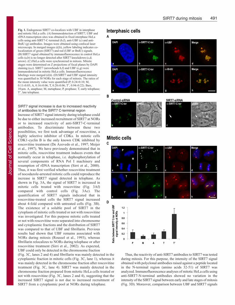
SIRT7 signal increase is due to increased reactivityof antibodies to the SIRT7 C-terminal regionIncrease of SIRT7 signal intensity during telophase couldbe due to either increased recruitment of SIRT7 at NORsor to increased reactivity of anti-SIRT7-C-terminalantibodies. To discriminate between these twopossibilities, we first took advantage of roscovitine, ahighly selective inhibitor of CDKs. In mitotic cellsCDK1-cyclin B is the only known CDK inhibited byroscovitine treatment (De Azevedo et al., 1997; Meijeret al., 1997). We have previously demonstrated that inmitotic cells, roscovitine treatment induces events thatnormally occur in telophase, i.e. dephosphorylation ofseveral components of RNA Pol I machinery andresumption of rDNA transcription (Sirri et al., 2000).Thus, it was first verified whether roscovitine treatmentof nocodazole-arrested mitotic cells could reproduce theincrease in SIRT7 signal detected in telophase. Asshown in Fig. 3A, the signal of SIRT7 is increased inmitotic cells treated with roscovitine (Fig. 3Af)compared with control cells (Fig. 3Ac). Thequantification of SIRT7 signals indicated that inroscovitine-treated cells the SIRT7 signal increasedabout 4-fold compared with untreated cells (Fig. 3B).The existence of a soluble pool of SIRT7 in thecytoplasm of mitotic cells treated or not with roscovitinewas investigated. For this purpose mitotic cells treatedor not with roscovitine were separated into chromosomeand cytoplasmic fractions and the distribution of SIRT7was compared to that of UBF and fibrillarin. Previousresults had shown that UBF remains associated withNORs during mitosis (Roussel et al., 1993), whereasfibrillarin relocalizes to NORs during telophase or afterroscovitine treatment (Sirri et al., 2002). As expected,UBF could only be detected in the chromosome fraction(Fig. 3C, lanes 2 and 4) and fibrillarin was mainly detected in thecytoplasmic fraction in mitotic cells (Fig. 3C, lane 1), whereas itwas mainly detected in the chromosome fraction after roscovitinetreatment (Fig. 3C, lane 4). SIRT7 was mainly detected in thechromosome fraction prepared from mitotic HeLa cells treated ornot with roscovitine (Fig. 3C, lanes 2 and 4), suggesting that theincreased SIRT7 signal is not due to increased recruitment ofSIRT7 from a cytoplasmic pool at NORs during telophase.
Thus, the reactivity of anti-SIRT7 antibodies to SIRT7 was testedduring mitosis. For this purpose, the intensity of the SIRT7 signalobtained with polyclonal antibodies raised against a peptide locatedin the N-terminal region (amino acids 32-51) of SIRT7 wasanalyzed. Immunofluorescence analyses of mitotic HeLa cells usinganti-SIRT7-N-terminal antibodies showed no variation in theintensity of the SIRT7 signal between early and late stages of mitosis(Fig. 3D). Moreover, comparison between UBF and SIRT7 signals
Fig. 1. Endogenous SIRT7 co-localizes with UBF in interphaseand mitotic HeLa cells. (A) Immunodetection of SIRT7, UBF andrDNA transcription sites was obtained in fixed interphase HeLacells using anti-SIRT7-C-terminal (b,f), anti-UBF (c) and anti-BrdU (g) antibodies. Images were obtained using confocal lasermicroscopy. In merged images (d,h), yellow labeling indicates co-localization of green (SIRT7) and red (UBF or BrdU) signals.(B) SIRT7 signal obtained by immunofluorescence in control HeLacells (a,b) is no longer detected after SIRT7 knockdown (c,d;arrow). (C) HeLa cells were synchronized in mitosis. Mitoticstages were determined on Z projections of focal planes by DAPIstaining (a,e). SIRT7 (arrowheads b,f) and UBF (c,g) wereimmunodetected in mitotic HeLa cells. Immunofluorescencelabelings were merged (d,h). (D) SIRT7 and UBF signal intensitywas quantified in 30 NORs for each stage of mitosis. The ratios ofthe mean intensity value were quantified (P, 0.24±0.10; M,0.11±0.03; A, 0.16±0.06; T, 0.28±0.06; T’, 0.94±0.22). Bars,10 μm. A, anaphase; M, metaphase; P, prophase; T, early telophase;T’, late telophase.
Jour
nal o
f Cel
l Sci
ence
492
showed equivalent red and green labelings throughout mitosis (Fig.3D). Taken together, these results indicate that SIRT7 remainsmainly localized at NORs during mitosis, but that the reactivity ofantibodies to its C-terminal region varies, i.e. weak in early stagesand high in late stages of mitosis.
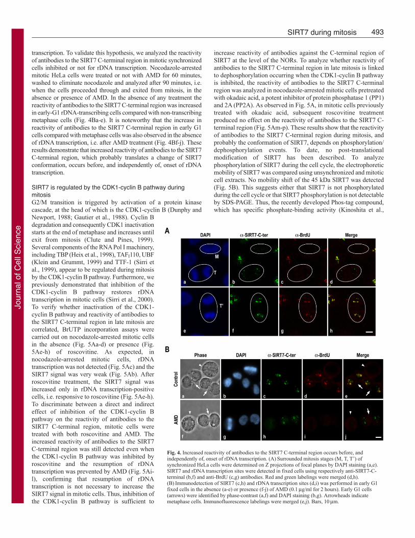
Increased reactivity of antibodies to the SIRT7 C-terminalregion occurs before, and independently of, onset of rDNAtranscriptionAs in cycling cells rDNA transcription is arrested at the beginningof mitosis and restored at telophase (Roussel et al., 1996), we
investigated whether the reactivity of antibodies to the SIRT7 C-terminal region at telophase is linked to the onset of rDNAtranscription in HeLa cells. As expected, rDNA transcription, revealedby BrUTP incorporation, was detected in interphase cells and in latetelophase (Fig. 4Ac,g) but not in metaphase and early telophase (Fig.4Ac). As for SIRT7, its increased signal was already detected in earlytelophase (Fig. 4Ab) when rDNA transcription was not yet detectable(Fig. 4Ac). The SIRT7 signal was maximum in late telophase andco-localized with rDNA transcription sites (Fig. 4Ae-h). Theseresults suggest that the reactivity of antibodies to the SIRT7 C-terminalregion at NORs in late mitosis does not depend on the onset of rDNA
Journal of Cell Science 122 (4)
Fig. 2. SIRT7 is recruited to pseudo-NORs via directinteraction with UBF. (A) GST and GST-SIRT7 pull-down assays were performed using whole HeLa cellextracts. 1% of extract used for each pull-down assay(Input), 5% of the proteins eluted after GST-SIRT7pull-down assays and 5% of the proteins eluted afterGST pull-down assays were submitted to 10% SDS-PAGE. Immunoblotting was performed using anti-PAF49, anti-PAF53, anti-RPA116/135, anti-RPA43,anti-TIF-1A/Rrn3, anti-UBF, anti-TTF-1 and anti-TBP antibodies. (B) GST and GST-SIRT7 pull-downassays were performed using 1 μg purified humanUBF1 or UBF2. (C) The pseudo-NOR containing cellline 3D-1 was stained with anti-SIRT7-C-terminaland anti-UBF antibodies (a-d). Red and greenlabelings were merged (d). Pseudo-NORs areidentified by arrowheads. Bar, 10 μm.
Fig. 3. Increase in SIRT7 signal corresponds toincreased reactivity of antibodies to the SIRT7 C-terminal region. (A) In nocodazole-arrested HeLacells treated (d-f) or not (a-c) with roscovitine(150 μM for 1 hour), SIRT7 and UBF wereimmunodetected using anti-SIRT7-C-terminal(c,f) and anti-UBF (b,e) antibodies. DNA wasstained by DAPI and merged with red and greenlabelings (a,d). (B) SIRT7 signal intensity wasquantified in 10 cells. The means of intensityvalue were quantified (Control: 0.25±0.06,Roscovitine: 0.83±0.15). (C) Cytoplasmic (lanes1 and 3) and chromosome (lanes 2 and 4)fractions prepared from nocodazole-arrestedHeLa cells treated (lanes 3 and 4) or not (lanes 1and 2) with roscovitine were submitted to 10%SDS-PAGE. Immunoblotting was performedusing anti-UBF, anti-SIRT7-N-terminal and anti-fibrillarin antibodies. (D) HeLa cells weresynchronized in mitosis and mitotic stages weredetermined by DAPI staining (a,d,g). Inimmunofluorescence assays, SIRT7 and UBFwere detected using anti-SIRT7-N-terminal (c,f,i)and anti-UBF (b,e,h) antibodies, respectively.Immunofluorescence labelings were merged withDAPI (a,d,g). Bars, 10 μm.
Jour
nal o
f Cel
l Sci
ence
493SIRT7 during mitosis
transcription. To validate this hypothesis, we analyzed the reactivityof antibodies to the SIRT7 C-terminal region in mitotic synchronizedcells inhibited or not for rDNA transcription. Nocodazole-arrestedmitotic HeLa cells were treated or not with AMD for 60 minutes,washed to eliminate nocodazole and analyzed after 90 minutes, i.e.when the cells proceeded through and exited from mitosis, in theabsence or presence of AMD. In the absence of any treatment thereactivity of antibodies to the SIRT7 C-terminal region was increasedin early-G1 rDNA-transcribing cells compared with non-transcribingmetaphase cells (Fig. 4Ba-e). It is noteworthy that the increase inreactivity of antibodies to the SIRT7 C-terminal region in early G1cells compared with metaphase cells was also observed in the absenceof rDNA transcription, i.e. after AMD treatment (Fig. 4Bf-j). Theseresults demonstrate that increased reactivity of antibodies to the SIRT7C-terminal region, which probably translates a change of SIRT7conformation, occurs before, and independently of, onset of rDNAtranscription.
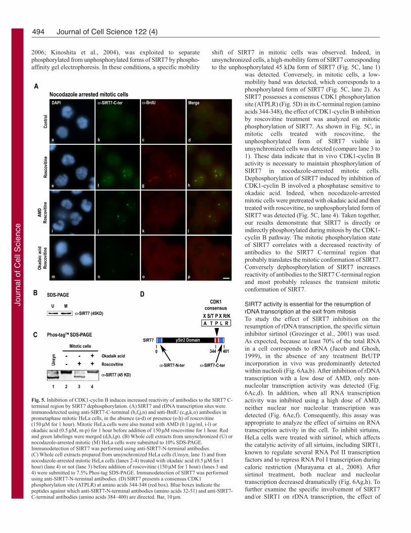
SIRT7 is regulated by the CDK1-cyclin B pathway duringmitosisG2/M transition is triggered by activation of a protein kinasecascade, at the head of which is the CDK1-cyclin B (Dunphy andNewport, 1988; Gautier et al., 1988). Cyclin Bdegradation and consequently CDK1 inactivationstarts at the end of metaphase and increases untilexit from mitosis (Clute and Pines, 1999).Several components of the RNA Pol I machinery,including TBP (Heix et al., 1998), TAFI110, UBF(Klein and Grummt, 1999) and TTF-1 (Sirri etal., 1999), appear to be regulated during mitosisby the CDK1-cyclin B pathway. Furthermore, wepreviously demonstrated that inhibition of theCDK1-cyclin B pathway restores rDNAtranscription in mitotic cells (Sirri et al., 2000).To verify whether inactivation of the CDK1-cyclin B pathway and reactivity of antibodies tothe SIRT7 C-terminal region in late mitosis arecorrelated, BrUTP incorporation assays werecarried out on nocodazole-arrested mitotic cellsin the absence (Fig. 5Aa-d) or presence (Fig.5Ae-h) of roscovitine. As expected, innocodazole-arrested mitotic cells, rDNAtranscription was not detected (Fig. 5Ac) and theSIRT7 signal was very weak (Fig. 5Ab). Afterroscovitine treatment, the SIRT7 signal wasincreased only in rDNA transcription-positivecells, i.e. responsive to roscovitine (Fig. 5Ae-h).To discriminate between a direct and indirecteffect of inhibition of the CDK1-cyclin Bpathway on the reactivity of antibodies to theSIRT7 C-terminal region, mitotic cells weretreated with both roscovitine and AMD. Theincreased reactivity of antibodies to the SIRT7C-terminal region was still detected even whenthe CDK1-cyclin B pathway was inhibited byroscovitine and the resumption of rDNAtranscription was prevented by AMD (Fig. 5Ai-l), confirming that resumption of rDNAtranscription is not necessary to increase theSIRT7 signal in mitotic cells. Thus, inhibition ofthe CDK1-cyclin B pathway is sufficient to
increase reactivity of antibodies against the C-terminal region ofSIRT7 at the level of the NORs. To analyze whether reactivity ofantibodies to the SIRT7 C-terminal region in late mitosis is linkedto dephosphorylation occurring when the CDK1-cyclin B pathwayis inhibited, the reactivity of antibodies to the SIRT7 C-terminalregion was analyzed in nocodazole-arrested mitotic cells pretreatedwith okadaic acid, a potent inhibitor of protein phosphatase 1 (PP1)and 2A (PP2A). As observed in Fig. 5A, in mitotic cells previouslytreated with okadaic acid, subsequent roscovitine treatmentproduced no effect on the reactivity of antibodies to the SIRT7 C-terminal region (Fig. 5Am-p). These results show that the reactivityof antibodies to the SIRT7 C-terminal region during mitosis, andprobably the conformation of SIRT7, depends on phosphorylation/dephosphorylation events. To date, no post-translationalmodification of SIRT7 has been described. To analyzephosphorylation of SIRT7 during the cell cycle, the electrophoreticmobility of SIRT7 was compared using unsynchronized and mitoticcell extracts. No mobility shift of the 45 kDa SIRT7 was detected(Fig. 5B). This suggests either that SIRT7 is not phosphorylatedduring the cell cycle or that SIRT7 phosphorylation is not detectableby SDS-PAGE. Thus, the recently developed Phos-tag compound,which has specific phosphate-binding activity (Kinoshita et al.,
Fig. 4. Increased reactivity of antibodies to the SIRT7 C-terminal region occurs before, andindependently of, onset of rDNA transcription. (A) Surrounded mitosis stages (M, T, T’) ofsynchronized HeLa cells were determined on Z projections of focal planes by DAPI staining (a,e).SIRT7 and rDNA transcription sites were detected in fixed cells using respectively anti-SIRT7-C-terminal (b,f) and anti-BrdU (c,g) antibodies. Red and green labelings were merged (d,h).(B) Immunodetection of SIRT7 (c,h) and rDNA transcription sites (d,i) was performed in early G1fixed cells in the absence (a-e) or presence (f-j) of AMD (0.1 μg/ml for 2 hours). Early G1 cells(arrows) were identified by phase-contrast (a,f) and DAPI staining (b,g). Arrowheads indicatemetaphase cells. Immunofluorescence labelings were merged (e,j). Bars, 10μm.
Jour
nal o
f Cel
l Sci
ence
494
2006; Kinoshita et al., 2004), was exploited to separatephosphorylated from unphosphorylated forms of SIRT7 by phospho-affinity gel electrophoresis. In these conditions, a specific mobility
shift of SIRT7 in mitotic cells was observed. Indeed, inunsynchronized cells, a high-mobility form of SIRT7 correspondingto the unphosphorylated 45 kDa form of SIRT7 (Fig. 5C, lane 1)
was detected. Conversely, in mitotic cells, a low-mobility band was detected, which corresponds to aphosphorylated form of SIRT7 (Fig. 5C, lane 2). AsSIRT7 possesses a consensus CDK1 phosphorylationsite (ATPLR) (Fig. 5D) in its C-terminal region (aminoacids 344-348), the effect of CDK1-cyclin B inhibitionby roscovitine treatment was analyzed on mitoticphosphorylation of SIRT7. As shown in Fig. 5C, inmitotic cells treated with roscovitine, theunphosphorylated form of SIRT7 visible inunsynchronized cells was detected (compare lane 3 to1). These data indicate that in vivo CDK1-cyclin Bactivity is necessary to maintain phosphorylation ofSIRT7 in nocodazole-arrested mitotic cells.Dephosphorylation of SIRT7 induced by inhibition ofCDK1-cyclin B involved a phosphatase sensitive tookadaic acid. Indeed, when nocodazole-arrestedmitotic cells were pretreated with okadaic acid and thentreated with roscovitine, no unphosphorylated form ofSIRT7 was detected (Fig. 5C, lane 4). Taken together,our results demonstrate that SIRT7 is directly orindirectly phosphorylated during mitosis by the CDK1-cyclin B pathway. The mitotic phosphorylation stateof SIRT7 correlates with a decreased reactivity ofantibodies to the SIRT7 C-terminal region thatprobably translates the mitotic conformation of SIRT7.Conversely dephosphorylation of SIRT7 increasesreactivity of antibodies to the SIRT7 C-terminal regionand most probably releases the transient mitoticconformation of SIRT7.
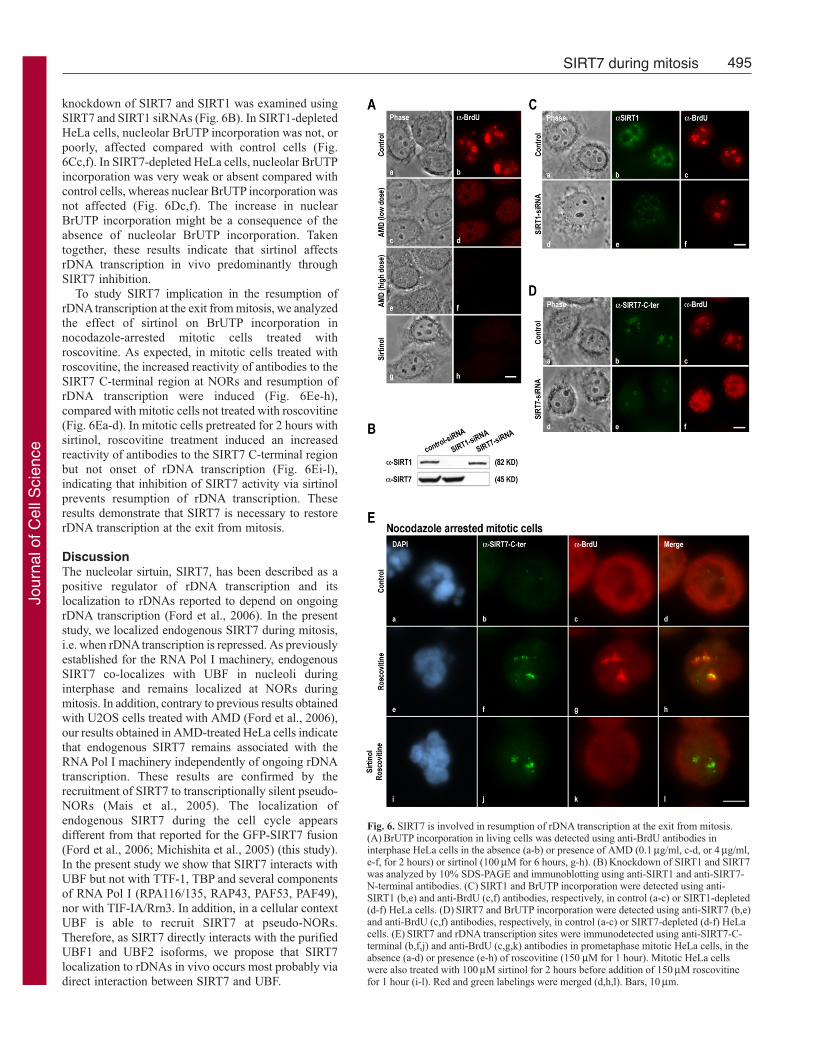
SIRT7 activity is essential for the resumption ofrDNA transcription at the exit from mitosisTo study the effect of SIRT7 inhibition on theresumption of rDNA transcription, the specific sirtuininhibitor sirtinol (Grozinger et al., 2001) was used.As expected, because at least 70% of the total RNAin a cell corresponds to rRNA (Jacob and Ghosh,1999), in the absence of any treatment BrUTPincorporation in vivo was predominantly detectedwithin nucleoli (Fig. 6Aa,b). After inhibition of rDNAtranscription with a low dose of AMD, only non-nucleolar transcription activity was detected (Fig.6Ac,d). In addition, when all RNA transcriptionactivity was inhibited using a high dose of AMD,neither nuclear nor nucleolar transcription wasdetected (Fig. 6Ae,f). Consequently, this assay wasappropriate to analyze the effect of sirtuins on RNAtranscription activity in the cell. To inhibit sirtuins,HeLa cells were treated with sirtinol, which affectsthe catalytic activity of all sirtuins, including SIRT1,known to regulate several RNA Pol II transcriptionfactors and to repress RNA Pol I transcription duringcaloric restriction (Murayama et al., 2008). Aftersirtinol treatment, both nuclear and nucleolartranscription decreased dramatically (Fig. 6Ag,h). Tofurther examine the specific involvement of SIRT7and/or SIRT1 on rDNA transcription, the effect of
Journal of Cell Science 122 (4)
Fig. 5. Inhibition of CDK1-cyclin B induces increased reactivity of antibodies to the SIRT7 C-terminal region by SIRT7 dephosphorylation. (A) SIRT7 and rDNA transcription sites wereimmunodetected using anti-SIRT7-C-terminal (b,f,j,n) and anti-BrdU (c,g,k,o) antibodies inprometaphase mitotic HeLa cells, in the absence (a-d) or presence (e-h) of roscovitine(150 μM for 1 hour). Mitotic HeLa cells were also treated with AMD (0.1 μg/ml, i-l) orokadaic acid (0.5 μM, m-p) for 1 hour before addition of 150 μM roscovitine for 1 hour. Redand green labelings were merged (d,h,l,p). (B) Whole cell extracts from unsynchronized (U) ornocodazole-arrested mitotic (M) HeLa cells were submitted to 10% SDS-PAGE.Immunodetection of SIRT7 was performed using anti-SIRT7-N-terminal antibodies.(C) Whole cell extracts prepared from unsynchronized HeLa cells (Unsyn, lane 1) and fromnocodazole-arrested mitotic HeLa cells (lanes 2-4) treated with okadaic acid (0.5μM for 1hour) (lane 4) or not (lane 3) before addition of roscovitine (150 μM for 1 hour) (lanes 3 and4) were submitted to 7.5% Phos-tag SDS-PAGE. Immunodetection of SIRT7 was performedusing anti-SIRT7-N-terminal antibodies. (D) SIRT7 presents a consensus CDK1phosphorylation site (ATPLR) at amino acids 344-348 (red box). Blue boxes indicate thepeptides against which anti-SIRT7-N-terminal antibodies (amino acids 32-51) and anti-SIRT7-C-terminal antibodies (amino acids 384–400) are directed. Bar, 10 μm.
Jour
nal o
f Cel
l Sci
ence
495SIRT7 during mitosis
knockdown of SIRT7 and SIRT1 was examined usingSIRT7 and SIRT1 siRNAs (Fig. 6B). In SIRT1-depletedHeLa cells, nucleolar BrUTP incorporation was not, orpoorly, affected compared with control cells (Fig.6Cc,f). In SIRT7-depleted HeLa cells, nucleolar BrUTPincorporation was very weak or absent compared withcontrol cells, whereas nuclear BrUTP incorporation wasnot affected (Fig. 6Dc,f). The increase in nuclearBrUTP incorporation might be a consequence of theabsence of nucleolar BrUTP incorporation. Takentogether, these results indicate that sirtinol affectsrDNA transcription in vivo predominantly throughSIRT7 inhibition.
To study SIRT7 implication in the resumption ofrDNA transcription at the exit from mitosis, we analyzedthe effect of sirtinol on BrUTP incorporation innocodazole-arrested mitotic cells treated withroscovitine. As expected, in mitotic cells treated withroscovitine, the increased reactivity of antibodies to theSIRT7 C-terminal region at NORs and resumption ofrDNA transcription were induced (Fig. 6Ee-h),compared with mitotic cells not treated with roscovitine(Fig. 6Ea-d). In mitotic cells pretreated for 2 hours withsirtinol, roscovitine treatment induced an increasedreactivity of antibodies to the SIRT7 C-terminal regionbut not onset of rDNA transcription (Fig. 6Ei-l),indicating that inhibition of SIRT7 activity via sirtinolprevents resumption of rDNA transcription. Theseresults demonstrate that SIRT7 is necessary to restorerDNA transcription at the exit from mitosis.
DiscussionThe nucleolar sirtuin, SIRT7, has been described as apositive regulator of rDNA transcription and itslocalization to rDNAs reported to depend on ongoingrDNA transcription (Ford et al., 2006). In the presentstudy, we localized endogenous SIRT7 during mitosis,i.e. when rDNA transcription is repressed. As previouslyestablished for the RNA Pol I machinery, endogenousSIRT7 co-localizes with UBF in nucleoli duringinterphase and remains localized at NORs duringmitosis. In addition, contrary to previous results obtainedwith U2OS cells treated with AMD (Ford et al., 2006),our results obtained in AMD-treated HeLa cells indicatethat endogenous SIRT7 remains associated with theRNA Pol I machinery independently of ongoing rDNAtranscription. These results are confirmed by therecruitment of SIRT7 to transcriptionally silent pseudo-NORs (Mais et al., 2005). The localization ofendogenous SIRT7 during the cell cycle appearsdifferent from that reported for the GFP-SIRT7 fusion(Ford et al., 2006; Michishita et al., 2005) (this study).In the present study we show that SIRT7 interacts withUBF but not with TTF-1, TBP and several componentsof RNA Pol I (RPA116/135, RAP43, PAF53, PAF49),nor with TIF-IA/Rrn3. In addition, in a cellular contextUBF is able to recruit SIRT7 at pseudo-NORs.Therefore, as SIRT7 directly interacts with the purifiedUBF1 and UBF2 isoforms, we propose that SIRT7localization to rDNAs in vivo occurs most probably viadirect interaction between SIRT7 and UBF.
Fig. 6. SIRT7 is involved in resumption of rDNA transcription at the exit from mitosis.(A) BrUTP incorporation in living cells was detected using anti-BrdU antibodies ininterphase HeLa cells in the absence (a-b) or presence of AMD (0.1 μg/ml, c-d, or 4 μg/ml,e-f, for 2 hours) or sirtinol (100 μM for 6 hours, g-h). (B) Knockdown of SIRT1 and SIRT7was analyzed by 10% SDS-PAGE and immunoblotting using anti-SIRT1 and anti-SIRT7-N-terminal antibodies. (C) SIRT1 and BrUTP incorporation were detected using anti-SIRT1 (b,e) and anti-BrdU (c,f) antibodies, respectively, in control (a-c) or SIRT1-depleted(d-f) HeLa cells. (D) SIRT7 and BrUTP incorporation were detected using anti-SIRT7 (b,e)and anti-BrdU (c,f) antibodies, respectively, in control (a-c) or SIRT7-depleted (d-f) HeLacells. (E) SIRT7 and rDNA transcription sites were immunodetected using anti-SIRT7-C-terminal (b,f,j) and anti-BrdU (c,g,k) antibodies in prometaphase mitotic HeLa cells, in theabsence (a-d) or presence (e-h) of roscovitine (150 μM for 1 hour). Mitotic HeLa cellswere also treated with 100 μM sirtinol for 2 hours before addition of 150 μM roscovitinefor 1 hour (i-l). Red and green labelings were merged (d,h,l). Bars, 10 μm.
Jour
nal o
f Cel
l Sci
ence
496
SIRT7 immunofluorescence analyses during mitosis usingantibodies directed against the SIRT7 C-terminal region revealeda weak SIRT7 signal during the early mitotic stages (from prophaseto anaphase) and a high SIRT7 signal in late telophase, unlike UBF,which presents a constant level of immunofluorescence labelingduring mitosis. An equivalent increase of SIRT7 signal is alsoinduced by treatment using the CDK inhibitor roscovitine.Interestingly, the comparison between chromosomal andcytoplasmic proteins prepared from mitotic HeLa cells treated ornot with roscovitine shows that SIRT7 remains fully associated withNORs during mitosis, as does UBF, revealing that increase of SIRT7signal during telophase is not due to increased recruitment of SIRT7at NORs. Immunofluorescence analyses using antibodies directedagainst either the N-terminal or C-terminal region of SIRT7 showedthat only the reactivity of antibodies to the SIRT7 C-terminal regionchanges during mitosis. Conversely, the reactivity of antibodies tothe SIRT7 N-terminal region is high and unchanged. These resultsindicate that SIRT7 most probably undergoes a conformationalrearrangement during mitosis.
It is well established that local and/or global conformationalchanges can be induced by modifying protein phosphorylation(Johnson and Lewis, 2001). Thus, the mitotic rearrangement of theN-terminal domain of histone H3 induced by phosphorylationmodifies its accessibility to antibodies (Sauvé et al., 1999). Inhibitionof the CDK1-cyclin B pathway at the exit from mitosisconcomitantly induces dephosphorylation of several componentsof the RNA Pol I machinery (Heix et al., 1998; Kuhn et al., 1998;Sirri et al., 2000) and onset of rDNA transcription. The concomitantinhibition of the CDK1-cyclin B pathway by roscovitine and rDNAtranscription by AMD in mitotic cells led us to conclude thatinhibition of CDK1-cyclin B is sufficient to increase reactivity ofantibodies to the SIRT7 C-terminal region independently of rDNAtranscription. Moreover, experiments performed on mitotic cellspretreated with okadaic acid led us to propose that the increasedreactivity of antibodies to the SIRT7 C-terminal region at the exitfrom mitosis is linked to dephosphorylation. Indeed, our resultsshow that SIRT7 is undoubtedly phosphorylated during mitosis bythe CDK1-cyclin B pathway. At present, we cannot concludewhether CDK1-cyclin B kinase directly phosphorylates SIRT7 orwhether it activates another kinase responsible for SIRT7phosphorylation. Interestingly, since SIRT7 presents a consensusCDK1 phosphorylation site at amino acids 344-348 in its C-terminalregion, we can speculate that SIRT7 could be directlyphosphorylated by CDK1-cyclin B. To verify if SIRT7 is indeedphosphorylated in vivo by CDK1-cyclin B during mitosis, ectopicexpression of a SIRT7 form mutated at the CDK1 phosphorylationsite would have been pertinent; however, the mislocalization ofexogenous SIRT7 does not make this possible. Interestingly, SIRT2has been reported to be phosphorylated by CDK1-cyclin B in itsC-terminal region, and this phosphorylation is required for themitotic function of SIRT2 (North and Verdin, 2007). Thus it istempting to suggest that SIRT7 might itself be phosphorylated byCDK1-cyclin B in its C-terminal region and that thisphosphorylation could induce a conformational change of SIRT7,impairing the reactivity of antibodies to its C-terminal region duringmitosis. Indeed, as anti-SIRT7-C-terminal antibodies are directedagainst a peptide downstream of the CDK1 phosphorylation site,we can exclude that anti-SIRT7-C-terminal antibodies preferentiallyrecognize the dephosphorylated form of SIRT7. The mitoticregulation of SIRT7 by the CDK1-cyclin B pathway is in agreementwith previous results concerning the mitotic repression of the RNA
Pol I machinery. Thus, the phosphorylation of SL1, TTF-1 andSIRT7 modify, respectively, the interaction between SL1 and UBF(Heix et al., 1998; Kuhn et al., 1998), the chromatin-binding affinityof TTF-1 (Sirri et al., 1999), and the conformation of SIRT7.
Our results indicate that the conformational rearrangement ofSIRT7 occurs independently of and before the onset of rDNAtranscription. Thus, we investigated the potential role of SIRT7 inthe resumption of RNA Pol I transcription at the exit from mitosis,and first analyzed the effect of sirtinol on rDNA transcription: ininterphasic HeLa cells inhibition of sirtuins by sirtinol affects bothnuclear and nucleolar BrUTP incorporation. To date, only SIRT7 wasreported to function as activator in the in vivo regulation of rDNAtranscription (Ford et al., 2006) whereas SIRT1 was reported to repressrDNA transcription during caloric restriction (Murayama et al., 2008).Repression of nuclear transcription may be due to sirtinol inhibitionof SIRT1, which is reported to be a regulator of several nucleartranscription factors (Brunet et al., 2004; Cohen et al., 2004; Luo etal., 2001; Motta et al., 2004; Vaziri et al., 2001; Yeung et al., 2004),but other sirtuins could be implicated. As for nucleolar transcription(i.e. rDNA transcription), the absence of BrUTP incorporationdemonstrates that nucleolar SIRT7 is inhibited in vivo by sirtinoltreatment. Inhibition of SIRT1 should have increased nucleolartranscription. The fact that such is not the case demonstrates eitherthat SIRT1 does not function as a major repressor, or that SIRT7plays a predominant role as activator on rDNA transcription. To verifythis hypothesis, SIRT7 and SIRT1 were knocked down by specificsiRNAs. As expected, SIRT7 knockdown significantly decreasesnucleolar transcription, whereas unexpectedly SIRT1 knockdowndoes not clearly increase nucleolar transcription. The role of SIRT1as repressor is probably not of major importance in HeLa cells duringinterphase even if it could be important at the onset of mitosis whenrDNA transcription is switched off, as previously suggested (Ford etal., 2006). Taken together, these results indicate that inhibition ofrDNA transcription by sirtinol treatment is most probably due toinhibition of SIRT7, even if involvement of other sirtuins cannot becompletely ruled out. To verify the role of SIRT7 on the resumptionof rDNA transcription, mitotic cells were concomitantly treated withroscovitine, which induces onset of rDNA transcription, and withsirtinol, which inhibits SIRT7. Sirtinol treatment impairs resumptionof rDNA transcription without affecting reactivity of antibodies tothe SIRT7 C-terminal region. Consequently, these results not onlyconfirm the positive regulation role of SIRT7 on rDNA transcription(Ford et al., 2006) but also reveal that the activity of SIRT7 plays anindispensable role in restoring rDNA transcription at the exit frommitosis.
Our results lead us to propose the following model. Duringmitosis, rDNA transcription is repressed and SIRT7, as well asseveral components of the RNA Pol I machinery, is phosphorylatedby the CDK1-cyclin B pathway. SIRT7 phosphorylation in its C-terminal region could change the conformation of SIRT7, makingantibodies weakly reactive to the C-terminal region of SIRT7.Inhibition of the CDK1-cyclin B pathway increases the reactivityof antibodies against the C-terminal region of SIRT7 before theonset of rDNA transcription. Nevertheless, we cannot conclude thatthe increased reactivity of the SIRT7 antibodies is due only to aconformational change of SIRT7, resulting from itsphosphorylation/dephosphorylation. Indeed, dephosphorylation ofseveral components of the RNA Pol I machinery at the exit frommitosis could induce a conformational change of the RNA Pol Imachinery, which could modify the position of SIRT7 inside theRNA Pol I machinery, increasing the reactivity of antibodies to
Journal of Cell Science 122 (4)
Jour
nal o
f Cel
l Sci
ence
497SIRT7 during mitosis
SIRT7. Interestingly, the N-terminal and C-terminal regions of yeastsirtuin Hst2 and mammalian SIRT2 play a role in the regulation oftheir enzymatic activity (Zhao et al., 2003), suggesting thatrearrangement of SIRT7 could regulate SIRT7 activity at the exitfrom mitosis. Thus, during mitosis SIRT7 would be in an inactiveform, whereas at the exit from mitosis SIRT7 would be inducedinto a more active form by a conformational rearrangement thatwould be essential to restore rDNA transcription.
Materials and MethodsCell culture and drugsHeLa cells were cultured in minimum essential medium (MEM) with GlutaMAX(Invitrogen) supplemented with 10% fetal calf serum (FCS). Stable GFP-SIRT7 HeLacells were obtained by co-transfection of GFP-SIRT7 and pBABE-PURO (Addagene)at a 10:1 ratio, using effectene (Qiagen) according to the manufacturer’s instructions,and were selected with 10 μg/ml puromycin. GFP-SIRT7 HeLa cells were culturedas HeLa cells. Clone 3D-1 was maintained and transfected as previously described(Prieto and McStay, 2007). For synchronization in mitosis, HeLa cells wereaccumulated in prometaphase by nocodazole treatment (0.04 μg/ml for 4 hours),selectively harvested by mechanical shock, washed and resuspended in nocodazole-free medium for 60 or 90 minutes. The drugs used were AMD (Sigma) at 0.1 or 4μg/ml for 2 hours, roscovitine (Calbiochem) at 150 μM for 1 hour, okadaic acid(Calbiochem) at 0.5 μM for 2 hours and sirtinol (Calbiochem) at 100 μM for 2 or 6hours. For immunofluorescence labeling and BrUTP incorporation assays, cells weregrown as monolayers on glass slides or coverslips and transfected or treated 24 hourslater. Synchronized mitotic cells were transferred onto glass slides coated with poly-L-lysine.
Plasmid constructs and antibodiesHuman SIRT7 (hSIRT7) cDNA (clone IMAGE 5087554) was PCR-amplified andsubcloned into the EcoRI/SalI sites of the pMIGR1 retroviral vector provided byS.A. Leibovitch (INRA, Montpellier, France) to generate the GFP-SIRT7 constructand into the EcoRI/NotI sites of the pGEX4T1 (Amersham) to obtain the GST-SIRT7construct. SIRT7-YFP was obtained by cloning hSIRT7 cDNA into the Gateway entryvector pENTR-D-TOPO (Invitrogen), and recombined into pdEYFP (Simpson et al.,2000).
Anti-SIRT7-C-terminal rabbit polyclonal antibodies were first provided by I.Horikawa (National Institute of Health, Bethesda, USA) and subsequently raisedagainst the same peptide (GWFGRGCTKRTKRKKVT) by Eurogentec. HumanRPA43, PAF49, PAF53 and TIF-1A/Rrn3 antibodies were raised in sheep immunizedwith full-length recombinant proteins produced using baculovirus or Escherichia coliexpression systems. UBF antibodies used in Fig. 2C and supplementary material Fig.S2B were described in (Mais et al., 2005). The other antibodies were anti-SIRT7-N-terminal (S5947, Sigma-Aldrich), anti-UBF (F-9, Santa Cruz Biotechnology), anti-RPA116/135 (N-17, Santa Cruz Biotechnology), anti-SIRT1 (Abcam), anti-TBP (N-12, Santa Cruz Biotechnology), anti-TTF-1 (Sirri et al., 1999), anti-BrdU (Sigma-Aldrich) and Texas Red-, FITC- or horseradish peroxidase-conjugated secondaryantibodies (Jackson ImmunoResearch Laboratories).
ImmunofluorescenceThe cells were fixed in 4% paraformaldehyde for 20 minutes at room temperature(RT) and permeabilized with 0.5% Triton X-100 for 5 minutes at RT. They were thenwashed with PBS and incubated with primary antibodies at RT for 60 minutes. Theantibodies were revealed with Texas Red- and/or FITC-conjugated secondaryantibodies. Alternatively, the cells were fixed with methanol for 20 minutes at –20°C,air-dried for 5 minutes and rehydrated with PBS for 5 minutes before incubation withantibodies. They were then incubated with DAPI to visualize DNA and mounted withthe antifading solution AF1 (Citifluor). Fluorescent microscopy was performed usinga CCD camera Leitz DMRB. Optical sections (0.4 μm thick) were examined on aLeica SPD2 AOBS confocal microscope (Leica Microsystem) with a 63�, 1.32 NAPlanApo lens and acquired with a Micromax CCD camera (Princeton Instruments,France). An argon laser adjusted to 488 nm was used for the fluorescein signal, akrypton laser adjusted to 568 nm for Texas Red and a blue laser diode at 405 nm forDAPI. Images were assembled using Adobe Photoshop. SIRT7 and UBF signalintensity was quantified in Fig. 1C and Fig. 2B using ImageJ software. Cell stainingand image capture in Fig. 2C and supplementary material Fig. S2B were as describedin (Prieto and McStay, 2007).
siRNA-mediated mRNA knockdownThe siControl RISC-free siRNA and SIRT1 SMARTpool siRNA were purchased fromDharmacon. SIRT7 siRNA duplexes targeting 5�-GCCUGAAGGUUCUAAAGAA(sense) and 5�-UUCUUUAGAACCUUCAGGC (antisense) were synthetized byEurogentec. siRNA duplexes (15 nM) were transfected using INTERFERinTM
(Polyplus-transfection) according to the manufacturer’s instructions 24 and 48 hoursafter cell seeding.
Pull-down experimentsFor in vitro pull-down assays, GST-SIRT7 was overexpressed in E. coli BL21 DE3in StabyTMSwitch auto-inducible medium (Eurogentec) according to themanufacturer’s instructions. The GST protein was induced for 3 hours with 0.1 mMisopropylthiogalactoside in E. coli. Lysates were obtained by enzymatic reaction withlysozyme (100 μg/ml) and DNase I (20 ng/μl) and clarified by ultracentrifugation at65,000 g for 30 minutes. The clarified lysates were incubated with glutathione-sepharose beads (Amersham) for 2 hours at 4°C. GST-fusion proteins bound toglutathione-sepharose beads were washed four times with wash buffer (50 mM Tris-HCl, pH 8, 1 M NaCl, 10% glycerol and complete) and once with binding buffer[50 mM Tris-HCl, pH 7.4, 150 mM NaCl, 1 mM EDTA, 0.2% NP40, 10% glyceroland complete; complete corresponds to a cocktail of protease inhibitors (Roche)].Proteins of HeLa cells were extracted using lysis buffer (50 mM Tris-HCl, pH 7.4,500 mM NaCl, 1 mM EDTA, 1% NP40, 10% glycerol and complete). Aftercentrifugation at 16,000 g for 15 minutes, the supernatants were adjusted to the bindingbuffer conditions and these whole-cell lysates were incubated with GST and GST-SIRT7 bound to glutathione-sepharose beads. Otherwise, 1 μg of human UBF1 andUBF2, produced as previously described (McStay et al., 1997), was diluted in thebinding buffer and incubated with GST and GST-SIRT7 bound to glutathione-sepharose beads. After gentle shaking overnight at 4°C, the beads were centrifugedat 500 g for 2 minutes and washed five times in wash buffer (50 mM Tris-HCl, pH7.4, 300 mM NaCl, 1 mM EDTA, 0.2% NP40, 10% glycerol and complete). Proteinscorresponding to cell lysates and proteins bound to beads were resuspended in SDSloading buffer, boiled for 5 minutes, resolved by 10% SDS-PAGE and analyzed byimmunoblotting.
Chromosome and cytoplasmic fractionsMitotic HeLa cells were resuspended in 20% FCS for 20 minutes at RT. Cells werethen mechanically disrupted by passage through a G22 needle until chromosomesappeared scattered, as assessed by phase microscopy. The pellet containing thechromosome fraction and the supernatant containing the cytoplasmic fraction werenormalized to an equal volume of SDS loading buffer for quantitative comparison.
Phos-tag SDS-PAGEProteins were resolved by 7.5% SDS-PAGE containing 100 μM Phos-tag ligand(AAL-107, NARD Institute, Japan) and 100 μM MnCl2 as previously described(Kinoshita et al., 2006). Gels were run at 40 mA for 1.5 hours, rinsed once for 10minutes in transfer buffer containing 1 mM EDTA to chelate MnCl2 and once in thesame buffer without EDTA. Proteins were then submitted to immunoblotting.
ImmunoblottingProteins resolved by SDS-PAGE were transferred to nitrocellulose membranes(Protran, Schleicher and Schuell) and incubated with the following antibodies: anti-SIRT1, anti-SIRT7-N-terminal, anti-UBF, anti-TBP, anti-TTF-1, anti-RPA116/135,anti-RPA43, anti-PAF49, anti-PAF53 or anti-TIF-1A/Rrn3. The membranes were thenincubated with suitable horseradish peroxidase-conjugated secondary antibodies andimmunoreactivity was detected by chemiluminescence (Pierce).
BrUTP incorporationThe assay described in (Valdez et al., 2004) was slightly modified as follows.Coverslips seeded with HeLa cells were briefly rinsed with hypotonic KH buffer (30mM KCl and 10 mM Hepes, pH 7.4) and incubated with KH buffer containing 10mM BrUTP for 10 minutes in a 5% CO2 incubator at 37°C. The cells were rinsedthree times with culture medium and incubated for 30 minutes in MEM containing20% FCS. They were rinsed with PBS, fixed in methanol for 20 minutes at –20°C,air-dried for 5 minutes and rehydrated with PBS for 5 minutes. BrUTP incorporationwas detected using an anti-BrdU antibody. Alternatively, RNA Pol I activity wasdetected in fixed cells in conditions set up to preferentially reveal RNA Pol I aspreviously described (Roussel et al., 1996). Briefly, HeLa cells seeded as monolayerson glass slides were weakly fixed with ethanol/acetone for 5 minutes at 4°C and airdried. The cells were then incubated in assay solution (100 mM Tris-HCl, pH 7.9,12 mM 2-mercaptoethanol, 150 mM sucrose, 12 mM MgCl2, 0.6 mM ATP, CTP andGTP each, and 0.12 mM BrUTP) at 37°C for 15 minutes. The cells were post-fixedin 2% paraformaldehyde for 20 minutes at RT. BrUTP incorporation was detectedby immunofluorescence labeling using an anti-BrdU antibody.
The authors are grateful to I. Horikawa and S. A. Leibovitch forgenerously providing constructs or antibodies, to C. Chamot and A.Jobart-Malfait for technical support in confocal microscopy and to A.L. Haenni for critical reading of the manuscript. A.G. is the recipientof an undergraduate grant from Ministère de l’Education Nationale, dela Recherche et de la Technologie. This study was supported in partby grants from the Centre National de le Recherche Scientifique andthe Association pour la Recherche sur le Cancer (contrat 3303). B.M.was supported by the MRC, UK. Deposited in PMC for release after6 months.
Jour
nal o
f Cel
l Sci
ence
498
ReferencesBlander, G. and Guarente, L. (2004). The Sir2 family of protein deacetylases. Annu. Rev.
Biochem. 73, 417-435.Boisvert, F. M., van Koningsbruggen, S., Navascues, J. and Lamond, A. I. (2007). The
multifunctional nucleolus. Nat. Rev. Mol. Cell. Biol. 8, 574-585.Brunet, A., Sweeney, L. B., Sturgill, J. F., Chua, K. F., Greer, P. L., Lin, Y., Tran, H.,
Ross, S. E., Mostoslavsky, R., Cohen, H. Y. et al. (2004). Stress-dependent regulationof FOXO transcription factors by the SIRT1 deacetylase. Science 303, 2011-2015.
Carmo-Fonseca, M., Mendes-Soares, L. and Campos, I. (2000). To be or not to be inthe nucleolus. Nat. Cell Biol. 2, E107-E112.
Chen, D., Dundr, M., Wang, C., Leung, A., Lamond, A., Misteli, T. and Huang, S.(2005). Condensed mitotic chromatin is accessible to transcription factors and chromatinstructural proteins. J. Cell Biol. 168, 41-54.
Clute, P. and Pines, J. (1999). Temporal and spatial control of cyclin B1 destruction inmetaphase. Nat. Cell Biol. 1, 82-87.
Cohen, H. Y., Miller, C., Bitterman, K. J., Wall, N. R., Hekking, B., Kessler, B., Howitz,K. T., Gorospe, M., de Cabo, R. and Sinclair, D. A. (2004). Calorie restriction promotesmammalian cell survival by inducing the SIRT1 deacetylase. Science 305, 390-392.
De Azevedo, W. F., Leclerc, S., Meijer, L., Havlicek, L., Strnad, M. and Kim, S. H.(1997). Inhibition of cyclin-dependent kinases by purine analogues: crystal structure ofhuman cdk2 complexed with roscovitine. Eur. J. Biochem. 243, 518-526.
Denu, J. M. (2005). The Sir 2 family of protein deacetylases. Curr. Opin. Chem. Biol. 9,431-440.
Dryden, S. C., Nahhas, F. A., Nowak, J. E., Goustin, A. S. and Tainsky, M. A. (2003).Role for human SIRT2 NAD-dependent deacetylase activity in control of mitotic exitin the cell cycle. Mol. Cell. Biol. 23, 3173-3185.
Dunphy, W. G. and Newport, J. W. (1988). Unraveling of mitotic control mechanisms.Cell 55, 925-928.
Ford, E., Voit, R., Liszt, G., Magin, C., Grummt, I. and Guarente, L. (2006). MammalianSir2 homolog SIRT7 is an activator of RNA polymerase I transcription. Genes Dev. 20,1075-1080.
Gautier, J., Norbury, C., Lohka, M., Nurse, P. and Maller, J. (1988). Purified maturation-promoting factor contains the product of a Xenopus homolog of the fission yeast cellcycle control gene cdc2+. Cell 54, 433-439.
Gilbert, N., Lucas, L., Klein, C., Menager, M., Bonnet, N. and Ploton, D. (1995). Three-dimensional co-location of RNA polymerase I and DNA during interphase and mitosisby confocal microscopy. J. Cell Sci. 108, 115-125.
Grozinger, C. M., Chao, E. D., Blackwell, H. E., Moazed, D. and Schreiber, S. L. (2001).Identification of a class of small molecule inhibitors of the sirtuin family of NAD-dependent deacetylases by phenotypic screening. J. Biol. Chem. 276, 38837-38843.
Grummt, I. (2003). Life on a planet of its own: regulation of RNA polymerase I transcriptionin the nucleolus. Genes Dev. 17, 1691-1702.
Guarente, L. (2000). Sir2 links chromatin silencing, metabolism, and aging. Genes Dev.14, 1021-1026.
Hadjiolov, A. A. (1985). The Nucleolus and Ribosome Biogenesis. New York: Springer-Verlag.
Heix, J., Vente, A., Voit, R., Budde, A., Michaelidis, T. M. and Grummt, I. (1998).Mitotic silencing of human rRNA synthesis: inactivation of the promoter selectivity factorSL1 by cdc2/cyclin B-mediated phosphorylation. EMBO J. 17, 7373-7381.
Jacob, S. T. and Ghosh, A. K. (1999). Control of RNA polymerase I-directed transcription:recent trends. J. Cell. Biochem. Suppl. 32-33, 41-50.
Johnson, L. N. and Lewis, R. J. (2001). Structural basis for control by phosphorylation.Chem. Rev. 101, 2209-2242.
Jordan, P., Mannervik, M., Tora, L. and Carmo-Fonseca, M. (1996). In vivo evidencethat TATA-binding protein/SL1 colocalizes with UBF and RNA polymerase I when rRNAsynthesis is either active or inactive. J. Cell Biol. 133, 225-234.
Kim, D. H., Sarbassov, D. D., Ali, S. M., Latek, R. R., Guntur, K. V., Erdjument-Bromage, H., Tempst, P. and Sabatini, D. M. (2003). GbetaL, a positive regulator ofthe rapamycin-sensitive pathway required for the nutrient-sensitive interaction betweenraptor and mTOR. Mol. Cell 11, 895-904.
Kinoshita, E., Takahashi, M., Takeda, H., Shiro, M. and Koike, T. (2004). Recognitionof phosphate monoester dianion by an alkoxide-bridged dinuclear zinc(II) complex.Dalton Trans. 8, 1189-1193.
Kinoshita, E., Kinoshita-Kikuta, E., Takiyama, K. and Koike, T. (2006). Phosphate-binding tag, a new tool to visualize phosphorylated proteins. Mol. Cell. Proteomics 5,749-757.
Klein, J. and Grummt, I. (1999). Cell cycle-dependent regulation of RNA polymerase Itranscription: the nucleolar transcription factor UBF is inactive in mitosis and early G1.Proc. Natl. Acad. Sci. USA 96, 6096-6101.
Kuhn, A., Vente, A., Doree, M. and Grummt, I. (1998). Mitotic phosphorylation of theTBP-containing factor SL1 represses ribosomal gene transcription. J. Mol. Biol. 284, 1-5.
Leung, A. K., Gerlich, D., Miller, G., Lyon, C., Lam, Y. W., Lleres, D., Daigle, N.,Zomerdijk, J., Ellenberg, J. and Lamond, A. I. (2004). Quantitative kinetic analysisof nucleolar breakdown and reassembly during mitosis in live human cells. J. Cell Biol.166, 787-800.
Liszt, G., Ford, E., Kurtev, M. and Guarente, L. (2005). Mouse Sir2 homolog SIRT6is a nuclear ADP-ribosyltransferase. J. Biol. Chem. 280, 21313-21320.
Luo, J., Nikolaev, A. Y., Imai, S., Chen, D., Su, F., Shiloh, A., Guarente, L. and Gu,W. (2001). Negative control of p53 by Sir2alpha promotes cell survival under stress.Cell 107, 137-148.
Mais, C., Wright, J. E., Prieto, J. L., Raggett, S. L. and McStay, B. (2005). UBF-bindingsite arrays form pseudo-NORs and sequester the RNA polymerase I transcriptionmachinery. Genes Dev. 19, 50-64.
McStay, B., Sullivan, G. J. and Cairns, C. (1997). The Xenopus RNA polymerase Itranscription factor, UBF, has a role in transcriptional enhancement distinct from that atthe promoter. EMBO J. 16, 396-405.
Meijer, L., Borgne, A., Mulner, O., Chong, J. P., Blow, J. J., Inagaki, N., Inagaki, M.,Delcros, J. G. and Moulinoux, J. P. (1997). Biochemical and cellular effects ofroscovitine, a potent and selective inhibitor of the cyclin-dependent kinases cdc2, cdk2and cdk5. Eur. J. Biochem. 243, 527-536.
Michishita, E., Park, J. Y., Burneskis, J. M., Barrett, J. C. and Horikawa, I. (2005).Evolutionarily conserved and nonconserved cellular localizations and functions of humanSIRT proteins. Mol. Biol. Cell 16, 4623-4635.
Mostoslavsky, R., Chua, K. F., Lombard, D. B., Pang, W. W., Fischer, M. R., Gellon,L., Liu, P., Mostoslavsky, G., Franco, S., Murphy, M. M. et al. (2006). Genomicinstability and aging-like phenotype in the absence of mammalian SIRT6. Cell 124, 315-329.
Motta, M. C., Divecha, N., Lemieux, M., Kamel, C., Chen, D., Gu, W., Bultsma, Y.,McBurney, M. and Guarente, L. (2004). Mammalian SIRT1 represses forkheadtranscription factors. Cell 116, 551-563.
Murayama, A., Ohmori, K., Fujimura, A., Minami, H., Yasuzawa-Tanaka,K., Kuroda, T., Oie, S., Daitoku, H., Okuwaki, M., Nagata, K. et al. (2008).Epigenetic control of rDNA loci in response to intracellular energy status. Cell 133,627-639.
North, B. J. and Verdin, E. (2004). Sirtuins: Sir2-related NAD-dependent proteindeacetylases. Genome Biol. 5, 224.
North, B. J. and Verdin, E. (2007). Mitotic regulation of SIRT2 by cyclin-dependentkinase 1-dependent phosphorylation. J. Biol. Chem. 282, 19546-19555.
North, B. J., Marshall, B. L., Borra, M. T., Denu, J. M. and Verdin, E. (2003). Thehuman Sir2 ortholog, SIRT2, is an NAD+-dependent tubulin deacetylase. Mol. Cell 11,437-444.
Prescott, D. M. and Bender, M. A. (1962). Synthesis of RNA and protein during mitosisin mammalian tissue culture cells. Exp. Cell. Res. 26, 260-268.
Prieto, J. L. and McStay, B. (2007). Recruitment of factors linking transcription andprocessing of pre-rRNA to NOR chromatin is UBF-dependent and occurs independentof transcription in human cells. Genes Dev. 21, 2041-2054.
Roussel, P., Andre, C., Masson, C., Geraud, G. and Hernandez-Verdun, D. (1993).Localization of the RNA polymerase I transcription factor hUBF during the cell cycle.J. Cell Sci. 104, 327-337.
Roussel, P., Andre, C., Comai, L. and Hernandez-Verdun, D. (1996). The rDNAtranscription machinery is assembled during mitosis in active NORs and absent in inactiveNORs. J. Cell Biol. 133, 235-246.
Ruggero, D. and Pandolfi, P. P. (2003). Does the ribosome translate cancer? Nat. Rev.Cancer 3, 179-192.
Russell, J. and Zomerdijk, J. C. (2005). RNA-polymerase-I-directed rDNA transcription,life and works. Trends Biochem. Sci. 30, 87-96.
Sauvé, D. M., Anderson, H. J., Ray, J. M., James, W. M. and Roberge, M. (1999).Phosphorylation-induced rearrangement of the histone H3 NH2-terminal domain duringmitotic chromosome condensation. J. Cell Biol. 145, 225-235.
Scheer, U. and Rose, K. M. (1984). Localization of RNA polymerase I in interphase cellsand mitotic chromosomes by light and electron microscopic immunocytochemistry. Proc.Natl. Acad. Sci. USA 81, 1431-1435.
Schmelzle, T. and Hall, M. N. (2000). TOR, a central controller of cell growth. Cell 103,253-262.
Simpson, J. C., Wellenreuther, R., Poustka, A., Pepperkok, R. and Wiemann, S. (2000).Systematic subcellular localization of novel proteins identified by large-scale cDNAsequencing. EMBO Rep. 1, 287-292.
Sirri, V., Roussel, P. and Hernandez-Verdun, D. (1999). The mitotically phosphorylatedform of the transcription termination factor TTF-1 is associated with the repressed rDNAtranscription machinery. J. Cell Sci. 112, 3259-3268.
Sirri, V., Roussel, P. and Hernandez-Verdun, D. (2000). In vivo release of mitotic silencingof ribosomal gene transcription does not give rise to precursor ribosomal RNAprocessing. J. Cell Biol. 148, 259-270.
Sirri, V., Hernandez-Verdun, D. and Roussel, P. (2002). Cyclin-dependent kinases governformation and maintenance of the nucleolus. J. Cell Biol. 156, 969-981.
Stefanovsky, V. Y., Pelletier, G., Hannan, R., Gagnon-Kugler, T., Rothblum, L. I. andMoss, T. (2001). An immediate response of ribosomal transcription to growth factorstimulation in mammals is mediated by ERK phosphorylation of UBF. Mol. Cell 8,1063-1073.
Valdez, B. C., Henning, D., So, R. B., Dixon, J. and Dixon, M. J. (2004). The TreacherCollins syndrome (TCOF1) gene product is involved in ribosomal DNA gene transcriptionby interacting with upstream binding factor. Proc. Natl. Acad. Sci. USA 101, 10709-10714.
Vaziri, H., Dessain, S. K., Ng Eaton, E., Imai, S. I., Frye, R. A., Pandita, T. K., Guarente,L. and Weinberg, R. A. (2001). hSIR2(SIRT1) functions as an NAD-dependent p53deacetylase. Cell 107, 149-159.
Yeung, F., Hoberg, J. E., Ramsey, C. S., Keller, M. D., Jones, D. R., Frye, R. A. andMayo, M. W. (2004). Modulation of NF-kappaB-dependent transcription and cell survivalby the SIRT1 deacetylase. EMBO J. 23, 2369-2380.
Zhao, K., Chai, X., Clements, A. and Marmorstein, R. (2003). Structure andautoregulation of the yeast Hst2 homolog of Sir2. Nat. Struct. Biol. 10, 864-871.
Journal of Cell Science 122 (4)
Jour
nal o
f Cel
l Sci
ence