involucrin expression in the corneal epithelium: an
TRANSCRIPT
Involucrin Expression in the Corneal Epithelium: AnEssential Role for Sp1 Transcription Factors
Gautam Adhikary,1 James F. Crish,1 Ramamurthy Gopalakrishnan,1 Frederic Bone,1 andRichard L. Eckert1,2,3,4
PURPOSE. Identifying the mechanism(s) that regulate gene ex-pression during the transition of the limbal stem cell to adifferentiated superficial cell is an important area of interest inthe corneal epithelium.
METHODS. However, the factors that regulate gene expressionduring this process are not well understood. In the presentstudy, the human involucrin (hINV) gene was used as a modelto study gene expression in the corneal epithelium. Expressionwas studied in normal human corneal epithelial cell culturesand hINV promoter transgenic mice.
RESULTS. Studies in cultured cells revealed that an Sp transcrip-tion factor–binding site, located in the upstream regulatoryregion of the hINV promoter, is essential for optimal hINVgene expression. Mutation of this site reduces promoter activ-ity. Expression of Sp1 results in an Sp1-dependent increase inactivity, whereas expression of dominant-negative Sp1 inhibitspromoter activity. Gel mobility shift analysis showed the inter-action of Sp1 and Sp3 with the Sp DNA element. Treatment ofthe corneal epithelial cells with 12-O-tetradecanoylphorbol-13-acetate increased hINV gene expression and this response isassociated with increased nuclear factor binding of Sp1 andSp3 to the Sp DNA response element. Promoter mutagenesisstudies in transgenic mice confirmed the importance of the Spsite, as removal of this site by promoter truncation or pointmutation resulted in a complete loss of in vivo corneal epithe-lial cell gene expression.
CONCLUSIONS. These studies provide in vivo evidence that Sptranscription factor input is absolutely necessary for activationof involucrin gene expression in the differentiating cornealepithelium. (Invest Ophthalmol Vis Sci. 2005;46:3109–3120)DOI:10.1167/iovs.05-0053
During the process of corneal epithelial cell differentiation,proliferating stem cells, located in the limbus of the eye,
give rise to transient amplifying cells and superficial cells thatpopulate the multilayered corneal epithelium.1–3 Ultimately,
the superficial cells are lost from the corneal surface.4 Thisprocess is associated with specific changes in the expression ofgenes that are necessary to maintain corneal epithelial cellintegrity and function. Involucrin is a marker of the transitionfrom proliferation to differentiation.5 Activation of involucrinexpression is associated with the exit of the stem cell from thelimbal compartment, and involucrin is maximally expressed inthe differentiated, superficial cell layer. There is intense inter-est in elucidating the mechanisms that regulate the transitionfrom stem cell to transient amplifying cell to differentiated celland the mechanisms that regulate gene expression during thisprocess. However, there is only limited information regardingthe mechanisms that regulate gene expression during thisprocess. We have developed the involucrin gene as a model tomonitor this process, with a goal of providing understandingregarding the transcriptional mechanisms that regulate geneexpression during corneal epithelial cell maturation.
Involucrin is a rod-shaped, �-helical structural protein6 thatserves as a substrate for the formation of covalent interprotein�-(�-glutamyl)lysine isopeptide bonds.7,8 It is produced as asoluble cytosolic protein.7 However, during the terminal stagesof differentiation in surface epithelial cells, it becomes co-valently cross-linked at sites on the inner face of the plasmamembrane, where it serves as a scaffold protein to stabilize cellstructure.6,9,10 Type I transglutaminase,11 which catalyzes for-mation of interprotein �-(�-glutamyl)lysine bonds, catalyzes theformation of interprotein covalent cross-links between involu-crin and other proteins.12,13 Both involucrin and type I trans-glutaminase are present in corneal epithelial cells, and assem-bly of covalently cross-linked structures most likely plays a roleduring the corneal epithelial cell life cycle.5,14
In the present study, we used two models as part of oureffort to identify mechanisms that regulate the transition dur-ing corneal epithelial cell differentiation: primary human cor-neal epithelial cultures, for in situ analysis of promoter func-tion, and a series of human involucrin (hINV) promotertruncation and point mutation transgenic mice, to confirm thein vivo physiologic relevance of the cell-culture–based studies.Studies with cultured human corneal epithelial cells showedthat Sp1 and Sp3 interact with the involucrin Sp responseelement to regulate involucrin gene expression. Moreover,mutation of the Sp site, or expression of dominant-negative Sp1confirmed that involucrin expression is Sp1 factor dependent.Parallel studies in transgenic mice confirmed that the involu-crin promoter distal regulatory region (DRR), which containsthe Sp site, is absolutely necessary for corneal expression.Additional studies showed that mutation of the DRR Sp siteeliminated involucrin gene expression in the corneal epithe-lium but not in other surface epithelia. These studies implicateSp transcription factors as key regulators of corneal epithelialcell gene expression during the transition from stem cell tosuperficial cell in the corneal epithelium.
MATERIALS AND METHODS
Chemicals and Reagents
Keratinocyte serum-free medium (KSFM), trypsin, Hanks’ balanced saltsolution, and gentamicin were purchased from Invitrogen (Carlsbad,
From the Departments of 1Physiology and Biophysics, 2Biochem-istry, 3Oncology, and 4Dermatology, Case School of Medicine, Cleve-land, Ohio.
Supported was supported by grants from the National Institutes ofHealth (RLE). This work used the facilities of the Visual SciencesResearch Center at Case Western Reserve University (National EyeInstitute Grant P30 EY11373), and the Skin Diseases Research Centerof Northeast Ohio (National Institute of Arthritis and Musculoskeletaland Skin Diseases Grant AR39750). GA was supported by a NationalEye Institute postdoctoral research training fellowship T32 EY07157from the Visual Sciences Training Program.
Submitted for publication January 14, 2005; revised March 1 andApril 21, 2005; accepted April 26, 2005.
Disclosure: G. Adhikary, None; J.F. Crish, None; R. Go-palakrishnan, None; F. Bone, None; R.L. Eckert, None
The publication costs of this article were defrayed in part by pagecharge payment. This article must therefore be marked ”advertise-ment“ in accordance with 18 U.S.C. §1734 solely to indicate this fact.
Corresponding author: Richard L. Eckert, Department of Physiol-ogy/Biophysics, Room E532, Case School of Medicine, 2109 AdelbertRoad, Cleveland, OH 44106-4970; [email protected].
Investigative Ophthalmology & Visual Science, September 2005, Vol. 46, No. 9Copyright © Association for Research in Vision and Ophthalmology 3109

CA); phorbol ester (12-O-tetradeconylphorbol-13-acetate [TPA]) anddimethyl sulfoxide from Sigma-Aldrich (St. Louis, MO); the pGL2-Basicplasmid and a chemiluminescent luciferase assay system from Promega(Madison, WI); and [�-32P]ATP from Perkin Elmer Life Sciences (Bos-ton, MA). The hINV-specific polyclonal antibody was generated aspreviously described.13 Transcription factor-selective rabbit polyclonalantibodies specifying Sp1 (SC-59, diluted 1:1000 for immunoblot) andSp3 (SC-644, diluted 1:1000 for immunoblot) were obtained from SantaCruz Biotechnology (Santa Cruz, CA), as were blocking peptides foreach antibody (SC-59P, SC-644P). The mouse monoclonal �-actin anti-body was from Sigma-Aldrich and was used diluted 1:3000 for immu-noblot assays. Secondary antibodies include HRP-conjugated goat anti-mouse IgG, and HRP-conjugated goat anti-rabbit IgG, both from SantaCruz Biotechnology. The hINV promoter reporter plasmids were con-structed in pGL2-Basic, as previously described.15,16 Recombinant hu-man involucrin (rhINV) was produced by cloning the hINV codingsequence into pRSET-B to yield phINV(1-585). This vector was used toproduce histidine-tagged hINV in bacterial strain BL21. The recombi-nant protein (rhINV) was then purified with a nickel metal affinitycolumn and used to produce a rabbit polyclonal antibody that detectshINV but not murine (m)INV.13 Recombinant murine involucrin (rm-INV) was produced with a similar system.
Immunoblot Method
For immunoblot, an identical number of protein equivalents wereelectrophoresed on denaturing and reducing 6% polyacrylamide gelsand transferred to nitrocellulose membrane. The membrane wasblocked with 5% nonfat dry milk and then incubated with the appro-priate antibodies. Antibody binding was visualized with chemilumines-cence detection technology.
Plasmids
Construction of the involucrin promoter luciferase reporter plasmidshas been described.17,18 The human Sp1-encoding plasmid was pro-vided by Jon Horowitz (Duke Medical Center, Durham, NC).19
pcDNA3-HA-dnhSp1 (HA-tagged dominant-negative human Sp1) wasconstructed by fusing an HA epitope upstream of amino acids 619 to785 of human Sp1 (GenBank accession no. BC062539; http://www.ncbi.nlm.nih.gov/Genbank; provided in the public domain by the Na-tional Center for Biotechnology Information, Bethesda, MD) by poly-merase chain reaction and then cloning the resultant BamHI/EcoRIfragment into pcDNA3. Amino acids 619 to 785 encode the threeC-terminal zinc fingers of Sp1 but lack the amino terminal transcrip-tional activation domains.20,21
Human Corneal Epithelial Cell Culture
Human eyes were obtained from the Cleveland Eye Bank, at 5 to 10hours after death, from donors 40 to 75 years of age. The tissueprocurement protocol was approved by the Case Western Reserve/University Hospitals Institutional Review Board. Eyes were obtainedfrom subjects who had agreed to donate these organs, and all proce-dures followed the principles articulated in the Helsinki Declaration.The methods for obtaining corneal epithelial cells and culturing thesecells has been described.22 Cells were routinely passaged at a split ratioof 1:3 in early cultures and at 1:5 in established cultures. Cultures thatwere 50% to 70% confluent were used for experimentation.
hINV Promoter Activity
For hINV promoter studies, 6 �L of a transfection reagent (Fugene-6;Roche Diagnostics, Indianapolis, IN) was mixed with 94 �L of KSFMand incubated at 25°C for 10 minutes. This mixture was then added to2 �g of hINV promoter reporter plasmid and incubated at 25°C for 20minutes, followed by direct addition to cultures containing 2 mL ofKSFM. For cotransfection experiments, the involucrin reporter plasmidand Sp119 and Sp3 expression plasmids were used at the levels indi-cated in the Figure 6. The final DNA concentration in all groups wasmaintained constant by addition of empty expression vector. In someexperiments, at 24 hours after transfection, 2 mL of fresh media was
added containing 0 or 50 ng TPA per milliliter. After an additional 24hours, the cells were washed with phosphate-buffered saline (pH 7.5)and scraped into 200 �L of cell lysis buffer,16 and luciferase activitywas assayed immediately. All assays were performed in triplicate, andeach experiment was repeated a minimum of three times.
Nuclear Extract Preparation and Gel MobilityShift Assay
Sixty percent confluent human corneal epithelial cells, growing inKSFM, were incubated with 0 or 50 ng/mL TPA for 24 hours. The cellswere then washed with phosphate-buffered saline, and total cell ex-tract was prepared in the presence of proteinase inhibitors,16 andnuclear extracts were prepared according to Schreiber et al.23 in buffercontaining 5 �g/mL leupeptin, 5 �g/mL aprotinin, and 1 mM phenyl-methylsulfonyl fluoride (PMSF). Protein content was measured with aprotein assay reagent (DC; Bio-Rad, Hercules, CA). Identification ofbinding to the hINV promoter Sp site was detected with an electro-phoretic mobility shift assay.15 Nuclear extract (3 �g) was incubatedfor 25 minutes at room temperature in a total volume of 20 �Lcontaining 20 mM HEPES (pH 7.5), 10% glycerol, 50 mM KCl, 2 mMMgCl2, 0.5 mM EDTA, 0.5 mM dithiothreitol (DTT), 1 �g/mL poly(dI-dC), 0.1 mg/mL bovine serum albumin, and 50,000 cpm radioactive,double-stranded, 32P-labeled Sp site oligonucleotide (5�-ATTC-GATCGGGGCGGGGCGAGC). The Sp-binding site is italic. For compe-tition studies, nonradioactive competitor oligonucleotide was added tothe DNA-binding reaction. For the gel mobility supershift assay, Spfactor specific antibodies (2 �g) were added to the reaction mixtureand incubated at 4°C for 45 minutes. The 32P-labeled probe was thenadded, and the incubation was continued for an additional 20 minutesat room temperature. Protein-DNA complexes were then resolved innondenaturing 6% polyacrylamide gels, and the position of the com-plex was determined by autoradiography.
Production and Identification of hINVGene-Positive Mice
Mouse embryos from a B6CBA x B6CBA mating were injected witheach hINV transgene and implanted into surrogate mothers, as previ-ously described.24–26 In each construct, the authentic relationshipbetween the promoter and upstream regulatory region and the struc-tural gene is maintained. Founder animals were identified by tail blot-ting of DNA using an hINV-specific probe. Expression of the transgeneis monitored in tissue by assaying for the presence of hINV proteinusing an hINV-specific antibody that does not cross-react with mouseinvolucrin.13 A minimum of four separate transgenic lines was exam-ined for each DNA construct.
Involucrin Expression in TransgenicMouse Cornea
Mice were killed, and the central corneal epithelium was excised by acircumferential cut with fine scalpel under a microscope. Internal eyefluid was removed by scraping and the corneal epithelium was mincedinto small pieces and homogenized in 200 �L of buffer A (1� PBS, 1%NP-40, 0.5% sodium deoxycholate, 0.1% SDS, 1 mM PMSF, 0.1 mMsodium orthovanadate, 5 �g/mL leupeptin, and 5 �g/mL aprotinin) at4°C. The lysate was incubated for 30 minutes on ice and centrifuged at12,000 rpm, and the supernatant was stored at �80°C. To detect hINVprotein, equivalent quantities of protein from each sample were elec-trophoresed on an 8% gel and transferred to nitrocellulose for immu-noblot assays. The hINV level was monitored by immunoblot.13 hINVexpression was also measured by immunohistology of sections derivedfrom the central cornea. The sections were fixed and then incubatedwith hINV-specific antibody followed by peroxidase-linked secondaryantibody.25 �-Actin was used as a control for normalization of theamount of protein in each lane.
3110 Adhikary et al. IOVS, September 2005, Vol. 46, No. 9

RESULTS
Involucrin Expression: A Critical Role for an SpFactor DNA-Binding Site
To begin examining the mechanisms regulating hINV expres-sion during corneal epithelial cell maturation, we developedtransgenic mice that encode selected segments of the hINVpromoter. Each transgene encodes a segment of the promoterupstream regulatory region linked to the hINV protein codingregion (Fig. 1A). Previously identified functional domains, in-cluding the distal (DRR) and proximal (PRR) regulatory re-gions, are indicated for each construct.15 Transgene expres-sion in corneal epithelia was monitored by immunoblot or
immunohistology using an hINV-specific antibody.15,24 Four to10 independent transgenic mouse lines were tested for eachconstruct. To monitor for transgene expression, total cell ex-tracts were prepared from the central corneal epithelium ofmice harboring each transgene, and samples were electropho-resed for immunoblot. Figure 1B shows that deletion of theDRR (nucleotides �2473/�1956)15 results in a complete lossof hINV expression in the corneal epithelium. Moreover, fur-ther truncation of the promoter to nucleotide positions�1956, �1336, �986, or �41 does not restore the lost ex-pression. P3.4B (�41) is a basal promoter construct that en-codes only the hINV promoter transcription start site.24–26
Figure 1C shows immunohistochemical staining of corneal
FIGURE 1. The DRR is necessary forin vivo corneal epithelial expressionof involucrin. (A) Mouse embryoswere injected with each of the indi-cated transgenes. hINV H6B encodesthe full-length (2473 nucleotides) up-stream regulatory region. ConstructsHa5.5B, A4.3B, K4B, and P3.4B en-code 1956, 1336, 986, and 41 nucle-otides of the upstream regulatory re-gion, respectively. hINV P3.4B,which lacks all upstream sequenceexcept the minimal promoter (�41/�1), encodes only the basal pro-moter and serves as a negative con-trol. Black rectangle: involucrinprotein coding sequence; arrow:start site and direction of transcrip-tion. The distal (DRR) and proximal(PRR) regulatory regions are indi-cated, as are functionally importanttranscription-factor–binding sitesthat interact with AP1, Sp1, andC/EBP transcription factors.15 (B) De-tection of hINV transgene expressionin the corneal epithelium. Centralcorneal epithelium was harvestedfrom individual transgenic mouselines. Total cell extracts were pre-pared in sample buffer and electro-phoresed on an 8% denaturing andreducing polyacrylamide gel, trans-ferred to nitrocellulose, and incu-bated with an antibody that specifi-cally detects hINV. Antibody bindingwas visualized using chemilumines-cence detection reagents. Four to 10independently derived transgeniclines were tested for each constructwith identical results. No signal wasdetected in nontransgenic mice (notshown). The �-actin level was moni-tored as a loading control. (C) Immu-nohistochemical localization of hINVprotein in sections derived from thecentral corneal epithelium of hINVtransgenic mice. Sections were pre-pared from central corneal epithe-lium and then incubated with anti-hINV27 followed by peroxidase-conjugated goat anti-rabbit IgG.Asterisk: the extent of the cornealepithelium. (D) The hINV-specificantibody does not detect murine in-volucrin (mINV). rhINV and rmINV (500 ng per lane), extracts prepared from BxPC-3 cells (a human-derived pancreatic cancer cell line that doesnot express involucrin), normal epidermal keratinocytes, and normal corneal epithelial cells and extracts from the central corneal epitheliumharvested from mice that are nontransgenic, nontransgenicCE, or transgenic, hINV H6B(�2473)CE, for hINV. Cell and tissue extracts wereelectrophoresed at 20 �g protein/lane on a 6% acrylamide gel and then incubated with anti-hINV antibody13,27 (top). The primary anti-hINVantibody was incubated in the absence (� rhINV) or presence (� rhINV) of 2 �g/mL recombinant hINV as a specific antibody competitor. A parallelblot was incubated with anti-�-actin to confirm appropriate loading.
IOVS, September 2005, Vol. 46, No. 9 Gene Regulation in the Corneal Epithelium 3111

epithelial sections with anti-hINV. In mice encoding the full-length hINV promoter H6B (�2474), hINV is detected in themurine corneal epithelium. Consistent with the immunoblotfindings, no corneal epithelial expression was observed forconstructs �1956, �1336, �986, or �41. The present studyrelied on the ability to detect hINV transgene expression inmice that express murine involucrin. For this purpose, wegenerated an antibody that specifically detects hINV.13 Asshown in Figure 1D, the antibody detected recombinant hINV(hINV), in extracts from cultured human corneal and epider-mal epithelial cells and in tissue extracts of corneal epitheliumderived from the central corneal epithelium of the hINV trans-genic mouse line, hINV H6B(�2473). In contrast, no signalwas detected in cells that did not express involucrin (includingBxPC-3 cells, an involucrin-negative human pancreatic cancercell line) or in extracts prepared from central corneal epithe-lium derived from mice lacking the involucrin transgene (non-transgenicCE). Moreover, the antibody does not detect recom-binant murine involucrin (rmINV). Finally, supplementation ofthe primary antibody incubation mixture with an excess ofrecombinant hINV (�rhINV) efficiently eliminates detection.Thus, the antibody detects human but not murine involucrin,making it an appropriate reagent for detection of hINV in themurine corneal epithelium.
The above results suggest that the DRR, nucleotides �2473/�1956, is necessary for corneal epithelial hINV gene expres-sion. However, these findings do not demonstrate whether theDRR alone is sufficient for this expression. To assess thispossibility, we compared expression of P3.4B, the basal pro-moter construct with DRR-P3.4B. As shown in Figure 2A, inDRR-P3.4B, the DRR is cloned immediately upstream of thebasal promoter at position �41. Figure 2B shows that theP3.4B construct does not drive expression. In contrast, DRR-P3.4B drives expression, which is comparable to that observedfor the full-length promoter construct H6B (�2473). This find-ing suggests that the DRR is both necessary and sufficient todrive corneal epithelial expression in vivo.
We next examined the role of specific nucleotide motifswithin the DRR. An analysis of the DRR sequence24 reveals thepresence of an Sp factor–binding site.15,18,29 Because Sp fac-tors have been shown to be essential in regulating cytokeratinK3 expression in cultured rabbit corneal epithelial cells,30 wedesigned a transgene to determine whether mutation of thehINV promoter Sp-binding site results in altered gene expres-sion. For this purpose, we generated multiple transgenicmouse lines encoding the DRR(Sp1m)-P3.4B construct andcompared the expression with that observed for DRR-P3.4B(Fig. 3A). The sequence of the Sp site mutation is shown inFigure 3B (underscored). Figure 3C confirms that DRR-P3.4Bdrives appropriate corneal epithelial involucrin expression. Incontrast, mutation of the Sp site resulted in a complete loss oftransgene expression. As expected, no expression was ob-served in nontransgenic (NT) mice. Figure 3D shows a com-parison of the pattern of expression detected by immunohis-tologic methods. This analysis indicated that DRR-P3.4B drivesnormal differentiation-dependent corneal epithelial expressionand showed that no staining is observed in DRR(Sp1m)-P3.4Bmice or nontransgenic mice. A remarkable feature of this reg-ulation was the complete absence of expression of theDRR(Sp1m) construct, suggesting a strict requirement for theSp site for involucrin expression in the corneal epithelium invivo.
Sp Factors in the Mouse Corneal Epithelium
The above in vivo studies strongly support the hypothesis thatinvolucrin expression requires Sp factors to be present in thecorneal epithelium. We therefore tested for the presence of
Sp1 and Sp3, two Sp family members that are expressed inmany tissues. Tissue samples from the mouse central cornealepithelium were prepared for monitoring Sp1 and Sp3 contentby immunoblot. For comparison, parallel samples were pre-pared from mouse liver. As shown in Figure 4, both Sp1 andSp3 were detected by the corresponding specific antibodies,and in each case the signal was efficiently competed by addi-tion of antibody-specific blocking peptide (Fig. 4, BP). The Sp3blocking peptide only partially blocked binding to the majorSp3 band (asterisk), presumably because of the abundance ofthis band. In addition, several individual Sp1- and Sp3-immu-noreactive bands were detected. This result is as expected, asthe single Sp1 isoform is known to be phosphorylated, glyco-sylated, acetylated, and proteolytically processed, and Sp3 ex-ists as four isoforms that are each differentially acetylated andsumoylated.31–34
FIGURE 2. The hINV promoter DRR is necessary for in vivo cornealepithelial cell involucrin expression. (A) Mouse embryos were injectedwith each of the indicated transgenes, and a minimum of six individualtransgenic lines were tested for each construct. DRR-P3.4B encodesnucleotides �2473/�1950 of the hINV upstream regulatory region.This segment is linked directly to the minimal hINV promoter. P3.4Bencodes only the hINV gene minimal promoter. The positions of theDRR and PRR and C/EBP, Sp1, and AP1 sites are indicated.15,28 (B)Detection of hINV protein expression in the corneal epithelium. Cen-tral corneal epithelium was harvested from each independently de-rived transgenic mouse line. Total cell extracts were prepared insample buffer and electrophoresed on an 8% denaturing and reducingpolyacrylamide gel, transferred to nitrocellulose and incubated with anhINV-specific antibody as in Figure 1. The �-actin level was monitoredas a loading control.
3112 Adhikary et al. IOVS, September 2005, Vol. 46, No. 9

hINV Expression in Cultured Human CornealEpithelial Cells: Sp Site-DependentPromoter Activity
To gain additional insights regarding the Sp-related mecha-nisms that regulate involucrin expression, we used human
corneal epithelial cell cultures. hINV promoter activity isknown to be TPA-dependent in cultured human corneal epi-thelial cells.22 To examine the role of the DRR and the Sp sitein this regulation, normal human corneal epithelial cells weretransfected with pINV(�2473/�2088), which encodes thehINV promoter DRR segment linked to luciferase, and thentreated with TPA. The DRR is the DNA segment that is neces-sary for hINV transgene expression in mice (see Fig. 2). TPAtreatment resulted in a threefold increase in hINV promoteractivity (Fig. 5). In contrast, mutation of the Sp site resulted insubstantially reduced basal promoter activity and reduced ac-tivation in response to TPA.
Sp1 and Sp3 Regulation of hINV PromoterActivity in Cultured Corneal Epithelial Cells
To gain insight into the role of individual Sp factors, culturedhuman corneal epithelial cells were transfected with the full-length hINV promoter-luciferase reporter construct in the pres-ence of Sp1 or Sp3. We examined the effect of Sp1 and aminoterminal-truncated Sp3 (�N-Sp3), since these are known to
FIGURE 3. The DRR Sp1 site was shown to be essential for in vivocorneal epithelial cell involucrin expression. (A) Mouse embryos wereinjected with each of the indicated transgenes. DRR-P3.4B encodesnucleotides �2473/�1950 of the hINV upstream regulatory region.This segment is linked directly to the minimal hINV promoter.DRR(Sp1m)-P3.4B is identical, except that the Sp1 site is mutated. Thepositions of the DRR, PRR, and transcription-factor–binding sites areindicated.15,28 (B) The authentic (Sp1) and mutated (Sp1m) Sp1 sitesencoded by the DRR-P3.4B and DRR(Sp1m)-P3.4B constructs, respec-tively. The Sp1 site is indicated in bold24,26 and altered residues inSp1m are underscored. (C) Detection of hINV protein expression inthe corneal epithelium. Central corneal epithelium was harvested fromeach transgenic mouse line. Total cell extracts were prepared insample buffer and electrophoresed on an 8% denaturing and reducingpolyacrylamide gel, transferred to nitrocellulose and incubated with anantibody that specifically detects hINV as in Figure 1. Six indepen-dently transgenic lines were tested for each construct with identicalresults. No signal was detected in nontransgenic mice (NT). The�-actin level was monitored as a loading control. (D) Immunohisto-chemical localization of hINV protein in sections derived from thecentral corneal epithelium of hINV transgenic mice. Sections wereprepared from central corneal epithelium and then incubated withanti-hINV followed by peroxidase-conjugated goat anti-rabbit IgG.24
FIGURE 4. Sp1 and Sp3 are expressed in murine corneal epithelialcells. Total cell extracts were prepared from murine corneal epithe-lium and from murine liver. Equivalent quantities of protein wereelectrophoresed in parallel sets of lanes and transferred to nitrocellu-lose for immunoblot with anti-Sp1 or anti-Sp3. Sp1 or Sp3 blockingpeptide (BP) was included in the antibody incubations as indicated.The antibody (0.5 �g) was preincubated with 2.5 �g of blockingpeptide. Asterisk: an abundant band, with only partial competitionfrom the blocking peptide. The �-actin was blotted as a loading con-trol.
IOVS, September 2005, Vol. 46, No. 9 Gene Regulation in the Corneal Epithelium 3113

have opposing regulatory actions in several systems.35 Figure6A shows that Sp1, an Sp factor that is known to function as atranscriptional activator,35–37 increased hINV promoter activ-ity. In contrast, expression of �N-Sp3 reduced promoter activ-ity (Fig. 6B). Moreover, increasing concentrations of amino-terminal Sp3 reduced Sp1-dependent promoter activation (Fig.6C). In addition, parallel experiments indicated that �N-Sp3inhibited the TPA-dependent increase in promoter activity (notshown), suggesting that the TPA-dependent increase is in-duced by Sp1.
This regulation predicts that Sp1 and Sp3 should beexpressed in cultured primary corneal epithelial cell cul-tures. To confirm expression, we prepared total cell extractsand monitored Sp1 and Sp3 level by immunoblot. Endoge-nous Sp1 and Sp3 were present in cultured human cornealepithelial cells, and the level of each was increased bytransfection with the corresponding expression vector (Fig.6D). Sp1 and Sp3 are known to be posttranslationally mod-ified in tissues; thus, multiple immunoreactive bands arepresent.37 The band that displayed the most obvious in-crease in each case is indicated by an asterisk. Since approx-imately 20% of cells were transfected in these experiments(not shown), the vectors delivered substantial levels of Sp1and Sp3 to the transfected cells.
Dominant-Negative Sp1 Inhibition of theTPA- and Sp1-Dependent Increase inhINV Promoter Activity
As demonstrated in Figure 6, human corneal epithelial cellsexpressed substantial levels of endogenous Sp1 and Sp3. Wenext initiated experiments designed to manipulate the impactof endogenous Sp factors. We predicted that interfering withthe function of these factors would reduce hINV promoteractivity and provide additional evidence of a role for Sp factors.Our initial approach was to construct and use a vector encod-ing dominant-negative Sp1. Previous studies indicate that theamino terminal region of Sp1 encodes the activation domainand that the carboxyl terminus encodes three zinc fingersinvolved in DNA binding.20,37 Expression of the Sp1 fragmentencoding only the zinc finger region does not activate tran-scription31; moreover, this fragment functions as a dominant-negative inhibitor of Sp1-dependent transcription.38 To assessthe role of endogenous Sp factors in regulating involucrinpromoter activity, we transfected cells with pINV�2473 orpINV(�2473/�2088) in the presence or absence of dominant-negative human Sp1 (dnhSp1). Expression of dnhSp1 pro-duced a concentration-dependent reduction in promoter activ-ity (Fig. 7A), suggesting that input from endogenous Sp1 isnecessary for promoter activity. However, it is also clear thatdnhSp1 does not completely suppress activity, making it likelythat Sp factors are not the only transcription regulator involvedin maintaining hINV expression.
As a second method of assessing the role of endogenous Spfactors, we used mithramycin A (MMA). MMA is an agent thatinhibits protein binding to G/C-rich motifs, such as Sp ele-ments, and is a well-characterized inhibitor of Sp factor ac-tion.39 We therefore treated keratinocytes with MMA and mon-itored the effects on hINV promoter activity. Keratinocyteswere transfected with pINV�2473 or pINV(�2473/�2088)and then treated in the presence or absence of 200 nM MMA.After 24 hours, the cells were harvested, and extracts wereprepared for luciferase assay. MMA treatment reduced activityof both pINV�2473 and pINV(�2473/�1088) by 40% to 50%(Fig. 7B). In addition, mutation of the Sp1 site resulted in areduction in overall activity and a loss of MMA-dependentsuppression. These findings are consistent with a role for theDRR Sp-binding site in mediating the effects of Sp1. Moreover,it should be noted that MMA treatment produced a 30% to 50%reduction in endogenous hINV protein and mRNA levels, indi-cating that expression of the endogenous gene was also onlypartially suppressed by this treatment (not shown).
Sp1 and Sp3 Binding to the hINV PromoterSp Site
The results of functional studies described herein suggest thatan increase in Sp1 factor level leads to increased transcriptionand predicts that this may be due to increased Sp factorbinding to the hINV promoter Sp1 element. Because TPAincreases hINV gene expression in corneal epithelial cells,22
nuclear extracts were prepared from cells grown for 24 hoursin the absence or presence of TPA. We hypothesized that theincreased expression may be associated with increased Spfactor binding to the Sp DNA element. To assess the status ofSp factor binding to the Sp site, we incubated a 32P-labeledoligonucleotide encoding an Sp consensus binding site (Sp1c;Fig. 8A) with nuclear extracts prepared from nontreated andTPA-treated human corneal epithelial cells. Electrophoresis ofthese mixtures on nondenaturing gels revealed the presence ofshifted bands (Sp; Fig. 8B). The intensity of these bands in-creased in cells treated with TPA. Moreover, competition forSp binding was induced by addition of a 10- or 100-fold molarexcess of nonradioactive Sp1c oligonucleotide. Non-Sp se-
FIGURE 5. Regulation of hINV promoter activity in human cornealepithelial cells. The hINV promoter DRR luciferase reporter construct,pINV(�2473/�2088), encoding an intact or mutant Sp1 site, wastransfected into 40% confluent cultures of normal human cornealepithelial cells. After 24 hours, the cells were treated with 50 ng/mLTPA. After an additional 24 hours, the cells were harvested and assayedfor luciferase activity with a fluorometer. Error bars, �SEM. Data arethe mean of results in four separate experiments.
3114 Adhikary et al. IOVS, September 2005, Vol. 46, No. 9

quence oligonucleotides or oligonucleotides in which the Spsite is scrambled (Sp1c-m) do not compete for binding (Fig.8B). To demonstrate interaction of Sp1 and Sp3 with theSp-binding element, we performed a gel mobility supershiftassay. Addition of antibody specific for Sp1 resulted in a com-plete loss of binding to DNA, whereas addition of anti-Sp3produced a supershifted band (asterisk; Fig. 8C), results thatprovide compelling evidence for an interaction of Sp1 and Sp3with the hINV promoter Sp factor–binding site. The positionsof the bands that presumably correspond to Sp1 and Sp3,based on the gel supershift analysis, are indicated in Figure 9C.Incubation with anti-IgG did not supershift bands, indicatingthat the anti-Sp1- and anti-Sp3-dependent changes in bandmobility were specific.
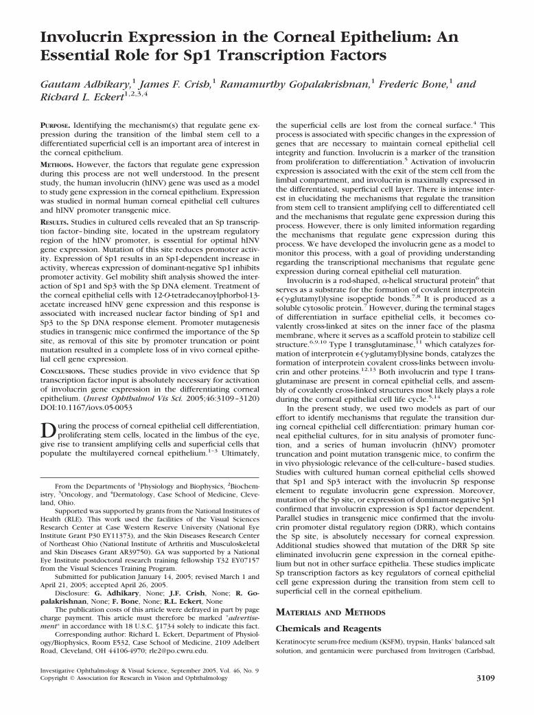
The increased binding to the Sp element in response to TPAtreatment could be due to enhanced expression of Sp1 and Sp3or to enhanced movement of Sp1 to the nucleus. To assess thispossibility, we performed immunoblots with total cell andnuclear extracts prepared from TPA-treated and nontreatedcorneal epithelial cells. Figure 9A shows that TPA treatment for24 hours did not alter the total cellular level of Sp1 or Sp3.However, this treatment did produce a change in cytosolic andnuclear Sp1 and Sp3 levels. The cytosolic level of these factorswas decreased in TPA-treated cells, and the nuclear level ofthese factors, which was relatively low in the absence of TPA
treatment, increased in TPA-treated cells (Figs. 9B, 9C). Thelevel of Sp1 and Sp3 in the cytosol was substantially greater, ona per cell basis, than in the nucleus; therefore, a modestapparent reduction in cytoplasmic Sp1 and Sp3 level resultedin a substantial increase in nuclear level. These blots furthershow that numerous Sp1 and Sp3 immunoreactive bands weredetected, a finding that is consistent with observations in othersystems.40
Effect of the Mutation of the Sp1 Site on hINVExpression in Other Surface Epithelia
Involucrin is expressed in a wide range of surface epithelia.14
To assess whether the hINV promoter Sp-binding site isuniquely necessary for corneal epithelial hINV expression, weexamined the effects of the Sp-binding site mutation on hINVexpression in other surface epithelia. Epithelial sections offootpad, cervix, epidermis, and esophagus were prepared fromDRR(Sp1m)-P3.4B transgenic mice. The structure of theDRR(Sp1m)-P3.4B construct used to produce these mice isshown in Figure 3A. Involucrin expression was monitored byimmunoblot and by immunohistology. Figure 10A shows thatinvolucrin was detected in the epithelial cells derived fromfootpad, epidermis, and esophagus of DRR(Sp1m)-P3.4B mice,as measured by immunoblot. Analysis of cervical tissue by
FIGURE 6. Sp1 and �N-Sp3 regula-tion of hINV promoter activity. (A)Sp1 increased hINV promoter activ-ity. Cells were transfected with 0.5�g of pINV�2473 in the presence ofincreasing levels of Sp1 expressionvector. The total plasmid content ofthe transfection was equalized to 2�g total DNA by addition of emptyvector (EV). (B) Differential regula-tion by Sp1 and Sp3. Corneal epithe-lial cells were transfected with 1 �gof pINV�2473 in the presence of 1.0�g of EV or the Sp1 or �N-Sp3 ex-pression vector. After 24 hours, theindicated groups were treated with50 ng TPA/mL; and, after an addi-tional 24 hours, the cells were har-vested and assayed for luciferase ac-tivity. Data are the mean � SEM ofresults in four separate experiments.(C) Opposing action of Sp1 and �N-Sp3. Cells were transfected with 0.5�g of pINV�2473 and the indicatedlevel of EV or Sp1 or �N-Sp3-encod-ing expression vector. After 24hours, the cells were harvested andassayed for promoter-dependent lu-ciferase activity. Error bars, �SEM.Data are the mean � SEM of resultsin three separate experiments. (D)Sp1 and Sp3 immunoblots. Cellswere transfected with empty expres-sion vector (�) or expression vectorencoding Sp1 or �N-Sp3 (�). After24 hours, the cells were harvestedfor preparation of total extracts. Theextracts were immunoblotted withrabbit anti-Sp1 (dilution � 1:1000) orrabbit anti-Sp3 (dilution � 1:1000).Parallel blots were incubated withanti-�-actin as a gel-loading control.Asterisks: band displaying the mostobvious increase in Sp1- or Sp3-trans-fected cells.
IOVS, September 2005, Vol. 46, No. 9 Gene Regulation in the Corneal Epithelium 3115

immunoblot was not feasible because of the small amount oftissue. Figure 10B confirms that the pattern of expression wasappropriate in each of the four tissues. That is, involucrin
expression was detected in the suprabasal layers in each epi-thelium. Thus, in contrast to the response in the cornealepithelium, mutation of the Sp-binding site does not have as
FIGURE 7. Dominant-negative Sp1 and mithramycin treatment reduce hINV promoter activity. (A) The hINV promoter full-length promoterconstruct, pINV�2473 (0.25 �g), and the DRR luciferase reporter construct, pINV(�2473/�2088; 0.25 �g) were transfected into 40% confluentcultures of normal human corneal epithelial cells in the presence of the indicated number of micrograms of human dominant-negative Sp1-encodingplasmid (dnhSP1). After 24 hours, the cells were harvested and assayed for luciferase activity. Error bars, �SEM. Data are the mean of results infour separate experiments. (B) Mithramycin A treatment reduces hINV promoter activity. The hINV promoter full-length promoter construct,pINV�2473, and the DRR luciferase reporter constructs, pINV(�2473/�2088) and pINV(�2473/�2088), encoding an intact or mutant Sp1 site(Sp1m), were transfected into 40% confluent cultures of normal human corneal epithelial cells. After 24 hours, the cells were treated with 200 nMMMA. After an additional 24 hours, the cells were harvested and assayed for luciferase activity using a fluorometer. Error bars, �SEM. Data are themean results in four separate experiments.
FIGURE 8. Complex formation atthe hINV promoter Sp1 site. (A) TheSp1c and Sp1c-m oligonucleotidesused for gel mobility shift assay. TheSp1 site is indicated in bold andmatches the hINV Sp1 site.18,29 (B)Nuclear extract (NE) was preparedfrom human corneal epithelial cellstreated in the presence and absenceof TPA (50 ng/mL) for 24 hours. Nu-clear extract, prepared from eachgroup of cells, was incubated withdouble-stranded 32P-end-labeled Sp1cfor 25 minutes at room temperature.Some reactions were supplementedwith a 10- or 100-fold molar excess ofnonradioactive Sp1c oligonucleotide.The reaction mixtures were thenfractionated on nondenaturing gelsand bands were visualized by autora-diography. Sp indicates migration ofthe putative Sp factor complexes andFP indicates migration of 32P-end-la-beled Sp1c free probe. An oligonu-cleotide encoding a mutated Sp1binding site 5�-ATTCGATCGGTCAA-GGGCGAGC (Sp1 site underscored,mutated residues in bold) did notcompete for binding. (C) Supershiftanalysis reveals the presence of Sp1and Sp3 at the Sp binding site. Ex-tracts, prepared as just described,were incubated with Sp1c-P32 in thepresence of anti-IgG, anti-Sp1, or an-ti-Sp3. The complexes were then
electrophoresed on a nondenaturing gel and band mobility was visualized by autoradiography. Similar results were observed in each of fourseparate experiments. Asterisk: migration of the Sp3 supershifted band.
3116 Adhikary et al. IOVS, September 2005, Vol. 46, No. 9
dramatic an impact on hINV expression in other surface epi-thelia.
DISCUSSION
Role of the Involucrin Promoter Distal RegulatoryRegion Sp Factor–Binding Site in InvolucrinExpression in the Corneal Epithelium
Identifying mechanisms that govern tissue-specific and differ-entiation-appropriate gene expression in the corneal epithe-lium is an important area of interest. In this epithelium, stemcells give rise to daughter cells that then differentiate to formthe superficial layers of the tissue. This process is marked byprofound changes in cell morphologic and biochemistry. Sev-eral genes have been described that are markers of this pro-cess. These include cytokeratins K3 and K12,30,41,42 MUC1 andMUC4,43 lactate dehydrogenase,44 and involucrin.22 Knowl-edge regarding the mechanisms that guide expression of thesegenes is limited and is confined to conclusions derived fromthe use of cell-culture–based models that have not been con-firmed in vivo. Thus, a central goal of the present study was togain information regarding the mechanisms that drive involu-crin expression using both in vivo and cell-culture–based systems.
The most effective method of confirming cell-culture–basedstudies is the use of transgenic mouse models. Previous studieshave shown that this method can be successfully applied to thestudy of involucrin gene expression.24–26 To identify elementsresponsible for cornea epithelial gene expression, transgenicmice were generated encoding the intact full-length involucrinpromoter and various promoter truncation and point mutants.These studies revealed that deletion of the promoter segmentspanning nucleotides �2473/�1953 results in a complete ab-sence of corneal epithelial involucrin gene expression. Toassess whether the sequences that reside in this segment aresufficient for corneal expression, the �2473/�1953 DRR seg-ment was isolated and linked directly to the involucrin minimalpromoter in the absence of other regulatory elements. Analysis
of these mice revealed a normal level and pattern of involucrinexpression, suggesting that sequences within this segment arenot only necessary, but are also sufficient to drive appropriateinvolucrin gene expression in the corneal epithelium.
Analysis of the sequence of the �2473/�1953 segment, theso called DRR,17 identified several candidate transcription-fac-tor–binding sites that could participate in this regulation, in-cluding an Sp-factor–binding site.24 Previous studies, using cellculture models, suggest a role for Sp factors in regulation ofcorneal epithelial gene expression. For example, Sp factorsplay a role in regulation of expression of cytokeratin K3,45
lactate dehydrogenase,44 �5 integrin,46 and cytokeratin 4.47
We hypothesized that the Sp-factor–binding site within thehINV promoter DRR may be necessary for involucrin expres-sion in the corneal epithelium. Indeed, mutation of this siteresulted in a complete loss of in vivo corneal epithelial expres-sion, suggesting that Sp factor input is necessary for in vivoinvolucrin expression. To our knowledge this is the first in vivoconfirmation that Sp factor input is essential for expression ofa gene in the corneal epithelium.
Sp1: A Selective Role in the Corneal Epithelium?
An important issue is the particular role of Sp1 in the cornealepithelium. Involucrin is expressed in several surface epithelia,including the cervix, epidermis, oral cavity, and esophagus.14
It is possible that removing the Sp binding site could eliminateinvolucrin expression in all these epithelia. Previous studieshave shown that the involucrin transgenes encoding the full-length involucrin upstream regulatory region (nucleotides�2473/�1) are expressed in all murine surface epithelia in apattern that mirrors the expression pattern of endogenoushINV in human tissues.24–26 We therefore assayed whetherelimination of the Sp-binding site resulted in a selective loss oftransgene expression only in the corneal epithelium, or if thismutation produced a generalized loss of expression. Analysis ofthe pattern of expression of the transgene encoding a mutatedSp-binding site shows that the Sp site mutation selectively
FIGURE 9. TPA treatment alters nu-clear Sp factor levels. (A, B) Total celland cytosolic extracts were preparedfrom cells treated for 24 hours with50 ng/mL TPA. The extracts werethen electrophoresed on an 8% acryl-amide gel and transferred to nitrocel-lulose for immunoblot with anti-Sp1,anti-Sp3, or anti-�-actin. Complexeswere then visualized by incubationwith the appropriate secondary anti-body and chemiluminescence detec-tion. (C) Nuclear extracts were pre-pared from cells treated for 24 hourswith 50 ng/mL TPA. The extractswere then electrophoresed on 8%acrylamide denaturing gel and trans-ferred to nitrocellulose for immuno-blot. The blots were then incubatedwith anti-Sp1 or anti-Sp3. Complexeswere then visualized by incubationwith the appropriate secondary anti-body and chemiluminescence detec-tion. For optimal visualization, thenuclear extract was exposed to filmseveral times longer than the totaland cytosolic extracts. This reflectsthe overall lower abundance of thesefactors in the nucleus.
IOVS, September 2005, Vol. 46, No. 9 Gene Regulation in the Corneal Epithelium 3117

eliminates transgene expression in the cornea and not in othersurface epithelia (i.e., cervix, epidermis, and esophagus). Thisfinding indicates that Sp transcription factors are uniquelynecessary for involucrin expression in the corneal epithelium.The effect of mutating the Sp-binding site is in contrast to theimpact of mutating the AP1-5 site. AP1-5 is an AP1 transcrip-tion-factor (junB, junD, Fra-1, and Fra-2)–binding site located in
the DRR, adjacent to and immediately upstream of the Sp-factor–binding site.15,24 Previous studies have shown that mu-tation of AP1-5 results in a loss of transgene expression inepidermis, esophagus, and cervix.25 Our recent study indicatesthat mutation of the AP1-5 site also abolishes expression in thecorneal epithelium.48 Taken together, these findings suggestthat AP1 transcription factor input is essential for expression ofinvolucrin in many surface epithelia, but that Sp factors areuniquely necessary for expression in the corneal epithelium.
Regulatory Role of Sp Factors
The Sp-factor–related regulation of involucrin gene expressionin corneal epithelium is likely to be complicated. Our in vivotransgenic studies showed that mutation of the Sp site withinthe hINV promoter DRR results in a complete loss of in vivocorneal epithelial hINV expression. Additional studies, usingcultured normal human corneal epithelial cells, showed thatmutation of the Sp site causes a reduction in both basal andTPA-stimulated promoter activity. TPA treatment is known toincrease involucrin promoter activity in cultured corneal epi-thelial cells.22 Thus, Sp factor function is necessary for bothbasal and regulated involucrin promoter activity. In addition,expression of a dominant-negative form of Sp138 suppressesSp1- and TPA-dependent promoter activation, and treatmentwith mithramycin A, an agent that selectively binds to G/C-richDNA and prevents Sp factor binding, reduced Sp1- and TPA-dependent promoter activity and also reduced endogenousinvolucrin gene expression. Moreover, treatment with TPAresulted in an increased nuclear Sp factor level and increasedbinding of Sp factors to the hINV promoter Sp-binding site,responses that are correlated with increased hINV promoteractivity. The finding that Sp factor level can alter involucrinexpression is consistent with a previous study showing thatoverexpression of Sp1 activates expression of the endogenousinvolucrin gene in fibroblasts, a cell type that does not nor-mally express involucrin in vivo.29 Based on these findings, onecould hypothesize that the absolute level of Sp1 present in thetissue directly influences the level of involucrin expression.However, this is clearly not the case. In fact, as shown in thepresent study, mouse liver, a tissue that does not expressinvolucrin, expressed more Sp1 than the corneal epithelium.Moreover, other surface epithelial tissues also expressed highlevels of Sp1,18 yet mutation of the Sp binding site within thehINV promoter did not eliminate expression in these tissues(Fig. 10).
These results suggest that the unique role of Sp factors inthe corneal epithelium must be explained by other mecha-nisms. Potential levels of differential regulation may includedifferences in the covalent posttranslational modification ofSp1 and Sp3. These covalent modifications, which includephosphorylation (Sp1), glycosylation (Sp1), acetylation (Sp1,Sp3), and sumoylation (Sp3), are known to influence Sp factoractivity.40 Parallel studies indicate that the Sp1, present incorneal extracts, is extensively phosphorylated and glycosy-lated (not shown); however, the impact of these modificationson function is not presently known. In addition, differentialinteraction with coregulators may play an important role. Sp1is known to interact with a wide range of regulators in othertissues.33,49 The precise nature of the Sp1 and Sp3 interactionwith the Sp DNA element may be important, as gel mobilitysupershift studies suggested that Sp1 may anchor Sp3 to the Spbinding site (i.e., addition of an Sp1-specific antibody to a gelshift mixture resuled in a loss of both Sp1 and Sp3 binding, but,in contrast, addition of an anti-Sp3 antibody resulted in a loss ofthe putative Sp3 band, but only minor changes in the apparentlevel of bound Sp1). Sp1 and Sp3 are known to regulatedifferentially the expression of other target genes50 and ourpresent study suggests that Sp1 and an amino-terminal trun-
FIGURE 10. The hINV promoter Sp1 site has a unique role in thecorneal epithelium. Mouse embryos were injected with theDRR(Sp1m)-P3.4B transgene. The structure of the construct is shownin Figure 3A. Expression of the transgene was monitored by immuno-blot and/or immunohistology using a hINV-specific antibody. Surfaceepithelium from footpad, epidermis, and esophagus were harvested,and total cell extracts were prepared in sample buffer. Samples wereelectrophoresed on 8% denaturing and reducing polyacrylamide gels,transferred to nitrocellulose, and incubated with hINV-specific anti-body. For immunohistochemical localization, sections were preparedand then incubated with anti-hINV followed by peroxidase-conjugatedgoat anti-rabbit IgG.24 The samples were derived from the same sixindependently derived transgenic mouse lines used in Figure 3.
3118 Adhikary et al. IOVS, September 2005, Vol. 46, No. 9

cated form of Sp340 differentially regulate involucrin promoteractivity. This antagonistic relationship between Sp1 and thistruncated form of Sp3 has been noted in other systems.50–52
The mechanism responsible for this inverse regulatory relation-ship is not known. However, Sp1 and Sp3 factors can formheterodimers37; thus, it is possible that an Sp1/Sp3 het-erodimer has altered activity.41,53 Additional studies are underway to assess these possibilities.
References
1. Cotsarelis G, Cheng SZ, Dong G, Sun TT, Lavker RM. Existence ofslow-cycling limbal epithelial basal cells that can be preferentiallystimulated to proliferate: implications on epithelial stem cells. Cell1989;57:201–209.
2. Lavker RM, Dong G, Cheng SZ, Kudoh K, Cotsarelis G, Sun TT.Relative proliferative rates of limbal and corneal epithelia: impli-cations of corneal epithelial migration, circadian rhythm, and su-prabasally located DNA-synthesizing keratinocytes. Invest Oph-thalmol Vis Sci. 1991;32:1864–1875.
3. Sun TT, Lavker RM. Corneal epithelial stem cells: past, present, andfuture. J Investig Dermatol Symp Proc. 2004;9:202–207.
4. Ladage PM, Jester JV, Petroll WM, Bergmanson JP, Cavanagh HD.Vertical movement of epithelial basal cells toward the cornealsurface during use of extended-wear contact lenses. Invest Oph-thalmol Vis Sci. 2003;44:1056–1063.
5. Chen Z, de Paiva CS, Luo L, Kretzer FL, Pflugfelder SC, Li DQ.Characterization of putative stem cell phenotype in human limbalepithelia. Stem Cells. 2004;22:355–366.
6. Eckert RL, Green H. Structure and evolution of the human involu-crin gene. Cell. 1986;46:583–589.
7. Rice RH, Green H. Presence in human epidermal cells of a solubleprotein precursor of the cross-linked envelope: activation of thecross-linking by calcium ions. Cell. 1979;18:681–694.
8. Rice RH, Green H. The cornified envelope of terminally differen-tiated human epidermal keratinocytes consists of cross-linked pro-tein. Cell. 1977;11:417–422.
9. Kalinin AE, Kajava AV, Steinert PM. Epithelial barrier function:assembly and structural features of the cornified cell envelope.Bioessays. 2002;24:789–800.
10. Eckert RL, Yaffe MB, Crish JF, Murthy S, Rorke EA, Welter JF.Involucrin: structure and role in envelope assembly. J Invest Der-matol. 1993;100:613–617.
11. Phillips MA, Qin Q, Mehrpouyan M, Rice RH. Keratinocyte trans-glutaminase membrane anchorage: analysis of site-directed mu-tants. Biochemistry. 1993;32:11057–11063.
12. Kim IG, McBride OW, Wang M, Kim SY, Idler WW, Steinert PM.Structure and organization of the human transglutaminase 1 gene.J Biol Chem. 1992;267:7710–7717.
13. Robinson NA, LaCelle PT, Eckert RL. Involucrin is a covalentlycrosslinked constituent of highly purified epidermal corneocytes:evidence for a common pattern of involucrin crosslinking in vivoand in vitro. J Invest Dermatol. 1996;107:101–107.
14. Banks-Schlegel S, Green H. Involucrin synthesis and tissue assem-bly by keratinocytes in natural and cultured human epithelia. J CellBiol. 1981;90:732–737.
15. Welter JF, Crish JF, Agarwal C, Eckert RL. Fos-related antigen(Fra-1), junB, and junD activate human involucrin promoter tran-scription by binding to proximal and distal AP1 sites to mediatephorbol ester effects on promoter activity. J BiolChem. 1995;270:12614–12622.
16. Efimova T, LaCelle P, Welter JF, Eckert RL. Regulation of humaninvolucrin promoter activity by a protein kinase C, Ras, MEKK1,MEK3, p38/RK, AP1 signal transduction pathway. J Biol Chem.1998;273:24387–24395.
17. Welter JF, Eckert RL. Differential expression of fos and jun familymembers c-fos, fosB, Fra-1, Fra-2, c-jun, junB and junD duringhuman epidermal keratinocyte differentiation. Oncogene. 1995;11:2681–2687.
18. Banks EB, Crish JF, Welter JF, Eckert RL. Characterization of hu-man involucrin promoter distal regulatory region transcriptionalactivator elements-a role for Sp1 and AP1 binding sites. Biochem J.1998;331:61–68.
19. Udvadia AJ, Templeton DJ, Horowitz JM. Functional interactionsbetween the retinoblastoma (Rb) protein and Sp-family members:superactivation by Rb requires amino acids necessary for growthsuppression. Proc Natl Acad Sci USA. 1995;92:3953–3957.
20. Kadonaga JT, Courey AJ, Ladika J, Tjian R. Distinct regions of Sp1modulate DNA binding and transcriptional activation. Science.1988;242:1566–1570.
21. Kadonaga JT, Carner KR, Masiarz FR, Tjian R. Isolation of cDNAencoding transcription factor Sp1 and functional analysis of theDNA binding domain. Cell. 1987;51:1079–1090.
22. Adhikary G, Crish J, Lass J, Eckert RL. Regulation of involucrinexpression in normal human corneal epithelial cells: a role foractivator protein one. Invest Ophthalmol Vis Sci. 2004;45:1080–1087.
23. Schreiber E, Matthias P, Muller MM, Schaffner W. Rapid detectionof octamer binding proteins with “mini-extracts”, prepared from asmall number of cells. Nucleic Acids Res. 1989;17:6419.
24. Crish JF, Zaim TM, Eckert RL. The distal regulatory region of thehuman involucrin promoter is required for expression in epider-mis. J Biol Chem. 1998;273:30460–30465.
25. Crish JF, Bone F, Banks EB, Eckert RL. The human involucrin genecontains spatially distinct regulatory elements that regulate expres-sion during early versus late epidermal differentiation. Oncogene.2002;21:738–747.
26. Crish JF, Howard JM, Zaim TM, Murthy S, Eckert RL. Tissue-specificand differentiation-appropriate expression of the human involu-crin gene in transgenic mice: an abnormal epidermal phenotype.Differentiation. 1993;53:191–200.
27. LaCelle PT, Lambert A, Ekambaram MC, Robinson NA, Eckert RL.In vitro cross-linking of recombinant human involucrin. Skin Phar-macol Appl Skin Physiol. 1998;11:214–226.
28. Agarwal C, Efimova T, Welter JF, Crish JF, Eckert RL. CCAAT/enhancer-binding proteins: a role in regulation of human involu-crin promoter response to phorbol ester. J Biol Chem. 1999;274:6190–6194.
29. Banks EB, Crish JF, Eckert RL. Transcription factor Sp1 activatesinvolucrin promoter activity in non-epithelial cell types. BiochemJ. 1999;337:507–512.
30. Chen TT, Wu RL, Castro-Munozledo F, Sun TT. Regulation of K3keratin gene transcription by Sp1 and AP-2 in differentiating rabbitcorneal epithelial cells. Mol Cell Biol. 1997;17:3056–3064.
31. Courey AJ, Tjian R. Analysis of Sp1 in vivo reveals multiple tran-scriptional domains, including a novel glutamine-rich activationmotif. Cell. 1988;55:887–898.
32. Jackson SP, Tjian RO. Glycosylation of eukaryotic transcriptionfactors: implications for mechanisms of transcriptional regulation.Cell. 1988;55:125–133.
33. Bouwman P, Philipsen S. Regulation of the activity of Sp1-relatedtranscription factors. Mol Cell Endocrinol. 2002;195:27–38.
34. Berg JM. Sp1 and the subfamily of zinc finger proteins with gua-nine-rich binding sites. Proc Natl Acad Sci USA. 1992;89:11109–11110.
35. Philipsen S, Suske G. A tale of three fingers: the family of mamma-lian Sp/XKLF transcription factors. Nucleic Acids Res. 1999;27:2991–3000.
36. Black AR, Black JD, Azizkhan-Clifford J. Sp1 and kruppel-like factorfamily of transcription factors in cell growth regulation and cancer.J Cell Physiol. 2001;188:143–160.
37. Suske G. The Sp-family of transcription factors. Gene. 1999;238:291–300.
38. Petersohn D, Thiel G. Role of zinc-finger proteins Sp1 and zif268/egr-1 in transcriptional regulation of the human synaptobrevin IIgene. Eur J Biochem. 1996;239:827–834.
39. Kaluz S, Kaluzova M, Stanbridge EJ. Expression of the hypoxiamarker carbonic anhydrase IX is critically dependent on SP1activity: identification of a novel type of hypoxia-responsive en-hancer. Cancer Res. 2003;63:917–922.
40. Sapetschnig A, Koch F, Rischitor G, Mennenga T, Suske G. Com-plexity of translationally controlled transcription factor Sp3 iso-form expression. J Biol Chem. 2004;279:42095–42105.
41. Li L, He S, Sun JM, Davie JR. Gene regulation by Sp1 and Sp3.Biochem Cell Biol. 2004;82:460–471.
IOVS, September 2005, Vol. 46, No. 9 Gene Regulation in the Corneal Epithelium 3119

42. Liu CY, Zhu G, Westerhausen-Larson A, et al. Cornea-specificexpression of K12 keratin during mouse development. Curr EyeRes. 1993;12:963–974.
43. Gipson IK, Argueso P. Role of mucins in the function of thecorneal and conjunctival epithelia. Int Rev Cytol. 2003;231:1–49.
44. Hernandez-Quintero M, Garcia-Villegas R, Castro-Munozledo F. Dif-ferentiation-dependent increases in lactate dehydrogenase activityand isoenzyme expression in rabbit corneal epithelial cells. ExpEye Res. 2002;74:71–82.
45. Alarcon R, Koumenis C, Geyer RK, Maki CG, Giaccia AJ. Hypoxiainduces p53 accumulation through MDM2 down-regulation andinhibition of E6-mediated degradation. Cancer Res. 1999;59:6046–6051.
46. Larouche K, Leclerc S, Salesse C, Guerin SL. Expression of thealpha 5 integrin subunit gene promoter is positively regulated bythe extracellular matrix component fibronectin through the tran-scription factor Sp1 in corneal epithelial cells in vitro. J BiolChem.2000;275:39182–39192.
47. Opitz OG, Rustgi AK. Interaction between Sp1 and cell cycleregulatory proteins is important in transactivation of a differentia-tion-related gene. Cancer Res. 2000;60:2825–2830.
48. Adhikary G, Crish JF, Bone F, Gopalakrishnan R, Lass J, Eckert RL.An involucrin promoter AP1 transcription factor binding site isrequired for expression of involucrin in the corneal epithelium invivo. Invest Ophthalmol Vis Sci. 2005;46:1219–1227.
49. Billon N, Carlisi D, Datto MB, et al. Cooperation of Sp1 and p300in the induction of the CDK inhibitor p21WAF1/CIP1 duringNGF-mediated neuronal differentiation. Oncogene. 1999;18:2872–2882.
50. Hagen G, Muller S, Beato M, Suske G. Sp1-mediated transcriptionalactivation is repressed by Sp3. EMBO J. 1994;13:3843–3851.
51. Kumar AP, Butler AP. Transcription factor Sp3 antagonizes activa-tion of the ornithine decarboxylase promoter by Sp1. NucleicAcids Res. 1997;25:2012–2019.
52. Li JM, Datto MB, Shen X, Hu PP, Yu Y, Wang XF. Sp1, but not Sp3,functions to mediate promoter activation by TGF-beta throughcanonical Sp1 binding sites. Nucleic Acids Res. 1998;26:2449–2456.
53. Tang S, Bhatia B, Zhou J, et al. Evidence that Sp1 positively and Sp3negatively regulate and androgen does not directly regulate func-tional tumor suppressor 15-lipoxygenase 2 (15-LOX2) gene expres-sion in normal human prostate epithelial cells. Oncogene. 2004;23:6942–6953.
3120 Adhikary et al. IOVS, September 2005, Vol. 46, No. 9