investigation of the physiological basis of the rind
TRANSCRIPT
RA
Investigation of the physiological basis of the
rind disorder oleocellosis in Washington navel
orange (Citrus sinensis [L'l Osbeck)
Toby George Knight
B.Ag.Sc. (Hort.Sc.)
Submitted in fulfilment of requirements for the degree of
Doctor of PhilosoPhY
Deparünent of Horticulture, viticulture and oenolory
The UniversitY of Adelaide
June,2002

Table of Contents
Abstract.. """' iDeclaration.............. ""' iii
Acknowledgments..... """""""""" iv
List of Abbreviations............... ""' vi
List of Figures......... "' viii
List of P1ates............ ""' ixList of Tables...... """""' x
Chapter 1
General Introduction............. 1
Chapter 2Literature Review....
2. 1. The fruit..........2.1.1. Botanical classification .'.... " "' "'2.1.2. Rind anatomy ..... -......
2.2. The oil gland
2.2.1. Gland development.........'....2.2.2. Gland anatomY
2.3.Therind oils2.3.1. Oil synthesis and accumulation '"""""'2.3.2. Oil composition..... ".....2.3.3. Oil ProPerties ....'.....'.
2.4. Oleocellosis.........2.4.1. TerminologY2.4.2. Cause2.4.3. SymPtoms2.4.4. Artificial induction. .
2.4.5. Oleocellosis PhYsiolory2.4.6. Fruit susceptibility to oleocellosis """"""2.4.7 . Oleocellosis control2.4.8. Fruit treatments affecting oleocellosis ''""''"'
2.5. Summary..........'.
5
5
5
5
7
7
8
9
.910
10
11
11
11
l1l3t415
16
18
20
2.6. Aims.
Chapter 3
Oil giand development: structure and quantification """""3. 1. Introduction.......
3.1.1. Background.3.1.2. Aims ...........
2t
22
221,)
23
3 .2. Matenals and Methods...
3.2.1. Plant material .'....'.....'.
3.2.2. Gland counts
3.2.3. Microscopy ...........3.2.4. Gland age survey
3.2.5. Image analysis.....
3 .2.6. Statistical analYsis
3.3. Results................3.3. 1. Anatomical gland development .
3.3.2. Timing of gland development
3.3.3. The mature gland
3.4. Discussion...........
3.5. Conclusion
Chapter 4Rind oil visualisation: method development........... """41
2727
30
36
40
4. 1. Introduction........4.1.I. Background.41 2 Aims
4.2. Matenals and Methods... "......4.2.1. Light microscopy (LM).............
4.2.2. Scanning electron microscopy (SEM)
4.2.3. Fluorescence microscopy...........
4.3. Results4.3.1. Lieht microscoPY (LM)...4.3.2. Scanning electron microscopy (SEM)
.41
.42
.43
4l
43
..46
..47
..49
..49
..51
534.3.3. Fluorescence mlcrocopy ..'.......
4.4. Discussion...............4.4.1. Method development4.4.2. Rind oil observations
4.5. Conclusion ..........
Chapter 5Inducing oleocellosis in the laboratory
5.1. Introduction5.1.1. Background5.I.2. Aims...........
5.2. Materials and Methods...5.2.1. Preliminary induction methods test ....
5.2.2. Oil method refinement5.2.3. Penetrometer method refinement
5.3. Results.................5.3.1. Preliminary induction methods test
5.3.2. Oil method refinement
58
58
63
64
.65
.65..65
66
6767
7t72
73
73
78
835 . 3. 3. Penetrometer method refinement'.'. -...
5.4. Discussion...........5 .4.1. Mechanical induction.5.4.2. Oil induction...........' -.
5.5. Conclusion..........
86
86
89
91
Chapter 6Oleocellosis: macroscopic and microscopic development............................... 92
6.1.Introduction......... "' '926.1.1. Background..........'.... ""' 92
6.1.2.4ims........ "" 93
6.2.Matenals and Methods """ ' 94
6.2.1. Symptom time course """"' 94
6.2.2.Mcroscopy...........'.. "' """" 97
6.3. Resu1ts............... " "" "101
6.3.1. Symptoms ""1016.3.2.Rindsurface damage """"'1056.3.3.Rind sub-surface damage...'....'.". """ """ ""108
6.4. Discussion........... "" """"'I246.4.1. Symptoms ""' "1246.4.2. Oleocellosis mechanism.............. """"1256.4.3. Oil effects. "1286.4.4. The link between microscopic damage and symptoms """' 130
6.5. Conclusion.......... " 133
Chapter 7General Discussion ""'134
7.1. Addressing limitations of current control practices... """""'1347.2.More factors influencing fruit susceptibility to oleocellosis"....'...... . .. . 136
7.3. Developing new strategies for oleocellosis control..................... . .. . ..139
7.4.Oleocellosisdiagnosis.........'. ' """"""1417.5. Summary............ """"""""742
Appendix 1: Video Pro programs.......... ""'143Appendix 2: Publications ........ "'145Appendix 3: Conference presentations """'146Appendix 4: Industry communication.....-.. """"""""'151
Bibliography ............ """""""""'152Personat Communications..... ""'165

z
Abstract RA
Oleocellosis is a rind disorder of citrus fruit, which produces an unattractle
surface blemish, and causes significant economic losses to the Australia¡ citrus
industry. It is caused by phytotoxic oils released from oil glands located in the rind,
as a result of mechanical damage. In this study, microscopy investigations into the
oil glands, localisation of the rind oils and the development of oleocellosis have
been carried out in Washington navel orange ftvit (Citnls sinensis [L.] Osbeck)'
Changes in the structure, size and the number of oil glands located in the rind
were assessed in the developing fnrit from pre-anthesis to fruit maturity' Gland
initiation was restricted to early fruit development, but glands continued to develop
and reached maturity by fruit size 30 to 50 mm diameter. Mature glands continued
to enlarge with ftiit growth, but their final size and form varied within each fruit.
Mature fruit had between 8,000 and 12,000 oil glands. Glands were found to
develop from a cluster of cells adjacent to the fruit epidermis, into a structure
consisting of a central cavity surrounded by several layers of epithelial cells. All
glands were joined to the fruit epidermis by a 'stalk'. Gland cavity formation
appeared to involve schizogenY.
Methods of tissue preparation were investigated in an effort to retain the
essential oil contained within the glands. The modification of chemical fixation
methods for improved rind oil retention gave limited success based on light
microscopy observations. However, the examination of cryofixed material with
scanning electron microscopy and fresh tissue with multi-photon microscopy were
promising techniques for oil visualisation in orange rind tissue.
Oleocellosis was artificially induced in mature fruit under controlled conditions.
This was achieved by mechanically damaging the fruit or applying rind oils to the
fruit surface. Based on a range of criteria, penetrometer damage and dlimonene
treatment were chosen as the optimal methods for inducing oleocellosis These
methods were considered to simulate the two forms of naturally occurring
oleocellosis; namely gland rupture and oil transfer between fruit.
Following induction, oleocellosis development was examined using a detailed
time course assessment of surface symptoms and microscopic rind damage. Based
on these observations, the events leading to oleocellosis blemish formation were
proposed. Mechanical damage resulted in rupture of the epidermis above the
glands and release of oil to the ft¡it surface. Surface oil appeared to infiltrate the
rind via the cuticle, and via the ruptured epidermis in injured fn¡it. Once in the rind,
the phytotoxic oils caused rapid cell content degeneration and cell collapse, with
early stages of.ultrastructural damage detected within six hours of induction. The
resulting blemish, which was cha¡acterised by rind collapse and darkening,
developed substantially within three days and was attributed to the sub-surface rind
damage.
ll

Declaration
This thesis contains no material which has been accepted for the award of any
other degree or diploma in universþ or other tertiary institution and, to the
best of my knowledge and belief, contains no material previously published or
written by another person, except where due reference has been made in the text.
I give consent to this copy of my thesis, when deposited in the Universþ library,
being available for loan and photocopyrng.
Toby George Krìight.
Date: 14.6.O>
ru

Acknowledgments
Thanks to my supenrisors, Andreas Klieber and Margaret Sedgley, for their
ongoing support and encouragement.
This study was funded by the Australian Research Council through the Strategic
Partnership with Industry - Research and Training (SPIRT) Scheme. Thanks to
our industry partner, the Citrus Board of South Australia, for their support and for
enabling me to travel to the International Society for Citriculture 2000 Congress in
Orlando, Florida. Special thanks to Andrew C¡reen.
Thanks to Gueue Bros. Pty. Ltd., Mypolonga, for their contribution of fruit.
Thanks to atl my Mcroscopy Lab mates over the years, especially Merran
Matthews, Raelene Mbus and Leanne Pound, for their friendship and support,
during the countless hours on the microtome and weekends in the darkroom. Also
to all my other friends from FIVO, who have made it such an enjoyable place to
work.
Special thanks to Meredith Wallwork for her unwavering support on every level,
and to peter Kolesik for his advice and constant humour. Thanks also to Meredith,
Chris Ford and Bob Barrett for their proof reading prol¡/ess.
Thanks to the staff of The Centre for Electron Mcroscopy and Microstructural
Analysis (CEMMSA), especially Lyn Waterhouse, for their technical advice and
instruction, and for making the dungeon always a fun place to be.
Thanks to the University of Adelaide, for enabling me to travel to the Sixth
International Botanical Microscopy Meeting, St Andrews, Scotland, with a
Research Abroad Scholarship. To Chris Hawes, Oxford Brookes University, for
lv
allowing me to visit, and Rebecca Morden, Royal Mcroscopical Society, for her
help and humour.
To all others who have helped with this project, incuding:
. Michelte Lorimer, Biometrics SA, for her support with statistics'
. Brian Loveys, Sue Maffei and Sue Johnson, cslRo Plant Industry.
o Barry Tugwell and other staffof SARDI Postharvest Horticulture'
o Karen Churchill and Lynn Jarvis for technical support for Video Pro'
o Sharon Clapham and other staffof the Image and Copy Centre, Waite Campus
Last but definitely not least. To my family, for seeing me through the many years of
study. fuid to my friends, who have always provided a welcome distraction. Gold
stars to Tony, PennY and Matt.
v

List of Abbreviations
a-pinene
p-myrcene
p-glucan
'cCBSA
CEMMSA
CLSM
cm
CSIRO
CW
ER
ESEM
et ø1.
FEGSEM
Fig.
ob
GA
GMA
h
HCr
kg
kPa
kvLM
LSD
M
min
mm
ms-'
alpha-pinene
beta-myrcene
beta-glucan
degree(s) Celsius
Citrus Board of South Australia
centre for Electron Microscopy and Microstructural Analysis
confocal laser scanning microscope or microscopy
centimetre(s)
Commonwealth S cientifi c and Industrial Research Organisation
Calcofluor White
endoplasmic reticulum
environmental scanning electron microscope or microscopy
and others
field emission gwr scanning electron microscope or microscopy
Figure
grarn(s)
gibberellic acid
glycol-methacrylate
hour(s)
hydrochloric acid
kilogram(s)
kiloPascal(s)
kilovolt(s)
light microscope or microscopy
least signifi cant difference
mole(s)
minute(s)
millimetre(s)
metres per second, Per second
vl
N
nm
NR
P
Pa
PA
PAS
Pers. Comm.
PIRSA
PPO
RORP
SARDI
SBB
SEM
TBO
TEM
pm
pl
USA
UV
VPD
Newton(s)
nanomehe(s)
Nile Red
probability
Pascal(s)
Procure-Araldite
periodic acid-Schiffls
Personal Commr¡nication
Primary Industries and Resowces South Australia
polphenol oxidase
rind oil release pressure
South Australian Research and Development Institute
Sudan Black B
scanning electron microscope or microscopy
Toluidine Blue O
transmission electron microscope or microscopy
micrometre(s)
microlitre(s)
United States of America
ultraviolet
vapour pressr¡re deficit
vu
List of Figures
Figure 2.1. Cross-section of mature lemon rind' """"' """" 6
Figure 2.2. Oleocellosis symptoms on mature navel orange fn¡it. -.-..-- 12
Figure 3.1. Changes in oil gland densþ and total gland number per fruit with fruit
growth. ' "" 31
Figure 3.2. Glandage survey in ftiit stages Ato E' """""32
Figure 3.3. Mature gland volume in fruit stages A to E' "" 34
Figure 5.1. preliminary induction methods test. The effect of induction method and
induction time on blemish severity at days I and 3. ...........74
Figure 5.2. preliminary induction methods test. The effect of induction method and
inductiontime onblemish areaatdays I and 3" """"""""77Figure 5.3. Oil method refinement. The effect of dJimonene concentration on
blemish severity at days I and 3. """""' 79
Figure 5.4. Oil method refinement. The effect of citral concentration and 100% d-
limonene on blemish severity at days I and 3' """ """""" 79
Figure 5.5. Oil method refinement. The effect of dlimonene concentration on
btemish ueaatdays 1 and 3.....".. ' " """82
Figure 5.6. Oil method refinement. The effect of citral concentration and 100% d-
limoneneonblemish areaaldays1and3' """"82
Figure 5.7. penetrometer method refinement. The effect of induction time on
blemish severitY at daYs I and 3.
Figure 5.8. Penetrometer method refinement. The effect of induction time on
blemish areaat days 1 and 3...... .'
Figure 6.1. The relationship between time and rind collapse' """"""Figure 6.2. The relationship between time and rind discolouration.
Figure 6.3. The relationship between time and blemish area"""""
.84
102
r02
103
vln

List of Plates
Plate 3.1. Light micrographs of the gland developmental series. .-..-.--28
Plate3.Z.Light micrographs depicting the gland stalk and gland form' .....-....-..--29
plate 3.3. Micrographs depicting mature gland anatomy and cavity formation....35
Plate 4.1. Rind oil visualisation using light microscopy.. 50
Plate 4.2. Rind oil visualisation using scanning electron microscopy................-..52
Plate 4.3. Rind oil visualisation using light fluorescence microscopy. ......'....-....'54
plate 4.4. Rind oil visualisation using confocal laser scanning microscopy. ........55
Plate 4.5. Rind oil visualisation using multi-photon microscopy..........................57
Plate 5.1. Blemish severity scoring system........
Plate 5.2. Oleocellosis symptoms induced in the preliminary induction methods
test. 75
Plate 5.3. Oleocellosis symptoms induced in the oil method refinement
experiments. .................. 80
Plate 5.4. Tlpical oleocellosis blemish resulting from penetrometer induction....85
Plate 6.1. Modified blemish severity scoring system. 96
PIate 6.2. Rind profile image of healtþ rind tissue. 100
Ptate 6.3. Dissecting microscope images of fruit swface damage and oleocellosis
symptoms..... 106
Plate 6.4. Scanning electron micrographs of the fruit surface following mechanical
damage, with the t07
Plate 6.5. Light micrographs of the oleocellosis-damaged rind...........................109
Plate 6.6. Scanning electron micrograph of the oleocellosis-damaged rind,
resulting from oil treatment. 110
Plate 6.7. Multi-photon microscope images of healthy and oleocellosis-damaged
rind 111
Plate 6.8. Rindprofile images of healtþ and oleocellosis-damaged rind........."113
Plate 6.9. Transmission electron micrographs of rind ultrastructure in an untreated
70
fruit sample. .....................
Plate 6.10. Transmission electron micrographs of rind ultrastructure in an oil-
tl4
treated fruit sample, after 30 minutes
Plate 6.11. Transmission electron micrographs of rind ultrastructure in an oil-treated fruit sample, after six hours...
Plate 6.12. Transmission electron micrographs of rind ultrastructure in an oil-treated fruit sample,after two daYs.
116
.118
D(
119

plate 6.13. Transmission electron micrographs of rind ultrastructure in an oil-
treated frtrit sample, after ten days.
Plate 6.14. Transmission electron micrographs of rind ultrastructure in a
penetrometer-damaged fruit sample, after three days'
121
123
List of Tables
Table 4.1. Summary of fixation, dehydration and embedding schedules for LM
Methods I to 7. """"""' 43
Table 6.1. Variance strata for blemish collapse, colour and area at day 3, expressed
as percentage of total variation. Experiment 1, fruit han¡ested from five
treãs and different fruit assessed at each time. Experiment 2, ftuitharvested from one tree and same fruit assessed at each time. Sample
refers to replicate treatments on the one fnrit' 104
x
Chapter L
General Introduction
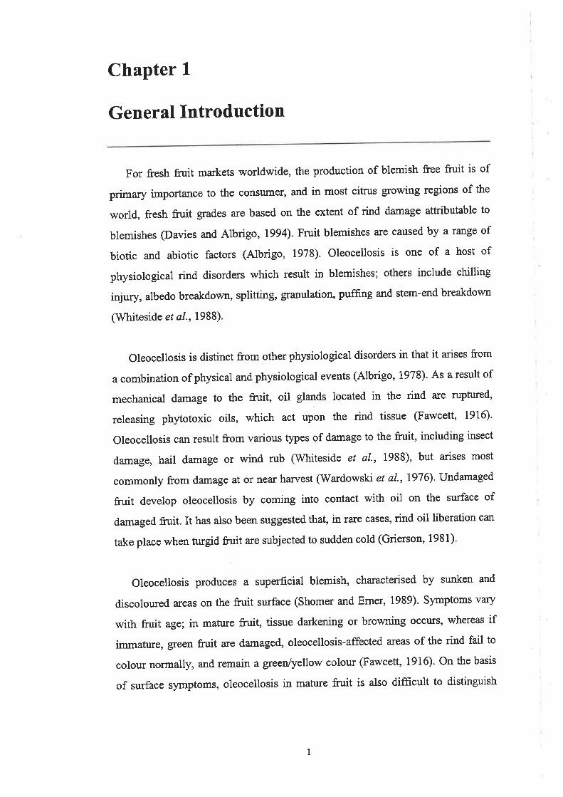
For fresh fruit markets worldwide, the production of blemish free fruit is of
prima¡y importance to the consumer' and in most citrus growing regions of the
world, fresh fruit grades are based on the extent of rind damage attributable to
blemishes (Davies and Albrigo, 1994). Fruit blemishes are caused by a range of
biotic and abiotic factors (Albrigo, 1973). Oleocellosis is one of a host of
physiological rind disorders which result in blemishes; others include chilling
injury, albedo breakdown, splitting, granulation, puffing and stem-end breakdown
(Whiteside et al., 1 988).
Oleocellosis is distinct from other physiological disorders in that it arises from
a combination of physical and physiological events (Albrigo, l97S)- As a result of
mechanical damage to the fruit, oil glands located in the rind are ruptured,
releasing ph¡otoxic oils, which act upon the rind tissue (Fawcett, 1916).
Oleocellosis can result from various types of damage to the fruit, including insect
damage, hail damage or wind rub (Whiteside el ø/., 1988), but arises most
commonly from damage at or near harvest (Wardowski e/ al',1976)' Undamaged
fruit develop oleocellosis by coming into contact 'ù/ith oil on the surface of
damaged fruit. It has also been suggested that, in rare cases, rind oil liberation can
take place when turgid fruit are subjected to sudden cold (Grierson, 1981)'
Oleocellosis produces a superficial blemish, characterised by sunken and
discoloured areas on the fruit surface (Shomer and Erner, 1989)' Symptoms vary
with fruit age; in mature fruit, tissue darkening or browning occurs, whereas if
immature, green fruit are damaged, oleocellosis-affected areas of the rind fail to
colour normally, and remain a greenlyellow colour (Fawcett, 1916). On the basis
of surface symptoms, oleocellosis in mature fruit is also difficult to distinguish
1
from other rind disorders such as rind staining (Eaks, 1964) and pitting (du
Plessis, 1978).
Oleocellosis is considered to be a major concern for the South Australian
citrus industry, which accoturts for 3lYo of Aushalia's production, as well as 46 o/o
of exports valued at approximately A$180 million (BanV Tugwell, Chief
Scientist, SARDI; Pers. Comm., 2001). It has been estimated that oleocellosis can
affect up to l}Yo of a crop (Andrew Green, Technical Officer, CBSA; Pers.
Comm., 1993). Oleocellosis is often detected during grading and fruit can be
discarded prior to packing, but delayed symptom detection can necessitate further
fruit discard and repacking at destination markets (Green, Pers. Comm., 1998). In
addition, rind damage associated with oleocellosis can predispose the fruit to
pathogen infection during storage.
In recent years in Australia, the oversupply of domestic markets has placed an
emphasis on the export of fresh fruit, to help maintain the industry's viability. In
lgg2, after many years of negotiations over quarantine issues, South Australian
growers were grcnted access to fresh fruit markets in the USA. In trial shipments
of navel oranges to the USA, it was estimated that A$1 million was lost due to
repacking costs incured as a result of the presence of oleocellosis-like blemishes.
Today, the industry's export drive continues to be led by navel orange exports to
the USA. Two million cartons of oranges in total, particularly navels, are exported
to the USA and Japan each year. Reports indicate that a high incidence of
oleocellosis-like blemishes was again observed during the 2000 season (Green,
Pers. Comm.,2001).
Oleocellosis control strategies currently employed by growers include careful
fruit handling at all stages of production and the use of oleocellosis prediction
tests prior to han¡est. A range of practices have been implemented to minimise
mechanical damage to the fruit; for example, the training of pickers to handle fruit
with care, and the design of packing lines to remove all potential impact points.
Oleocellosis prediction is based upon the premise that fn¡it with a high turgor are
2

more likely to develop oleocellosis (Eaks, 1955). For this reason, varieties that
bear through winter, such as 'Washington navel orange, are more prone to
oleocellosis. The two measurements used to predict oleocellosis Íue pressure
gradient and 'rind oil release pressure' or RORP (Cahoon et al-, 1964). Pressure
gradient is the difference between fruit and wet-bulb temperatr.ues, and RORP is
measured with a hand-held pressure tester, or penetrometer. Fruit are considered
'safe' to pick when the pressure gradient is above 2 "C and RORP is above 3 kg
(Feutrill, 1997). A comparison between 1993 and 1994 USA navel orange exports
shows the potential of adjusting harvesting practices to reduce oleocellosis.
Oleocellosis incidence dropped from 15% to 6%o as a result of harvesting fruit in
the afternoon, when humidity and fruit turgor were low (Tugwell and Chvyl,
1995). However, delaying han¡est to the afternoon is not practical from a
commercial standpoint. A recent study in South Australian orchards showed that
picking navel oranges would frequently be delayed and on some days not possible
when adhering to presswe gradient guidelines (Loveys et a1.,1998). Studies have
also questioned the effectiveness of RORP as a test for fruit susceptibility to
oleocellosis (Gillespie and Tugwell, I97I; Erner, 1982; Loveys et al',1998), but
investigation of new tests has shown no improvement on cunent methods (Loveys
et aL.,1998).
Oleocellosis has been observed to vary greatly from season to season.
Variability between orchards has also been reported (Edwatds et al., 1994;
Tugwell and Chvyl, 1995), but studies have failed to produce a correlation
between oleocellosis and specific management practices used in Australian
orchards (Edwards et al-, 1994; Loveys et al., 1993). Factors such as fruit age,
variety and position in the tree canopy have been reported to influence fruit
susceptibility to oleocellosis, but the reasons for these effects have not been
explored. Similarly, it has been suggested recently that preharvest gibberellic acid
(GA) application may reduce oleocellosis incidence (Loveys et al., 1998), but
such an effect has not been explained in terms of physiological changes to the
fruit.
J

In simple terms, oleocellosis can be considered to comprise both physical and
physiological elements. To date, control practices for oleocellosis have focussed
upon minimising mechanical damage to the fruit. This has been achieved by the
implementation of careful handling practices at all stages of production, and also
by picking fruit which are considered less susceptible to damage, due to low
turgor. Despite the current precautions implemented by the industry, oleocellosis
is still occuring at r¡nsatisfactory levels and compromising the quality of fresh
citrus fruit for domestic and export markets. An improved understanding of the
physiological basis of oleocellosis is required to overcome the limitations of
current control Practices.
4

Chapter 2
Literature Review
2.1. The fruit
2.1.1. Botanical classification
Citrus sinensis (L.) Osbeck or'sweet orange', belongs to the subgenus Citnts,
of the genus Citrus, of the subfamily Aurantoideae, of the family Rutaceae
(Swingle, 1943). V/ithin Aruantoideae, the genus Citrus belongs to the 'True
Citrus Fruit' group, within which the subgenus Cinzts is classified by its edible
fruits (Swingle, 1943). Further subdivisions within subgenus Citrus have been
made by Tanaka (1961). The sweet oranges have been divided into the common
oranges, acidless oranges, pigmented oranges and navel oranges (Hodgson, 1967).
The navel oranges are identified by the presence of "a small and rudimentary
secondary fruit embedded in the apex of the primary fruit" (Hodgson, 1967).
Some characteristics of the ''Washington' navel fruit include: deep orange colour,
globose to ellipsoid shape, medium sized cal1x, medium to large navel, medium
rind thickness and conspicuous oil glands (Webber, 1943). With respect to
flavour, sweetness, fruit size and appearance, and relative seedlessness, it is
considered "the most important variety of the sweet orange" (V/ebber, 1943).
2.1.2. Rind anatomy
The rind or pericarp consists of three layers: the epicarp, h¡poderm and
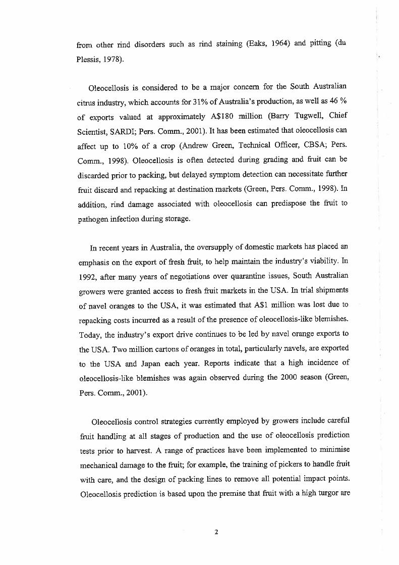
mesocarp (Fig. 2.1) (Bartholomew and Reed, 1943). The epicarp is the cuticle-
covered epidermal layer, composed of isodiametric' and 'polygonal' cells, and
laterally flattened cells above the oil glands in mature fruit. The hypoderm,
located beneath the epicarp comprises two or more layers of compacted
collenchyma cells. The outer mesocarp is composed of thin-walled parenchyma
cells, in which the oil glands are embedded.
5
¿:
M.E'
Figure 2.1. Cross-section of mature lemon rind. E, epicarp; Hy,
hypodermis; O to M, outer mesocarp; Og, oil gland; M to M', inner
mesocarp; E', endocarp.(Adapted from: Bartholomew and Reed, 1943).
6

The epicarp, h¡poderm and outer mesocarp constitute the coloured portion of
the mature fruit, or the 'flavedo'. The inner mesocarp, comprising branching cells
and large intercellular spaces is referred to as the colourless 'albedo'.
2.2.Th'e oil gland
Secretory cavities occur naturally in all species of the family Rutaceae (Fahn,
lgTg). As pointed out by Schneider (1968), the structures are more complex than
mere cavities, and are commonly refened to as 'oil glands'.In Citnts species, oil
glands have been documented in various parts of the plant, including the stem
(Webber and Fawcett,1935; Schneider, 1955), leaves (Schneider, 1968; Thomson
et al., 1976; Turner et al., 1998), all parts of the flower except the stamens
@artholomew and Reed, 1943), and the fruit (Tunell and Klotz, 1940; Ford,
1942; Bartholomew and Reed, 7943; Scott and Baker, 1947; Bain, 1958;
Schneider, 1968; Holtzhausen, 1969; Shomer, 1980; Bosabalidis and Tsekos,
1982a, 1982b). For the oil glands of the fruit, characteristics such as shape, size,
arangement and position of the glands, and kind, amount and aroma of the oils
are thought to be highly variable among citrus fruit types (Hodgson, 1967).
2.2.1. Gland development
In the fruit, oil glands have been observed as early as the ovary stage, in
Eueka lemon (Ford, 1942), Valencia orange (Schneider, 1968), Washington navel
orange (Holtzhausen, 1969) and Mediterranean mandarin (Bosabalidis and
Tsekos, I982a). Gland formation in Citrus species has been reported by some to
be confined to early stages of fruit development (Ford, 1942; Bain, 1958), whilst
others have suggested continuous formation with fruit growth (Schneider, 1968).
Glands have also been reported to enlarge with fruit growth (Bain, 1958;
Holtzhausen,1969).
7

2.2.2. Gland anatomY
2.2.2.1. Gland ontogenY
According to Bartholomew and Reed (L943), early studies by Sieck (1895)
and Biermann (1896) suggested that citrus oil glands originated from the
differentiation of four polyhedral cells from the sr¡rounding tissue. Anatomical
examination of the floral ovaries of C. deliciosa,has shown glands to differentiate
from a pair of meristematic cells, one epidermal and one unde¡lying @osabalidis
and Tsekos,I982a). The two initial cells were reported to be precwsors to the two
parts of the differentiated glandular complex; the globular/oval gland situated in
the parenchyma, and the 'conical stalk' joining the gland to the epidermis.
Ontogeny of secretory cavities ín Citrus (Bosabalidis and Tsekos, 1982a) appears
to show similarities to those in Eucalyptus (Carc and Ca:r, 1970).
2.2.2.2. The mature gland
Secretory cavities are considered to consist of "relatively large intercellular
spaces (lumina or lacunae) lined by an epithelium of secretory cells" (Fahn, 1979).
Observations of the mature oil glands of Citrus appe¿ìr to be consistent with this
description; glands are composed of a central cavity surrounded by several layers
of radially flattened epithelial cells (Thomson et aI., 1976; Shomer, 1980;
Bosabalidis and Tsekos, 7982a, I982b; Turner et al., 1998). Epithelial cells are
thought to be gradually modified into a protective sheath of cells with thickened
cell walls, refened to as 'boundary' or 'envelope' cells (Thomson e/ al., 1976;
Bosabalidis and Tsekos, 1982b).
2.2.2.3. Cavity formation
There is contradiction in the literature regarding the origin of the gland cavity;
schizogenous vs. lysigenous. 'schizogeny' refers to the separation of walls of
neighbouring cells, and 'lysigeny' to the disintegration of cells, to form the
intercellular space (Fahn, 1979). Using electron microscopy, Heinrich (1966;
1969) reported lysigeny in the glands of C. medica and C. limon, as did
Bosabalidis and Tsekos (19S2b) in C. delicíos¿. Thomson et al. (7976), concluded
that the cavities of the foliar glands of C. sinensl's formed schizogenously, but did
8

not exclude the possibility of lysigeny occurrin g at Iater stages of development. In
the most recent study, Turner et al. (1998) examined the effect of a range of
fixatives on the folia¡ glands of C. limon, and demonstrated artefactual swelling
and bursting of inner epithelial cells due to hl.potonic fixatives. In a later review
of cavity formation in Cítrus and related species, Turner (1999) pointed out the
difficulties in distinguishing between fixation artefact and a true lysigenous
process, suggesting that schizogeny be favoured in cases where there is dispute.
2.3. The rind oils
2.3.1. Oil synthesis and accumulation
The inner epithelial cells of citrus oil glands appear to be secretory in nature
(Thomson et al., 7976; Bosabalidis and Tsekos, 1982b). The majority of
transmission electron microscopy (TEM) studies have reported the major site of
essential oil synthesis to be the plastids (Heinrich, 1966; Shomer, 1980;
Bosabalidis and Tsekos, 1982b), however various mechanisms of oil secretion
have been proposed. Heinrich (1966) suggested that lipids in the plastids were
released into the cavity following cell lysis; however, more recent studies have
suggested that an active secretion takes place (Shomer, 1980; Bosabalidis and
Tsekos, 1982b). Shomer (1980) suggested that oil transfer from the cytoplasm into
the wall takes place via ectoplast invagination. Bosabalidis and Tsekos (1982b)
added to these observations by suggesting that oil movement from the plastids is
facilitated by ER elements which fuse to the plasmalemma, depositing the oil into
the apoplast, through which it is driven into the cavity.
For the microscopic examination of rind oils, it is important to take the
methods of tissue preparation into consideration. This issue was addressed by
Turner et al. (1998) in their study of C. hmon foliar glands. On the basis of light
microscopy (LM) and TEM observations, better lipid retention was achieved in
osmium vapour fixed or cryofixed sarnples compared to those prepared with
conventional chemical fixation procedures.
9
2.3.2. Oil composition
Essential oils largely consist of volatile, low molecular weight terpenes (Fahn,
1990). In C. sinensz,s, 116 oil components have been identified (Shaw, 1979). The
monoterpene d-limonene constitutes more than. 90%o of total oil by weight,
followed by p-myrcene and c-pinene, also terpenes. Other major components are
aldehydes including neral and geranial, alcohols including linalool and terpineol,
esters, ketones and non-volatiles (Shaw, 1979). d-limonene is the most abundant
oil component of most Citrus species (Kesterson and Hendrickson, 1962; Attaway
et al., 1967; Sawamura et al., 1984; Daito and Morinaga, 1984; Ruberto et al.,
L997 ;Loveys et al., 1998; Sun and Petracek, 1999).
The effect of frt¡it maturation on the essential oils of citrus fruit has been well
documented (Kesterson and Hendrickson,Ig62; Attaway et a1.,7967; Scora et al.,
1968; Chandler et al., 1976; Casas and Rodrigo, 1981; Daito and Morinaga,
1984). In V/ashington navel orange, an increase in total rind oil has been shown
late in the season (Scora et al., 1968). In the study by Scora et al. (1968), the
levels of 18 major oil components were measured from eight months post-
anthesis; showing a general increase in o-pinene, decrease in p-myrcene and
linalool, and negligible changes in dlimonsne levels. A biosynthetic relationship
between d-limonene and linalool has been suggested (Attaway et al.,1967).
2.3.3. Oil properties
Monoterpenes are important agents of insect toxicity (Taiz and Zeiger,1998).
Citrus oil components possess pesticidal (Don-Pedro, 1996) and antifungal
properties (Ben-Yehoshua ¿/ al., 1992). Essential oils also supply raw materials
for various industries, such as resin for paper and textiles, pharmaceutical
products, perfumes and spices (Fahn, 1979).
l0

2.4. Oleocellosis
2.4.1. Terminology
In the literature, oleocellosis has been referred to by a range of names including
'Oil spOtting', 'Oleo', 'bruising', 'gfeen spOt' and 'gas burn' (GrierSOn, 1986)'
However, discrepancies in the use of some names are obvious. The term 'rind oil
spot' has been used to describe oleocellosis (Sawamura et al., 1984; Sawamura el
dl., 1987; Sawamura et aL, 1988), as well as blemishes induced by chilling
(Kanlayanarat et al., 1988b, 1988a) and high temperatures (Chikaizumi, 2000).
The Japanese tefin 'Kohansho' has also been loosely used to describe oleocellosis
(Tugwell, Pers. Comm., 1998), but a number of studies into Kohansho have shown
characteristics that suggest the two disorders are not synonymous (Hasegawa and
Iba, 1981; Manago, 1988; Hasegawa and Yano, 1992 Yano et al., 1997).
Hasegawa and Iba (1981) reported that Kohansho is not a result of fruit injury, but
is largely dependent on storage temperature.
2.4.2. Cause
Terpenes and sesquiterpenes are highly phytotoxic (Soule and C¡rierson, 1986)'
Oleocellosis is caused by phytotoxic rind oils, which are released from oil glands
following mechanical damage and act upon the rind tissue (Fawcett, 1916). In the
earliest documented study of oleocellosis, Fawcett (1916) induced oleocellosis
symptoms or 'green spotting' in lemon fruit, by either applylng pressure to the fruit
surface or by appþing oils to the surface of uninjured fruit. In subsequent studies,
oleocellosis has been induced by various mechanical and oil methods, see Section
2.4.4.
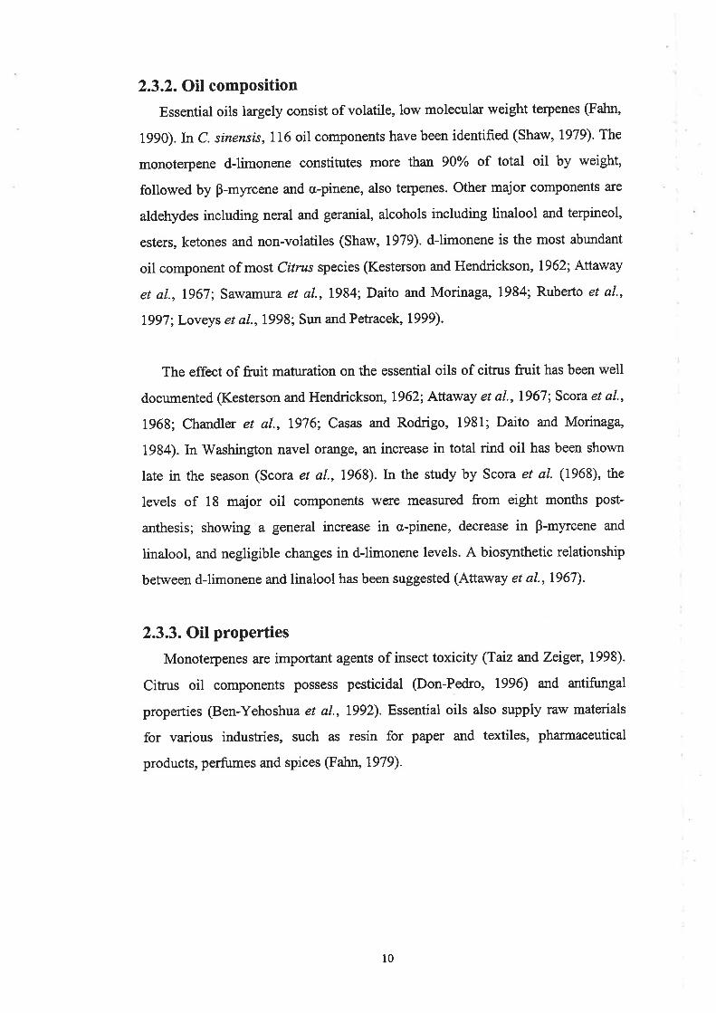
2.4.3. Symptoms
Oleocellosis produces a superficial blemish, characterised by collapse and
discolouration of the rind tissue (Shomer and Erner, 1989). Oleocellosis symptoms
are depicted in Figure 2.2. Tissue collapse between glands causes glands to appear
more prominent on the fruit surface (Fawcett, 1916; Mustard, 1954). Oleocellosis
is a particular problem in immature green fiuit, as oleocellosis-damaged areas fail
to colour normally (Fawcett, I9l6;Miller and Winston,1943; Cahoon et al',1964;
11
X'igure 2.2. Oleocellosis symptoms on mature navel orange fruit.Oleocellosis damage to mature fruit, causes rind darkening (A).
Oleocellosis damage to immature fruit; damaged areas fail to colour
normally (B).(Fhotos: Barrj' Tugwell)
t2

Wardowski et aI., 1976; Shomer and Erner, 1989). Slight symptoms have been
observed to develop as early as 20 minutes after oil application in lemon fruit
(Fawcett, 1916). In mandarin fnrit, symptoms have been monitored over four days,
with rind undulation detected by 18 hours and 'necrotic spots' by 36 hours,
forming lesions by 48 hours (Williams and Wild, 1996). In navel oranges,
symptoms have been observed to develop within two to three days of oil
application (Wild, 1998).
2.4.4. Artifrcial induction
2.4.4.1. Mechanical induction
Normal picking and packing procedures have been used to induce oleocellosis
in Washington navel oranges (Levy et a1.,1979). Normal handling practices have
also been simulated, by dropping Atrit from a standard height onto a hard surface
such as the back of a flat-bed truck (Cahoon et aI., 1964) or a picking trailer
(Loveys et aI., l99S). The penetrometer has been used to induce oleocellosis in
Shamouti orange fruit @rner,1982; Shomer and Erner, 1989). Pin-pricking glands
has also been shown to produce severe rind injury in citrus fruit (Sawamura et al',
1e84).
2,4.4.2. Oil induction
Rind oits have been applied to fruit by bending excised rind segments
(Sawamura et aL.,1984; Sawamura et al., 1987; Sawamura et al., 1988; Williams
and Wild, 1996), applyrng single drops @rodrick, 1970; Chikaizrtrnt,2000) or by
smearing aqueous emulsions (Shomer and Erner, 1989). In the most recent study,
oil was applied by attaching oil-treated cardboard discs to the fruit surface with
adhesive tape (Wild and Williams, 1992). In navel orange fruit, typical symptoms
have also been induced by injecting oils into the rind (Loveys et al., 1998)'
Previous observations of oil-induced oleocellosis suggest that linalool is highly
phytotoxic, moreso than dJimonene (Wild and Williams, 1992), but that both
components are more damaging than o,-pinene, n-nonanol, n-nonanal and carvone
(Sawamura et al., 1984).
13
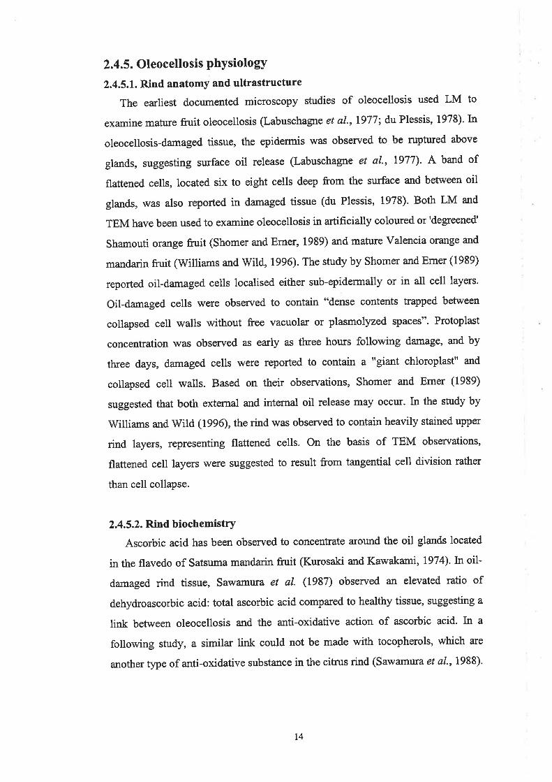
2.4.5. Oleocellosis PhYsiologY
2.4.5.1. Rind anatomy and ultrastructure
The earliest documented microscopy studies of oleocellosis used LM to
examine mature fruit oleocellosis (Labuschagne et al.,1977; du Plessis, 1978). kl
oleocellosis-damaged tissue, the epidermis was observed to be ruptwed above
glands, suggesting swface oil release (Labuschagne et al., 1977). A band of
flattened cel|s, located six to eight cells deep from the surface and between oil
glands, was also reported in damaged tissue (du Plessis, 1978). Both LM and
TEM have been used to examine oleocellosis in artificially coloured or'degreened'
Shamouti orange fruit (Shomer and Erner, 1939) and mature Valencia orange and
mandarin fruit (V/illiams and Wild, 1996). The study by Shomer and Erner (1989)
reported oil-damaged cells localised either sub-epidermally or in all cell layers.
Oil-damaged cells were obsen¡ed to contain "dense contents trapped between
collapsed cell walls without free vacuolar or plasmolyzed spaces". Protoplast
concentration was observed as early as three hours following damage, and by
three days, damaged cells were reported to contain a "giant chloroplast" and
collapsed cell walls. Based on their observations, Shomer and Erner (1989)
suggested that both external and intemal oil release may occur. In the study by
Williams and Wild (1996), the rind was observed to contain heavily stained upper
rind layers, representing flattened cells. On the basis of TEM observations,
flattened cell layers were suggested to result from tangential cell division rather
than cell collapse.
2.4.5.2. Rind biochemistry
Ascorbic acid has been observed to concentrate around the oil glands located
in the flavedo of Satsuma mandarin fruit (Kwosaki and Kawakami,1974).In oil-
damaged rind tissue, Sawamura et al. (1937) obsenred an elevated ratio of
dehydroascorbic acid: total ascorbic acid compared to healtþ tissue, suggesting a
link between oleocellosis and the anti-oxidative action of ascorbic acid. In a
following study, a similar link could not be made with tocopherols, which are
another type of anti-oxidative substance in the citrus rind (Sawamura et a1.,1988).
t4

Oleocellosis, along with dessication and pathogen infection, has also been shoïvn
to alter the terpene content of oil glands (Scora and Adams,1973).
2.4.6. Fruit susceptibitity to oleocellosis
2.4.6.1. Fruit turgor
Fruit susceptibility to oleocellosis has been linked to frtlit turgor; turgid fruit
are more likely to develop oleocellosis (Eaks, 1955). Prior to this finding,
oleocellosis incidence was related to orcha¡d conditions (Fawcett, 1916; Miller
and Winston, 1943; Mustard, 1954). Fawcett (1916) reported that oleocellosis
occr¡rred predominantly on lemon fruit that were wet, in a moist atmosphere, or
just picked. Similarly, Mustard (1954) reported increased oleocellosis in Persian
lime fruit that were wet from rain or dew. The conclusions of Eaks (1955) were
based on observations of wet vs. dry fruit, snapped vs. clipped fruit, storage
temperature and dwation. In a later study in lemons, a quantitative relationship
was established between oleocellosis and factors affecting fruit turgor, such as soil
moisture and vapor.u pressure deficit CVPD) of the atrnosphere (Cahoon et al.,
1964). A relationship was also established with rind oil release pressure (RORP),
which was measured with a fruit pressure tester, and considered to integrate both
soil and climatic conditions. In following studies, a similar relationship was
established in Florida lemons (Oberbacher, 1965), Califomian oranges and lemons
(Eaks, 1963) and Florida limes (Pantastico et al., 1966). The fruit pressure tester
or 'penetrometer' used for RORP assessment was originally developed for fruit
maturity assessment (Magness and Taylor, 1925). Christ (1967) described
modifications of the pressure tester for use on citrus fruit, and recommended
methods for detecting oil release. Fruit turgor estimation is the basis for current
prediction tests for oleocellosis, see Section 2.4.7.7.
2.4.6.2. Fruit age and size
Wardowski et al. (1976) made the general statement that small-sized fruit
tended to be more susceptible to oleocellosis than larger sizes. In lime fruit, lower
RORP values (higher turgor) have been obtained in smaller fruit (Pantastíco et al.,
1966). In lemon fruit, immatwe fruit have been suggested to be more susceptible
15
to gland rupture due to the 'fragility' of the rind (Cahoon et al., 1964), or the
rough surface and exposed glands (Oberbacher, 1965).
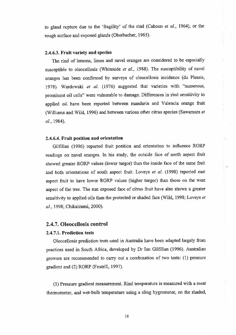
2.4.6.3. Fruit variety and species
The rind of lemons, limes and navel oranges are considered to be especially
susceptible to oleocellosis (Whiteside et a/., 1988). The susceptibility of navel
oranges has been confirmed by sr.rveys of oleocellosis incidence (du Plessis,
1973). Wardowski et al. (1976) suggested that varieties with "numerous,
prominent oil cells" were vulnerable to damage. Differences in rind sensitivity to
applied oil have been reported between mandarin and Valencia orange fruit
(Williams and Wild, 1996) and between various other citrus species (Sawamwa er
al.,1984).
2.4.6.4. Fruit position and orientation
Gilfrllan (1996) reported fruit position and orientation to influence RORP
readings on navel oranges. ln his study, the outside face of north aspect fruit
showed greater RORP values (lower turgor) than the inside face of the same fruit
and both orientations of south aspect fruit. Loveys et al. (1998) reported east
aspect fruit to have lower RORP values (higher turgor) than those on the west
aspect of the tree. The sun exposed face of citrus fruit have also shown a greater
sensitivity to applied oils than the protected or shaded face (Wild, 1998; Loveys et
al., 1998; Chikaizumi, 2000).
2.4.7 . Oleocellosis control
2.4.7 .1. Prediction tests
Oleocellosis prediction tests used in Australia have been adapted largely from
practices used in South Africa, developed by Dr Ian Gilfrllan (1996). Australian
grorwers are recommended to carry out a combination of ¡wo tests: (1) pressure
gradient and (2) RORP (Feutrill, 1997).
(1) Pressure gradient measu¡ement. Rind temperature is measured with ameat
thermometer, and wet-bulb temperature using a sling hygrometer, on the shaded,
t6

south aspect of the tree. This test is satisfied when fruit temperature exceeds wet-
bulb temperature by 3'C for green fruit (or 2"C for coloured fruit).
(2) RORP measurement. This measurement is caried out assuming the
required pressure gradient has been met. RORP is measured using a penetrometer
with an 8 mm diameter tip, on at least 20 fruit not exposed to the sun that day
(inside fruit or south aspect fruit prefened). It is said to be safe to pick when
RORP is greater than 3 kg.
A recent study focussed on developing improved susceptibility tests (Loveys
et a1.,1998). In this study, a transparent, acrylic penetrometer tip was shown to
give more acct¡rate RORP readings than the conventional brass tip. An electronic
penetrometer was also shown to produce rind firmness readings with lower
associated errors than RORP readings. However, RORP was judged to be a poor
indicator of fruit susceptibility to oleocellosis. Previous studies support this
finding (Gillespie and Tugwell, l97l; Erner, 1982). Pressr¡¡e gradient and VPD
were considered to be 'reasonable' indicators of oleocellosis susceptibility, but
major delays in harvesting navel oranges in a South Australian orchard occurred
when adhering to pressure gradient guidelines. Another test for oleocellosis
susceptibility was also tested by Loveys et al. (1998), in which fruit were dropped
from a height of 500 mm onto a fïlter paper on a flat surface, and the amount of oil
released onto the paper was measured. At low temperatures, larger amounts of oil
were released from the fruit, however amount of oil released did not conelate with
resulting oleocellosis.
2.4.7.2. Careful fruit handling
Studies have shown that oleocellosis can be avoided, even in turgid fruit, if the
fruit is handled with care. Eaks (1955) reported that oleocellosis could be avoided
in wet lime fruit if they were clipped and handled carefully rather than snapped at
harvesting. Other studies have also suggested that oleocellosis can be avoided ifhandled carefully, even early in the morning, when fruit turgor is high @antastico
et al., 1966; Loveys et a1.,1998).
t7

Industry recommendations aim to minimise mechanical damage to fruit at all
stages of handling (Feutrill, 1997). For example, pickers should be trained and
picking bags clean and free of sand or dirt, and fruit should be transported by a
good carrier, over graded roads, with reduced tyre pressure. Indicator papers have
been used to detect oil release from fruit (Grierson, 1958). More recently, the
'instrumented sphere' has been adopted in Australian packing sheds; this device
measures impact mass whilst travelling along the packing line (Jim Hill,
Consultant, PIRSA Rural Solutions; Pers. Comm., 1998). Fruit injury along the
packing line is reduced by removing potential impact points; in the USA the
maximum acceptable limit is 150 g, whilst in South Australia, a limit of 70 g is
used (Hill, Pers. Comm., 1998).
2.4.8. Fruit treatments affecting oleocellosis
2.4.8.1. Plant growth hormones
a. Ethylene
Etþlene is a plant growth regulator that is termed the 'ripening hormone' for
many plants (Abeles, 1973). In citnrs, preharvest etþlene has been shown to
reduce oleocellosis in postharvest fiuit, possibly by enhancing rind colouration
(Levy et al., 1979; Erner, t982). However, Levy et al. (1979) also reported a
corresponding decrease in RORP (increase in turgor), possibly related to reduced
stomatal conductance. Preharr¡est ethylene may also facilitate reduced fruit
damage by loosening fruit (El-Zeftawi, 1977). Postharvest etþlene is used to
degteen citrus fruit, but can accentuate oleocellosis, as damaged areas remain
green (Levy et aL.,1979).
b. Gibberellins
In citrus, gibberellins are thought to play an important role in the control of
flowering and fruit development (Monselise, 1979). In Australia, preharvest GA
(in the form of GAr) is widely applied to delay fruit senescence and reduce the
severity of albedo breakdown, another rind disorder (Monselise et al., 1976).ln
l8

navel oranges, it has been suggested that preharvest GA treatment may also be
effective in reducing oleocellosis (Loveys et a1.,1998).
2.4.8.2. Storage conditions
a. Storage temperature
In mature fruit, temperatures of 10'C and below have been shown to inhibit
blemish development in navel oranges (Wild, 1998), as has cold disinfestation at
1"C prior to lO'C storage (Edwards et a1.,1994). However, cwing fruit at arrbient
temperature prior to cold disinfestation has been reported to increase the levels of
oleocellosis in lemons @redebon and Edwards,1992).
b. Storage atmosphere
Low oxygen storage has been shown to reduce darkening of the oleocellosis-
damaged rind of navel oranges, but darkening recommenced once fruit were
retumed to a normal atmosphere (Wild, 1998). Low oxygen (5-12%) combined
with very low carbon dioxide levels (0-2%) have been shown to minimise
oleocellosis in lemons (Calero et a1.,1931). High carbon dioxide levels have also
been reported to prevent the development of oleocellosis, rind pitting and
membranosis in 'Primofiori' lemons (Artés et a1.,1993).
2.4.8.3. Miscellaneous treatments
Other methods reported to reduce rind sensitivity to applied oils include:
mixing rind oil with potassium hydroxide (KOH) and ethanol (Wild, 1998),
applying commercial citrus waxes Briteseal and, Zidvat or sodium and potassium
silicates to the fruit (Wild, 1998), and exposing fruit to ultraviolet light for five
minutes or more (Loveys et a1.,1998).
l9

2.5. Summary
This review highlights the need for further investigation of oleocellosis from a
physiological perspective, and points to a number of avenues of investigation.
Oleocellosis results from damage to the oil glands and the effect of released
oils on the rind tissue. An understanding of the anatomy of the glands, as well as
the timing of gland development and oil accumulation in the fruit is required.
Anatomical examinations of the glands have been caried out for a range of Ci*us
species and in different plant parts, but not in navel orange fruit. In addition, some
contention currently exists regarding the timing of gland development in citrus
fruit.
Seasonal fluctuations in the rind oils have been examined by quantitative
methods, but the use of microscopy techniques to examine oil accumulation has in
the past been limited. This may be explained by the inability of conventional
tissue fixation methods to reliably retain oil within the glands. To enable rind oil
visualisation, additional method development is still required'
Our understanding of oleocellosis physiology is limited and largely based
upon a small number of microscopy studies. Previous studies have shown
significant discrepancies in their findings, which have been based on LM and
TEM observations. A detailed examination of oleocellosis, facilitated by the use
of varied microscopy techniques is required.
This research will help to explain the limitations of current oleocellosis control
practices and reveal possible alternatives. An improved understanding of factors
influencing oleocellosis will also help to explain previous reports of oleocellosis
variability and the effect of different fruit treatments on the expression of the
disorder.
20
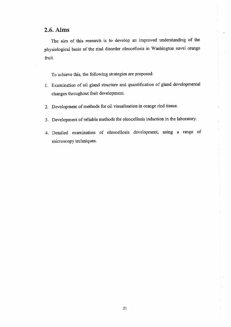
2.6. Aims
The aim of this research is to develop an improved understanding of the
physiological basis of the rind disorder oleocellosis in 'Washington navel orange
frnit.
To achieve this, the following strategies are proposed:
l. Examination of oil gland structure and quantification of gland developmental
changes throughout fruit development.
2. Development of methods for oil visualisation in orange rind tissue.
3. Development of reliable methods for oleocellosis induction in the laboratory.
4. Detailed examination of oleocellosis development, using a range of
microscopy techniques.
2l
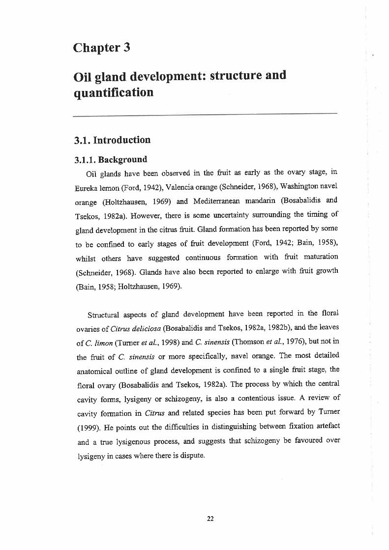
Chapter 3
Oil gland development: structure and
quantification
3.1. Introduction
3.1.1. Background
Oil glands have been observed in the fruit as early as the ovary stage, in
Eureka lemon (Ford, 1942), Valencia orange (Schneider, 1968), Washington navel
orange (Holtzhausen, 1969) and Meditenanean mandarin @osabalidis and
Tsekos, L982a). However, there is some rurcertainty surrounding the timing of
gland development in the citrus fruit. Gland formation has been reported by some
to be confined to early stages of fruit development (Ford, 1942; Bain, 1958),
whilst others have suggested continuous formation with fruit maturation
(Schneider, 1968). Glands have also been reported to enlarge with fruit growth
@ain, 1958; Holtzhausen, 1969).
Structural aspects of gland development have been reported in the floral
ovaries of Citrus deliciosa @osabalidis and Tsekos, 1982a, 1982b), and the leaves
of C. limon (Turner et a\.,199S) and C. sinensis (Thomson et aL.,7976), but not in
the fruit of C. sinensrs or more specifically, navel orange. The most detailed
anatomical outline of gland development is confined to a single fruit stage, the
floral ovary @osabalidis and Tsekos, 1982a). The process by which the central
cavity forms, lysigeny or schizogeny, is also a contentious issue. A review of
cavity formation in Citrus and related species has been put forward by Turner
(1999). He points out the difficulties in distinguishing between fixation artefact
and a true lysigenous process, and suggests that schizogeny be favoured over
lysigeny in cases where there is dispute.
22

3.1.2. Aims
The aim of this study was to examine oil gland anatomy and quantify gland
developmental changes in the frr¡it of Washington navel orange, from pre-anthesis
to maturity. By establishing an anatomical outline of gland development, the timing
of gland initiation, development and enlargement in relation to fnrit growth was
investigated, and mature gland anatomy was examined at different fruit ages.
3.2. Materials and Methods
3.2.I. Plant material
Fruit were collected from two 23 year old Washington navel orange trees on
poncirus trifoliata rootstock, located in the University of Adelaide, Waite Campus
Alverstoke orcha¡d (34o97'S, 138"63'E). The trees were positioned three metres
apart, in a north-south facing row. Fruit were harvested at monthly intewals from
pre-anthesis to fruit maturity (eight months). At each harvest, six fn¡it were picked
from each tree, representing three sizes x two replicates. Each harvest was carried
out at the same time of day to avoid possible discrepancies due to diurnal changes.
The equatorial and polar diameters of each fn¡it were measured. An average of
the two diameters is used when referring to fürit diameter. Fruit surface area was
extrapolated according to Turrell (1946). By this method, the fruit is considered a
spheroid, and the difference between polar and equatorial diameters is incorporated
into an equation to calculate surface area.
3.2.2. Gland counts
Gland densþ was measured on 9 mm to 88 mm diameter fruit. Within a sector
of 180 mm2 along the fruit equator (or an adjusted area for fruit smaller than this
dimension), glands visible from the surface using a dissecting microscope were
counted. Glands with deeper-seated cavities, difficult to detect from surface
examination, were counted by dissection of the tissue along the flavedo/albedo
boundary. The addition of these two counts gave total number of glands within the
sector. For all fruit, gland density r¡ras expressed as number of glands per 180 mm2.
23

Total number of glands per fnrit was estimated using gland density and fruit
surface area values
Total gland number: gland density x (fnrit surface areall80 mm1.
3.2.3. Microscopy
3.2.3.1. Tissue samPling
Samples for microscopy were collected from each fn¡it. A minimum of six rind
tissue samples was collected from the equatorial region of each fruit (three for each
tissue preparation method).
3.2.3.2. Light microscoPy (LM)
a. Tisnte preparation
Two tissue processing methods were employed: aldehyde fixation and aldehyde
fixation plus osmium tetroxide postfixation.
The first method was similar to that described by Feder and O'Brien (1968).
Rind samples of approximately 5 x 5 mm were fixed in 3Yo glutaraldehyde (25%
solution, Unilab, Aubum, Austratia) in 0.025M phosphate buffer, pH 7.0,
overnight at 0-4"C. Samples were rinsed in phosphate buffer and dehydrated
through an alcohol series: methoxy-ethanol, ethanol, propanol and butanol.
Samples were infiltrated overnight in a l:1 mixture of butanol:glycol-methacrylate
(GMA) (Sigma, St. Louis, USA) (prepared by mixing 93 ml 2-hydroxyethyl
methacrylate, T ml polyethylene glycol 400 and 0.69 benzoyl peroxide), followed
by three changes of 100% GMA over one week. Samples were embedded in
gelatin capsules (size '00', Parke Davis Capsugel Division, Caringbah, Australia)
and polymerisation achieved at 60'C. Sections (4 pm thickness) were cut with a
Reichert-Jung}O1} Supercut Mcrotome using glass knives, placed on glass slides
and stained with periodic acid-Schiffs @AS) (Schiffs reagent, BDH, Poole,
England) for carbohydrates and counterstained with Toluidine Blue O (TBO)
(Aldricb Mlwaukee, USA) to provide detail of cell structure (O'Brien and
McCully, 1981).
24

In the second method, rind samples of approximately 1 x 1 mm were fixed in a
solution of 4% formaldehyde (paraformaldehyde powder, ProSciTech,
Thuringowa, Australia), I.25% glutaraldehyde (EM grade, 25% solution,
ProSciTech) and 4Yo sucrose, in phosphate buffered saline (PBS), pH 7 '2,
overnight at O-4"C. After two short rinses in PBS, samples were postfixed in
phosphate buffered 1% osmium tetroxide (ProSciTech) for one hour. Samples
were dehydrated through an acetone series: 70, 90, 95, l00o/o, and infiltrated
overnight in a 1:1 mixture of acetone:Procure-Araldite (PA) resin @rocure-
Araldite Embedding Kit, ProSciTech), followed by three eight hour changes of
IOO%PA resin. PA resin was prepared according to Mollenhauer (1964). Samples
were flat embedded and polymerisation achieved at 70"C. Sections (1 pm
thickness) were cut with a Reichert-Jung Ultacut E Microtome using glass
knives, placed on glass slides and stained with TBO to provide detail of cell
structure (O'Brien and McCully, 1981).
Histochemical staining for lignin using Phloroglucinol-HCl (May and Baker,
Dagenham, England) (Jensen, 1962) and suberin using Sudan Black B (SBB)
(Sigma) (O'Brien and McCully, 1981) was also employed. All material was
observed with a Zeiss Axiophot Photomicroscope, using transmitted light.
b. Sample selection
Five fruit stages were chosen on the basis of size, to represent complete fruit
development. The five fruit stages selected were: (A) floral ovary (at full bloom),
@) immatwe green coloured fruit 10 mm diameter, (C) immature green fruit 32
mm, (D) immature green ftút 52 mm, (E) mature ora¡rge coloured fruit 88 mm.
For each fruit stage, samples were serial sectioned and used in a sr:rvey of gland
age and image analysis of gland volume. In addition, 15 'in between' fruit sizes
were sectioned for general anatomical observation.
3.2.3.3. Transmission electron microscopy (TEM)
Ultra-thin sections (70 nm in thickness) of aldehyde plus osmium tetroxide
postfixed tissue were collected on a diamond knife, using a Reichert-Jung Ultracut
25

E Microtome. Sections were transferred to collodion coated copper grids, 200
mesh thin bar. Sections were stained with uranyl acetate and lead citrate (O'Brien
and McCully, 1981), and observed under an accelerating voltage of 80 kV with a
Philips CM 100 TEM.
3.2.4. Gland age survey
From each of the five fruit stages, a sample of 12 to 40 glands was selected,
and glands were classed according to their stage of development. Using LM, the
complete set of sections comprising each gland was examined to avoid
discrepancies in gland classification.
3.2.5.Image analysis
From each of the five fruit stages, the volume of five mature glands was
measured using the image analysis program Video Pro 32 version 3.42 (Leading
Edge Pty. Ltd., Adelaide, Australia). For Video Pro programs see Appendix 1.
The gland perimeter was defined by the outermost epithelial cells of the gland and
included the gland stalk. Gland volumes were calculated from an equation
incorporating area and distance between serial sections. For example, the volume
between two sections I and 2 was:
Volume 1 (pm'): Area I (r.nt') x Distance between sections 1 and 2 (pm)'
3.2.6. Statistical analysis
Gland volume data were analysed using the statistical package Genstat for
Windows version 4.1 (Lawes Agricultural Trust, IACR Rothamsted). A
completely randomised design was applied to the data and Analysis of Variance
(ANOVA) tables, and tables of means were generated. Statistical difference due to
fruit age was tested at the 95%o confidence level.
26

3.3. Results
3.3.1. Anatomical gland development
From LM observations of aldehyde fixed tissue samples, a series of six stages
was chosen to outline anatomical gland development in Washington navel orange
fruit. These were as follows: Stage 1, a cluster of up to ten cells adjacent to the
epidermis. These gland initials were distinguished from sunounding parenchyma
cells as slightly radially elongated and lacking starch (Plate 3.14)' Stage 2, a
larger cell cluster of ten to 30 cells @late 3.18). Stage 3, a larger cluster showing
the start of differentiation into boundary and inner cells (Plate 3.lC). Stage 4, a
differentiated gland with flattened boundary cells enclosing polyhedral shaped
inner cells. Nuclei of the inner cells appear enlarged (Plate 3.1D). Stage 5, a
differentiated gland with flattened boundary cells enclosing polyhedral shaped
inner cells whose walls appeü to form the cavity (Plate 3.1E). Stage 6, a mature
gland with an expanded central cavity (Plate 3.1F). Differences in the appearance
of surrounding parenchyma cells can be attributed to the differences in fruit age.
This outline was used to quantiff gland development in relation to fruit
development. The anatomy of mature, cavity-containing glands (stages 5 and 6)
was examined more closely in aldehyde plus osmium tetroxide postfixed tissue
(Plates 3.34 and 3.38).
At each stage of gland development, glands were joined to the fruit epidermis
by a stalk-like structure. The stalk was often conical in shape (Plate 3.24). The
structure was solid rather than funnel-like, as revealed by tangential sectioning of
the rind tissue @late 3.2lr_). The stalk appeared to be an extension of the irurer
gland cells, unintemrpted by bonndary cells, which met the epidermis at its apex.
Stalk cells were round to oblong shaped, and were smaller and different in form to
other cells of the gland complex and su¡rounding parenchyma cells of the rind.
27
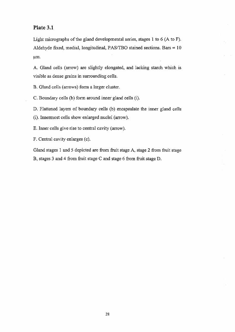
Plate 3.1
Light micrographs of the gland developmental series, stages 1 to 6 (A to F).
Aldehyde fixed, medial, longitudinal, PAS/TBO stained sections. Bars = 10
pm.
A. Gland cells (arrow) are slightly elongated, and lacking starch which is
visible as dense grains in sr¡¡rounding cells.
B. Gland cells (arrows) form a larger cluster.
C. Boundary cells (b) form around inner gland cells (i).
D. Flattened layers of boundary cells (b) encapsulate the inner gland cells
(i). Innermost cells show enlarged nuclei (arrow).
E. Inner cells give rise to central cavity (arrow).
F. Central cavity enlarges (c).
Gland stages 1 and 5 depicted are from fruit stage A, stage 2 from fruit stage
B, stages 3 and 4 from fruit stage C and stage 6 from frr¡it stage D.
28

A
lã: ro-
Y- :f ú-

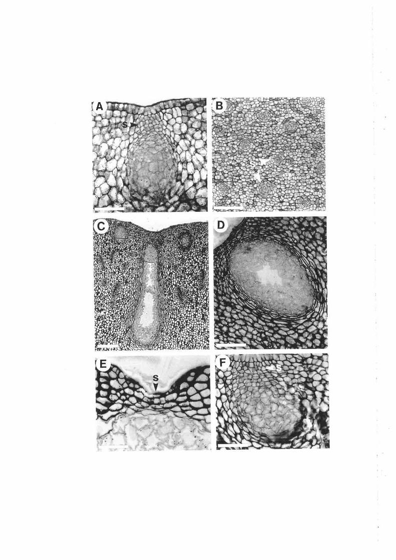
Plate 3.2
Light micrographs depicting the gland stalk and gland form. Aldehyde
fixed, PAS/TBO stained sections.
A and B. Longitudinal (A) and transverse @) sections through the gland
stalk. A, A conical-shaped stalk (s) joins the gland to the fruit epidermis. B,
A transverse section 16 pm from the fruit surface shows gland stalks are
solid structures, appearing as round clusters of cells (s). Bars: 10 ¡rm.
C and D. Medial, longitudinal sections through mature (stage 6) glands of
different forms. C, Elongated gland. Bar: 50 pm. D, Oval-shaped gland.
Bar: 20 ¡tm.
E and F. Medial, longitudinal sections through the stalks of mature glands
(s). E, In a mature fruit, stage E. F, In an immatr:¡e fruit, stage B. Bars : 10
F¡rn.
29
t+tA

3.3.2. Timing of gland development
Glands were present in the pre-anthesis floral ovary. Glands at various stages
of development were observed in the youngest sample collected, a flower bud of 8
mm length from the base of the calyx to petal tip, with an ovary of approximately
2.6 nnin diameter. The most advanced stage of gland development observed in
this sample was stage 4.
Gland initiation was for¡nd to be restricted to early stages of fruit development.
Gland counts under the dissecting microscope showed an exponential decline in
gland density up until a fruit size of approximately 20 cm2 surface area (25 mm
diameter). From this time onwards, a more gradual decline was evident (Fig.
3.14). Considering the possible effects of fruit growth on gland density values,
total gland nr¡rnber per fruit was assessed. Total gland number per fruit appeared
to show a slight initial increase, but remained fairly constant with increasing fruit
size, concentrated around 8000 to 12000 glands per fruit (Fig' 3.18). The
approximate total gland number of a mature, 88 mm diameter frnit (247 cÍl
surface area) was similar to that evident in an immatue, 20 mm diameter fuit (13
cm2 surface area). This confirms that gland initiation was confined to early stages
of fruit development, until a fruit size of approximately 20 mm diameter. Results
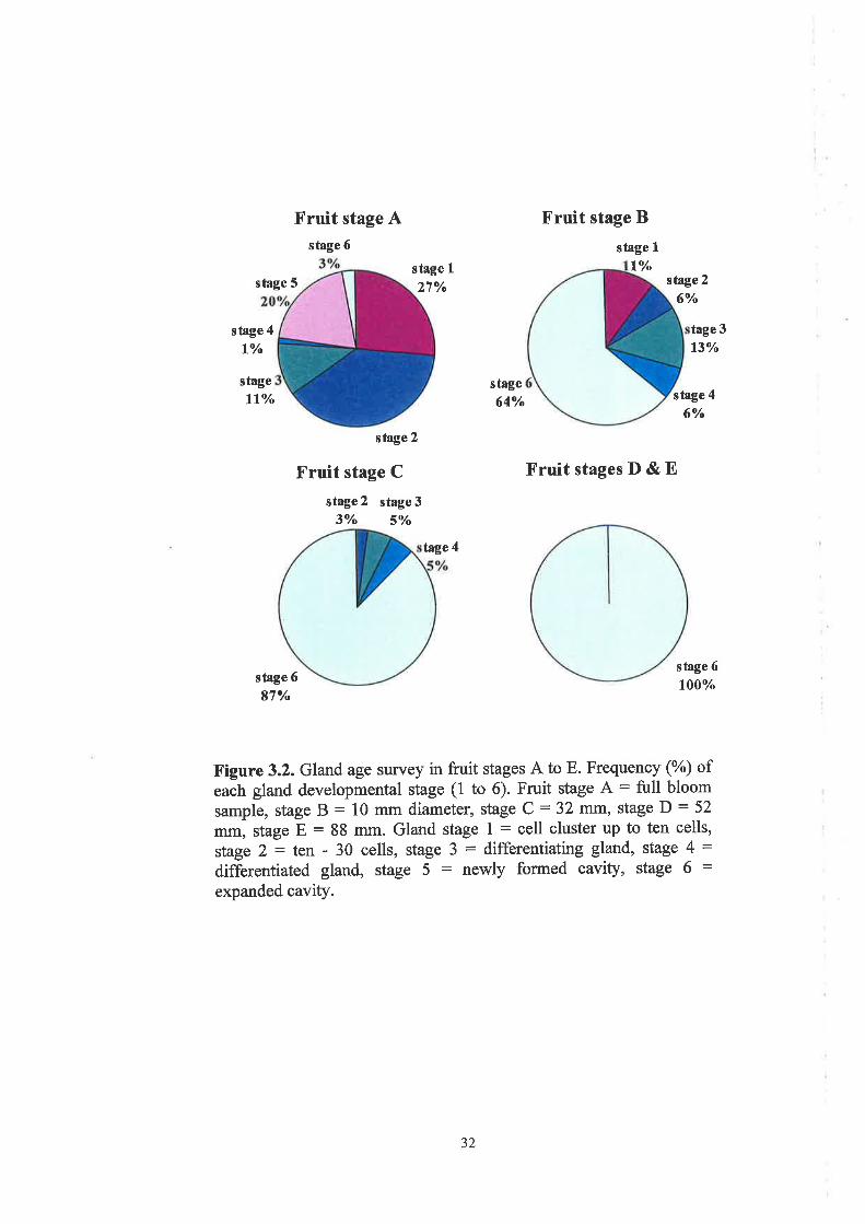
of the gland age survey supported this finding, with gland initials (stage 1)
comprising 27%o of the gland population in the floral ovary at full bloom (stage
A), declining to 1I%o in a 10 mm fruit (stage B) and disappearing thereafter (Fig.
3.2).
In the floral ovary, a high proportion of glands contained newly formed
cavities (20%) and some showed a more advanced stage of cavity opening (3%)
(Fig. 3.2). The proportion of mature glands increased from23%o in the floral ovary
(A), to 64Yo in a 10 mm fruit (B), 87% in a32 mm fruit (c) and to 100% in a 52
mm fruit (D) (Fig. 3.2). Hence, all glands reached maturity in an immature, green
fruit,3z to 52 mm in diameter.
30

6000
cl
! sooo
a-t 4000¡r(Ð
Y ¡oooq)
i 2000
f; rooorh
0
100 150 200
AB CrPuit surface area (cm2)
50
E
^ ^l^
25000
20000
15000
10000
5000
L€)
li(¡)
GI
rh
B y=1524.8ln(x)+5460.412 = 0.3041 ì
^
AA ^
^A A
100 150 200
0
50AB D
Fiuit surface area (cm2)
Figure. 3.1. Changes in oil gland density (A) and total gland number
per fruit (B) with fruit growth (represented by fruit surface area). The
five fruit stages A to E are labelled.
Af =0.9592
=11
31
Fruit stage Astage 6
Fruit stage B
stage 1
Lt/"stage 5
stage I27Vo
farge4
stage260/o
stage 4
lt/"
stage
llVo
tage 3
13Y'
stage
64Vo stage 4
6Vo
stage 6
l00Vo
stage 2
Fruit stage C
stage 2 stage 33o/o svo
FruitstagesD&E
stage 6
87Y"
X'igure 3.2. Gland age survey in fi:uit stages A to E. Frequency (yù ofeach gland developmental stage (1 to 6). Fruit stage A: full bloom
sample, stage B : 10 mm diameter, stage C : 32 mm, stage D : 52
mm, stage E : 88 mm. Gland stage I : cell cluster up to ten cells,
stage 2: ten - 30 cells, stage 3 = differentiating gland, stage 4 :differentiated gland, stage 5 : newly formed cavity, stage 6 :expanded cavity.
32
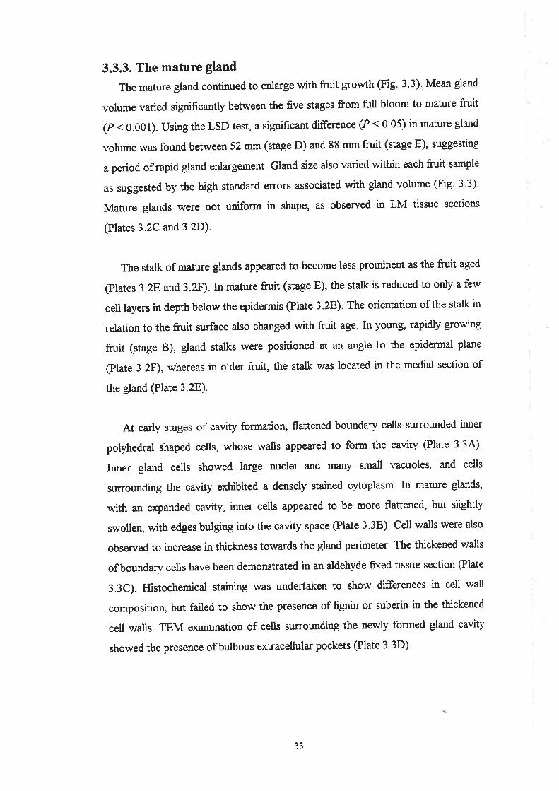
3.3.3. The mature gland
The mature gland continued to enlarge with frtrit grou/th @ig. 3.3). Mean gland
volume varied significantþ between the five stages from full bloom to mature fruit
(P < 0.001). using the LSD test, a significant difference (P < 0.05) in mature gland
volume was found between 52 mm (stage D) and 88 mm fruit (stage E), suggesting
a period of rapid gland enlargement. Gland size also varied within each fnrit sample
as suggested by the high standard errors associated with gland volume @ig. 3.3).
Mature glands \ilere not uniform in shape, as observed in LM tissue sections
(Plates 3.2C and3.2D).
The stalk of mature glands appeared to become less prominent as the fruit aged
@lates 3.28 and3.zF).In mature fruit (stage E), the stalk is reduced to only a few
cell layers in depth below the epidermis @late 3.28). The orientation of the stalk in
relation to the fruit surface also changed with fn¡it age. In young, rapidly growing
fruit (stage B), gland stalks were positioned at an angle to the epidermal plane
@late 3.2F), whereas in older fruit, the stalk was located in the medial section of
the gland @late 3.28).
At early stages of cavity formation, flattened boundary cells surrounded inner
polyhedral shaped cells, whose walls appeared to form the cavity (Plate 3 3A)'
Inner gland cells showed large nuclei and many small vacuoles, and cells
surrounding the cavity exhibited a densely stained cytoplasm. In mature glands,
with an expanded cavity, inner cells appeared to be more flattened, but slightly
swollen, with edges bulging into the cavity space @late 3.3B). Cell walls were also
observed to increase in thickness towards the gland perimeter. The thickened walls
of boundary cells have been demonstrated in an aldehyde fixed tissue section (Plate
3.3C). Histochemical staining was undertaken to show differences in cell wall
composition, but failed to show the presence of lignin or suberin in the thickened
cell walls. TEM examination of cells surrounding the newly formed gland cavity
showed the presence ofbulbous extracellular pockets (Plate 3.3D).
33

,
1.5
I
0.5
0
(.)
q)
õcË
â0
cËq)
Ir,là
c
Fruit stage
Figure 3.3. Mature gland volume in fruit stages A to E. Fruit stage A
= full bloom sample, stage B = 10 mm diameter, stage C = 32 rtttrt,
stage D = 52 ttrfiI¡t, stage E = 88 mm. Means and standard errors
represented. Letters (a and b) represent significant differences (P <0.05) between fruit stages.
EBA D
b
aaa a
34

Plate 3.3
Micrographs depicting mature gland anatomy and cavity formation.
A and B. Light micrographs of aldehyde plus osmium tetroxide postfixed,
medial, longitudinal, TBO stained sections through the gland cavity. A,
Gland with a newly formed cavity (arrow), equivalent to stage 5. B, Gland
with an expanded cavity (c), equivalent to stage 6. Bars: 10 pn.
C. Light micrograph of an aldehyde fixed, longitudinal, PAS/TBO stained
section through a mature (stage 6) gland: gland cavity (c), inner gland cells
(i), boundary cells (b) and surrounding parenchyma cells þ). Bar:20 pm.
D. Transmission electron micrograph showing bulbous extracellular pockets
(arrows) along the walls of cells sr.urounding the newly formed gland cavity
(c). Bar: I Frm.
35

o
TD
ì
i'i
6¡
,,"JY
."
j ..
I ..n
+/u
r'"'.f
* ',i
l¡.,;'
I
a
o
3.4. Discussion
In this study, the effect of different tissue fixation methods used has been
taken into consideration. Aldehyde fixed tissue facilitated the collection of large
tissue samples and serial sectioning, which was required to establlsh the
anatomical gland developmental series, and to carry out the gland age survey and
gland volume measr¡rements. Aldehyde fixation has been reported to cause
artefactual swelling and rupture of inner epithelial cells in the foliar glands of C.
limon (Turner et a1.,1998). For this reason, an improved fixation protocol was
used to prepare tissue for the examination of mature gland anatomy and cavity
formation. In addition, glands were not damaged dwing sample collection and
have been captured in medial section.
Until this study, gland development had not been examined in fruit of the
Washington navel orange. Gland development from the stage of cavity formation
onwards has been examined in the foliar glands of V/ashington navel orange
(Thomson et aI., 1976), but the most detailed anatomical outline of gland
development has been achieved in the ovaries of C. deliciosa or Mediteranean
mandarin (Bosabalidis and Tsekos, 1982a). LM observations in the present study
were similar to those made in C. deliciosa ovanes (Bosabalidis and Tsekos,
1982a).In both studies, gland initiation and cavity formation clearly occr¡¡red as
two separate events. The ea¡liest detection of gland initiation as a small cluster of
cells at the fruit surface supports Bosabalidis and Tsekos (I982a), who reported
that glands differentiated from a pair of meristematic cells, one epidermal and one
underþing. This also corroborates earlier descriptions of citrus gland
differentiation by Bartholomew and Reed (1943) and Schneider (1968).
To date, the presence of a gland 'stalk' has been described only by Bosabalidis
and Tsekos (1982a). In their study, they considered the two initial cells to be
precursors to the two parts of the differentiated glandular complex; the
globular/oval gland situated in the parenchyma, and the 'conical stalk' joining the
gland to the epidermis. Epidermal cells referred to as 'oil-gland cover cells', have
also been reported previously in the fruit (Scott and Baker,1947; Safran, 1980)
36

and bark (Schneider, 1955) of Citrus species. Oil-gland cover cells were reported
by Scott and Baker (1947) to have aî aÍea proportional to the underlying gland
and appear somewhat flattened and polygonal in surface view. In the present
study, the gland stalk was observed in all differentiation stages of Washington
navel orange glands, as well as at different fruit ages. The stalk structure was less
apparent in older fruit, which may be explained by a lateral flattening above oil
glands @artholomew and Reed, 1943) or by the continual growth of cells in the
epidermal and hypodermal layers (Bain, 1958), causing a bulging effect around
the gland stalk. The inconspicuous nattue of the stalk in matwe fruit may explain
why the majority of past studies have failed to describe an epidermal attachment
of the oil glands in mature citrus fn¡it. In addition, if tissue sections are being
examined, the stalk can go unnoticed if a longitudinal tissue section does not pass
directly through it, as pointed out by Bosabalidis and Tsekos (I982a).
Gland initiation, development, and enlargement were all quantified with
respect to fruit growth. Gland counts were localised to the equatorial region of the
fruit, to correspond to microscopy samples. Gland initiation was found to be
restricted to early fruit development, ceasing at a fruit size of approximately 20
mm diameter. This agrees with findings in Eureka lemon (Ford, 1942) and
Valencia orange, where gland initiation was observed only in the 'cell division'
stage of fruit development, which continued to a fruit size of approximately 16
mm (Bain, 1958). Howover, findings refute the statement of Schneider (1968)
who suggested that oil glands continued to form as the citrus fruit matured.
Similarly, it has been suggested that oil glands form in the very young stems of C.
sinensis (V/ebber and Fawcett, 1935). Glands continued to develop, until all had
reached maturity in an immatr.re green frvit, 32 to 52 mm in size. This finding
contradicts Holtzhausen (1969), who inferred that glands in Washington navel
orange fruit continued to develop up until fruit ripeness.
Once mature, glands continued to enlarge with fruit growth, as has been
reported previously in Washington navel orange (Holtzhausen, 1969), as well as
Valencia orange (Bain, 1958). The period of rapid gland enlargement observed
37

between fruit sizes of 52 and 88 mm roughly coincides with the period of cell
enlargement in the rind of Washington navel orange fruit described by Bouma
(1959). Mature glands of different sizes were also found within each fruit
examined, which may be explained by the non-uniform timing of both gland
initiation and maturation in relation to fruit development. Gland shape was also
non-uniform. Previously, the oil glands of Citrus have been described as varying
from "oblate to spherical or ovoidal to pyriform" (Bartholomew and Reed, 1943).
It has also been suggested that glands a¡e located at different depths in the rind
(Bartholomew and Reed, 1943; Ting and Attaway, 1971).In this study, although
all gtands were found to link to the epidermis by the gland stalk, gland cavities did
occur at different depths due to size differences alone, often extending into the
albedo layer.
Matwe gland anatomy consisted of a central cavity surrounded by a multi-
layered epithelium of radially flattened cells (Thomson et al., 1976; Shomer,
1980; Bosabalidis and Tsekos, 1982a; Turner et a1.,1998). Observations suggest
that inner gland cells are modified into bor¡ndary cells, as has been reported
previously in the glands of Citrus (Thomson et al., 1976; Bosabalidis and Tsekos,
19S2b). The innermost gland cells appeared typical of what have been described
as 'secretory parenchyma' cells, which usually possess thin and extremely
permeable walls, allowing movement of secretion into and out of the cell
(Mauseth, 1988). Boundary cells possessed thickened walls, but did not appear to
contain lignin, a common component of plant secondary walls (Mauseth, 1988).
Similarly, the foliar glands of C. limon have been observed not to contain lignin or
suberin (Turner et a1.,1998).
An interesting feature of the mature gland is the mechanism by which the
secreted material is isolated from the rest of the plant body. Fahn (1979) suggested
that this may occur by deposition of waterproofing materials (usually lignin or
suberin), viscosity of the secreted material, compactness of the wall, or by the
absence of intercellular spaces. To our knowledge, previous microscopy studies of
the oil glands of Citrus have not addressed this matter, except Thomson er ¿/.
38
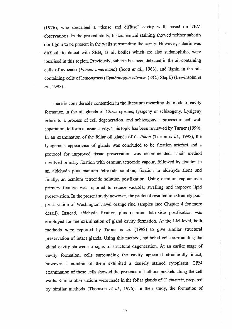
(1976), who described a "dense and diffuse" cavity wall, based on TEM
observations. In the present study, histochemical staining showed neither suberin
nor lignin to be present in the walls surrounding the cavity. However, suberin was
difficult to detect with SBB, as oil bodies which are also sudanophilic, were
localised in this region. Previously, suberin has been detected in the oil-containing
cells of avocado (Persea americana) (Scott et al., 1963), and lignin in the oil-
containing cells of lemongrass (Cymbopogon citratus (DC.) Stapf.) (Lewinsohn e/
al.,1998).
There is considerable contention in the literature regarding the mode of cavity
formation in the oil glands of Citrus species; lysigeny or schizogeny. Lysigeny
refers to a process of cell degeneration, and schizo gerty a process of cell wall
separation, to form a tissue cavity. This topic has been reviewed by Turner (1999).
In an examination of the foliar oil glands of C. limon (Turner et al., 1998), the
lysigenous appearance of glands was concluded to be fixation artefact and a
protocol for improved tissue preservation w¿ts recornmended. Their method
involved primary fixation with osmium tetroxide vapour, followed by fixation in
an aldehyde plus osmium tetroxide solution, fixation in aldehyde alone and
finally, an osmium tetroxide solution postfixation. Using osmium vapour as a
primary fixative was reported to reduce vacuolar swelling and improve lipid
preservation. In the present study however, the protocol resulted in extremely poor
preservation of Washington navel orange rind samples (see Chapter 4 for more
detail). Instead, aldehyde fixation plus osmium tetroxide postfixation was
employed for the examination of gland cavity formation. At the LM level, both
methods were reported by Tumer et al. (1998) to give similar structural
preservation of intact glands. Using this method, epithelial cells sr¡¡rowrding the
gtand cavity showed no signs of structural degeneration. At an earlier stage of
cavity formation, cells surrorurding the cavity appeared structurally intact,
however a number of them exhibited a densely stained cytoplasm. TEM
examination of these cells showed the presence of bulbous pockets along the cell
walls. Similar obseruations were made in the folia¡ glands of C. sinensls, prepared
by similar methods (Thomson et al., 1976). In their study, the formation of
39

bulbous extracellular pockets was used as evidence of a schizogenous cavity
formation.
3.5. Conclusion
Gland initiation was restricted to early stages of fruit development, and all
glands matured in an immature fruit, but continued to enlarge throughout fruit
growth. All glands were joined to the fruit epidermis by a stalk, irrespective of
their stage of development or location of the cavity within the rind. LM and TEM
observations suggest that glands of Washington navel orange fruit are
anatomically similar to those of other Citrus species, and that early stages of
cavity formation involve schizogeny.
40

Chapter 4
Rind oil visualisation: method development
4.L. Introduction
4.1.1. Background
Microscopic examination of oils in plant and animal tissues is limited due to
difficulties in retaining lipids with conventional tissue fixation protocols. In a
typical tissue fixation schedule for light or electron microscopy, tissues are fixed
in a primary aldehyde fixative, rinsed in buffer, postfixed in osmium tetroxide,
dehydrated, and infiltrated with a plastic resin, which is then polymerised using
heat, catalysts or ultraviolet radiation (Dykstra, 1992). Primary aldehyde fixation
(commonly glutaraldehyde or paraformaldehyde) assures the rapid stabilisation of
proteins, but does not effectively preserve lipid (Dykstra, 1992). Postfixation with
osmium tetroxide (OsOo) stabilises both proteins and lipids, in particular cell
membranes and other fatty components (Roland and Vian, 1991). Osmium
oxidises the double bonds of unsaturated lipids, leading to the formation of
monoesters, diesters and dimeric monoesters @ykstra, 1992). Usrng osmium
postfixation, oils have been preserved to some extent in the foliar oil glands of
Citrus sinensis (Thomson et aI-, 1976) and C. limon (Turner et a1.,1998)' and in
other plant secretory structures, such as the oil idioblasts of Persea americana
(Platt and Thomson, 1992) and Annona muricata (Bakker and Gerritsen, 1990).
However, osmium postfixation does not prevent lipid loss during dehydration
(Hayat, 1989).
Modifications to conventional methods are required to improve oil retention.
Such a method has been suggested by Turner et al. (1998) in their study of the
foliar oil glands of C. limon 'iVhen compared with standa¡d double fixation, they
reported that using osmium vapour primary fixation achieved "enhanced lipid
retention, both within the epithelial cells and in the central storage space" of
4t
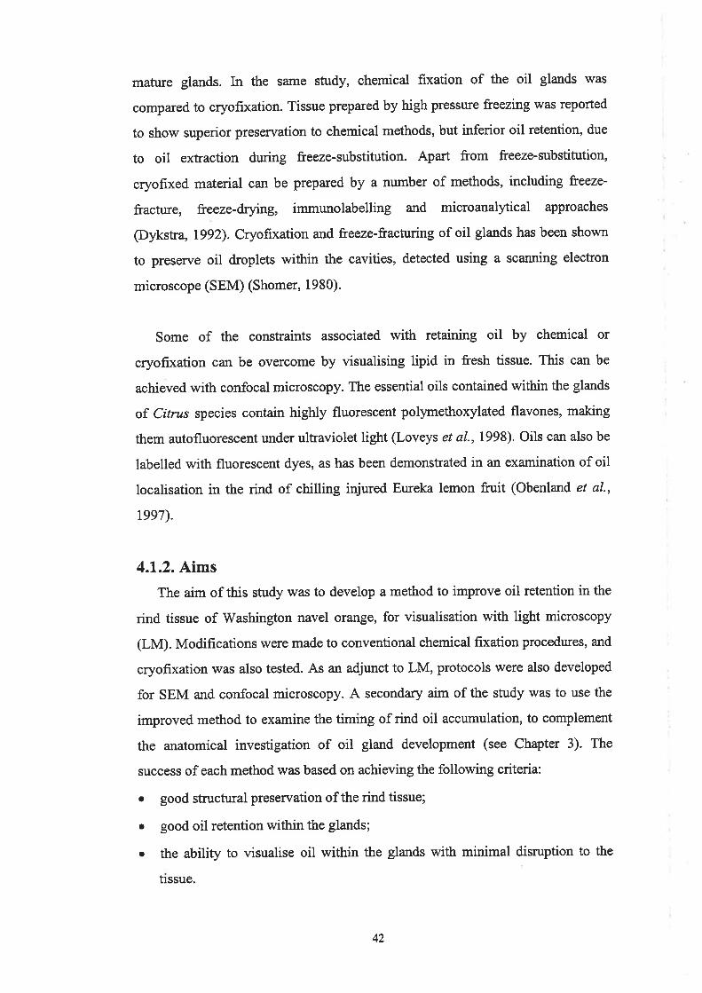
mafi¡¡e glands. In the same study, chemical fixation of the oil glands was
compared to cryofixation. Tissue prepared by high pressure freezíng was reported
to show superior preservation to chemical methods, but inferior oil retention, due
to oit extraction during freeze-substitution. Apart from freeze-substitution,
cryofixed material can be prepared by a number of methods, including heeze-
fracture, freeze-drying, immt¡nolabelling and microanalytical approaches
(Dykstra, 1992). Cryofixation and freeze-fracturing of oil glands has been shown
to preserve oil droplets within the cavities, detected using a scanning electron
microscope (SEM) (Shomer, 1980).
Some of the constraints associated with retaining oil by chemical or
cryofixation can be overcome by visualising lipid in fresh tissue. This can be
achieved with confocal microscopy. The essential oils contained within the glands
of Citrus species contain highly fluorescent polymethoxylated flavones, making
them autofluorescent under ultraviolet light (Loveys et a1.,1998). Oils can also be
labelled with fluorescent dyes, as has been demonstrated in an examination of oil
localisation in the rind of chilling iqiured Eureka lemon fruit (Obenland et al.,
reeT).
4.1.2. Aims
The aim of this study was to develop a method to improve oil retention in the
rind tissue of V/ashington navel orange, for visualisation with light microscopy
(LM). Modifications were made to conventional chemical fixation procedwes, and
cryofixation was also tested. As an adjunct to LM, protocols were also developed
for SEM and confocal microscopy. A secondary aim of the study was to use the
improved method to examine the timing of rind oil accumulation, to complement
the anatomical investigation of oil gland development (see Chapter 3). The
success of each method was based on achieving the following criteria:
o good structural preservation of the rind tissue;
o good oil retention within the glands;
o the ability to visualise oil within the glands with minimal disruption to the
trssue.
42

4.2. Materials and Methods
4.2.1. Light microscopy (LM)
schedules for LM tissue preparation are summarised in Table 4.1
Table 4.1. Summary of fixation, deþdration and embedding schedules
for LM Methods I to7.
Method Primary firation Post-Iixation Dehydration Ernbedding
1
2
3
4
5
6
7
Aldehyde
Osmium vapour
Osmiumvapour
Aldehyde
Aldehyde
SBB staining
Slam freezing
Aqueous osmium (lh)
Aldehyde/osmium solutiorL aldehyde, aqueous
osmiumAldehyde/osmium solutio¡! aldehyde, aqueous
osm¡umAqueous osmium (4h)
Aqueous osmium (4h)
Aldehyde
Acetone (70-l007o) PA
Ethanol (70-1000/o) PA
Ethanol (10-100%) Spun
Elhanol (50-100þ
Ethanol (50-100%)
Erhanol (30-100o/o)
Methanol
PA
GMA
GMA
Lowicryl
4.2.l.l.Method I - Osmium postfixation and Procure-Araldite embedding
Tissue preparation was carried out according to the second method outlined in
Section 3.2.3.2. Rind samples of approximately 1 x 2 mm were fixed in a solution
of yoformaldehyde, 7.25o/o glutaraldehyde and 4olo sucrose, in phosphate buffered
saline (PBS), pH 7.2, overnight at 0-4'C. After two short rinses in PBS, samples
were postfixed in phosphate buffered 7yo osmium tetroxide for one hour. Samples
were dehydrated through an acetone series (70, 90, 95, I00yo), with each step
consisting of thfee 20 minute changes, plus an additional one hour of 100%
acetone. Samples viere infiltrated overnight in a l:1 mixture of acetone.Procure-
Araldite (PA) resin, followed by three eight hour changes of 100% resin, all on
rotator. Samples were flat embedded and polymerisation achieved at 70"C.
4.2.l.2.Method 2 - osmium vapour fixation and ProcureAralditeembedding
Tissue fixation was similar to that of Turner et al. (1998). Rind samples of
approximately I x 2 mmwere placed on moistened filter paper in an enclosed petri
dish and exposed to vapours from a pool of aqueous 2olo osmium tetroxide, at
room temperature for 30 minutes. They were then placed in a chilled solution of
2.5Yo glutaraldehyde, 2Yo formaldehyde and 0.lolo aqueous osmium tetroxide in
0.05M Pipes buffer, pH 6.8, for one hour, and then transferred to the same
43

solution minus the osmium at 0-4'C overnight. The following morning, samples
were postfixed in a solution of lYo osmium tetroxide in 0.05M Pipes buffer at 0-
4"C for one hour. Deþdration \¡/as slightly modified from Method 1, with samples
dehydrated at o-4"c through an ethanol series (70, 90, 95, l00yo), with each step
consisting of three 20 minute changes, plus an additional one hour of 100%
ethanol. Samples were infiltrated overnight in a l:1 mixture of ethanol: PA resin,
followed by three eight hour changes of 100% resin. Samples were embedded as in
Method l.
4.2.l.3.Method 3 - Osmium vapour fixation and Spurr embedding
Tissue fixation was conducted as described in Method 2. Dehydration,
infiltration and embedding methods were adapted from those outlined by Spun
(1969). Samples were dehydrated at 0-4'C through a gradual ethanol series (10,
20, 30, 40, 50, 60, 70,80, 90 and 100%), with each step consisting of two 10
minute changes, plus an additional one hour of 100% ethanol. Samples were
infiltrated overnight in a l:l mixture of ethanol:Spurr resin, followed by three eight
hour changes of l}Oyo resin. Samples were flat embedded and polymerisation
achieved at 70"c. A 'firm' resin was prepared according to the method of Spurr
(1969). Osmium postfixed material was also processed.
Initially, blocks were found to be brittle and difficult to section. Softer blocks
.were not obtained by reducing polymerisation temperature or extending
polymerisation time. Problems were rectified by changing the flexibiliser
component of Spurr resin from DER 736 (Spun kit, Sigma, St. Louis, USA) to
DER 732 (Spurr's embedding kit, ProSciTech, Thuringowa, Australia).
4.2.l.4.Method 4 - Modified osmium fixation and Procure-Araldite
embedding
Rind samples of approximately I x 2 mm were used. Samples were fixed in a
solution of 2.5Yo glutaraldehyde and 2%o îormaldehyde in 0.05M Pipes buffer, pH
6.8, for 24 hours at O-4"C. After two short rinses in Pipes buffer, samples were
postfixed in l%o osmium tetroxide in 0.05M Pipes buffer at 0'4"C for four hours.
After two further rinses in Pipes buffer, samples were dehydrated through an
44

ethanol series (50, 70, 90,95, l}Oyo), with each step consisting of three 20 minute
changes, plus an additional one hour of 100% ethanol. Samples were infiltrated for
24 hours in a l:1 mixture of ethanol:PA resin. Infiltration in 100% resin was
carried out in several steps: overnight, eight hours, 24 hours, 24 hours and eight
hours, all at 0-4'C. Samples were embedded according to previous methods'
4.2.1.5. Method 5 - Modified osmium fixation and glycol-methacrylate
embedding
Rind samples of approximately 2 x 2 mm were used, and tissue was prepared
according to Method 4, replacingPA resin with glycol-methacrylate (GMA). GMA
preparation and embedding was carried out as outlined in Section 3.2.3.2.
4.2.1.6. Method 6 - Sudan Black B staining and glycol-methacrylate
embedding
Rind tissue was injected with the lipid stain Sudan Black B (SBB) (Sigma) in
7Oo/o ethanol for ten minutes prior to sample collection. This method is similar to
one previously observed to stain oil in the glands of navel orange rind tissue (Susan
Johnson, Technical Officer, CSIRO Plant Industry; Pers. Comm., 2000)' Samples
of approximately 3 x 3 mm were rinsed briefly in 50 and 30Yo ethanol and fixed
overnight in3Yo glutaraldehyde in 0.025M phosphate buffer at 0-4'C (Feder and
O'Brien, 1963). Samples were rinsed in phosphate buffer and dehydrated at 0-4'C
through an ethanol series (30, 40, 50, 60, 70, 80, 90, 95, l00yo), with each step
lasting one hour. Samples were infiltrated in a 1.1 mixture of ethanol:GMA
overnight, followed by two changes of 1009/o GMA for two days each- Samples
were embedded in GMA as in Method 5
4.2.1.7. Method 7 - Cryofixation, freeze-substitution and low temperature
embedding
Rind samples of 0.5 mm width and length were frozen by impact onto a liquid
nitrogen cooled copper block using a MM 80 metal mirror cryofixation system
attached to a Leica EM CPC cryopreparation unit. Frozen samples \ilere
transferred to the chamber of a Reichert AFS automalic freeze-substitution system
at -80'C and substituted in methanol for 36 hours (Monaghan and Robertson,
1990). Samples were warïned 4'C per hour to -45"C, and infiltrated with Lowicryl
45

HM 20 (Chemische Werke Lowi, Waldkraiburg, Germany). The resin was
polymerised by ultraviolet light for 48 hours at'45"C'
4.2.L.8. Microtomy and histochemistry
For each method, sections were collected from a minimum of four samples. For
pd Spun and Lowicryl embedded material, sections (1 pm thickness) were cut on
a glass knife with a Reichert-Jung Ultracut E Mcrotome, placed on glass slides
and stained with Toluidine Blue O (TBO) (Sigma) (O'Brien and McCully, 1981) or
SBB (Sigma) (Bronner, 1975). For GMA embedded material, sections (4 pm
thickness) were cut on a glass knife with a Reichert-Jung 2050 Supercut
Microtome, placed on glass slides, and stained with periodic acid-Schitrs (PAS)
and TBO (Otsrien and McCully, 1981), or with SBB (O'Brien and McCully,
l98l). All material was observed with aZeiss Axiophot Photomicroscope, using
transmitted light.
4.2.2. Scanning electron microscopy (SEM)
4.2.2.1" Environmental SEM (ESEM)
Rind samples of approximately 2 x 3 mm were examined using an ElectroScan
ESEM.
4.2.2.2. Field emission gun SEM (FEGSEM)
a. Chemicalfixation
Rind samples of 2 x 2 mm were fixed according to Section 4.2.1'1. Samples
were deþdrated through an acetone series and critical-point dried with COz.
Samples v¡ere mounted on aluminium stubs with sticþ tabs (ProSciTech),
carbon/gold coated and observed using a Philips XL 30 FEGSEM, at an
accelerating voltage of l0 kV.
b. Freeze-fracture
Rind samples of approximately 2 x 3 mm were attached to a clamping holder,
oriented to allow a longitudinal fracture through a selected gland. Samples were
plunge frozen in a liquid nitrogen slush, and transferred under vacuum to the
prechamber of an o>dford CT 1500 IIF cryotransfer system, maintained at
46

approximately -130'C, where they were fractured with the end of a scalpel blade.
After fracturing, the samples were sublimed for five minutes at -92"C and 5 x 10's
pa, recooled to -l lO'C, and coated with gold palladium. Samples were transferred
to the cold stage mounted in the Philips )(f, 30 FEGSEM, and examined at
approximately -190'C.
c. Cryofixation
Rind samples of approfmately 2 x 3 mm $/ere mounted onto aluminium stubs
with carbon paint. Samples were cryofixed according to the above protocol, and
sublimed for approximately three minut es at -92"C and 5 x 10⁻⁵ Pa, recooled to
-110'C, and coated with gold palladium. Samples were examined as above'
4.2.3. Fluorescence microscopy
4.2.3.1. Light fluorescence microscopy
. Rind sections approximately I mm thick and l0 mm long were stained with the
fluorescent lipid dye Nile Red (NR) (0.01%) (Sigma) (Obenland et al-, 1997). The
NR stain solution was prepared in either ethanol or in a mixture of polyethylene
glycol (PEG) 400 and gQYo glycerol @rundrett et al., 1991). Sections were
mounted in 75Yo glycerol on glass slides, and observed with a Zeiss Axiophot
photomicroscope, using incident-light fluorescence, and a filter combination of
excitation 450 - 490 nm and emission 520 nm long pass'
4.2.3.2. Confocal laser scanning microscopy (CLSM)
a. Method IRind tissue was torn, rather than razor sectioned, to obtain intact glands, and
then trimmed into sections approúmately 2 mm thick and 15 mm long. Sections
were mount ed in 7 5Yo glycerol on glass cover slips and examined using a Bio-Rad
MRC-1000 UV CLSM system, attached to a Nikon Diaphot 300 inverted
microscope. A kryptor/argon laser was used to excite specimens at 488/30 nm and
images were collected using emission frlter 522135 nm.
4'.1

b. Method 2
Rind sections approximately 2 mm thick and 15 mm long were collected'
Sections were stained for two hours with aqueous Safranin O (Australian Biostain,
Traralgon, Australia), to highlight cell walls. Sections were rinsed and
counterstained for two hours with 0.01% NR (Sigma), prepared according to
Brundrett et at. (1991). Sections were mounted as above. NR fluorescence was
observed using the same settings as above, and Safranin O using excitation 568/10
nm and emission filter 605/32 nm.
4.2.3.3. Multi-photon microscopy
Rind sections approximately 2 mm thick and 15 mm long were collected'
Sections were stained for l8 hours \¡r/ith 0.01% NR (Sigma) prepared as above,
rinsed, and counterstained for five minutes in 0.01% Calcofluor White M2R (CW)
(Sigma) prepared in distilled water (Hughes and McCully, 1975). Calcofluor stains
mixedlinkage p-glucans (Wood et a1.,1983) and was used to highlight cell walls.
Tissue sections were mounted as for CLSM, and examined using a Bio-Rad
Radiance 2000 MP visualising system attached to a Nikon Eclipse TE300 inverted
microscope. A Coherent Mira900-F titanium.sapphire ultrafast laser was used to
excite the specimen at 800 nm (or 830 nm) and images were collected using
emission filters 62O1LOO nm (for NR) and 450/80 nm (for CW).
48

4.3. Results
4.3.1. Light microscopy (tM)4.3.l.l.Method 1 - Osmium postfixation and Procure'Araldite embedding
Rind tissue showed good preservation (Plate 4.14). Some glands showed
lightly stained oil within their cavity (Plate 4.18), whilst others showed poorly
preserved gland contents. Glands disrupted during tissue preparation showed
empty oil cavities.
4.3.l.2.Method 2 - Osmium vapour fixation and Procure-Araldite embedding
The quality of tissue preservation \ilas inferior to that obtained with Method 1
(plate 4.lC), and sectioning damage due to fragility of the tissue occurred
frequently. Oil was evident in all gland cavities and frequently stained da¡ker than
that observed in Method 1, but glands showed more signs of damage. The best
preserved gland showed an oil-filled cavity with an intact lining, darkly stained
inner gland cells, and slight damage to boundary cell layers (Plate 4.1D)- Glands
disrupted dwing tissue preparation showed empty oil cavities.
4.3.l.3.Method 3 - Osmium vapour fixation and Spurr embedding
The quality of tissue preservation rù¡as slightly improved from Method 2,
appearing similar to that obtained with Method 1 @late 4.lE)' however,
sectioning damage due to fragility of the tissue occurred frequently. Darkly
stained oil in the gland cavities was observed, but gland structure was poorly
preserved @late 4.1F). Osmium postfixed samples showed better overall rind
preservation than osmium vapour fixed samples, although glands showed similar
artefactual damage (Plate 4.1G and 4.1H). The best preserved gland showed
darkly stained oil in the gland cavity and inner gland cells @late 4.1H).
49

Plate 4.1
Rind oil visualisation using light microscopy. Longitudinal, TBO stained
sections (A to H).
A. Aldehyde and osmium postfixed tissue, embedded in PA resin. Bar = 200
luÎ.
B. Higher magnification of the gland from A. Lightly stained oil in the
cavity (c). Bar: 100 pm.
C. Osmium vapour fixed tissue, embedded in PA resin. Bar:200 pm.
D. Higher magnification of the gland from C. Darkly stained oil in the
cavity (c) and inner gland cells (i), and damage to boundary cells (arow).
Ba¡: 100 ¡rm.
E. Osmium vapour fixed tissue, embedded in Spurr resin. Bat = 200 ¡rm.
F. Higher magnification of the gland from E. Darkly stained oil in the cavity
(c), and damage to gland cells (arrows). Bar: 100 Fm.
G. Aldehyde and osmium postfixed tissue, embedded in Spurr resin. Bar :200 pm.
H. Higher magnification of the gland from G. Darkly stained oil in the
cavity (c) and inner gland cells (i), and damage to boundary cells (arows).
Bar: 100 ¡rm.
50
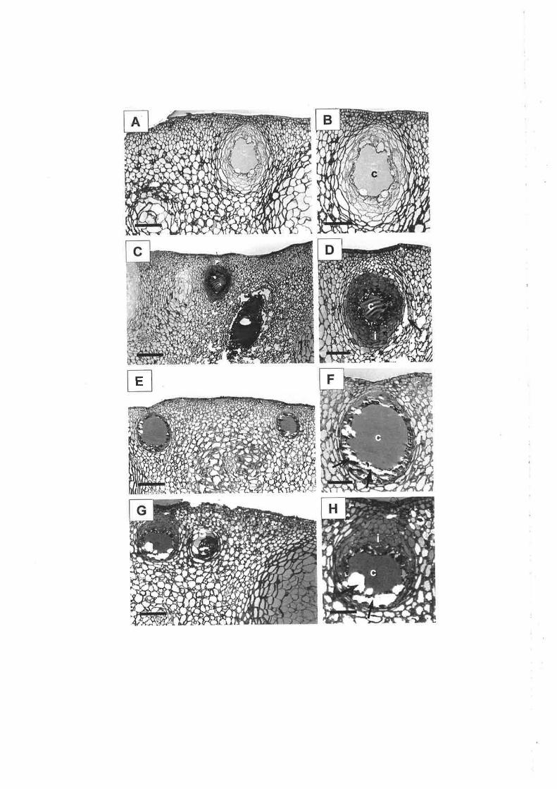
E

4.3.l.4.Methods 4, 5 and 6
Sections from tissue prepared by these methods have not been presented due to
poor preservation. Modified osmium fixation and PA embedding (Method 4)
produced significant sectioning damage, and all glands were excessively damaged,
or were completely absent, with oil retention difficult to assess. Embedding
similarly fixed tissue in GMA (Method 5) improved rind preservation, but glands
appeared distorted and oil was not present in the gland cavities. Staining tissue
with SBB and embedding in GMA (Method 6) produced the worst preseruation of
all chemical fixation procedures.
4.3.1.5. Method 7 - Cryofixation, freeze-substitution and low temperature
embedding
The small size of samples required for slam freezing resulted in glands either
being not present in samples or damaged dr:ring tissue preparation.
4.3.1.6. Histochemistry
SBB frequently produced very strong staining of PA resin, hampering the
visualisation of black stained lipid bodies. Another drawback of SBB for oil
staining was its preparation in ethanol, which is a lipid solvent. TBO was
preferred as it did not excessively stain the resin, provided a good contrast stain,
and showed the oils.
4.3.2. Scanning electron microscopy (SEM)
4.3.2.1. Environmental SEM
Technical problems encountered with the ESEM examination of fresh tissue
samples meant that images could not be obtained. Extreme 'flaring' of the on-
screen image was thought to be a result of the sample contacting the detector or
the effect of oil volatilisation from the rind tissue.
4.3.2.2. Field emission gun SEM
In chemically fixed tissue, the gland cavity was largely emPtY, however, small
spherical oil bodies were associated with the inner surface of the gland cavity
(plate 4.2A)- Oil bodies were also present inside inner gland cells, dissected
51

Plate 4.2
Rind oil visualisation using scanning electron microscopy. Longitudinal
gland cross-sections.
A. Chemically fixed tissue, showing a largely empty gland cavity (c).
Spherical oil bodies (arrow) associated with the inner surface of the cavity.
Morphology of the epicuticular wax (ew) was also evident. Bar: 50 pm.
B. Higher magnification of the gland depicted in A. Spherical oil bodies
(arrows) inside inner gland cells, dissected during tissue preparation. Bar:50 pn.
C. Freeze-fractured tissue, showing an uneven fracture line. Boundary cells
(b), and inner gland cells (i) were identifiable, but gland oil could not be
detected. Bar: 100 ¡rm.
D. Cryofixed tissue, showing an oil-filled cavity (c). Bar:200 Frm.
52


during tissue preparation (Plate 4.28).In freeze-fractured tissue, oil could not be
visualised due to the rough fractures achieved @late 4.2C), however, in tissue
which was sectioned fresh and cryofixed, oil within the gland cavity was
successfully preserved and visualised. The cavity of mature glands appeared to be
tulIof oil (Plate 4.2D).
4.3.3. F luorescence microcopy
4.3.3.1. Light fluorescence microscopy
In dissected glands, NR stained oil bodies v/ere detected around the gland
perimeter (Plates 4.34 and 4.38). Oil bodies were also detected deeper in the
gland cavity, and appeared to be associated with the cavity lining (Plate 4.3C).
Closer inspection of glands suggested that oil was not present in the flattened
boundary cells of the gland (Plate 4.3D). Oil bodies did not appear to be present
outside the gland. Both NR preparations (ethanol and PEG 400 plus glycerol)
resulted in intense oil fluorescence.
4.3.3.2. Confocal laser scanning microscopy (CLSM)
a. Method IOil was autofluorescent under UV (excitation 351 nm/emission 460 nm), but
was more clearly visualised when stained with NR, and examined at excitation
488 nm, with emission filter 522 rmt. Rind tissue tearing resulted in a gland half-
embedded in tissue and half exposed. Oil bodies ïvere present over the exposed
surface of the gland (Plate 4.4A), and were approximately 25 - 30 pm in width-
Longitudinal optical sections were collected up to approximately 500 pm into the
I mm wide gland. An optical section close to the gland centre showed oil bodies
localised aror¡nd the perimeter @late 4.48). Faint fluorescent bodies were also
visible deeper into the gland cavity, but could not be visualised. Oil was not
present within the gland cavþ. Oil bodies did not appear to be present outside the
gland.
53

Plate 4.3
Rind oil visualisation using light fluorescence microscopy. Longitudinal
gland cross-sections.
A. Fluorescing oil bodies localised around the gland perimeter at the cut
surface. Gland cavity (c) out of focus. Gland attachment to the epidermis
also evident (arrow). Bar: 100 pm.
B. Fluorescing oil bodies localised around the gland perimeter at the cut
sruface (a:rows). Gland cavity (c) out of focus. Bar : 100 ¡rm.
C. Same gland as depicted in B. Change of microscope focus shows
fluorescing oil bodies deeper into the gland cavity (c). Cut edge is out of
focus (arrows). Bar: 100 pm.
D. Basal region of a gland. Fluorescing oil bodies present in inner gland
cells (i), but not in flattened boundary cells (b), or in the parenchyma cells
þ) surrounding the gland. Bar: 100 pm.
54

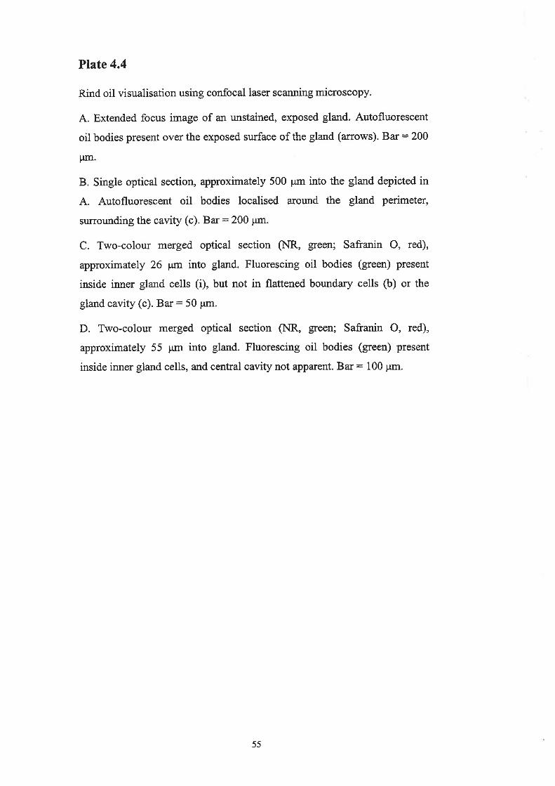
Plate 4.4
Rind oil visualisation using confocal laser scanning microscopy.
A. Extended focus image of an unstained, exposed gland. Autofluorescent
oil bodies present over the exposed surface of the gland (arows). Bar = 200
pm.
B. Single optical section, approximately 500 pm into the gland depicted in
A. Autofluorescent oil bodies localised around the gland perimeter,
surrounding the cavity (c). Bar = 200 pm.
C. Two-colour merged optical section (NR, green; Safranin O, red),
approximately 26 ¡rm into gland. Fluorescing oil bodies (green) present
inside irurer gland cells (i), but not in flattened boundary cells (b) or the
gland cavity (c). Bar: 50 pm.
D. Two-colour merged optical section (NR, green; Safranin O, red),
approximately 55 pm into gland. Fluorescing oil bodies (green) present
inside inner gland cells, and central cavity not apparent. Bar = 100 pm.
55
A

b. Method 2
Glands were slightly damaged ínrazor sectioned tissue samples. In one gland,
an optical section 26 Wn from the cut surface showed oil bodies clearly associated
with inner gland cells, but not present in boundary cells (Plate 4.4C). Oil was also
not present in the gland cavity. In another gland, in which slightly deeper optical
sections were obtained (55 pm from the cut surface), oil bodies were again
associated with inner gland cells, and a cavity was not conspicuous @late 4.4D).
Oil bodies did not appear to be present outside the gland.
4.3.3.3. Multi-photon microscopy
Oil bodies were detected to a depth beyond 180 pm from the cut surface,
localised to inner gland cells but not present in boundary cells @late 4.54). A
higher magnification optical section thr.ough the inner gland cells showed a dense
distribution of oil bodies (Plate 4.58). The composite image of a z-series of
optical sections through another gland showed boundary cells to be oil-free, and a
dense distribution of oil bodies inside the gland cavity (Plate 4.5C). It appeared
that the entire cavity space was filled with oil. A small number of NR labelled
bodies were detected outside the gland. Calcofluor provided a more intense
labelling of cell walls than Safranin O.
56
Plate 4.5
Rind oil visualisation using multi-photon microscopy.
A. Two-colour merged optical section (NR, red; CW, green), approximately
150 ¡rm into damaged gland. Fluorescing oil bodies (red) present inside
inner gland cells (i), but not in flattened bor:ndary cells (b) or the gland
cavity (c). Bar: 50 Frm.
B. Two-colow merged optical section (NR, red; C\ù/, green), approximately
180 pm into damaged gland. Fluorescing oil bodies (red) closely associated
with the polyhedral shaped inner gland cells. Ba¡: 10 pm.
C. Two-colour merged composite image of a z-series of 16 optical sections,
1 pm apart, through a gland (NR, red; CW, green). Fluorescing oil bodies
(red) filled the entire cavity space (c). Flattened boundary cells of the gland
(b) appeared oil-free, and epicuticular wa"x (ew) stained strongly with the
lipid stain NR. Bar: 250 Fm.
57

4.4. Discussion
4.4.1. Method development
4.4.1.1. Light microscopy (LIvt)
Rind tissue fixed with aldehyde alone has been shown to result in empty gland
cavities (see Chapter 3). In this study, aldehyde fixed and osmium postfixed tissue
(Method 1) retained some oil, as in previous studies of citrus oil glands using
similar fixation protocols (Thomson et al., 1976; Turner et al., 1998). Primary
fixation with osmium vapour, shown to improve lipid preservation by Turner e/
al. (1998), was used as the starting point for method development in this study. In
the majority of samples, osmium vapour fixation (Method 2) achieved enhanced
lipid retention compared to osmium postfixation (Method 1), as reported in the
foliar glands of C. limon (Tumer et a1.,1998). However, this was accompanied by
a major compromise in structural preservation, not previously observed in the leaf
tissue of C. limon (Tumer et a1.,1993). Poor structural preservation with osmium
vapour primary fixation is explained by the fact that osmium tetroxide penetrates
fresh tissue very slowly, and has very poor cross-linking capabilities compared to
the aldehydes (Dykstra, 1992). Despite the fact that small (1 mm) rind samples
were used, osmium vapour would have more easily diffused into the thin leaf
tissue. In addition, some glands observed in this study were greater than 500 pm
in width, compared to 200 ¡.tm foliar glands (Turner et a1.,1998). The mixture of
osmium and aldehyde introduced after primary fixation may itself have
contributed to fixation problems. Glutaraldehyde and osmium tetroxide were
observed to react, and form a fine, dense precipitate of reduced osmium, which
has been reported to ruin specimens (Hayat, 1989). The use of this fixative
mixture has not been reported widely, and has been shown to produce inferior
fixation of the petiolar hairs of Lycopersicon esculentun (Mersey and McCully,
1978). In poorly preserved rind tissue, artefactual damage was commonly
localised to the glands. This observation suggested that the thickened walls of the
gland borurdary cells may have acted as a mechanical ba¡rier to the penetration of
fixatives. Similar fixation problems have been reported in plant tissues with a
thick cuticle (O'Brien and McCully, 1981).
58

To improve tissue preservation, the embedding medium was changed from PA
to Spurr resin (Method 3). Spurr resin was chosen as it was used in the original
osmium vapour fix study by Turner et al. (1998), and has also been used in the
other major anatomical studies of citrus oil glands (Bosabalidis and Tsekos,
1982a; Thomson et a|.,1976). Spun also has a lower viscosity than Araldite resins
(Spurr, 1969), and was expected to better infiltrate samples. However, this did not
appear to be the case, with tissue preservation appearing to be slightly poorer.
Intact tissue sections of both osmium vapour fixed and osmium postfixed material
were fowrd to be more difficult to obtain when embedded in Spurr resin.
Extending the dr¡¡ation of osmium postfixation (Method 4) did not improve
lipid fixation as expected, but resulted in inferior tissue preservation. The use of
another embedding medium, GMA (Method 5), slightly improved tissue
preservation, but removed all traces of the oil. This is not surprising, considering
GMA has been described as a 'lipid solvent' (Hayat, 1989). Polymerisation
problems also occuned with osmium treated tissue, as GMA requires an oxygen
free environment for pol¡rmerisation @ykstra, 1992). A final test for an alternative
to osmium vapour fixation involved staining tissue with SBB prior to aldehyde
fixation. SBB is the most effective of the lipid colourants (Casselman, 1959). This
method was adapted from another study, which showed it to retain oil to some
extent in the glands of the navel orange rind (Johnson, Pers. Comm., 2000). The
ability of SBB to retain lipid may be explained by the fact that Sudan dyes
partition into lipid, but are also capable of reacting chemically, by virtue of their
two amino groups (Gr¡rr, 1960). Unfortunately, past observations were not
duplicated in this study, with poor tissue preservation hampering lipid
visualisation. It is likely that tissue was damaged by the 70o/o ethanol in which the
stain was prepared.
Changes were also made to dehydration steps to improve oil retention, and to
staining techniques, to enhance oil visualisation. For all modified chemical
fixation methods, ethanol was used instead of acetone, as it was considered a
preferred dehydration agent for lipid retention (V/anner et a1.,1981). Dehydration
59

rwas modified to reduce the chance of lipid removal, by lowering the concentration
of the first dehydration step and shortening the duration in each dehydrating
solution. Dehydration was also carried out at 0-4"C to reduce lipid loss (Hayat,
1989), as rù,'as resin infiltration in some methods. However dehydration and
infiltration for Spurr embedded samples was carried out at room temperature, to
avoid condensation, which hampers polymerisation (Dykstra, 1992). Section
staining with TBO was preferred over SBB as it showed a greater specificity for
the tissue, rather than the epoxy resin. TBO also effectively showed the oil
retained within the glands, as has been demonstrated previously in LM studies of
the oil glands of Citrus (Turner et al., 1998) and also Eucalyptus (Jocelyn
Carpenter, Electron Microscopist, The University of Melbourne; Pers. Comm.,
te99).
The ability to obtain intact glands with oil-filled cavities was limited by the
small size of samples required for chemical fixation. Most chemical fixatives
penetrate at least 0.5 mm into a sample within one hour, enabling the fixation of 1
mm thin samples (Dykstra, 1992), however, in the previous anatomical
investigation of the oil glands of V/ashington navel orange (Chapter 3), glands of
up to 1 mm in width were observed. Consequently, damaged glands with empty
cavities were frequently observed in this study. Size limitations for samples also
limited the success of cryofixation; samples of less than 0.2 mm were required for
slam freezing effectiveness (Dykstra, 1992). As a result, no intact cryofixed
glands were obtained. The use of slam freezing has been reported previously in
apple tissue, although here, it was reported to produce inferior structural
preservation to plunge freezíng and conventional freezing (Golding et a1.,1995).
High pressure freezing, on the other hand, has provided some success in oil gland
fixation (Turner et a1.,199S). Unlike most cryotechniques, which freeze tissue to
no more than 15 ¡rm depth, high pressure freezing is an exception, enabling
fixation to approximately 0.5 mm, analogous to chemical fixation (Dykstra, 1992).
High pressure freezing is considered the only feasible method to fix large plant
tissues without pretreatment (Kaeser et al., 1989). Unfor¡rnately, high pressure
freezingequipment was not available for use in this study.
60
4.4.1.2. Scanning electron microscopy (SEM)
SEM is routinely used to examine tissue morphology. In the present study,
SEM allowed the examination of fresh, chemically fixed and cryofixed material.
Observations of fresh material using ESEM were hampered due to the naflrre of
the material being examined. It is likely that volatilisation of the rind oils from the
tissue contaminated the atmosphere of the ESEM specimen chamber, and affected
the electron beam movement to the detector, preventing image collection.
Both chemical and cryofixation methods produced well preserved tissue for
observation with FEGSEM, allowing a good comparison of oil retention, which
was not possible with light microscopy. In glands which were dissected dwing
fresh tissue preparation, oil was largely removed from the gland cavity by
chemical fixation, as observed by light microscopy. However, in cryofixed tissue,
oils were retained and appeared largely unaffected by fresh tissue sectioning.
Based on these observations, freeze-fracture was expected to be the optimal
method, as it involved fixation prior to gland dissection or fracture, minimising
possible structural damage and oil removal. Unfortunately, the inability to produce
a smooth fracture surface after several attempts prevented the visualisation of
cavity oil using this method. Clean fractures, such as those previously achieved in
the rind tissue of grapefruit (Petracek et al., 1995) were not duplicated. Of the
methods tested, sectioning of fresh tissue to expose gland cavities, followed by
plunge freezíngto fix the oils, was the optimal SEM method for oil visualisation.
4.4.1.3. Fluorescence microscopy
Confocal microscopy methods used ìvere the most successful for oil
visualisation in fresh rind tissue. To obtain real images of gland oils in fresh
tissue, both good staining and optical penetration of the tissue were required. Prior
to tissue examination with the confocal microscope, the ability of the fluorescent
dye NR to stain lipids was tested using light fluorescence microscopy. Brightly
fluorescent oil bodies were clearly distinguishable around the gland perimeter, but
it was not clear whether this was real or could be attributed to dye localisation in
cells at the cut surface. The ability to optically section into the tissue with the
6l

confocal microscope removed the necessity for gland dissection. However, the
ability to prepare intact glands close to the cut sr¡rface was extremely difficult, and
most glands, when examined closely, appeared to be slightly damaged. Intact
glands were obtained by tearing the rind tissue, but this method often caused
excessive damage to the interconnecting tissue. Interestingly, intact glands were
frequently observed to show poor staining of the oil in the gland cavity. Based on
these observations, it appeared likely that slight disruption of the gland was
beneficial, in that it provided improved stain penetration.
Inrazor sectioned tissue stained for þid with NR and counterstained for cell
walls (with Safranin O), optical penetration of up to 55 gn from the cut surface
was achieved using CLSM. Using this method, the localisation of oil bodies to
inner gland cells, as observed using epifluorescence, was confirmed. Boundary
cells appeared oil-free and oil was also not detected inside the cavity. Given that
gland damage occurred, and tissue was not fixed, it is likely that oil was removed
from the gland cavity whilst tissue samples were immersed in the staining
solutions. As a result, oil bodies observed were largely intracellular.
Using CLSM, the ability to capture images further from the cut surface was
limited by sample opacity and inhomogeneity, and limitations of the working
distance of the objective lens (Pawley, 1995). Clearing the tissue can overcome
such obstacles, but this was not used because of its tendency to remove lipid.
Instead, optical limitations were largely overcome by the use of the multi-photon
microscope. Advantages of the two-photon system over single-photon system are
that it utilises infra red light, which reduces photobleaching, cytotoxic effects and
also light scattering, resulting in improved sensitivity and deeper optical
sectioning (Pawley, 1995). Improved optical penetration of up to 180 pm into the
gland was achieved using the multi-photon microscope. With thicker tissue slices
and improved optical penetration, oils within the cavity were retained. ln one
gland, the large central cavity was filled with oil.
62

4.4.2. Rind oil observations
In previous light and transmission electron microscopy (TEM) studies of
mature citrus oil glands, oil has been reported to be localised in the gland cavity
and also the inner epithelial cells su¡rounding the cavity (Thomson et al.,1976;
Bosabalidis and Tsekos, 1982b; Turner et aL.,1998). The same observations \¡/ere
made in this study for glands of orange rind tissue prepared for LM. Cryofixed
tissue examined using SEM, and fresh tissue examined using the multi-photon
microscope also showed the oil-filled cavities of mature glands. However, for
fresh material in which oil had been extracted from the cavity, the presence of oil
bodies within the inner gland cells could be observed more clearly using confocal
methods. Observations of fresh tissue in this study support previous reports in
fixed tissue which suggest that the inner epithelial cells of the glands are secretory
in nature (Thomson et al., 1976; Bosabalidis and Tsekos, 1982b). Following oil
synthesis, oils are believed to be actively secreted into the cavity space (Shomer,
1980; Bosabalidis and Tsekos, 1982b). In this study, SEM examination of
chemically fixed tissue showed similar results to those of Shomer (1980), who
observed nr¡merous oil droplets associated with the inner surface of the cavity wall
in freeze-fractured glands of Lisbon lemon.
Confocal microscopy (CLSM and multi-photon) observations in this study
clearly contradict the frndings from a previous confocal microscopy study of oil
localisation in the rind of chilling injured Er:¡eka lemon fruit (Obenlard et al.,
1997). In their study, Obenland et al. (1997) prepared tissue using a similar
protocol, but in healtþ tissue, found large amounts of oil, in the form of discrete
oil bodies, outside the glands. It seems likely that during the collection of 25 mm
long and 2.5 mm thick rind samples for their examination, gland damage occured
without detection, resulting in oil spillage into surrounding tissues. The possibility
of rind oil occurring naflually outside of the glands is unlikely due to its
phytotoxicity (see Chapters 5 and 6). Indeed, the function of these glands, at least
in part, is to isolate these substances from the rest of the plant (Fahn,7979).
63

4.5. Conclusion
This study provides a detailed appraisal of methods for oil retention and
visualisation in the oil glands of orange rind tissue, with the use of LM, SEM,
CLSM and multi-photon microscopy. For LM, methods which appeared to
improve oil retention compromised structural preseration of the rind tissue.
Characteristics of the glands themselves, namely their size and the thickness of
their outer walls, also limited the success of fixation. Based on our selection
criteria, a reliable method for rind oil visualisation with LM was not achieved. A
fi¡rther study into oil accumulation in the developing fruit was not undertaken. As
alternatives to LM, SEM showed the effectiveness of cryofixation of gland oils,
whilst fluorescence microscopy showed the ability to preserve rind oils without
tissue fixation. Although these techniques were revealed to be promising for oil
visualisation, further work is required to develop optimal methods. In conclusion,
rind oil localisation is best examined routinely in serial sections using LM or
TEM, augmented by techniques such as cryoSEM and multi-photon microscopy.
64

Chapter 5
Inducing oleocellosis in the laboratory
5.1. Introduction
5.1.1. Background
Fawcett (1916) was the first author to attribute the oleocellosis blemish to the
action of rind oils. In his study, oleocellosis ìwas reported to develop as a result of
swface oil release following fiuit injury, as well as the transfer of oil from injwed
fruit to the surface of uninjured ft¡it. Since these early findings, oleocellosis has
been artificially induced by both mechanical damage and oil application to the
fruit.
To induce oleocellosis, some studies have mechanically damaged fruit in a
non-uniform manner, by using or simulating normal handling practices (Catroon er
al., 1964; Levy et al., 1979; Loveys et al., 1998). The penetrometer, used
commercially to measure rind oil release pressure (RORP) prior to hanrest, has
also been used to induce oleocellosis (Erner, 19821' Shomer and Erner, 1989). The
penetrometer is favowed over normal handling practices, in that it allows the user
to control the applied pressure, enabling a more uniform and reliable induction.
However, like other mechanical methods, the penetrometer has been reported to
rely heavily on fruit turgor (Shomer and Erner, 1989). In their study on Shamouti
oranges, Shomer and Erner (1989) reported that penetrometer pressure did not
selectively rupture the oil glands in fruit which had lost a significant amount of
moisture, but created excess tissue damage, particularly to the albedo layer. Like
the penetrometer, pin pricking glands achieves localised damage, but does not rely
on a high fruit turgor. In a study by Sawam:uÍa et al. Q98\, pin pricking
individual glands produced severe rind injury in all of the seven Citrus species
examined, including Citrus sinensis. This method has also been successfully
tested on South Australian Washington navel oranges (Loveys et a1.,1998).
65

In comparison to mechanical methods, inducing oleocellosis with rind oils
overcomes the requirement of high fruit turgor and allows the observer to attribute
rind damage entirely to oil effects. In previous studies; oil has been applied to the
fruit surface by bending excised rind segments (Sawamr.ra et al.,1984; Sawamrua
et al., 1987; Sawamwa et a1.,1988; Wiltiams and Wild, 1996), applying oil drops
(Brodrick, 1970; Chikaizumi, 2000) and smearing aqueous oil solutions (Shomer
and Emer, 1989). A more standardised method of oil application has been
developed by'Wild and Williams (1992), in which oil-treated discs are attached to
the fruit surface with adhesive tape.
Oleocellosis symptoms have been assessed by such measures as number of
spots (Catrooî et al., 1964; Levy et al., 1979; Erner, 1982; Loveys et al', 1998)
and spot size (Levy et al., 1979 Erner, 1982; Sawamura et al-, 1984). In
experiments using large volumes of fruit, percentage of fruit showing symptoms
has also been used (Eaks, 1955). The measure of 'number of spots' is considered
representative of severity in disorders where numerous spots develop naturally,
and has been used to assess pitting (Petracek et al., 1998). However, in
oleocellosis, number of spots is a direct result of impacts to the fruit. Such a
measure is considered misleading where oleocellosis has been induced by random
mechanical damage, as apperirs to be the case in many previous studies (Cahoon er
al.,1964;Levy et a1.,1979; Loveys et a1.,1998). For the purposes of this study,
blemish area or 'spot size' and blemish severity are considered to be informative
measurements, where oleocellosis has been induced in a uniform manner. The
term 'severity' is used to describe the extent of rind collapse and discolouration
associated with the blemish. There is no indication that an equivalent measwe of
severity has been used in previous studies.
5.1.2. Aims
The aim of this study was to develop successful methods for oleocellosis
induction by both mechanical means and oil application, in order to simulate the
two forms of naturally occurring oleocellosis; namely gland rupture and oil
66

transfer between fruit. For mechanical induction, conditions for maximising firrit
turgor were tested. For oil induction, various oil components and concentrations
\¡/ere assessed. The success of each method was based on fulfilment of the
following criteria:
o must produce a localised blemish;
. must be reproducible;
o must produce an adequate blemish area to allow macroscopic assessment and
the collection of rind samples for microscopy examination;
o must produce a blemish severity which increases at a tate which allows a
detailed time sequence examination of changes;
o must be relatively easy to achieve.
In addition to these objectives, RORP values obtained from penetrometer
induction were compared to the threshold value currently used by growers to
predict oleocellosis.
5.2. Materials and Methods
5.2.L. Preliminary induction methods test
5.2.1.1. Plant material
Fruit were taken from 23 year old Valencia orange trees on Troyer citrange
rootstock, located in the University of Adelaide, Waite Campus Alverstoke
orchard (34o97'S, 138"63'E). Rows were N-S oriented, and the trees were spaced
at approximately 5.3 x 5.9 metres. Fruit were harvested on October 6,1997.
Fruit sampling was based on uniformity of fruit size, and to a lesser extent, fruit
colour. Polar and equatorial diameter (mm) and fresh weight (g) values were
recorded for each fn¡it. A uniform colour was considered important as colour has
been reported to influence oleocellosis blemish perception (Levy et al., 1979;
Erner, 1982).
67

Fruit were picked from the east aspect of the tree, as these fruit normally show
a higher turgor (Loveys et al., 1998), indicating a higher susceptibility to
mechanical oil gland rupture. Fruit were also picked at 9:00 am to avoid a decline
in turgor due to increasing temperature and decreasing relative humidity (and
increasing vapouf pressure deficit) throughout the day (Cahoon et a1.,1964). The
close proximity of the orchard to the laboratory allowed the rapid transfer of fnrit,
and minimal turgor loss between harvest and treatment. Mechanical damage to
harvested fn¡it was reduced by the use of secateurs and careful handling.
5.2.L.2. Induction methods
a. Pin prick methods
A pin prick was used to rupture a single oil gland. Multiple pin pricks were
used to rupture glands within aî areaof approximately I cm'on the rind surface.
b. Penetrometer
A penetrometer with an 11 mm diameter tip, and 0 to 13 kg pressure gauge
was used (Effegr, Alfonsine, Italy). A similar tool was used in studies by Cahoon et
aI. Q96\ and Eaks (1968), but a smaller tip was used by Shomer and Erner
(1939). All RORP values \ryere converted from kilograms into pressure units, a
standard unit to allow correlation between studies which have used different sized
penetrometer tips. For example, a mass reading of 3 kg is equivalent to a force of
29.4 N, when assuming acceleration (due to gravity) to be 9.8 ms-2. With an I I
mm diameter penetrometer tip, this produces a pressure of approximately 309 lcPa.
A standard pressure could not be guaranteed to cause gland rupture in all fruit,
as RORP varied somewhat between trees and also between fruit on the same tree.
Instead, standard damage was achieved by applylng RORP to each fruit'
c. Oil methods
Cold-pressed orange oil (Tropico Fruits Pty. Ltd., South Australia), and two
components of orange oil, dlimonene and linalool (Sigma, St. Louis, USA) were
used. In a method similar to rWild (1998), 15 pl of l00yo concentrations of each of
the three oils was pipetted onto 6 mm cardboard discs (Antibiotic Assay discs,
68

Whatman International Ltd., Maidstone, England), which were attached to the fruit
surface with adhesive tape. Non-phytotoxic sunflower oil was also included as a
control oil treatment.
All of the methods were applied to each fruit around the equatorial region.
Induction was carried out at 0, 6 and 2l hours after harvest. Time of induction
after harvest was varied to assess the effect of fruit turgor on mechanical and oil
induction methods. The times of induction were chosen on the basis of
convenience with respect to the transport of fruit. Following induction, fruit were
stored at ambient temperature (20'C).
5.2.1.3. Symptom assessment
Blemish severity and area were assessed on day 1 and 3 following induction.
Adequate symptom development was expected by two to three days on the basis of
past observations of Washington navel oranges (Wild, 1998).
Blemish area (in mm'¡ was measured using a grid sheet transparency. For
mechanical methods, the area of oil spread outside of the damage site was
assessed. For oil application, the area of oil spread outside of the oil disc
application site was assessed.
Blemish severity was assessed using a scoring system, which took both rind
collapse and discolouration into consideration. A single observer was used to score
blemishes, and standard blemish images were used to improve consistency (Plate
s.l).
5.2.L.4. Experimental design
Sixty fnrit were harvested in total, from five trees. Treatments were randomly
allocated to the twelve fruit from each tree: three induction times x two assessment
times x two replicates.
69

Plate 5.1
Blemish severity scoring system. Bars: 1 cm.
A. 0 - nil.
B. 1-veryslight.
C. 2 - slight.
D. 3 - medium.
E. 4 - high.
F. 5 - extreme.
70

l.fàt
.*
t\tt:a.a

5.2.1.5. Statistical analYsis
Blemish severity and area data were analysed using the statistical package
Genstat for Windows version 4.1 (Lawes Agricultural Trust, IACR Rothamsted).
A split plot design was applied to the data and Analysis of Variance (AI\IOVA)
tables used to test for statistical significance at the 95o/o cottfrdence level. Data
were also analysed using Restricted Maximum Likelihood analysis (REML), in
cases where there \¡/as non-uniform replication between treatments. This was due
to a small number of cases where oil discs did not adhere closely to the fn¡it.
5.2.2. OiI method refinement
5.2.2.1. Experimental plan
Fruit were harvested during the 1998 season from 23 year old Washington
navel orange trees on Poncirus trifoliata rootstock located in the University of
Adelaide, Waite Campus Alverstoke orchard (34o97'S, 138"63'E). Fruit sampling,
oil induction and symptom assessment were carried out as in the preliminary
experiment. Repeat assessments were made on individual fruit at days I and 3.
5.2.2.2. Oleocellosis induction methods
In experiment l, ten fruit were harvested on May 6, 1998, and each füiit was
treated with dJimonene concentrations of 25,50 and 100%.
In experiment2, six fruit were han¡ested on June 10, 1998, and each frr¡it was
treated with dJimonene concentrations of 60, 70, 80 and90Yo'
In experiment 3, six fruit were harvested on June 26, 1998, and each fruit was
treated with 100% d-limonene and the oil component citral at concentrations of 20,
40,60,80 and l00Yo.
1L
5.2.3. Penetrometer method refinement
5.2.3.1. Experimental Plan
Fruit were harvested from the same trees used above. Fruit sampling,
penetrometer induction and symptom assessment were ca¡ried out as in the
preliminary experiment. In each experiment, RORP rù/as measured on four fruit
immediately after harvest and at each of the induction times, to give an indication
of changes in fruit tr.ugor. Repeat assessments \¡/ere made on individual fruit at
days 1 and 3.
5.2.3.2. Oleocellosis induction methods
In experiment 1., 12 frvit were han¡ested on lN,day 26, 1998, at 8:30 am, and
stored in plastic bags on ice, in a styrofoam box. Both plastic bags and low
temperature storage were used to minimise moisture loss and maintain turgor.
In experiment2, nine fruit were harvested on June 10 at 10:00 am. Trees were
deep watered 24 hours prior to hanrest, and following harvest each fruit was
wrapped in moist paper towel and stored in a styrofoam box. The objective was to
achieve a higher turgor at harvest and maintain turgor more effectively by keeping
the fruit surface wet and cool. In the first two experiments, induction was caried
out at 2,4 and 8 hours after harvest.
In experiment 3, nine fruit were harr¿ested on June 30 at 12:30 pm, slightly
later in the day, due to rainy conditions. Experiment 3 was otherwise identical to
the preceding experiment, except earlier induction times of 1, 3 and 5 hours were
used, to test the effect of inducing damage closer to the time of harvest.
72

5.3. Results
5.3.1. Preliminary induction methods test
5.3.1.1. Blemish severity
The single pin prick treatment had a negligible effect and the penetrometer
produced very low severity scores, whilst all other methods produced slight to
medium severity blemishes (Fig. 5.1). These differences explain why the method
effect was significant (P < 0.001). The pin prick produced no symptoms in 90% of
samples by day 3. The multiple pin prick gave the highest severity scores of the
mechanical methods, with a mean score of approximately 2.5, or slight to
medium. d-limonene and orange oil produced a similar mean severity to the
muttþle pin prick treatment. Linalool gave the most severe blemishes, with a
mean of approximately 3, or medium. At day 3,80yo of blemishes were classed as
score 3 or 4 and l3Yo as score 5. Linalool typically produced a blemish with a
darker coloured border (Plate 5.2A). By day 3, linalool also produced gland
collapse, unlike the other oil treatnents where collapse was localised between
glands only. Sunflower oil, the non-phytotoxic oil used as the control, produced
no signs ofblemish.
Induction at 21 hours produced lsss severe blemishes than induction at 0 or 6
hours, for all oil methods and for the multiple pin prick treatment. Penetrometer
showed the opposite trend, which may explain the significant interaction between
method and induction time (P < 0.001). Assessment time had a negligible effect
for mechanical methods, but for oil methods, mean severity increased from day I
to 3 by a score of l. The interaction between method and assessment time was
also significant (P < 0.001).
73

rnI
q)li9ch
¡iq)
c)(t)
raI
(¡)LIIA
Lq)
q)(h
0
Pin Mpin Lim
Induction methodLin Orange
Figure 5.1. Preliminary induction methods test. The effect ofinduction method and induction time on blemish severity at days Iand 3. Induction methods include penetrometer (pen), pin prick (pin),multiple pin prick (mpin), d-limonene (lim), linalool (lin) and orange
oil (orange). Sixty fruit from five trees, treated with three inductiontimes (0, 6 and 21 hours), two assessment times (days 1 and 3) and
replicated twice. Means and standard errors represented.
4
3
t
1
05
4
3
,,
1
Pen
Day L
Day 3
74

Plúe 5.2
Oleocellosis symptoms induced in the preliminary induction methods test.
A. A fruit treated with linalool (left), d-limonene and orange oil (right).
Linalool typically produced a blemish with a darker coloured border
(arrow). Bar: I cm.
B. A fruit treated with multþle pin pricking. Area of rind damage due to oils
is localised around the margins of the damage zone (dz). Bar: 1 cm.
C. The d-limonene blemish (lim) was þpically larger than linalool (lin) and
orange oil (orange) blemishes. Bar: 1 cm.
75
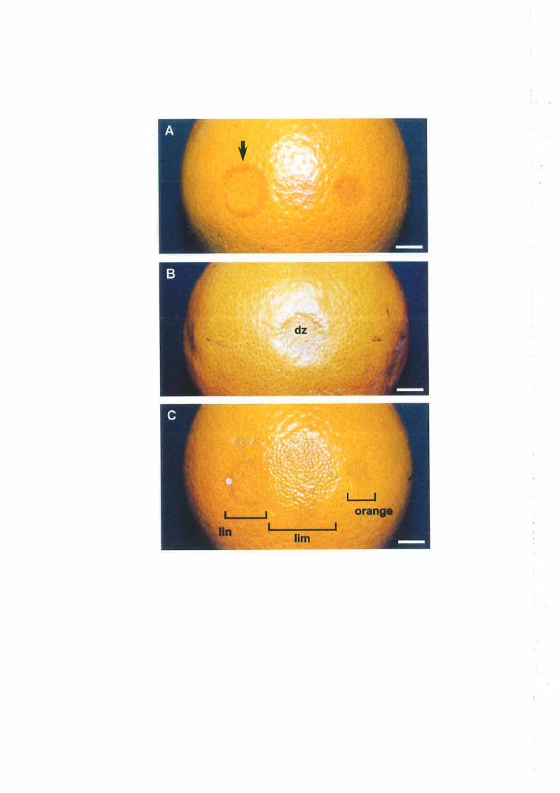
ì. *.1
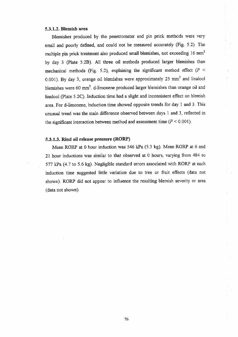
5.3.L.2. Blemish area
Blemishes produced by the penetrometer and pin prick methods were very
small and poorly defined, and could not be measured accurately (Fig. 5.2). The
multiple pin prick treatment also produced small blemishes, not exceeding 16 mm2
by day 3 @late 5.28). All three oil methods produced larger blemishes than
mechanical methods @ig. 5.2), explarning the significant method effect (P <
0.001). By day 3, orange oil blemishes were approúmately 25 mm2 and linalool
blemishes were 60 mm'. d-limonene produced larger blemishes than orange oil and
linalool @late 5.2C).Induction time had a slight and inconsistent effect on blemish
area. For d-limonene, inductiontime showed opposite trends for day I and 3. This
unusual trend was the main difference observed betvveen days I and 3, reflected in
the significant interaction between method and assessment time (P < 0.001).
5.3.1.3. Rind oil release pressure (RORP)
Mean RORP at 0 hour induction was 546 kPa (5.3 kg). Mean RORP at 6 and
2l hour inductions was similar to that observed at 0 hours, varying from 484 to
577 kPa (4.7 to 5.6 kg). Negligible standard erors associated with RORP at each
induction time suggested little variation due to tree or fruit effects (data not
shown). RORP did not appear to influence the resulting blemish severity or area
(data not shown).
'76
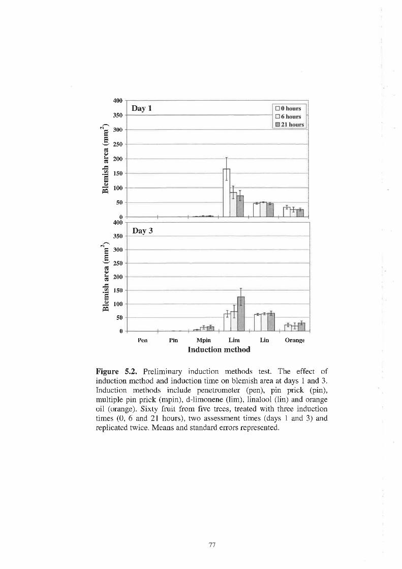
400
3s0
N- 300
5 zsoq)
h 2oo
.9 rsO
é)
= 100t*i
50
0400
350
ê¡Ê 300
- 250cÉ(¡)
h 2oo
.9 1s0
.9 roo
50
0
Mpin Lim
Induction methodLin Orange
Figure 5.2. Preliminary induction methods test. The effect ofinduction method and induction time on blemish area at days 1 and 3.
Induction methods include penetrometer (pen), pin prick (pin),multiple pin prick (mpin), d-limonene (lim), linalool (lin) and orangeoil (orange). Sixty fruit from five trees, treated with three inductiontimes (0, 6 and 21 hours), two assessment times (days 1 and 3) andreplicated twice. Means and standard errors represented.
PinPen
Day 1
Day 3
77

5.3.2. Oil method refinement
5.3.2.1. Blemish severity
In experiment 1, 100% d-limonene produced much more severe blemishes
than 50% and25o/o d-limonene (Fig. 5.3), explaining the significant concentration
effect (P < 0.001). A d-limonene concentration of 25olo produced no symptoms by
day 3. A concentration of 50%o gave a mean score of less than 1 by day 3, with
70%o of fruit showing no symptoms. A concentration of 100% gave a mean score
of 2.5 by day 3, or slight to medium. A very minor severity increase was observed
between days 1 and 3.
In experiment 2, d-limonene concentrations of 90Yo and 80% produced more
severe blemishes than 60%o and 70% @íg. 5.3), explaining the significant
concentration effect (P < 0.001). Concentrations of 60% and70o/o produced mean
severity scores of less than 1, with the majority of fruit showing no symptoms by
day 3 (83% md 67Yo, respectively). A concentration of 80% gave a low mean
score of 1.5, whilst 90% d-limonene produced the most severe blemishes, with a
mean score of 2.5 at day 3, the same as pure d-limonene in experiment l. Aconsistent increase in severity from day 1 to 3 was also observed, explaining the
significant assessment time effect (P : 0.008).
In experiment 3, all citral concentrations produced much higher severity scores
than 100% d-limonene (Fig. 5.4), explaining the significant oil treatnent effect (P
< 0.001). d-limonene gave a mean score of only 1 at day 3, and a maximum score
of 2, or slight. d-limonene produced less severe blemishes than in the preliminary
experiment and experiment 1. d-limonene also showed a high standard error,
which was attributable to fruit differences. Cifral rwas very damaging to the rind;
20%o cítral gave a mean score of 2.5 by day 3, and blemish severity increased with
citral concentration. A dramatic increase in severity from day I to 3 was obsenred,
explaining the significant assessment time effect (P < 0.001). Citral also caused
gland collapse, with the entire blemish area appearing to be depressed into the rind
(Plate 5.34).
78

4
3
2
1
¡,nI
OLI(t)
þo,¡
o)U)
5
0
25Exp 1
50 100 60 70 80
Exp 2
Limonene concentration (Vo)
90
5
0
Citral20 Citrat 40 Citral60 Citral S0 Citral 100 Lim100
Oil component and concentration (7o)
Figure 5.4. Oil method refinement. The effect of citral concentrationand IÙOVI d-limonene on blemish severity at days I and 3. Experiment3, Six fruit treated with six oil treatments (citral 20, 40,60, 80, IOOVo
and d-limonene lÙOVo). Repeat assessments made at days 1 and 3.
Means and standard errors represented.
Figure 5.3. Oil method refinement. The effect of d-limoneneconcentration on blemish severity at days 1 and 3. Experiment 1, Ten
fruit treated with three d-limonene concentrations (25, 50 and IOOVo).
Experiment 2, Six fruit treated with four d-limonene concentrations(60, 70, 80 and 90Vo). Repeat assessments made at days 1 and 3.
Means and standard errors represented.
rnI
é)Lq)at)
Lq)
(¡)U)
4
3
.,
1
trday L
@day 3
trday1@day 3
79

Plate 5.3
Oleocellosis symptoms induced in the oil method refinement experiments.
A. Citral produced severe rind collapse, including gland collapse. Two
blemishes resulting from 100% citral (arrows). Bar = 1 cm.
B. A concentration of 100% produced the most advanced d-limonene
blemish, with a severity score of 4 and an area of 476 mm2 at day 3. Drawn
lines delineate the blemish boundaries from two oil disc application sites.
Bar: 1 cm.
C. d-limonene at 600lo concentration produced small blemishes, with a mean
area of 16 mm2 (arows). Bar: 1 cm.
D. dlimonene at 90% produced large blemishes, with a mean area of 166
mm2 (outlines). Bar: 1 cm.
80

> \
>
-
<t
<l!''

5.3.2.2. Blemish area
Blemish area showed a similar trend to blemish severity in all three oil
experiments (Figs. 5.5 and 5.6). In experiment l, I00oÁ d-limonene produced
much larger blemishes than 50% and 25%o d-limonene (Fig. 5.5), explaining the
significant concentration effect (P < 0.001). A concentration o125o/o produced no
s¡rmptoms, and 50o/o d-limonene produced small blemishes, with a mean area of
23 mmz at day 3. Pure d-limonene produced a mean blemish area exceeding 200
mm2, but also showed high standard errors due to fruit variation. Differences
between day 1 and 3 were negligible. The most advanced blemish observed was
produced by 100% d-limonene and had a severity rating of 4, or high level, and
was 476 mmt in area (Plate 5.3B).
In experiment 2, a gradual area increase was associated with increasing d-
limonene concentration (Fig. 5.5), and the concentration effect was significant (P
:0.002). 4 concentration of 60% d-limonene gave the smallest mean a¡ea of 16
mm' lPlate 5.3C) whilst 90% d-limonene gave a mean area of 166 mm2 (Plate
5.3D). A consistent increase in area between days I and 3 was also observed,
explaining the significant assessment time effect (P:0.003).
In experiment 3, all citral concentrations produced much larger blemishes than
rc}% d-limonene (Fig. 5.6), explaining the significant oil treatment effect (P <
0.001). Blemish area also appeared to increase consistently from day 1 to 3,
explaining the significant assessment time effect (P: 0.03).
8l
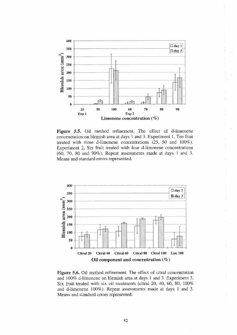
400
350
Ng 300
I zsoGq)
þ zoo
.9 150
9 rooÊa
50
0
25Exp I
50 100 60 70 80Exp2
Limonene concentration (7o\
90
400
350
'E roo
5 zso
é)
h 200
f rso
q.) 100
E
Figure 5.5. Oil method refinement. The effect of d-limoneneconcentration on blemish area at days 1 and 3. Experiment 1, Ten fruittreated with three d-limonene concentrations (25, 50 and IOOVo).
Experiment 2, Six fruit treated with four d-limonene concentrations(60, 70, 80 and 9OVo). Repeat assessments made at days 1 and 3.
Means and standard errors represented.
Citral20 Citral40 Citral60 Citral S0 Citral 100 Lim 100
Oil component and concentration (Vo)
Figure 5.6. Oil method refinement. The effect of citral concentrationand l00%o d-limonene on blemish area at days 1 and 3. Experiment 3,
Six fruit treated with six oil treatments (citral 20, 40,60, 80, IOOVo
and dlimonene l0o7o). Repeat assessments made at days 1 and 3.
Means and standard errors represented.
50
0
3
trdayL
trday1@ùay 3
82

5.3.3. Penetrometer method refinement
All penetrometer experiments showed similar blemish induction success;
experiment 2 was chosen to be representative of oleocellosis induction with the
penetrometer, and results from this experiment a¡e outlined below'
5.3.3.1. Blemish severity
All induction times produced a mean severity scofe of less than 1 (or very
slight) and little or no difference between days I and 3 (Fig. 5.7). High proportions
of fruit showed no blemish development;67% for 2 hour, 78%o for 4 hour and 56%o
for 8 hour induction. The most severe blemish observed had a score of a (high) and
resulted from the 2 hour induction.
5.3.3.2. Blemish area
In all penetrometer experiments, blemish area was difficult to measure because
of the weak symptoms. At day 3, mean blemish area va¡ied from 65 to 86 mm2
@ig. 5.8). High standard errors observed were attributed to fn¡it variation.
Plate 5.4 demonstrates the appearance of the oleocellosis blemish achieved with
a penetrometer, three days after induction.
5.3.3.3. Rind oil release pressure (RORP)
Mean RORP at harvest varied from 526 kPa (5.1 kg) in experiment 3 to 588
kPa (5.7 kg) in experiment 2. For all experiments, mean RORP at delayed
induction times was similar to that observed at 0 hour induction (data not shown).
The most notable effect of fruit storage was observed in experiment 3, where
RORP declined from 536 kPa (5.2 kg) to 433l<Pa@ zkg) after five hours storage.
83

4
3
2
I
rnI
IL9at)
L(¡)
c)ct)
5
0
2 4 IInduction time (hours after harvest)
Figure 5.7. Penetrometer method refinement. The effect of inductiontime on blemish severity at days 1 and 3. Nine fruit, treated with three
induction times (2, 4 and 8 hours). Repeat assessments made at days 1
and 3. Means and standard errors represented.
trday IBday3
T T T
248Induction time (hours after harvest)
Figure 5.8. Penetrometer method refinement. The effect of inductiontime on blemish area at days 1 and 3. Nine fruit, treated with three
induction times (2,4 and 8 hours). Repeat assessments made at days 1
and 3. Means and standard errors represented.
400
350
alE 3oo
v 250q)
h 2oo
.9 1s0
I looÉ
50
0
trday 1
Eday3
84

Plate 5.4
Typical oleocellosis blemish resulting from penetrometer induction'
Blemish arises in area of oil spread (o) away from the penetrometer
application zone (paz). Bar: 1 cm.
85


5.4. I)iscussion
5.4.1. Mechanical induction
In the preliminary experiment, none of the mechanical methods used could be
recommended for oleocellosis induction based on the selection criteria used. All
methods failed to produce an adequate blemish area. The most severe blemishes
produced were by the multiple pin prick treatnent, but were classed as only slight.
The inconsistent effect of induction time was attributed to variation due to tree or
fruit effects. Unexpectedly, there was little change in blemish severity from day I
to 3, but only a low level of blemish was found overall.
The single pin prick rarely produced blemish symptoms on Valencia orange
fruit (C. sinensis). The pin prick was observed to release a small volume of oil,
which showed little visible surface spread from the site of damage. These findings
are in complete contrast to those of Sawamura et al. (1984), who induced rind
injury in fruit of seven Citrus species using a single pin prick. rn C. sinensis,
severe rind injury, defined as spots ) 16 mm in diameter, vr'as induced. In some
species, the pin prick produced more severe injury than application of the fruit's
own rind oils. This was explained to be due to the entry of the volatiles into the
flavedo tissue, compared to the volatile loss of low-boiling point compounds
associated with swface oil application. Discrepancies between studies may be
partially explained by fruit storage conditions; enhanced volatile loss was more
likely in the present study, in detached fruit under 20'C conditions, compared to
fruit on the tree during winter months in Japan. However, Sawamtua et al. (1984)
did report less severe rind injury from the pin prick later in the season, which may
explain the poor response of mature fruit in the present study. The effect of fruit
age was not explored further, but it is unlikely that it is related to changes in oil
yield or composition, as both have been shown to remain fairly stable at later
stages ofthe season (Scora et aL.,1968).
For multiple pin prick and penetrometer methods, multiple glands were
ruptured and a greater volume of oil released onto the fruit surface, but the degree
86
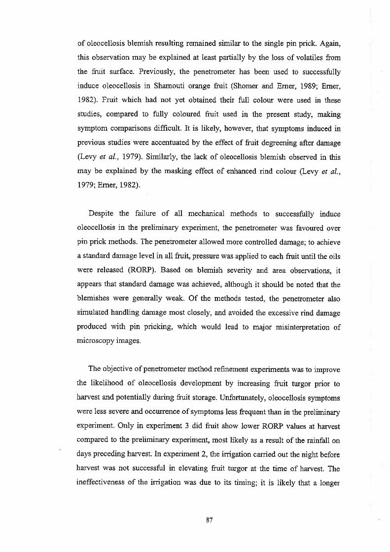
of oleocellosis blemish resulting remained similar to the single pin prick. Again,
this observation may be explained at least partially by the loss of volatiles from
the fruit surface. Previously, the penetrometer has been used to successfully
induce oleocellosis in Shamouti orange fruit (Shomer and Erner, 1989; Erner,
1982). Fruit which had not yet obtained their full colour were used in these
studies, compared to fully coloured fruit used in the present study, making
symptom comparisons difficult. It is likely, however, that symptoms induced in
previous studies were accentuated by the effect of fruit degreening after damage
(Levy et al., 1979). Similarly, the lack of oleocellosis blemish observed in this
may be explained by the masking effect of enhanced rind colour (Levy et al.,
1979;Emer, 1982).
Despite the failure of all mechanical methods to successfully induce
oleocellosis in the preliminary experiment, the penetrometer was favoured over
pin prick methods. The penetrometer allowed more controlled damage; to achieve
a standard damage level in all fruit, pressure was applied to each ftiit until the oils
were released (RORP). Based on blemish severity and area observations, it
appears that standard damage was achieved, although it should be noted that the
blemishes were generally weak. Of the methods tested, the penetrometer also
simulated handling damage most closely, and avoided the excessive rind damage
produced with pin pricking, which would lead to major misinterpretation of
microscopy images.
The objective of penetrometer method refinement experiments was to improve
the likelihood of oleocellosis development by increasing fruit trugor prior to
harvest and potentially during fruit storage. Unfortr¡nately, oleocellosis symptoms
were less severe and occurrence of symptoms less frequent than in the preliminary
experiment. Only in experiment 3 did fruit show lower RORP values at harvest
compared to the preliminary experiment, most likely as a result of the rainfall on
days preceding harvest. In experiment 2, the irrigation carried out the night before
harvest was not successful in elevating fruit turgor at the time of harr¡est. The
ineffectiveness of the irrigation was due to its timing; it is likely that a longer
87

irrigation, carried out further in advance of han¡est would have been more
effective. Information on the effect of inigation timing on RORP is limited, but
Cahoon et al. (1964) have shown that a 14 day interval between irrigations is
preferred to 7 days in obtaining same-sized lemon fn¡it with a lower turgor, to
reduce oleocellosis susceptibility at harvest.
Storing fn¡it in water saturated conditions following harvest has been shown to
increase fruit turgor (Cahoon et aI., 1964). Cahoon et al. (7964) reported that
lemon fruit stored for 16 hours under ambient temperature showed a slight
decrease in weight and dramatic decrease in turgor, whilst fruit stored in a water
saturation chamber at 28 "C with their stems in water, showed an increase in both
fruit weight and turgor. In contrast, fruit stored under ambient conditions for up to
2l hours in the present study showed only a slight decrease in turgor (increase in
RORP). In follow-up experiments, storing fruit under modified conditions for
moisture retention showed little improvement to storing fn¡it at ambient
temperature. The most notable effects were observed in experiment 3, where
RORP was reduced from 536 kPa (5.2 kg) at harvest to 433 k}a (.2 kg) after five
hours storage, by wrapping frr¡it in moist paper towel and storing in a styrofoam
box. A comparison of findings suggests that storage time r¡/as not a contributing
factor, but that our storage conditions for moisture retention were inadequate
compared to those of Cahoon et al. (1964).
Commercially, RORP is used as a predictive test for oleocellosis in the field
(Cahoon et al., 1964). Australian citrus growers use 3 kg RORP with an 8 mm
diameter penetrometer tip (585 kPa) as a threshold value, above which oleocellosis
is considered not likely to occur under normal handling conditions (Feutrill, 1997).
In the present study, mean RORP at han¡est was consistently close to or greater
than this ttreshold value, varying from 526 to 588 kPa (5.1 to 5.7 kg). However,
oleocellosis was observed to develop in fruit with RORP values as high as
approximately 770 kPa (7.5 kg). Based on this observation, it appears likely that
oleocellosis symptoms have the potential to develop as long as oil is released from
the glands. If pressure applied to the fruit surface is greater than RORP, this can be
achieved. An exception, however, is for fruit with exceedingly low turgor, in which
88

it is not possible to cause selective gland rupture without greater mechanical
damage to the rind, as reported by Shomer and Erner (1989).
5.4.2. Oil induction
Studies into the phytotoxicity of different rind oil components to the rind of
orange fruit have found that d-limonene and linalool are major contributors to
oleocellosis (Sawamura et al., L984; Wild and Williams, 1992).In the preliminary
experiment, 700yo concentrations of dJimonene, linalool and orange oil all
produced an adequate blemish severity for our experimental purposes. Linalool
produced the most severe blemishes, often displaying a darkened border and in
severe cases, collapsed glands. In comparison, dlimonene and orange oil produced
less severe blemishes, with rind collapse localised between glands only, as is
evident in naturally occurring oleocellosis. A comparison of identical (100%)
concentrations of d-limonene and linalool in this study appears to support Wild and
Williams (1992), who suggested that linalool is highly ph¡otoxic, more so than d-
limonene. It was expected that lQlYo dJimonene would induce a blemish similar to
that of natural oleocellosis, as it is the major component of orange oil, comprising
between 83 and 97Yo of the total oil in the mature fruit of C. sinensis (Shaw,
1979). Linalool however, makes up a smaller proportion of the total oil, in the
order of 0.3 to 5.3Yo (Shaw, 1979).
Induction time did not have a consistent effect on blemish severity, and
differences observed were attributed to fn¡it variation, as separate frr¡it were used
for each induction time. For all oil treatments, there was a considerable increase in
blemish severity between days I and 3. This suggests that physiological changes in
the rind due to oil action take place over a number of days. These changes were
examined in detail in a time sequence study of oleocellosis development, outlined
in Chapter 6.
The largest blemishes were produced by dJimonene. The inconsistent effect of
induction time was again attributed to variation between fruit, and may have been
avoided if repeat measurements had been made on single fn¡it. If induction time is
not considered in the data assessment, mean blemish a¡ea due to d-limonene was
89
only slightly larger than linalool. In the study by Sawamura et al. (1984), a similar
blemish size was also produced by d-limonene and linalool on C. sinensis fruit. In
their study, blemish size was reported to be in excess of 16 mm diameter, which
equates to approximately 200 mmt, compared to 50 to 160 mm2 observed in our
study. The change in area between days I and 3 was slight for all oil treatments,
suggesting the rapid spread of oil from the application site within one day. Indeed,
at the time of induction, oil was observed to spread outwards from the oil disc
almost immediately following its application. From the preliminary experiment, d-
limonene was chosen to be the preferred oil treatment for use in further studies. d-
limonene blemishes were of adequate severity and ample areaby day 3, progressed
in severity from day I to 3, and were most similar in appearance to naturally
occurring oleocellosis of the treatments examined.
The objective of oil method refinement experiments was to test a broader range
of dJimonene concentrations, and an additional oil component not tested in
preliminary experiment, citral. In experiments I and 2, both blemish severity and
area results showed consistency. In experiment l, l00yo d-limonene was greatly
preferred to concentrations of 25 and 50ol0, which produced weak blemishes or no
blemish at all. In experiment 2, 90Yo was the only d-limonene concentration in the
range of 60 to 90% which gave a similar blemish to 100% d-limonene with respect
to severity and area. In experiment 3, 100% dJimonene produced weaker
blemishes, which may be partially explained by the masking effect of enhanced peel
colour @rner, 1982).In the same experiment, citral concentrations of 20 to l00yo
devastated the rind, giving larger and more severe blemishes than the 100% d-
limonene treatment. Citral blemish appearance was uncharacteristic of naturally
occurring oleocellosis, showing extreme tissue collapse and rind indentation,
greater than that resulting from 100% linalool. Observations suggest that citral is
an extremely ph¡otoxic component of rWashington navel orange rind oil. Citral,
which occurs naturally as a mixture of the two aldehydes neral and geranial, occurs
at even lower levels than linalool, in the order of 0.05 to 0.2%o (Shaw, 1979).
Valencia orange fruit were used in preliminary experiments due to availability,
but for all method refinement experiments, lvere replaced with the variety of
90
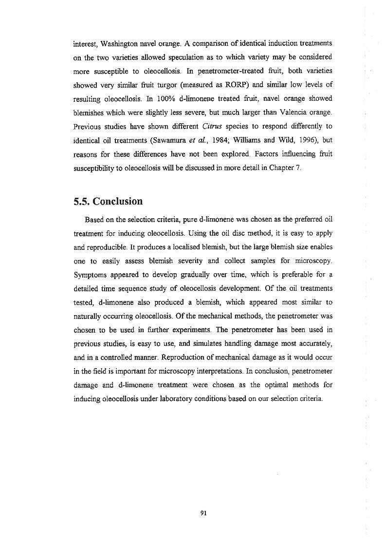
interest, Washington navel orange. A comparison of identical induction treatments
on the two varieties allowed speculation as to which variety may be considered
more susceptible to oleocellosis. In penetrometer-treated fruit, both varieties
showed very similar fruit turgor (measured as RORP) and similar low levels of
resulting oleocellosis. In 100% dJimonene treated fruit, navel orange showed
blemishes which were slightly less severe, but much larger than Valencia orange.
Previous studies have shown different Citnts species to respond differently to
identical oil treatments (Sawamura et al., 1984; Williams and Wild, 1996), but
reasons for these differences have not been explored. Factors influencing fruit
susceptibility to oleocellosis will be discussed in more detail in Chapter 7.
5.5. Conclusion
Based on the selection criteria, pure dJimonene was chosen as the prefened oil
treatment for inducing oleocellosis. Using the oil disc method, it is easy to apply
and reproducible. It produces a localised blemish, but the large blemish size enables
one to easily assess blemish severity and collect samples for microscopy.
Symptoms appeared to develop gradually over time, which is preferable for a
detailed time sequence study of oleocellosis development. Of the oil treatments
tested, dJimonene also produced a blemish, which appeared most similar to
naturally occurring oleocellosis. Of the mechanical methods, the penetrometer was
chosen to be used in further experiments. The penetrometer has been used in
previous studies, is easy to use, and simulates handling damage most accurately,
and in a controlled manner. Reproduction of mechanical damage as it would occur
in the field is important for microscopy interpretations. In conclusion, penetrometer
damage and dJimonene treatment were chosen as the optimal methods for
inducing oleocellosis under laboratory conditions based on our selection criteria.
9t

Chapter 6
Oleocellosis: macroscopic and microscopicdevelopment
6.1. Introduction
6.1.1. BacþroundOur understanding of oleocellosis physiology is limited, and based largely on a
small number of microscopy studies. The natural mode of oil release from the
glands is not understood. Mechanical damage to the fruit has been observed to
result in surface oil release (Fawcett, 1916; Cahoon et a1.,1964; Eaks, 1968), and
based upon such observations, oleocellosis is considered to be caused by the efflect
of the oil released onto the fruit surface (Whiteside et al., 1988). Surface oil
release following mechanical damage to the fruit was confirmed by early light
microscope (LM) studies, which reported epidermal rupture above glands
(Labuschagne et aL.,1977; du Plessis, 1978). However, the possibility of oil release
into the rind tissue has also been suggested, based on the presence of sub-surface
rind damage (Shomer and Erner, 1989). Such a proposition is also supported by
Loveys et al. (1998), who observed symptoms to develop in fn¡it despite the
removal of surface oil, and also induced symptoms by injecting oil into the rind.
The nature of the reaction between the phytotoxic oils and the rind tissue is
also ill-defined in the literature. Using LM, oleocellosis rind damage was first
described as a band of flattened cells located six to eight cells deep from the
surface and between glands (du Plessis, 1978). In later microscopy studies, tissue
postfixation with osmium tetroxide enabled researchers to observe the dense
contents of oil-damaged cells (Shomer and Erner, 1989;V[illiams and Wild, 1996).
Using this improved tissue fixation method, oleocellosis has been examined in
degreened Shamouti orange fruit (Shomer and Erner, 1989) and mature Valencia
orange fruit (Williams and Wild, 1996). These studies show significant
discrepancies based on both LM and transmission electron microscope (TEM)
92
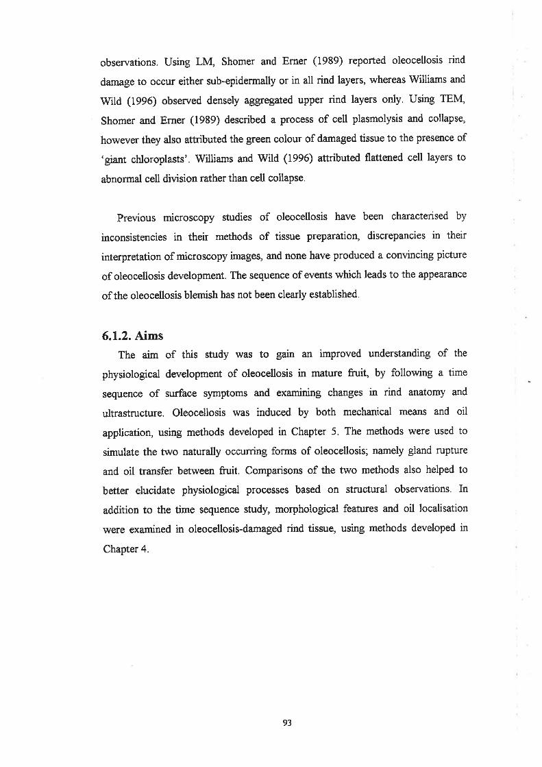
observations. Using LM, Shomer and Erner (1939) reported oleocellosis rind
damage to occur either sub-epidermally or in all rind layers, whereas Williams and
Wild (1996) observed densely aggregated upper rind layers only. Using TEM,
Shomer and Erner (1989) described a process of cell plasmolysis and collapse,
however they also attributed the green colour of damaged tissue to the presence of
'giant chloroplasts'. Williams and Wild (1996) attributed flattened cell layers to
abnormal cell division rather than cell collapse.
Previous microscopy studies of oleocellosis have been characterised by
inconsistencies in their methods of tissue preparation, discrepancies in their
interpretation of microscopy images, and none have produced a convincing picture
of oleocellosis development. The sequence of events which leads to the appearance
of the oleocellosis blemish has not been clearly established.
6.1.2. Aims
The aim of this study was to gain an improved understanding of the
physiological development of oleocellosis in mature fruit, by following a time
sequence of surface symptoms and examining changes in rind anatomy and
ultrastructure. Oleocellosis was induced by both mechanical means and oil
application, using methods developed in Chapter 5. The methods were used to
simulate the two naturally occurring forms of oleocellosis; namely gland rupture
and oil transfer between fnrit. Comparisons of the two methods also helped to
better elucidate physiological processes based on structural observations. In
addition to the time sequence study, morphological features and oil localisation
were examined in oleocellosis-damaged rind tissue, using methods developed in
Chapter 4.
93
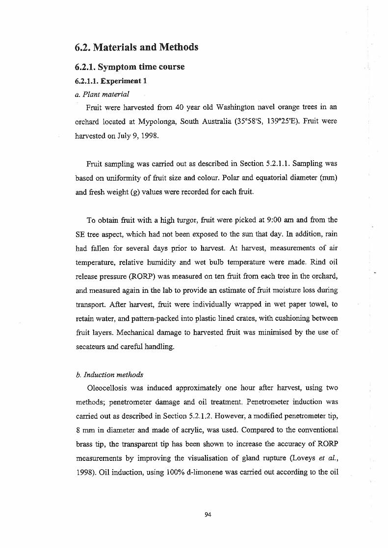
6.2. Materials and Methods
6.2.1. Symptom time course
6.2.1.1. Experiment 1
a. Plant material
Fruit were harvested from 40 year old Washington navel orange trees in an
orchard located at Mlpolonga, South Australia (35o58'S, 139'25'E). Fruit were
harvested on July 9,1998.
Fruit sampling was ca¡ried out as described in Section 5.2.1.1. Sampling was
based on ruriformity of fruit size and colour. Pola¡ and equatorial diameter (mm)
and fresh weight (g) values were recorded for each fruit.
To obtain fruit with a high turgor, fruit were picked at 9:00 am and from the
SE tree aspect, which had not been exposed to the sun that day. In addition, rain
had fallen for several days prior to harvest. At harvest, measurements of air
temperature, relative humidity and wet bulb temperatrue were made. Rind oil
release pressure (RORP) rùras measured on ten fruit from each tree in the orchard,
and measured again in the lab to provide an estimate of fruit moisture loss dr:ring
transport. After harvest, fruit were individually wrapped in wet paper towel, to
retain water, and pattern-packed into plastic lined crates, with cushioning between
fruit layers. Mechanical damage to harvested fruit was minimised by the use of
secateurs and careful handling.
b. Induction methods
Oleocellosis was induced approximately one hour after harvest, using two
methods; penetrometer damage and oil treatment. Penetrometer induction was
carried out as described in Section 5.2.1.2. However, a modified penetrometer tip,
8 mm in diameter and made of acrylic, was used. Compared to the conventional
brass tip, the transparent tip has been shown to increase the accuracy of RORP
measurements by improving the visualisation of gland rupture (Loveys et al.,
1993). Oil induction, using 100% d-limonene was caried out according to the oil
94

disc method described in Section 5.2.1.2. Each method was replicated twice on
each fruit, and replicate treatments referred to as 'samples'.
c. Symptom assessment
Following induction, symptoms were assessed at eight time intervals over th¡ee
days: 30 mins, lh, 2h, 6h, lzh, 24h, 48h, 72h. Nine fruit were assessed at each
time interval (three from each tree).
Blemish area (in mm) was measured as described in Section 5.2.1.3. Blemish
severity lvas measured using a modified scoring system. Rind collapse and colour
(discolouration) were assessed using two separate subjective scales (outlined
below), rather than one (as in Section 5.2.1.3). This allowed assessment of the
individual timing of collapse and discolouration events. Plate 6.1 shows the six
standard blemish images used for assessment in Chapter 5, assigned with severity
scores according to the modified system.
collapse0-nilI - very slight2 - slight3 - medium4 - high
colour0- nilI - very slight2 - slight3 - medium4 - high5 - extreme
d. Experimental design
Seventy-two mature fnrit were harvested in total, from three trees. Treatments
were randomly allocated to the twenty-four fn¡it from each tree: eight assessment
times x three replicates.
e. Statistical Analysis
Rind collapse, colour and blemish area data were analysed using the statistical
package Genstat for Windows version 4.1 (Lawes Agricultural Trust, IACR
Rothamsted). A split plot design was applied to the data and Analysis of Variance
(ANOVA) tables used to test for statistical significance at the 95Yo confrdence
level. Variance components were also analysed at the 72hout time interval.
95

Plate 6.1
Modified blemish severity scoring system. Bars : I cm.
A. 0,0 (no collapse, no discolor¡ration).
B. 1,0 (very slight collapse, no discolouration).
C. 2,1 (slight collapse, very slight discoloruation).
D. 3,2 (medium collapse, slight discolouration).
E. 3,4 (medium collapse, high discolowation).
F. 4,5 (high collapse, extreme discolouration).
96
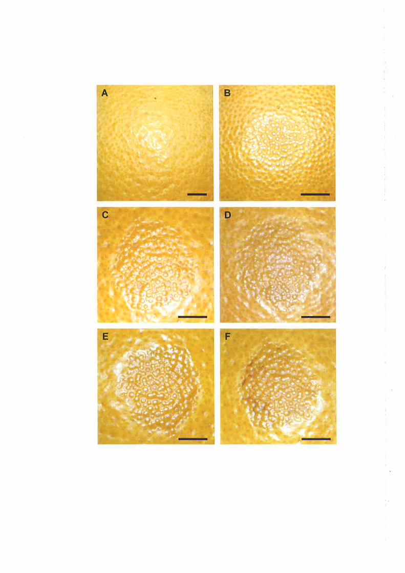

6.2.1.2. Experiment 2
A modified time course assessment was carried out to reduce variation
observed in experiment 1. To achieve this, 15 mature fn¡it were collected from one
tree, and all fruit were assessed at each time interval. Fruit were harvested from a
23 yeu old Washington navel orange tree on Poncirus trifoliata rootstock located
in the University of Adelaide, Waite Campus Alverstoke orchard (34o97'5,
138o63'E). Fruit were harvested on August 25, 1998. Data collected in experiment
2 were used for symptom assessment. No microscopy observations of this material
were made.
6.2.2. Microscopy
6.2.2.1. Tissue sampling
In experiment l, rind samples were collected at each of the eight time intervals,
and prepared for LM and TEM examination. In penetrometer-damaged fruit, rind
samples were taken from three areas: the area of penetrometer application, the area
of oil spread outside the perimeter of penetrometer damage, and an unaffected area
(healthy tissue control). In oil-treated fruit, similar samples were taken, from the
area of oil disc application, the area of oil spread outside of the disc perimeter, and
an unaffected area (healtþ tissue control).
6.2.2.2. Light microscopy (LM)
a. Tissue preparation
Tissue preparation was carried out according to methods outlined in Section
3.2.3.2. Two tissue processing methods were employed: aldehyde fixation and
aldehyde fixation plus osmium tetroxide postfixation.
In the first method, rind samples of 5 x 5 mm were fixed overnight in
glutaraldehyde, dehydrated through an alcohol series and embedded in glycol-
methacrylate (GMA). As microscopy samples were collected at staggered time
intervals over three days, samples were stored after fixation in methoxy-ethanol to
allow bulk processing. Sections (4 pm thickness) were cut with a Reichert-Jung
2050 Supercut Mcrotome using glass knives, placed on glass slides and stained
97

with periodic acid-Schiffs (PAS) and Toluidine Blue O (TBO) (O'Brien and
McCully, 1981).
In the second method, rind samples of I x 1 mm were fixed overnight in a
formaldehyde/glutaraldehyde solution, postfixed in osmium tetroxide, dehydrated
through an acetone series and embedded in Procure-Araldite @A) resin. Samples
were stored after primary fixation in washing buffer, to allow bulk processing.
Sections (l pn thickness) were cut with a Reichert-Jung Ultracut E Microtome,
placed on glass slides and stained with TBO (O'Brien and McCully, 1981).
Histochemical staining for lignin using Phloroglucinol-HCl (Jensen, 1962) and
suberin using Sudan Black B (SBB) (O'Brien and McCully, 1981) was also
employed. All material was observed with a Zeiss Axiophot Photomicroscope,
using transmitted light.
b. Sample selection
For aldehyde fixed tissue, serial sections were collected from approximately
20 samples. For aldehyde plus osmium postfixed material, survey sections were
collected from approximately 25 samples. Approximately 100 sections were
collected from each sample. The latter material was preferred for rind damage
observations. Fifteen samples were selected for TEM preparation, to achieve
intact ulka-thin sections for six samples.
6.2.2.3. Transmission electron microscopy (TEM)
a. Tíssue preparation
Ultra-thin sections (70 nm in thickness) were collected on a diamond knife,
using a Reichert-Jung Ultracut E Microtome. Sections were transferred to
collodion coated copper grids, 200 mesh thin ba¡. Sections were stained with
uranyl acetate and lead citrate (O'Brien and McCully, 1981), and observed under
an accelerating voltage of 80 kV with a Philips CM 100 TEM.
98
b. TEM assessment
Montage images of the rind profile u/ere created with the analySlS program
(Soft Imaging Systems, Muenster, Germany). Plate 6.2 shows the rind profile of a
healthy, untreated tissue sample. Flavedo tissue was divided into three layers:
epidermis, hypodermis and cortex. Within each layer, a minimum of th¡ee cells
was examined. The cell wall, cytoplasm, nucleus and vacuoles of each cell were
observed (Mauseth, 1988). The main components of the cytoplasm assessed for
integrity included the plasmalemma, plastids and mitochondria. This method
enabled a standard comparison of ultrastructural damage between samples. TEM
was used to compare rind damage between induction methods, and to examine
oleocellosis development in oil-treated fruit.
6.2.2.4. S cannin g electron microscopy (SEIvf)
Healtþ and oleocellosis-damaged tissue samples were prepared for
cryofixation according to the method outlined in Section 4.2.2.2. In addition,
cryofixed samples were examined in backscatter mode to enable elemental
detection.
6.2.2.5. Confocal microscopy
Healtþ and oleocellosis-damaged tissue samples rùiere prepared for multi-
photon microscopy according to Section 4.2.3.3.
99

Plúe 6.2
Rind profile image of healtþ rind tissue. Image merged from a sequence of
TEM images. The rind profile was divided into three layers: epidermis,
h¡podermis and cortex. Bar: 5 pm.
100

Epidermis
Hypodermis
Cortex

6.3. Results
6.3.1. Symptoms
6.3.1.1. Time course assessment
a. Blemish severity
Rind collapse was first detected at t how following induction in the
penetrometer-damaged fruit and 6 hours in oil-treated fruit (Fig. 6.1). The period
of most rapid collapse for penetrometer-damaged fruit was within the first 12
hours, with significant increases (P < 0.05) observed between 2 and 6, and agaun
between 6 and 12 hows. Oil-freated fruit also showed a significant increase
between 6 and 12 hours, and also at later stages, between 24 and 48, and 48 and
72 hours. By day 3, mean collapse scores were 2.3 for oil-treated fruit and 1.5 for
p enetrometer-damaged fruit.
Rind discolouration was first detected at 2 hours following induction in oil-
treated fruit and 12 hours in penetrometer-damaged fruit (Fig. 6.2). Oil-treated
fruit showed a mean score of 1 by 6 hows, compared to penetrometer-damaged
fruit, which showed a maximum score of 0.3 at day 3. In oil-treated fruit, a
significant increase (P < 0.05) was observed between 2 arrd 6 hours, and24 and 48
hours, resulting in a mean colour score of 2.3 by day 3. The mean scores for both
collapse and discolouration were considered to be low given the scoring scales of
0 to 4 and 0 to 5, respectively. This was attributed to the high proportion of
samples which did not develop blemish.
b. Blemish area
For blemish area, a similar trend was observed for both induction methods
(Fig. 6.3). A significant area increase (P < 0.05) was observed between 12 and 24
hours for both methods, as well as between 2 and 6 hows for oil-treated fruit. By
day 3, oil-treated fruit showed a larger mean blemish area than penetrometer-
damaged fü¡it, with mean scores of 229 mm2 and 173 mtl?, respectively.
However, there was little change in mean blemish area after day 1.
AA
l0l

3
.,
rüI
q)L<¿U)
{)U)
cc
(,
4
LSD +-pen+-oil
I
6 t2 18 24 30 36 42 48 54
Time after induction (hours)60 66 72
5
0
0 6 t2 18 24 30 36 42 48 54 60 66 72
Time after induction (hours)
Figure 6.2, The relationship between time and rind discolouration.Colour score: 0 (nil), I (very slight),2 (slight), 3 (medium),4 (high), 5
(extreme). Pen : penetrometer induction, oil : oil induction. Fifteenfruit, assessed at each of eight time intervals from zero to three days.Means represented. LSD = 0.33.
I LSD ,+p.Ei+oil i
0
0
Figure 6.1. The relationship between time and rind collapse. Collapsescore: 0 (nil), I (very slight), 2 (slight), 3 (medium), 4 (high). Pen:penetrometer induction, oil : oil induction. Fifteen fruit, assessed ateach of eight time intervals from zero to three days. Meansrepresented. LSD : 0.26.
r,f)t
€)L(,øt
L
U
4
3
.,
1
102
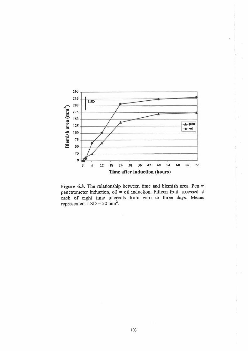
250
225
,-. 200êl
Ensv lso
E t2scÉ
ã loo
Eis€)
Êâ s0
25
0
I
LSD
+Pen¡>oil
6 t2 18 24 30 36 42 48 s4 60 66 72
Time after induction (hours)0
Figure 6.3. The relationship between time and blemish area. Pen =penetrometer induction, oil : oil induction. Fifteen fruit, assessed at
each of eight time intervals from zero to three days. Meansrepresented. LSD : 50 mm2.
103

c. Symptom variation
Variance components were assessed at 72 hours, and compared between
experiment I and experiment 2 (Table 6.1). In experiment 1, fruit effects were
responsible for a high proportion of variation in area and collapse. The tree effect
was nil or negligible for all responses. The method effect was notable for all
responses, but particularly colour, where it accounted for 78o/o of variation. The
variation between replicate samples was larger than expected for all responses. In
experiment 2, a tree stratum was not present in the analysis, as fruit were picked
from one tree only. The fn¡it effect was responsible for negligible variation, much
reduced from experiment l. The method effect showed a similar trend to
experiment 1, and accounted for most of the va¡iation in colour (85%). Replicate
sample variation also showed a similar trend, and accounted for most of the
variation in collapse (73%) and area (68yo), but to a lesser extent colour change
(ts%).
Table 6.1. Variance strata for blemish collapse, colour and area at day
3, expressed as percentage of total variation. Experiment 1, füritharvested from five trees and different fruit assessed at each time.Experiment 2, fruit harvested from one tree and same fruit assessed at
each time. Sample refers to replicate treatments on the one fruit.
Stratum component Variation of score (7o)Colour Area
Experiment ITreeFruitMethod
0
25l460
007823
I253737
Experiment 2FruitMethod
225
73
0
85
15
J
29ó8
d. Rind oil release pressure (RORP)
As observed in Chapter 5, RORP at induction showed very little variation
between fnrit, varying between 164 and 245 kPa (data not shown). In general,
RORP values were lower than those observed in Chapter 5, suggesting a higher
104
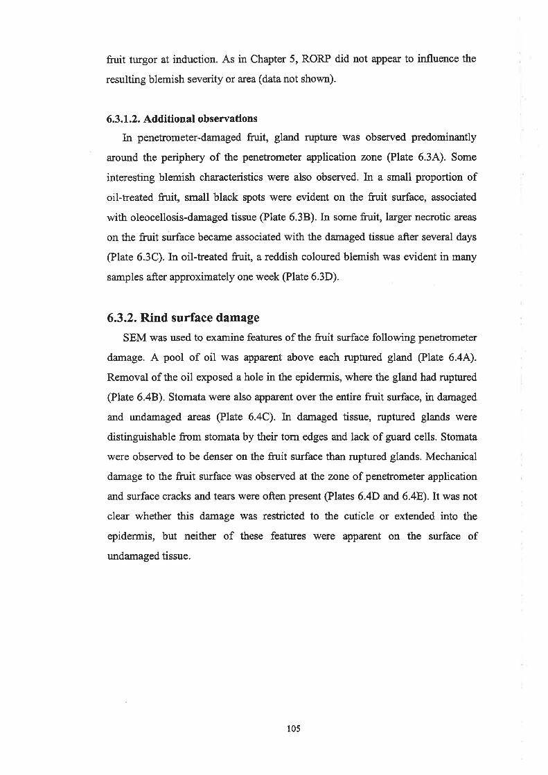
fruit turgor at induction. As in Chapter 5, RORP did not appear to influence the
resulting blemish severity or area (data not shown).
6.3.1.2. Additional observations
In penetrometer-damaged fruit, gland rupture was observed predominantly
around the periphery of the penetrometer application zone (Plate 6.34). Some
interesting blemish characteristics were also observed. In a small proportion of
oil-treated fruit, small black spots were evident on the fruit surface, associated
with oleocellosis-damaged tissue @late 6.38). In some fruit, larger necrotic areas
on the fruit surface became associated with the damaged tissue after several days
(Plate 6.3C). In oil-treated fruit, a reddish coloured blemish was evident in many
samples after approximately one week (Plate 6.3D).
6.3.2. Rind surface damage
SEM was used to examine features of the fruit surface following penetrometer
damage. A pool of oil was apparent above each ruptured gland (Plate 6.4A).
Removal of the oil exposed a hole in the epidermis, where the gland had rupttued
@late 6.48). Stomata were also apparent over the entire fruit surface, in damaged
and undamaged areas (Plate 6.4C). In damaged tissue, ruptured glands were
distinguishable from stomata by their torn edges and lack of guard cells. Stomata
were observed to be denser on the fruit surface than ruptured glands. Mechanical
damage to the fruit surface was observed at the zone of penetrometer application
and surface cracks and tears were often present (Plates 6.4D and 6.48).It was not
clear whether this damage was restricted to the cuticle or extended into the
epidermis, but neither of these features were apparent on the surface of
undamaged tissue.
105

Plate 6.3
Dissecting microscope images of fruit surface damage and oleocellosis
symptoms.
A. Gland rupture and oil release (a:rows) predominantly around the
perimeter of the penetrometer application zone (paz). Bar:2 mm.
B. Black spots evident on the swface of a small proportion of oil-treated
fruit, possibly necrotic guard cells. Bar:0.5 mm.
C. Necrotic areas (arrows) developed around the edges of the oleocellosis
blemish in a small proportion of fruit, after several days. Bar = 5 mm.
D. Red pigmentation observed in some extreme blemishes after
approximately one week. Bar:5 mm.
106

fte
a
t
útr"1f
#'a
¡l}a
a
aa

Plate 6.4
Scanning electron micrographs of the fruit surface following mechanical
damage, with the penetrometer.
A. Oil pool on the fruit surface, above a ruptured gland. Bar = 100 ¡rm'
B. Holes in the epidermis resulting from gland rupture showed non-uniform
shape and rough edges. Bar: 20 Wn.
C. Stomata (s) were present over the entire fruit surface. Bar:20 Fm.
D. Surface cracks resulted from penetrometer action upon the rind. Bar :
200 pm.
E. Surface tears and cracks resulted from penetrometer action upon the rind.
Bar: 50 pm.
t07
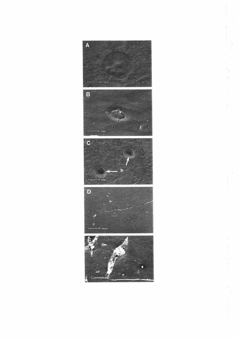
___ -_ -1 t00 | ilr
A

6.3.3. Rind sub-surface damage
6.3.3.1. Rind anatomy
Untreated rind tissue showed no signs of cell collapse @late ó 5A). In
penetrometer-damaged fruit, epidermal rupture above glands was observed (Plate
6.58). In aldehyde fixed tissue, oil-damaged cells showed distortion or collapse
@lates 6.5C and 6.5D). In both penetrometer-damaged @late 6.5C) and oil-
treated tissue @late 6.5D), oil-damaged cells were localised between glands and a
number of layers below the epidermis. In osmicated tissue, oil-damaged cells were
more clearly distinguishable from healthy cells by their degenerated cell contents
@lates 6.5E and 6.5F). Unlike aldeþde fixed tissue, a difference in the localisation
of oil damage was observed between penetrometer-damaged and oil-treated fruit.
In penetrometer-damaged tissue, the majority of samples showed damaged cells to
be localised a number of cell layers below the epidermis @late 6.5E). However, in
oil-treated fruit, damaged cells were located in all cell layers from the epidermis
downwards @late 6.5F). Samples taken from the zone of oil application and the
zone of oil spread appeared similar. Histochemical staining of aldehyde fixed tissue
failed to show the presence of lignin and suberin in oleocellosis-damaged tissue. In
cryofixed samples examined using SEM, oil-damaged cells appeared to be severely
collapsed. In oil-treated fn¡it, a thick band of collapsed cells, incorporating all cell
layers from the epidermis downwards was observed @late 6.6). X-ray micro-
analysis of cryofixed samples showed no elemental difference between healthy
tissue and oleocellosis-damaged tissue.
6.3.3.2. Oil localisation
Untreated rind tissue showed no sign of major oil deposits outside of the glands
@late 6.7A). However, small, fluorescent bodies in the upper rind layers were
most likely plastid or cytoplasm localised oil bodies. Tissue was examined l8 hours
after penetrometer damage, but no oil was apparent in tissue between the glands.
After three days however, the degenerated contents of oil-damaged cells observed
in osmicated tissue sections were also detected in fresh tissue @late 6.78).
Degenerated cell contents that were stained by both cell wall (green) and lipid (red)
stains, appeared yellodorange in colour.
108

Plate 6.5
Light micrographs of the oleocellosis-damaged rind. Longitudinal, aldehyde
fixed, PAS/TBO stained sections (A to D). Longitudinal, aldehyde plus
osmium tetroúde postfixed, TBO stained sections @ and F).
A. Untreated fir¡it sample. Bar: 100 ¡m.
B. Ruptured epidermis (arrow) above a gland (g), resulting from
penetrometer damage. Bar = 100 prm.
C. Penetrometer-damaged fnrit sample, showing sub-epidermal cell collapse
(arrows). Bar: 100 pm.
D. Oil-treated fn¡it sample, showing sub-epidermal cell collapse (arrows).
Bar: 100 pm.
E. Penetrometer-damaged fnrit sample, showing sub-epidermal cell content
degeneration (arrows), below intact upper rind layers. Bar: 100 pm
F. Oil-treated fruit sample, showing cell content degeneration in all layers,
including the epidermis (arrows). Bar: 100 pm.
109

BA
!:
I
c
t\l
\7 t
Plate 6.6
Scanning electron micrograph of the oleocellosis-damaged rind, resulting
from oil treatment. Longitudinal rind cross-section showed a thick band of
collapsed cells, incorporating all cell layers from the epidermis downwards.
Bar: 50 pm.
110

\\
B0 Um
a-'F\.
"[)
Ii
')
-.a, -'
-C,
t> _-r_4ra_
::-=+q:
Plate 6.7
Multi-photon microscope images of healthy and oleocellosis-damaged rind.
Two-colour merged optical sections of fresh tissue, stained with Calcofluor
White for cell walls (green), and Nile Red for lipid (red).
A. Untreated tissue. Bar: 30 pm.
B. Penetrometer-damaged tissue after three days, showing degenerated
contents of oil-damaged cells (arrows), stained by both stains and labelled a
yellow/orange colour. Bar:30 ¡m.
111
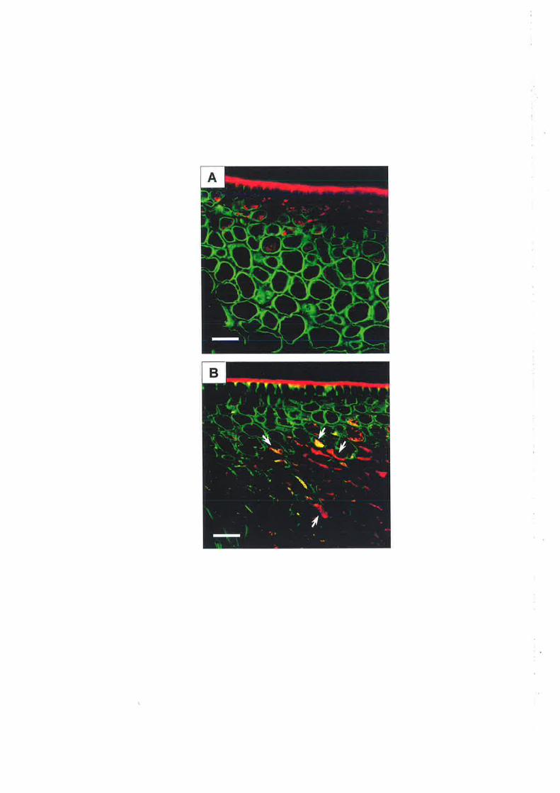

6.3.3.3. Rind ultrastructure
Rind samples from untreated fruit, penetrometer-damaged frtrit and oil-treated
fruit were examined using TEM. For each sample, the rind profile (Plate 6.8) and
ultrastructr¡ral features of individual cells @lates 6.9 to 6.14) were examined.
a. Untreatedfr"it
The cuticle appeared vesicular @late 6.94). Epidermal cells contained one or two
main vacuoles, often with smaller vacuoles also present (Plate 6.8A). The
protoplast was located a¡ound the cell edge. The tonoplast and plasmalemma were
intact and continuous in most cells. In some cells, the plasmalemma had vesicles
associated with the walls @late 6.98). The nuclei showed an intact nuclear
envelope, often with very slight bulges between inner and outer envelopes (Plate
6.9C). Plastids, in the form of chromoplasts, were usually round to oval shaped
and showed an intact outer membrane, and folded inner membrane (Plate 6.9D).
Plastids contained plastoglobuli, which v¡ere generally lightly stained.
Mitochondria showed an intact double membrane, including intemal cristae @late
6.9E). Round to oval shaped oil bodies were present in epidermal cells, often
closely associated with vacuole edges or in the cytoplasm. Oil movement was
observed between the cytoplasm and vacuole (Plate 6.9F). Multivesicular bodies
were present in some cells, often located between the plasmalemma and cell wall
(Plate 6.9G). Hlpodermal cells were similar in ultrastructural appearance to
epidermal cells. Some contained a main vacuole, while others contained a few
smaller sized vacuoles. Cortex cells also showed intact organelles and membrane
systems. Plasmodesmata were observed between all cell types, on occasions
causing constrictions in cell walls, particularly in hypodermal cells @late 6.9H).
b. Oil-treatedfruit, 30 minutes, no symptoms
The rind profile image of this sample (Plate 6.88) appeared similar to that of
the untreated sample (Plate 6.84). The cuticle appeared similar to that in untreated
fruit, as did the membranes in epidermal cells. Some cells showed intact tonoplast
and plasmalemma, whereas in others the tonoplast was not continuous, and the
plasmalemma appeared to be detached from the cell wall or was missing in
Lt2

Plate 6.8
Rind profile images of healtþ and oleocellosis-damaged rind. Each image
merged from a sequence of TEM images. Bats : 5 ¡rm. See over page.
A. Untreated fruit sample.
B. Oil-treated fruit sample, 30 minutes, no symptoms (0,0).
C. Oil-treated fruit sample, six hours, very slight blemish (2,0).
D. Oil-treated fruit sample, two days, medium level blemish (3,4).
E. Oil-treated fruit sample, ten days, extreme blemish (4,5).
F. Penetrometer-damaged fruit sample, three days, medium level blemish
(3,2).
r13

;¡ÉÈr
-

-ú
-r.t-ttl \
\

Plate 6.9
Transmission electron micrographs of rind ulhastructure in an r.urtreated
fruit sample.
A. The cuticle appeared vesicular. Bar: I ¡rm.
B. Plasmalemma (arrows) with vesicles between it and the cell wall (cw).
Bar:0.5 pm.
C. Nucleus with an intact envelope and very slight swelling between inner
and outer membranes (arrows). Bar: 1 pm.
D. Plastid (chromoplast) with intact outer membrane, folded inner
membrane (i) and lightly stained plastoglobuli (p). Bar: 1 ¡rm.
E. Mitochondrion with intact double membrane and cristae. Bar -- 0.2 pm.
F. Oil (o) in the cytoplasm adjacent to vacuole (v). Bar:0.5 Fm.
G. Multivesicular body (mb), located between the plasmalemma and cell
wall (cw). Bar:0.2 pm.
H. Constrictions in the cell wall at the site of plasmodesmata (arrows). Bar:
0.5 pn.
LL4
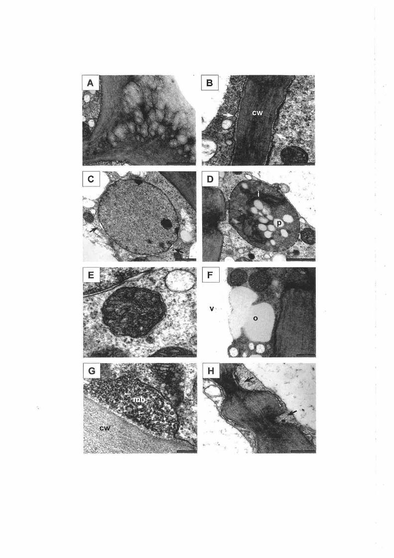
o
.Tt
TD

sections (Plate 6.104). The majority of nuclei, mitochondria and plastids appeared
similar to those in untreated tissue. Some nuclei showed bulging in their envelope,
although on occasions more advanced stages of degeneration were observed (Plate
6.108). Plastids contained intact double membranes, but some degeneration of
membranes and internal structure was also observed @late 6.10C). Oil bodies were
larger and more numerous than in untreated tissue, and were located in vacuoles
and cytoplasm, or appeared to be moving between the two, as observed in
untreated tissue. Oil bodies appeared to be more closely associated with organelles;
for example, mitochondria @late 6.10D). The plasmaleÍtma, tonoplast and nuclei
of hypodermal cells appeared similar to those of epidermal cells. The double
membrane of mitochondria appeared to be intact in some but slightly diffi¡se in
others (Plate 6.10E). Plastids showed signs of membrane and internal degeneration
(Plate 6.10F). Oil bodies were present in many hypodermal cells, again associated
with the vacuole and organelles, and also present in intercellular spaces between
hypodermal cells. The ultrastructural appearance of cortex cells varied at this
stage. Some cells at layer seven appeared more damaged than those atlayer four or
five. In the least damaged cells, mitochondria, plastids and even tonoplast and
plasmalemma appeared intact. In the most damaged cell observed, the vacuole was
not intact and the cell contained debris, but some mitochondria remained intact. Oil
bodies were less evident in cortex cells than epidermal and hypodermal cells, but
were again closely associated with organelles, and were present in intercellular
spaces between cortex cells, to a depth of approximately nine cell layers (Plate
6.10G). Small oil bodies were also observed in close association with
plasmodesmata between adjoining cortex cells @late 6.10H).
c. Oil-treated/ruit, six hours, very slight blemish
Six hours after oil treatment, only very slight surface symptoms were observed
in this frtrit sample, with slight rind collapse but no discolouration. However, the
rind profile image at 6 hours @late 6.SC) was dramatically different to 30 minutes
(Plate 6.88). Cells showed various stages of degeneration. The cuticle appeared
similar to that in untreated fruit. Epidermal cells were at various stage of
degeneration. In some cells, many vesicles were present and the c¡oplasm had
il5

Plate 6.10
Transmission electron micrographs of rind ultrastructure in an oil-treated
fruit sample, after 30 minutes.
A. Plasmaleilrma absent and cell wall (cw) edges diffuse. Bar:0.5 pn.
B. Nucleus with significant swelling of nuclear envelope (arrows). Bar: 0.5
l.rtTt.
C. Degenerated plastid. Bar: 0.5 ¡un.
D. Mitochondrion (m) with adjacent oil bodies (o). Bar:0.2 Fm.
E. Mitochondrion with intact but slightly diffuse membrane. Bar = 0.2 pn.
F. Plastid showing signs of outer membrane and intemal degeneration
(arrows). Bar: 0.5 pn.
G. Intercellular spaces filled with oil (o). Bar: 5 Frm.
H. Oil bodies (o) in close association with a plasmodesma. Ba¡: I pm.
116

#
G
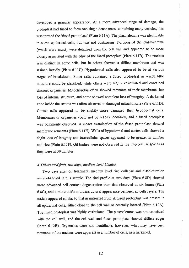
developed a granular appearance. At a more advanced stage of damage, the
protoplast had fused to form one single dense mass, containing many vesicles; this
was termed the 'fused protoplast' @late 6.114). The plasmalemma was identifiable
in some epidermal cells, but was not continuous. Portions of the plasmalemma
(which were intact) were detached from the cell wall and appeared to be more
closely associated with the edge of the fused protoplast @late 6.118). The nucleus
was distinct in some cells, but in others showed a diffi.lse membrane and was
stained heavily @late 6.11C). Hypodermal cells also appeared to be at various
stages of breakdown. Some cells contained a fused protoplast in which little
structure could be identified, while others were highly vesiculated and contained
discreet organelles. Mitochondria often showed remnants of their membrane, but
loss of internal structure, and some showed complete loss of integrity. A darkened
zone inside the stroma was often observed in damaged mitochondria (Plate 6.11D).
Cortex cells appeared to be slightly more damaged than hypodermal cells.
Membranes or organelles could not be readily identified, and a fused protoplast
was commonly observed. A closer examination of the fused protoplast showed
membrane remnants @late 6.11E). Walls of hypodermal and cortex cells showed a
slight loss of integrity and intercellular spaces appeared to be greater in number
and size @late 6.11F). Oil bodies were not observed in the intercellular spaces as
they were at 30 minutes.
d. Oil-treatedfruit, two days, medium level blemish
Two days after oil treatment, medium level rind collapse and discolouration
were observed in this sample. The rind profile at two days (Plate 6.8D) showed
more advanced cell content degeneration than that observed at six hours (Plate
6.8C), and a more uniform ultrastructural appearance between all cells layers. The
cuticle appeared similar to that in untreated fruit. A fused protoplast was present in
all epidermal cells, either close to the cell wall or centrally located @late 6.12A).
The fused protoplast was highly vesiculated. The plasmalemma was not associated
with the cell wall, and the cell wall and fused protoplast showed diffi¡se edges
(Plate 6.128). Organelles were not identifiable, however, what may have been
remnants of the nucleus were apparent in a number of cells, as a darkened,
rt'Ì

Plate 6.11
Transmission electron micrographs of rind ultrastructwe in an oil-treated
fruit sample, after six hours.
A. Highly vesiculated fused protoplast. Bar: 1 pm.
B. The plasmalemma (a:row), detached from the cell wall (cw) and
surrounding the fused protoplast (fp). Bar: 0.2 pm.
C. Nucleus, with diffuse envelope and densely osmiophillic appearance. Bar
:0.5 pm.
D. Mitochondria (m) showing diffuse membranes and loss of internal
structure. Bar:0.2 Un.
E. Membrane remnants trapped inside the fused protoplast. Bar:0.2 pm.
F. An expanded intercellula¡ space (is). Bar :2 pm.
118

-
I
Plúe 6.12
Transmission electron micrographs of rind ultrastructure in an oil-treated
fruit sample, after two days.
A. Highly vesiculated fused protoplast, located centrally in the cell. Bar: 1
pm.
B. Diffuse edges of the fused protoplast (fp). Bar: 0.2 pm.
C. Darkened region of the fused protoplast, possibly nucleus remains. Bar:
0.2 ¡tm.
D. Membrane remnants (arrow) trapped inside the fused protoplast. Bar =
0.2 ¡un.
E. Cell wall thickening and slight folding in epidermal cells. Bar: I pm.
F. Cell wall folding in cortex cells. Bar: I Ftm.
119

A B
c -
E
<E
rrmorphous region in the fused protoplast @late 6.12C). Membrane remnants were
apparent within the fused protoplast @late 6.12D). Epidermal cell walls showed
signs of thickening and to a lesser extent, folding @late 6.128). Hypodermal and
cortex cells also contained a fused protoplast, appearing ribbonlike in lower cortex
cell layers. Cell wall thickening and folding was also evident in hypodermal and
cortex cells (Plate 6.12F). Intercellular spaces were present in hypodermal and
cortex cell layers.
e. Oil-treatedÍruit, ten days, extreme blemish
Ten days after oil treatment, high level rind collapse and extreme discolouration
were observed in this fruit sample. The rind profile at ten days @late 6.8E) showed
more advanced cell content degeneration than that observed at two days @late
6.8D), and increased cell wall thickening and folding, particularly of hypodermal
cells. The cutin appeared less vesicular and slightly more flattened than in earlier
samples (Plate 6.134). A fused protoplast was present in all epidermal cells, either
close to the cell wall or centrally located, as at two days. Oil bodies were observed
within the fused protoplast and external to the protoplast (Plate 6.13B), and being
secreted from the fused protoplast @late 6.13C). Epidermal cell walls were thicker
than those observed at two days. Hypodermal and cortex cells also contained a
fused protoplast and oil bodies. Cells exhibiting these features \ryere present to a
depth of approximately seventeen layers in this sample. In addition, oil movement
via plasmodesmata was also observed @late 6.13D). Hypodermal cells showed
extreme folding (Plate 6.13E) and thickening, highlighted by constrictions at the
site of plasmodesmata. Cortex cells also showed folding and to a lesser extent,
thickening (Plate 6. I3F).
f. Penetrometer-damaged.fruit, three dnys, medium level blemish
Three days after induction, surface symptoms in penetrometer-damaged fruit
were less advanced than oil-treated fnrit. In this sample, medium level rind collapse
and only very slight discolouration were observed. The rind profile of
penetrometer-damaged fruit showed oil-damaged cells localised to the cortex layer
@late 6.8F). The cuticle appeared similar to that in untreated fruit. Epidermal cells
r20

Plate 6.13
Transmission electron micrographs of rind ultrastructu¡e in an oil-treated
fruit sample, after ten days.
A. The cuticle appeared less vesicular than earlier samples. Bar: I pm.
B. Oil bodies located inside vesicles of the fused protoplast, and outside the
protoplast (arows). Bar: I pm.
C. Oil (arrow) traversing vesicles and fused protoplast (fp). Bar = 0.2 pm.
D. Oil (arrow) adjacent to cortex cell wall (cw) plasmodesma. Bar: 0.5 pm.
E. Cell wall thickening and folding, in hlpodermal cells. Bar: 1 pm.
F. Cell wall thickening and folding, in cortex cells. Bar: 1 pm.
t2l
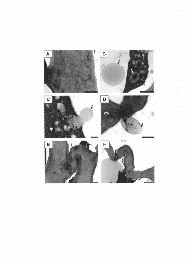
B1
t
-

contained numerous vacuoles and a main vacuole, which often appeared to be
compartmentalised. The plasmalemma and tonoplast were both present, but
fragmented (Plate 6.144). Similarly, the double envelope of nuclei was frequently
discontinuous (Plate 6.148). The double membrane of mitochondria appeared to
be intact in some cells, while in others, small, osmiophilic bodies were associated
with the mitochondria. Similar osmiophilic bodies were also present around the
cell edge, between the plasmalemma and wall (Plate 6.14C).In more damaged
cells, densely osmiophilic bodies \¡/ere also present in the stroma of plastids, as
distinct from lighter-staining plastoglobuli (Plate 6.14D). Hlpodermal cells had a
similar appearance to epidermal cells. Osmiophilic bodies were less evident, but
again associated with mitochondria and plastids. Vesicles also appeared to be
associated with some organelles. Both epidermal and h¡'podermal cells also
showed abundant rough ER @late 6.148). Cortex cells adjacent to hypodermal
cells were dramatically different in ultrastnrctr¡ral appearance, resembling cortex
cells at two days after oil treatment. All cortex cells appeared to be oil-damaged,
containing a fused protoplast, and exhibiting thickened and folded cell walls
(Plates 6.14F and 6.14G). The cell wall between hlpodermal (intact) and cortex
cells (damaged) appeared to be structurally sound, and plasmodesmata were
observed between these cell layers (Plate 6.14H).
122
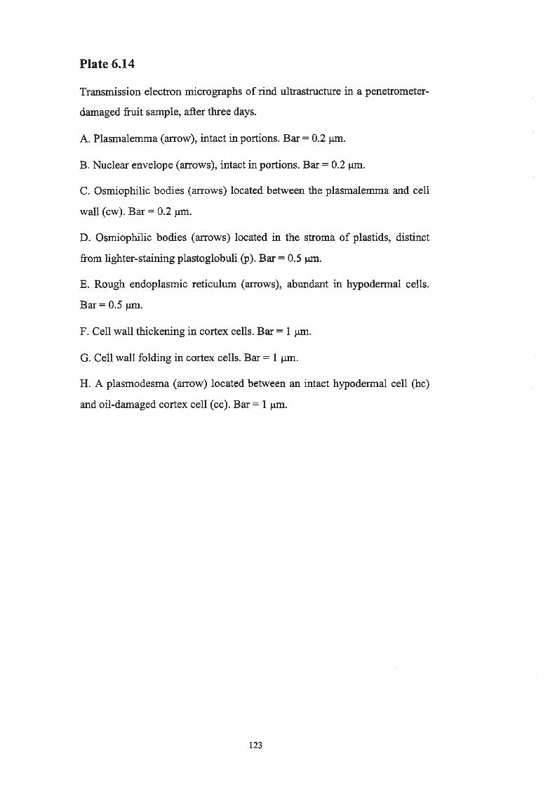
Plate 6.14
Transmission electron micrographs of rind ultrastructure in a penetrometer-
damaged fruit sample, after three days.
A. Plasmalemma (arrow), intact in portions. Bar: 0.2 ptn.
B. Nuclear envelope (arrows), intact in portions. Bar: 0.2 p;rn.
C. Osmiophilic bodies (a:rows) located between the plasmalemma and cell
wall (cw). Bar: 0.2 ¡tm.
D. Osmiophilic bodies (arrows) located in the stroma of plastids, distinct
from lighter-staining plastoglobuli þ). Bar: 0.5 pn.
E. Rough endoplasmic reticulum (arrows), abundant in hlpodermal cells.
Bar = 0.5 pm.
F. Cell wall thickening in cortex cells. Bar: L pm.
G. Cell wall folding in cortex cells. Bar: I pm.
H. A plasmodesma (anow) located between an intact hypodermal cell (hc)
and oil-damaged cortex cell (cc). Ba¡: 1 pm.
t23

-1,à
=E

6.4. Discussion
6.4.1. Symptoms
In mature, colowed fruit, oleocellosis symptoms consist of both collapse and
darkening of the fruit surface between the oil glands (Shomer and Erner, 1989).
Symptoms have been reported to develop within a week of harr¡est in lime fruit
(Eaks, 1955), and within two to three days in navel oranges (rù/ild, 1998).
Observations of oleocellosis in Valencia orarige and mandarin fruit have
suggested that rind collapse precedes discolouration (T[illiams and V/ild, 1996).
However, rurtil this study, oleocellosis symptom development has not been
examined in detail. Measuring blemish severity using a scoring system enabled
the quantitative assessment of blemish development. The timing of blemish
development appeared to differ between physical and oil induced oleocellosis.
Over a period of three days, penetrometer-damaged fruit primarily showed tissue
collapse and only slight discolouration, compared to oil-treated fruit which
showed a similar rate of collapse and discolouration. Observations suggested that
physiological development may differ between mechanical and oil induced forms
of oleocellosis. Microscopic examination proved this to be the case. Blemish a¡ea
appeared to show a rapid increase up to 24 hours. However, area could not be
accurately measured until symptoms had developed, at approximately 24 hours.
Blemish area remained fairly constant after this time, suggesting rapid oil spread
following induction.
Statistical analysis of the effects of time and method on blemish severity and
area identified variability between trees, fruit and even different zones on the same
fruit. In experiment 2,the tree effect was removed and repeat measures were made
on each fruit, however variation between induction zones on the same fririt
remained high. This variation may be explained by the effect of fruit orientation,
which was not taken into account during sampling. Similar observations were
made by Wild (1998), who showed the sun exposed face of navel orange fruit to
be more sensitive to applied oil thar¡ the protected face. Future experiments should
take this effect into account during experimental design.
t24

6.4.2. Oleocellosis mechanism
The mode of oil release following gland rupture is unclear. Swface oil release
has been observed (Fawcett, 1916; Cahoon et aL.,1964; Eaks, 1968), however, the
likelihood of intemal oil release has also been suggested (Shomer and Erner,
1989; Loveys et aI., 1998). Some doubt also exists over previous microscopy
findings due to poor tissue preservation and misinterpretation of images. Using
LM, epidermal rupture above glands has been observed in mecharrically damaged
rind tissue (Labuschagne et a1.,1977; du Plessis, 1978). However, tissue sections
presented in these studies showed excessive damage, possibly artefactual,
attributable to poor tissue preservation or sectioning damage. Improved tissue
presenration \¡/as achieved in a study by Shomer and Erner (1989), who suggested
that "oleocellosis-damaged cells were the result of either external or intemal
contamination by essential oil". In their study, light micrographs depicted a
ruptured gland, which appeared to open into the flavedo tissue above the gland.
With our knowledge that all glands are attached to the fruit sr.¡¡face by a 'stalk'
(see Chapter 3), it appears that misinterpretations were made, as medial sections
of glands were not examined.
In the present study, fruit were mechanically damaged with a penetrometer;
surface oil release was detected using multiple microscopy methods and no
evidence of internal oil release was established. Confocal microscopy, while a
useful tool for oil visualisation as it avoids tissue fixation (see Chapter 4), did not
allow oil to be detected outside of the glands in damaged tissue. As a result,
microstructural rind damage v/as used as the basis of ow examination.
Penetrometer-damaged tissue prepared for LM examination rù/as well preserved.
Tissue was serially sectioned and the medial sections of glands were examined.
Gland rupftue was only obsen¡ed to occur at the junction of the apical region of
the gland and the epidermis. These findings were supported by our prior
knowledge that all glands were linked to the epidermis by a stalk (see Chapter 3).
In mature fruit, the gland is positioned very close to the fruit surface, facilitating
easy surface oil release. The gland stalk is not hollow and does not open to the
fruit surface, which would explain the need for mechanical force to facilitate oil
125

release (Ting and Attaway, 1971). Serial sections of penetrometer-damaged tissue
revealed no other structural disruption to the gland that may have facilitated oil
release into the tissue. Internal oil release may be prevented by the layers of
thickened boundary cells which enclose the gland. CToSEM findings verified the
damage observed in tissue sections, highlighting the presence of holes in the fruit
epidermis at the site of gland ruptures, and pools of oil.
The proposition that surface oil is responsible for the oleocellosis blemish was
supported by an additional experiment in which oil released to the fruit surface was
removed immediately, with absorbent tissue, and symptoms failed to develop (data
not shown). Similar results have been reported in ftiit washed in water at 32"C
within two minutes of oil release @ckert and Eaks, 1989). These findings however
are in direct contrast to those of Loveys et al. (1998). In their study, firrit were
dropped onto a filter paper and liberated oil was absorbed, howeve¡ some fruit
were observed to develop symptoms. The most likely explanation for some fruit
developing symptoms was that surface oil was not removed completely.
Mcrostructural rind damage was used to determine the route of oil movement
following surface release. Past microscopy studies of oleocellosis have used
aldehyde fixation for tissue preparation, and identified sub-surface cell deformation
or flattening in physically damaged fruit (Labuschagne et al., 1977; du Plessis,
1978). In the present study, similar observations were made in both physically
damaged and oil-treated tissue fixed in aldehyde. In osmicated tissue, however,
rind damage appeared more extensive in oil-treated ft¡it, with oil-damaged cells
occurring in all rind layers from the epidermis downwards. Observations suggested
that aldehyde fixation alone may lead to misinterpretation. Primary aldehyde
fixation followed by osmium postfixation was preferred as it allowed observation
of the protoplasm as well as cell walls.
Given that gland rupture results in surface oil release, it was expected that
mechanically damaged fn¡it would show identical rind damage to surface oil-
treated fruit, but this was not the case. In penetrometer-damaged fruit, only a very
small proportion of samples showed cell content degeneration in all layers of the
126

rind, and the majority of these were collected at the edge of the penetrometer
application zone, where surface oil release was observed to occur. The majority of
samples showed cells with degenerated contents localised several layers below the
epidermis. Closer inspection using TEM showed damage to be localised to cortex
cells, whilst the cuticle, and epidermal and hypodermal cells appeared structurally
intact. These findings expand on those of Shomer and Erner (1989), who reported
identical rind damage from penetrometer and surface oil treatments, occurring
either several layers under the epidermis or in all cell layers.
Previously, separate studies have reported surface oil release and sub-epidermal
rind damage, but a link between the two phenomena had not been identified. Based
on our LM and TEM observations, it appears that oil may filter into the rind tissue
via the ruptured epidermis, move into the cortex layer alongside the outer gland
walls, and spread laterally. This proposition is supported by the suggestion that
gland boundary cell walls are less permeable to oil (Thomson e/ al., 1976; Shomer,
l9S0). Oil movement in the cortex may also be aided by more numerous
intercellula¡ spaces. Oil localisation to the cortex, however, is difficult to explain.
Plasmodesmatal connections were observed between intact hypodermal and oil-
damaged cortex cells. Collenchymatic hypodermal cells had thicker walls than
cortex cells, but did not possess suberised or lignified secondary walls which may
have restricted oil movement between the two cell layers. Such depositions have
been observed in the endodermis of citrus roots, separating hypodermal and cortex
cells (Walker et al., 1934). An alternative proposition is that surface oil enters
lower rind layers through other openings in the fn¡it surface, such as the cracks and
tears caused by mechanical damage. This would be similar to the mechanism of
water spot described by Scott and Baker (L947), in which water enters the rind via
stomata or microscopic cracks formed during tissue expansion.
Interestingly, sub-epidermal cell breakdown has also been associated with the
citrus rind disorders 'preharvest peel pitting' (Medeira et al., 1999) and 'rind-oil
spot' (Chikaianmi,2000). The rind oils were suggested as a possible cause for both
disorders, but the underlying mechanisms could not be clearly established. The
'rind-oil spot' described by Chikaizumi (2000) showed similarities to oleocellosis
r27
with respect to symptoms, ruptured glands and sub-epidermal damage, but differed
in that it was consistently associated with high radiation exposure. Medeira et al.
(1999) drew a correlation between the sub-surface damage associated with pitting
and that observed in chilling injury of eggplant, which has been attributed to
localised dehydration (Rhee and lwata, 1982). In the present study, cell
ultrastructural degeneration is not considered to result from dehydration, as it
occurred as early as six hours following penetrometer damage, which paralleled
timing in oil-treated fn¡it.
6.4.3. OiI effects
Prior to the examination of ultrastructural rind damage, the quality of tissue
preservation was assessed in untreated tissue using TEM. Organelles appeared
intact, and cell ultrastructure was simila¡ to that of mature, deep orange coloured
or 'after-ripe' navel orange fn¡it, described by Thomson and Platt-Aloia (1976).
Plastids, in the form of chromoplasts, were identified by their abundant
plastoglobuli and lack of gtana (Mauseth, 1988). Nuclei were intact, with some
showing slight expansion of the perinuclear space, thought to be a sign of nuclear
activity, rather than degeneration (Thomson and Platt-Aloia, 7976). Intracellular
oil bodies were observed in untreated tissue, located in the cytoplasm, vacuole and
often observed moving between the two. Thomson and Platt-Aloia (1976) also
observed lipid in the cytoplasm, generally associated with the tonoplast. Lipid
associated with mitochondria and plastids was also reported at an earlier stage of
ripening.
Details of oil localisation in the oleocellosis-damaged rind were provided to
some extent by TEM. Following oil treatment, lipid bodies \ryere clearly
distinguished in all layers of the rind, in greater abundance than in untreated tissue.
Oil appeared to move indiscriminantly through the rind tissue, with observations
suggesting both apoplastic (oil within intercellular spaces) and symplastic
movement (oil bodies associated with plasmodesmata). The presence of lipid
bodies in intercellular spaces to a depth of approximately nine cell layers only 30
minutes after surface oil treatment also suggested rapid oil penetration into the
rind. It appeared that indirect oil effects may include the restriction of gaseous
128
diffi.lsion, by filling intercellular spaces and damaging stomata, potentially hastening
senescence.
Oil-damaged cells appeared to undergo a loss of membrane integrity, followed
by degeneration of cell contents. Ultrastructural changes were similar to those
observed in oleocellosis of degreened Shamouti orange fruit (Shomer and Erner,
1989). In that study, concentration of the protoplast was observed three hours
after oil treatment. Earlier signs of degeneration were detected in the present study,
due to the time sequence sample collection. Degeneration of membranes was
observed as early as 30 minutes after treatment. Oil bodies were also closely
associated with deformed mitochondria and plastids at this time. This was not
observed in untreated tissue, and may have indicated a direct targetting of
organelles by the oil, not previously described. By six hours, the degeneration of
membranes had induced the fusion of protoplasmic elements, although nuclei and
mitochondria appeared to maintain their integrity for longer prior to protoplast
collapse. In some cells, the fused protoplast at the cell centre was observed to
remain attached to the cell wall and plasmodesmata by 'Hechtian strands' (Oparka
et al., 1994). Using confocal microscopy, the fused protoplast was observed to
take up both lipid and cell wall stains. Disruption of the plasmalemma at eaily
stages of oleocellosis development helps distinguish it from senescence processes,
in which the plasmalemma remains intact until later stages to maintain homeostasis
(Nooden and Leopold, 1988). The physiological development of oleocellosis may
be more aptly described as necrosis, which is caused by poisons or other external
factors (Nooden and Leopold, 1988).
Shomer and Erner (1989) misinterpretted the fused protoplast of oil-damaged
cells to be a 'giant chloroplast', resulting from chloroplast fusion. In their study,
the giant chloroplast was reported to contain more starch and denser grana than
undamaged cells. In the present study, the large structures apparent in the fused
protoplast did not show the refractory nature of starch bodies, and instead
appeared to be vesicular. The presence of grana seems unlikely, considering that
prior to damage cells contained chromoplasts, which do not contain a substantial
graîa system. In addition to their ultrastructural observations, Shomer and Erner
r29

(1989) speculated that giant chloroplasts may produce larger amounts of
chlorophyll than normal chloroplasts. Based on the degenerative state of cells
described, the possibility of normal cellular metabolism, such as chlorophyll
synthesis, is unlikely. Naturally occurring chloroplast fusion is extremely rare, but
chloroplast aggregation has been correlated to reduced growth activity or cell
death at low temperatures (Kimball and Salisbury, 1973).
Cell wall thickening and collapse \¡/as observed to take place in oil-damaged
cells. Cell wall thickening is most likely attributable to swelling. Cell collapse,
which appeared to result from folding of the cell walls, was not detected until
protoplast fusion had occurred. However, the fact that parenchymatic cortex cells
showed signs of collapse before the thicker-walled collenchymatic hypodermal
cells, suggests that collapse resulted from loss of physical strength (Labuschagne er
al., 1977; du Plessis, 1978; Shomer and Erner, 1989). Rather than cell collapse,
flattened cell layers have also been attributed to a change from radial to tangential
division (Williams and Wild, 1996). Williams and Wild (1996) likened the process
to that observed in wounding; however, inadequate evidence was provided to
support their case. In the present study, TEM observations showed no evidence of
dividing cells, by radial or tangential division. With respect to wounding, suberin
and lignin, which are commonly formed at early stages of the wounding response
(Larson, 1994), were not present in damaged tissues.
6.4.4. The link between microscopic damage and symptoms
6.4.4.1. Rind collapse
Rind collapse in oleocellosis-damaged tissue is localised between glands. This
feature helps to distinguish it from disorders such as pitting (Labuschagne et al.,
1977; Petracek et al., 1998) and chilling injury (Underhill et al., 1995), which
show collapsed glands. Dehydration is also known to produce collapsed glands
(Williams and Wild, 1996). Rind collapse due to oleocellosis was originally
attributed to the breakdown of surface cells by oil (Fawcett, 1916), but LM studies
linked it to sub-surface cell collapse (du Plessis, 1978). Microscopy observations of
this study support this proposition. Oil-damaged cells were localised between
glands in all rind samples, which may be explained by the fact that gland boundary
130
cells are considered to be impermeable to oil (Thomson et al., 1976: Shomer,
1930). TEM observations in the present suggested that outermost boundary cells
may be penetrated by oil, but their mechanical strength prevents collapse from
occurring. Walls of boundary cells are thicker than those of other gland cells, and
surrounding parenchyma cells of the rind, however compositional differences are
yet to be ascertained (see Chapter 3).
6.4.4.2. Rind discolouration
Past studies have attributed rind discolouration to changes in the fn¡it cuticle or
epidermis (Labuschagne et al., 1977; Sawamura el al., 1984; Williams and Wild,
1996). Williams and Wild (1996) reported small necrotic spots on the surface of
mandarin fruit 36 hours after oil treatment, which joined to form necrotic regions
between the glands. Similar 'necrotic spots' were observed in a very small
proportion of oil-treated fn¡it examined in the present study; however, blemish
colour was observed to develop independently of these surface spots. The size and
distribution of these structures on the fn¡it surface suggest that they were most
likely oil-damaged stomata, or more specifically, necrotic guard cells. Similar
spots, caused by darkening around the stomata have also been observed in citrus
fruit as a result of non-phytotoxic spray oils (Veierov et al.,1979).
Based on the time sequence examination of ultrastructural oleocellosis
development, it seems likely that surface discolouration is a result of sub-surface
damage, or more specifically, cell content degeneration and cell wall changes. No
symptoms were observed in a rind sample showing early signs of degeneration and
slight collapse of cortex cells. However, significant discolouration was evident in a
rind sample showing cells with a fused protoplast and collapsed and thickened cell
walls, in all rind layers. The much reduced rind discolouration observed in
penetrometer-damaged fnrit compared to oil-treated fnrit may be explained by the
fact that upper layers of the flavedo, the coloured portion of the rind, remained
intact. Previously, similar sub-epidermal cell damage has been used to explain the
chlorotic spots of peel pitting (Medeira et a1.,1999).
l3l

Gven the rapid breakdown in cellular compartmentalisation, it is also possible
that browning reactions involving enzymes such as poþhenol oxidase @PO) and
peroxidase are involved. PPO has been implicated in the tissue browning of various
fruit disorders, including superficial scald in apples (Bain and Mercer, 1963) and
pericarp browning in litchi (Joubert and Van Leþeld, 1975). PPO catalyses the
oxidation of phenols, which are sequestered in the vacuole (Mayer and Harel,
1979), but are released upon tonoplast breakdown, which was observed at early
stages of oleocellosis development. The involvement of PPO is also supported by
the fact that oleocellosis-damaged tissues have been reported to show reduced
darkening under low oxygen storage conditions (Wild, 1998). The reddish colour
observed in extreme oleocellosis samples is most likely due to oxidation products
of pigments and other compounds. Similar reddish discolouration has been
associated with zebra skin and other age-related disorders in senescent citrus fruit
(Taverner et al., 2001).
In immature fn¡it, oleocellosis-damaged tissues remain gfeen after degreening,
whilst healthy tissue colours normally (Fawcett, 1916; Cahoon et al., 1964;
Wardowski et al., 1976; Shomer and Erner, 1989). Ultrastructural differences
between oleocellosis in mature fruit and degreened fnrit (Shomer and Erner, 1989)
have not been established, and the explanation of 'giant chloroplasts' has been
refuted in this study. Based on the evidence provided by Shomer and Erner (1989),
it appears likely that damaged areas retain their green colour, due to the sub-
epidermal localisation of damaged cells and the possible impairment of chloroplast
differentiation in otherwise intact upper rind layers. However, another study has
also demonstrated what appears to be wound periderm tissue in the immature fruit
oleocellosis resulting from compression damage (Giudice and Catara, 1972). It is
clear that further microscopy investigation of immature fn¡it oleocellosis is
required.
t32
6.5. Conclusion
This study provides a detailed time course of oleocellosis development using
varied microscopy techniques. Surface oil, released following gland rupture, was
confirmed to be the cause of oleocellosis. Surface oil infiltrated the rind tissue via
the cuticle or the ruptured epidermis. In surface oil-treated fn¡it, all cell layers were
damaged by the oils, whilst in mechanically damaged fn¡it, oil-damaged cells were
localised sub-epidermally, below intact epidermal and hypodermal cell layers. Oil
movement within the rind tissue and oil action upon the tissue were rapid. The oils
produced tissue necrosis; damaged cells underwent cell content degeneration and
collapse, but showed no signs of lignification or suberisation. Early signs of
ultrastructural degeneration were detected within six hours of oil treatment. The
resulting surface blemish developed substantially within three days. Rind collapse
was attributed to sub-surface cell collapse, and rind darkening to the dense
degenerated contents and thickened walls of these collapsed cells.
133

Chapter 7
General Discussion
The resea¡ch presented in this thesis has contributed to an improved
understanding of the physiological basis of oleocellosis. Findings have been based
on a time sequence examination of oleocellosis development, combined with
examinations of oil gland structure and development, and oil localisation, in
healthy V/ashington navel orange fn¡it. The method of tissue preparation was
found to be important in all microscopy studies, particularly for rind oil
visualisation, but also for anatomical observation of the oil glands and
oleocellosis-damaged rind. A range of microscopy techniques were used; the
traditional techniques of light microscopy (LM) and transmission electron
microscopy (TEM) were complemented by more recently developed methods such
as cryofixation for scanning electron microscopy (SEM), and state-oÊthe-art
methods such as confocal laser scanning microscopy (CLSM) and multi-photon
microscopy.
7.L. Addressing limitations of current control practices
A sequence of events constituting oleocellosis development has been presented
in this study. It is as follows:
1. Mechanical damage causes gland rupture.
2. Gland rupture leads to the release of oil onto the fruit surface.
3. Surface oil infiltrates the rind, causing tissue necrosis.
4. Tissue necrosis is responsible for the swface blemish.
This study was instigated as a result of concerns over the limitations of the
current oleocellosis control practices, which target the identification of early
stages of oleocellosis development only. The aim of current methods is to
minimise gland ruptrue by handling fruit with care and picking fruit with low
134
turgor. Despite the need for careful fruit handling practices, it will always be
impossible to rigidly enforce such practices at all stages of delivery between the
field and the marketplace. To predict oleocellosis, fnrit turgor is estimated using a
combination of two measurements: pressure gradient and rind oil release pressure
(RORP) (Feutrill, 1997). These tests have proved to be inconvenient and
unreliable. Pressure gradient has been judged as a 'reasonable' indicator of fruit
susceptibility to oleocellosis, however major delays in harvesting navel oranges
have occurred when adhering to pressure gradient guidelines (Loveys et aL.,1998).
RORP has been judged as a poor indicator of fnrit susceptibility to oleocellosis
(Gllespie and Tugwell, 7971;Erner, 1982;Loveys et al., 1998).
Poor correlation between RORP and oleocellosis has been attributed to the fact
that RORP only assesses oil glands "on the surface of the fruit" (Loveys et al.,
1998); however, findings from the present study question this proposition. In
mature Washington navel orange fürit, all glands appeared to be positioned close
to the fruit surface. In younger fnrit, gland cavities were observed to occur at
different depths in the rind, however, all glands were observed to join the epidermis
by a'stalk'. This attachment was later confirmed as the route of oil movement to
the fn¡it surface.
The relationship between RORP and oleocellosis was not examined per se as a
part of this study. Instead, RORP was applied to each fn¡it to achieve a standard
level of damage. Oleocellosis symptoms \¡/ere observed to develop in fruit with
high RORP values (low turgor), close to or in excess of the 585 kPa RORP
threshold used to predict oleocellosis prior to harvest. Observations suggest that
oleocellosis is likely to develop as long as gland rupture and oil release occur.
Hence, to avoid gland rupture completely, the RORP threshold value must be
representative of the mærimum impact pressure to which the fruit will be exposed.
For the current RORP threshold of 585 lcPa (or 3 kg) to be effective, careful fü¡it
handling is therefore vital.
135

7.2.More factors influencing fruit susceptibility tooleocellosis
Limitations of using fruit turgor to determine fruit susceptibility to oleocellosis
have been discussed. Previous studies have also repofed oleocellosis to vary with
fruit age, variety and position in the tree canopy. It is not likely that such
differences are all attributable to fruit turgor. Findings from this study, combined
with a detailed review of the literature, enable speculation as to which intrinsic
fruit characteristics may influence fruit susceptibility to oleocellosis.
In previous studies, 'fruit susceptibility' to oleocellosis has been discussed in
terms of differences in RORP values (Cahoon et al., 1964) or differences in fruit
response to applied oils (V/ild, 1998). However, for an accurate evaluation of past
findings, it is important to recognise exactly what these two parameters represent.
RORP can be more accwately regarded as a measure of gland rupture
susceptibility, whilst response to applied oils can be more accurately regarded as a
measure of rind sensitivity to oil. These two measwes take into consideration
different aspects of oleocellosis development, but can provide valuable
information when used together.
Gland rupture susceptibility has been reported to vary with fruit orientation
and position in the tree canopy (Gillfillan, 1996; Loveys et a1.,1998) and fruit age
(Pantastico et al., 1966). The effect of fruit position and orientation has been
suggested to be due to sun exposure effects (Gillfillan, 1996). The outer face of
fruit, and fruit positioned on the north or west aspect of the tree are more exposed
to the sun, and show higher RORP values (lower turgor). The effect of fruit age,
however, is not so easily explained by ttugor and is more likely attributed to
structural changes in the rind tissue associated with fruit matr:ration. Mechanical
propefies of cells are determined not only by turgor pressure, but also by cell wall
strength (Kays, 1991). The mechanical behaviour of tissues is determined by such
factors as cell wall strength, intercellular bonding between neighbotrring cells, and
intercellular space between the cells (Kays, 1991).
136

In the present study, the examination of changes in gland size, age and
numbers in developing fruit allowed a detailed correlation between fruit age and
susceptibility to oleocellosis. Mature glands were present in floral ovary tissue,
and increased in proportion as fruit developed, until all reached maturity in fruit
size 30 to 50 mm. Mature glands are considered to contain oil, as rapid oil
synthesis has been reported to take place as soon as the cavity forms (Bosabalidis
and Tsekos, 1982). These findings imply that oil is present in the floral ovary,
which suggests that oleocellosis has the potential to occur in all fruit from this
stage of development onwards. Smaller fruit showed more densely packed glands
in the rind, which suggests that more glands may be damaged upon impact.
However, larger fruit had much larger glands, with gland volume increasing with
fruit age, especially between fruit of 50 to 90 mm diameter. A larger gland volume
implies a larger oil holding capacity. This is supported by the fact that rind oil
yield has been shown to increase with the development of navel orange fruit
(Scora et a1.,1968). A previous study has suggested that 2 pl represents eight to
ten glands of oil. In this study, the mean gland volume in mature fruit was
approximately 1.1 mm3, which suggests a much larger oil holding capacity. It has
been shown that 0.01 cm' of oil applied to the fruit surface is sufficient to cause
oleocellosis (Fawcett, 191 6).
Rind sensitivity to oil has been shown to vary with fruit species (Sawamura er
al., 1984; Williams and \Mild, 1996) and fruit orientation (Wild, 1998).
Observations in the present study supported previous reports of the phytotoxicity
of linalool and d-limonene (V/ild and Williams,1992), and also showed the minor
oil component citral to be very damaging to the rind. Although d-limonene is the
major oil component in most Citrus species, oil composition has been reported to
vary with fruit species and variety (Scora and Torrisi, 1966;, Attaway et al., 1967),
fruit orientation (Daito and Morinaga, 1984), and fruit age (Scora et aL.,1968). Oil
compositional differences may help to explain rind sensitivity observations. The
study by Sawamwa et al. (1984), which investigated the response of seven fruit
species to each of their rind oils, suggested that differences in response were likely
attributable to differences in oil composition.
137

Rind sensitivity to oil may also be influenced by the epicuticular wær layer of
the fruit. In their comparison of mandarin and Valencia orange fruit, V/illiams and
Wild (1996) observed oleocellosis symptoms to be more severe and develop more
rapidly in mandarin fruit, with a 'smoothed' wÐry cuticle, compared to Valencia
orange fruit with a 'more stn¡ctured' cuticle. Based on the understanding of
oleocellosis development gained from the present study, it is possible that a
thickened cuticle may inhibit surface oil movement into the rind, and as a result
reduce oleocellosis damage. Epicuticular wax thickness may also influence
oleocellosis through its ability to impart protection against mechanical damage
(El-Obnani and Coggins Jr., 1985b). 'Wæ< content, composition and morphology
have been shown to change with fruit maturation (El-Otmani and Coggins Jr.,
1985a; Sala, 2000). However, it has been suggested that shading of the fruit delays
these changes (El-Otmant, et al., 1989). This may help to explain differences in
rind sensitivity due to fruit orientation or position in the canopy.
With an improved knowledge of fruit characteristics that influence
susceptibility to oleocellosis, there is potential to manipulate these characteristics
to make frtrit less susceptible. It may be possible to achieve this by the application
of specific preharvest fruit treatments, or in time, by conventional breeding or
molecular approaches. Such strategies would augment cunent preventative
measures, which address fruit turgor only. Preliminary reports have suggested that
preharvest gibberellic acid (GA) application may be effective in reducing
oleocellosis occrrrence (Loveys et a1.,1998). GA application is known to delay
fruit senescence, and has been reported to reduce the occwrence of various
senescence-related disorders such as albedo breakdown, puffing, water spot and
rind staining, and also mould wastage. These observations are likely to be
explained by the fact that the application of GA reduces rind softening (Coggins
Jr. and Lewis, 1965), subsequently making the rind more resistant to puncture
(Bevington, 1973). However, it is important to note that GA application also
delays changes in rind oil composition (Scora et al., 1968; Fuh, 1988) and
epicuticular wax (El-Otmani and Coggins Jr., 1986; Zaragoza et al., 1996)
associated with fruit maturation. Studies into GA highlight the fact that fruit
138
treatments frequently produce a multi-faceted physiological response. Futu¡e
studies into potential treatments for oleocellosis control need to assess treatment
effects in terms of overall fruit quality.
7.3. Developing new strategies for oleocellosis control
The ability to avoid gland ruptue remains the best prevention method for
oleocellosis. Findings from this study suggest that control strategies may however
be implemented at later stages of the disorder's development. Given that gland
rupture causes surface oil release, removing oil from the fruit surface may prevent
oleocellosis from developing. In lemon fruit, Fawcett (1916) showed that oil
acting on the fruit surface for as little as eight seconds showed a "slight effect"
within 24 hours. In their review of postharvest citrus disorders, Eckert and Eaks
(1989) suggested that oleocellosis could be avoided by washing fruit in water at
32'C within two minutes of oil release. The proposition of washing fruit to
remove oil contradicts current recommendations that fruit must not be picked or
handled when wet. However, it is interesting to note recent observations by Israeli
citrus growers, which suggest that oleocellosis incidence is reduced when fruit a¡e
picked in the rain, and handled carefully (Yair Erner, Senior Researcher, The
Volcani Center, Israel; Pers. Comm., 2000). Since careful handling of fruit during
picking has been aided by the use of cotton gloves, the ability of such gloves to
absorb released oils may also be worth investigating. Further research is required
to test the optimal methods for removing oil, and the timing of oil removal to
avoid oleocellosis blemish development.
If not removed, surface oil infiltrates the rind. Ultrastructural examination of
oil-treated fruit showed the epicuticular wær layer of the fruit to be no barrier to
surface oil penetration. The ability to reduce oil entry into the rind is theoretically
possible by application of a protective coating to the fruit prior to han¡est.
Previously, application of commercially available citrus \¡/Ð(es Briteseal and
Zivdar has been shown to give some protection against surface applied oil (Wild,
1993). In contemplating the properties required for a substance to act as a barrier
to oil, the mechanism by which the oil glands naturally sequester the oils from the
139

rest of the plant body may give some clues. Observations suggest that this
mechanism is not facilitated by the presence of specialised cell walls.
Histochemical tests were carried out to detect lignin and suberin, but neither were
found to be present in the cavity walls enclosing the oil resenroir, or in any other
cells of the oil gland for that matter. Future research into the formulation of fruit
surface coatings and their effectiveness in reducing oleocellosis may be
worthwhile, although it should be recognised that such a treatment may be less
successful in mechanically damaged fruit compared to fruit exposed to surface oil.
Observations of mechanically damaged tissue suggest that oil may not enter the
rind directly via the cuticle and epidermis, but may infiltrate the tissue at the site
of damage, the ruptured epidermis. The potential detrimental effects of fruit
coatings also need to be considered; for example, fruit waxing is loown to inhibit
gaseous exchange, and has been linked to another rind disorder, postharvest
pitting (Petracek et a1.,1998).
Once in the rind, the phytotoxic oils were observed to cause rapid and
devastating damage. Previous studies suggest that little can be done to retard the
reaction between the oils and the rind tissue. The 'inactivation' of rind oils has
been shown to be largely unsuccessful, and also inapplicable to the commercial
situation (Wild, 1998). Of the various compounds tested, including sodium
hydroxide, chlorine dip, ethanol, and dilute hydrochloric acid, Wild (1998) fotutd
that only one treatment, a mixture of KOH and ethanol, reduced the ability of oil
to produce oleocellosis symptoms. The same study also showed that low
temperature and low oxygen storage, both unachievable in a commercial situation,
provided only a transient inhibition ofblemish development (Wild, 1998).
Knowledge of the effect of low oxygen on oleocellosis, combined with
ultrastructural observations in this study, suggest the involvement of pol¡phenol
oxidase (PPO). PPO catalyses the oxidation of phenols, located in the cell
vacuole, and it has been implicated in the tissue browning associated with various
fruit disorders. Enzymology work is required to confirm the assumption; with this
information, the prevention of tissue browning by enzyme inhibition may be
140

possible in the future. Such a treatment would be more effective and commercially
practical in controlling oleocellosis than the manipulation of fruit storage
conditions. Inhibition of PPO activity has already been demonstrated in litchi
pericarp, with sulphw dioxide fumigation and dilute hydrochloric acid dipping
(Zauberman et al., 1991). Similar inhibition mechanisms may occur naturally.
Ascorbic acid, a natrnally occurring anti-oxidant, has been suggested to inhibit
tissue browning by reducing phenolic substances (Joubert and Van Lelyveld,
1975). As an altemative to the inhibition of tissue browning, studies have also
shown that perception of the oleocellosis blemish may be manipulated. For
example, the severity of 'gteen spotting' associated with immature fruit
oleocellosis can be avoided by artificial rind colouration with preharvest ethylene
(Levy et al.,1979; Erner, 1982).
7 .4. Oleocellosis diagnosis
The effective and dependable control of oleocellosis relies upon accurate
diagnosis. On the basis of symptoms, in particular tissue browning, oleocellosis in
mature fruit may be difficult to distinguish from many other prevalent rind
disorders such as rind staining, pitting and chilling injury. As stated by Grierson
(1986); "the association of particular symptoms with a variety of causes will
continue to complicate diagnosis of the physiological disorders of citrus". Since
this time, the increased application of microscopy to studies of rind disorders has
aided the characterisation of disorders such as pitting (Petracek et al., 1998;
Medeira et a1.,1999) and chilling injury (Underhill et a1.,1995). Previous studies
have predominantly used LM and TEM, and have based their findings largely
upon the timing and localisation of structu¡al damage to the rind. Oleocellosis
shows similarities to previous descriptions of rind-oil spot (Chikaizumi,2000) and
preharvest pitting (Medeira et al., 1999). Studies have also suggested that rind
breakdown (Klotz and Fawcett, 1935), chilling injury (Underhill et al., 1995),
rind-oil spot (Chikaizwi,2000) and preharvest pitting (Medeira et a1.,1999) may
all be caused to some extent by the release of rind oils from the oil glands.
t4t
o
Differentiation between disorders is also hampered by the fact that
terminology used to describe rind disorders is not uniform. Currently, terminology
appears to be based upon a number of parameters, including the type of rind
damage þitting, breakdown), symptom form (zebra-skin, stem-end breakdown) or
symptom cause (chilling injury, brush burn). The term 'oleocellosis' can literally
be translated as "oil cell disease", but it appears that this description may be apt in
referring to a number of rind disorders. Improved methods of characterisation are
required to find the link between blemish symptoms and their cause. The approach
taken and the range of microscopy techniques used to investigate oleocellosis in
this study will provide a useful foundation for future studies in this field.
7.5. Summary
A more complete picture of the sequence of events comprising oleocellosis
development has been presented. The ability to avoid gland rupture remains
the most reliable method for preventing oleocellosis, however, observations
suggest that oleocellosis can be prevented as long as oil is removed from the
fruit surface following injury.
a Our understanding of fruit susceptibility to oleocellosis has been improved. In
future, the manipulation of fruit characteristics, such as rind structure, oil
composition and epicuticular wax attributes, may enable growers to develop
fruit less prone to oleocellosis.
a Evidence suggests that the microscopy methods used in this study will prove
valuable for futr:re investigations into the cause and control of this and other
problematic rind disorders of citrus fruit.
142
Appendix L: Video Pro programs
Setup program
program control open
program control enable
program control
data close
program control
data column clear
data column I identifier
data column 2 code
data column 3 area
data column 4length
data column 5 breadth
program control disable
program control
data units microns
data scale 1.0
data aspect 1.0
data new
data column * record
progrcm control enable
data code 0
program run tlaneas
end tkdata
' Set up the data
"'Wait.."
'Close any open data files
"Enter a ne\¡/ data file name."
'Area of gland
'Length of gland
'Ividth of gland
"Enter a new data file name.
'Calibration units
' Scale in the X direction
' Scale in the Y direction
'Begin coding the data at 0
r43

Measurement program
program control disable
program control
image read
program control
program control
data identifier
data code
progr¿ìm control enable
edit draw cover
edit draw size 8
program control
edit line
program control
binary hole fill
binary erode
binary clean
binary dilate
data column * protect
data column 3 record
data column 4 record
data column 5 record
measure field
data code increment 1
program control disable
program control
program query
program control enable
progr¿rm control
proglam pause
progrÍrm run tkmeas
end tkmeas
"Select an image to analyse
"'Wait..."
"Enter the specimen ID
'imagename
' Larger pen in case the edges are drawn in
"Loop around the entire gland
"'Wait..."
' and record measurements of the gland features
'in columns 3,4 and 5
'Measure gland area,length and width
'Assigns new number to gland in next measu¡ement
"Answer the query.."
"Open a new file?" 'nur orangedataprogr¿rm or
"Click Run to continue or Quit to finish."
144

Appendix 2: Publications
o Knight, T.G., Klieber, A. and Sedgley, M. (2001). The relationship between
oil gland and fruit development in Washington navel orange (Citrus sinensis
L. Osbecþ . Annals of Botany 88: 1039-1047.
Submitted to Annals of Botany:
o Knight, T.G., Klieber, A. and Sedgley, M. Structural basis of the rind disorder
oleocellosis in Washington navel orange (Citras sinensis L. Osbeck).
In preparation for International Journal of Plant Sciences'.
o Knight, T.G., Klieber, A. and Sedgley, M. Visualisation of citrus rind oils
using light, electron and fluorescence microscopy techniques.
r45

Knight, T. G., Klieber, A. & Sedgley, M. (2001). The relationship between oil
gland and fruit development in Washington navel orange (Citrus sinensis L.
Osbeck). Annals of Botany, 88(6), 1039-1047.
NOTE:
This publication is included in the print copy
of the thesis held in the University of Adelaide Library.
It is also available online to authorised users at:
http://dx.doi.org/10.1006/anbo.2001.1546

Knight, T., Sedgley, M. & Klieber, A. (1999, March). The development of
secretory cavities in the fruit of Citrus sinensis cv. Washington navel. In Sixth
International Botanical Microscopy Meeting. Meeting conducted at the
University of St Andrews, Scotland.
NOTE:
This publication is included in the print copy
of the thesis held in the University of Adelaide Library.

International Society of Citriculture Congress 2000' 3-7 December 2000'
Orlando, Florida
Featured Poster Award, Postharvest section.
Poster abstract:
Oleocellosis ofNavel oranses under the nucroscoDe
T. Knight*, A. Klieber and M. Sedgley* presenter
The citrus rind disorder oleocellosis appears as an unatffactive surface blemish
characterised by tissue collapse and discolor¡ration. It is caused by the
phytotoxicity of the essential oil released from the ruptured glands in the rind.
Oleocellosis is responsible for major fresh fruit losses, and affects many Citrus
species, but Navel oranges are particularly sensitive.
Cunent oleocellosis control strategies focus on minimising gland rupture.
Methods of reducing physical damage to the fruit have been implemented
throughout the handling chain, from field to market. Turgid fruit are considered to
be prone to gland rupture. Harvest guidelines based on fruit turgor and fieldconditions affecting it have been developed, but these are often unreliable and
impractical. Despite rind oils being the causal agent of oleocellosis, there are
conflicting reports on the mechanism of oil release from the glands and ourunderstanding of the natu¡re of oil action on fruit rind tissues is limited.This review will explore the physiological basis of oleocellosis in the fruit of the'Washington Navel orange, by observations of symptom development and
microscopic examination of oleocellosis-damaged tissues using various forms ofmicroscopy.
147

International Society of Citriculture Congress 2000, 3-7 December 2000,Orlando, Florida
Paper abstract:
The Involvement of the Oil Gland in Pathogen Resistance of Citrus Fruit
A. Klieberx, T. Knight and S. Ben-Yehoshua**The Volcani Centre, PO Box 6, Bet-Dagan 50250,Israel* presenter
Citrus fruit have a unique skin that contains many oil glands. These glands and theoil contained within them plays an important role in defending fruit againstpathogenic and pest attack. The oils also contribute to the natural aroma of citrusfruit and are indeed often recovered as perfume agents and aroma compounds forfood and industrial applications. From a posthawest and distribution perspectivethe antifungal properties of the oil gland are of utmost importance. Knowledgeand management of the fruit's own defence mechanism allows reduced use ofsynthetic fungicides which the consumer increasingly shuns and which the fungiare gradually becoming resistant to. It also allows the exploitation of naturalmechanisms to minimise losses from spoilage. Natural mechanisms in themselvesdo not mean increased safety of consumers; however, a range of citrus oilsincluding citral are considered to be GRAS compounds and are already widelyused.This review will explore oil gland formation and development as the citrus fruitdevelops, oil localisation, content and composition in the frr:it, effect of oils onfungi, fungal invasion pathways of the citrus rind, and potential strategies to useoils as an antifungal mechanism in the postharvest phase.
148
Knight, T., Klieber, A. & Sedgley, M. (2001, September). Olecellosis of Navel
oranges under the microscope. In The Australasian Postharvest Conference
(Poster Abstract). Conference conducted in Adelaide, South Australia.
NOTE:
This publication is included in the print copy
of the thesis held in the University of Adelaide Library.

17th Australian Conference on Electron Microscopy' 4-8 February 2002,Adelaide, South Australia
ln preparation. Poster abstract:
Aoplication of microscopy tools to the examination of citrus rind blemish
T. Knight*, A. Klieber and M. Sedgley* presenter
Oleocellosis is a physiological rind disorder of citrus fruit, which produces an
wrattractive swface blemish. Oleocellosis is caused by phytotoxic oils, released
from oil glands located in the rind following mechanical damage. To improve ourunderstanding of oleocellosis physiology, healthy and oleocellosis-damaged rindtissue collected from navel orange fruit have been examined using a range ofmicroscopy techniques, including light, electron and confocal microscopy.On the basis of light microscopy observations, the anatomy of the oil gland was
found to be similar to that previously described for Citrus. Glands were composed
of a central oil-accumulating cavity surrounded by a sheath of epithelial cells. Ofparticular interest was the attachment of all glands to the fruit epidermis, by a
structure which has been termed the gland 'stalk'.Conventional tissue fixation methods used to preserve rind structure \¡/ere found tobe limiting in their ability to retain the oil within the glands. As a result, variousmicroscopy techniques were tested, with the aim of visualising the rind oils.Chemically fixed, cryofixed and fresh tissue samples \ryere examined. Of themethods tested, cryofixation combined with scanning electron microscopeobservation and confocal microscopy were found to be promising techniques.Microscopy techniques developed in previous studies were applied to theexamination of oleocellosis. Oleocellosis development at the cellular level wasexamined over a time course of 0 to 3 days. Electron microscopy was a valuabletool in our assessment. CToSEM was used to examine the damaged fruit srrface,and showed the presence of oil pools, released from ruptured oil glands. Oversubsequent days, TEM observation of cell ultrastn¡cture allowed the assessment ofearly developmental stages of the disorder. In addition, TEM images were mergedto create rind 'profile' images, which provided a broader picture of rind damageprogression.
150

Appendix 4: Industry communication
Industry publications
o New work to solve citrus rind disorder. The Grower, July 1998, p.25-26.
Citrus Board of South Australia annual review meetings
o September 1998
o July 2000
o July 2001
Regional .Cittgroup' meetings
o April 1997 - Waikerie and Beni, SA.
- Mldura, Vic.
- Griffith, NSW.
o November 2000 - Berri and Loxton, SA.
151

Bibliography
Abeles, F.B. (1973). Etþlene in Plant Biology. Academic Press, New York.
Albrigo, L.G. (1978). Occurrence and identification of preharvest fruit blemishesin Florida citrus orchards. Proceedings of the Florida State HorticulturalSociety 91:78-81.
Artés, F., Escriche, A.J. and Marín, J.G. (1993). Treating 'Primofori' Lemons inCold Storage with Intermittent Warming and Carbon Dioxide. HortScience28:819-821.
Attaway, J.4., Pieringer, A.P. and Barabas, L.J. (1967). The origin of citrus flavorcomponents - III. Phytochemistry 6:25-32.
Bain, J.M. (1958). Morphological, anatomical and physiological changes in thedeveloping fruit of the Valencia orange Citrus sinensis (L.) Osbeck.
Australian Journal of Botany 6: l-24.
Bain, J.M. and Mercer, F.J. (1963). The submicroscopic cytology of superficialscald, a physiological disease of apples. Australian Journal of BiologicalScience 16;442-449.
Bakker, M.E. and Gerritsen, A.F. (1990). Ultrastructure and Development of OilIdioblasts inAnnona muricataL. Annals of Botany 66 673-686.
Bartholomew, E.T. and Reed, H.S. (1943). General morphology, histology andphysiology. In: The Citrus Industry. Volume I. History, Botany and Breeding,Vol. I (Eds. H.J. V/ebber and L.D. Batchelor) University of Califomia Press,
Los Angeles. pp. 669-717.
Ben-Yehoshua, S., Rodov, V., Kim, J.J. and Carmeli, S. (1992). Preformed andinduced antifiurgal materials of citrus fruit in relation to the enhancement ofdecay resistance by heat and ultraviolet light. Journal of Agricultural andF ood ChemistrV 40: 1277 -122I.
Bevington, K. (1973). Effect of gibberellic acid on rind quality and storage ofcoastal navel oranges. Australian Journal of Experimental Agriculture andAnimal Husbandry 13: 196-199.
Biermann, M. (1896). Beiträge zur Kenntnis der Entwickelungsgeschichte derFrüchte von Citrus vulgaris Risso urd anderer Citrusarten. J.C.C. Bruns,Berne, Switzerland.
152

Bosabalidis, A. and Tsekos, I. (1982a). Ultrastructural studies on the secretory
cavities of Citrus deliciosa Ten. I. Early stages of the gland cells
differentiati on. Protoplasma ll2: 5 5 -62.
Bosabalidis, A. and Tsekos, I. (1982b). Ultrastructural studies on the secretory
cavities of Citrus deliciosa Ten. II. Development of the essential oil-accumulating central space of the gland and process of active secretion.
Protoplasma ll2; 63-7 0.
Bouma, D. (1959). The development of the fruit of the Washington Navel orange.
Australian Journal of Agricultural Research l0: 804-817.
Brodrick, H.T. (1970). Investigations into blemishes on Citrus Fruits - Part III.The development of early and late injwies in relation to rind oils. South
African Citrus Journal44l: 19-26.
Bronner, R. (1975). Simultaneous demonstration of lipids and starch in plant
tissues. Stain Technology 50: 1-4.
Brundrett, M.C., Kendrick, B. and Peterson, C.A. (1991). Efficient Lipid Stainingin Plant Material with Sudan Red 7B or Fluoral Yellow 088 in Polyethylene
Glycol-Glycerol. Biotechnic & Histochemistry 66 : 1 1 I - I 1 6.
Catroon, G.4., Grover, B.L. and Eaks, I.L. (1964). Cause and Control ofOleocellosis on Lemons. Proceedings of the American Society forHorticultural Science 84: I 88-198.
Calero, F.4., Guillén, M.C. and Escriche, A. (1981). Physiological disorders inthe storage of lemon fruits. In: Proceedings of IV Intemational CitrusCongress, Toþo, Japan, Vol. 2 (Ed. K. Matsumoto) International Society ofCitriculture. pp. 7 68-7 7 2.
Car, D.J. and Carr, S.G.M. (1970). Oil glands and ducts in Eucalyplzs L'Herit. II.Development and structure of oil glands in the embryo. Australian Journal ofBotany 18: t9I-272.
Casas, A. and Rodrigo, M.I. (1981). Changes in the Limonin MonolactoneContent During Development of Washington Navel Oranges. Journal of the
Science of Food and Agriculture 32:252-256.
Casselman, B.V/.G. (1 959). Histochemical technique.'Wiley, London.
Chandler, 8.V., Nicol, K.J. and von Biedermann, C. (1976). Factors Controllingthe Accumulation of Limonin and Soluble Constituents Within OrangeFruits. Journal of the Science of Food and Agrículture2T:866-876.
153

Chikaizumi, S. (2000). Mechanisms of Rind-Oil Spot Development in 'Encore'(Cítrus nobilis Lour. X C. deliciosa Ten.) Fruit. Journal of the Japanese
So ciety for Horticultural Science 69 : I 49 -l 5 5.
Christ, A. (1967). The rind-tester, a new device to measure the presswe which isrequired to break the oil cells of citrus nnd. South African Journal ofAgricultural Science l0: 7 4I -7 46.
Coggins Jr., C.'W. and Lewis, L.N. (1965). Some Physical Properties of the NavelOrange Rind as Related to Ripening and to Gibberellic Acid Treatments.
American Society of Horticultural Scìence 86; 272-27 9.
Daito, H. and Morinag4 K. (1984). Changes in the Volatile Flavor Components ofSatsuma Mandarin Fruit dr¡ring Maturation. Journal of the Japanese Society
for Horticultural Science 53: 147-L49.
Davies, F.S. and Albrigo, L.G. (1994). Citrus. CAB International, Wallingford.
Don-Pedro, K.N. (1996). Fumigant Toxicity of Citruspeel Oils against Adult and
Immature Stages of Storage Insect Pests. Pesticide Science 47:213-223.
du Plessis, S.F. (1978). Skilprobleme by sitrus: oleoselloseen Gepokte skil. The
Citrus and Sub-tropical Fruit Journal534: 12-16.
Dykstra, M.J. (1992). Biological Electron Microsopy. Theory, Techniques and
Troubleshooting. Plenum Press, New York.
Eaks, I.L. (1955). The Physiological Breakdown of the Rind of Lime Fruits AfterHan¡est. Proceedings of the American Society þr Horticultural Science 66:r4t-745.
Eaks, I.L. (1964). The effect of harvesting and packing house procedures on rindstaining of central Califomian 'Washington' navel oranges. Proceedings ofthe Amerí can S o ciety for Horticultural Science 85 245 -25 6.
Eaks, I.L. (1968). Rind disorders of oranges and lemons in California. In:Proceedings of First International Citrus Symposium, Riverside, USA, Vol. 3(Ed. H.D. Chapman) University of California. pp. 1343-7354.
Eckert, J.V/. and Eaks, I.L. (1989). Posthan¡est Disorders and Diseases of CitrusFruits. In: The Citrus Industry. Volume V. Crop Protection, Postharvest
Technology, and Early History of Citrus Research in California, Vol. 5 (Eds.
W. Reuther, E.C. Calavan and G.E. Carman) The Regents of the Universityof Califomia, Riversid e. pp. l7 9 -260.
Edwards, M., Predebon, S., Dale, M. and Buchanil, G. (1994). The effect of colddisinfestation treatment on the quality of V/ashingfon Navel oranges inSunraysia. Australian Journal of Experimental Agriculture 34: 515-519.
t54

El-Otmani, M., Arpaia, M., Coggins Jr., C"W., Pehrson Jr-, J.E. and O'Connell,
N.V. (19S9). Developmental Changes in'Valencia' Orange Fruit Epicuticular
Wax in Relation to Fruit Position on the Tree. Scientia Horticulturae 4l: 69'81.
El-Otmani, M. and Coggins Jr., C.'W. (1985a). Fruit Age and Growth Regulator
Effects on the Quantity and Structr¡re of the Epicuticular V/ax of'Washington' Navel Orange Fruit. Journal of the American Society forHorticultural Science ll0: 37 I -37 8.
El-Otmani, M. and Coggins Jr., C.'W. (1985b). Fruit Development and GrowthRegulator Effects on Normal Alkanes of ''Washington' Navel Orange FruitEpicuticular Wax. Journal of Agricultural and Food Chemistry 33: 656-663.
El-Otmani, M. and Coggins Jr., C.W. (1986). Fruit Age and Gibberellic AcidEffect on Epicuticular Wax Accumulation, Respiration, and InternalAtmosphere of Navel Orange Fruit. Journal of the American Socíety forH orticultural Science lll : 228-232.
El-Zeftawi, B.M. (1977). Factors affecting pigment levels during re-greening ofValencia orange. Journal of Horticultural Science 52 127-134.
Erner, Y. (19S2). Reduction of oleocellosis damage in Shamouti orange peel withethephon prehanrest spray. Journal of Horticultural Science 57:129-133.
Fahn, A. (1979). Secretory Tissues in Plants, lst ed. Academic Press, London.
Fahn, A. (1990). Plant Anatomy, 4th ed. Pergamon Press, Oxford.
Fawcett, H.S. (1916). A spotting of citrus fruits due to the action of oil liberated
from the rind. California Agricultural Experiment Station Bulletin 266. pp.2s9-270.
Feder, N. and O'Brien, T.P. (1968). Plant microtechnique: some principles and
new methods. American Journal of Botany 55:.1'23-142.
Feutrill, C. (1997). National Cittgroups Column. Australian Citrus News, June
1997,p.7 .
Ford, E.S. (1942). Anatomy and histology of the Eureka lemon. Botanical Gazette
104: 288-305.
Fuh, B.-S. (1988). Changes in the Lipid Composition in the Flavedo Tissues ofNaruto (Citrus medioglobosa) during Storage, and the Effects of GrowthRegulators and Storage Temperature. Journal of the Japanese Society þrHorticultural Science 57 : 529-537 .
155

Gillespie, K. and Tugwell, B.L. (1971). Predicting the development of oleocellosis
by measuring the Rind Oil Rupture Pressu¡e of citrus prior to harvest.
Department of Agriculflre, South Australia, Fruit Packaging Storage and
Transport Repof 6/71.
Gillfillan, L (1996). Studies on Reducing Oleocellosis Damage in the Palmer
Navel Orange (Citrus sinensis [L.] Osbecþ Dwing Harvest. In: Proceedings
of VIII International Citrus Congress, Sun City, South Africa, Vol. 2 (Eds. B.
Manicom, J. Robinson, S.F. du Plessis, P. Joubert, J.L. van Zyl a¡¡d S' du
Preez) Intemational Society of Citriculture. pp. 1 007- 1 0 1 2.
Giudice, V.L. and Catara, A. (1972). Oleocellosi da contatto in frutti di agrumi
Annali dell'Ins tituto Sp erimentale p er l'Agrumicoltur a 5: 339 -3 43 .
Golding, J., Sarafis, V., McGlasson, B., Nailon, J. and Cribb, B. (1995).
Ultrastructural cytology of superficial scald in Granny Smith apples. In:
Proceedings of Australasian Posthan¡est Horticulture Conference, Melboume,Australia (Eds. C. Frisina, K. Mason and J. Faragher) Institute forHorticultural Development, Department of Natural Resowces and
Environment . pp. 17 -21.
Grierson, $/. (1953). Indicator Papers for Detecting Damage to Citrus FruitAgricultrue Experiment Station, University of Florida, Report 5-102.
Grierson, W. (1981). Physiological disorders of citrus fruits. In: Proceedings of1981 International Citrus Congress, Tokyo, Japan, Vol. 2 (Ed. K.Matsumoto) International Society of Citriculture. pp. 7 64-7 67 .
Grierson, W. (1986). Physiological Disorders. In: Fresh Citrus Fruits (Eds. W.F.V/ardowski, S. Nagy and V/. Grierson) Van Nostrand Reinhold Company,
New York. pp. 361-378.
Gurr, E. (1960). Encyclopedia of microscopic stains. Leonard Hill Books Ltd.,London.
Hasegaw4 Y. and Iba, Y. (1981). Kohansho: A physiological disorder of the rindof citrus fruits during storage in Japan. In: Proceedings of IV InternationalCitrus Congress, Tokyo, Japan, Vol. 2 (Ed. K. Matsumoto) InternationalSociety of Citriculftre. pp. 77 4-7 7 6.
Hasegawa, Y. and Yano, M. (1992). Kohansho (a physiological disorder of the
rind) on the 'Kiyomi' tangor. In: Proceedings of VII International Citrus
Congress, Acireale, Italy, Vol. 3 (Eds. E. Tribulato, A. Gentile and G.
Reforgiato) Intemational Society of Citriculture. pp. ll 17 -l I20.
Hayat, M.A. (1989). Principles and Techniques of Electron Microscopy, 3rd ed.
CRC Press, Hong Kong.
156
Heinrich, G. (1966). Licht- und elektronenmikroskopische Untersuchr¡ngen anr
Genese der Exkrete in den lysignen Exkreträumen von Citrus medica. Flora156:451-456.
Heinrich, G. (1969). Elektronenmikroskopische Beobachtungen NrEntstehungsweise der Exkretbehälter von Ruta graveolens, Citrus limon and
Poncirus trífoliata. Oesterreichische Botanische Zeitschrift ll7: 397'403 .
Hodgson, R.W. (1967). Horticultural Varieties of Citrus. In: The Citn¡s Industry.
Volume I. History, World Distribution, Botany and Varieties, Vol. I (Eds' V/'Reuther, H.J. rWebber and L.D. Batchelor) University of Califomia, Divisionof Agricultural Sciences, Riverside. pp. 43 1 -59 1.
Holtzhausen, L.C. (1969). Observations on the developmg fuit of Citrus sinensis
cultivar rü/ashington Naval from anthesis to ripeness. Department .ofAgricultural Technical Seruices, Pretoria. Technical Commr¡nication No. 9 1 .
Hughes, J. and McCully, M.E. (1975). The use of an optical brightener in the
study of plant structure. Staín Technology 50 319-321.
Jensen, V/.4. (1962). Botanical histochemistry. W.H. Freeman and Co., San
Francisco.
Joubert, A.J. and Van Lelyveld, L.J. (1975). An investigation of pre-harvest
browning of litchi peel. Phytophylactica 7 : 9 -l 4.
Kaeser, W., Koyro, H.-W. and Moor, H. (1989). Cryofixation of plant tissues
without pretreatment. Journal of Micros copy 154l. 27 9-288.
Kanlayanarat, S., Oogaki, C. and Gemma, H. (1988a). Biochemical and
Physiological Characteristics as Related to the Occr¡rrence of Rind Oil Spot
ín Citrus hassalat Journal of the Japanese Socieþ for Horticultural Science
57:521-528.
Kanlayanarat, S., Oogaki, C. and Gemma, H. (1988b). Occurrence of Rind-OilSpot of Hassaku (Citrus hassalu Hort. ex Tanaka) Fruits Stored under
Different Temperatures and Relative Humidities. Journal of the Japanese
Society for Horticultural Science 57 : 573-520.
Kays, S.J. (1991). Postharvest Physiology of Perishable Plant Products (Ed. S.J.
Kays) Van Nostrand Reinhold Company Inc., New York.
Kesterson, J.V/. and Hendrickson, R. (1962). The composition of valencia orange
oil as related to fruit maturity. American Perfumer and Cosmetics 77:21-24.
Kimball, S.L. and Salisbury, F.B. (1973). Ultrastructural changes of plants
exposed to low temperatures. American Journal of Botany 60: 1028-1033.
157

KJotz, L.J. and Fawcett, H.S. (1935). Rind Breakdown of Navel Orange.
Caliþrnian Citrography 20: 124.
Kurosaki, T. and Kawakami,I'K. (1974). Histochemical and Biochemical Studies
on Satsuma Mandarins I. Seasonal Fluctuations in the Distribution and
Concentration of Ascorbic Acid within Pulp and Peel Tissues during Growth
of Satsuma Mandarin Fruit. Journal of the Japanese Societyfor HorticulturalScience 43: 189-193.
Labuschagne, J.L., Holtztrausen,L.C. and du Plessis, S.F. (1977). 'N Anatomiese
ondersoek na die voorkoms van oleocellosis en gepokte skil by Citrussinensis (L.) Osbeck. The Citrus and Sub-tropical Fruit Journal52T:7-14.
Larson, P.R. (1994). The Vascular Carnbium: Development and Structure, lst ed.
(Ed. P.R. Larson) Springer-Verlag, Berlin.
LeW, Y., Greenberg, J. and Ben-Anat, S. (1979). Effect of Etþlene-ReleasingCompounds on Oleocellosis in 'Washington' Navel Oranges. Scientia
Horticulturae ll: 61-68.
Lewinsohn, E., Dudai, N., Tadmor, Y., Katzír,I., Ravid, U., Putievsky, E. and
Joel, D.M. (1998). Histochemical Localization of Citral Accumulation inLemongrass Leaves (Cymbopogon citratus (DC.) Stapf., Poaceae). Annals ofBotany 81:35-39.
Loveys, 8., Tugwell, 8.L., Bevington, K. and Wild, B.L. (1998). NewApproaches to Combating Oleocellosis. Horticultural Research and
Development Corporation (HRDC) Final Report, Project CT 403.
Magness, J.R. and Taylor, G.F. (1925). An improved tyrpe of pressure tester forthe determination of fruit maturity. USDA Circular 350. p. 8.
Manago, M. (1938). Studies on 'Kohansho': a physiological disorder of fruit rindin Hassaku (Citrus hassalat Hort. ex Tanaka). Journal of the Japanese
So ciety for H orticultural Science 57 : 29 5 -303 .
Mauseth, J.D. (19SS). Plant Anatomy, lst ed. Benjamin/Cummings Pub. Co.,
California.
Mayer, A.M. and Harel, E. (1979). Polyphenol oxidases in plants. Phytochemistry18:193-215.
Medeira, M.C., Maia, M.I. and Vitor, R.F. (1999). The First Stages of Pre-harvest
'Peel Pitting' Development in 'Encore' Mandarin. An Histological and
Ultrastructural Study. Annals of Botany 83: 667-673.
Mersey, B. and McCully, M.E. (1978). Monitoring of the course of fixation ofplant cells. Journal of Miuoscopy ll4:49-76.
158
Miller, E.V. and 'Winston, J.R. (1943). Green spotting in relation to time of day
that early oranges are picked. Citrus Industry 24: l4-I5.
Mollenhauer, H.H. (L964). Plastic embedding mixtr¡¡es for use in electron
microscopy . Stain Technology 39: 11 1-114.
Monaghan, P. and Robertson, D. (1990). Freeze-substitution \ilithout aldehyde or
osmium fixatives : ultrastruchue and implications for immunocytochemistry.
Journal of Microscopy 158: 355-363.
Monselise, S.P. (1979). The use of growth regulators in citriculttue; a review.
Scientia Horticulturae ll : I 51 -l 62.
Monselise, S.P., W'eiser, M., Shafir, N., Goren, R. and Goldschmidt, E.E. (1976).
Creasing of orange peel - physiology and control. Journal of HorticulturalScience 51: 341-351.
Mustard, M.J. (1954). Oleocellosis or rind-oil spot on Persian limes. Proceedings
of the Florida State Horticultural Society 67:224-226.
Nooden, L.D. and Leopold, A.C. (1988). Senescence and Aging in Plants
Academic Press, San Diego.
Obenland, D.M., Margosan, D.4., Houck, L.G. and A*9, L.H. (1997). Essential
Oils and Chilling Injury in Lemon. HortScience 32: 108-111.
Oberbacher, M.F. (1965). A method to predict the post-harvest incidence ofoleocellosis on lemons. Proceedings of the Florida State HorticulturalSociety 78:237-240.
O'Brien, T.P. and McCully, M.E. (1981). The study of plant structure. Principlesand selected methods. Termarcarphi Pty Ltd, Melboume.
Oparka, K.L., Prior, D.A.M. and Crawford, J.'W. (1994). Behaviour of plasma
membrane, cortical ER and plasmodesmata during plasmolysis of onionepidermal cells. Plant Cell Environment l7:163-177.
Pantastico, E.8., Grierson,'W. and Soule, J. (1966). Peel injury and rind colour of'Persian' limes as affected by hanesting and handling methods. Proceedings
of the Florida State Horticultural Society 79:338-343.
Pawley, J.B. (1995). Handbook of biological confocal microscopy, 2nd ed.
Plenum Press, New York.
Petracek, P.D., Montalvo, L., Dou, H. and Davis, C. (1998). Postharvest Pitting of'Fallglo'Tangerine. Journal of the American Societyfor Horticultural Science
123:130-135.
159

Petracek, P.D., Wardowski, W.F. and Brown, G.E. (1995). Pitting of Grapefruit
that Resembles Chilling Injury. Hort Science 30 : 7 422-1 426.
Platt, K.A. and Thomson, W.W. (1,992). Idioblast oil cells of avocado:
distribution, isolation, ultrastructure, histochemistry, and biochemistry.International Journal ofPlant Sciences 153: 301-310.
Predebon, S. and Edwards, M. (1992). Cr:ring to prevent chilling itjoty duringcold disinfestation and to improve the extemal and internal quality of lemons.
Aus tralian Journal of Exp erimental Agriculture 32 : 233 -23 6.
Rhee, J.K. and lwata, M. (19S2). Histological Observations on the Chilling Injuryof Eggplant Fruit dtring Cold Storage. Journal of the Japanese Society forHorticultural Science 5l 237 -243.
Roland, J.C. and Vian, B. (1991). General Preparation and Staining of ThinSections. In: Electron Microscopy of Plant Cells (Eds. J.L. Hall and C.
Hawes) Academic Press, London. pp. l-66.
Ruberto, G., Biondi, D., Rapisarda, P., Renda, A. and Starantino, A. (1997).
Essential Oil of Cami, a New Citrus Hybrid. Journal of Agricultural andFood Chemistry 45: 3206-3210.
Safran, H. (19S0). Improved methods for demonstration of the isolated epidermrs
of citrus firrit, for use in development and disorder studies of the peel.
Scientia Horticulturae 12: 347 -3 50.
Sala, J.M. (2000). Content, chemical composition and morphology of epicuticularwax of Fortune mandarin fruits in relation to peel pitting. Journal of the
Science of Food and Agriculture 80:1887-1894.
Sawamura, M., Kuriyama, T. and Li, Z. (1988). Rind spot, antioxidative activityand tocopherols in the flavedo of citrus fruits. Journal of HorticulturalScience 63 717-721.
Sawamura, M., Manabe,T., Kuriyama, T. and Kusunose, H. (1987). Rind spot
and ascorbic acid in the flavedo of citrus fruits. Journal of HorticalturalScience 62:-263-267.
Sawamura, M., Manabe, T., Oonishi, S., Yasuoka, K. and Kusunose, H. (1984).
Effects of rind oils and their components on the induction of rind spot incitrus species. Journal of Horticultural Science 59:575-579.
Schneider, H. (1955). Ontogeny of lemon tree ba¡k. American Journal of Botany42:893-905.
160

Schneider, H. (1968). The Anatomy of Citrus. In: The Citrus Industry. Volume II.Anatomy, Physiology, Genetics and Reproduction, Vol. 2 (Eds. rW. Reuther,
H.J. 'Webber and L.D. Batchelor) Universþ of California, Division ofAgricultural Sciences, Riverside. pp. 1-85.
Scora, R.W. and Adams, C. (1973). Effect of oleocellosis, dessication, and fungalinfection upon the terpenes of individual oil glands in Citrus latipes.
P hyt o ch emi s try 12 : 23 47 -23 5 0.
Scora, R.V/., Coggins Jr., C.'W. and Knapp, J.C.F. (1968). Influence of Matwationupon the Essential Rind Oil Components in Citras sinensis.In: Proceedings
of First Intemational Citrus Symposium, Riverside, USA, Vol. 3 (Ed. H.D.Chapman) University of Califomia. pp. Il7 l-ll7 6.
Scora, R.'W. and Torrisi, S. (1966). Relation of Taxonomic, Climatic and Tissue
Maturity Factors to the Essential Oil Constituents in Leaves and Fruits in the
Aurantioid eae. American So ciety for Horticultural Science 88 : 262-27 I .
Scott, F.M. and Baker, K.C. (1947). Anatomy of \Mashington Navel orange rind inrelation to water spot. Botanical Gazette 108:459-475.
Scott, F.M., Bystrom, B.G. and Bowler, E. (1963). Persea americana, mesocarp
cell structure, light and electron microscope study. Botanical Gazette 124:
423-428.
Shaw, P.E. (1979). Review of Quantitative Analyses of Citrus Essential Oils.Journal of Agricultural and Food Chemistry 27:246-257.
Shomer, I. (1980). Sites of production and accumulation of essential oils in citrus
fruits. In: Proceedings of Seventh European Congress on ElectronMicroscopy, The Hague, The Netherlands, Vol. 2. pp.256-257.
Shomer, L and Erner, Y. (1989). The Nature of Oleocellosis in Citrus Fruits.Botanical Gazette 150: 281-288.
Sieck, W. (1895). Die schizolysigenen Sekretbehälter.wissenschaftliche Botanik 27: 197 -242.
Jahrbucher fur
Soule, J. and Grierson, W. (1986). Anatomy and Physiology. In: Fresh CitrusFruit (Eds. W.F. 'Wardowski, S. Nagy and W. Grierson) Van Nostrand
Reinhold Company Inc., New York. pp.379-395.
Spurr, A.R. (1969). A Low-Viscosity Epoxy Resin Embedding Medium forElectron Microscopy. Journal of Ultrastructure Research 26: 31-43.
Sun, D. and Petracek, P.D. (1999). Grapefruit Gland Oil Composition Is Affectedby Wax Application, Storage Temperature, ffid Storage Time. Journal ofAgricultural and Food Chemistry 47:2067-2069.
161

Swingle, W.T, (1943). The Botany of Citrus and Its Wild Relatives of the Orange
Subfamily. In: The Citrus Industry. Volume I. History, Botany and Breeding,Vol. 1 (Eds. H.J. Webber and L.D. Batchelor) University of California Press,
Los Angeles. pp. 129-47 4.
Taiz, L. and Zeiger, E. (1998). Plant Physiology, 2nd ed. Sinauer Associates,Sunderland.
Tanaka, T. (1961). Citrologia. Citrologia Supporting Foundation, Osaka.
Tavemer, P., Tugwell, B.L. and Wild, B. (2001). A Guide to the CommonPosthan¡est Diseases and Disorders of Navel oranges and mandarins grown inInland Australia. South Australian Research and Development Institute(SARDI), Adelaide.
Thomson, W.W. and Platt-Aloia, K. (1976). Ultrastructure of the Epidermis ofDeveloping, Ripening, and Senescing Navel Oranges. Hilgardia 44 61-82.
Thomson, W.W., Platt-Aloia, K.A. and Endress, A.G. (1976). Ultrastructure of oilgland development in the leaf of Citras sinensis L. Botanical Gazette 137:330-340.
Ting, S.V. and Attaway, J.A. (1971). Citrus Fruits. In: The Biochemistry of Fruitsand their Products, Yol. 2 (Ed. A.C. Hulme) Academic Press, London. pp.
107-169.
Tugwell, B.L. and Chvyl, W.L. (1995). The effect of storage temperature ondevelopment of rind blemish on'Washington Navel oranges. Ir: Proceedingsof Australasian Postharvest Horticulture Conference, Melbourne, Australia(Eds. C. Frisina, K. Mason and J. Faragher) Institute for HorticulturalDevelopment, Department of Natural Resor¡rces and Environment. pp. 105-108.
Turner, G.V/. (1999). A brief history of the lysigenous gland hypothesis. TheBotanical Review 65: 76-88.
Turner, G.Vf., Berry, A.M. and Gifford, E.M. (1998). Schizogenous secretorycavities of Citrus limon (L.) Burm. F. and a reevaluation of the lysigenousgland concept. International Journal of Plant Sciences 159: 75-88.
Turrell, F.M. (1946). Tables of Surfaces and Volumes of Spheres and of Prolateand Oblate Spheroids, and Spheroidal Coefficients, lst ed. University ofCalifornia Press, Los Angeles.
Turell, F.M. and Klotz, L.J. (1940). Density of stomata and oil glands andincidence of water spot in the rind of W'ashington navel orange. BotanicalGazette l0l: 862-871.
r62

Underhill, S.J.R., Mclauchlan, R.L. and Eaks, I.L. (1995). 'Eweka' LemonChilling Injury. Hort Science 30 309 -312.
Veierov, D., Erner, Y., Shomer, I. and Aharonson, N. (1979). Early evaluation oforange peel blotches caused by spray olls. Phytoparasitica 7:79-88.
'Walker, R.R., Sedgley, M., Blesing, M.A. and Douglas, T.J. (1984). Anatomy,Ultrastructure and Assimilate Concentrations of Roots of Citrus GenotypesDiffering in Ability for Salt Exclusion. Journal of Experimental Botany 35:t48t-t494.
'Wanner, G., Formanek, H. and Theimer, R.R. (1981). The Ontogeny of LipidBodies (Spherosomes) in Plant Cells. Planta l5l: 109-123.
Wardowski, W.F., McCornack, A.A. and Grierson, W. (1976). Oil spotting(Oleocellosis) of Citrus Fruit. Institute of Food and Agricultural Sciences,
University of Florida, Florida Cooperative Extension Service, Circular 410.
Webber, H.J. (1943). Cultivated Varieties of Citrus. In: The Citrus Industry.Volume I. History, Botany and Breeding, Vol. 1 (Eds. H.J. Webber and L.D.Batchelor) University of California Press, Los Angeles. pp. 475-668.
W'ebber, H.J. and Fawcett, H.S. (1935). Comparative histology of healtþ andpsorosis-affected tissue s of Citrus sinensis . Hilgardia 9: 7 l-93 .
Whiteside, J.O., Garnsey, S.M. and Timmer, L.W.(1988). Compendium of CitrusDiseases. APS Press, St. Paul.
Wild, B.L. (1998). New method for quantitatively assessing susceptibility ofcitrus fruit to oleocellosis development and some factors that affect itsexpression . Australian Journal of Experimental Agriculture 38:- 279-285.
Wild, B.L. and V/illiams, M.H. (1992). Oils Ain't Oils in Citrus Rinds. AustralianCitn¡s News, Jlulrte 1992,pp. 9-1 1.
V/illiams, M.H. and Wild, B.L. (1996). Development of oleocellosis in 'Valencia'oranges and mandarins. Unpublished.
'Wood, P.J., Fulcher, R.G. and Stone, B.A. (1983). Studies on the Specificity ofInteraction of Cereal Cell V/all Components with Congo Red and Calcofluor.Specific Detection and Histochemisûry of (1-3), (l-4), -ß-D-Glucan. Journalof Cereal Science 1: 95-110.
Yano, T., Ninomiya, Y. and Shimizu, Y. (1997). Anatomical Observation onUlcerous Kohansho and Similar Disorders on the Peel Surface of Yuzu(Citrus junos Sieb. ex Tanaka) Fruit. Journal of the Japanese Society forHortianltural Science 66: 489-493.
163
Zaragoza, S., Almela, V., Tadeo, F.R., Primo-Millo, E. and Agusti, M. (1996).
Effectiveness of calcium nitrate and GA, on the control of peel-pitting of'Fortune' mandarin. Journal of Horticultural Science 7 I : 321 -326.
Zauberman, G., Ronen, R., Akerman, M., Weksler, 4., Rot, I. and Fuchs, Y(1991). Post-harvest retention of the red colou¡ of litchi fruit pericarp
Scientia Horticulturae 47 ; 89-97 .
t64

Personal Communications
Carpenter, Ms. J.
FEI AustraliaAccount Manager, AustralasiaFitroy North, Victoria 3068
Formerþ : Electron MicroscoPistThe Universþ of Melbourne, School of Botany
Erner, Dr. Y.Senior ResearcherThe Volcani Center, Dept. ofHorticultureBet Dagan 50250Israel
Green, Mr. A.Technical OfficerCitrus Board of South Australia (CBSA)Adelaide, South Australia 5001
Hill, Mr. J.
ConsultantPrimary Industries and Resources South Australia (PIRSA),
Rural SolutionsLoxton, South Australia 5333
Johnson, Mrs. S.
Technical OfficerC ommonwealth S cientifi c and Industrial Research Organisation (C SIRO),
Plant Industry, Horticulture UnitGlen Osmond, South Australia 5064
Loveys, Dr. B.Affliate ProfessorCommonwealth Scientific and Industrial Research Organisation (C SIRO),
Plant Industry, Florticulture UnitGlen Osmond, South Australia 5064
Tugwell, Mr. B.L.Chief ScientistSouth Australian Research and Development Institute (SARDI),
P o stharvest HorticultureGlen Osmond, South Australia 5064
165