investigation of selected properties of a resin-based root
TRANSCRIPT

Investigation of selected properties of a
resin-based root canal filling material
-an in vitro study
A thesis submitted to the University of Manchester for the degree of
Doctor of Philosophy
in the Faculty of Medical and Human Sciences
2014
Fatma K. Asheibi
School of Dentistry

2
List of Contents
List of Contents ....................................................................................................... 2
List of Tables ........................................................................................................... 7
List of Figures .......................................................................................................... 8
List of Abbreviations ............................................................................................. 11
Abstract ................................................................................................................. 14
Declaration ............................................................................................................ 15
Copyright Statement .............................................................................................. 16
The Author ............................................................................................................ 17
Dedication ............................................................................................................. 19
Acknowledgement ................................................................................................. 20
Chapter 1
Review of the Literature ...................................................................................... 21
1.1 Introduction ............................................................................................. 22
1.2 Role of obturation .................................................................................... 23
1.3 Obturation materials ................................................................................ 24
1.3.1 Gutta percha ..................................................................................... 25
1.3.2 Resin-based root canal filling materials ............................................. 26
1.4 Comparison between gutta percha and Resilon ........................................ 29
1.4.1 Biocompatibility of Resilon in comparison with gutta percha ............ 29
1.4.2 Quality of obturation with Resilon in comparison with gutta percha .. 31
1.4.3 Leakage resistance of Resilon in comparison with gutta percha......... 33
1.4.4 Ability of Resilon to reinforce root canal treated teeth in comparison
with gutta percha ............................................................................................ 42
1.4.5 Bond strength of Resilon in comparison with gutta percha ................ 45
1.4.6 Clinical outcome after obturation with Resilon in comparison with
gutta percha .................................................................................................... 48
1.5 Adhesion to dentine ................................................................................. 49
1.5.1 The concept of monoblock ................................................................ 51
1.5.1.1 Primary monoblocks ..................................................................... 52

3
1.5.1.2 Secondary monoblocks.................................................................. 52
1.5.1.3 Tertiary monoblocks ..................................................................... 52
1.5.2 Problems with dentine bonding ......................................................... 54
1.5.2.1 Polymerisation shrinkage .............................................................. 54
1.5.2.2 Configuration factor (C-factor) ...................................................... 54
1.5.2.3 Time factor ................................................................................... 54
1.5.2.4 Structure of radicular dentine ........................................................ 56
1.5.2.5 Bonding to deep areas in the root canal system .............................. 56
1.5.2.6 The effect of medicaments and irrigation solution used in endodontic
treatment ..................................................................................................... 57
1.5.3 The effect of the smear layer ............................................................. 58
1.6 Homogeneity of the root filling using cold lateral condensation or thermal
obturation techniques ......................................................................................... 60
1.7 Methods of assessment of quality of obturation ........................................ 62
1.8 Causes of failure of endodontic treatment ................................................ 65
1.9 Non-surgical retreatment .......................................................................... 67
1.9.1 Removal of root filling materials ...................................................... 67
1.9.2 Comparison between different techniques in removal of gutta percha
and Resilon ..................................................................................................... 68
1.9.3 Possible reasons for the contrasting results in retreatment studies...... 73
1.9.3.1 Time allowed for setting of the sealers .......................................... 73
1.9.3.2 The use of Gates-Glidden drills ..................................................... 74
1.9.3.3 Technique of obturation ................................................................ 74
1.9.3.4 The use of solvents ........................................................................ 74
1.10 Methods of assessment of residual filling materials .................................. 75
Chapter 2
Statement of the Problem and Aims & Objectives ............................................. 76
2.1 Statement of the problem ......................................................................... 77
2.2 Aims and Objectives ................................................................................ 80

4
Chapter 3
Methodologies ...................................................................................................... 83
3.1 Volume of voids and remaining material using Micro-computed
Tomography (Micro-CT) .................................................................................... 84
3.1.1 Introduction ...................................................................................... 84
3.1.2 Nikon Metris custom bay micro-CT .................................................. 85
3.1.3 Reconstruction of images .................................................................. 88
3.1.4 Visualisation and analysis of images ................................................. 90
3.2 Assessment of push-out bond strength using a universal testing machine . 94
3.2.1 Introduction ...................................................................................... 94
3.2.2 Preparation of samples for the push-out test ...................................... 95
3.2.3 Push-out test using the Zwick universal testing machine ................... 96
3.2.4 Scanning electron microscope examination of the debonded surface . 98
3.3 Evaluation of root fracture resistance using a universal testing machine ... 99
3.3.1 Introduction ...................................................................................... 99
3.3.2 Preparation of the samples for mechanical testing ........................... 100
3.3.3 Zwick universal testing machine ..................................................... 100
3.4 Evaluation of leakage resistance using a dye leakage method ................. 104
3.4.1 Introduction .................................................................................... 104
3.4.2 Dye leakage method ....................................................................... 104
Chapter 4
Micro-CT evaluation of voids in the filling material of single-rooted teeth
obturated with different techniques .................................................................. 108
4.1 Abstract ................................................................................................. 109
4.2 Introduction ........................................................................................... 110
4.3 Materials and Methods ........................................................................... 112
4.4 Results ................................................................................................... 117
4.5 Discussion ............................................................................................. 119
4.6 Conclusion ............................................................................................. 121
4.7 Acknowledgement ................................................................................. 121

5
Chapter 5
Effect of calcium hydroxide and its combination with iodoform on the bond
strength of Resilon ............................................................................................. 122
5.1 Abstract ................................................................................................. 123
5.2 Introduction ........................................................................................... 124
5.3 Materials and Methods ........................................................................... 126
5.4 Results ................................................................................................... 131
5.5 Discussion ............................................................................................. 134
Chapter 6
Micro-CT evaluation of the effectiveness of the combined use of rotary and
hand instrumentation in removal of Resilon..................................................... 136
6.1 Abstract ................................................................................................. 137
6.2 Introduction ........................................................................................... 138
6.3 Materials and methods ........................................................................... 140
6.4 Results ................................................................................................... 145
6.5 Discussion ............................................................................................. 148
6.6 Conclusion ............................................................................................. 151
6.7 Acknowledgement ................................................................................. 152
Chapter 7
Fracture resistance of Resilon-filled roots following different retreatment
techniques ........................................................................................................... 153
7.1 Abstract ................................................................................................. 154
7.2 Introduction ........................................................................................... 155
7.3 Materials and methods ........................................................................... 157
7.4 Results ................................................................................................... 163
7.5 Discussion ............................................................................................. 166
7.6 Acknowledgment ................................................................................... 168

6
Chapter 8
Leakage resistance of Resilon-filled roots following different retreatment
techniques ........................................................................................................... 169
8.1 Abstract ................................................................................................. 170
8.2 Introduction ........................................................................................... 172
8.3 Materials and methods ........................................................................... 174
8.4 Results ................................................................................................... 178
8.5 Discussion ............................................................................................. 181
Chapter 9
General Discussion, Conclusions and Future Work Suggestions ..................... 184
9.1 General Discussion ................................................................................ 185
9.2 Conclusions ........................................................................................... 193
9.3 Suggestions for Future Work ................................................................. 195
References ........................................................................................................... 196
Appendix ............................................................................................................. 237
Word Count 46,719

7
List of Tables
Table 1.1 Composition of gutta percha (17) ........................................................... 26
Table 1.2 Composition of Resilon cone and sealer ................................................. 28
Table 1.3 Studies which found that Resilon had a significantly better leakage
resistance than gutta percha .................................................................... 39
Table 1.4 Studies which found that Resilon had a significantly lower leakage
resistance than gutta percha .................................................................... 40
Table 1.5 Studies which found that Resilon and gutta percha had an equivalent
leakage resistance .................................................................................. 41
Table 3.1 Micro-CT settings used to scan the samples ........................................... 88
Table 4.1 Means and standard deviations of the overall percentage of voids ........ 118
Table 4.2 Means and standard deviations of the percentage of voids in each third of
the root canal ....................................................................................... 118
Table 5.1 Means and standard deviations (SD) of bond strength of Resilon (MPa)
............................................................................................................ 131
Table 5.2 Modes of bond failure .......................................................................... 132
Table 6.1 Means and standard deviations (SD) of the overall percentage volume of
remaining material in the tested groups ................................................ 145
Table 6.2 Means and standard deviations (SD) of the percentage volume of
remaining material in each third of the root canal in the tested groups .. 146
Table 7.1 Means and standard deviations (SD) of bucco-lingual and mesio-distal
dimensions (mm) of the roots of all subgroups ..................................... 158
Table 7.2 Means, standard deviations (SD) and confidence intervals (CI) of force
(Newton) required to fracture for all subgroups .................................... 164
Table 8.1 Number of roots in each group according to the leakage scores. ........... 178

8
List of Figures
Figure 1.1 Classification of adhesives according to the adhesion strategy and
number of steps of application (134) .................................................... 51
Figure 1.2 Classification of endodontic monoblocks (136) .................................... 53
Figure 2.1 A flow chart illustrates the outline of this thesis.................................... 81
Figure 2.2 An ethical approval letter for the use of extracted human teeth in this
research ............................................................................................... 82
Figure 3.1 Nikon micro-CT housed in a customised bay used to scan the samples.
Controls (encircled) are used to orientate the sample (242). ................. 86
Figure 3.2 A sample (root) is attached to a stage which is rotated in front of an X-ray
source .................................................................................................. 87
Figure 3.3 2-dimensional image of a scanned sample before reconstruction .......... 89
Figure 3.4 Reconstruction of the acquired projections using CT Pro 3D software .. 90
Figure 3.5 Visualisation and segmentation of the reconstructed data using Avizo 6.3
Standard software. Tools used for segmentation are brush (blue circle),
magic wand (yellow circle) and threshold tool (red circle). .................. 91
Figure 3.6 2-dimensional slice of the reconstructed images ................................... 92
Figure 3.7 A virtual 3D image created using Avizo Standard software .................. 93
Figure 3.8 A serveyor used to help in vertical alignment of the roots before
embedding in acrlyic resin ................................................................... 96
Figure 3.9 A root slice mounted in a universal testing machine to apply the push-out
test ....................................................................................................... 97
Figure 3.10 Force (N)/displacement (mm) curve showing a gradual increase in the
force followed by a sharp drop indicating bond failure ......................... 98
Figure 3.11 A root embedded in acrylic resin is mounted into a jig in a universal
testing machine .................................................................................. 101
Figure 3.12 Zwick/Roell Z020 universal testing machine .................................... 102
Figure 3.13 Load/displacement curve displayed by the universal testing machine
showing a gradual increase in the force (N) with a sharp drop indicating
fracture .............................................................................................. 103

9
Figure 3.14 A root embedded in a resin block is being sectioned horizontally using a
diamond wheel saw ......................................................................... 105
Figure 3.15 A stereomicroscope used to examine the samples to detect the extent of
dye penetration ................................................................................ 106
Figure 3.16 Slices from a representative sample examined under a microscope (x25)
to detect dye penetration through the filled root canal. The dye is
visible at 2 mm (A), 4 mm (B) and 6 mm (C), but not at 8 mm (D)
section. ............................................................................................ 107
Figure 4.1 Visualisation of reconstructed images showing virtual 2D slices of filling
(A) gutta percha/cold lateral condensation, (B) Resilon/cold lateral
condensation, (C) gutta percha/thermal compaction, (D) Resilon/thermal
compaction. The arrows indicate voids within the filling material and the
scale bars are 5 mm long. ................................................................... 115
Figure 4.2 3D images of the root canal fillings in (A) gutta percha/cold lateral
condensation, (B) Resilon/cold lateral condensation, (C) gutta
percha/thermal compaction, (D) Resilon/thermal compaction. The blue
colour within the filling material indicates voids; the scale bars are 6-8
mm long. ........................................................................................... 116
Figure 5.1 Schematic representation for the set-up for push-out bond strength test
(A). Image for a root slice in the universal testing machine (B). ......... 129
Figure 5.2 Image of a root slice before performing the push-out test (A). The filling
material (Resilon) was extruded from the root canal after performing the
test (B: apical aspect, C: coronal aspect). ........................................... 129
Figure 5.3 A bar chart for the means of the bond strength of the groups. The error
bars represent standard deviations. ..................................................... 132
Figure 5.4 SEM images illustrate the types of Resilon-to-dentine bond failure. (A)
Adhesive failure shows canal surface clean of filling material. (B)
Cohesive failure shows surface covered with sealer. Filler particles of the
sealer can be observed (arrows). (C) Mixed failure shows areas clean and
others covered with sealer. (D) A higher magnification (x1000) of a
section from (A) (square) shows some open dentinal tubules (open
arrows) and others occluded with fractured resin tags (solid arrows). . 133

10
Figure 6.1 Reconstructed three-dimensional images show roots (a) filled with gutta
percha (b) before retreatment (A) and the remaining gutta percha after
removal (B). ...................................................................................... 147
Figure 7.1 Groups and subgroups allocation according to filling materials and
retreatment techniques ....................................................................... 160
Figure 7.2 A longitudinal section of a sample illustrating root (a) filled with gutta
percha (b), surrounded by a thin layer of silicone (c) and embedded in
acrylic resin (d) .................................................................................. 162
Figure 7.3 A bar chart showing means and standard deviations of maximum force
(Newton) at fracture for subgroups (A) no retreatment (B) retreated with
hand K-files and refilled with the same material in the first treatment (C)
retreated with ProTaper retreatment files and refilled with the same
material in the first treatment ............................................................. 164
Figure 7.4 Mode of fracture: (A) bucco-lingual, (B) oblique................................ 165
Figure 8.1 A line graph illustrates the increase in dye penetration with time in all
groups. ............................................................................................... 179
Figure 8.2 A representative sample (from the no retreatment group after one week)
visualized under magnification (x25) shows the dye (blue colour) could
be clearly seen at 2 mm (A), 4 mm (B) and 6 mm (arrow) (C), but not at
8 mm (D). The sample was given a grade 3. ....................................... 180

11
List of Abbreviations
A Area
Amps Amperes
BHI Brain-heart infusion
BisGMA Bisphenol-A-glycidyldimethacrylate
BSD Backscattered detector
C Cold lateral condensation
CBCT Cone beam computed tomography
CEJ Cement-enamel junction
C-factor Configuration factor
CH Calcium hydroxide
CH1 Calcipast1
CIH Clinical Impression of Healing
cm Centimetre
CT Computed tomography
E faecalis Enterococcus faecalis
EBDS Electron backscattered diffraction system
EDS Energy dispersive spectroscopy
EDTA Ethylenediaminetetraacetic acid
Ed Elastic modulus of dentine
Ef Elastic modulus of filling material
F Force
FDA US Food and Drug Administration
FEGSEM Field emission gun scanning electron microscope
GP Gutta percha

12
h Thickness of the slice
HEMA Hydroxy-ethyl-methacrylate
IO Iodoform
kg Kilogram
kV Kilo Voltage
MAF Master apical file
Micro-CT Micro-computed tomography
mL Millilitre
mm Millimetre
MMPs Matrix metalloproteinases
MPa Megapascals
MTAD Mixture of a tetracycline isomer, an acid and a detergent
N Newton
NaOCl Sodium hypochlorite
NiTi Nickel-titanium
PAI Periapical Index
PCL Polycaprolactone
PECS Precision etching and coating system
r Radius of the canal at the apical aspect
R Radius of the canal at the coronal aspect
R Resilon
rpm Revolution per minute
SD Standard deviation
SEM Scanning electron microscopy
T Thermal compaction
TEM Transmission electron microscope

13
UDMA Urethane dimethacrylate
VP Vitapex
°C Centigrade
µm Micrometre
2D Two dimensions
3D Three dimensions

14
Abstract
Following chemo-mechanical cleaning of the root canal system, the provision of a
fluid-tight seal is one of the main requirements for successful endodontic treatment.
Gutta percha with a sealer has been considered as the gold standard root canal filling
for many years. However, it does not have all the properties of an ideal root canal
filling. A resin-based root filling, Resilon, has been introduced which has the
advantage of bonding to the root canal dentine forming a “monoblock”. Different
properties of Resilon have been investigated, but some properties using different
obturation techniques and in retreatment need further investigation.
The aim of this project was to investigate selected properties of Resilon in primary
endodontic treatment (quality of obturation using either cold lateral condensation or
thermal compaction, and push-out bond strength following the use of different
intracanal medicaments) and secondary endodontic treatment (removability using a
combination of hand and rotary instrumentation, fracture resistance and leakage
resistance following different removal techniques).
Using micro-CT, the volume of voids in root canals obturated with Resilon in
comparison with gutta percha using either cold lateral condensation or thermal
compaction was investigated. The results showed that there was no significant
difference between the two materials regardless of the obturation technique.
The use of Vitapex and iodoform was found to significantly reduce the bond strength
of Resilon to dentine in comparison with calcium hydroxide and its aqueous
combination with iodoform (Calcipast1).
The effectiveness of the combined use of hand K-files and ProTaper retreatment files
in removal of Resilon using either cold lateral condensation or thermal compaction
was compared to that of gutta percha. Micro-CT assessment showed that Resilon
resulted in significantly more remaining material than gutta percha when thermal
compaction was used.
Fracture resistance of retreated roots filled with Resilon was found to be not
significantly different from those filled with gutta percha irrespective to the removal
technique (either hand K-files or ProTaper retreatment files). Using the same
retreatment techniques, dye leakage resistance of root canals re-filled with Resilon
was compared with that of primarily treated root canals. The results showed that
there was no significant difference in leakage resistance between re-treated and
primarily treated root canals.
Obturation with Resilon was shown to have no significant advantage over gutta
percha in terms of quality of obturation and fracture resistance in retreated roots.
More investigation of the clinical performance of Resilon is required before it can be
considered as a replacement for gutta percha.

15
Declaration
No portion of the work referred to in the thesis has been submitted in support of an
application for another degree or qualification of this or any other university or other
institute of learning.
Fatma Asheibi
2014

16
Copyright Statement
i. The author of this thesis (including any appendices and/or schedules to this thesis)
owns certain copyright or related rights in it (the “Copyright”) and s/he has given
The University of Manchester certain rights to use such Copyright, including for
administrative purposes.
ii. Copies of this thesis, either in full or in extracts and whether in hard or electronic
copy, may be made only in accordance with the Copyright, Designs and Patents Act
1988 (as amended) and regulations issued under it or, where appropriate, in
accordance with licensing agreements which the University has from time to time.
This page must form part of any such copies made.
iii. The ownership of certain Copyright, patents, designs, trade marks and other
intellectual property (the “Intellectual Property”) and any reproductions of copyright
works in the thesis, for example graphs and tables (“Reproductions”), which may be
described in this thesis, may not be owned by the author and may be owned by third
parties. Such Intellectual Property and Reproductions cannot and must not be made
available for use without the prior written permission of the owner(s) of the relevant
Intellectual Property and/or Reproductions.
iv. Further information on the conditions under which disclosure, publication and
commercialisation of this thesis, the Copyright and any Intellectual Property and/or
Reproductions described in it may take place is available in the University IP Policy
(see http://documents.manchester.ac.uk/DocuInfo.aspx?DocID=487), in any relevant
Thesis restriction declarations deposited in the University Library, The University
Library’s regulations (see http://www.manchester.ac.uk/library/aboutus/regulations)
and in The University’s policy on Presentation of Theses.

17
The Author
I finished my high school in 1998 with an Excellent grade and achieved second place
in the high school unified national exam. I graduated from the School of Dentistry at
University of Garyounis in 2004 with a general grade of Excellent. I was given an
Outstanding Achievement Award from the School of Dentistry. I then worked at the
same school as a teaching assistant in the Conservative Dentistry department till June
2008.
I was awarded a scholarship from the Ministry of Higher Education in Libya to
complete my postgraduate study. I joined a one-year full-time MSc programme in
Endodontics at the University of Manchester in September 2008. I got my degree
with distinction in 2009. I was awarded another scholarship by the Ministry of
Higher Education to study PhD. I enrolled in a three-year Non-Clinical PhD
programme in Endodontics in April 2010. In June 2012, I won the Friends of the
Hebrew University Prize for the best oral presentation for research in the
postgraduate presentation day at the School of Dentistry. I volunteered as an Enquiry
Based Learning (EBL) tutor at the University of Manchester from September to
December 2012.
I have been the lead author of the following papers:
Fatma Asheibi, Alison Qualtrough, Anthony Mellor, Philip J. Withers, Tristan Lowe
(2014). Micro-CT evaluation of the effectiveness of the combined use of rotary and
hand instrumentation in removal of Resilon. Dental Materials Journal, 33(1): 1–6.

18
Fatma Asheibi, Alison Qualtrough, Anthony Mellor, Philip J. Withers, Tristan Lowe
(2014). Micro-CT evaluation of voids in the filling material of single-rooted teeth
obturated with different techniques. Journal of Research and Practice in Dentistry,
Vol. 2014 (2014), Article ID 556901, 1-10.

19
Dedication
In The Name of Allah
And His Blessings
The all knowing, The most wise
I would like to dedicate my work to my beloved parents, the light of my eyes.
Whatever I am is due to their hard work, prays and love. Thank you for teaching me
to believe in myself, in Allah and in my dreams. There are not enough words to
express my appreciation.
This thesis is also dedicated to my loving husband who experienced all the ups and
downs of my research. All I have and will accomplish are only possible due to his
endless support and encouragement. It is also dedicated to my little angel, the spring
of my life, Marya.
I would also like to dedicate this work to my lovely brothers and sisters who were
always there for me whenever I need.

20
Acknowledgement
I would like to express my sincere gratitude and appreciation to my supervisors,
Alison Qualtrough and Anthony Mellor for their patient guidance and invaluable
advice they have provided during my study. I would also like to thank Dr. Nick
Silikas for his help and support as my academic advisor.
My thanks are also extended to Professor Philip Withers and Dr. Tristan Lowe from
the School of Materials for their help and advice during my work on micro-CT. Big
thanks are due to Mr. Brian Daber and Mrs Margaret Stockberger for their assistance
and help throughout my study.
Special thanks to Dr. Muhanad Hatamleh and Dr. Haitham Elbishari for their advice
and help with the use of laboratory equipment, to my friends Nesreen Salim, Eman
Abuhajar, and Mariam Elmokhtar who made my time in Manchester enjoyable and
exceptional.

21
Chapter 1
Review of the Literature

22
1.1 Introduction
Endodontology is a word derived from Greek language and it means “the knowledge
of what is inside the tooth” (1). It is “concerned with the study of the form, function
and health of, injuries to and diseases of the dental pulp and periradicular region,
their prevention and treatment; the principle disease being apical periodontitis,
caused by infection” (2).
Endodontic treatment was first considered in 1728 when Fauchard described a
procedure to treat a tooth with an abscess. A cavity was made in the tooth to drain
the pus, and then the tooth was left open for two to three months. After that, the pulp
chamber was filled with lead foil. Bourdet, in 1757, described what was called
“intentional endodontic treatment”, in which the affected tooth was dislocated to cut
the nerve and then replaced in its socket. This procedure had also been described
hundreds of years earlier by the Arabian physician Avicenna (3).
The first root canal filling was performed by Edward Hudson in 1809 using gold foil.
Other filling materials used at that time were paraffin, amalgam and metals. During
the 1850s, there was an attempt to fill the root canals with a firm core and cement.
The core material consisted of plugs of wood soaked in creosote which was
cemented with Hill’s stopping and chloroform or eucalyptus oil. Hill’s stopping
contained gutta percha, powdered glass, quick lime, feldspar, and metal fillings. This
was the first use of gutta percha in a root canal filling (3). Using a sealer with a gutta
percha cone to fill the root canal was proposed by Rickert in 1925. This procedure
was then improved by the use of a tool to condense the gutta percha cones laterally
and create a space for extra points (4).

23
In 1931, Rickert and Dixon postulated the “hollow tube theory”. They explained
that if a root canal contained gaps, fluids can enter into these spaces and undergo
enzymatic degradation. The products of this breakdown then can move into the
periapical tissues and may cause inflammation (4). This theory formed the basis for
obturating root canals.
1.2 Role of obturation
The role of bacteria in the establishment and progression of endodontic lesions is
well documented (5). Thorough chemo-mechanical cleaning of the root canal system
results in healing of most periapical lesions (6). The root canal system should then be
obturated in three dimensions with an inert material that creates a fluid-tight seal to
maintain the achieved level of disinfection and to prevent percolation of bacteria (7).
Total disinfection of the root canal system is not possible because of the
inaccessibility of chemo-mechanical cleaning and the complexity of the root canal
network (8). Therefore, some micro-organisms may remain in the root canal or be
trapped in dentinal tubules. The obturation material hinders the release of these
residual micro-organisms and prevents the in-flow of nutrients. In most cases, the
presence of the micro-organisms appears not to influence the outcome of the root
canal treatment (9). Although obturation may not be the most critical step in
endodontic treatment, it should be performed to the highest standards.
Leakage of bacteria and fluids from the oral environment into the root canal can
occur not only before or during endodontic treatment, but also after the treatment is
finished. This leakage may lead to failure of the endodontic treatment (10). Leakage
can occur by: dissolution of the sealer by saliva, percolation of saliva in the gap

24
between either the sealer and the root canal walls, the sealer and the core material, or
through voids in the obturating materials (11).
Voids within the filling materials, which are not linked to gaps at the periphery of the
filling, could be considered less clinically significant because micro-organisms, if
present, are restrained in an unfavourable environment and do not have access to a
nutritional supply, unless connected with peripheral gaps. Peripheral gaps along the
dentine-sealer or core material-sealer interfaces may jeopardize the outcome of root
canal treatment. This is because they may act as a pathway that permits sealer
dissolution and passage of micro-organisms through the filled root canal to the
periradicular tissues. Furthermore, if residual micro-organisms remain trapped in the
dentinal tubules after treatment is finished, the peripheral gaps may form channels
through which the micro-organisms can get access to nutrients and initiate or
continue the inflammatory process (12).
1.3 Obturation materials
Many of the materials that are currently used in root canal therapy have been
thoroughly investigated. Knowing the properties of the materials used can help
practitioners in selecting the appropriate one for each case. Introducing new
materials and improving the existing ones can be aided by studying their clinical
performance (13). There are several types of root canal filling materials, such as
gutta percha, resin-based fillings (e.g. RealSeal, EndoRez), silicone-based fillings
(e.g. GuttaFlow) and glass ionomer-based fillings (e.g. ActiV GP). In the following
section selected properties of gutta percha and RealSeal system are reviewed.

25
1.3.1 Gutta percha
Gutta percha has long been the obturation material of choice of many dentists. The
first use of gutta percha in dentistry is credited to Dr. Asa Hill (3). Numerous
materials have been introduced with claims that they are better than gutta percha.
However, to date no material has been as widely acknowledged as a root filling like
gutta percha, since it has many of the properties of an ideal root canal filling which
are (14):
1. Easy to manipulate with sufficient working time.
2. Dimensionally stable.
3. Provides a three-dimensional seal with an ability to conform to root canal
irregularities.
4. Biocompatible.
5. Not affected by tissue fluids.
6. Radiopaque.
7. Does not cause tooth discolouration.
8. Can be easily removed from the canal if retreatment is required.
9. Have antimicrobial properties.
10. Sterile.
Gutta percha is extracted from the mazer wood tree. The two main components of
gutta percha are zinc oxide and gutta percha (Table 1.1). It is the trans-isomer of
polyisoprene and has two crystalline phases: alpha and beta. The form that exists
naturally is the alpha form, which if heated above 65°C, changes to an amorphous
form. When the amorphous form is cooled, the resulting phase depends on the rate of
cooling. Very slow cooling (0.5°C per hour) results in the reformation of the alpha

26
phase, whereas fast cooling results in recrystallization in the beta form. The gutta
percha which is used in endodontic treatment is in the beta phase (15). However, for
thermoplastic obturation techniques, the alpha phase gutta percha is preferred
because it undergoes less shrinkage on cooling (16).
Table 1.1 Composition of gutta percha (17)
Component Percentage
Zinc oxide 65%
Gutta percha 20%
Radiopacifiers 10%
Plasticisers 5%
1.3.2 Resin-based root canal filling materials
The lack of adhesion of gutta percha to root canal dentine or conventional sealers has
led to the introduction of resin-based root canal filling materials. These materials are
capable of bonding to methacrylate-based sealers which in turn bond to the root
canal wall forming a “monoblock” which may enhance the seal and strengthen the
root canal treated tooth (18).
Resilon was introduced in 2004 under the name RealSeal®
(Pentron Corp.,
Wallingford, CT, USA) (19). A Resilon cone is comprised of a matrix which is
composed of co-polymer of polycaprolactone (PCL) and urethane dimethacrylate
(UDMA), bioactive glass and radiopaque fillers (bismuth oxychloride and barium

27
sulphate). The dimethacrylate component enables Resilon to be bonded to various
dual-cure methacrylate-based resin sealers e.g. Epiphany and RealSeal (20).
The form and handling properties of Resilon are similar to gutta percha. It is
available in ISO standardised cones in 0.04 and 0.06 tapers. It is also available in
pellets which can be used for backfilling using warm thermoplastic injection
techniques. It can be removed for retreatment by softening with heat or dissolving
with solvents (21, 22). The melting point of Resilon is the same as that of gutta
percha (60°C) (23). It has a two-step, self-etching adhesive system.
PCL, which is the main component of Resilon, is a biocompatible biodegradable
polymer that has previously been used as a biomedical material in drug delivery (24).
It has strong mechanical properties, low viscosity and a low melting point. It has a
good resistance to water, solvents, oil and chlorine. Total degradation of this
polymer may take up to two years. Its chemical structure enables compatible
blending with a wide range of polymers while maintaining many of its own
properties (24). The thermoplasticity of Resilon is derived from PCL, whereas its
ability to bond to methacrylate-based sealers is because of the dimethacrylate
monomers which are blended into the polymer (25). Tay et al. (26, 27) reported that
PCL was susceptible to alkaline and enzymatic degradation via ester bond-cleaving
enzymes which might be present in saliva or in the extracellular fluid of endodontic
micro-organisms.
PCL has a low glass transition temperature (-60°C). Glass transition is a unique
property of polymers. A polymer is hard and brittle as glass at a temperature below
its glass transition temperature. This is called the “glassy state”. When the

28
temperature is elevated above the glass transition temperature, the polymer becomes
soft and pliable. This is called the “rubbery state” (25).
The resin-based sealer which is used in conjunction with Resilon is a lightly filled,
dual-cure, methacrylate-based material. The matrix is a mixture of bisphenol-A-
glycidyldimethacrylate (BisGMA), urethane dimethacrylate (UDMA), ethoxylated
BisGMA and hydrophilic difunctional methacrylates. The fillers in the sealer are
calcium hydroxide, barium glass, barium sulphate and silica (70% by weight) (28).
Table 1.2 summarises the composition of Resilon core material and its sealer.
Table 1.2 Composition of Resilon cone and sealer
Organic component Inorganic component
Resilon cone Polycaprolactone and
UDMA
Bioactive glass, bismuth
oxychloride and barium sulphate
Resilon sealer BisGMA, UDMA,
ethoxylated BisGMA and
hydrophilic difunctional
methacrylates
Calcium hydroxide, barium
glass, barium sulphate, and silica

29
The acid and primer are combined in one bottle, eliminating one step in the adhesion
process (self-etch primer). The primer, which is an aqueous solution of an acidic
monomer, penetrates the smear layer and demineralises the dentine. The primer is
composed of a sulphonic acid terminated monomer, hydroxyethyl methacrylate
(HEMA), and a polymerisation initiator (29). Paper points are used to remove the
volatile carrier part in the primer. The sealer is then applied and polymerised after
completion of root canal filling. There is no washing step in this system. Therefore,
the smear layer is incorporated into the hybrid layer (30).
1.4 Comparison between gutta percha and Resilon
The literature contains many studies which compare Resilon with gutta percha.
These studies have been reviewed and categorized according to the property under
investigation: biocompatibility, quality of obturation, leakage resistance, root
fracture resistance, bond strength and treatment outcome.
1.4.1 Biocompatibility of Resilon in comparison with gutta percha
As filling materials may come into direct contact with the periapical tissues, their
biocompatibility should be carefully assessed. Resilon has been found to be non-
toxic and has been approved by the US Food and Drug Administration (FDA). Skin
sensitisation to Epiphany sealer was also tested and was found to be insignificant
(28).
Bodrumlu et al. (31) compared the biocompatibility of Resilon to that of gutta
percha. Both materials were directly implanted into the subcutaneous tissue of rats.
Severe inflammation was caused by both materials by the seventh day, but this then

30
subsided by the 60th
day and the materials were well tolerated by the surrounding
tissues. When testing for biocompatibility, the long term harmful reaction of tissues
to a material is more important than the short term reaction. The results revealed that
the inflammatory responses to Resilon were comparable to gutta percha and
therefore it has an acceptable level of biocompatibility. This is in agreement with
studies by Gracia et al. (32), Key et al. (33), Scotti et al. (34) and Onay et al. (35)
who showed that a Resilon/Epiphany root canal filling had a satisfactory tissue
response.
In a study by Donadio et al. (36) using an in-vitro cell culture, Resilon showed
significantly more viable cells than both gutta percha and Activ GP at any test time.
In contrast, Economides et al. (37) reported that the cytotoxicity of Resilon
significantly increased with time and it was more than that of gutta percha.
Brasil et al. (38) evaluated periapical repair following chemo-mechanical preparation
and root filling with Resilon in comparison with gutta-percha in dog’s teeth (38).
Radiographic and histological examination showed regenerative changes in both
groups. The biocompatibility of Resilon was considered equivalent to gutta percha.

31
1.4.2 Quality of obturation with Resilon in comparison with gutta
percha
James et al. (39) compared the contents of canals filled with gutta percha/AH-26
sealer to canals filled with Resilon/Epiphany using a continuous wave compaction
technique by assessing the percentage of core material, sealer, voids and debris. The
roots were cross-sectioned (2, 4 and 6 mm from the apex) and examined using
scanning electron microscopy. The results showed no significant difference between
the two groups. This is in agreement with a study by Gulsahi et al. (40) in which the
roots were also sectioned, but were examined using a stereomicroscope. On the other
hand, a study by Hammad et al. (41) reported that gutta percha/Tubliseal sealer (zinc
oxide eugenol-based) group showed the lowest percentage of voids compared with
Resilon, GuttaFlow and EndoRez. In this study, a more accurate method of
assessment, micro-computed tomography (micro-CT), was used.
Epley et al. (42) investigated the completeness of root canal obturation with gutta
percha in comparison with Resilon using either cold lateral condensation or
continuous wave of compaction. The filled roots were cross-sectioned and examined
using a stereomicroscope. They found that a gutta percha/cold lateral condensation
group showed significantly more voids than a gutta percha/thermal condensation
group, Resilon/cold lateral condensation and Resilon/thermal condensation.
The surface area of voids and gaps in root canals filled with Resilon was compared
with gutta percha/AH Plus and gutta percha/MetaSEAL (a resin-based sealer) (43).
The obturated roots were cross-sectioned and the slices were digitally photographed
using a stereomicroscope. Analysis of the images revealed no significant difference
in the area of voids and gaps between the tested materials. A sample from each group

32
was then examined by SEM. Small voids could be seen within the AH Plus and
MetaSEAL sealers in the coronal sections, whereas in the apical sections, no voids
were detected. The opposite was observed in the Resilon-filled sections; no gaps
were seen in the coronal sections, whereas the apical sections contained gaps. This
was explained by the restricted accessibility of the self-etch primer of Resilon to the
apical area.
Alicia Karr et al. (44) compared the flow of Resilon into lateral canals and root canal
depressions by continuous wave condensation with that of gutta percha. A split-tooth
model was used in which lateral grooves and depressions were prepared at 1, 3, 5,
and 7 mm from the working length. There were six groups in the study. Three groups
were filled with gutta percha using System B at three levels (3, 4 and 5 mm from the
working length). The other three groups were filled with Resilon at the same levels
in the gutta percha groups. The results showed that Resilon and gutta percha flowed
similarly into lateral canals and depressions. However, when the System B plugger
was inserted into the canal to 3 or 4 mm, gutta percha filled the lateral grooves 1 mm
from the apex significantly better than Resilon. It was observed that no extrusion
beyond the working length occurred in either groups. Also, there was no space
between the filling using System B and backfill using Obtura ΙΙ.
The effectiveness of filling lateral canals with Resilon was compared with two
brands of gutta percha: Obtura gutta percha (Obtura Spartan, Fenton, USA) and
EndoFlow gutta percha (Endopoints Indústria e Comércio Ltda. Paraíba do Sul,
Brazil) (45). Artificial root canals were used in which lateral canals were made at
three levels (4, 8 and 12 mm from the apex). The roots were filled with the materials
using Obtura ΙΙ. The filled roots were examined using digital radiographs which

33
were analysed using software. Resilon and EndoFlow gutta percha filled the lateral
canals more effectively than Obtura gutta percha at the three examined levels.
Karabucak et al. (46) also found that Resilon showed a significantly deeper
penetration into lateral canals than standard gutta percha. Different brands of gutta
percha may contain different percentages of gutta percha polymer which may
influence plasticity and flow (47).
1.4.3 Leakage resistance of Resilon in comparison with gutta
percha
The provision of a fluid-tight seal after completion of root canal cleaning and
shaping is crucial for the success of the treatment (7). Failure can occur due to
leakage of fluids and micro-organisms through poorly obturated root canals (10).
Microbial leakage by Streptococcus mutans and Enterococcus faecalis in gutta
percha-filled root canals was compared with that of Resilon-filled root canals using
cold lateral condensation and thermal obturation techniques (48). A split-chamber
model was used which consisted of upper and lower chambers. Bacteria were placed
in the upper chamber in which the coronal part of the root projected through an
opening at the lower end. The apical part of the root was immersed in a broth in the
lower chamber which contained a pH indicator. Bacteria could only reach the lower
chamber through the filled root canal. After completion of the leakage test, which
was performed over a period of 30 days, one sample from the Resilon group and
another from the gutta percha group were selected. They were sectioned vertically
and the dentine-filling interface was examined by scanning electron microscopy
(SEM). The results of the microbial test showed that Resilon had significantly better
leakage resistance than gutta percha regardless of the technique of obturation. The

34
findings from the SEM examination showed a gap between the gutta percha and the
resin sealer (AH-26) which formed resin tags into the dentinal tubules. No gap could
be seen in the Resilon sample. This finding was also observed by Nawal et al. (49),
but was contradicted by De-Deus et al. (50) who found that significantly more gaps
between the sealer and dentine were seen in Resilon than in gutta percha/AH Plus.
Perdigão et al. (51) also reported the presence of wide interfacial gaps in gutta
percha/AH-26 filled root canals, but no resin tags could be seen. They also found
areas of cohesive separation in the RealSeal sealer. Resin tags were rarely seen in the
apical area of Resilon-filled root canals.
Another study by Shipper et al. (52) used a dog model to compare the ability of gutta
percha/AH-26 in preventing apical periodontitis with that of Resilon. Cold lateral
condensation and continuous wave of compaction techniques were used in this
experiment. The obturated teeth were inoculated coronally with micro-organisms
from the dog’s teeth before sealing the access cavity. After 14 weeks, histological
examination was performed. 82% of the gutta percha-filled roots showed mild
inflammation compared to only 19% of the Resilon-filled roots, which was a
statistically significant difference. This is in agreement with a study by Leonardo et
al. (53) in which histological examination of dog’s teeth was also used. No
significant difference was found between the two techniques of obturation with both
filling materials. This finding was also reported by Verissimo et al. (54), but was
contradicted by Silveira et al. (55) who found that gutta percha used with cold lateral
condensation leaked significantly more than with continuous wave of compaction
technique.

35
The results from the studies by Shipper et al. (48, 52) were confirmed by subsequent
studies which showed the superior sealing ability of Resilon, despite different
methods of assessment (49, 54-63). The superiority of Resilon may be attributed to
the formation of a monoblock, in which the core material, sealer, and dentine are
closely adhered in the form of a single unit. In contrast, gutta percha has no adhesion
to the sealer which may lead to leakage through the gap between gutta percha and
the sealer (48).
In contrast to the aforementioned studies, studies by Santos et al. (64), Hirai et al.
(65), Onay et al. (66), Pasqualini et al. (67), Paque and Sirtes (68) and Saleh et al.
(69), reported that Resilon permitted significantly more leakage than gutta percha. In
the studies by Hirai et al. (65) and Paque and Sirtes (68), only the apical 5 mm and 4
mm respectively, was assessed by fluid filtration. Santos et al. (64), who also used
the fluid filtration method, found that the sealing ability of both materials
significantly improved after storage in 100% humidity at 37°C for 180 days. This
finding was unexpected as it was shown that the sealing ability of the filling
materials decreased after long-term storage (68, 70). Resilon leaked significantly
more than gutta percha/AH Plus at both periods of observation (immediate and 180-
day intervals). It was also found that a sound coronal restoration significantly
reduced leakage.
Paque and Sirtes (68) found that the apical sealing ability of Resilon and gutta
percha/AH Plus showed no significant difference when assessed immediately,
whereas after 16 months, the sealing ability of Resilon was significantly lower than
that of gutta percha/AH Plus. The deterioration of the seal of Resilon over time was
also reported by Kokorikos et al. (71).

36
Using bacterial leakage tests, Saleh et al. (69) found that in the absence of the smear
layer, Resilon leaked significantly more than gutta percha/AH Plus. SEM
examination revealed that bacteria colonised the sealer-dentine and gutta percha-
sealer interfaces in the gutta percha group, whereas they colonised the sealer-dentine
interface and within the sealer in the Resilon group.
In the study by Pasqualini et al. (67), a new homogenous method for sequence
detection was used to assess the existence of E. faecalis microleakage through root
canals filled with gutta percha or Resilon. This method is known as “One Cut Event
AmplificantioN (OCEAN)”. In this method, an anchor bonds to the DNA of E.
faecalis and then a fluorescent labelled amplifier is attached to them. An enzyme,
endonuclease, causes fragmentation of the ternary structure. The amplifier is broken
into two segments releasing the fluorescent label which is used as an indicator for the
presence of E. faecalis.
Jack and Goodell (72) compared leakage through root canals filled with Resilon with
those filled with gutta percha/Roth sealer (a zinc oxide eugenol-based sealer) using a
fluid filtration model. The results showed that gutta percha had significantly better
leakage resistance than Resilon. However, in the gutta percha group, a 4 mm intra-
orifice barrier was placed which was not placed in the Resilon group.
The suboptimal performance of Resilon in comparison with gutta percha reported in
these studies may be explained by several factors that make the adhesion to root
canal dentine challenging. These problems are discussed later in this review.
On the other hand, other studies have shown that Resilon and gutta percha/AH Plus
or AH-26 have equivalent ability to prevent leakage (71, 73-84). Tay et al. (85) used

37
SEM to detect gaps at the interfaces between dentine, sealer and filling materials.
This revealed gaps in canals filled with Resilon and gutta percha. A similar finding
was reported by Pawińska et al. (20). Tay et al. (85) also used the silver tracer
penetration technique which revealed silver traces along the sealer-gutta percha
interface and the sealer-hybrid layer interface in Resilon when examined by a
transmission electron microscope (TEM).
Shokouhinejad et al. (79) reported that there was no significant difference in the
sealing ability of Resilon in comparison with gutta percha when the smear layer was
removed using ethylenediaminetetraacetic acid (EDTA) or a mixture of a
tetracycline isomer, an acid and a detergent (MTAD). The smear layer removal was
found to have no significant effect on the sealing ability of the filling materials (86).
The coronal and apical sealing ability of Resilon was compared with that of gutta
percha/AH Plus using a dye leakage test (87). The external surface of the filled root
was coated with two layers of ethyl cyanoacrylate leaving the access cavity and
apical foramen uncovered. The teeth then were immersed in methylene blue dye for
two days. Afterwards, the roots were sectioned vertically and they were scanned for
dye penetration. The results showed that both groups had equivalent coronal sealing
ability. The apical leakage resistance of Resilon was significantly better than gutta
percha.
Keçeci et al. (88) evaluated the leakage resistance of various combinations of
Resilon and gutta percha with AH Plus and Epiphany sealers using cold lateral
condensation or thermal obturation techniques using a glucose penetration test for 90
days and then a fluid filtration test. Results from the glucose penetration showed that
the combination of Resilon/AH Plus/cold lateral had significantly better sealing

38
ability than all of the other combinations. However, the fluid filtration model did not
detect any significant difference between the tested groups.
Shemesh et al. (89) reported that a glucose penetration test revealed that Resilon had
significantly more apical leakage than gutta percha/AH-26, whereas the fluid
filtration method did not show any significant difference. Another study by Shemesh
et al. (83) also used glucose penetration and fluid filtration to compare the coronal
leakage resistance of Resilon with that of gutta percha/AH-26, but both methods
showed no significant difference between the two materials.
The ability of Resilon and gutta percha to entomb E. faecalis in the dentinal tubules
in bovine root dentine was investigated (90). Blocks of bovine root dentine were
inoculated with E. faecalis and then filled with Resilon, gutta percha/zinc oxide
eugenol-based sealer or gutta percha/resin-based sealer. The filled blocks were
incubated for four weeks in brain-heart infusion (BHI) agar. At one, two, three and
four weeks, some dentine blocks were taken for analysis. Filling materials were
removed and dentine chips from the canal walls were examined. Results showed that
all groups had regrowth of the bacteria with no significant difference between them.
However, Resilon showed a significant reduction in bacterial regrowth over time.
Tables 1.3, 1.4 and 1.5 provide a summary of the studies that compared leakage
resistance of root canals filled with Resilon to those filled with gutta percha.

39
Table 1.3 Studies which found that Resilon had a significantly better leakage
resistance than gutta percha
Study Method of
assessment Sample
size Duration
Shipper et al. 2004 (48) Bacterial leakage 156 30 days
Shipper et al. 2005 (52) Bacterial leakage
(animal study) 56 14 weeks
Silveira et al. 2007 (91) Dye penetration 54 5 days
Leonardo et al. 2007 (53) Bacterial leakage
(animal study) 60 90 days
Sagsen et al. 2006 (61) Fluid filtration 36 7 days
Stratton et al. 2006 (59) Fluid filtration 140 Immediate
Nawal et al. 2011 (49) Bacterial leakage 40 30 days
Bodrumlo and Tunga 2006
(63) Dye penetration 42 3 days
Bodrumlo and Tunga 2007
(57) Dye penetration 72 Centrifuge for
5 minutes
Tunga and Bodrumlu 2006
(62) Fluid filtration 66 3 hours
Verissimo et al. 2007 (92) Dye penetration 70 7 days
Shashidhar et al. 2011 (60) Bacterial leakage 90 30 days
Ishimura et al. 2007 (93) Dye penetration 28 30 days
Wedding et al. 2007 (56) Fluid filtration 46 90 days
Aptekar and Ginnan 2006
(58) Dye penetration 105 3 months

40
Table 1.4 Studies which found that Resilon had a significantly lower leakage
resistance than gutta percha
Study Method of
assessment Sample
size Duration
Santos et al. 2010 (64) Fluid filtration 82 180
Hirai et al. 2010 (65) Fluid filtration 64 8 minutes
Onay et al. 2009 (66) Fluid filtration 120 8 minutes
Pasqualini et al. 2008 (67) Bacterial leakage 88 47 days
Paque and Sirtes 2007 (68) Fluid filtration 90 Immediate/1
6 months
Shemesh et al. 2006 (89)* Glucose
leakage/Fluid
filtration
120 56 days/8
weeks
Saleh et al. 2008 (69) Bacterial leakage 110 135 days
Jack and Goodell 2008 (72) Fluid filtration 34 Immediate
*In this study, the fluid filtration method revealed no significant difference between the two
filling materials

41
Table 1.5 Studies which found that Resilon and gutta percha had an equivalent
leakage resistance
Study Method of
assessment Sample
size Duration
de Almeida-Gomes et
al. 2010 (94) Bacterial leakage 140 Coronal: 15 weeks,
apical: 30 days Fransen et al. 2008
(74) Bacterial leakage 73 65 days
Karapinar-Kazandag et
al. 2010 (76) Glucose leakage 120 3 weeks
Williamson et al. 2009
(75) Bacterial leakage 70 40 days
Biggs et al. 2006 (80) Fluid filtration 96 Immediate
Raina et al. 2007 (84) Fluid filtration 22 7 days
Oddoni et al. 2008
(87)* Dye leakage 24 48 hours
Shokouhinejad et al.
2010 (79) Bacterial leakage 100 60 days
Baumgartner et al.
2007 (95) Bacterial leakage 36 50 days
Kaya et al. 2007 (78) Glucose leakage 156 30 days
Shemesh et al. 2007
(88) Glucose leakage
and fluid
penetration
50 Glucose test: 4
weeks followed by
fluid filtration
Shin et al. 2008 (90) Bacterial leakage 160 4 weeks
De-Deus et al. 2007
(82) Bacterial leakage 80 9 weeks
Kokorikos et al. 2009
(71) Fluid filtration 98 1 year
Tay et al. 2005 (85) Dye leakage 24 3 hours
Pitout et al. 2006 (96) Bacterial and dye
leakage 110 Bacterial: 3 months,
dye: 72 hours
*In this study, the two filling materials showed similar coronal seal, but Resilon had a
significantly better apical seal than gutta percha

42
The contradictory results from leakage studies may be attributed to differences in the
study design: the type of tooth, the type of sealer used with gutta percha, time
allowed for setting of sealer, method of leakage evaluation and concentration of
sodium hypochlorite (NaOCl) used for irrigation. Different concentrations of NaOCl
have been used (0.5% to 5.25%). It was reported that 5.25% NaOCl caused a
significant reduction in flexural strength and modulus of elasticity of dentine in
comparison with saline and 0.5% NaOCl (97). This may have a negative effect on
the bond strength and leakage resistance of the filling materials. A study by Kececi
et al. (88) used two different methods of assessment: glucose penetration and fluid
filtration tests. Although the same samples were examined by both techniques and
gave quantitative measurements, the results were different, as previously explained.
1.4.4 Ability of Resilon to reinforce root canal treated teeth in
comparison with gutta percha
Root fracture is a commonly cited cause of endodontic treatment failure (98). Root
canal treated teeth may be more susceptible to fracture because of a reduction in the
amount of remaining tooth structure. Root canal treatment inherently requires
removal of tooth structure to create straight line access and to permit thorough
cleaning and shaping of the root canal system (99).
The strength of a root canal treated tooth is directly proportional to the quantity and
quality of remaining tooth structure. The possibility for tooth fracture increases as
tooth structure is removed (100). It was found that root canal preparation alone
decreased tooth stiffness by only 5%, whereas the removal of coronal tooth structure
in a mesio-occluso-distal (MOD) cavity reduced tooth stiffness by 60% (101).
Gutmann (102) showed that dentine dehydration and alteration in collagen cross-

43
linking in root canal treated teeth resulted in a 14% drop in strength and toughness.
Adding to these changes the forces that are applied during obturation, the potential
for fracture of root canal treated teeth is real.
The resistance to fracture of endodontically treated teeth filled with Resilon was
compared with those filled with gutta percha/AH-26 (18). Two techniques of
obturation were used: cold lateral condensation and a continuous wave compaction
technique. A loading fixture with a rounded end (2 mm in diameter) was applied
along the long axis of the root at the canal orifice using a Universal Testing Machine.
The results showed that root canals filled with Resilon had significantly higher
fracture load values than those filled with gutta-percha regardless of the technique of
obturation. Similar results were shown by other subsequent studies (103-105). There
was no significant difference in fracture resistance between roots filled using cold
lateral condensation and those filled using a continuous wave compaction technique
with both materials. The mode of fracture in most of the roots was in a bucco-lingual
direction, a finding which was also reported by Hammad et al. (104) in their study.
A different methodology was used in the studies by Teixeira et al. (18) and Hammad
et al. (104). In the study by Hammad et al. (104), the full length of root was
surrounded by a silicon tubing to resemble the periodontal tissues, and was then
embedded in resin. In the study by Teixeira et al. (18), 9 mm of the coronal part of
the roots were exposed and there was no silicone tubing. The vertical force in the
study by Hammad et al. (104) study was applied using a sharp spreader inserted into
the canal orifice at a speed of 10 mm/minute, whereas in the study by Teixeira et al.
(18) it was applied using a loading fixture with a spherical end at a speed of 1

44
mm/minute. Despite these differences in methodology, both studies showed similar
results.
In contrast to the studies by Hammad et al. (104) and Teixeira et al. (18), Ulosoy et
al. (106), who used a methodology similar to that used in the study by Teixeira et al.
(18), reported that the Resilon group showed significantly lower fracture loads than
gutta percha/AH-26 sealer group. This result was also reported by Hanada et al.
(107) when a vertical force was applied at the centre of the filling material using a
rod with a flat end. In the latter study, it was observed that roots which were
prepared to a master apical file (MAF) of size 80 had significantly lower fracture
resistance than those prepared to an MAF of size 40. This finding highlighted the
importance of the retaining dentine thickness to improve the fracture resistance of
root canal treated teeth.
On the other hand, other studies (108-113) showed no significant difference in
reinforcing root canal treated teeth between the Resilon and gutta percha/epoxy
resin-based sealer. Nagas et al. (110) also investigated the effect of intra-orifice
filling on fracture resistance. It was found that placing 3 mm of resin-modified glass
ionomer or fibre-reinforced composite in the root canal orifice significantly
improved the fracture resistance.
Wilkinson et al. (114) evaluated the resistance to cervical fracture of extracted
immature sheep incisors filled with Resilon, a hybrid composite, a flowable
composite or gutta percha/AH Plus sealer. The apical part of the root was restored
with mineral trioxide aggregate (MTA) to create an apical plug and then backfilled
with one of the tested materials (using a thermo-plasticised injection technique in the
Resilon and gutta percha groups). The roots were then subjected to forces

45
perpendicular to the long axis at the cement-enamel junction (CEJ) using a sharp tip
in a Universal Testing Machine. The lowest resistance to fracture was showed by the
gutta percha group and the highest was exhibited by the hybrid composite group. A
significant difference was found only between the gutta percha and the hybrid
composite groups. The Resilon group showed higher fracture loads than those of
gutta percha, but this was not statistically significant.
Another study by Stuart et al. (115) investigated the effect of the same materials
used in the study by Wilkinson et al. (114) in reinforcing simulated immature human
canines and premolars. The force was applied at 130° to the long axis of the tooth at
the CEJ using a sharp tip in a Universal Testing Machine. Although the flowable
composite group showed the highest mean values of forces required for cervical
fracture, it was not significantly different from other filling materials. This is in
agreement with Hemalatha et al. (116).
1.4.5 Bond strength of Resilon in comparison with gutta percha
The modulus of elasticity of dentine is about 16,000 megapascals (MPa) (117). In
order to prevent debonding of composite resin as a result of stress concentration at
the interface with dentine, the composite material should have a modulus of
elasticity similar to that of dentine. Composites that are highly filled (70-80 volume
%) have moduli of elasticity similar to dentine (12,000-16,000 MPa). As gutta-
percha, whose modulus of elasticity is only 77 MPa, does not bond to dentine, it
cannot strengthen root canal treated teeth. It was suggested that Resilon, which
bonds to the root canal dentine, may have the ability to reinforce root canal treated

46
teeth (25). This claim has been investigated by many studies which showed
conflicting results.
The cohesive strength and stiffness of Resilon and gutta percha was compared under
dry conditions and after one month water storage to verify if they have the ability to
reinforce roots (25). Twenty specimens of each material were prepared using a dog-
bone shaped mould. Ten dry specimens were pulled to failure. The other ten were
stored in water at 37°C for one month then they were subjected to the same test to
which the dry samples were subjected to. The results showed that the cohesive
strength and stiffness of both materials are relatively low. The modulus of elasticity
of Resilon under dry conditions was 86.58 MPa which is far below that of dentine.
Therefore, the ability of Resilon to strengthen endodontically treated roots was
questioned. This is in agreement with a study by Grande et al. (118) who reported
that the flexural strength and flexural modulus of gutta percha and Resilon are too
low to strengthen root canal treated teeth.
Gesi et al. (119) evaluated the interfacial strength and displacement resistance
between Resilon/Epiphany and intra-radicular dentine and compared them to those
of gutta percha/AH Plus using thin-slice push-out tests as a reflection of the ability
of the filling to strengthen the roots. Both materials showed low interfacial strengths.
This result challenges the concept of reinforcement of roots filled with Resilon. An
SEM was then used for examination of the failed slices. It was observed that slices in
gutta percha group failed exclusively at the gutta percha-sealer interface, whereas
Resilon slices detached along the sealer-dentine interface with presence of fractured
resin tags. In some Resilon slices, unexpectedly, dislocation occurred at the Resilon-
Epiphany interface. This was explained by the low concentration of dimethacrylate

47
in the Resilon matrix which is the part that is involved in bonding to the Epiphany
sealer.
On the other hand, Skidmore et al. (120) reported that the bond strength in roots
filled with Resilon was significantly higher than those filled with gutta percha/Kerr
Pulp Canal sealer. This was also shown by Onay et al. (121). In the study by
Skidmore et al. (120), the roots were cross-sectioned and slices from the coronal and
middle thirds were used to measure the push-out bond strength using a Universal
Testing Machine. The mode of failure was inspected using an SEM and this showed
that dislodgment occurred predominately along the sealer-dentine interface in both
groups. The sealer that was used in this study was Kerr Pulp Canal Sealer (a zinc-
oxide/eugenol based sealer), whereas in the study by Gesi et al. (119), AH Plus was
used (an epoxy resin-based sealer). It was reported that zinc-oxide/eugenol based
sealers had lower bond strength to dentine than epoxy resin-based sealers (122).
Therefore, this may explain the contradictory results obtained from the two studies
despite their similar methodology. Another explanation is that the coronal surface in
the Resilon groups in the Gesi et al. study (119) was light cured for 40 seconds
which resulted in rapid polymerisation and restricted stress relief by resin flow
leading to interfacial stress concentration and debonding.
In contrast to the study by Skidmore et al. (120), Shokouhinejad et al. (123), Ureyen
Kaya et al. (124), De-Deus et al. (125), Sly et al. (126) and Fisher et al. (127) all
reported that Resilon showed significantly lower bond strength than gutta percha
combined with AH-26, AH Plus, Ketac-Endo (glass ionomer-based) or Epiphany
sealers. Shokouhinejad et al. (128) reported that the bond strength of Resilon was not
affected by irrigation with 5.25% NaOCl + EDTA or 1.3% NaOCl + MTAD. When

48
irrigation with 5.25% NaOCl + EDTA was used, gutta percha/AH-26 showed
significantly higher bond strength than all of the other groups.
Ungor et al. (129) compared the bond strength of Resilon/Epiphany, gutta
percha/AH Plus, Resilon/AH Plus and gutta percha/Epiphany using push-out tests.
The bond strength of gutta percha/Epiphany was significantly higher than all of the
other groups. Resilon/Epiphany showed no significant difference from gutta
percha/AH Plus. Examination of the failed sections using a stereomicroscope
revealed that the failure was mainly adhesive (at the sealer-dentine interface).
1.4.6 Clinical outcome after obturation with Resilon in comparison
with gutta percha
Few studies have investigated the clinical outcome of teeth obturated with Resilon
(20, 130, 131). Conner et al. (130) and Pawińska et al. (20) reported only on the
outcome of endodontic treatment of teeth obturated with Resilon/Epiphany without
comparing it to gutta percha. To date, there is only one study which compared the
clinical outcome of endodontic treatment using Resilon to that of gutta percha (131).
The study evaluated clinical symptoms and assessed immediate and recall
radiographs (at 12-25 months) using the Periapical Index (PAI); a 5-point scale in
which 1 represents teeth with healthy apical periodontium, and 5 indicates the
presence of radiolucency and radiating expansion of bony structures (132). The
results indicated that the two obturation materials had no significant difference in the
clinical outcome.
Conner et al. (130) investigated the clinical outcome of root canal treatment of teeth
obturated with Resilon. Immediate post-operative radiographs were compared to

49
radiographs at one-year follow up. Two evaluation methods were used to determine
the healing rate: the PAI and the Clinical Impression of Healing (CIH) where the
observed teeth received one of three ratings: healed, healing or not healed/not
healing according to a direct visual comparison between the immediate post-
operative and follow-up radiographs. It was found that the healing rates of Resilon-
filled teeth were within the range of success rate of endodontic treatment using gutta
percha reported in the literature. In this study, there was no clinical assessment and
the review period was relatively short.
Pawińska et al. (20) carried out a case-series study which investigated the outcome
of endodontic treatment using Resilon as a root filling material. Clinical examination
and radiographic assessment were performed immediately after the treatment and
then at 6 and 12 months recall. It was concluded that Resilon yielded a positive
treatment outcome. However, this study was preliminary with a limited number of
teeth treated and reviewed for a short period of time.
1.5 Adhesion to dentine
The use of bonded resin materials has been restricted to restorative dentistry.
Recently, adhesive materials have been introduced in endodontic treatment in an
attempt to improve both the coronal and apical seal (30). Bonding to dentine
involves the use of etching agents to enable hydrophobic materials to attach to the
dentine surface. During the etching procedure, the smear layer is removed, dentine is
demineralised and the collagen matrix is exposed. Then a primer, which is a
monomer incorporated in a volatile carrier, such as acetone or alcohol, is applied to
the etched dentine surface. The carrier infiltrates the demineralised dentine and

50
carries the primer into the exposed dentinal tubules and collagen matrix. Air dryness
of the dentine evaporates the volatile carrier leaving the resinous material behind
(30).
The next step is the application of an adhesive, which is an unfilled or lightly filled
resin, to the dentine surface and light curing. The adhesive co-polymerises with the
resin which has infiltrated the collagen matrix and dentinal tubules. The resin which
has penetrated the dentinal tubules forms “resin tags”. The layer of the collagen
matrix infiltrated by resin material is referred to as the “hybrid layer” which is 2-5
µm in thickness (30).
A hydrophobic resin material can be bonded to dentine by the process of
hybridisation. Contrary to the common belief, resin tags have only a minor role in
dentine adhesion, about 15%. It was found that the micromechanical retention given
by the hybrid layer in the inter-tubular dentine contributes to the bond strength more
than the resin tags (133).
Dentine bonding was first explained by Nakabayashi et al. in 1982 (30). The
procedure involved three steps: etching and rinsing, priming, and adhesion. To
simplify the bonding procedure, two-step etch-and-rinse, two-step self-etch and one-
step self-etch bonding have been introduced (Figure 1.1) (134). However, the bond
strength resulting from these simplified processes has been shown to be less than that
created by three-step total etch adhesives (135).

51
Figure 1.1 Classification of adhesives according to the adhesion strategy and
number of steps of application (134)
1.5.1 The concept of monoblock
The term “Monoblock” means that the root filling material and the root canal wall
chemically adhere to each other forming a single unit (136). There are two
requirements for a successful monoblock. First, the materials that form a monoblock
should have the ability to bond to one another, as well as to the substrate. Second,
the modulus of elasticity of the materials should be similar to that of the substrate
(136). Monoblocks formed in root canal spaces are classified as primary, secondary
or tertiary depending on the number of interfaces between the substrate and the core
material (Figure 1.2) (136).

52
1.5.1.1 Primary monoblocks
A primary monoblock is created when there is only one interface that extends
circumferentially between the material and the root canal wall (136).
1.5.1.2 Secondary monoblocks
A secondary monoblock is formed when there are two circumferential interfaces, one
between the sealer and dentine and the other between the sealer and the core
material. This type of monoblock is classically perceived in contemporary
endodontic obturations and fibre post adhesion (136).
1.5.1.3 Tertiary monoblocks
A tertiary monoblock is created when there is a third circumferential interface
between the substrate and the core material. EndoRez system (gutta percha cones
coated with resin) and ActiV GP (gutta percha cones coated with glass ionomer
fillers) are considered as tertiary monoblocks. Fibre posts that have an external layer
of either silicate or unpolymerised resin to provide proper fitting to the root canal are
another example of tertiary monoblocks (136).

53
Figure 1.2 Classification of endodontic monoblocks (136)

54
1.5.2 Problems with dentine bonding
1.5.2.1 Polymerisation shrinkage
Polymerisation of resin materials is associated with shrinkage which results in forces
that are often greater than the bond strength, leading to separation at the areas of
weakest bond (134). This separation results in gaps through which micro-organisms
can infiltrate and proliferate.
1.5.2.2 Configuration factor (C-factor)
The C-factor is the ratio of bonded to unbonded resin surface area (137). The greater
the C-factor, the greater the stress is on the bonded surfaces from contraction forces.
The unbonded surfaces allow relief of these forces. It was reported that a C-factor of
less than three is favourable for bonding (138). In the root canal system, the C-factor
may be more than 1000 (139) as there are few unbonded surfaces. Therefore,
debonding is frequently detected along the dentine-sealer interface (139). The use of
dual-cured sealers can provide more time for stress relief via resin flow (140).
1.5.2.3 Time factor
Weakening of the bond strength with time was observed in a number of studies (134,
141). There are several factors which lead to deterioration of the bond strength:
microleakage (142), nanoleakage, biodegradation of the hybrid layer (143), non-
polymerised resin and functional stress (134). If a resin material comes into contact
with fluids, plasticisation occurs. Plasticisation is a condition in which the resin
material absorbs fluids and swells leading to deterioration in its mechanical

55
properties. Furthermore, contact with fluids can cause hydrolysis of the resin
material.
Biodegradation of hybrid layers was hypothesised to occur in a cascade of events
(143). The first stage begins during acid etching of the dentine to remove the smear
layer and expose the underlying collagen fibres. In the second stage, resins that had
penetrated the dentine matrix through water-filled channels within the hybrid layer
are extracted. During the third stage, collagen fibres are attacked by enzymes such as
esterases and matrix metalloproteinases (MMPs). To avoid such biodegradation,
total penetration and curing of resins are essential. In addition, blocking the action of
the enzymes at the resin-dentin interface might be required. Although acid-etching
reduces the collagenolytic activity of the enzymes, some residual action remains.
Even more reduction in the enzymatic activity occurs when the collagen fibres, to
which the MMPs are attached, are infiltrated by the resin. However, if the collagen
fibres are inadequately infiltrated by the resin, or if resin hydrolysis occurs, MMP
activity can be recovered (144). Hashimoto et al. (145) reported that the activity of
MMPs could be inhibited by chlorhexidine. If chlorhexidine could be incorporated
into etchants, primers or adhesives, it might preserve the durability of the resin-
dentine bond.
Simplified adhesives were reported to have more bond deterioration than three-step
etch-and-rinse adhesives (134). Since weakening of the bond strength was first
observed at three months (134), all clinical studies of bonding should be more than
three months to be valid (30).
The depth of penetration of resin into the demineralised dentine is an important
factor for the stability and strength of the bond. Over-etching may result in deeply

56
demineralised dentine that cannot be fully infiltrated by the resin material.
Incomplete infiltration results in passage of fluids between the hybrid layer and the
unaltered dentine. This is called “nanoleakage” because it occurs through spaces
that are only 20-100 nm wide, whereas spaces that permit microleakage are 10-20
µm wide (143). Nanoleakage may lead to faster bond deterioration (134). It can also
occur as a result of the presence of residual water at the dentine-resin interface.
1.5.2.4 Structure of radicular dentine
The apical one-third of the root contains fewer dentinal tubules than the coronal
dentine (146) and, as a result, fewer resin tags are formed during the adhesion
procedure. Since it has been reported that the hybrid layer contributes more to the
bond strength than resin tags (133), radicular dentine can be considered more
favourable for bonding as it contains more intertubular dentine for hybrid layer
formation (147).
Studies have shown conflicting results about the bond strength in the apical one-third
of the root compared to the middle and coronal thirds. Some studies revealed higher
bond strengths (148, 149), some lower bond strengths (150, 151) and others slight
variation (152, 153).
1.5.2.5 Bonding to deep areas in the root canal system
The application of adhesive materials in deep parts of the root canal can be very
challenging. Improper drying of the root canal walls may leave water droplets that
may become entrapped within the adhesive material and act as crack propagation
areas (136). This occurs due to the formation of “water trees” as described by Tay

57
and Pashley (154) to reflect the similar appearance of water trees that occur in the
insulating material around underground electrical cables. These are microscopic tree-
like water channels that project from the surface of the hybrid layer extending
through the adhesive layer to reach the interface between the adhesive and Resilon.
Water-trees were detected when the resin-dentine interface was stained with a silver
nitrate tracer and examined using an SEM. The formation of these water channels in
the polymerised adhesive material provides pathways for the movement of water
across the interface with the dentine leading to hydrolytic degradation of the bond
(154).
For successful bonding, the primer must be placed accurately on the dentine wall.
This may not be possible in the apical area of the root canal. The use of compressed
air to dry the primer may result in subcutaneous emphysema (155). Paper points may
not eliminate the volatile carrier part of the primer properly which may have a
negative effect on the bond (156).
1.5.2.6 The effect of medicaments and irrigation solution used in
endodontic treatment
The most commonly used irrigant solution in endodontic treatment is NaOCl, which
is a strong oxidizing agent. Oxygen remaining on the root canal walls can cause a
reduction in bond strength (157, 158), inhibition of resin polymerisation (158) and
an increase in microleakage (159). The same effect may occur if hydrogen peroxide
is used (160). To overcome this problem, a reducing substance, such as ascorbic acid
or sodium ascorbate, may be used after NaOCl to neutralise its effect. It has been
reported that the bond strength returned to its original level following the application
of these reducing agents (157). Alternatively, a non-oxidising irrigant, such as

58
chlorhexidine, can be used as an irrigant if the root canal is to be obturated with an
adhesive filling material.
Several materials containing eugenol are used in root canal treatment as sealers and
temporary filling materials. Eugenol can interfere with the polymerisation of resin
material (161). The root canal should be cleaned mechanically and a three-step etch-
and-rinse adhesive system should be used to remove any eugenol that may have
adhered to the dentine wall or got incorporated in the smear layer (30).
Calcium hydroxide is frequently used as an inter-appointment medicament.
Complete removal of calcium hydroxide may not be possible and this may also
interfere with successful bonding to dentine by acting as a barrier and also
neutralising the acidic primer in self-etching adhesive systems (30).
1.5.3 The effect of the smear layer
Instrumented root canal surfaces are covered with a layer composed of organic and
inorganic material and may contain micro-organisms (162). This layer is the smear
layer which was first identified by Eick et al. (163) using scanning electron
microscopy. It extends few micrometres into the dentinal tubules forming dentine
plugs and is not present on unprepared surfaces. Removal of the smear layer before
obturation has been a subject of a long debate.
Safavi et al. (164) suggested that the smear layer should be maintained to restrict
penetration of micro-organisms and their toxins into the dentinal tubules. Micro-
organisms remaining after preparation of the root canal may be entombed into the

59
dentinal tubules by the smear layer. It was reported that removal of the smear layer
caused a significant increase in apical leakage (165).
On the other hand, other authors support its removal for many reasons. Meryon and
Brook (166) explained that the smear layer harbours micro-organisms which can
survive, proliferate and compromise the outcome of endodontic treatment. The
organic components of the smear layer may serve as a substrate for micro-organisms
(167). Its loose attachment to the dentine walls and its degradation by micro-
organisms can create an avenue through which leakage may occur (166).
Effective disinfection of the root canal walls and dentinal tubules may be limited by
the presence of the smear layer (168). However, Ørstavik and Haapasalo (169)
reported that the action of disinfectants during root canal preparation was delayed
and not totally abolished when the smear layer was not removed.
The smear layer may act as a barrier against proper adaptation of the filling material
to the root canal walls (170). Okşan et al. (171) reported that the smear layer
interfered with penetration of the sealer into the dentinal tubules. Removal of the
smear layer was reported to decrease coronal leakage (172). However, Saleh et al.
(69) reported that removal of the smear layer did not improve leakage resistance.
The current recommendation is to remove the smear layer before obturation (162).
Removal of the smear layer can be performed by the alternative use of NaOCl,
which dissolves the organic components of the smear layer, and a chelating agent
which remove the inorganic components (173). The chelating agents which have
been used are EDTA, the most commonly used, MTAD and citric acid. As NaOCl
was found to be deactivated by EDTA and citric acid, it is recommended that NaOCl

60
is used throughout root canal preparation and then a final rinse of EDTA or citric
acid is applied (173). Ultrasonic instruments combined with NaOCl can also be used
for removal of the smear layer (162).
1.6 Homogeneity of the root filling using cold lateral
condensation or thermal obturation techniques
Following thorough cleaning of the root canal, a three dimensional seal of the root
canal system should be achieved (7). The filling material should be homogenous
with no voids that may permit leakage of micro-organisms or fluids. One of the
potential drawbacks of cold lateral condensation is that it relatively poorly replicates
the root canal surface and tends to form voids and spreader tracts between gutta
percha points (174).
Wu et al. (175) investigated the percentage of gutta percha-filled canal area in
mandibular premolars obturated using cold lateral condensation or warm vertical
compaction (175). The obturated roots were cross sectioned at 2 and 4 mm from the
apex. Using a microscope, images were obtained and analysed. The study showed
that the warm vertical compaction group had a significantly higher percentage of
gutta percha-filled canal areas than the cold lateral condensation group. This finding
was consistent with the results obtained by Kandaswamy et al. (176) and Anbu et al.
(177). Spiral CT was used in the studies by Kandaswamy et al. (176) and Anbu et al.
(177) to evaluate the volume of the filling material.
Epley et al. (42) evaluated the completeness of obturation of root canals filled with
gutta percha/Roth sealer (zinc-oxide/eugenol based) or Resilon/Epiphany sealer
inserted into the canal using cold lateral condensation or continuous wave

61
compaction techniques. The roots were horizontally sectioned at three levels (1, 3
and 5 mm from the apex) and digitally photographed using a stereomicroscope. The
images were then analysed using software to measure the surface area of the voids.
The results showed that, at one of the three tested levels (3 mm from the apex), the
gutta percha/cold lateral condensation group had significantly more voids than the
other groups. No significant difference was found between the groups at the other
tested levels.
The homogeneity of root canal fillings carried out by undergraduate dental students
using cold lateral condensation and warm vertical compaction techniques in
simulated root canals was investigated (178). Digital radiographs were taken in two
projections and the images were analysed. The plastic blocks were then sectioned
and examined using a light microscope. Both assessment methods showed that warm
vertical compaction resulted in a more homogenous filling with significantly fewer
voids than cold lateral condensation. This agrees with the previously mentioned
studies by Epley et al. (42), Kandaswamy et al. (176) and Wu et al. (175). On the
other hand, a study by Keçeci et al. (179) showed no significant difference in the
area of voids and filling material between the two techniques of obturation.
The percentage of obturated volume was investigated in teeth filled with gutta percha
and AH plus sealer using different obturation techniques (177). Four techniques were
compared: cold lateral condensation, Thermafil, Obtura ΙΙ and System B. Spiral CT
was used to measure the obturated volume in each canal. The results showed that
System B and Thermafil resulted in the highest percentage of obturated volume
which was significantly higher than Obtura ΙΙ and lateral compaction. The latter

62
technique showed the least percentage of obturated volume. All fillings in this study
contained some voids.
1.7 Methods of assessment of quality of obturation
Assessment of the quality of obturation has been carried out using various methods
such as radiographs (178), sectioning the roots and examining the filling using a
stereomicroscope (40, 42, 175, 179), scanning electron microscopy (39), dye leakage
tests (58), radioactive isotopes tests (180), electrochemical tests (181, 182), fluid
filtration testing (175), bacterial leakage testing (48), and glucose leakage testing
(76), a split-tooth model (44), computed tomography (CT) (176, 177) or micro-CT
(41).
Radiographs are not reliable as they give a two-dimensional view of the filled tooth
and therefore, superimposed structures can make it difficult to detect voids in the
filling material. Sectioning the roots, which was the most commonly used technique
for void detection, may result in removal of some of the filling material which may
appear like voids (177).
Microbial leakage studies may be more biologically relevant than dye and
radioisotopes tests, but they do not give quantitative measurements of leakage and
require long observation periods (11). In addition, it may be difficult to maintain an
aseptic environment throughout the test and the results may vary according to the
bacterial species used (183).
Radioisotope and electrochemical methods are less frequently used because they
involve a radiation hazard and require complicated equipment (183). The glucose

63
filtration test was introduced by Xu et al. (183). Glucose was used as a tracer
because it has a small molecular size and is a nutrient for bacteria. Xu et al. (183)
suggested that if glucose could pass through the filled root canal from the oral cavity,
residual bacteria could proliferate and eventually cause periapical inflammation.
Therefore, it was thought that glucose might be more clinically relevant than other
tracers. In this method, quantitative measurement of leakage of glucose was carried
out using spectrophotometry.
The dye leakage method is the most popular method in evaluation of leakage (183).
Different dyes have been used such as methylene blue dye (63), India ink (91) and
ammoniacal silver nitrate (85). Air entrapped in the voids, when not performed under
reduced air pressure, may interfere with the penetration of the dye resulting in a
failure to reveal the actual size of the voids (184). However, other studies (185, 186)
reported that the application of a vacuum before dye penetration had a little or no
effect on the extent of dye penetration. It was reported that when the dye comes in
contact with some filling materials, its colour may be lost (187).
The amount of dye leakage is evaluated by one of two methods. Clearing the roots
and measuring the linear extent of the dye penetration using a stereomicroscope is
one method (54, 55). The other method is by cross-sectioning the roots and
examining them using a stereomicroscope to confirm the presence or absence of
leakage (58) or longitudinal-sectioning and measuring the extent of dye penetration
using a stereomicroscope (57). A new dye penetration assessment method was
introduced by Ishimura et al. (93) which permitted quantitative measurements of
leakage. In this method, a spectrophotometer was used to measure the concentration
of the dye passed through the obturated root and released into water.

64
The fluid filtration method, which was developed by Derkson et al. (188) and
modified later by Wu et al. (189) and Abramovitz et al. (190), can be time
consuming. However, it was shown to have advantages over other leakage tests:
It allows quantitative measurements of fluid percolation through the filled
canals.
It does not require tracers, therefore avoiding problems related to affinity to
dentine and molecular size.
Samples are kept intact which allows repeated measurements over a course of
time.
It does not require a special material such as in the dye, glucose or bacterial
penetration tests.
The use of micro-CT in endodontic research has recently increased. Its main
advantage is that the root canal system can be visualised without destroying the
samples. It has been used to understand root canal morphology, evaluate the effect of
instrumentation on root canal shape and assess the quality of obturation materials
and techniques. More details about micro-CT are presented in Chapter 3.

65
1.8 Causes of failure of endodontic treatment
Endodontic treatment often fails when it is performed below accepted standards. The
implications of micro-organisms in pulpal and periapical diseases are well
documented (5). Periapical disease may persist or emerge following completion of
root canal treatment as a result of intra-radicular infection, extra-radicular infection
or non-microbial factors (191).
The main cause of failure is persistent or recurrent microbial infection (192, 193). It
was reported that some parts of the root canal system may remain untouched during
endodontic disinfection procedures (194, 195). Additionally, procedural errors such
as ledges, perforations or instrument separation may impede proper chemo-
mechanical cleaning (196). Untreated canals may contain necrotic tissues and micro-
organisms. Residual micro-organisms may be entombed by the filling material and
eventually die. However, some may get nutrients from seepage of tissue fluids if the
obturation material does not provide a fluid-tight seal. Subsequently, these micro-
organisms can proliferate and gain access to the periapical tissues leading to
continuation or emergence of periapical inflammation (191).
The root canal system may be re-contaminated after endodontic treatment via
coronal leakage and this has been reported to be an important cause of failure of
endodontic treatment. This leakage may occur due to number of causes (10):
Delay in providing a permanent coronal restoration. Temporary filling
materials have accepted sealing properties. However, they dissolve in oral
fluids and therefore may allow leakage over time.
Breakage of the coronal restoration and/or tooth.

66
Recurrent caries that causes exposure of the root filling material.
Leaving an apical root canal filling of insufficient length and/or density when
preparing a post space.
It has been demonstrated that oral bacteria can penetrate the entire obturated root
canal system that had been exposed coronally within a period of less than 30 days
(197). Therefore, root canal treated teeth that have been exposed coronally for 30
days or more should be retreated (191).
Extra-radicular infection may also be implicated in post-treatment disease.
Contaminated instruments or dentine debris may be pushed into the periapical tissues
during instrumentation and result in persistent periapical infection (198).
Overextension of filling materials has been reported to negatively affect the
prognosis of endodontic treatment specifically in teeth with pre-operative periapical
periodontitis (199). Impairment in prognosis may be attributed to the over-
instrumentation and contamination of the periapical tissues rather than the filling
materials per se (199, 200). Failure of endodontic treatment has also been attributed
to non-microbial factors such as a foreign body reaction or a periapical cyst (201).
Extra-radicular infection and non-microbial factors do not respond to non-surgical
retreatment. As the cause of treatment failure is usually made after surgical
treatment, it is recommended to retreat failed endodontically treated teeth before
surgical intervention to exclude the most common cause of failure which is intra-
radicular infection (191).

67
1.9 Non-surgical retreatment
Non-surgical retreatment is usually the preferred initial choice in treating failed
endodontically treated teeth as it has a greater chance to eliminate any intra-radicular
infection than surgical retreatment (202). It comprises removal of the old root filling
material to allow access to any residual micro-organisms and permit further cleaning
and disinfection of the root canal system (203). Efficient removal of previous filling
materials can ensure intimate contact between the sealer and the root canal walls to
prevent leakage of micro-organisms (204). Until now, it has not been confirmed that
incomplete removal of root filling materials will result in the failure of retreatment.
However, the effective removal of root filling materials would appear to be crucial in
order to expose any residual micro-organisms that may be responsible for post-
treatment disease (205).
1.9.1 Removal of root filling materials
Several techniques have been used to remove existing filling materials from root
canals such as hand files, nickel-titanium (NiTi) engine-driven files or ultrasonic
instruments with or without the adjunctive use of solvents or heat. The use of hand
files can be a very tiresome and time-consuming procedure, especially if the filling
material is well-condensed (206). Therefore, a combination of two or more
techniques may be more effective and time-saving (207). Furthermore, it is
recommended to use a technique of preparation that is different from the one used in
the first or any subsequent retreatment in order to improve cleaning and therefore
success rate. It was reported that if the same technique of preparation was used, it
was unlikely that uncleaned areas would be reached and cleaned on the second

68
treatment (208). It was suggested that during retreatment, root canal preparation
should be performed to a size larger than the last one used in the previous treatment
in order to reduce the amount of residual material (209-211).
1.9.2 Comparison between different techniques in removal of gutta
percha and Resilon
Despite the high success rate in endodontic treatment, cases do fail and require
retreatment. During this procedure, the old root filling material is removed to allow
disinfection, cleaning and refilling of the root canal system. Different materials used
in filling root canals can be encountered during retreatment and have different
removability.
To date, the literature has not reported which removal technique is the most
effective. However, most studies agree that no technique results in complete removal
of the root filling material (21, 206, 207, 209, 212-216). Different removal
techniques of filling materials have been investigated and compared with respect to
two main aspects: effectiveness and time required for removal.
Several studies have demonstrated the effectiveness of NiTi rotary instruments in
removal of filling materials from the root canal (207, 213, 217). Their use can reduce
patient and operator fatigue (218). On the other hand, there is a higher risk of
instrument fracture than using hand files (214).
Duarte et al. (219) evaluated the efficacy of K-hand files and ProTaper Universal
rotary files in the removal of a root canal filling after periods of storage of one week
or six years. Root canals were filled with gutta percha/Endofill (zinc-oxide eugenol-
based sealer) and were stored in saline. Following removal of the filling material,

69
digital radiographs were taken and the roots were split longitudinally and examined
using a clinical microscope. Digital images were obtained and analysed. The results
showed no significant difference between the two removal techniques, irrespective
of the age of the filling. This is in agreement with studies by Fenoul et al. (220),
Barrieshi-Nusair (221) and Gergi and Sabbagh (222), but contradicts studies by de
Carvalho Maciel and Zaccaro Scelza (223) and Saad et al. (224) which reported
superior effectiveness with rotary instruments. On the other hand, Hammad et al.
(21) showed that hand K-files removed gutta percha more effectively than ProTaper
retreatment files.
In the study by Duarte et al. (219), it was observed that removal of a one-week-old
root filling resulted in significantly more residue in the middle third of the root canal
than with a 6-year-old filling. This was explained by the presence of stronger
adhesion between the sealer and the root canal walls in the one-week-old filling. The
other two-thirds of the root canal showed no significant difference in remaining
material. This was attributed to the circular cross-section of the apical third that
enabled better contact of instruments with the root canal wall. In the coronal third,
the straight line access assisted material removal. However, when comparison was
made between root canal thirds, the apical third showed significantly more remnants
than the other two thirds in both groups. This was consistent with studies by Masiero
and Barletta (213), Gergi and Sabbagh (222), Somma et al. (215), Hassanloo et al.
(209) and Fenoul et al. (220).
Schirrmeister et al. (205) compared the effectiveness of Hedstrom hand files and
RaCe rotary files in the removal of gutta percha and Resilon without the use of any
solvent. The filling materials were removed after one week of storage. The roots

70
were then cleared and digital images were obtained using a stereomicroscope.
Analysis of the images showed significantly more gutta percha remained than
Resilon, regardless of the instruments used for removal. This agrees with studies by
Hammad et al. (21) Cunha et al. (225), Bodrumlu et al. (226), Pinto de Oliveira et
al. (207) and Marfisi et al. (216), but contradicts studies by Zarei et al. (227) and
Hassanloo et al. (209) which reported that removal of Resilon resulted in
significantly more residual material than gutta percha. On the other hand, a study by
Fenoul et al. (220) reported no significant difference in removability between
Resilon and gutta percha.
Although the use of RaCe rotary instruments in the study by Schirrmeister et al.
(205) resulted in less remaining material than Hedstrom files, this was not
statistically significant. By contrast, Schirrmeister et al. (214), using the same
method of assessment, reported that RaCe rotary files resulted in significantly
cleaner canal walls than Hedstrom hand files and FlexMaster files. In the study by
Schirrmeister et al. (205), there was no significant difference in time required for
removal of Resilon and gutta percha using either method of removal. However,
Hedstrom files removed both materials significantly faster than RaCe instruments.
This was contradictory to the study by Schirrmeister et al. (214) which showed Race
to be faster than Hedstrom files.
Pinto de Oliveira et al. (207) investigated the effectiveness of K3 and Liberator
rotary instruments with chloroform in removal of gutta percha/AH-26 and Resilon.
Following two weeks of storage, the filling materials were removed. The roots were
split vertically and imaged using a digital camera. The results showed that the K3
rotary system removed gutta percha/AH-26 and Resilon/Epiphany more effectively

71
than Liberator rotary files. Roots obturated with Resilon/Epiphany and retreated with
K3 files showed significantly less remaining material than gutta percha groups
retreated with either files. Resilon/Epiphany was removed faster than gutta
percha/AH-26 using either file systems. This is in agreement with studies by Ezzie et
al. (228) and Bodrumlu et al. (229), but disagrees with studies by Schirrmeister et al.
(205) and Marfisi et al. (216) which reported no significant difference in the time of
removal of Resilon and gutta percha.
Marfisi et al. (216) evaluated the efficacy of ProTaper retreatment files, Mtwo
retreatment files and Twisted files in removal of gutta percha/AH Plus and Resilon.
The filled roots were stored for two weeks before removal of the filling materials.
Cone beam computed tomography (CBCT) was used to assess the remaining
material. The roots were then sectioned vertically and examined using an optical
microscope. No significant differences in residual material were observed between
the instruments used for removal of the filling material. Nevertheless, Resilon was
more effectively removed than gutta percha irrespective of which rotary files were
used. Mtwo files removed filling materials significantly faster than the other two
systems.
The efficacy of the combined use of chloroform and ProFile 0.06 rotary files in
removal of gutta percha/AH Plus and Resilon was compared with the use of heat and
ProFile 0.06 (228). The obturated roots were stored for three weeks before removal
of the filling materials. Examination of the remaining material was carried out by
sectioning the roots and using a stereomicroscope to obtain digital images for
analysis. SEM was used to examine representative specimens. The results showed
that the use of ProFile instruments with chloroform resulted in cleaner canal walls

72
than the use of the instruments with heat. The same finding was observed by the use
of SEM. Resilon required significantly less time for removal and showed cleaner
canal surfaces in the apical one-third than gutta percha regardless of the method of
removal.
Hassanloo et al. (209) compared the effectiveness of removal of Resilon/Epiphany
using K3 0.04 rotary files to that of gutta percha/AH Plus. After preparation of the
root canals, the roots were embedded in resin blocks with fixation rods to help in
reassembling. The roots were sectioned vertically and digital images were taken to
be used as a baseline for the subsequent images. The roots were reassembled, filled
with the filling materials and stored for eight weeks in an anaerobic incubator.
Afterwards, the filling materials were removed with K3 rotary files either with or
without chloroform. The roots were disassembled and examined using a dissecting
microscope. The Resilon/Epiphany group showed significantly more residual
material than the gutta percha/AH Plus group regardless of the method of removal.
This agrees with a study by Zarei et al. (227), but contradicts other studies which
showed less remaining material in roots filled with Resilon/Epiphany than those
filled with gutta percha/AH Plus (21, 205, 207, 216, 225, 228). Residues were
significantly more in the apical third than the middle and coronal thirds in all groups.
In this study, it was reported that significantly less residual material was apparent
when the root canals were instrumented to two sizes larger than the last size used in
first treatment. Nonetheless, extensive apical enlargement can increase the risk of
canal transportation and apical perforation.
In the study by Hassanloo et al. (209), retreatment time was significantly longer in
roots obturated with Resilon/Epiphany than those filled with gutta percha/AH Plus.

73
This is in agreement with a study by Iizuka et al. (230), but is contrary to the studies
by Ezzie et al. (228), Pinto de Oliveira et al. (207), which reported significantly
shorter time required for Resilon removal than gutta percha. On the other hand,
Cunha et al. (225), Schirrmeister et al. (205) found no significant difference in time
for removal.
1.9.3 Possible reasons for the contrasting results in retreatment
studies
The different results obtained in the study by Hassanloo et al. (209) may be
explained by several differences in study design:
1.9.3.1 Time allowed for setting of the sealers
In the study by Hassanloo et al. (209), the Epiphany sealer was allowed to set for
eight weeks in an anaerobic incubator, whereas in the studies by Pinto de Oliveira et
al. (207), Ezzie et al. (228), Schirrmeister et al. (205) it was left to set for 1-3 weeks
in an aerobic environment. It was reported that complete setting of the Epiphany
sealer takes about 30 minutes in an anaerobic field, but in an aerobic environment it
may take one week with a layer of unset sealer on the surface (231). Therefore, full
setting of Epiphany sealer may be questioned in studies that did not allow enough
time for sealer setting. If the sealer did not set completely, its removal would be
relatively easier than with a set filling, and as a result fewer residues would be
observed (209).

74
1.9.3.2 The use of Gates-Glidden drills
Gates-Glidden drills were used to remove only 2 mm of root canal filling in the
study by Hassanloo et al. (209), while in the studies by Pinto de Oliveira et al. (207)
and Cunha et al. (225) they were used to remove 5-6 mm of the filling material, thus
leaving approximately 4-5 mm of apical filling. This amount of apical filling
material may not be enough to compare effectiveness of retreatment techniques.
Gates-Glidden drills would have removed some root canal dentine along with the
filling material resulting in a clean root canal surface. Additionally, enlarging the
coronal and middle thirds of the root canal would have assisted in removal of filling
material in the apical third (209).
1.9.3.3 Technique of obturation
Cold lateral condensation was used to obturate the root canals in the study by
Hassanloo et al. (209), while in the studies by Ezzie et al. (228) and Schirrmeister et
al. (205) warm vertical compaction was used. The latter technique may require lesser
amount of sealer than the former one. Hence, there would be lesser sealer to be
eliminated.
1.9.3.4 The use of solvents
Chloroform was used only at the beginning of removal of the root filling material in
the study by Hassanloo et al. (209), but it was placed several times in the root canal
during retreatment in the studies by Ezzie et al. (228), Pinto de Oliveira et al. (207)
and Cunha et al. (225). This would have facilitated removal of filling material and
resulted in less remaining material.

75
1.10 Methods of assessment of residual filling materials
Several methods have been used to assess the effectiveness of removal of filling
materials in endodontic retreatment such as radiographs and digital imaging (213,
219, 225, 232). These methods only give a two-dimensional image of a three-
dimensional structure. The two-dimensional view gives inaccurate information as the
root canal contents can superimpose.
Another method of assessment is longitudinal splitting of roots and viewing the
remaining material using a stereomicroscope, obtaining digital images and analysing
them with special software (207, 220, 226-228, 233, 234). Disadvantages of this
method are that some material may be lost during sectioning of roots and it gives a
two-dimensional image of the residual materials.
A clearing method has also been used in which the roots are made transparent and
then the residual materials are imaged using a stereomicroscope. The images
obtained are analysed using image analyser software (205, 214, 218). It was claimed
that solutions used in this method had no influence on the remaining materials inside
the root canals (214).
Recently, CT has gained increasing popularity in endodontic research (12, 41, 235).
It creates a three-dimensional image of the root canal content without destroying the
samples. Intact samples can then be used for further research. Micro-CT has also
been used in endodontic research which creates images with much better resolution
than CT (approximately 1,000,000 times smaller voxels) (236). Other features of
micro-CT are discussed in Chapter 3.

76
Chapter 2
Statement of the Problem and Aims & Objectives

77
2.1 Statement of the problem
Following completion of chemo-mechanical preparation, the root canal system
should be filled in three dimensions with a material that provides a fluid-tight seal.
Gutta percha has been the filling material of choice for many years, but it has some
limitations, most significantly of which is that not bond to any type of sealer (28). As
many sealers shrink upon setting, gaps or voids may be created through which
microleakage can occur (48, 49). Studies show that gutta percha is not capable of
preventing leakage should the coronal seal fail (48, 49, 56) and the provision of a
high quality coronal seal may be as important as the quality of gutta percha filling
itself in prevention of re-infection of the root canal system (237).
A newly introduced resin-based material, Resilon (Pentron Corp., Wallingford, US),
has been claimed to bond to the root canal dentine, strengthen the root (18, 104) and
provide a better seal than gutta percha (48, 56). It has the same handling properties
as gutta percha and can be placed into the root canals using cold lateral condensation
(41, 64) or thermal methods (23, 42).
Properties of Resilon have been thoroughly investigated (18, 23, 32, 39, 41).
However, most of the studies are related to its use in cold lateral condensation. As
the use of thermal obturation is increasing (238), investigation of the quality of
obturation with Resilon when it is placed into the canal using this method is timely.
Few studies have been conducted using thermal obturation. Epley et al. (42)
investigated the completeness of obturation in roots filled with Resilon in
comparison with gutta percha using thermal and cold lateral condensation. In this

78
study, the filled roots were sectioned which may potentially lead to loss of some of
the filling material and creation of a space that may mimic voids.
During endodontic procedures, several materials are used which may affect the bond
between Resilon and the root canal dentine. It has been reported that 5% NaOCl
(157), chlorhexidine (239) and CH (240) resulted in a reduction in the bond strength
between Resilon and dentine. There has been no report on the effect of other
medicaments, such as Vitapex and iodoform, on the bond of Resilon to dentine.
When endodontic retreatment is indicated, it is essential that the filling material can
be effectively removed to permit access to any residual or re-introduced micro-
organisms (202). Many studies have been conducted to compare removability of
Resilon in comparison with gutta percha (21, 205, 207, 209, 216, 225, 228).
However, no study has investigated if there is a relation between the success of
removal of these materials and the method of obturation (i.e. cold lateral
condensation or thermal obturation). In addition, most of these studies compared the
effectiveness of either rotary to hand instrumentation (21, 205) or compared different
rotary systems (207, 216, 228). The use of hand instruments in combination with
rotary files for effective removal of filling materials has been advocated (21), but no
study has investigated the effectiveness of a combination.
Root treated teeth may become susceptible to fracture as a result of extensive loss of
coronal tooth structure (101) and changes in physical properties of dentine (102). As
gutta percha does not bond to the root canal dentine, it does not provide any
resistance to fracture to the root canal treated teeth (25). The effect of Resilon in
increasing resistance of root canal treated teeth to fracture has been investigated (18,

79
103, 104, 108, 110). All of these studies investigated Resilon in primary endodontic
treatment. In retreatment, the roots are potentially even more susceptible to fracture
as a result of further removal of dentine. Also, it is not known whether the technique
used for removal of the filling materials have any effect on the fracture resistance of
the root.
One of the important requirements of a root filling material is its ability to resist
leakage and prevent re-infection of the root canal system (10). Several studies have
compared the sealing ability of Resilon with that of gutta percha (48, 64, 65, 75). All
of these studies investigated roots that had been filled with the tested materials as in
primary endodontic treatment. After retreatment, any remaining material in the root
canals may interfere with the bonding of Resilon to dentine and affect its leakage
resistance. Investigation of the relation between the retreatment technique and the
leakage resistance of the filling material is needed.

80
2.2 Aims and Objectives
The overall aim of this research is to investigate some of the properties of Resilon in
primary and secondary endodontic treatment. The specific objectives are to:
evaluate the three-dimensional quality of obturation with Resilon in
comparison with gutta percha using either cold lateral condensation or
continuous wave of compaction filling techniques.
assess the effect of calcium hydroxide and its oil- and aqueous-based
combinations with iodoform on the bond strength of Resilon to root canal
dentine.
investigate the effectiveness of the combined use of hand K-files and
ProTaper retreatment files in removal of Resilon in comparison with gutta
percha using micro-CT.
investigate the fracture resistance of Resilon-refilled roots following
retreatment using either hand K-files or ProTaper retreatment files.
evaluate the leakage resistance of Resilon-refilled root canals following
retreatment using either hand K-files or ProTaper retreatment files.
The outline of this thesis is illustrated by a flow chart in Figure 2.1. The use of
extracted human teeth in this research was approved by the Ethics Committee at the
University of Manchester (Figure 2.2).

81
Figure 2.1 A flow chart illustrates the outline of this thesis

82
Figure 2.2 An ethical approval letter for the use of extracted human teeth in this
research

83
Chapter 3
Methodologies

84
3.1 Volume of voids and remaining material using Micro-
computed Tomography (Micro-CT)
3.1.1 Introduction
The word tomography is a word derived from two Greek words: tomos, which means
slice, and graphein, which means to write and is defined as “to draw a section of an
object” (241). Computed tomography (CT) imaging was initially developed in the
early 1970s. It is based on similar technology to that used in dental radiography.
However, rather than taking just one 2D radiograph, the tooth is rotated through 360°
during which time it is exposed to radiation. Generally, over 1000 images are
collected per scan. The series of radiographs is then used to reconstruct a 3D image
(242).
Clinical CT scanners construct images composed of 1 mm3
volume elements
(voxels), whereas micro-CT scanners, introduced in the early 1980s, have a much
greater resolution, generating voxels in the range of 5–50 μm, or approximately
1,000,000 times smaller than that produced by CT (236).
CT was the first radiological modality in which digital images could be produced
instead of the directly provided analogue images. It creates grey-scale images of a
series of sequential slices rather than superimposed images of complete object
sections (243). It has been used in different fields of dentistry: implant treatment
planning, temporo-mandibular joint examination and assessment of large cysts and
tumours (244).
The main advantage of CT is that any external and internal features such as voids or
cracks can be visualised with the samples remaining intact (21, 235, 245). An added

85
benefit is that the results are reproducible and can be accurately correlated to
histological sections (246, 247). It has been used in various applications in
endodontic research: in understanding root canal anatomy (248), evaluation of root
canal morphology after instrumentation (249), assessing quality of obturation
materials and techniques (41, 176), investigating the effect of ultrasonic removal of
separated instruments on root canal walls (250), and assessing the effectiveness of
various filling removal techniques in root canal retreatment (21).
3.1.2 Nikon Metris custom bay micro-CT
A Nikon micro-CT (225kV 3 µm source) (X-tek Systems Ltd., Tring, England) was
used in this study. It is housed in a large customised walk-in bay (Figure 3.1). The
size of this area and a heavy duty sample manipulator allow large specimens (up to
1m length and 100 kg weight) to be scanned. Magnified images can be recorded on
the 2000 pixel (200 micron pixel pitch) detector allowing observation of fine
differences in contrast. The X-ray attenuation is adjusted by changing the target
material and accelerating voltage. A range of materials can be used as the target
material for the X-ray source, such as copper, molybdenum, silver and tungsten. The
interchangeable sources enables the use of the machine at energies ranging from
~40-230kV, which permits scanning of a wide variety of samples from biological
specimens to high density metals (242).

86
Figure 3.1 Nikon micro-CT housed in a customised bay used to scan the samples.
Controls (encircled) are used to orientate the sample (242).
Scan times range from 20 to 120 minutes. Resolution varies with the sample size.
For samples up to 1 cm, the resolution is approximately 5 microns and for the largest
samples (~20 cm), it is around 100 microns. Samples larger than 20 cm can be
imaged by linking scans which can then be reconstituted during the reconstruction
stage (242).
The sample is attached to a stage which is rotated through 360° in front of the X-ray
source (Figure 3.2). This stage also allows the connection of a range of special rigs
designed for in-situ 4D analysis (compression, tension and temperature). Two work
stations are connected to the custom bay: one for data collection and the other for
adjusting the X-ray parameters and the resulting image. The micro-CT settings used

87
are presented in Table 3.1. Voltage, current and time are optimised to get the best
resolution. Contrast range can be adjusted to be the same for all samples.
Figure 3.2 A sample (root) is attached to a stage which is rotated in front of an X-ray
source

88
Table 3.1 Micro-CT settings used to scan the samples
Parameter Value
Voltage 93 kV
Current 135 micro Amps
Projections 1901 radiographs
Exposure time 1000 seconds
Gain 16
Sample-source distance 2 cm
Detector-sample distance 138 cm
3.1.3 Reconstruction of images
The acquired projections (Figure 3.3) were in the form of 2D 32 bit files which were
reconstructed using CTPro 3D software (Version XT 2.2, Metris, Hertfordshire, UK)
(Figure 3.4). The software allowed examination of the image set to look for aberrant
X-rays, corrupt images, slippage of the object or any other scanning errors which
may affect the reconstruction. If such defects were found, the samples were
rescanned. Other set up corrections such as, beam hardening and noise reduction,
were also established. The output volume was then defined by determining the
region of interest to be reconstructed. The files produced after the reconstruction
were:
Recon.xml: contains the parameters of the reconstruction.
Recon.vgi: contains description of volume graphics (used to import the CT
volume file into the volume viewing software VGStudio MAX).
Recon.vol: contains the data of volume graphics (the 3D reconstructed
volume). It is usually a large file.

89
Recon.log: log of activity for this reconstruction.
Recon.xtekct: saved parameter file with the settings used to generate the
volume.
VGStudio MAX 2.1 (Volume Graphics GMBH, Heidelberg, Germany) software was
then used to convert the data to 16 bit files. This made them more convenient to
visualise and analyse due to the reduced size of the files.
Figure 3.3 2-dimensional image of a scanned sample before reconstruction

90
Figure 3.4 Reconstruction of the acquired projections using CT Pro 3D software
3.1.4 Visualisation and analysis of images
A computer programme Avizo 6.3 Standard version (Visualization Sciences Group,
Berlin, Germany) was used to visualize and analyze the data (Figure 3.5). 2D
horizontal slices of the samples were viewed (Figure 3.6). This software has the
advantage of permitting analysis of the images on grey scale and not just black and
white (251). This makes it easy to view and differentiate between materials with
different radiopacities. Different thresholding strategies were tested and one strategy
was used to segment all samples. Different tools (Figure 3.5) like the brush, magic
wand and threshold tool were used during segmentation (251). Using a line profile of
the grey values, threshold values were determined to be: <30000 signify voids,
>30000 signify filling material. However, when this was applied using the threshold
tool, there were some voids which were not selected. We then tested two strategies:
upper limits and lower limits for selecting those voids using the brush tool. The

91
resulting volumes, using the two strategies, were then plotted. The scatter per sample
was found to be about 0.1%. The brush tool was then used at a range which is
intermediate between the upper and lower limits. The volume of each material
(including voids) was then calculated using a function in the software (measure
materials statistics) which provides the volume of each material. Virtual 3D images
of the root and filling were created using the same software (Figure 3.7).
Figure 3.5 Visualisation and segmentation of the reconstructed data using Avizo 6.3
Standard software. Tools used for segmentation are brush (blue circle), magic wand
(yellow circle) and threshold tool (red circle).

92
Figure 3.6 2-dimensional slice of the reconstructed images

93
Figure 3.7 A virtual 3D image created using Avizo Standard software

94
3.2 Assessment of push-out bond strength using a universal
testing machine
3.2.1 Introduction
The push-out bond test was initially used to evaluate bonding between bone and
orthopaedic implants (252). Its first application in dental research was to investigate
bonding of restorative materials to dentine (253). The material under investigation
was packed into a cylindrical cavity prepared in slices of coronal dentine and then
subjected to a push-out test (253). Since then, the test has been widely used in
endodontic research to evaluate the bond strength of posts and filling materials to
tooth tissue and to other materials (254). It is considered to be the best method of
measurement of adhesion currently available (254).
The push-out test allows precise standardisation of the specimens with application of
forces perpendicular to the dentinal tubules thus simulating the clinical situation
(255). The bond strength is calculated from the force required to extrude the material
from the root canal space and the interfacial surface area between the dentine wall
and the material (256).
Although it has been shown that the push-out test is not suitable for thermoplastic
materials such as gutta percha, which can deform under testing (25), the test has been
used to measure the bond strength of different sealers (119, 257) with similar results
found when the canal was entirely filled with the sealer alone (258, 259). Therefore,
as the failure occurs at the interface rather than within the core material, the
deformation of the core material does not negate the value of the test in comparing
different filling materials (254).

95
3.2.2 Preparation of samples for the push-out test
The method of sample preparation and testing was adopted from previous studies
(119, 120, 260). After completion of root canal treatment, the teeth were decoronated
to the level of the cemento-enamel junction using a low-speed diamond wheel saw
under continuous water coolant (Model 650, South Bay Technology inc., California,
USA). A surveyor (Autenal Medizintechnik GmbH, Essen, Germany) (Figure 3.8)
was used to help in aligning the roots vertically. The roots were then embedded in
acrylic resin. After complete setting of the acrylic resin, the roots were sliced, using
the diamond wheel saw, perpendicular to the long axis. The thickness of each slice
was 2 mm, unlike that in the studies by Gesi et al. (119) and Skidmore et al. (120)
who used 1 mm thick slices. This thickness was chosen because it has been reported
that the formula of push-out bond strength works well for slices that are greater than
1.1 mm in thickness (261). The roots were sliced to the level of the apical third,
where the canal size is very small and not appropriate to be used for the test. The
canal shape was examined under a microscope to choose slices with a circular canal
shape. Two slices per root were selected. The surface area of the bonding interface
was calculated using the following formula (260):
22 )()( rRhrRA
, where π is the constant 3.14, R is the coronal radius of the canal, r is the apical
radius of the canal, and h is the slice thickness, all measured in millimetres (mm).
The formula used in the studies by Gesi et al. (119) and Skidmore et al. (120) is for
the surface area of a cylinder. As the root canal is tapered, the formula used in this
study is for a truncated cone.

96
Figure 3.8 A surveyor used to help in vertical alignment of the roots before
embedding in acrylic resin
3.2.3 Push-out test using the Zwick universal testing machine
Each slice was mounted into the universal testing machine (Figure 3.9) with the
coronal aspect facing the support jig so that the forces are applied in apico-coronal
direction to avoid any interference from the canal taper. Two cylindrical plungers
were used (0.78 and 0.50 mm in diameters) to apply force to the filling material
without touching the canal walls. The root slices were placed on a support jig with a
hole to allow extrusion of the filling material. The force was applied at a speed of 0.5
mm/minute until debonding occurred which was manifested by a sudden drop in the
force as displayed by the force/displacement curve (Figure 3.10).

97
The bond strength (megapascals [MPa]) was calculated using a modified formula
(261):
, where F is the force (Newtons), A is the interfacial surface area (mm2), dE is the
elastic modulus of dentine (16000 MPa) (117), and fE is the elastic modulus of the
filling (Resilon) (86.58 MPa) (25).
Figure 3.9 A root slice mounted in a universal testing machine to apply the push-out
test

98
Figure 3.10 Force (N)/displacement (mm) curve showing a gradual increase in the
force followed by a sharp drop indicating bond failure
3.2.4 Scanning electron microscope examination of the debonded
surface
A Philips XL30 field emission gun scanning electron microscope (FEGSEM), with a
resolution of 3 nm, was used to examine the root canal wall after bond failure. This
device has a range of detectors. A backscattered detector (BSD) is used to produce
topographical, alignment and atomic number contrast. There is also an energy
dispersive spectroscopy (EDS) analytical system which can identify elements from
boron to uranium. Information about the granular and sub-granular orientation,
boundary distribution and texture can be obtained by the electron backscattered
diffraction system (EBDS). The two systems (EDS and EBDS) can be linked to help
in phase identification (242).

99
Representative samples were selected. The slices were sectioned in a perpendicular
direction to the root surface to allow visualization of the root canal wall. The canal
wall was coated with gold/palladium at approximately 5 nm thickness using a PECS
(precision etching and coating system).
3.3 Evaluation of root fracture resistance using a universal
testing machine
3.3.1 Introduction
Root fracture is one of the most frustrating complications of endodontic treatment
and usually results in extraction of the tooth (262). It is disappointing to both the
patient and the dentist as it often occurs after all endodontic and prosthodontic
treatment have been completed (263). Vertical root fracture represents about 2 to 5%
of crown/root fractures with the highest incidence occuring in root canal treated teeth
in patients who are more than 40 years old (264).
Vertical root fracture is usually characterized by a complete or incomplete fracture
line which extends along the root surface to the apex (263). The most susceptible
teeth are the maxillary premolars and the mesial roots of the mandibular molars
(265). The narrow mesio-distal dimension compared with the bucco-lingual in these
teeth makes it more susceptible to fracture especially after removal of additional
tooth structure during root canal and post preparation (263).
The etiology of vertical root fracture is usually iatrogenic with the two main factors
being root canal treatment procedures (e.g. excessive canal enlargement, excessive
pressure during compaction of filling material) and the use of intra-radicular posts

100
(263). Diagnosis of vertical root fracture can be challenging. However, there are
some differentiating parameters, such as a narrow, deep, isolated pocket associated
with the affected tooth and a sharp pain on biting of hard food (265). When a post
crown dislodges more than one time, a vertical root fracture should be suspected
(265).
3.3.2 Preparation of the samples for mechanical testing
The experimental design and the method of sample preparation for the mechanical
test used in the present experiment have been used in previous studies (104, 266,
267). After performing the root canal treatment, the roots were immersed in molten
wax. Acrylic resin was mixed to a thin consistency and poured into a mould in which
the roots were embedded. After complete setting of the resin, the blocks were
quickly submerged in hot water and the resulting socket and the root surface were
cleaned from any remnants of wax. A low-viscosity silicone was injected into the
acrylic socket and the roots were repositioned into the socket resulting in a thin layer
of silicone (0.22-0.32 mm) surrounding the root to resemble the periodontal
ligaments (268).
3.3.3 Zwick universal testing machine
The resin blocks contianing the embedded root were mounted into a jig (Figure 3.11)
and a round tip (2.5 mm in diameter) was selected to apply the force directly on the
root canal orifice. The Zwick universal testing machine (Figure 3.12) was set to
apply compressive foce (Newton) at a speed of 1 mm/minute until fracture occurred.
The maximum force at fracture was recorded when there was a sudden and sharp
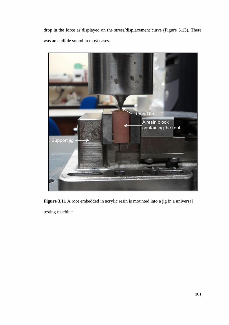
101
drop in the force as displayed on the stress/displacement curve (Figure 3.13). There
was an audible sound in most cases.
Figure 3.11 A root embedded in acrylic resin is mounted into a jig in a universal
testing machine

102
Figure 3.12 Zwick/Roell Z020 universal testing machine
.

103
Figure 3.13 Load/displacement curve displayed by the universal testing machine
showing a gradual increase in the force (N) with a sharp drop indicating fracture

104
3.4 Evaluation of leakage resistance using a dye leakage
method
3.4.1 Introduction
Leakage through the root canal system has been investigated using a wide range of
methods, including dye leakage, fluid filtration, glucose leakage, radioactive
isotopes, electrochemical, and bacterial leakage. Dye leakage is considered the most
widely used method for leakage assessment (183). Different types of dyes have been
used, for example methylene blue (87), India ink (269), rhodamine B fluorescent dye
(270) and basic fuchsine (271).
The use of a vacuum to prevent interference with dye penetration due to entrapped
air has been suggested (189). However, it has been reported that the application of a
vacuum has an insignificant effect on the extent of dye penetration (185).
Visualization of the extent of dye penetration into the root canal has been performed
either by clearing or sectioning the root. The clearing method involves rendering the
root transparent by decalcification and dehydration (272). In the sectioning
technique, the root is sectioned either longitudinally (57) or horizontally (58) to
examine the presence of dye under a microscope.
3.4.2 Dye leakage method
The teeth were decoronated. After performing root canal retreatment, the root
surface was covered with two layers of nail varnish. Each root was submerged in 2%
methylene blue dye in a plastic container where the root was kept upright to facilitate
the passage of dye in coronal-apical direction.

105
There were three immersion periods: 1 day, 7 days and 30 days. After each period,
10 roots from every group were removed from the dye and washed under running
water. The roots were then embedded in acrylic resin so that they could be easily
clamped while being sectioned by a diamond wheel saw under water coolant at 2, 4,
6 and 8 mm from the coronal surface (Figure 3.14). After sectioning, the root canal
was examined under a stereomicroscope microscope (x25) (MEIJI Techno Co. Ltd.,
Tokyo, Japan) (Figure 3.15) to detect the presence of dye (Figure 3.16).
Figure 3.14 A root embedded in a resin block is being sectioned horizontally using a
diamond wheel saw

106
Figure 3.15 A stereomicroscope used to examine the samples to detect the extent of
dye penetration

107
Figure 3.16 Slices from a representative sample examined under a microscope (x25)
to detect dye penetration through the filled root canal. The dye is visible at 2 mm
(A), 4 mm (B) and 6 mm (C), but not at 8 mm (D) section.

108
Chapter 4
Micro-CT evaluation of voids in the filling material
of single-rooted teeth obturated with different
techniques
Fatma Asheibi, Alison J.E. Qualtrough, Anthony Mellor,
Philip J. Withers, Tristan Lowe
Journal of Research and Practice in Dentistry
Vol. 2014 (2014), Article ID 556901, 10 pages
(Appendix: Published Paper 1)

109
4.1 Abstract
Aim: The aim of this study was to compare the volume of voids in a resin-based root
canal filling (Resilon) with gutta percha using either cold lateral condensation or
continuous wave of compaction filling techniques using computed X-ray micro-
tomography (micro-CT).
Methodology: A total of 56 extracted upper anterior human teeth were decoronated,
the canals shaped using rotary ProTaper files to a final size of F3 and randomly
allocated into 4 groups (n=14) according to the filling material and obturation
technique; Group 1: gutta percha/cold lateral (GP/C), Group 2: Resilon/cold lateral
(R/C), Group 3: gutta percha/thermal (GP/T), Group 4: Resilon/thermal (R/T).
Thermal filling was performed using System B and Obtura ΙΙ. The filled roots were
then scanned using micro-CT and the data were subsequently processed using Avizo
6.3 Standard version software. Statistical analysis was performed using the Kruskal-
Wallis test with multiple pairwise comparisons.
Results: The total percentage volume of voids in GP/C (2.40%) and R/C (1.88%)
were significantly higher than GP/T (1.04%) and R/T (1.29%) (P<0.001). A
significantly higher percentage of voids was present in the coronal third compared
with middle and apical thirds (P<0.001).
Conclusion: Thermal obturation resulted in a root filling with significantly fewer
voids than the cold lateral condensation technique regardless of the obturation
material used. Most of the voids located in the coronal third which emphasizes the
importance of obtaining a good coronal seal following endodontic treatment.
Key words: computed tomography (CT); Resilon; voids

110
4.2 Introduction
Optimal obturation leads to successful endodontic treatment (273). Incomplete filling
of a well-cleaned and prepared root canal system could compromise the outcome of
treatment (273). The ideal obturation material should provide good adaptation to the
canal walls and fill any irregularities with the full length of the canal being densely
filled with a homogenous mass of obturation material (7).
Gutta percha (GP) has been the material of choice for obturation for many years as it
possesses favourable properties such as dimensional stability, good working
properties, ease of removal, biocompatibility and radiopacity (23). One of the
limitations of GP is that it does not bond to any type of sealer (28). As many sealers
shrink upon setting, there is a potential for gaps or voids creation via which
microleakage may occur (48, 56).
A resin-based material, Resilon, was introduced in 2004 (19). It has been claimed
that it bonds to the root canal dentine, strengthens the root when used as an
obturation material, (18, 104, 120, 274) and provides a better seal than GP (48, 56).
It has similar handling properties as GP and can be placed into root canals using
either cold lateral condensation (41, 64) or thermal obturation (42, 46).
Many properties of Resilon have been investigated such as leakage resistance (48),
root strengthening (18, 104) and quality of obturation (41). However, most of these
studies relate to it when used with cold lateral condensation and very few have
compared it with thermal obturation (42). As the use of heated filling techniques is
increasing (238), investigation of the quality of obturation with Resilon using this
method in comparison with cold lateral condensation is timely.
The few studies which have investigated Resilon using thermal obturation in
comparison with cold lateral condensation have used a variety of assessment
methods which may result in inaccuracies. In one study (42), the filled roots were
sectioned and examined under a stereomicroscope. This process may lead to loss of
some of the filling material and creation of spaces that may appear as voids. In the

111
present study, micro-CT was used as it gives highly accurate three-dimensional
analysis of voids (246).
The aim of this study was to compare the percentage volume of voids in root canals
filled with Resilon and with GP using cold lateral condensation and continuous wave
of compaction techniques. The null hypothesis was that there would be no significant
difference in the percentage volume of voids between the four combinations of the
two materials and obturation techniques.

112
4.3 Materials and Methods
This study was approved by the ethics committee at the University of Manchester
(Reference number: UREC5-10275). Seventy upper anterior human teeth were
collected, disinfected with 5% sodium hypochlorite (NaOCl) (Procter and Gamble,
Weybridge Surrey, UK) for 10 minutes and stored in normal saline. Exclusion
criteria were: roots that were carious, fractured, resorbed or immature. Roots with
sclerosed canals, with more than one canal or those with root curvature >10°, as
determined by Schneider (275), were also excluded.
Preparation
From the initial sample, a total of 56 teeth (28 lateral incisors, 16 central incisors and
12 canines) were selected according to the aforementioned criteria. The teeth were
decoronated and the length of the roots was reduced to 14 mm using a diamond
wheel saw with water cooling (central incisors, lateral incisors and canines were kept
separate to facilitate in their allocation into groups for obturation). The working
length was determined to be 1 mm short of the apical foramen using a size 10 K-file
(VDW GmbH, Munich, Germany).
The root canals were prepared using rotary ProTaper files to the final size of F3
(Dentsply Maillefer, Ballaigues, Switzerland) and irrigated with 1% NaOCl. After
completion of the preparation, the root canals were irrigated with 5 mL 17%
ethylene-diamine-tetra-acetic acid (EDTA) (PPH Cerkamed, Sandomierska, Poland)
and 10 mL distilled water.
Obturation of root canals
The roots were randomly allocated into four groups using a stratified sampling
method (n=14: 7 lateral incisors, 4 central incisors and 3 canines) according to the
filling material and obturation technique.
Group 1 (GP/C) was filled with ProTaper GP F3 (Dentsply Maillefer, Ballaigues,
Switzerland) and AH Plus sealer (Dentsply DeTrey GmbH, Konstanz, Germany)
using cold lateral condensation. A master cone was selected and trimmed to fit the

113
canal to the working length with tug-back. Then, the canal was dried using paper
points (Diadent, Chungcheongbuk-do, Korea). The sealer was carried into the canal
using the master cone. A finger spreader (Dentsply Maillefer, Ballaigues,
Switzerland) was used to compact the GP cone and create a space for accessory
points. When obturation was completed, the GP points were cut to the coronal end of
the root using a heated instrument and the coronal part was compacted using a
Machtou plugger (Dentsply Maillefer, Ballaigues, Switzerland).
Group 2 (R/C) was filled with Resilon/RealSeal (Pentron Corp., Wallingford, US)
sealer using cold lateral condensation according to the manufacturers’ instructions. A
Resilon point (30/0.06) was used as the master cone which was trimmed to fit the
canal to the working length with tug-back. After drying the root canal with paper
points, RealSeal primer was applied. RealSeal sealer was carried to the canal on the
master cone. Cold lateral condensation was performed as previously described. The
coronal surface was light cured for 40 seconds.
Group 3 (GP/T) was filled with ProTaper GP/AH Plus sealer using a continuous
wave of compaction technique. The sealer was carried to the canal using the master
cone. A System B plugger of fine-medium size (SybronEndo, Glendora, USA) was
selected and a rubber stopper was fitted 5 mm short of the working length, marking
the binding point. The heat source was adjusted to 200°C. The plugger was activated
and used to cut the excess GP and then inserted into the canal along the master cone
until the binding point was reached. The heat source was then deactivated and a firm
pressure was maintained for 10 seconds. A one-second heat burst was activated and
the plugger was withdrawn. The canal was then backfilled with GP pellets (Obtura
Spartan, Earth City, Missouri, US) using Obtura III at 200°C (Obtura Spartan, Earth
City, Missouri, US).
Group 4 (R/T) was obturated with Resilon/RealSeal sealer using the continuous
wave of compaction technique described above. The temperature setting in system B
and Obtura III was set to 150°C according to the manufacturers’ instructions.
All roots were stored for 72 hours at 37°C and 100% humidity in air-tight containers.

114
Scanning of the roots
The roots were scanned in a random order using a 225kV 3 µm source Nikon micro-
CT housed in a walk-in enclosure (Nikon X-tek Systems Ltd., Tring, England).
Using a sample-source distance of 2 cm and a detector-sample distance of 138 cm,
magnified images were collected on a 2000 pixel (200 micron pixel pitch) detector
allowing observation of fine differences in contrast. The X-ray attenuation could be
adjusted by changing the target material and accelerating voltage. For this scan,
copper was used as the target material. The micro-CT settings were: 93 kV, 135
micro Amps, 1901 projections (radiographs), 1000 seconds exposure, gain 16.
The raw data were reconstructed using CTPro 3D software (Version XT 2.2, Metris,
Hertfordshire, UK). An effective voxel size of 7.7-9.3 µm was obtained. Voids were
observed in 2-dimensional (2D) (Figure 4.1) and 3-dimensional (3D) images (Figure
4.2) using Avizo 6.3 Standard version (Visualization Sciences Group, Berlin,
Germany). A number of grey-scale thresholding values were tried and the upper and
lower limit values demonstrated that the scatter per sample was found to be about
0.1% volume of voids. After segmentation, Avizo software was used to measure the
total volume of voids. The percentage volume of voids was calculated using the
following equation:
Statistical analysis
Test of normality showed that data were not normally distributed and therefore, non-
parametric tests were used. Analysis of data was carried out using the Kruskal-
Wallis test with multiple pairwise comparisons. The software used was SPSS
Version 20.0 (IBM Corp., Armonk, US) with the level of significance set at 5%.
100x canalme of the Total volu
sme of voidTotal volu

115
Figure 4.1 Visualisation of reconstructed images showing virtual 2D slices of filling
(A) gutta percha/cold lateral condensation, (B) Resilon/cold lateral condensation, (C)
gutta percha/thermal compaction, (D) Resilon/thermal compaction. The arrows
indicate voids within the filling material and the scale bars are 5 mm long.

116
Figure 4.2 3D images of the root canal fillings in (A) gutta percha/cold lateral
condensation, (B) Resilon/cold lateral condensation, (C) gutta percha/thermal
compaction, (D) Resilon/thermal compaction. The blue colour within the filling
material indicates voids; the scale bars are 6-8 mm long.

117
4.4 Results
The means and standard deviations (SD) of the overall percentage (%) of voids are
shown in Table 4.1. The GP/C (gutta percha/cold lateral) group showed the highest
overall mean value (2.40%), whereas the GP/T (gutta percha/thermal) group had the
lowest overall value (1.04%). The Kruskal-Wallis test indicated that there was a
statistically significant difference between the groups (P<0.001). The multiple
pairwise comparisons showed that Groups GP/C and R/C (Resilon/cold lateral) had
significantly higher percentage of voids than Groups GP/T and R/T
(Resilon/thermal) (P<0.001 for GP/C versus GP/T and P=0.007 for R/C versus R/T).
Table 4.2 shows the means and standard deviations of the percentage of voids in
each third of the root canal. In the coronal and the middle thirds, the results were
similar to those for the overall percentage of voids (GP/C and R/C showed
significantly higher percentage of voids than GP/T and R/T) (P<0.05). In the apical
third, there was no significant difference between the groups. When the thirds were
compared for each group, the coronal third showed significantly higher percentage of
voids than the middle third, which had significantly higher percentage of voids than
the apical third (P<0.001). The percentage of voids in the coronal third in relation
with the overall percentage of voids was 68.8% in GP/C, 75.5% in R/C, 70.2% in
G/T and 73.7% in R/T.

118
Table 4.1 Means and standard deviations of the overall percentage of voids
Group
(n=14) Overall voids %
mean (SD)
GP/C 2.40a
(0.66)
R/C 1.88a
(0.61)
GP/T 1.04b
(0.44)
R/T 1.29b
(0.44)
GP/C: gutta percha/ cold lateral condensation
R/C: Resilon/cold lateral condensation
GP/T: gutta percha/thermal compaction
R/T: Resilon/thermal compaction Different superscript letters indicate statistically significant differences
Table 4.2 Means and standard deviations of the percentage of voids in each third of
the root canal
Group
(n=14)
Coronal voids %
mean (SD)
Middle voids %
mean (SD)
Apical voids %
mean (SD)
GP/C 1.65 (0.60) 0.66 (0.22) 0.09 (0.05)
R/C 1.42 (0.57) 0.44 (0.21) 0.02 (0.01)
GP/T 0.73 (0.30) 0.27 (0.20) 0.04
(0.03)
R/T 0.95 (0.40) 0.26 (0.12) 0.08 (0.06)
GP/C: gutta percha/ cold lateral condensation
R/C: Resilon/cold lateral condensation
GP/T: gutta percha/thermal compaction
R/T: Resilon/thermal compaction
The differences between the thirds in all groups are statistically significant. GP/C and R/C have significantly higher volume of voids than GP/T and R/T in the coronal and middle thirds.

119
4.5 Discussion
Traditional methods used to evaluate the quality of root fillings allow only partial
evaluation of the root canal content. Radiographs give a two-dimensional view of a
three-dimensional structure. Sectioning the roots to view them using a scanning
electron microscope or a stereomicroscope could result in loss of some of the
material which may mimic voids. Further, the chance of intersecting a void is
statistical and the shape of the voids may mean that volume fractions are not
properly inferred from 2D sections. The clearing technique is time consuming (177)
and the long periods of immersion in alcohol may affect the physical properties of
gutta percha (276). Recently, the use of computed tomography (CT) (176, 177, 235)
and micro-CT (277-279) in dental research has gained increasing popularity. It gives
an accurate three-dimensional view of the root canal content without destroying the
samples which can be used for further research (41). In the present study, the voxel
size obtained was 7.7-9.3 µm, which is better than that used in previously published
studies (12, 41).
The null hypothesis was rejected as the results showed a statistically significant
difference in the volume of voids between the tested groups. The cold lateral
condensation technique with both materials showed a significantly higher percentage
of voids in comparison with the thermal technique. This agrees with previous studies
(176-178) which compared the two techniques using gutta percha. In cold lateral
condensation, the resulting mass is not homogenous and there may be spaces present
between the points. By contrast, the thermal technique produces a more homogenous
mass with fewer voids and better adaptation of the filling to the canal walls (177,
178). However, during condensation with pluggers, air could be entrapped. None of
our samples were void-free. This finding has also been reported in previous studies
(39, 40, 42).
Our study showed that the GP/C group had a higher percentage of voids than the R/C
group, but this was not statistically significant. This is consistent with other studies
(39, 40, 43), but is in contrast with that found by Hammad et al. (41). In the latter
study (41), the lowest percentage of voids was found in the GP group as compared

120
with Resilon, GuttaFlow and EndoRez. The sealer used with GP was Tubliseal (a
zinc-oxide eugenol-based sealer), whereas in the present study AH Plus sealer (a
resin-based sealer) was used. It has been reported that GP showed volumetric
expansion when it came into contact with eugenol which resulted in a better seal of
the obturation (280). In addition, the voxel size used in the present study was lower
than that used in the study by Hammad et al. (41) which improved detection of
smaller voids. This may explain the fewer voids in the GP group reported by
Hammad et al. (41).
In contrast with the findings of Hammad et al. (41), the results of the present study
showed that the coronal third in all groups contained a significantly higher
percentage of voids than the middle and apical thirds. This may have a clinical
significance if the coronal seal lost as it may accelerate penetration of fluids through
the root canal filling and dissolution of the sealer. In both obturation techniques used
in this study, the apical third of the canal was filled with a master cone. In the cold
lateral condensation technique, the remainder of the canal was filled with accessory
points which were packed using a spreader leading to a greater risk of void creation
between the accessory points in the middle and coronal thirds as opposed to the
apical third. Similarly, in the thermal obturation technique, the middle and coronal
thirds were obturated with a softened filling material in which air bubbles may be
entrapped during condensation. This may explain the significant difference in the
percentage of voids between the thirds.
Voids inside the filling materials, which are not linked to the periphery of the filling,
could be considered less clinically significant than peripheral gaps because micro-
organisms, if present, are restrained in an unfavourable environment and do not have
access to a nutritional supply. Peripheral gaps along the dentine-sealer or core
material-sealer interfaces may jeopardize the outcome of root canal treatment (273).
This is because they may act as a pathway that permits sealer dissolution and passage
of micro-organisms through the filled root canal to the periradicular tissues.
Furthermore, if residual micro-organisms remain trapped in the dentinal tubules after
treatment is finished, the peripheral gaps may form channels through which the
micro-organisms can get access to nutrients and initiate or continue the inflammatory
process.

121
This is an in vitro study and further in vivo investigations are indicated. Variations in
the canal anatomy of the human teeth may affect the obturation quality. Ideally,
contralateral teeth from the same individual should be used (281). However, this is
not practical in research settings and limits the sample size. Therefore, upper anterior
teeth were used in this study and each group contained equal number of central
incisors, lateral incisors and canines (stratified sampling). It would be beneficial in
the future to extend the testing to a wider range of root curvatures as this would be
more clinically relevant.
4.6 Conclusion
The results of the present study show that the percentage of voids in the fillings is
low (1.04-2.40%), regardless of the filling technique. However, the significant
difference shown highlights the importance of the use of continuous wave of
compaction technique in reducing the volume of voids in the root canal filling. In
addition, the significantly higher percentage of voids in the coronal third in
comparison with the middle and the apical thirds emphasizes the significance of
obtaining a good coronal seal after completion of endodontic treatment.
4.7 Acknowledgement
The authors deny any conflicts of interest. We are grateful to the EPSRC whose
grant funding (EP/I02249X, EP/F007906 and EP/F028431) enabled the purchase and
support of the imaging equipment in the Henry Moseley X-ray Imaging Facility.

122
Chapter 5
Effect of calcium hydroxide and its combination with
iodoform on the bond strength of Resilon
(Submitted to the Journal of Adhesive Dentistry)

123
5.1 Abstract
The aim of this study was to investigate the effect of calcium hydroxide (CH) and its
aqueous- or oil-based combinations with iodoform on Resilon bond strength. Fifty
extracted human upper anterior teeth were selected. Root canal treatment was
performed using ProTaper rotary files. The teeth were allocated into five groups
(n=10): Group 1 (control), no medicament; Group 2, CH; Group 3, aqueous CH and
iodoform (Calcipast1); Group 4, oil-based CH and iodoform (Vitapex); Group 5,
iodoform. After one week storage, the medicaments were removed and the canals
were obturated with Resilon. The teeth were embedded in acrylic resin and sectioned
into 2 mm slices. The slices were mounted in a Universal Testing Machine and
loaded until bond failure occurred. The mean bond strength of the control (18.68
MPa), CH (15.36 MPa) and Calcipast1 (16.82 MPa) groups were significantly higher
than that of the Vitapex (5.13 MPa) and iodoform (5.46 MPa) groups (P≤0.001).
Vitapex and iodoform decreased the bond strength of Resilon. It may be
recommended to avoid the use of these medicaments if Resilon is the obturation
material.
Key words: bond strength, calcium hydroxide, iodoform, Resilon, Vitapex

124
5.2 Introduction
The main goal of chemo-mechanical preparation is to disinfect the entire root canal
system (282). The complexity of the root canal system and limited access of the
disinfection methods to intra-canal spaces restrict the total removal of micro-
organisms (283). The main function of intra-canal medicaments during endodontic
treatment is to disinfect the root canal (284, 285). The most commonly used
medicament is calcium hydroxide (CH) due to its biocompatibility and activity
against a wide range of micro-organisms (286). However, there are conflicting
reports regarding its effectiveness against Enterococcus faecalis, a commonly
isolated bacterium from failed endodontically-treated roots. Some studies have
reported significant antibacterial activity against E faecalis (287-290), while others
have reported that it is not effective (291-294).
CH can be mixed with different vehicles. Some of these are oil-based, such as
Vitapex (Neo Dental Chemical Products Co. Ltd., Tokyo, Japan), which contains
40.4% iodoform, 30.3% calcium hydroxide and 22.4% silicone oil (295). Others are
aqueous, such as Calcipast1 (PPH Cerkamed, Sandomierska, Poland), which
promote rapid ion release and are easier to remove than oil-based vehicles (286).
Adhesive filling materials, such as Resilon (Pentron Corp., Wallingford, USA), have
been introduced in endodontic treatment in an attempt to improve the coronal and
apical seal (48). Resilon contains a matrix (co-polymer of polycaprolactone and
urethane dimethacrylate), bioactive glass and radiopaque fillers (28). It can bond to
the root dentine and form a “monoblock” (28). This bond to dentine can be
weakened by materials used during endodontic treatment such as sodium
hypochlorite (157), chlorhexidine (239) and some intra-canal medicaments (240).
The total removal of intra-canal medicaments from the root canal may not be
possible (296). The remnants may interfere with the bond of adhesive filling
materials by acting as a physical barrier. In addition, the high pH of CH can
neutralise the acidic primer in self-etching adhesive systems (30). The effect of CH
on the bond strength of Epiphany sealer has been investigated (297, 298) and also
compared to the effect of chlorhexidine (240). The literature does not contain any

125
reports on the effect of other medicaments used in endodontic treatment on the bond
strength of Resilon.
Therefore, the aim of this study was to investigate the effect of intra-canal
medicaments (CH, Calcipast1, Vitapex, and iodoform) on the bond strength of
Resilon to dentine. The null hypothesis was that there would be no significant
difference in the bond strength of Resilon to dentine after the use of these
medicaments.

126
5.3 Materials and Methods
This study was approved by the Ethics Committee of the University of Manchester
(UREC5-10275). Upper anterior human teeth were collected, disinfected for 10
minutes with 5% sodium hypochlorite (NaOCl) and stored in normal saline. The
roots were inspected under illumination and magnification to exclude those with
cracks, caries or resorption. Fifty teeth (10 central incisors, 30 lateral incisors and 10
canines) were included in the study.
Teeth preparation
Access cavities were prepared and the canals were cleaned and shaped using rotary
ProTaper files (Dentsply Maillefer, Ballaigues, Switzerland) to a final size of F4.
5 mL 1% NaOCl (Procter and Gamble, Weybridge Surrey, UK) was used for
irrigation. After completion of the canal preparation, the canals were irrigated with
5 mL 17% ethylene-diamine-tetra-acetic acid (EDTA) (PPH Cerkamed,
Sandomierska, Poland) and finally 10 mL distilled water. The canals were dried
using paper points (Diadent, Chungcheongbuk-do, Korea) and the teeth were then
randomly assigned into five groups using stratified sampling (n=10, 2 central
incisors, 6 lateral incisors and 2 canines) according to the type of intra-canal
medicament:
Group 1 (control): no medicament.
Group 2 (CH): Calcipast (PPH Cerkamed, Sandomierska, Poland).
Group 3 (CH1): Calcipast1, aqueous combination of CH and iodoform (PPH
Cerkamed, Sandomierska, Poland).
Group 4 (VP): Vitapex, silicone oil-based combination of CH and iodoform (Neo
Dental Chemical Products Co. Ltd., Tokyo, Japan).
Group 5 (IO): Iodoform (PPH Cerkamed, Sandomierska, Poland).
CH, CH1 and Vitapex were placed into the canals using plastic tips. Iodoform was
applied into the canals using a lentulo spiral filler (Dentsply Maillefer, Ballaigues,

127
Switzerland) until the canals were totally filled with the medicament to the level of
the canal orifice. After the canal was filled with the medicament, the access cavity
was sealed with a temporary filling (Coltosol, Coltène, Altstätten, Switzerland) and
the teeth were stored for one week in a moist environment at 37°C.
Obturation of the root canals
The intra-canal medicaments were removed from the canals using a size 25 hand K-
file and agitated irrigation with 5 mL 1% NaOCl using a 10 mL syringe and a 27
gauge needle. The removal of iodoform and Vitapex was assisted by the use of
ethanol (Whatman International Ltd, Maidstone, UK) according to the
manufacturer's recommendations. The removal of the medicaments was considered
complete when the canal walls appeared clean under magnification (X2.0)
(Orascoptic, Middleton, USA) and no material could be seen on the file. Paper points
were also used to detect any remaining medicament. The root canals were then
irrigated with 5 mL 17% EDTA and 10 mL distilled water.
The root canals were obturated with Resilon/RealSeal sealer (Pentron Corp.,
Wallingford, US) using System B (SybronEndo, Glendora, USA) and Obtura III
(Obtura Spartan, Earth City, Missouri, USA). A master cone which fitted to the
working length with tug-back was selected. After drying the canals with paper
points, RealSeal primer was applied. The master cone was covered with the sealer
and inserted into the canal. The heat source was set to 150°C. The system B plugger
(fine-medium size) was inserted into the canal along the master cone to 5 mm short
of the working length. The heat application was then stopped and apical pressure was
maintained for about 10 seconds. A one-second heat burst was activated and the
plugger was withdrawn. Backfilling of the canal was performed using Obtura III at
150°C. The coronal surface was light cured for 40 seconds.
The access cavity was closed with temporary filling and the teeth were stored for one
week in a moist environment at 37°C.

128
Preparation for the push-out test
The teeth were decoronated to the level of the cemento-enamel junction using a low-
speed diamond wheel saw under continuous water coolant (Model 650, South Bay
Technology inc., California, USA). The roots were embedded in acrylic resin
(Metrodent Ltd, Huddersfield, UK) and then horizontally sectioned into 2 mm thick
slices. Only slices with circular canal shape were included (2 slices per root, n=20
slices per group). The thickness of each slice was measured with a digital caliper
(Mitutoyo Corp. Kawasaki, Japan), and all were within 1.9–2.2 mm. The interfacial
area of the root filling was calculated using the formula (260):
22 )()( rRhrRA
, where π is the constant 3.14, R is the coronal radius of the canal, r is the apical
radius of the canal, and h is the slice thickness, all measured in millimetres (mm).
The slices were marked to ensure that the load was applied in apical-coronal
direction to avoid any interference owing to root canal taper. Two flat ended
cylindrical plungers with diameters of 0.78 and 0.50 mm were used to apply the
force on the filling material. Each slice was positioned carefully so that the plunger
contacted only the filling material (Figure 5.1). A support jig with a hole was
selected to provide enough clearance for the filling material when it was extruded
from the slice. Compressive loading was performed using a Universal Testing
Machine (Zwick GmbH & Co. KG, Germany). Loading was applied at a speed of 0.5
mm/minute until a sharp and instant drop of the applied force was observed on the
load/displacement curve which indicated bond failure. Failure was also manifested
by extrusion of the filling material from the root slice (Figure 5.2).
The bond strength (megapascals [MPa]) was computed using a modified formula
(261):
fd EEAF /)/(

129
where F is the force (Newton), A is the interfacial surface area (mm2), Ed is the
elastic modulus of dentine (16000 MPa) (117), and Ef is the elastic modulus of the
filling (Resilon) (86.58 MPa) (25).
Figure 5.1 Schematic representation for the set-up for push-out bond strength test
(A). Image for a root slice in the universal testing machine (B).
Figure 5.2 Image of a root slice before performing the push-out test (A). The filling
material (Resilon) was extruded from the root canal after performing the test (B:
apical aspect, C: coronal aspect).

130
Analysis of Failure Modes
Both sides of each slice were examined under a stereomicroscope microscope
(MEIJI Techno Co. Ltd., Tokyo, Japan) at x25 magnification to determine the mode
of bond failure. Three failure modes were identified: (1) adhesive failure at the
filling material/ dentine interface, (2) cohesive failure within the filling material, and
(3) mixed failure in both the filling material and dentine interface.
Scanning electron microscope (SEM) evaluation
A representative sample from each group was randomly selected for SEM
examination. Each sample was sectioned perpendicular to the root surface to allow
visualisation of the root canal wall after bond failure. The samples were coated with
gold/palladium at approximately 5 nm thickness using a PECS (precision etching
and coating system). They were then scanned using the Philips XL30 FEG SEM
(field emission gun scanning electron microscope) (FEI, Tokyo, Japan).
Statistical Analysis
SPSS Version 20.0 (IBM Corp., Armonk, US) was used to analyse the data.
A normality test showed that the data were not normally distributed and therefore,
non-parametric tests were used. The significance level was set at ≤0.05. Data were
analysed using the Kruskal-Wallis test with multiple pairwise comparisons.

131
5.4 Results
The means and standard deviations of the bond strength (MPa) of Resilon for all
groups are presented in Table 5.1 and Figure 5.3. The control group showed the
highest bond strength (18.68 MPa) and VP group showed the lowest bond strength
(5.13 MPa). The Kruskal Wallis test showed that there was a significant difference
between the groups (P≤0.001). The control, CH and CH1 groups had significantly
higher bond strengths than VP and IO groups (P≤0.001). There was no significant
difference between the first three groups (control, CH, CH1) and neither between the
last two groups (VP, IO).
As shown in Table 5.2, the mode of bond failure was predominately adhesive in the
groups VP (60%) and IO (55%). In the control and CH1 groups, the main failure
mode was mixed (55% and 45%, respectively), whereas in CH group, it was
cohesive (50%). SEM examination of the root canal wall after bond failure showed
the three failure modes (Figure 5.4).
Table 5.1 Means and standard deviations (SD) of bond strength of Resilon (MPa)
Group (n=20) Mean bond strength
(MPa) SD
Control 18.68a
3.92
Calcipast 15.36a
5.85
Calcipast 1 16.82a
3.74
Vitapex 5.13b
1.78
Iodoform 5.46b
1.53
Different superscript letters indicate statistical significant difference

132
Figure 5.3 A bar chart for the means of the bond strength of the groups. The error
bars represent standard deviations.
Table 5.2 Modes of bond failure
Group (n=20)
Adhesive n (%)
Cohesive n (%)
Mixed n (%)
Control 3 (15) 6 (30) 11 (55) Calcipast 2 (10) 10 (50) 8 (40) Calcipast 1 4 (20) 7 (35) 9 (45) Vitapex 12 (60) 1 (5) 7 (35) Iodoform 11 (55) 2 (10) 7 (35)
Groups
Control Calcipast Calcipast+1 Vitapex Iodoform
Bo
nd
str
en
gth
(M
Pa)
0
5
10
15
20
25

133
Figure 5.4 SEM images illustrate the types of Resilon-to-dentine bond failure. (A)
Adhesive failure shows canal surface clean of filling material. (B) Cohesive failure
shows surface covered with sealer. Filler particles of the sealer can be observed
(arrows). (C) Mixed failure shows areas clean and others covered with sealer. (D) A
higher magnification (x1000) of a section from (A) (square) shows some open
dentinal tubules (open arrows) and others occluded with fractured resin tags (solid
arrows).

134
5.5 Discussion
The results of the present study showed that the use of the intra-canal medicaments,
Vitapex and iodoform, resulted in significantly lower bond strengths of Resilon to
dentine in comparison with CH and CH1. Therefore, the null hypothesis was
rejected. The effect of CH on the bond strength of Resilon has been previously
investigated (240, 297, 298). There has been no previous investigation of the bond
strength of Resilon when CH has been combined with iodoform using different
vehicles.
The significantly reduced bond strength of Resilon after the use of Vitapex and
iodoform may be attributed to their composition. Both Vitapex and iodoform contain
silicone oil (295) which may make removal of these medicaments difficult (299) and
may therefore interfere with the bonding of Resilon resulting in lower bond
strengths. This may also explain the mode of failure in these groups which was
predominately at the root filling-dentine interface (adhesive failure).
Our study showed that CH had no significant effect on the bond strength of Resilon.
This was in agreement with a previous study (298) and disagreed with some others
(240, 297). Barbizam et al. (240) reported that CH significantly reduced the bond
strength of Epiphany sealer to dentine. In their study, the coronal and middle thirds
of the root canals were prepared using a cylindrical post drill after preparing the
canals with RACE Ni Ti files. The resulting shape of the canal may interfere with
proper removal of the intra-canal medicaments. In addition, the canal was filled with
the sealer without using Resilon core. This may explain the different results.
Slices with canals that are not circular in shape were excluded to avoid non-uniform
stress distributions during testing and inaccurate measurements (120, 261). The
slices which were used in the test were mainly from the middle third of the root. In
the coronal third, the canal shape was oval, and in the apical third, the canal size was
very small. Removal of the iodoform and Vitapex was assisted by alcohol as per the
manufacturer’s recommendation. However, as a final rinse with alcohol results in
dry canal and the dentine surface needs to be moist for effective bonding (300), the
canals were irrigated with distilled water as a final rinse.

135
Bond strength tests are the best measure of adhesion currently available (254).
However, the push out bond strength test has some limitations with regard to the
diameter of pins used, modulus of elasticity of the filling material tested and the
specimen thickness (261). In the present study, efforts were made to minimise these
limitations. The diameters of the pins used in the test were chosen so that they were
just slightly smaller than the canal diameter and the ratio between the pin diameter
and the canal diameter was less than 0.85. Therefore, the stress was concentrated
near the dentine-filling interface (254, 261). In calculating of the bond strength, we
used the correction factor fd EE / suggested by Chen et al. (261) because the
modulus of elasticity of Resilon (86.58 MPa) (25) is lower than that of the dentine
(16000 MPa) (117). The thickness of the root slices was standardised at 2 mm as it
has been reported that the formula of push-out bond strength works well for slices
that are greater than 1.1 mm in thickness (261).The formula to calculate the
interfacial surface area used in some previous studies (120, 127) was for a curved
surface of a cylinder and in others (301) was not stated. However, the root canal
walls are tapered and therefore we used the formula for a curved surface of a
truncated cone (260). Bond strength is only one aspect of the quality of root canal
seal. Further investigation of the effect of the medicaments on other properties of
Resilon is required.
In conclusion, our study showed that Vitapex and iodoform significantly decreased
the bond strength of Resilon in comparison with CH and CH1. Within the limitations
of this in vitro study, it is recommended to avoid the use of Vitapex and iodoform if
the canal is to be obturated with Resilon.

136
Chapter 6
Micro-CT evaluation of the effectiveness of the
combined use of rotary and hand instrumentation in
removal of Resilon
Fatma Asheibi, Alison J.E. Qualtrough, Anthony Mellor,
Philip J. Withers, Tristan Lowe
Dental Materials Journal 2014; 33(1): 1–6
(Appendix: Published Paper 2)

137
6.1 Abstract
This study compares the effectiveness of ProTaper rotary files with ProTaper
retreatment and K-files in the removal of Resilon or gutta percha (GP) from canals
filled either by cold lateral condensation or thermal obturation using micro-CT.
Ninety-six teeth were prepared using ProTaper files and allocated into four groups
(n=24): Group-1 was filled with GP/AH Plus and Group-2 with Resilon/RealSeal
using cold lateral condensation. Group-3 was filled with GP/AH Plus and Group-4
with Resilon/RealSeal using System B and Obtura II. The roots were scanned by
micro-CT. Each group was divided into two subgroups (n=12): A, retreated using
ProTaper files and B, using ProTaper retreatment and K-files. The roots were
scanned to calculate the volume of the remaining material. With thermal obturation,
roots filled with Resilon had significantly more remaining material than GP.
Obturation using thermal technique resulted in significantly less remaining material
than cold condensation except Resilon retreated using ProTaper retreatment and K-
files.
Key words: Computed tomography (CT), Gutta percha, ProTaper, Retreatment,
Resilon

138
6.2 Introduction
Periradicular diseases may persist or emerge following the completion of root canal
treatment as a result of intra-radicular infection, extra-radicular infection or non-
microbial factors (191). The main cause of failure is persistent, or recurrent
microbial infection (192, 193). Non-surgical retreatment is usually the preferred
option for teeth in which endodontic treatment has failed as there is a greater
opportunity to eradicate any intra-radicular source of infection compared with a
surgical approach (202). Retreatment comprises removal of the old root filling
material to allow access to any residual micro-organisms and permit further cleaning
and disinfection of the root canal system (202). Additionally, efficient removal of
previous filling materials can facilitate intimate contact between the sealer and the
root canal walls, thus potentially preventing microleakage (204).
Gutta percha is the most commonly used root canal filling due to its favourable
properties such as dimensional stability, biocompatibility and ease of removal (23).
However, it does not bond to sealer (28). A resin-based root canal filling, Resilon,
was launched in 2004 (19). It is capable of bonding to methacrylate-based sealers
and as such may result in the prevention of microleakage and strengthening of the
root treated tooth (18, 92).
Root filling materials can be removed by a variety of techniques such as the use of
stainless steel or nickel-titanium hand files, nickel-titanium rotary files or ultrasonic
instruments with or without the adjunctive use of solvents or heat (207, 209). Studies
have compared the effectiveness of either rotary to hand instrumentation (21, 205) or
compared different rotary systems (207, 216). The combined use of hand instruments

139
and rotary files for effective removal of filling materials has been advocated (21), but
no study has investigated the effectiveness of a combination of the two.
Several studies have investigated the ease of removal of Resilon compared with
gutta percha (21, 207, 209). So far, however, there has been no published data
related to the relationship between removal success and method of filling technique
(i.e. cold lateral condensation versus thermal obturation).
The aim of this study, therefore, was to investigate the effectiveness of rotary
instrumentation and its combination with hand instrumentation in the removal of
gutta percha and Resilon placed into root canals by either cold lateral condensation
or continuous wave of compaction technique using micro-CT. The null hypothesis
was that there would be no significant difference in the removability between the
tested materials, techniques of obturation and techniques of removal.

140
6.3 Materials and methods
Ethical approval for this study was obtained from the Ethics Committee at the
University of Manchester. Upper anterior human teeth were collected, placed in 5%
sodium hypochlorite (NaOCl) for 10 minutes and then stored in normal saline.
Examination of the roots was performed under magnification (x40) to exclude roots
with cracks, fracture, resorption, caries or immature apices or those with a root
curvature of more than 10°, as described by Schneider (275).
Preparation of teeth
Ninety-six upper anterior teeth (48 central incisors, 24 lateral incisors and 24
canines) were selected according to the criteria noted above. Teeth were decoronated
and root lengths were reduced to 14 mm using a diamond wheel saw with water
cooling (Model 650, South Bay Technology inc., California, USA). Central incisors,
lateral incisors and canines were kept separate to facilitate in their allocation into
groups for obturation. The working length was determined to be 1 mm short of the
apical foramen using a size 10 K-file (VDW GmbH, Munich, Germany). A glide
path was established and the canals were prepared using rotary ProTaper Universal
files (Dentsply Maillefer, Ballaigues, Switzerland) to size F3. The motor used was an
X-smart (Dentsply Maillefer, Ballaigues, Switzerland) with a contra angle hand-
piece. The canals were irrigated with 1 mL 1% NaOCl (Procter and Gamble,
Weybridge Surrey, UK) after each filing. When the preparations were completed, the
root canals were irrigated with 5 mL 17% ethylene-diamine-tetra-acetic acid (EDTA)
(PPH Cerkamed, Sandomierska, Poland) and 10 mL distilled water.

141
Obturation of root canals
The roots were randomly allocated into four groups using stratified sampling (n=24,
12 central incisors, 6 lateral incisors and 6 canines) based on the material and
technique of obturation:
Group 1 (GP/C) was filled with ProTaper gutta percha (Dentsply Maillefer,
Ballaigues, Switzerland) and AH Plus sealer (Dentsply DeTrey GmbH, Konstanz,
Germany) using cold lateral condensation. A suitable master cone (fitted the canal to
the working length with tug-back) was selected and the canal was dried using paper
points. The sealer was applied using the master cone. A finger spreader (Dentsply
Maillefer, Ballaigues, Switzerland) was used to compact the cone and accessory
points were inserted into the resulting space until obturation was completed. The
gutta percha points were cut to the coronal end of the root using a heated instrument
and the coronal part was condensed using a Machtou plugger (Dentsply Maillefer,
Ballaigues, Switzerland).
Group 2 (R/C) was filled with Resilon/RealSeal (Pentron Corp., Wallingford, USA)
sealer using cold lateral condensation. A master cone of size 30/0.06 was selected.
RealSeal primer was applied and the excess was removed using paper points. The
sealer was applied using the master cone. Lateral condensation was completed as
described for Group 1. The coronal surface was light cured for 40 seconds as
recommended by the manufacturers.
Group 3 (GP/T) was filled with ProTaper gutta percha/AH Plus sealer using
continuous wave of compaction technique. A System B plugger of fine-medium size
(SybronEndo, Glendora, USA) was used and a rubber stopper was adjusted to 5 mm

142
short of the working length. The temperature was set to 200°C in the touch mode.
The excess GP was cut using the hot plugger which was then inserted alongside the
master cone until the rubber stopper reached a reference point. The heat application
was then stopped and apical pressure was maintained for 10 seconds. The plugger
was withdrawn after one-second activation of the heat source. Backfilling was
performed using Obtura III at 200°C (Obtura Spartan, Earth City, Missouri, USA).
Group 4 (R/T) was obturated with Resilon/RealSeal sealer using System B and
Obtura III as explained for Group 3. Temperature in System B and Obtura III was set
to 150°C according to the manufacturers’ instructions. All roots were stored in air-
tight containers for a week at 37°C and 100% humidity. All the root fillings were
carried out by one operator.
Scanning of the roots
Teeth were scanned in random order using a Nikon micro-CT (225kV 3 µm source)
(X-tek Systems Ltd., Tring, England) housed in a customised bay. Images were
collected on a 2000 pixel detector using a sample-source distance of 2 cm and a
sample-detector distance of 138 cm which allowed observation of fine differences in
contrast. The target material could be changed which allowed adjustment of the X-
ray attenuation by accelerating voltage. Copper was chosen as the target material.
The micro-CT settings were: 93 kV, 135 micro Amps, 1901 projections
(radiographs), 1000 seconds exposure, gain 16.

143
Retreatment
Each group was divided into two subgroups (n=12, 6 central incisors, 3 lateral
incisors and 3 canines). In both subgroups, the coronal 2 mm of the filling was
removed using Gates Glidden drills (3 and 4) (Dentsply Maillefer, Ballaigues,
Switzerland). Then, two drops of eucalyptus oil (PPH Cerkamed, Sandomierska,
Poland) were placed in the space made available. In subgroup A, the filling material
was removed using rotary ProTaper Universal files (F1, F2 and F3)
(at 300 rpm) and in subgroup B using rotary ProTaper Universal retreatment files
(D1, D2 and D3) (Dentsply Maillefer, Ballaigues, Switzerland) (at 500 rpm as per
the manufacturer’s recommendation) followed by hand K-files (25 and 30).
ProTaper Universal and retreatment files were used at the following penetration
depths: F1 and D1 to the coronal third, F2 and D2 to the middle third and F3 and D3
to the apical third. In subgroup B, hand K-files (25 and 30) were used to the full
working length in a circumferential quarter-turn push-pull motion. The criteria for
completion of the treatment were clean canal walls and no material on the files as
observed by the naked eye. When retreatment completed, irrigation was performed
using 3 mL 1% NaOCl, 5 mL 17% EDTA and finally, 10 mL distilled water. Root
canals were dried using paper points. All the retreatment work was carried out by
one operator.
Scanning of the roots
The Nikon micro-CT was used to scan the roots for the second time using the same
settings as for the first scan. The raw data from the first and second scan were
reconstructed using CTPro 3D software (Version XT 2.2, Metris, Hertfordshire,

144
UK). A voxel size of 7.7-9.3 µm was obtained. Using Avizo 6.3 Standard version
(Visualization Sciences Group, Berlin, Germany), the total filling and the remaining
material were viewed in three-dimensional images. The total volume of filling was
measured from the data of the first scan and the total volume of the remaining
material from the second scan. The percentage of the volume of the remaining
material was then calculated.
Statistical analysis
SPSS Version 20.0 (IBM Corp., Armonk, US) was used with the level of
significance set at p≤0.05. Data were found to be not normally distributed and
therefore, nonparametric tests were used. Analysis of data was carried out using
Kruskal-Wallis test with multiple pairwise comparisons.

145
6.4 Results
Table 6.1 shows means and standard deviations of the overall percentage volume of
remaining material. Resilon/cold lateral group (R/C) retreated using ProTaper
Universal rotary files showed the highest percentage volume of remaining material
(3.94%) and gutta percha/thermal group (GP/T) retreated using ProTaper Universal
rotary files showed the lowest percentage (1.34%). When the thermal obturation
technique was used, teeth filled with Resilon were found to have significantly more
remaining material than gutta percha when they were removed using ProTaper
Universal retreatment files and hand K-files (P=0.028) and using ProTaper Universal
files (P=0.013). There was no significant difference between Resilon and gutta
percha when the cold lateral condensation technique was used.
Table 6.1 Means and standard deviations (SD) of the overall percentage volume of
remaining material in the tested groups
P: ProTaper Universal files, PH: ProTaper retreatment files and hand K-files. The two obturation
techniques are significantly different except with Resilon when removed by PH. Resilon is
significantly different from gutta percha when continuous wave of compaction technique used
irrespective to the removal technique.

146
On comparison of the techniques of obturation, the continuous wave of compaction
resulted in significantly less remaining material than the cold lateral condensation
technique in all groups except the Resilon group retreated using ProTaper Universal
retreatment files and hand K-files (P=0.004 for the gutta percha group retreated
using ProTaper Universal files and P=0.018 for the Resilon group retreated using
ProTaper Universal files). There was no significant difference between the
retreatment techniques used. As shown in Table 6.2, the remaining material in the
apical third was found to be significantly more than in the middle and the coronal
thirds in all groups. Figure 6.1 illustrates reconstructed three-dimensional images for
a representative sample filled with gutta percha before and after retreatment.
Table 6.2 Means and standard deviations (SD) of the percentage volume of
remaining material in each third of the root canal in the tested groups
P: ProTaper Universal files, PH: ProTaper retreatment files and hand K-files. The apical third is
significantly different from the other two thirds in all groups.

147
Figure 6.1 Reconstructed three-dimensional images show roots (a) filled with gutta
percha (b) before retreatment (A) and the remaining gutta percha after removal (B).

148
6.5 Discussion
The present study showed that when Resilon was removed significantly more
material remained than in teeth filled with gutta percha when a continuous wave of
compaction technique was used. It was also found that obturation using the cold
lateral condensation technique resulted in significantly more remaining material than
the continuous wave of compaction technique after removal in all groups except the
Resilon group retreated using ProTaper Universal retreatment files and hand K-files.
Removability of Resilon in comparison with gutta percha has been previously
investigated using different retreatment techniques (21, 209, 228). However, no
study has compared the outcome of removal of the two root filling materials when
placed into the canal using different techniques. In addition, the effectiveness of the
combined use of hand and rotary instrumentation has not been examined before.
Very few studies have used micro-CT to measure the volume of the remaining
material (21). In this study, micro-CT was used as it gives an accurate three-
dimensional image of the remaining material.
Resilon matrix is composed of co-polymers of polycaprolactone and urethane
dimethacrylate. The dimethacrylate component enables Resilon to be bonded to
various dual-cure methacrylate-based resin sealers which in turn bond to the root
canal dentine forming a “monoblock” (20, 136). By contrast, gutta percha, which is
composed of zinc oxide and gutta percha (17), does not bond to any type of sealers
(28). This may explain the significantly greater volume of Resilon remaining
compared with gutta percha found when the thermal obturation technique was used.
Similar results have been reported in previous studies (209, 227), but contradictory
results have also been found (21, 207). It has been reported that complete setting of

149
RealSeal sealer takes about 30 minutes in an anaerobic environment, but may take
one week in an aerobic field (231). In the study by Hammad et al. (21), the time
allowed for the sealer to set was 72 hours, whereas in this study, the roots were
stored for a week. Therefore, the low volume of Resilon reported in the former study
(21) may be attributed to the incomplete setting of the sealer.
But why was the difference between Resilon and gutta percha significant only with
the thermal obturation technique and not with the cold lateral obturation technique?
It has been reported that softened Resilon flows better than gutta percha into lateral
canals (46). Therefore, there would be more residual Resilon than gutta percha in the
lateral canals after removal. In addition, it has been found that eucalyptus oil is more
effective on thermoplastic gutta percha than conventional gutta percha (22) and this
may explain the results obtained in this study.
It was found that obturation using cold lateral condensation resulted in significantly
more residual material than the continuous wave of compaction after removal in all
groups except the Resilon group retreated using ProTaper Universal retreatment files
and hand K-files. The latter technique may require a smaller quantity of sealer and
form a more homogenous mass than the former (177, 209). Hence, there would be a
lesser amount of sealer to be eliminated and easier removal of the homogenous mass.
When ProTaper Universal retreatment files and hand K-files were used, the
difference between the two obturation techniques with Resilon was not significant.
This may be attributed to the equal effectiveness of this technique in removal of
Resilon with both obturation techniques. Previous studies which compared the
effectiveness of hand with rotary instrumentation reported contrasting results (21,
219, 224). This may be due to differences in the rotary systems tested, type of hand

150
file and time allowed for setting of sealers. The current study found no significant
difference in the effectiveness of removal between ProTaper Universal rotary files
and the combined use of ProTaper Universal retreatment files and hand K-files. The
retreatment in the ProTaper Universal groups was performed up to a file size F3,
whose tip size is 30, and to size 30 hand K-file in the combined ProTaper Universal
retreatment files and hand K-files groups. The standardisation of the size of the
apical enlargement may explain the insignificant difference found.
The results of this study showed that the remaining material in the apical third was
significantly more than the middle and coronal thirds in all tested groups. This was
in agreement with previous studies which investigated different retreatment
techniques (209, 215). During retreatment, the material in the coronal and middle
thirds is more accessible via both mechanical and chemical removal techniques than
the material in the apical third. In addition, the material may be pushed to the apical
third during removal. Furthermore, the root canal anatomy in the apical third is
complicated with lateral canals and ramifications which make complete removal of
the filling material very difficult (302).
Micro-CT was used in this study as it has been shown that it gives a highly accurate,
non-destructive, three-dimensional view of the internal structure of the root (12, 41).
Other methods of assessment used in earlier studies such as radiographs, splitting or
clearing of roots may not be accurate as they only yield partial evaluation of the
remaining material (205, 207, 219).
Variations in the anatomy of the human teeth such as lateral canals and other root
canal ramifications may influence the removal of the filling material. However, the

151
roots were randomly allocated into groups and therefore they were equally affected.
As this is an in vitro study, caution should be exercised when these results are
extrapolated to a clinical situation. The percentages of the remaining material shown
in this study, although significantly different, are actually small (1.34–3.94%) and
clinical studies are needed to see if these differences are clinically significant.
6.6 Conclusion
The current findings show that retreatment of teeth filled with Resilon resulted in
more residual material than those filled with gutta percha when a thermal
compaction technique was used. This may highlight the need for meticulous care if
this resin-based root filling material needs to be removed. Removal of Resilon and
gutta percha from teeth filled using cold lateral condensation resulted in more
residual material than those filled using thermal compaction. The combined use of
rotary and hand instrumentation may not be superior to rotary instrumentation alone
if the size of apical enlargement in retreatment, in relation with the primary
treatment, is considered.

152
6.7 Acknowledgement
The authors deny any conflicts of interest. We are grateful to the EPSRC whose
grant funding (EP/I02249X, EP/F007906 and EP/F028431) enabled the purchase and
support of the imaging equipment in the Henry Moseley X-ray Imaging Facility.
We also thank Dr. Tanya Walsh for her helpful advice on the statistical analysis.

153
Chapter 7
Fracture resistance of Resilon-filled roots following
different retreatment techniques
(Accepted for publication in the Journal of Research and Practice in
Dentistry)

154
7.1 Abstract
Aim: The aim of this study was to investigate whether endodontically retreated teeth
filled with Resilon are more resistant to fracture than those filled with gutta percha
following hand or rotary removal techniques.
Methodology: Seventy-two upper anterior human teeth were selected, decoronated
and reduced to 12 mm. The canals were shaped using rotary ProTaper files and
randomly assigned into two groups (n=36). Obturation was performed using gutta
percha/AH Plus in Group 1 and Resilon/RealSeal in Group 2. The roots were stored
for one week at 37°C. The roots were then randomly allocated into three subgroups:
A (n=10), no further treatment was performed. In B (n=13), the filling was removed
using hand K-files and in C (n=13) using ProTaper retreatment files. Subgroups B
and C were refilled using the same materials as used in the primary treatment and
stored for one week. Three roots from each of the four subgroups were left unfilled
to act as a control (n=12). All roots were embedded in resin and subjected to fracture
in a universal testing machine. Data were analysed using Kruskal-Wallis with
multiple pairwise comparisons.
Results: Resilon-refilled roots showed no significance difference in fracture
resistance compared with gutta percha-refilled roots regardless of the retreatment
technique. There was no significant difference in fracture resistance between the two
retreatment techniques.
Conclusion: When used as a root filling material, Resilon was found not to increase
the fracture resistance of primarily and previously root filled teeth regardless of the
retreatment technique.
Key words: Gutta percha; ProTaper; Resilon; retreatment; root fracture

155
7.2 Introduction
Root fracture is one of the causes of endodontic treatment failure and usually results
in tooth extraction (263). Endodontic treatment inherently requires the removal of
tooth structure in order that a straight line access is created and to permit thorough
cleaning and shaping (99). The strength of the treated tooth is directly proportional to
the quantity and quality of remaining tooth structure. The more tooth structure is
removed, the likelihood of tooth fracture increases (100).
Theoretically, if the root canal filling material bonds to the root dentine, in addition
to providing a good seal, fracture resistance of the root may be improved (28). Gutta
percha has favourable properties, such as dimensional stability, biocompatibility and
removability which make it the most commonly used obturation material (23).
However, its main limitation is that it does not bond to the root dentine or to any
sealer (28). Therefore, it does not provide any resistance to fracture of the root canal
treated teeth (25, 104).
A thermoplastic synthetic polymer-based root filling material, Resilon, was
introduced in 2004 (19). It is comprised of a matrix (co-polymer of polycaprolactone
and urethane dimethacrylate), bioactive glass and radiopaque fillers (bismuth
oxychloride and barium sulphate) (28). It is produced in points, as is gutta percha,
and can be used to obturate the canal using either cold lateral condensation or warm
filling techniques. If indicated, it can be removed using heat or solvents as with gutta
percha (21). The literature indicates that Resilon has some promising properties
compared with gutta percha in that it bonds to the dentine (28), provides more
favourable resistance to leakage (48) and improves resistance to fracture (18).

156
The effect of Resilon in increasing fracture resistance of root canal treated teeth has
been investigated in primary root canal treatment (104, 274), but its significance in
retreatment cases has not been addressed nor have the effects of different techniques
for its removal as a filling material. In cases of retreatment, roots may become even
more susceptible to fracture as a result of further removal of dentine. Furthermore,
any remaining material may interfere with the bonding of Resilon to the dentine wall
(30).
Therefore, the aim of this study was to investigate the fracture resistance of
previously root treated roots refilled with Resilon in comparison with gutta percha
following retreatment using either hand K-files or ProTaper retreatment files. The
null hypothesis is that there would be no significant difference in fracture resistance
between roots refilled with Resilon compared with gutta percha using the two
different retreatment techniques.

157
7.3 Materials and methods
The use of extracted human teeth in this study was approved by the Ethics
Committee at the University of Manchester. Eighty-five upper anterior teeth were
disinfected in 5% sodium hypochlorite (NaOCl) (Procter and Gamble, Weybridge
Surrey, UK) for 10 minutes and stored in normal saline. Teeth were carefully
examined under illumination and magnification (x40) to exclude any roots with
cracks or fractures, resorption, immature apices, caries or roots with curvature more
than 5°, as described by Schneider (275) The experimental design and the method of
samples preparation for the mechanical test used in the present experiment have been
used in previous studies (104, 267, 303).
Teeth preparation and obturation
The crowns were removed and the length of the roots was reduced to 12 mm using a
diamond wheel saw with water cooling (Model 650, South Bay Technology inc.,
California, USA). Teeth were identified (central incisors, lateral incisors and
canines) to aid in their allocation into groups for obturation. Bucco-lingual (BL) and
mesio-distal (MD) dimensions of the roots were measured and roots with
comparable dimensions were selected (Table 7.1). From the initial sample, 72 roots
(28 central incisors, 22 lateral incisors and 22 canines) were suitable for the study.

158
Table 7.1 Means and standard deviations (SD) of bucco-lingual and mesio-distal
dimensions (mm) of the roots of all subgroups
Group Control Gutta percha Resilon
*A **B ***C A B C Mean BL
dimension (SD) 6.81
(0.36) 6.67
(0.33) 6.91
(0.48) 6.96
(0.58)
6.97
(0.46) 6.71
(0.39) 6.85
(0.38)
Mean MD
dimension (SD) 4.86
(0.39) 4.59
(0.41) 4.69
(0.43) 4.75
(0.48) 4.62
(0.39) 4.84
(0.42) 4.68
(0.34)
*A: No retreatment
**B: Retreated with hand K-files and refilled with the same material in the first treatment
***C: Retreated with ProTaper retreatment files and refilled with the same material in the first
treatment
A size 10 K-file (VDW GmbH, Munich, Germany) was used to determine the
working length, which was set at 1 mm short of the apical foramen. Root canals were
prepared using rotary ProTaper files (Dentsply Maillefer, Ballaigues, Switzerland) to
a final size of F3. 1% NaOCl was used as an irrigant. When preparation was
finished, the canals were irrigated with 17% ethylene-diamine-tetra-acetic acid
(EDTA) (PPH Cerkamed, Sandomierska, Poland) and distilled water. The roots were
randomly allocated into two groups using stratified sampling (n=36: 14 central
incisors, 11 lateral incisors and 11 canines) according to the filling material: Group 1
was filled with ProTaper gutta percha (GP) (Dentsply Maillefer, Ballaigues,
Switzerland) and AH Plus sealer (Dentsply DeTrey GmbH, Konstanz, Germany) and
Group 2 was filled with Resilon/ReaSeal sealer (Pentron Corp., Wallingford, US).
The root canals were dried using paper points (Diadent, Chungcheongbuk-do,
Korea). In Group 1, a ProTaper GP master cone was selected (fitted the canal to the
working length with tug-back), coated with the sealer and inserted into the canal.
The cone was compacted using a System B plugger of fine-medium size
(SybronEndo, Glendora, USA) with the temperature set at 200°C. Light vertical

159
pressure was applied for 10 seconds and then the plugger was withdrawn by
applying a one-second heat burst. The canal was backfilled using Obtura III (Obtura
Spartan, Earth City, Missouri, USA) at 200°C.
Group 2 was filled with Resilon/ReaSeal sealer using the thermal technique as
described for Group 1. The primer was applied into the canal after drying and the
excess was removed using a paper point. The sealer was carried on a Resilon master
cone (30/0.06). As per the manufacturers’ instructions, the temperature was adjusted
to 150°C for System B and Obtura III. After completion of obturation, the coronal
part was light cured for 40 seconds. All roots were stored for one week at 37°C and
100% humidity in air-tight containers.
Retreatment
The roots in each group were randomly assigned into three subgroups (Figure 7.1):
A (n=10: 4 central incisors, 3 lateral incisors, 3 canines): no further treatment was
performed.
B (n=13: 5 central incisors, 4 lateral incisors, 4 canines): the root filling material was
removed using hand K-files.
C (n=13: 5 central incisors, 4 lateral incisors, 4 canines): the root filling material was
removed using ProTaper retreatment files (Dentsply Maillefer, Ballaigues,
Switzerland).

160
Figure 7.1 Groups and subgroups allocation according to filling materials and
retreatment techniques

161
In subgroup B, the coronal 2 mm of the filling was removed using Gates-Glidden
drills (Quality Endodontic Distributors Ltd, Peterborough, UK), then two drops of
eucalyptus oil (PPH Cerkamed, Sandomierska, Poland) were placed in the canal.
Hand K-files were then used in a circumferential quarter-turn push–pull filing
motion until the filling material was removed such that no material was seen on the
file or on the canal walls by naked eye. During removal, one or two drops of
eucalyptus oil were applied as required. The final K-file size used was 30.
In subgroup C, ProTaper retreatment D1 file was used to remove the coronal one-
third of the filling. The speed of the motor was set at 500 rpm according to the
manufacturers’ recommendations. Eucalyptus oil was applied as in the subgroup B.
D2 and D3 were then used to remove the filling from the middle and the apical thirds
respectively.
After completion of removal, the canals were irrigated with 3 mL 1% NaOCl, 5 mL
17% EDTA and finally with 10 mL distilled water. The root canals were re-filled
using the same materials and technique used in the first treatment of each subgroup.
Three roots (1 central incisor, 1 lateral incisor, 1 canine) from each of the subgroups
B and C were not refilled and assigned as a control group (n=12). Roots were stored
at 37°C and 100% humidity for one week in air-tight containers.
Mechanical testing
All roots were dipped into molten wax and then embedded in acrylic resin
(Metrodent Ltd, Huddersfield, UK). After the resin had set, it was immersed in hot
water for 10 seconds to melt the wax and get the root out. The root surface and the
resulting socket were cleaned from any remnants of wax and dried. Low-viscosity

162
vinyl polysiloxane material (3M ESPE, St. Paul MN, USA) was then poured into the
acrylic resin socket and the root was repositioned into the socket resulting in a thin
layer (0.22-0.32 mm) of silicone to simulate the periodontal ligaments (268) (Figure
7.2). The blocks of resin with the embedded roots were mounted into a Universal
Testing Machine (Zwick GmbH & Co. KG, Germany). A tip with a round end (2.5
mm in diameter) was used to apply a vertical loading force directly above the root
canal orifice. The force (Newton [N]) was applied at a speed of 1 mm/minute until
the root fractured. Fracture was considered as the point at which a sharp and instant
drop of the applied force was observed. The test was terminated when a 25% drop in
the maximum force recorded.
Figure 7.2 A longitudinal section of a sample illustrating root (a) filled with gutta
percha (b), surrounded by a thin layer of silicone (c) and embedded in acrylic resin
(d)

163
Statistical analysis
SPSS Version 20.0 (IBM Corp., Armonk, US) was used at a level of significance of
P≤ 0.05. Test of normality showed that data were not normally distributed and
therefore, non-parametric tests were used. Data were analysed using the Kruskal-
Wallis test with multiple pairwise comparisons.
7.4 Results
Means and standard deviations of the force required to fracture for all groups are
presented in Table 7.2 and Figure 7.3. There was no significant difference between
the four non-refilled control subgroups which showed similar results when compared
with all test subgroups and therefore they were pooled into one representative control
group. The results showed that the control group had significantly lower resistance to
fracture than all filled groups regardless of the filling material or the retreatment
technique. There was no significant difference in forces required to fracture between
roots filled with Resilon and those filled with gutta percha irrespective of the
retreatment technique (P>0.05). There was also no significant difference in the
forces between the two retreatment techniques regardless of the obturation material
(P>0.05). Mode of fracture was bucco-lingually in 74% of the samples and oblique
in 26% (Figure 7.4).

164
Table 7.2 Means and standard deviations (SD) of force (Newton) required to
fracture for all subgroups
Group Control
(non-refilled) Gutta percha Resilon
*A **B ***C
A B C
Mean SD (N)
169.36 10.01
261.06
63.78 281.44
67.12 245.42
24.39
366.38
72.75 339.52
61.63 289.89
57.19
*A: No retreatment
**B: Retreated with hand K-files and refilled with the same material in the first treatment
***C: Retreated with ProTaper retreatment files and refilled with the same material in the first
treatment
Figure 7.3 A bar chart showing means and standard deviations of maximum force
(Newton) at fracture for subgroups (A) no retreatment (B) retreated with hand K-
files and refilled with the same material in the first treatment (C) retreated with
ProTaper retreatment files and refilled with the same material in the first treatment.
Subgroups
A B C Control
Ma
xim
um
Fo
rce
(N
)
0
100
200
300
400
500Gutta percha
Resilon
Control

165
Figure 7.4 Mode of fracture: (A) bucco-lingual, (B) oblique

166
7.5 Discussion
This study showed that Resilon, as a root filling material, did not increase the
fracture resistance of retreated and non-retreated roots in comparison with gutta
percha. Properties of Resilon in retreatment, such as sealing ability and bond strength
(128, 304), have been investigated. However, fracture resistance of roots refilled
with Resilon has not been examined. Furthermore, the effect of either hand or rotary
removal techniques on fracture resistance has not been investigated.
It has been claimed that Resilon has a root strengthening effect which has been
attributed to its ability to form a monoblock with the dentine wall whereby Resilon
bonds to RealSeal sealer which in turn bonds to dentine (136). In the present study,
Resilon did not improve the fracture resistance of primarily treated and retreated
roots in comparison with gutta percha. This is in agreement with the findings of
other reported work related to primary endodontic treatment (108-110, 305) and
contradicts others (18, 103, 104). Some other studies reported that the fracture
resistance of Resilon-filled roots were significantly lower than gutta percha-filled
roots (106, 107). The contradiction may be due to differences in the samples
preparation and study design. The roots in both studies (106, 107) were embedded in
resin without the use of silicone tubing.
In order to prevent debonding of a resin material as a result of stress concentration at
the interface with dentine, the material should have a modulus of elasticity similar to
that of dentine (25). The modulus of elasticity of dentine is about 16,000 MPa (117),
whereas that of Resilon is about 86.58 MPa (25) which is far below the dentine.

167
Therefore, the ability of Resilon to strengthen endodontically treated roots has been
questioned (25, 306).
The use of silicone tubing around the root to simulate the cushioning effect of the
periodontal tissues was used because it has been shown that unusually high forces to
fracture might be necessary otherwise (303, 307, 308). The light-body condensation
reaction silicone was used in the present study because it has a modulus of elasticity
of 0.26 MPa (309) which is approximate to that of the human periodontal ligaments
(0.11-0.96 MPa) (310). Full coverage of roots with silicone and acrylic resin was
adopted to simulate clinical situation. 1% NaOCl was used as the irrigant in this
experiment. It has been shown that high concentrations of NaOCl can significantly
reduce the microhardness, flexural strength and modulus of elasticity of root canal
dentine (311, 312). As all root canals were irrigated with the same concentration of
NaOCl, all samples should be equally affected.
As this is an in vitro study, it does not take into consideration the in vivo tissue
response. In addition, the natural variations in the anatomy of human teeth may
influence the strength of the roots. However, the number of variables was decreased
by standardising factors, such as root length and final size of canal preparation, and
by equal allocation of upper anterior teeth in each group using stratified sampling
and selecting comparable bucco-lingual and mesio-distal dimensions.
In conclusion, the current findings add to a growing body of literature which
question the root strengthening effect of Resilon and indicate that it still could not be
a replacement of gutta percha. Further in vivo investigation is warranted.

168
7.6 Acknowledgment
The authors would like to thank Dr. Tanya Walsh for her helpful advice on the
statistical analysis of the data.

169
Chapter 8
Leakage resistance of Resilon-filled roots following
different retreatment techniques

170
8.1 Abstract
Aim: This study investigated the effect of hand and rotary retreatment techniques on
the leakage resistance of roots filled with Resilon.
Methods: A hundred human anterior teeth were decoronated leaving roots of 12
mm. The canals were shaped using ProTaper rotary files to size F3. Obturation was
performed with Resilon using System B and Obtura III. 5 roots were obturated with
a single Resilon cone without sealer to act as a positive control. After one week
storage in a moist environment at 37°C, the roots were allocated into 3 groups
(n=30): Group 1: no further treatment was performed, Group 2: retreated using hand
K-files and Group 3: retreated using ProTaper retreatment files. Refilling in groups 2
and 3 was performed as in the first treatment. All surfaces, apart from the coronal of
the roots were covered with two layers of nail varnish. Five roots, which were not
retreated, were completely covered with nail varnish to act as a negative control. The
roots were then immersed in 2% methylene blue dye and subdivided into three
groups according to the period of immersion in the dye: one day, one week and one
month. The roots were then cross-sectioned at 2, 4, 6 and 8 mm from the coronal
surface. Each section was examined under a stereomicroscope microscope at x25
magnification for examination of dye leakage. A score was given to each sample
according to the extent of dye leakage. Data were analysed using Kruskal Wallis test.
Results: There was no significant difference in the extent of dye leakage between
the non-retreated group and the groups retreated using either hand K-files or
ProTaper retreatment files. The leakage of dye increased significantly with time in
all groups.

171
Conclusion: Leakage resistance of Resilon-refilled root canals was not significantly
different from that through primarily treated root canals. The retreatment techniques
investigated in this study had no effect on the leakage resistance of Resilon which
deteriorated significantly over time.

172
8.2 Introduction
Following thorough chemo-mechanical cleaning, complete obturation and sealing of
the root canal system is required for success (7). Failure of the treatment can occur
due to persistent or secondary intra-radicular infection (191, 192). To eliminate
residual apical infection following endodontic treatment, retreatment, endodontic
surgery or an extraction are the options. The success rate at 4-6 years for retreatment
procedures is 83%-92% (313, 314), that for endodontic surgery is 71.8% (314).
The aim of retreatment is total removal of the filling material in order that any
micro-organisms persisting after the original treatment or having entered at a later
stage via coronal defects and periodontal pockets are accessed and eliminated (203).
Complete removal of the previous filling material is also essential to ensure intimate
contact between the sealer and the root canal walls in the re-filled tooth thus
preventing microleakage and avoid the risk of further failure (10).
Adhesive endodontic filling materials are potentially advantageous in that the root
dentine to obturation material seal is optimised (28). One such material is Resilon
which is composed of polycaprolactone, urethane dimethacrylate, bioactive glass and
radio-opaque fillers (20). Its form and handling properties are similar to gutta percha.
It is provided in ISO standardised cones and pellets for thermoplastic injection
techniques. It can be removed for retreatment using heat or solvents (21).
The ability of Resilon to bond to dentine has been claimed to improve the seal of the
root canal system (48). The leakage resistance of Resilon has been previously
investigated in primary treatment (48, 49) and retreatment (315). In retreatment,
filling materials are removed using a variety of techniques such as with stainless

173
steel or nickel-titanium hand files, nickel-titanium rotary files or ultrasonic
instruments with or without the adjunctive use of solvents or heat (207, 209).
Different techniques have been described to remove Resilon (21, 216), all of which
can result in residual material remaining adherent to the canal walls and thus
potentially resulting in leakage. There has been no previous investigation of the
effect of the retreatment technique on the leakage resistance of Resilon used as a
retreatment material.
The aim of this study therefore was to investigate the leakage resistance of Resilon
following retreatment using either hand K-files or ProTaper retreatment files. The
null hypothesis was that there would be no significant difference in the leakage
resistance between the two retreatment techniques at different time intervals.

174
8.3 Materials and methods
Ethical approval for the use of human teeth in this study was obtained from the
Ethics Committee of the University of Manchester. Extracted upper anterior human
teeth were collected, soaked in 5% sodium hypochlorite (NaOCl) for 10 minutes and
then stored in normal saline. The roots were examined under magnification to detect
any cracks, caries or resorption. A hundred teeth (31 central incisors, 49 lateral
incisors and 20 canines) were included in the study.
Preparation of teeth
The teeth were decoronated and root lengths were reduced to 12 mm using a
diamond wheel saw with continuous water cooling (Model 650, South Bay
Technology inc., California, USA). The working length was determined to be 1 mm
short of the apical foramen using a size 10 K-file (VDW GmbH, Munich, Germany).
The canals were prepared using rotary ProTaper Universal files (Dentsply Maillefer,
Ballaigues, Switzerland) to size F3. 5 mL 1% NaOCl (Procter and Gamble,
Weybridge Surrey, UK) was used for irrigation using a 10 mL syringe and a 27
gauge notched needle (Kendall Monoject, Mansfield, Massachusetts, USA) after
each filing. The root canals were finally irrigated with 5 mL 17% ethylene-diamine-
tetra-acetic acid (EDTA) (PPH Cerkamed, Sandomierska, Poland) and 10 mL
distilled water using the same needle as for NaOCl.
Root Canal obturation
Obturation was performed with Resilon/RealSeal sealer (Pentron Corp., Wallingford,
US) using System B (SybronEndo, Glendora, USA) and Obtura III (Obtura Spartan,

175
Earth City, Missouri, US). A Resilon master cone (30/0.06) was selected and fitted
to the canal length. The cone was accepted if “tug back” was felt. If the cone was
loose, it was trimmed with scissors. The canals were dried using size 30 paper points
(Diadent, Chungcheongbuk-do, Korea) and RealSeal primer was applied using a
small brush. The sealer was applied to the master cone. A System B plugger (fine-
medium size) at 150°C was inserted along the master cone to 5 mm short of the
working length. Heat application was then stopped and light apical pressure was
retained for 10 seconds. The plugger was withdrawn after application of a short heat
burst. Obtura III at 150°C was used for backfilling using a 23 gauge needle tip. The
coronal surface was light cured for 40 seconds. Five roots (2 central incisors, 2
lateral incisors, 1 canine) were obturated with master cones without the use of sealer
to work as positive control.
All roots were stored for one week in a moist environment at 37°C.
Retreatment
The roots were randomly assigned into three groups:
Group 1 (n=30: 9 central incisors, 15 lateral incisors, 6 canines): no further treatment
was performed.
Group 2 (n=30: 9 central incisors, 15 lateral incisors, 6 canines): the root filling
material was removed using hand K-files.
Group 3 (n=30: 9 central incisors, 15 lateral incisors, 6 canines): the root filling
material was removed using ProTaper retreatment files (Dentsply Maillefer,
Ballaigues, Switzerland).

176
In groups 2 and 3, the coronal 2 mm of the filling was removed Gates-Glidden drills
(sizes 2 and 3) (Dentsply Maillefer, Ballaigues, Switzerland), then two drops of
eucalyptus oil (PPH Cerkamed, Sandomierska, Poland) were applied into the
resulting space. In group 2, hand K-files were then used in a circumferential quarter-
turn push–pull filing motion to remove the filling material. During removal, one or
two drops of eucalyptus oil were applied as required. The final K-file size used was
30.
In group 3, Eucalyptus oil was used as in group 2. ProTaper retreatment D1, D2 and
D3 files (at 500 rpm) were used to remove the filling material from the coronal,
middle and apical thirds, respectively.
Retreatment was considered complete when no material was seen on the files or on
the canal walls using magnification loupes (X2.5) (Orascoptic, Middleton, USA).
The canals were irrigated with 3 mL 1% NaOCl, 5 mL 17% EDTA and finally with
10 mL distilled water. The root canals were re-obturated with Resilon using the
technique described in the first treatment. The roots were stored at 37°C in a moist
environment for one week.
The external surface of the roots, excluding the coronal aspect, was covered with two
layers of nail varnish. Five roots (2 central incisors, 2 lateral incisors, 1 canine) were
not retreated and totally covered (including the coronal surface) with the nail varnish
to act as a negative control.
The roots were then immersed in 2% methylene blue dye at 37°. Each group was
divided into three subgroups (n=10) according to the immersion period: 1 day, 7
days and 30 days. The roots in the negative control were kept in the dye for 30 days.

177
At each time interval, the roots were removed from the dye and rinsed under running
water. The roots were then horizontally sectioned using a diamond wheel saw at 2, 4,
6 and 8 mm from the coronal surface. After each sectioning, the root was rinsed with
distilled water and air-dried to remove any debris. A stereomicroscope microscope
(MEIJI Techno Co. Ltd., Tokyo, Japan) (x25) was used to examine the root for the
presence of dye. According to the extent of dye penetration, a score was given for
each sample:
Grade 0: No dye could be detected at the 2 mm section (no or <2 mm leakage).
Grade1: Dye was detected at the 2 mm section but not detected at the 4 mm section
(leakage <4 mm).
Grade 2: Dye was detected at the 4 mm section but not the 6 mm section (leakage <6
mm).
Grade 3: Dye was detected at the 6 mm section but not at the 8 mm section (leakage
<8 mm).
Grade 4: Dye was detected beyond the 8 mm section (Leakage >8 mm).
Statistical analysis
SPSS Version 20.0 (IBM Corp., Armonk, US) was used to analyse the categorical
data. The Kruskal-Wallis with multiple pair-wise comparisons was used to determine
if there was any significant difference in dye penetration between the retreatment
techniques and the time intervals for each group.

178
8.4 Results
Table 8.1 shows the number of samples in each group according to the extent of
leakage. All of the positive control group were graded 4 at the first day and the
negative control group had grade 0 at day 30. There was no significant difference in
the extent of dye penetration for each root canal between all test groups at each time
interval (P>0.05). There was significant difference in the extent of dye penetration
between the time intervals for each group (P<0.001). Dye penetration increased
significantly with time (Figure 8.1). Figure 8.2 shows a representative sample (from
the no retreatment group after one week) viewed under magnification (x25) to
measure the extent of dye penetration. The dye was visible at 2, 4 and 6 mm, but not
at 8 mm section.
Table 8.1 Number of roots in each group according to the leakage scores.
Score No retreatment
retreated by Hand K-
file retreated by rotary
ProTaper Control
One
day One
week One
month One
day One
week One
month One
day One
week One
month positive
One day negative
One month 0 6 - - 5 - - 4 - - - 5 1 4 2 - 4 - - 4 - - - - 2 - 5 - 1 5 - 2 3 - - - 3 - 3 4 - 4 2 - 5 1 - - 4 - - 6 - 1 8 - 2 9 5 -

179
Figure 8.1 A line graph illustrates the increase in dye penetration with time in all
groups.

180
Figure 8.2 A representative sample (from the no retreatment group after one week)
visualized under magnification (x25) shows the dye (blue colour) could be clearly
seen at 2 mm (A), 4 mm (B) and 6 mm (arrow) (C), but not at 8 mm (D). The
sample was given a grade 3.

181
8.5 Discussion
The results of this study show that the retreatment techniques investigated (hand K-
files and ProTaper retreatment files) had no significant effect on the leakage
resistance of roots re-filled with Resilon. There was also no significant difference in
the leakage resistance between the non-retreated and both retreated groups.
Therefore, the null hypothesis was accepted. The sealing ability of Resilon has been
previously investigated in primarily treated (48) and retreated teeth (315). However,
no previous investigation has been performed to evaluate the effect of the
retreatment technique on the sealing ability of Resilon.
Resilon can bond to methacrylate-based sealers which in turn bond to dentine to
form a “monoblock” (28). The insignificant difference in leakage resistance between
the non-retreated and retreated groups found in our study may be explained by the
ability of Resilon to bond to dentine in retreated root canals as well as in primary
treated roots. Similar results have been reported by Lin et al. (315) who also
illustrated by the use of environmental scanning electron microscopy (ESEM) that
Resilon formed resin tags in retreated roots.
Our study showed a significant increase in leakage with time in all groups. This is in
agreement with previous studies (68, 71). Resin materials swell due to water
absorption (plasticization) leading to deterioration in their mechanical properties
(134). This may result in weakening of the bond between Resilon and dentine over
time resulting in more leakage (142, 143).

182
Different methods have been used to evaluate leakage, including dyes (58, 92),
bacteria (48), fluid filtration (189), an electrochemical technique (316) and
radioactive isotopes (317). Although the clinical significance of dye penetration
studies have been questioned (184, 318), previous studies reported a consistent
correlation between dye penetration, which is the most popular method for
evaluation of leakage (183), and other leakage evaluation methods (319). It has been
reported that the dye leakage method is sufficiently valid provided that the
experimental conditions such as type of specimen, type of dye and duration of
immersion in the dye have been standardised (320). Previous dye leakage studies
have described use of a vacuum to eliminate any air bubbles that might interfere with
the dye penetration (321). However, it has been shown that there is no significant
difference in the degree of dye penetration between vacuumed and non-vacuumed
samples (322).
Visualisation of the extent of dye penetration has been performed using different
techniques, such as clearing (92), longitudinal sectioning (57) and spectrophotometry
(93). Clearing only allows detection of the dye between the filling material and the
canal wall (320). Longitudinal sectioning can result in the plane of section missing
dye penetration. Spectrophotometry, although allows quantitative assessment of dye
(93), cannot detect leakage that has not reached the apical foramen. In the present
study, we used horizontal sectioning of the root which is a recognised method (58,
320). Limitations of this method include loss of dentinal tissue and dye during
sectioning, that some dyes are water soluble and thus may be affected by the water
coolant. Also, sectioning only determines whether leakage is present or not in each
section rather than giving a quantitative measurement (320). However, it does allow

183
visualization of dye penetration along the root canal wall and within the obturation
material itself (320).
In conclusion, the current study showed that the leakage resistance of Resilon in
retreated root canals is not different from that in the primary treatment and therefore,
Resilon can be used to fill retreated root canals. It also suggests that Resilon would
not withstand leakage over time if the coronal seal was lost. The retreatment
technique has no significant effect on the leakage resistance of Resilon.

184
Chapter 9
General Discussion, Conclusions and Future
Work Suggestions

185
9.1 General Discussion
Endodontic treatment has significantly advanced over the last decade; the
introduction of different types of rotary instrumentation, use of magnification, and
developments in root canal filling materials. An outcome of, and the stimulus for,
such advances has been research, much of which has been directed towards
obturation materials.
The ideal root canal filling materials should be biocompatible, sterile, radiopaque,
dimensionally stable, easy to manipulate and remove for retreatment, have
antimicrobial properties, provide a three-dimensional seal and not cause
discolouration of the tooth (14). To date, no material has fulfilled all of these
requirements.
Obturation of the root canal with a combination of gutta percha and a sealer is almost
universally used. Gutta percha is considered to be the “gold standard” root canal
filling material against which new materials are compared. Although gutta percha
has many of the properties of the ideal filling material it has some deficiencies.
While it is dimensionally stable, the sealer used with it is not. Resin-based sealers
suffer from polymerization shrinkage (323), whereas zinc-oxide/eugenol-based,
glass ionomer-based and calcium hydroxide-based sealers are soluble in tissue fluids
(324). Therefore, a fluid-tight seal cannot be achieved especially if the coronal seal
fails (197). In addition, gutta percha has no antimicrobial properties (325) and does
not bond to any type of sealer (28).
In an attempt to overcome the previously stated shortcomings of gutta percha, other
root filling materials have been introduced such as silicone-based systems

186
(GuttaFlow), glass ionomer-based systems (ActiV GP) and resin-based systems
(RealSeal and EndoRez). In this dissertation, selected properties of the RealSeal
system (Resilon) were investigated.
This thesis presents five experiments. The first two investigated properties of
Resilon after obturation in primary treatment (quality of obturation and push-out
bond strength) and the other three investigated some properties in endodontic
retreatment (ease of removal, fracture resistance and leakage resistance).
The techniques and materials currently available for obturation result in fillings of
variable quality. As is the case for gutta percha, Resilon can be placed in the canal
using thermal techniques. In general, the literature shows that thermal obturation
techniques produce a more homogenous filling than cold lateral condensation with
fewer voids and better adaptation to the canal walls (7, 175, 178). The quality of
obturation has been found to be one of the main predictors for the outcome of the
endodontic treatment (273): the more voids in the filling, the more likely the filling
is to have a poor prognosis. The presence of voids and gaps in the root canal filling
provides a pathway through which micro-organisms and their toxins can gain access
to the periradicular tissues and initiate or continue the inflammatory process.
The quality of obturation with Resilon using thermal obturation in comparison with
gutta percha has been investigated (42). The method of evaluation involved
sectioning the roots and examining them under a stereomicroscope. There are
potential errors; some of the filling material might have been lost during sectioning
and the chance of traversing a void is statistical and the shape of the voids may mean
that volume fractions are not properly inferred from 2D sections. Hence, in the first

187
study (Chapter 4) of this research a highly accurate 3D analysis method, micro-CT,
was used to investigate the volume of voids in the obturation materials. Resilon and
gutta percha were compared using either cold lateral condensation or continuous
wave of compaction techniques. The results indicated that the two materials resulted
in filled canals with no significant difference in the percentage volume of voids. The
technique of obturation was the important factor. The thermal obturation technique
resulted in a more homogenous filling with a lower volume of voids than the cold
lateral condensation technique. In cold lateral condensation, separate cones of the
filling material are condensed in the canal with the use of a sealer on the canal wall
and on each cone, whereas in the thermal technique, the softened material is packed
into the canal in one increment after application of a sealer on the canal wall.
Much of the published work related to Resilon has been in vitro and to date, there is
only one randomised clinical trial (131), which investigated the clinical outcome of
obturation with Resilon compared with gutta-percha. The study assessed
postoperative and recall radiographs (healed versus non-healed) and evaluated the
clinical symptoms at recall appointments. It was found that the difference in the
treatment outcome between the two filling materials was not statistically significant.
This concurs with the results of the first study of this thesis, where the difference in
the quality of obturation, which is one of the factors that affect outcome, between the
two materials was found to be insignificant.
During analysis of the data and viewing the resulting 3D figures of the samples, it
was noticed that the shape and position of the voids were different between the two
obturation techniques (Figure 4.2). The voids were like channels between the cones
of the filling material which were parallel to the long axis of the root in the samples

188
obturated using cold lateral condensation. On the other hand, the voids were round
and isolated within the filling material in the samples obturated using thermal
obturation. The arrangement of the voids may have an effect on the speed of micro-
leakage if the coronal seal is lost. Leakage via the filling material might be
accelerated by the presence of voids parallel to the long axis of the root and
connected like a channel.
One of the main advantages of Resilon over gutta percha is that it bonds to the
dentine to form a “monoblock” (28). This is because of the dimethacrylate
component which bonds to methacrylate-based sealers which in turn bond to the
dentine (25). This bond can be affected by some materials used during root canal
treatment such as sodium hypochlorite (157), chlorhexidine (239) and calcium
hydroxide (240), which is the most commonly used intra-canal medicament (286).
Other medicaments are also used, such as Vitapex and iodoform whose effect on the
bond strength of Resilon has not been investigated and this was the aim of the
second study (Chapter 5). The results of this showed that Vitapex and iodoform
significantly reduced the bond strength of Resilon to dentine in comparison with
Calcipast and Calcipast1. This may be due to the oil component of the Vitapex and
iodoform which make it difficult to totally clean the canal of these medicaments.
The push-out test has been widely accepted as the best measurement of adhesion
currently available (254). However, it has some limitations related to the diameter of
punches used, sample orientation, modulus of elasticity of the filling material tested
and the specimen thickness (254, 261). The punch diameter should be just slightly
smaller than the canal diameter and not so small to puncture the filling material. The
ratio between the pin diameter and the canal diameter should be less than 0.85. This

189
ensures that the stress concentration is near to the dentine-filling interface (254, 261).
The angulation of the sample sectioning relative to the long axis of the root was
found to have a little effect on the bond strength, but deviations of up to 10° resulted
in great variations in the load profile (254). A correction factor fd EE / should be
used in the formula to calculate the bond strength if the modulus of elasticity of the
filling material is less than that of the dentine (261). The push-out formula works
well for samples which are more than 1.1mm thick (261).
When endodontic treatment fails, treatment is required. The options for treating
failed root canal treated teeth are orthograde retreatment, retrograde surgical
treatment or extraction. Orthograde re-root canal treatment is understood to be the
treatment of choice in the first instance with the aim of eradicating the most
common reason of failure, which is intra-radicular infection (192, 202). The first step
in retreatment is removal of the previous filling material. It is important to totally
remove the old filling so that the chemo-mechanical cleaning can reach any micro-
organisms that may have remained or returned after completion of the first treatment
(202).
Removal of the old filling material can be performed using several techniques, such
as stainless steel or nickel-titanium hand files, nickel-titanium rotary files or
ultrasonic instruments with or without the adjunctive use of solvents or heat (207,
209). The combined use of rotary and hand instrumentation has been advocated (21).
The third study in this dissertation (Chapter 6) aimed to investigate the effectiveness
of the use of hand K-files and ProTaper retreatment files in removal of Resilon in
comparison with gutta percha placed into root canals by either cold lateral
condensation or continuous wave of compaction technique using micro-CT. The

190
results of this study demonstrated that Resilon removal resulted in significantly more
remaining material than gutta percha removal when thermal obturation technique
was used. It was also found that the combination of hand K-file and ProTaper
retreatment files was not more effective than ProTaper Universal alone in removal of
the filling materials.
During endodontic treatment, tooth structure is removed to achieve straight line
access to the root canal system. As tooth structure is removed, the tooth becomes
weaker and may be subjected to fracture (100). Excessive forces during condensation
of the filling material and the use of intra-radicular posts are other factors which
increase the susceptibility of root fracture (263). Theoretically, filling materials
which can bond to the root canal walls improve the root fracture resistance (25). As
Resilon bonds to the root canal dentine, it would be expected that it increases the
root fracture resistance (18, 28). In retreatment, any remaining material may act as a
barrier and interfere with the bond between Resilon and root canal dentine and
hence, reduces its root strengthening effect. The aim of the fourth study (Chapter 7)
was to investigate the ability of Resilon to increase the fracture resistance of
previously root canal treated teeth following retreatment using either hand K-files or
ProTaper retreatment files. Regardless of the retreatment technique, the results
showed that Resilon was not superior to gutta percha in terms of increasing the
fracture resistance of the root after retreatment. The retreatment technique had no
significant effect on the fracture resistance.
Several studies have reported that Resilon has the ability to strengthen the root in
primarily treated roots (18, 103, 104), but others have found that it does not (106-
108). In order for a material to strengthen the root, its modulus of elasticity has to be

191
similar or approximate to that of the dentine, so that the load stresses are evenly
distributed (25, 306). As the elastic modulus of Resilon is about 86.58 MPa (25),
which is far below that of the dentine (approximately 16,000 MPa) (117), it
undergoes plastic deformation under stress rather than absorbing or transferring the
stress (25). Therefore, the ability of Resilon to strengthen the root has been
questioned.
One of the main aims for the introduction of resin-based root canal fillings is to
provide a fluid-tight seal (28). Micro-leakage can result in re-infection of the root
canal system and failure of the endodontic treatment (191, 192). Following
retreatment, remaining material can interfere with the bond between Resilon and the
root canal dentine. This can prevent intimate contact and result in leakage if the
coronal seal is lost. In Chapter 8 of this thesis, the dye leakage resistance of Resilon
following retreatment was found to be not significantly different from primarily
treated roots. This may be explained by the ability of Resilon to bond to the dentine
in the presence of remaining material as well as in primarily treated roots (315). The
retreatment technique was found to have an insignificant effect on the leakage
resistance.
Sealability experiments have been criticised because their clinical implications are
not clear and the heterogeneity of the methods employed makes comparison difficult
(184, 281). However, they are still important to test any new material as they provide
an understanding of the ability of the material to seal the root canal space.
During examination of the root slices to detect the extent of dye leakage, it was
noticed that the longer the specimens remained in the dye, the deeper was the

192
penetration of the dye through the dentinal tubules. This may highlight the
significance of providing a good coronal seal immediately after performing the
endodontic treatment to prevent the deep penetration of micro-organisms through the
dentinal tubules.
To summarise, obturation with Resilon showed no significant difference to
obturation with gutta percha in terms of quality of obturation and fracture resistance
in re-treated roots. Although it has been reported that both filling materials resulted
in comparable treatment outcomes (131), more clinical trials are needed before
Resilon is considered as a replacement for gutta percha, which has a well-recognised
research record over years.

193
9.2 Conclusions
Within the limitations of this in vitro research, the following conclusions can be
drawn:
Filling the root canal with Resilon results in obturation with volume of voids
insignificantly different from filling with gutta percha.
The cold lateral condensation technique results in a filling with significantly
more voids than the continuous wave of compaction technique regardless of
the filling material used.
The bond strength of Resilon is significantly reduced when the canal is
medicated with Vitapex (oil-based CH+iodoform) or iodoform in comparison
with CH or CH1 (aqueous CH+iodoform).
Removal of Resilon for retreatment results in significantly more remaining
material than gutta percha when continuous wave of compaction is used for
obturation.
When cold lateral condensation is used, significantly more remaining
materials are left after retreatment in comparison with continuous wave of
compaction.
Removal of the filling materials for retreatment using combination of hand
K-files and ProTaper retreatment files is not superior to ProTaper Universal
files alone if the size of apical enlargement in retreatment, in relation with the
primary treatment, is considered.
Resilon does not have the ability to increase the root fracture resistance in
primarily and previously root filled teeth in comparison with gutta percha,
irrespective to the retreatment technique.

194
Retreatment technique has no significant effect on root fracture resistance.
Sealing ability of Resilon in retreated root canals is not significantly different
from primarily treated ones.
The retreatment technique has no significant effect on the leakage resistance
of root canals re-filled with Resilon.
Leakage through Resilon-filled and re-filled root canals increases
significantly over time.

195
9.3 Suggestions for Future Work
The following aspects are suggested for further research:
1. The efficacy of Resilon needs to be investigated in vivo. Randomised clinical
studies to assess the short and long term treatment outcome of teeth obturated
with Resilon in comparison with gutta percha are required.
2. Investigation of the bond formation between Resilon and root canal dentine in
the apical area of the root canal, where the application of the self-etch primer is
difficult.
3. Clinical trials to evaluate the effect of remaining materials after removal of
Resilon on the outcome of the endodontic retreatment.

196
References
1. Bergenholtz G, Horsted-Bindslev P and Reit C. Textbook of Endodontology.
Second ed: Blackwell Munkssgaard; 2010.
2. Loest C. Quality guidelines for endodontic treatment: consensus report of the
European Society of Endodontology. International Endodontic Journal 2006;
39:921-30.
3. Cruse WP and Bellizzi R. A historic review of endodontics, 1689-1963, part
1. Journal of Endodontics 1980; 6:495-9.
4. Bellizzi R and Cruse WP. A historic review of endodontics, 1689-1963, part
3. Journal of Endodontics 1980; 6:576-80.
5. Kakehashi S, Stanley HR and Fitzgera RJ. Effects of surgical exposures of
dental pulps in germ-free and conventional laboratory rats. Oral Surgery Oral
Medicine Oral Pathology Oral Radiology and Endodontics 1965; 20:340-9.
6. Sjögren U, Figdor D, Persson S and Sundqvist G. Influence of infection at
the time of root filling on the outcome of endodontic treatment of teeth with
apical periodontitis. International Endodontic Journal 1997; 30:297-306.
7. Schilder H. Filling root canals in three dimensions. Dental Clinics of North
America 1967; 11:723-44.
8. Kandaswamy D, Venkateshbabu N, Gogulnath D and Kindo AJ. Dentinal
tubule disinfection with 2% chlorhexidine gel, propolis, morinda citrifolia
juice, 2% povidone iodine, and calcium hydroxide. International Endodontic
Journal 2010; 43:419-23.

197
9. Peters LB, Wesselink PR and Moorer WR. The fate and the role of bacteria
left in root dentinal tubules. International Endodontic Journal 1995; 28:95-9.
10. Saunders WP and Saunders EM. Coronal leakage as a cause of failure in root
canal therapy: a review. Dental Traumatology 1994; 10:105-8.
11. Siqueira JF, Rocas IN, Lopes HP and Uzeda M. Coronal leakage of two root
canal sealers containing calcium hydroxide after exposure to human saliva.
Journal of Endodontics 1999; 25:14-6.
12. Somma F, Cretella G, Carotenuto M, Pecci R, Bedini R, De Biasi M and
Angerame D. Quality of thermoplasticized and single point root fillings
assessed by micro computed tomography. International Endodontic Journal
2011; 44:362-9.
13. Gatewood RS. Endodontic materials. Dental Clinics of North America 2007;
51:695-712.
14. Grossman LI, Oliet S and Del Rio CE. Endodontic practice. Philadelphia:
Lea & Febiger; 1988.
15. Goodman A, Schilder H and Aldrich W. The thermomechanical properties of
gutta-percha 1:: II. The history and molecular chemistry of gutta-percha. Oral
Surgery, Oral Medicine, Oral Pathology 1974; 37:954-61.
16. Johnson W and Gutmann J. Obturation of the Cleaned and Shaped Root
Canal System. In: Cohen S, Hargreaves KM, editors. Pathways of the Pulp.
ninth ed. St. Louis: Mosby; 2006. p. 358.
17. Friedman CE, Sandrik JL, Heuer MA and Rapp GW. Composition and
physical properties of gutta-percha endodontic filling materials. Journal of
Endodontics 1977; 3:304-8.

198
18. Teixeira FB, Teixeira ECN, Thompson JY and Trope M. Fracture resistance
of roots endodontically treated with a new resin filling material. The Journal
of the American Dental Association 2004; 135:646-52.
19. Teixeira FB and Trope M. Gutta-percha--the end of an era? Alpha Omegan
2004; 97:16-22.
20. Pawińska M, Kierklo A and Marczuk-Kolada G. New technology in
endodontics-the Resilon-Epiphany system for obturation of root canals.
Advances in Medical Sciences 2006; 51:154-7.
21. Hammad M, Qualtrough A and Silikas N. Three-dimensional evaluation of
effectiveness of hand and rotary instrumentation for retreatment of canals
filled with different materials. Journal of Endodontics 2008; 34:1370-3.
22. Tanomaru-Filho M, Orlando TA, Bortoluzzi EA, Silva GF and Tanomaru
JMG. Solvent capacity of different substances on gutta-percha and Resilon.
Brazilian dental journal 2010; 21:46-9.
23. Miner MR, Berzins DW and Bahcall JK. A comparison of thermal properties
between gutta-percha and a synthetic polymer based root canal filling
material (Resilon). Journal of Endodontics 2006; 32:683-6.
24. Elzubair A, Elias CN, Suarez JCM, Lopes HP and Vieira MVB. The physical
characterization of a thermoplastic polymer for endodontic obturation.
Journal of Dentistry 2006; 34:784-9.
25. Williams C, Loushine RJ, Weller RN, Pashley DH and Tay FR. A
comparison of cohesive strength and stiffness of Resilon and gutta-percha.
Journal of Endodontics 2006; 32:553-5.
26. Tay FR, Pashley DH, Williams MC, Raina R, Loushine RJ, Weller RN,
Kimbrough WF and King NM. Susceptibility of a polycaprolactone-based

199
root canal filling material to degradation. I. Alkaline hydrolysis. Journal of
Endodontics 2005; 31:593-8.
27. Tay FR, Pashley DH, Yiu CKY, Yau JYY, Yiu-fai M, Loushine RJ, Weller
RN, Kimbrough WF and King NM. Susceptibility of a polycaprolactone-
based root canal filling material to degradation. II. Gravimetric evaluation of
enzymatic hydrolysis. Journal of Endodontics 2005; 31:737-41.
28. Teixeira FB, Teixeira ECN, Thompson J, Leinfelder KF and Trope M.
Dentinal bonding reaches the root canal system. Journal of Esthetic and
Restorative Dentistry 2004; 16:348-54.
29. Shenoy VU and Sumanthini M. Resilon-Epiphany Obturation System.
Journal of Contemporary Dentistry 2011; 1:30-2.
30. Schwartz RS. Adhesive dentistry and endodontics. Part 2: Bonding in the
root canal system - The promise and the problems: A review. Journal of
Endodontics 2006; 32:1125-34.
31. Bodrumlu E, Muglali M, Sumer M and Guvenc T. The response of
subcutaneous connective tissue to a new endodontic filling material. Journal
of Biomedical Materials Research Part B: Applied Biomaterials 2008;
84:463-7.
32. Garcia LFR, Marques AAF, Roselino LMR and Pires-de-Souza FC.
Biocompatibility evaluation of Epiphany/Resilon root canal filling system in
subcutaneous tissue of rats. Journal of Endodontics 2010; 36:110-4.
33. Key JE, Rahemtulla FG and Eleazer PD. Cytotoxicity of a new root canal
filling material on human gingival fibroblasts. Journal of Endodontics 2006;
32:756-8.

200
34. Scotti R, Tiozzo R, Parisi C, Croce MA and Baldissara P. Biocompatibility
of various root canal filling materials ex vivo. International Endodontic
Journal 2008; 41:651-7.
35. Onay EO, Ungor M and Ozdemir BH. In vivo evaluation of the
biocompatibility of a new resin-based obturation system. Oral Surgery, Oral
Medicine, Oral Pathology, Oral Radiology, and Endodontology 2007;
104:e60-e6.
36. Donadio M, Jiang J, Safavi KE and Zhu Q. Cytotoxicity evaluation of Active
GP and Resilon cones in vitro. Oral Surgery, Oral Medicine, Oral Pathology,
Oral Radiology, and Endodontology 2008; 106:e76-e9.
37. Economides N, Gogos C, Kolokouris I, Beltes P and Antoniades D.
Comparative study of the cytotoxic effect of Resilon against two cell lines.
Brazilian dental journal 2008; 19:291-5.
38. Brasil DS, Soares JA, Horta MCR, Ferreira CL, Nunes E, Chaves GG and
Silveira FF. Periapical Repair in Dog Teeth: Root Canal Adhesive Filling by
Using the Resilon System. Journal of Endodontics 2010; 36:482-8.
39. James BL, Brown CE, Legan JJ, Moore BK and Vail MM. An in vitro
evaluation of the contents of root canals obturated with gutta percha and AH-
26 sealer or Resilon and Epiphany sealer. Journal of Endodontics 2007;
33:1359-63.
40. Gulsahi K, Cehreli ZC, Onay EO, Tasman-Dagli F and Ungor M.
Comparison of the area of resin-based sealer and voids in roots obturated
with Resilon and gutta-percha. Journal of Endodontics 2007; 33:1338-41.

201
41. Hammad M, Qualtrough A and Silikas N. Evaluation of root canal
obturation: a three-dimensional in vitro study. Journal of Endodontics 2009;
35:541-4.
42. Epley SR, Fleischman J, Hartwell G and Cicalese C. Completeness of root
canal obturations: Epiphany techniques versus gutta-percha techniques.
Journal of Endodontics 2006; 32:541-4.
43. Akman M, Akman S, Derinbay O and Belli S. Evaluation of gaps or voids
occurring in roots filled with three different sealers. European Journal of
Dentistry 2010; 4:101-9.
44. Alicia Karr N, Baumgartner JC and Marshall JG. A comparison of gutta-
percha and Resilon in the obturation of lateral grooves and depressions.
Journal of Endodontics 2007; 33:749-52.
45. Tanomaru-Filho M, Sant'anna-Junior A, Bosso R and Guerreiro-Tanomaru
JM. Effectiveness of gutta-percha and Resilon in filling lateral root canals
using the Obtura II system. Brazilian Oral Research 2011; 25:205-9.
46. Karabucak B, Kim A, Chen V and Iqbal MK. The comparison of gutta-
percha and Resilon penetration into lateral canals with different thermoplastic
delivery systems. Journal of Endodontics 2008; 34:847-9.
47. Gurgel-Filho ED, Feitosa JPA, Teixeira FB, de Paula RCM, Silva Jr JBA and
Souza-Filho FJ. Chemical and X-ray analyses of five brands of dental gutta-
percha cone. International Endodontic Journal 2003; 36:302-7.
48. Shipper G, Ørstavik D, Teixeira FB and Trope M. An evaluation of microbial
leakage in roots filled with a thermoplastic synthetic polymer-based root
canal filling material (Resilon). Journal of Endodontics 2004; 30:342-7.

202
49. Nawal RR, Parande M, Sehgal R, Rao NR and Naik A. A comparative
evaluation of 3 root canal filling systems. Oral Surgery, Oral Medicine, Oral
Pathology, Oral Radiology, and Endodontology 2011; 111:387-93.
50. De-Deus G, Reis C, Giorgi KD, Brandão MC, Audi C and Fidel RAS.
Interfacial adaptation of the Epiphany self-adhesive sealer to root dentin.
Oral Surgery, Oral Medicine, Oral Pathology, Oral Radiology, and
Endodontology 2011; 111:381-6.
51. Perdigão J, Lopes MM and Gomes G. Interfacial adaptation of adhesive
materials to root canal dentin. Journal of Endodontics 2007; 33:259-63.
52. Shipper G, Teixeira FB, Arnold RR and Trope M. Periapical inflammation
after coronal microbial inoculation of dog roots filled with gutta-percha or
Resilon. Journal of Endodontics 2005; 31:91-6.
53. Leonardo MR, Barnett F, Debelian GJ, de Pontes Lima RK and Bezerra da
Silva LA. Root canal adhesive filling in dogs’ teeth with or without coronal
restoration: a histopathological evaluation. Journal of Endodontics 2007;
33:1299-303.
54. Verissimo DM, Sampaio do Vale M and Monteiro AJ. Comparison of apical
leakage between canals filled with gutta-percha/AH-Plus and the
Resilon/Epiphany System, when submitted to two filling techniques. Journal
of Endodontics 2007; 33:291-4.
55. Silveira FF, Soares JA, Nunes E and Mordente VLM. Negative influence of
continuous wave technique on apical sealing of the root canal system with
Resilon. Journal of Oral Science 2007; 49:121-8.

203
56. Wedding JR, Brown CE, Legan JJ, Moore BK and Vail MM. An in vitro
comparison of microleakage between Resilon and gutta-percha with a fluid
filtration model. Journal of Endodontics 2007; 33:1447-9.
57. Bodrumlu E and Tunga U. Coronal sealing ability of a new root canal filling
material. Journal of Canadian Dental Association 2007; 73:623-c.
58. Aptekar A and Ginnan K. Comparative analysis of microleakage and seal for
2 obturation materials: Resilon/Epiphany and gutta-percha. Journal of
Canadian Dental Association 2006; 72:245-d.
59. Stratton RK, Apicella MJ and Mines P. A fluid filtration comparison of
gutta-percha versus Resilon, a new soft resin endodontic obturation system.
Journal of Endodontics 2006; 32:642-5.
60. Shashidhar C, Shivanna V, Shivamurthy GB and Shashidhar J. The
comparison of microbial leakage in roots filled with resilon and gutta-percha:
An in vitro study. Journal of Conservative Dentistry 2011; 14:21-7.
61. Sagsen B, Er O, Kahraman Y and Orucoglu H. Evaluation of microleakage of
roots filled with different techniques with a computerized fluid filtration
technique. Journal of Endodontics 2006; 32:1168-70.
62. Tunga U and Bodrumlu E. Assessment of the sealing ability of a new root
canal obturation material. Journal of Endodontics 2006; 32:876-8.
63. Bodrumlu E and Tunga U. Apical leakage of Resilon obturation material.
The Journal of Contemporary Dental Practice 2006; 7:45-52.
64. Santos J, Tjaderhane L, Ferraz C, Zaia A, Alves M, De Goes M and Carrilho
M. Long-term sealing ability of resin-based root canal fillings. International
Endodontic Journal 2010; 43:455-60.

204
65. Hirai VHG, Neto UXD, Westphalen VPD, Perin CP, Cameiro E and Fariniuk
LF. Comparative analysis of leakage in root canal fillings performed with
gutta-percha and Resilon cones with AH Plus and Epiphany sealers. Oral
Surgery Oral Medicine Oral Pathology Oral Radiology and Endodontology
2010; 109:E131-E5.
66. Onay EO, Ungor M, Unver S, Ari H and Belli S. An in vitro evaluation of the
apical sealing ability of new polymeric endodontic filling systems. Oral
Surgery, Oral Medicine, Oral Pathology, Oral Radiology, and Endodontology
2009; 108:e49-e54.
67. Pasqualini D, Scotti N, Mollo L, Berutti E, Angelini E, Migliaretti G, Cuffini
A and Adlerstein D. Microbial leakage of gutta-percha and Resilon root canal
filling material: A comparative study using a new homogeneous assay for
sequence detection. Journal of Biomaterials Applications 2008; 22:337-52.
68. Paque F and Sirtes G. Apical sealing ability of Resilon/Epiphany versus
gutta-percha/AH Plus: immediate and 16-months leakage. International
Endodontic Journal 2007; 40:722-9.
69. Saleh IM, Ruyter IE, Haapasalo M and Ørstavik D. Bacterial penetration
along different root canal filling materials in the presence or absence of
smear layer. International Endodontic Journal 2008; 41:32-40.
70. De-Deus G, Namen F and Galan Jr J. Reduced long-term sealing ability of
adhesive root fillings after water-storage stress. Journal of Endodontics 2008;
34:322-5.
71. Kokorikos I, Kolokouris I, Economides N, Gogos C and Helvatjoglu-
Antoniades M. Long-term evaluation of the sealing ability of two root canal

205
sealers in combination with self-etching bonding agents. Journal of Adhesive
Dentistry 2009; 11:239-46.
72. Jack RM and Goodell GG. In vitro comparison of coronal microleakage
between Resilon alone and gutta-percha with a glass-ionomer intraorifice
barrier using a fluid filtration model. Journal of Endodontics 2008; 34:718-
20.
73. de Almeida-Gomes MF, Claudio MVM, Carvalho-Sousa Bruno GNL and
dos Santos Roberto Filho G. Ex vivo evaluation of coronal and apical
microbial leakage of root canal-Filled with gutta-percha or Resilon/Epiphany
root canal filling material. Indian Journal of Dental Research 2005; 21:98-
103.
74. Fransen JN, He J, Glickman GN, Rios A, Shulman JD and Honeyman A.
Comparative assessment of ActiV GP/glass ionomer sealer,
Resilon/Epiphany, and gutta-percha/AH plus obturation: a bacterial leakage
study. Journal of Endodontics 2008; 34:725-7.
75. Williamson AE, Marker KL, Drake DR, Dawson DV and Walton RE. Resin-
based versus gutta-percha-based root canal obturation: influence on bacterial
leakage in an in vitro model system. Oral surgery, oral medicine, oral
pathology, oral radiology, and endodontics 2009; 108:292-6.
76. Karapinar-Kazandag M, Tanalp J, Bayrak OF, Sunay H and Bayirli G.
Microleakage of various root filling systems by glucose filtration analysis.
Oral surgery, oral medicine, oral pathology, oral radiology, and endodontics
2010; 42:705-10.
77. Tay FR, Loushine RJ, Weller RN, Kimbrough WF, Pashley DH, Mak YF,
Shirley Lai CN, Raina R and Williams MC. Ultrastructural evaluation of the

206
apical seal in roots filled with a polycaprolactone-based root canal filling
material. Journal of Endodontics 2005; 31:514-9.
78. Kaya BU, Kececi AD and Belli S. Evaluation of the sealing ability of gutta-
percha and thermoplastic synthetic polymer-based systems along the root
canals through the glucose penetration model. Oral Surgery, Oral Medicine,
Oral Pathology, Oral Radiology, and Endodontology 2007; 104:e66-e73.
79. Shokouhinejad N, Sharifian MR, Aligholi M, Assadian H, Tabor RK and
Nekoofar MH. The sealing ability of resilon and gutta-parcha following
different smear layer removal methods: an ex vivo study. Oral Surgery, Oral
Medicine, Oral Pathology, Oral Radiology, and Endodontology 2010;
110:e45-e9.
80. Biggs SG, Knowles KI, Ibarrola JL and Pashley DH. An in vitro assessment
of the sealing ability of resilon/epiphany using fluid filtration. Journal of
Endodontics 2006; 32:759-61.
81. Baumgartner G, Zehnder M and Paqué F. Enterococcus faecalis type strain
leakage through root canals filled with Gutta-Percha/AH plus or
Resilon/Epiphany. Journal of Endodontics 2007; 33:45-7.
82. De-Deus G, Audi C, Murad C, Fidel S and Fidel RAS. Sealing ability of
oval-shaped canals filled using the System B heat source with either gutta-
percha or Resilon: an ex vivo study using a polymicrobial leakage model.
Oral Surgery, Oral Medicine, Oral Pathology, Oral Radiology, and
Endodontology 2007; 104:e114-e9.
83. Shemesh H, Van Den Bos M, Wu MK and Wesselink P. Glucose penetration
and fluid transport through coronal root structure and filled root canals.
International Endodontic Journal 2007; 40:866-72.

207
84. Raina R, Loushine RJ, Weller RN, Tay FR and Pashley DH. Evaluation of
the quality of the apical seal in Resilon/Epiphany and Gutta-Percha/AH Plus–
filled root canals by using a fluid filtration approach. Journal of Endodontics
2007; 33:944-7.
85. Tay FR, Loushine RJ, Weller RN, Kimbrough WF, Pashley DH, Mak Y-F,
Shirley Lai C-N, Raina R and Williams MC. Ultrastructural evaluation of the
apical seal in roots filled with a polycaprolactone-based root canal filling
material. Journal of Endodontics 2005; 31:514-9.
86. Shemesh H, Wu MK and Wesselink PR. Leakage along apical root fillings
with and without smear layer using two different leakage models: a two
month longitudinal ex vivo study. International Endodontic Journal 2006;
39:968-76.
87. Oddoni PG, Mello I, Coil JM and Antoniazzi JH. Coronal and apical leakage
analysis of two different root canal obturation systems. Brazilian Oral
Research 2008; 22:211-5.
88. Keçeci AD, Kaya BU and Belli S. Corono-apical leakage of various root
filling materials using two different penetration models—A 3-month study.
Journal of Biomedical Materials Research Part B: Applied Biomaterials
2010; 92:261-7.
89. Shemesh H, Wu M and Wesselink P. Leakage along apical root fillings with
and without smear layer using two different leakage models: a two-month
longitudinal ex vivo study. International Endodontic Journal 2006; 39:968-
76.

208
90. Shin S-J, Jee S-W, Song J-S, Jung I-Y, Cha J-H and Kim E. Comparison of
Regrowth of Enterococcus faecalis in Dentinal Tubules after Sealing with
Gutta-Percha or Resilon. Journal of Endodontics 2008; 34:445-8.
91. Silveira FF, Soares JA, Nunes E and Mordente VL. Negative influence of
continuous wave technique on apical sealing of the root canal system with
Resilon. Journal of oral science 2007; 49:121-8.
92. Verissimo D, Sampaio de Vale M and AJ. M. Comparison of apical leakage
between canals filled with gutta-percha/AH-Plus and the Resilon/Epiphany
system, when submitted to two filling techniques. Journal of Endodontics
2007; 33:291-4.
93. Ishimura H, Yoshioka T and Suda H. Sealing ability of new adhesive root
canal filling materials measured by new dye penetration method. Dental
materials journal 2007; 26:290-5.
94. de Almeida-Gomes Fabio MF, Claudio MVM, Carvalho-Sousa Bruno GNL
and dos Santos Roberto Filho G. Ex vivo evaluation of coronal and apical
microbial leakage of root canal-Filled with gutta-percha or Resilon/Epiphany
root canal filling material. Indian Journal of Dental Research 2010; 21:98-
103.
95. Baumgartner G, Zehnder M and Paqué F. Enterococcus faecalis type strain
leakage through root canals filled with gutta-percha/AH-Plus or
Resilon/Epiphany. Journal of Endodontics 2007; 33:45-7.
96. Pitout E, Oberholzer TG, Blignaut E and Molepo J. Coronal leakage of teeth
root-filled with gutta-percha or Resilon root canal filling material. Journal of
Endodontics 2006; 32:879-81.

209
97. Sim TPC, Knowles JC and Ng Y. Effect of sodium hypochlorite on
mechanical properties of dentine and tooth surface strain. International
Endodontic Journal 2001; 34:120-32.
98. Chai H and Tamse A. Fracture mechanics analysis of vertical root fracture
from condensation of gutta-percha. Journal of biomechanics 2012; 45:1673-
8.
99. Goodacre CJ. Five factors to be considered when restoring endodontically
treated teeth. Practical Procedures & Aesthetic Dentistry 2004; 16:457-62.
100. Sornkul E and Stannard JG. Strength of roots before and after endodontic
treatment and restoration. Journal of Endodontics 1992; 18:440-3.
101. Reeh ES, Messer HH and Douglas WH. Reduction in tooth stiffness as a
result of endodontic and restorative procedures. Journal of Endodontics 1989;
15:512-6.
102. Gutmann JL. The dentin-root complex: anatomic and biologic considerations
in restoring endodontically treated teeth. The Journal of prosthetic dentistry
1992; 67:458-67.
103. Baba SM, Grover SI and Tyagi V. Fracture resistance of teeth obturated with
Gutta percha and Resilon: An in vitro study. Journal of Conservative
Dentistry 2010; 13:61-4.
104. Hammad M, Qualtrough A and Silikas N. Effect of new obturating materials
on vertical root fracture resistance of endodontically treated teeth. Journal of
Endodontics 2007; 33:732-6.
105. Schafer E, Zandbiglari T and Schafer J. Influence of resin-based adhesive
root canal fillings on the resistance to fracture of endodontically treated roots:

210
an in vitro preliminary study. Oral Surgery Oral Medicine Oral Pathology
Oral Radiology & Endodontics 2007; 103:274-9.
106. Ulusoy ÖA, Genç Ö, Arslan S, Alaçam T and Görgül G. Fracture resistance
of roots obturated with three different materials. Oral Surgery, Oral
Medicine, Oral Pathology, Oral Radiology, and Endodontology 2007;
104:705-8.
107. Hanada T, Quevedo CGA, Okitsu M, Yoshioka T, Iwasaki N, Takahashi H
and Suda H. Effects of new adhesive resin root canal filling materials on
vertical root fractures. Australian Endodontic Journal 2010; 36:19-23.
108. Ribeiro FC, Souza-Gabriel AE, Marchesan MA, Alfredo E, Silva-Sousa YTC
and Sousa-Neto MD. Influence of different endodontic filling materials on
root fracture susceptibility. Journal of Dentistry 2008; 36:69-73.
109. Kazandag MK, Sunay H, Tanalp J and Bayirli G. Fracture resistance of roots
using different canal filling systems. International Endodontic Journal 2009;
42:705–10.
110. Nagas E, Uyanik O, Altundasar E, Durmaz V, Cehreli ZC, Vallittu PK and
Lassila LVJ. Effect of Different Intraorifice Barriers on the Fracture
Resistance of Roots Obturated with Resilon or Gutta-Percha. Journal of
Endodontics 2010; 36:1061-3.
111. Jainaen A, Palamara JEA and Messer HH. Effect of dentinal tubules and
resin-based endodontic sealers on fracture properties of root dentin. Dental
Materials 2009; 25:e73-e81.
112. Jainaen A, Palamara JEA and Messer HH. The effect of resin based sealers
on fracture properties of dentine. International Endodontic Journal 2009;
42:136-43.

211
113. Sagsen B, Er O, Kahraman Y and Akdogan G. Resistance to fracture of roots
filled with three different techniques. International Endodontic Journal 2007;
40:31-5.
114. Wilkinson KL, Beeson TJ and Kirkpatrick TC. Fracture resistance of
simulated immature teeth filled with resilon, gutta-percha, or composite.
Journal of Endodontics 2007; 33:480-3.
115. Stuart CH, Schwartz SA and Beeson TJ. Reinforcement of immature roots
with a new resin filling material. Journal of Endodontics 2006; 32:350-3.
116. Hemalatha H, Sandeep M, Kulkarni S and Yakub SS. Evaluation of fracture
resistance in simulated immature teeth using resilon and ribbond as root
reinforcements–An in vitro study. Dental Traumatology 2009; 25:433-8.
117. Kinney JH, Balooch M, Marshall GW and Marshall SJ. A micromechanics
model of the elastic properties of human dentine. Archives of Oral Biology
1999; 44:813-22.
118. Grande NM, Plotino G, Lavorgna L, Ioppolo P, Bedini R, Pameijer CH and
Somma F. Influence of different root canal-filling materials on the
mechanical properties of root canal dentin. Journal of Endodontics 2007;
33:859-63.
119. Gesi A, Raffaelli O, Goracci C, Pashley DH, Tay FR and Ferrari M.
Interfacial strength of Resilon and gutta-percha to intraradicular dentin.
Journal of Endodontics 2005; 31:809-13.
120. Skidmore LJ, Berzins DW and Bahcall JK. An in vitro comparison of the
intraradicular dentin bond strength of Resilon and gutta-percha. Journal of
Endodontics 2006; 32:963-6.

212
121. Onay EO, Ungor M, Ari H, Belli S and Ogus E. Push-out bond strength and
SEM evaluation of new polymeric root canal fillings. Oral Surgery, Oral
Medicine, Oral Pathology, Oral Radiology, and Endodontology 2009;
107:879-85.
122. Lee KW, Williams MC, Camps JJ and Pashley DH. Adhesion of endodontic
sealers to dentin and gutta-percha. Journal of Endodontics 2002; 28:684-8.
123. Shokouhinejad N, Sharifian MR, Jafari M and Sabeti MA. Push-out bond
strength of Resilon/Epiphany self-etch and gutta-percha/AH26 after different
irrigation protocols. Oral Surgery, Oral Medicine, Oral Pathology, Oral
Radiology, and Endodontology 2010; 110:e88-e92.
124. Üreyen Kaya B, Keçeci AD, Orhan H and Belli S. Micropush out bond
strengths of gutta percha versus thermoplastic synthetic polymer based
systems–an ex vivo study. International Endodontic Journal 2008; 41:211-8.
125. De-Deus G, Di Giorgi K, Fidel S, Fidel RAS and Paciornik S. Push-out bond
strength of Resilon/Epiphany and Resilon/Epiphany self-etch to root dentin.
Journal of Endodontics 2009; 35:1048-50.
126. Sly MM, Moore BK, Platt JA and Brown CE. Push-out bond strength of a
new endodontic obturation system (Resilon/Epiphany). Journal of
Endodontics 2007; 33:160-2.
127. Fisher MA, Berzins DW and Bahcall JK. An in vitro comparison of bond
strength of various obturation materials to root canal dentin using a push-out
test design. Journal of Endodontics 2007; 33:856-8.
128. Shokouhinejad N, Sabeti MA, Hasheminasab M, Shafiei F and Shamshiri
AR. Push-out bond strength of Resilon/Epiphany self-etch to intraradicular

213
dentin after retreatment: a preliminary study. Journal of Endodontics 2010;
36:493-6.
129. Ungor M, Onay EO and Orucoglu H. Push out bond strengths: the Epiphany–
Resilon endodontic obturation system compared with different pairings of
Epiphany, Resilon, AH Plus and gutta percha. International Endodontic
Journal 2006; 39:643-7.
130. Conner DA, Caplan DJ, Teixeira FB and Trope M. Clinical outcome of teeth
treated endodontically with a nonstandardized protocol and root filled with
resilon. Journal of Endodontics 2007; 33:1290-2.
131. Cotton TP, Schindler WG, Schwartz SA, Watson WR and Hargreaves KM. A
retrospective study comparing clinical outcomes after obturation with
Resilon/Epiphany or Gutta-Percha/Kerr sealer. Journal of Endodontics 2008;
34:789-97.
132. Ørstavik D, Kerekes K and Eriksen HM. The periapical index: a scoring
system for radiographic assessment of apical periodontitis. Dental
Traumatology 1986; 2:20-34.
133. Tagami J, Tao L and Pashley DH. Correlation among dentin depth,
permeability, and bond strength of adhesive resins. Dental Materials 1990;
6:45-50.
134. De Munck J, Van Landuyt K, Peumans M, Poitevin A, Lambrechts P, Braem
M and Van Meerbeek B. A critical review of the durability of adhesion to
tooth tissue: Methods and results. Journal of Dental Research 2005; 84:118-
32.

214
135. Inoue S, Vargas MA, Abe Y, Yoshida Y, Lambrechts P, Vanherle G, Sano H
and Van Meerbeek B. Microtensile bond strength of eleven contemporary
adhesives to dentin. Journal of Adhesive Dentistry 2001; 3:237-45.
136. Tay FR and Pashley DH. Monoblocks in root canals: a hypothetical or a
tangible goal. Journal of Endodontics 2007; 33:391-8.
137. Carvalho RM, Pereira JC, Yoshiyama M and Pashley DH. A review of
polymerization contraction: the influence of stress development versus stress
relief. Operative Dentistry 1996; 21:17-24.
138. Yoshikawa T, Sano H, Burrow MF, Tagami J and Pashley DH. Effects of
dentin depth and cavity configuration on bond strength. Journal of Dental
Research 1999; 78:898-905.
139. Tay FR, Loushine RJ, Lambrechts P, Weller RN and Pashley DH. Geometric
factors affecting dentin bonding in root canals: a theoretical modeling
approach. Journal of Endodontics 2005; 31:584-9.
140. Kim YK, Mai S, Haycock JR, Kim SK, Loushine RJ, Pashley DH and Tay
FR. The Self-etching Potential of RealSeal Versus RealSeal SE. Journal of
Endodontics 2009; 35:1264–9.
141. Hashimoto M, Ohno H, Kaga M, Endo K, Sano H and Oguchi H. In vivo
degradation of resin-dentin bonds in humans over 1 to 3 years. Journal of
Dental Research 2000; 79:1385-91.
142. Okuda M, Pereira PN, Nakajima M, Tagami J and Pashley DH. Long-term
durability of resin dentin interface: nanoleakage vs. microtensile bond
strength. Operative Dentistry 2002; 27:289-96.
143. Sano H. Microtensile testing, nanoleakage, and biodegradation of resin-
dentin bonds. Journal of Dental Research 2006; 85:11-4.

215
144. Hashimoto M, Ohno H, Kaga M, Endo K, Sano H and Oguchi H. Resin-tooth
adhesive interfaces after long-term function. American journal of dentistry
2001; 14:211-5.
145. Hashimoto M, Tjäderhane L, Tay FR, Ito S, Sano H and Pashley DH. Effect
of chlorhexidine on MMP activity of human dentin. Journal of Dental
Research 2005; 84:1697.
146. Mjör IA, Smith MR, Ferrari M and Mannocci F. The structure of dentine in
the apical region of human teeth. International Endodontic Journal 2001;
34:346-53.
147. Ferrari M, Mannocci F, Vichi A, Cagidiaco MC and Mjör IA. Bonding to
root canal: structural characteristics of the substrate. American journal of
dentistry 2000; 13:255-60.
148. Muniz L and Mathias P. The influence of sodium hypochlorite and root canal
sealers on post retention in different dentin regions. Operative Dentistry
2005; 30:533-9.
149. Gaston BA, West LA, Liewehr FR, Fernandes C and Pashley DH. Evaluation
of regional bond strength of resin cement to endodontic surfaces. Journal of
Endodontics 2001; 27:321-4.
150. Yoshiyama M, Matsuo T, Ebisu S and Pashley D. Regional bond strengths of
self-etching/self-priming adhesive systems. Journal of Dentistry 1998;
26:609-16.
151. Bouillaguet S, Troesch S, Wataha JC, Krejci I, Meyer JM and Pashley DH.
Microtensile bond strength between adhesive cements and root canal dentin.
Dental Materials 2003; 19:199-205.

216
152. Yoshiyama M, Carvalho RM, Sano H, Horner JA, Brewer PD and Pashley
DH. Regional bond strengths of resins to human root dentine. Journal of
Dentistry 1996; 24:435-42.
153. Burrow MF, Sano H, Nakajima M, Harada N and Tagami J. Bond strength to
crown and root dentin. American journal of dentistry 1996; 9:223-9.
154. Tay FR and Pashley DH. Water treeing--a potential mechanism for
degradation of dentin adhesives. American journal of dentistry 2003; 16:6-
12.
155. Eleazer PD and Eleazer KR. Air pressures developed beyond the apex from
drying root canals with pressurized air. Journal of Endodontics 1998; 24:833-
6.
156. Tay FR, Pashley DH and Yoshiyama M. Two modes of nanoleakage
expression in single-step adhesives. Journal of Dental Research 2002;
81:472-6.
157. Morris MD, Lee KW, Agee KA, Bouillaguet S and Pashley DH. Effects of
sodium hypochlorite and RC-prep on bond strengths of resin cement to
endodontic surfaces. Journal of Endodontics 2001; 27:753-7.
158. Ari H, Yasar E and Bellí S. Effects of NaOCl on bond strengths of resin
cements to root canal dentin. Journal of Endodontics 2003; 29:248-51.
159. Yiu CKY, García-Godoy F, Tay FR, Pashley DH, Imazato S, King NM and
Lai SCN. A nanoleakage perspective on bonding to oxidized dentin. Journal
of Dental Research 2002; 81:628-32.
160. Erdemir A, Ari H, Güngünes H and Belli S. Effect of medications for root
canal treatment on bonding to root canal dentin. Journal of Endodontics
2004; 30:113-6.

217
161. Macchi RL, Capurro MA, Herrera CL, Cebada FR and Kohen S. Influence of
endodontic materials on the bonding of composite resin to dentin. Dental
Traumatology 1992; 8:26-9.
162. Violich DR and Chandler NP. The smear layer in endodontics–a review.
International Endodontic Journal 2009; 43:2-15.
163. Eick JD, Wilko RA, Anderson CH and Sorensen SE. Scanning electron
microscopy of cut tooth surfaces and identification of debris by use of the
electron microprobe. Journal of Dental Research 1970; 49:1359-68.
164. Safavi KE, Spngberg LSW and Langeland K. Root canal dentinal tubule
disinfection. Journal of Endodontics 1990; 16:207-10.
165. Timpawat S, Vongsavan N and Messer HH. Effect of removal of the smear
layer on apical microleakage. Journal of Endodontics 2001; 27:351-3.
166. Meryon SD and Brook AM. Penetration of dentine by three oral bacteria in
vitro and their associated cytotoxicity. International Endodontic Journal
1990; 23:196-202.
167. George S, Kishen A and Song P. The role of environmental changes on
monospecies biofilm formation on root canal wall by Enterococcus faecalis.
Journal of Endodontics 2005; 31:867-72.
168. Yamada RS, Armas A, Goldman M and Lin PS. A scanning electron
microscopic comparison of a high volume final flush with several irrigating
solutions: Part 3. Journal of Endodontics 1983; 9:137-42.
169. Ørstavik D and Haapasalo M. Disinfection by endodontic irrigants and
dressings of experimentally infected dentinal tubules. Dental Traumatology
1990; 6:142-9.

218
170. Yang SE and Bae KS. Scanning electron microscopy study of the adhesion of
Prevotella nigrescens to the dentin of prepared root canals. Journal of
Endodontics 2002; 28:433-7.
171. Okşan T, Aktener BO, şen BH and Tezel H. The penetration of root canal
sealers into dentinai tubules. A scanning electron microscopic study.
International Endodontic Journal 1993; 26:301-5.
172. Saunders WP and Saunders EM. The effect of smear layer upon the coronal
leakage of gutta percha root fillings and a glass ionomer sealer. International
Endodontic Journal 1992; 25:245-9.
173. Zehnder M. Root canal irrigants. Journal of Endodontics 2006; 32:389-98.
174. Bowman CJ and Baumgartner JC. Gutta-percha obturation of lateral grooves
and depressions. Journal of Endodontics 2002; 28:220-3.
175. Wu MK, Kašt'áková A and Wesselink PR. Quality of cold and warm gutta-
percha fillings in oval canals in mandibular premolars. International
Endodontic Journal 2001; 34:485-91.
176. Kandaswamy D, Venkateshbabu N, Krishna RG, Hannah R, Arathi G and
Roohi R. Comparison of laterally condensed, vertically compacted
thermoplasticized, cold free-flow GP obturations–A volumetric analysis
using spiral CT. Journal of Conservative Dentistry 2009; 12:145-9.
177. Anbu R, Nandini S and Velmurugan N. Volumetric analysis of root fillings
using spiral computed tomography: an in vitro study. International
Endodontic Journal 2010; 43:64-8.
178. Peters CI, Sonntag D and Peters OA. Homogeneity of root canal fillings
performed by undergraduate students with warm vertical and cold lateral

219
techniques. Oral Surgery Oral Medicine Oral Pathology Oral Radiology and
Endodontology 2010; 110:E41-E9.
179. Keçeci AD, Çelik Ünal G and en BH. Comparison of cold lateral compaction
and continuous wave of obturation techniques following manual or rotary
instrumentation. International Endodontic Journal 2005; 38:381-8.
180. Haikel Y, Wittenmeyer W, Bateman G, Bentaleb A and Allemann C. A new
method for the quantitative analysis of endodontic microleakage. Journal of
Endodontics 1999; 25:172-7.
181. Von Fraunhofer JA, Fagundes DK, McDonald NJ and Dumsha TC. The
effect of root canal preparation on microleakage within endodontically
treated teeth: an in vitro study. International Endodontic Journal 2000;
33:355-60.
182. Von Fraunhofer JA, Kurtzman GM and Norby CE. Resin-based sealing of
root canals in endodontic therapy. General dentistry 2006; 54:243.
183. Xu Q, Fan M-w, Fan B, Cheung GS and Hu H-l. A new quantitative method
using glucose for analysis of endodontic leakage. Oral Surgery, Oral
Medicine, Oral Pathology, Oral Radiology, and Endodontology 2005;
99:107-11.
184. Wu MK and Wesselink PR. Endodontic leakage studies reconsidered. Part I.
Methodology, application and relevance. International Endodontic Journal
1993; 26:37-43.
185. Masters J, Higa R and Torabinejad M. Effects of vacuuming on dye
penetration patterns in root canals and glass tubes. Journal of Endodontics
1995; 21:332-4.

220
186. Roda RS and Gutmann JL. Reliability of reduced air pressure methods used
to assess the apical seal. International Endodontic Journal 1995; 28:154-62.
187. Wu MK, Kontakiotis EG and Wesselink PR. Decoloration of 1% methylene
blue solution in contact with dental filling materials. Journal of Dentistry
1998; 26:585-9.
188. Derkson GD, Pashley DH and Derkson ME. Microleakage measurement of
selected restorative materials: a new in vitro method. The Journal of
prosthetic dentistry 1986; 56:435-40.
189. Wu MK, Gee AJDE, Wesselink PR and Moorer WR. Fluid transport and
bacterial penetration along root canal fillings. International Endodontic
Journal 1993; 26:203-8.
190. Abramovitz I, Lev R, Fuss Z and Metzger Z. The unpredictability of seal
after post space preparation: a fluid transport study. Journal of Endodontics
2001; 27:292-5.
191. Siqueira JF. Aetiology of root canal treatment failure: why well-treated teeth
can fail. International Endodontic Journal 2001; 34:1-10.
192. Nair PNR, Sjogren U, Krey G, Kahnberg KE and Sundqvist G. Intraradicular
bacteria and fungi in root-filled, asymptomatic human teeth with therapy-
resistant periapical lesions: A long-term light and electron microscopic
follow-up study. Journal of Endodontics 1990; 16:580-8.
193. Lin LM, Skribner JE and Gaengler P. Factors associated with endodontic
treatment failures. Journal of Endodontics 1992; 18:625-7.
194. Lin LM, Pascon EA, Skribner J, Gängler P and Langeland K. Clinical,
radiographic, and histologic study of endodontic treatment failures. Oral
Surgery, Oral Medicine, Oral Pathology 1991; 71:603-11.

221
195. Siqueira Jr JF, Araujo MC, Garcia PF, Fraga RC and Dantas CJ. Histological
evaluation of the effectiveness of five instrumentation techniques for
cleaning the apical third of root canals. Journal of Endodontics 1997; 23:499.
196. Roda R and Gettleman B. Nonsurgical retreatment. In: Cohen S, Hargreaves
K, editors. Pathways of the Pulp: Mosby; 2006. p. 944.
197. Khayat A, Lee SJ and Torabinejad M. Human saliva penetration of coronally
unsealed obturated root canals. Journal of Endodontics 1993; 19:458-61.
198. Holland R, De Souza V, Nery MJ, De Mello W, Bernabé PFE and Ja Filho
O. Tissue reactions following apical plugging of the root canal with infected
dentin chips:: A histologic study in dogs' teeth. Oral Surgery, Oral Medicine,
Oral Pathology 1980; 49:366-9.
199. Sjögren U, Hägglund B, Sundqvist G and Wing K. Factors affecting the long-
term results of endodontic treatment. Journal of Endodontics 1990; 16:498-
504.
200. Friedman S. Treatment outcome and prognosis of endodontic therapy. In:
Orstavik D, Pitt-Ford T, editors. Essential Endodontology: Prevention and
Treatment of Apical Periodontitis. London: Blackwell Munksgaard Ltd;
2008. p. 408.
201. Nair PNR, Sjögren U, Schumacher E and Sundqvist G. Radicular cyst
affecting a root filled human tooth: a long term post treatment follow up.
International Endodontic Journal 1993; 26:225-33.
202. Friedman S. Considerations and concepts of case selection in the
management of post treatment endodontic disease (treatment failure).
Endodontic Topics 2002; 1:54-78.

222
203. Friedman S, Stabholz A and Tamse A. Endodontic retreatment--case
selection and technique. 3. Retreatment techniques. Journal of Endodontics
1990; 16:543-9.
204. Rawlinson A. Sealing root canals with low-viscosity resins in vitro: a
scanning electron microscopy study of canal cleansing and resin adaption.
Oral Surgery, Oral Medicine, Oral Pathology 1989; 68:330-8.
205. Schirrmeister JF, Meyer KM, Hermanns P, Altenburger MJ and Wrbas KT.
Effectiveness of hand and rotary instrumentation for removing a new
synthetic polymer based root canal obturation material (Epiphany) during
retreatment. International Endodontic Journal 2006; 39:150-6.
206. Sae-Lim V, Rajamanickam I, Lim BK and Lee HL. Effectiveness of ProFile.
04 taper rotary instruments in endodontic retreatment. Journal of Endodontics
2000; 26:100-4.
207. Pinto de Oliveira D, Vicente Baroni Barbizam J, Trope M and Teixeira FB.
Comparison between gutta-percha and resilon removal using two different
techniques in endodontic retreatment. Journal of Endodontics 2006; 32:362-
4.
208. Wilcox LR and Swift ML. Endodontic retreatment in small and large curved
canals. Journal of Endodontics 1991; 17:313-5.
209. Hassanloo A, Watson P, Finer Y and Friedman S. Retreatment efficacy of the
Epiphany soft resin obturation system. International Endodontic Journal
2007; 40:633-43.
210. Friedman S, Moshonov J and Trope M. Efficacy of removing glass ionomer
cement, zinc oxide eugenol, and epoxy resin sealers from retreated root
canals. Oral Surgery, Oral Medicine, Oral Pathology 1992; 73:609-12.

223
211. Friedman S, Moshonov J and Trope M. Residue of gutta percha and a glass
ionomer cement sealer following root canal retreatment. International
Endodontic Journal 1993; 26:169-72.
212. Viducic D, Jukic S, Karlovic Z, Božic Ž, Miletic I and Anic I. Removal of
gutta-percha from root canals using an Nd: YAG laser. International
Endodontic Journal 2003; 36:670-3.
213. Masiero AV and Barletta FB. Effectiveness of different techniques for
removing gutta percha during retreatment. International Endodontic Journal
2005; 38:2-7.
214. Schirrmeister JF, Wrbas KT, Meyer KM, Altenburger MJ and Hellwig E.
Efficacy of different rotary instruments for gutta-percha removal in root canal
retreatment. Journal of Endodontics 2006; 32:469-72.
215. Somma F, Cammarota G, Plotino G, Grande NM and Pameijer CH. The
effectiveness of manual and mechanical instrumentation for the retreatment
of three different root canal filling materials. Journal of Endodontics 2008;
34:466-9.
216. Marfisi K, Mercade M, Plotino G, Duran-Sindreu F, Bueno R and Roig M.
Efficacy of three different rotary files to remove gutta-percha and Resilon
from root canals. International Endodontic Journal 2010; 43:1022-8.
217. Hülsmann M and Bluhm V. Efficacy, cleaning ability and safety of different
rotary NiTi instruments in root canal retreatment. International Endodontic
Journal 2004; 37:468-76.
218. Tasdemir T, Er K, Yildirim T and Celik D. Efficacy of three rotary NiTi
instruments in removing gutta percha from root canals. International
Endodontic Journal 2008; 41:191-6.

224
219. Duarte MAH, Só MVR, Cimadon VB, Zucatto C, Vier-Pelisser FV and Kuga
MC. Effectiveness of rotary or manual techniques for removing a 6-year-old
filling material. Brazilian dental journal 2010; 21:148-52.
220. Fenoul G, Meless GD and Pérez F. The efficacy of R Endo® rotary NiTi and
stainless steel hand instruments to remove gutta percha and Resilon.
International Endodontic Journal 2010; 43:135-41.
221. Barrieshi-Nusair KM. Gutta-percha retreatment: effectiveness of nickel-
titanium rotary instruments versus stainless steel hand files. Journal of
Endodontics 2002; 28:454-6.
222. Gergi R and Sabbagh C. Effectiveness of two nickel titanium rotary
instruments and a hand file for removing gutta percha in severely curved root
canals during retreatment: an ex vivo study. International Endodontic Journal
2007; 40:532-7.
223. de Carvalho Maciel AC and Zaccaro Scelza MF. Efficacy of automated
versus hand instrumentation during root canal retreatment: an ex vivo study.
International Endodontic Journal 2006; 39:779-84.
224. Saad AY, Al-Hadlaq SM and Al-Katheeri NH. Efficacy of two rotary NiTi
instruments in the removal of gutta-percha during root canal retreatment.
Journal of Endodontics 2007; 33:38-41.
225. Cunha RS, De Martin AS, Barros PP, da Silva FM, de Castilho Jacinto R and
da Silveira Bueno CE. In vitro evaluation of the cleansing working time and
analysis of the amount of gutta-percha or Resilon remnants in the root canal
walls after instrumentation for endodontic retreatment. Journal of
Endodontics 2007; 33:1426-8.

225
226. Bodrumlu E, Uzun O, Topuz O and Semiz M. Efficacy of 3 techniques in
removing root canal filling material. Journal Canadian Dental Association
2008; 74:721.
227. Zarei M, Shahrami F and Vatanpour M. Comparison between gutta-percha
and Resilon retreatment. Journal of oral science 2009; 51:181-5.
228. Ezzie E, Fleury A, Solomon E, Spears R and He J. Efficacy of retreatment
techniques for a resin-based root canal obturation material. Journal of
Endodontics 2006; 32:341-4.
229. Bodrumlu E, Uzun Ö, Topuz Ö and Semiz M. Efficacy of 3 techniques in
removing root canal filling material. Journal of the Canadian Dental
Association 2008; 74:721-e.
230. Iizuka N, Takenaka S, Shigetani Y and Okiji T. Removal of resin-based root
canal filling materials with K3 rotary instruments: relative efficacy for
different combinations of filling materials. Dental materials journal 2008;
27:75-80.
231. Nielsen BA, Beeler WJ, Vy C and Baumgartner JC. Setting times of Resilon
and other sealers in aerobic and anaerobic environments. Journal of
Endodontics 2006; 32:130-2.
232. Ferreira JJ, Rhodes JS and Pitt Ford TR. The efficacy of gutta percha
removal using ProFiles. International Endodontic Journal 2001; 34:267-74.
233. Giuliani V, Cocchetti R and Pagavino G. Efficacy of ProTaper universal
retreatment files in removing filling materials during root canal retreatment.
Journal of Endodontics 2008; 34:1381-4.
234. Tasdemir T, Yildirim T and Çelik D. Comparative study of removal of
current endodontic fillings. Journal of Endodontics 2008; 34:326-9.

226
235. Barletta FB, de Sousa Reis M, Wagner M, Borges JC and Dall'Agnol C.
Computed tomography assessment of three techniques for removal of filling
material. Australian Endodontic Journal 2008; 34:101-5.
236. Kuhn JL, Goldstein SA, Feldkamp LA, Goulet RW and Jesion G. Evaluation
of a microcomputed tomography system to study trabecular bone structure.
Journal of orthopaedic research 1990; 8:833-42.
237. Ray HA and Trope M. Periapical status of endodontically treated teeth in
relation to the technical quality of the root filling and the coronal restoration.
International Endodontic Journal 1995; 28:12-8.
238. Lee M, Winkler J, Hartwell G, Stewart J and Caine R. Current trends in
endodontic practice: emergency treatments and technological
armamentarium. Journal of Endodontics 2009; 35:35-9.
239. Wachlarowicz AJ, Joyce AP, Roberts S and Pashley DH. Effect of
endodontic irrigants on the shear bond strength of epiphany sealer to dentin.
Journal of Endodontics 2007; 33:152-5.
240. Barbizam JV, Trope M, Teixeira EC, Tanomaru-Filho M and Teixeira FB.
Effect of calcium hydroxide intracanal dressing on the bond strength of a
resin-based endodontic sealer. Brazilian dental journal 2008; 19:224-7.
241. Möbus G and Inkson BJ. Nanoscale tomography in materials science.
Materials Today 2007; 10:18-25.
242. Manchester X-ray Imaging Facility. 2013 [onlline] [Accessed 28th
October
2013]; Available from: http://www.mxif.manchester.ac.uk/index.php/ctintro.
243. Nair MK and Nair UP. Digital and advanced imaging in endodontics: a
review. Journal of Endodontics 2007; 33:1-6.

227
244. Huumonen S, Kvist T, Gröndahl K and Molander A. Diagnostic value of
computed tomography in re treatment of root fillings in maxillary molars.
International Endodontic Journal 2006; 39:827-33.
245. Barletta FB, Rahde NM, Limongi O, Moura AA, Zanesco C and Mazocatto
G. In vitro comparative analysis of 2 mechanical techniques for removing
gutta-percha during retreatment. Journal of the Canadian Dental Association
2007; 73:65-e.
246. Jung M, Lommel D and Klimek J. The imaging of root canal obturation using
micro CT. International Endodontic Journal 2005; 38:617-26.
247. Rhodes JS, Ford TR, Lynch JA, Liepins PJ and Curtis RV. Micro computed
tomography: a new tool for experimental endodontology. International
Endodontic Journal 1999; 32:165-70.
248. Nielsen RB, Alyassin AM, Peters DD, Carnes DL and Lancaster J.
Microcomputed tomography: An advanced system for detailed endodontic
research. Journal of Endodontics 1995; 21:561-8.
249. Peters OA, Schönenberger K and Laib A. Effects of four Ni–Ti preparation
techniques on root canal geometry assessed by micro computed tomography.
International Endodontic Journal 2001; 34:221-30.
250. Madarati AA, Qualtrough AJE and Watts DC. A microcomputed tomography
scanning study of root canal space: changes after the ultrasonic removal of
fractured files. Journal of Endodontics 2009; 35:125-8.
251. Visualization Sciences Group. Avizo 6 users guide. 2010.
252. Nilles J, Coletti J and Wilson C. Biomechanical evaluation of bone‐porous
material interfaces. Journal of biomedical materials research 1973; 7:231-51.

228
253. Haller B, Thull R, Klaiber B and M. T. An extrusion test for determination of
bond strength to dentine. Journal of Dental Research 1991; 70:525 Abstract
No. 2070.
254. Pane ES, Palamara JE and Messer HH. Critical evaluation of the push-out
test for root canal filling materials. Journal of Endodontics 2013; 39:669-73.
255. Sousa-Neto M, Silva Coelho F, Marchesan M, Alfredo E and Silva-Sousa Y.
Ex vivo study of the adhesion of an epoxy-based sealer to human dentine
submitted to irradiation with Er: YAG and Nd: YAG lasers. International
Endodontic Journal 2005; 38:866-70.
256. Chandra N and Ghonem H. Interfacial mechanics of push-out tests: theory
and experiments. Composites Part A: Applied Science and Manufacturing
2001; 32:575-84.
257. Stiegemeier D, Baumgartner JC and Ferracane J. Comparison of push-out
bond strengths of Resilon with three different sealers. Journal of Endodontics
2010; 36:318-21.
258. Babb BR, Loushine RJ, Bryan TE, Ames JM, Causey MS, Kim J, Kim YK,
Weller RN, Pashley DH and Tay FR. Bonding of self-adhesive (self-etching)
root canal sealers to radicular dentin. Journal of Endodontics 2009; 35:578-
82.
259. Jainaen A, Palamara J and Messer H. Push-out bond strengths of the dentine–
sealer interface with and without a main cone. International Endodontic
Journal 2007; 40:882-90.
260. Nagas E, Cehreli ZC, Durmaz V, Vallittu PK and Lassila LV. Regional push-
out bond strength and coronal microleakage of Resilon after different light-
curing methods. Journal of Endodontics 2007; 33:1464-8.

229
261. Chen WP, Chen YY, Huang SH and Lin CP. Limitations of push-out test in
bond strength measurement. Journal of Endodontics 2013; 39:283-7.
262. Morfis A. Vertical root fractures. Oral Surgery, Oral Medicine, Oral
Pathology 1990; 69:631-5.
263. Tamse A. Vertical root fractures in endodontically treated teeth: diagnostic
signs and clinical management. Endodontic Topics 2006; 13:84-94.
264. Fuss Z, Lustig J, Katz A and Tamse A. An evaluation of endodontically
treated vertical root fractured teeth: impact of operative procedures. Journal
of Endodontics 2001; 27:46-8.
265. Cohen S, Blanco L and Berman L. Vertical root fractures: clinical and
radiographic diagnosis. J Am Dent Assoc 2003; 134:434-41.
266. Lertchirakarn V, Palamara JEA and Messer HH. Load and strain during
lateral condensation and vertical root fracture. Journal of Endodontics 1999;
25:99-104.
267. Wilcox LR, Roskelley C and Sutton T. The relationship of root canal
enlargement to finger-spreader induced vertical root fracture. Journal of
Endodontics 1997; 23:533-4.
268. Soares CJ, Pizi EC, Fonseca RB and Martins LR. Influence of root
embedment material and periodontal ligament simulation on fracture
resistance tests. Brazilian Oral Research 2005; 19:11-6.
269. Pollard B, Weller R and Kulild J. A standardized technique for linear dye
leakage studies: immediate versus delayed immersion times. International
Endodontic Journal 1990; 23:250-3.

230
270. Torabinejad M, Watson T and Pitt Ford TR. Sealing ability of a mineral
trioxide aggregate when used as a root end filling material. Journal of
Endodontics 1993; 19:591-5.
271. Barthel C, Moshonov J, Shuping G and Orstavik D. Bacterial leakage versus
dye leakage in obturated root canals. International Endodontic Journal 1999;
32:370-5.
272. Robertson D, Leeb IJ, McKee M and Brewer E. A clearing technique for the
study of root canal systems. Journal of Endodontics 1980; 6:421-4.
273. Ng YL, Mann V, Rahbaran S, Lewsey J and Gulabivala K. Outcome of
primary root canal treatment: systematic review of the literature–Part 2.
Influence of clinical factors. International Endodontic Journal 2008; 41:6-31.
274. Monteiro J, de Ataide IN, Chalakkal P and Chandra PK. In vitro resistance to
fracture of roots obturated with Resilon or gutta-percha. Journal of
Endodontics 2011; 37:828-31.
275. Schneider SW. A comparison of canal preparations in straight and curved
root canals. Oral Surgery, Oral Medicine, Oral Pathology 1971; 32:271-5.
276. Moller B and Orstavik D. Influence of antiseptic storage solutions on
physical properties of endodontic guttapercha points. Scandinavian Journal of
Dental Research 1985; 93:158-61.
277. Hatamleh MM and Watts DC. Porosity and color of maxillofacial silicone
elastomer. Journal of Prosthodontics 2011; 20:60-6.
278. Roggendorf M, Legner M, Ebert J, Fillery E, Frankenberger R and Friedman
S. Micro‐CT evaluation of residual material in canals filled with Activ GP or
GuttaFlow following removal with NiTi instruments. International
Endodontic Journal 2010; 43:200-9.

231
279. Zakizadeh P, Marshall SJ, Hoover CI, Peters OA, Noblett WC, Gansky SA
and Goodis HE. A novel approach in assessment of coronal leakage of
intraorifice barriers: a saliva leakage and micro-computed tomographic
evaluation. Journal of Endodontics 2008; 34:871-5.
280. Michaud RA, Burgess J, Barfield RD, Cakir D, McNeal SF and Eleazer PD.
Volumetric expansion of gutta-percha in contact with eugenol. Journal of
Endodontics 2008; 34:1528-32.
281. De-Deus G. Research that matters - root canal filling and leakage studies.
International Endodontic Journal 2012; 45:1063-4.
282. Siqueira JF, Jr., Rocas IN, Favieri A and Lima KC. Chemomechanical
reduction of the bacterial population in the root canal after instrumentation
and irrigation with 1%, 2.5%, and 5.25% sodium hypochlorite. Journal of
Endodontics 2000; 26:331-4.
283. Nair PN. Pathogenesis of apical periodontitis and the causes of endodontic
failures. Critical Reviews in Oral Biology & Medicine 2004; 15:348-81.
284. Sjogren U, Figdor D, Spangberg L and Sundqvist G. The antimicrobial effect
of calcium hydroxide as a short-term intracanal dressing. International
Endodontic Journal 1991; 24:119-25.
285. Shuping GB, Orstavik D, Sigurdsson A and Trope M. Reduction of
intracanal bacteria using nickel-titanium rotary instrumentation and various
medications. Journal of Endodontics 2000; 26:751-5.
286. Mohammadi Z and Dummer PM. Properties and applications of calcium
hydroxide in endodontics and dental traumatology. International Endodontic
Journal 2011; 44:697-730.

232
287. Lima RK, Guerreiro-Tanomaru JM, Faria-Junior NB and Tanomaru-Filho M.
Effectiveness of calcium hydroxide-based intracanal medicaments against
Enterococcus faecalis. International Endodontic Journal 2012; 45:311-6.
288. Blanscet ML, Tordik PA and Goodell GG. An agar diffusion comparison of
the antimicrobial effect of calcium hydroxide at five different concentrations
with three different vehicles. Journal of Endodontics 2008; 34:1246-8.
289. Han GY, Park S-H and Yoon TC. Antimicrobial activity of Ca (OH)2
containing pastes with Enterococcus faecalis in vitro. Journal of Endodontics
2001; 27:328-32.
290. Portenier I, Waltimo T, Ørstavik D and Haapasalo M. The susceptibility of
starved, stationary phase, and growing cells of Enterococcus faecalis to
endodontic medicaments. Journal of Endodontics 2005; 31:380-6.
291. Gomes BP, Souza SF, Ferraz CC, Teixeira FB, Zaia AA, Valdrighi L and
Souza-Filho FJ. Effectiveness of 2% chlorhexidine gel and calcium
hydroxide against Enterococcus faecalis in bovine root dentine in vitro.
International Endodontic Journal 2003; 36:267-75.
292. Weiger R, De Lucena J, Decker H and Löst C. Vitality status of
microorganisms in infected human root dentine. International Endodontic
Journal 2002; 35:166-71.
293. Cook J, Nandakumar R and Fouad AF. Molecular-and culture-based
comparison of the effects of antimicrobial agents on bacterial survival in
infected dentinal tubules. Journal of Endodontics 2007; 33:690-2.
294. Ballal V, Kundabala M, Acharya S and Ballal M. Antimicrobial action of
calcium hydroxide, chlorhexidine and their combination on endodontic
pathogens. Australian dental journal 2007; 52:118-21.

233
295. Fava LR and Saunders WP. Calcium hydroxide pastes: classification and
clinical indications. International Endodontic Journal 1999; 32:257-82.
296. Lambrianidis T, Margelos J and Beltes P. Removal efficiency of calcium
hydroxide dressing from the root canal. Journal of Endodontics 1999; 25:85-
8.
297. Bolhari B, Shokouhinejad N and Masoudi A. The effect of calcium
hydroxide on the bond strength of resilon/epiphany self-etch and assessment
of calcium hydroxide removal techniques: an ex-vivo study. Journal of
Dentistry (Tehran) 2012; 9:150-5.
298. Carvalho CN, Bauer J, Ferrari PHP, Souza SFC, Soares SP, Loguercio AD
and Bombana AC. Influence of calcium hydroxide intracanal medication on
bond strength of two endodontic resin-based sealers assessed by micropush-
out test. Dental Traumatology 2013; 29:73-6.
299. Nandini S, Velmurugan N and Kandaswamy D. Removal efficiency of
calcium hydroxide intracanal medicament with two calcium chelators:
volumetric analysis using spiral CT, an in vitro study. Journal of Endodontics
2006; 32:1097-101.
300. Van Meerbeek B, De Munck J, Yoshida Y, Inoue S, Vargas M, Vijay P, Van
Landuyt K, Lambrechts P and Vanherle G. Buonocore memorial lecture.
Adhesion to enamel and dentin: current status and future challenges.
Operative Dentistry 2003; 28:215-35.
301. De-Deus G, Di Giorgi K, Fidel S, Fidel RA and Paciornik S. Push-out bond
strength of Resilon/Epiphany and Resilon/Epiphany self-etch to root dentin.
Journal of Endodontics 2009; 35:1048-1050.

234
302. Vertucci FJ. Root canal morphology and its relationship to endodontic
procedures. Endodontic Topics 2005; 10:3-29.
303. Lertchirakarn V, Timyam A and Messer HH. Effects of root canal sealers on
vertical root fracture resistance of endodontically treated teeth. Journal of
Endodontics 2002; 28:217-9.
304. Lin ZM, Jhugroo A and Ling JQ. An evaluation of the sealing ability of a
polycaprolactone-based root canal filling material (Resilon) after retreatment.
Oral Surgery, Oral Medicine, Oral Pathology, Oral Radiology, and
Endodontology 2007; 104:846-51.
305. Sagsen B, Zortuk M, Ertas H, Er O, Demirbuga S and Arslan H. In vitro
fracture resistance of endodontically treated roots filled with a bonded filling
material or different types of posts. Journal of Endodontics 2013; 39:1435-7.
306. Grande NM, Plotino G, Lavorgna L, Ioppolo P, Bedini R, Pameijer CH and
Somma F. Influence of Different Root Canal–Filling Materials on the
Mechanical Properties of Root Canal Dentin. Journal of Endodontics 2007;
33:859-63.
307. Kishen A. Mechanisms and risk factors for fracture predilection in
endodontically treated teeth. Endodontic Topics 2006; 13:57-83.
308. Trope M and Ray HL. Resistance to fracture of endodontically treated roots.
Oral Surgery, Oral Medicine, Oral Pathology 1992; 73:99-102.
309. Jamani KD, Harrington E and Wilson HJ. Rigidity of elastomeric impression
materials. Journal of Oral Rehabilitation 1989; 16:241-8.
310. Yoshida N, Koga Y, Peng C, Tanaka E and Kobayashi K. In vivo
measurement of the elastic modulus of the human periodontal ligament.
Medical Engineering & Physics 2001; 23:567-72.

235
311. Slutzky-Goldberg I, Maree M, Liberman R and Heling I. Effect of sodium
hypochlorite on dentin microhardness. Journal of Endodontics 2004; 30:880-
2.
312. Sim T, Knowles J, Ng YL, Shelton J and Gulabivala K. Effect of sodium
hypochlorite on mechanical properties of dentine and tooth surface strain.
International Endodontic Journal 2001; 34:120-32.
313. Friedman S and Mor C. The success of endodontic therapy—healing and
functionality. Journal of the California Dental Association 2004; 32:493-503.
314. Torabinejad M, Corr R, Handysides R and Shabahang S. Outcomes of
nonsurgical retreatment and endodontic surgery: a systematic review. Journal
of Endodontics 2009; 35:930-7.
315. Lin ZM, Jhugroo A and Ling JQ. An evaluation of the sealing ability of a
polycaprolactone-based root canal filling material (Resilon) after retreatment.
Oral Surgery Oral Medicine Oral Pathology Oral Radiology & Endodontics
2007; 104:846-51.
316. Von Fraunhofer J, Kurtzman G and Norby C. Resin-based sealing of root
canals in endodontic therapy. General dentistry 2006; 54:243-6.
317. Haïkel Y, Wittenmeyer W, Bateman G, Bentaleb A and Allemann C. A new
method for the quantitative analysis of endodontic microleakage. Journal of
Endodontics 1999; 25:172-7.
318. Oliver CM and Abbott PV. Correlation between clinical success and apical
dye penetration. International Endodontic Journal 2001; 34:637-44.
319. Martell B and Chandler NP. Electrical and dye leakage comparison of three
root-end restorative materials. Quintessence international 2002; 33:30-4.

236
320. Lucena-Martin C, Ferrer-Luque C, Gonzalez-Rodriguez M, Robles-Gijón V
and Navajas-Rodríguez de Mondelo J. A comparative study of apical leakage
of Endomethasone, Top Seal, and Roeko Seal sealer cements. Journal of
Endodontics 2002; 28:423-6.
321. Spngberg LS, Aciemo TG and Yongbum Cha B. Influence of entrapped air
on the accuracy of leakage studies using dye penetration methods. Journal of
Endodontics 1989; 15:548-51.
322. Dickson SS and Peters DD. Leakage evaluation with and without vacuum of
two gutta-percha fill techniques. Journal of Endodontics 1993; 19:398-403.
323. Hammad M, Qualtrough A and Silikas N. Extended setting shrinkage
behavior of endodontic sealers. Journal of Endodontics 2008; 34:90-3.
324. Schäfer E and Zandbiglari T. Solubility of root‐canal sealers in water and
artificial saliva. International Endodontic Journal 2003; 36:660-9.
325. Tanomaru JMG, Pappen FG, Tanomaru Filho M, Spolidorio DMP and Ito
IY. In vitro antimicrobial activity of different gutta-percha points and
calcium hydroxide pastes. Brazilian Oral Research 2007; 21:35-9.

237
Appendix
Published Papers

238

239

240

241

242

243

244

245

246

247

248

249

250

251

252

253