investigation of acute stress impact on nursery …
TRANSCRIPT

Purdue UniversityPurdue e-Pubs
Open Access Dissertations Theses and Dissertations
January 2015
INVESTIGATION OF ACUTE STRESSIMPACT ON NURSERY PIGGASTROINTESTINAL FUNCTION ANDABILITY OF BIOACTIVE COMPONENTS OFGARLIC TO MITIGATE STRESS-INDUCEDPHYSIOLOGICAL CHANGESNathan Lane HornPurdue University
Follow this and additional works at: https://docs.lib.purdue.edu/open_access_dissertations
This document has been made available through Purdue e-Pubs, a service of the Purdue University Libraries. Please contact [email protected] foradditional information.
Recommended CitationHorn, Nathan Lane, "INVESTIGATION OF ACUTE STRESS IMPACT ON NURSERY PIG GASTROINTESTINALFUNCTION AND ABILITY OF BIOACTIVE COMPONENTS OF GARLIC TO MITIGATE STRESS-INDUCEDPHYSIOLOGICAL CHANGES" (2015). Open Access Dissertations. 1116.https://docs.lib.purdue.edu/open_access_dissertations/1116

i
INVESTIGATION OF ACUTE STRESS IMPACT ON NURSERY PIG
GASTROINTESTINAL FUNCTION AND ABILITY OF BIOACTIVE COMPONENTS
OF GARLIC TO MITIGATE STRESS-INDUCED PHYSIOLOGICAL CHANGES
A Dissertation
Submitted to the Faculty
of
Purdue University
by
Nathan Horn
In Partial Fulfillment of the
Requirements for the Degree
of
Doctor of Philosophy
December 2015
Purdue University
West Lafayette, Indiana
11111111111111111111

ii
For my wife Mandy and daughter Adeline

iii
ACKNOWLEDGEMENTS
The author would like to express appreciation to all students, staff, and faculty at
Purdue University who may have assisted with studies presented in this document.
Further appreciation is given to all employees at JBS United, Inc. and Biomatrix
International that may have contributed to dissertation research efforts.
The author would specifically like to express gratitude to the industry partners
that helped make this research possible. First, Dr. Guy Miller for his encouragement,
guidance, and friendship over the last several years. Appreciation beyond words is also
given to management at JBS United, Inc., specifically, Dr. Ron Moser, Dr. Doug Webel,
and John Swisher for their support and encouragement over the last four years.
Gratitude is given to Dr. Layi Adeola, director of this dissertation, for his
professional and personal guidance, mentoring, and support throughout the author’s
academic career. The author would also like to show appreciation to Dr. Kola Ajuwon for
opening up his lab and for his mentorship. Appreciation is also extended to other
advising committee members Dr. Frank Ruch and Dr. Susan Eicher for their
contributions. Although, countless Purdue staff members have helped the author along
his journey, a few stand out and include: Pat Jaynes, Jason Fields, Pat Jonas, and Marlene
Miller. The author has had the great privilege of working with several undergraduate
students, graduate students, and other scholars while at Purdue. Many made contributions

iv
to the research presented within this document, but special appreciation is given to: Dr.
Tayo Adedokun, Katherine McCormick, Hang Lu, Hui Yan, and Ali Gautier.
The author also wishes to expression appreciation to his wife, Mandy. Her
patience and support allowed for this dissertation to be possible. The kindness and
humor offered by the author’s daughter Adeline allowed for him to stay focused and kept
him motivated.
Most importantly, thanks be to God.

v
TABLE OF CONTENTS
Page
LIST OF TABLES ............................................................................................................. ix
LIST OF FIGURES .......................................................................................................... xii
ABSTRACT ..................................................................................................................... xiv
CHAPTER 1.LITERATURE REVIEW ............................................................................. 1
1.1. Introduction ............................................................................................................... 1
g1.2. Digestive physiology of the weaned pig ................................................................... 5
1.3. Mucosal immunity .................................................................................................... 8
1.4. Intestinal tight junctions .......................................................................................... 11
1.5. Methods for determining change in epithelial barrier function ............................. 13
1.6. IPEC-J2 cells as a model for nutritional immunology studies ................................ 16
1.7. Implications of weaning and heat stress on swine physiology .............................. 19
1.8. General review: phytochemical feed additives in swine and poultry production .. 28
1.9. Specific review: garlic as a phytochemical supplement ........................................ 34
1.10. Conclusions .......................................................................................................... 43
1.11. References ............................................................................................................ 44
CHAPTER 2. IMPACT OF ACUTE WATER AND FEED DEPRIVATION EVENTS
ddON GROWTH PERFORMANCE, INTESTINAL CHARACTERISTICS, AND
ddSERUM STRESS MARKERS IN WEANED PIGS ......................................................... 67
2.1. Abstract ................................................................................................................... 67
2.2. Introduction ............................................................................................................. 69
2.3. Materials and methods ........................................................................................... 70
2.4. Results .................................................................................................................... 74

vi
Page
2.5. Discussion .............................................................................................................. 76
2.6. Acknowledgments................................................................................................... 84
2.7. References ............................................................................................................... 85
CHAPTER 3. IMPACT OF ACUTE FEED AND WATER DEPRIVATION AT
ddWEANING AND SUBSEQUENT HEAT STRESS EVENT ON GROWTH
ddPERFORMANCE, SERUM STRESS MARKERS, AND ILEAL MUCOSA
ddCHARACTERISTICS IN NURSERY PIGS................................................................ 96
3.1. Abstract ................................................................................................................... 96
3.2. Introduction ............................................................................................................. 97
3.3. Materials and methods ............................................................................................ 99
3.4. Results ................................................................................................................... 103
3.5. Discussion ............................................................................................................. 105
3.6. Acknowledgements ............................................................................................... 113
3.7. References ............................................................................................................. 114
CHAPTER 4. GARLIC DIALLYL DISULFIDE AND DIALLYL TRISULFIDE
ddMITIGATES EFFECTS OF PRO-OXIDANT INDUCED CELLULAR STRESS
ddAND HAS IMMUNE MODULATORY FUNCTION IN LPS-STIMULATED
ddPORCINE EPITHELIAL CELLS ..................................................................................... 127
4.1. Abstract ................................................................................................................. 127
4.2. Introduction ........................................................................................................... 129
4.3. Materials and methods .......................................................................................... 131
4.4. Results ................................................................................................................... 134
4.5. Discussion ............................................................................................................. 135
4.6. Acknowledgments................................................................................................. 141
4.7. References ............................................................................................................. 142
CHAPTER 5. EXPRESSION OF CYTOKINE AND TIGHT JUNCTION GENES AND
ddILEAL MUCOSA MORPHOLOGY IN NURSERY PIGS IN RESPONSE TO
ddGARLIC DIALLYL DISULFIDE AND GARLIC DIALLYL TRISULFIDE
ddCOMPOUNDS ........................................................................................................... 154

vii
Page
5.1. Abstract ................................................................................................................. 154
5.2. Introduction ........................................................................................................... 156
5.3. Materials and methods .......................................................................................... 157
5.4. Results ................................................................................................................... 160
5.5. Discussion ............................................................................................................. 162
5.6. Acknowledgements ............................................................................................... 169
5.7. References ............................................................................................................. 171
CHAPTER 6. DETERMINATION OF THE ADEQUATE DOSE OF GARLIC
ddDIALLYL DISULFIDE AND DIALLYL TRISULFIDE FOR EFFECTING
ddCHANGES IN GROWTH PERFORMANCE, NUTRIENT AND E DIGESTIBILITY,
ddILEAL CHARACTERISTICS, AND SERUM IMMUNE PARAMETERS IN
ddBROILER CHICKENS .............................................................................................. 182
6.1. Abstract ................................................................................................................. 182
6.2. Introduction ........................................................................................................... 184
6.3. Materials and methods .......................................................................................... 185
6.4. Results ................................................................................................................... 190
6.5. Discussion ............................................................................................................. 192
6.6. Acknowledgements ............................................................................................... 198
6.7. References ............................................................................................................. 200
CHAPTER 7. ABILITY OF GARLIC-DERIVED DIALLYL DISULFIDE AND
ddDIALLYL TRISULFIDE SUPPLEMENTED BY ORAL GAVAGE TO MITIGATE
ddEFFECTS OF AN ACUTE POST-WEANING FEED AND WATER
ddDEPRIVATION EVENT IN NURSERY PIGS……………………………………..211
7.1. Abstract .................................................................................................................. 211
7.2. Introduction ............................................................................................................ 213
7.3. Materials and methods .......................................................................................... 213
7.4. Results ................................................................................................................... 219
7.5. Discussion ............................................................................................................. 221
7.6. Acknowledgments................................................................................................. 230

viii
Page
7.7. References ............................................................................................................. 231
CHAPTER 8. SUMMARY ............................................................................................. 245
APPENDICES
Appendix A Propagating IPEC-J2 cells .......................................................................... 251
Appendix B IPEC-J2 cell media recipes ......................................................................... 252
Appendix C Wester blot protocol ................................................................................... 253
VITA ............................................................................................................................... 256

ix
LIST OF TABLES
Table .............................................................................................................................. Page
Table 2-1. Ingredient composition of diets ....................................................................... 89
Table 2-2. Primers used for RT-PCR ................................................................................ 90
Table 2-3. Growth performance response of pigs to feed, water, or feed + water stressor
dddddd........................................................................................................................... 91
Table 2-4. Intestinal histology and serum cortisol and corticotrophin releasing factor
dddddddresponses of pigs to feed, water, or feed + water stressor .............................. 92
Table 2-5. Jejunal gene expression responses of pigs to feed, water, or feed + water
dddddddstressor ............................................................................................................ 93
Table 2-6. Ileal gene expression responses of pigs to feed, water, or feed + water stressor
ddddddddddd ........................................................................................................................ 94
Table 3-1. Ingredient composition of diets ..................................................................... 119
Table 3-2. Primers used for RT-PCR .............................................................................. 120
Table 3-3. Growth performance and ileal morphology responses of pigs exposed to a 24-
dddddd h post-weaning feed and water deprivation event........................................ 121
Table 3-4. Growth performance and ileal histology responses of pigs exposed to a heat
dddddddstress event subsequent to a post-weaning feed and water deprivation event
ddddddd....................................................................................................................... 122

x
Table Page
Table 3-5. Serum measurements of pigs exposed to a 24-h post-weaning feed and water
ddddddddeprivation event ........................................................................................... 123
Table 3-6. Serum measurements of pigs exposed to a heat stress event subsequent to a
dddddddpost-weaning feed and water deprivation event ........................................... 124
Table 3-7. Ileal mucosa gene expression of pigs exposed to a 24-h post-weaning feed and
dddddddwater deprivation event ................................................................................. 125
Table 3-8. Ileal mucosa gene expression of pigs exposed to a heat stress event subsequent
dddddddto a post-weaning feed and water deprivation event ..................................... 126
Table 4-1. Primers used for RT-PCR .............................................................................. 148
Table 4-2. Main effects of garlic diallyl disulfide (DADS) and diallyl trisulfide (DATS),
dddddddhydrogen peroxide, and LPS on cytokine and tight junction gene expression in
dddddddIPEC-J2 cells ................................................................................................. 149
Table 5-1. Ingredient composition of common diet ........................................................ 176
Table 5-2. Primers used for RT-PCR .............................................................................. 177
Table 5-3. Growth performance and ileal histology responses of pigs that received oral
dddddddgavage of DADS + DATS ............................................................................ 178
Table 5-4. Ileal mucosal gene expression responses of pigs that received oral gavage of
ddddddddDADS + DATS ........................................................................................... 179
Table 6-1. Composition of common grower diet ............................................................ 205
Table 6-2. Primer sequences (5’ to 3’) used in real-time PCR ....................................... 206

xi
Table Page
Table 6-3. Growth performance, ileal morphology, and nutrient and energy utilization of
dddddddbroiler chickens that received an oral gavage of graded levels of garlic diallyl
ddddddddisulfide (DADS) and diallyl trisulfide (DATS) .............................................. 207
Table 6-4. Ileal mucosal gene expression and serum natural antibody and complement
dddddddprofile of broiler chickens that received an oral gavage of graded levels of
dddddddgarlic diallyl disulfide (DADS) and diallyl trisulfide (DATS) ..................... 208
Table7- 1. Ingredient composition of diets ..................................................................... 237
Table 7-2. Main effects of feed + water deprivation and an oral gavage containing diallyl
ddddddddisulfide (DADS) and diallyl trisulfide (DATS) on growth performance of
dddddddpigs ................................................................................................................ 238
Table 7-3. Main effects of feed + water deprivation and an oral gavage containing diallyl
ddddddddisulfide (DADS) and diallyl trisulfide (DATS) on serum stress markers of
dddddddpigs ................................................................................................................ 239
Table 7-4. Simple effects of feed + water deprivation and an oral gavage containing
dddddddiallyl disulfide (DADS) and diallyl trisulfide (DATS) on ileal gene
dddddddexpression responses of pigs ......................................................................... 240

xii
LIST OF FIGURES
Figure ............................................................................................................................. Page
Figure 1-1. The tight junction complex ............................................................................ 65
Figure 1-2. Garlic chemistry and bioactivity .................................................................... 66
Figure 2-1. Effect of acute weaning stressors on ileal mast cell density .......................... 95
Figure 4-1. Main effects of garlic diallyl disulfide (DADS) and diallyl trisulfide
dddddddd(DATS), hydrogen peroxide, and LPS on trans-epithelial electrical resistance
dddddddd(TEER). ....................................................................................................... 150
Figure 4-2. Simple effects of garlic diallyl disulfide (DADS) and diallyl trisulfide
dddddddd(DATS), hydrogen peroxide, and LPS on SOD activity ............................ 151
Figure 4-3. Simple effects of garlic diallyl disulfide (DADS) and diallyl trisulfide
dddddddd(DATS), hydrogen peroxide, and LPS on catalase activity. ....................... 152
Figure 4-4. Simple effects of garlic diallyl disulfide (DADS) and diallyl trisulfide
dddddddd(DATS), hydrogen peroxide, and LPS on IL-8 secretion .......................... 153
Figure 5-1. Fitted broken-line plot of villus height as a function of DADS + DATS intake
ddddddddin nursery pigs. ............................................................................................ 180
Figure 5-2. Fitted broken-line plot of ZO-1 gene expression as a function of DADS +
ddddddddDATS intake in nursery pigs. ..................................................................... 181
Figure 6-1. Fitted broken-line plot of BW gain (A) and villus height (B) as a function of
ddddddddDADS + DATS intake in broiler chickens. ................................................ 209

xiii
Figure Page
Figure 6-2. Fitted broken-line plot of apparent total tract retention of DM, N, and E as a
dddddddfunction of DADS + DATS intake in broiler chickens. ................................ 210
Figure 7-1. Simple effects of feed + water deprivation and an oral gavage containing
dddddddddiallyl disulfide (DADS) and diallyl trisulfide (DATS) on total instances of
dddddddddinjectable medications and fall-off pig observations. ............................... 241
Figure 7-2. Simple effects of feed + water deprivation and an oral gavage containing
dddddddddiallyl disulfide (DADS) and diallyl trisulfide (DATS) on ileal morphology 1
ddddddddd post-weaning. ........................................................................................... 242
Figure 7-3. Simple effects of feed + water deprivation and an oral gavage containing
dddddddddiallyl disulfide (DADS) and diallyl trisulfide (DATS) on superoxide
dddddddddismutase (SOD) activity in ileal mucosa 1 d post-weaning. ..................... 243
Figure 7-4. Simple effects of feed + water deprivation and an oral gavage containing
dddddddddiallyl disulfide (DADS) and diallyl trisulfide (DATS) on ZO-1 protein in
ddddddddileal mucosa. ............................................................................................... 244

xiv
ABSTRACT
Horn, Nathan L. Ph.D., Purdue University, December 2015. Investigation of acute stress impact on nursery pig gastrointestinal function and ability of bioactive components of garlic to mitigate stress-induced physiological effects. Major Professor: Olayiwola Adeola. Experiments were conducted to determine the effect of post-weaning feed and water
deprivation on nursery pig growth performance, gastrointestinal function, and ability of
garlic-derived diallyl disulfide (DADS) and diallyl trisulfide (DATS) to mitigate
deprivation-induced effects. For the first experiment, the effects of a 24-h post-weaning
feed, water, or feed + water deprivation event on nursery pig growth and intestinal
characteristics were determined. Water deprivation more severely impacted nursery pig
growth and intestinal measurements compared to feed deprivation. The water
deprivation event resulted in an increase in serum stress markers and altered intestinal
morphology and tight junction gene expression during the first week post-weaning.
Furthermore, the acute post-weaning deprivation event impacted growth performance
throughout the nursery period and resulted in pigs 0.57 kg lighter at 28 d post-weaning.
A second experiment examined the interaction between a 24-h post-weaning feed +
water deprivation event and a subsequent cyclic heat stress event. The results showed
that the feed + water deprivation event reduced growth performance, increased serum
stress markers, decreased ileal similar to the first experiment. Growth performance

xv
and intestinal tight junction gene expression were decreased during the heat stress
period. Growth performance results showed a stress event interaction in which nursery
pig performance was poorest in pigs exposed to both stress events. Next, an in-vitro
experiment was conducted to determine if garlic-derived DADS + DATS could mitigate
hydrogen peroxide- and LPS-induced oxidant and endotoxin stress, respectively, in
porcine epithelial (IPEC-J2) cells. Results showed that the garlic-derived compounds
could mitigate oxidative stress by increasing superoxide dismutase and catalase activity.
Furthermore, DADS + DATS were immune modulatory and augmented the LPS-
induced increase in interleukin 8 (IL-8) secretion. Following the in-vitro evaluation,
two in-vivo pilot trials were conducted to identify the optimal dosage of DADS +
DATS and to evaluate the effect of graded doses of DADS + DATS on nursery pig and
broiler chicken performance and gastrointestinal function. Garlic-derived DADS +
DATS were supplemented to pigs and chickens by daily oral gavage for a period of 6 d.
The oral gavage of DATS + DATS did not impact nursery pig growth performance,
although ileal villus height was increased. Furthermore, there was a linear increase in
IL-8 and a decrease in zonula occludens 1 (ZO-1) ileal gene expression due to oral
DADS + DATS administration. The optimal dosage of DATS + DATS to maximize
ileal villus height was determined to be 1.7 mg per kg BW. For the broiler chicken trial,
DADS + DATS supplementation by oral gavage improved BW gain, ileal morphology,

xvi
and digestibility of DM, N, and E. The average optimal oral dose of DADS + DATS to
optimize BW gain and villus height in broiler chickens was 2.5 mg DADS + DATS per
kg BW. In the final experiment, the ability of a daily oral gavage of DADS + DATS to
mitigate effects of a post-weaning feed + water deprivation event in nursery pigs was
investigated. The post-weaning feed + water deprivation event reduced growth
performance, reduced ileal villus height, decreased activity of mucosal superoxide
dismutase, and decreased expression of occludin and ZO-1 tight junction genes in the
ileum. Oral supplementation of DADS + DATS partially mitigated the effects of the
feed + water deprivation event on ileal villus height and superoxide dismutase activity.
In conclusion, the overall results from these studies showed that post-weaning stress
events have short- and long-term implications on nursery pig growth performance and
intestinal characteristics. Additionally, garlic-derived DADS + DATS impact epithelial
cell oxidative and immune status, improve pig and chicken ileal morphology, and can
partially mitigate effects of an acute post-weaning feed + water deprivation event.

1
CHAPTER 1. LITERATURE REVIEW
1.1. Introduction
Following weaning pigs are exposed to environmental, nutritional, and
psychological stressors that have short- and long-term implications on gastrointestinal
function, health, and growth performance (Lalles, 2004). During the first week post-
weaning poor feed intake and lethargy lead to atrophy of intestinal architecture which
leads to a drastic decrease in brush border enzyme activity (Pluske et al., 1997).
Furthermore, information from the literature shows that ability to produce pancreatic
and brush border enzymes is not fully developed until 3 to 4 week of age (Pluske et al.,
1997). Post-weaning intestinal damage along with limited intestinal enzyme function in
young pigs leads to poor nutrient utilization, which is a causative factor of post-weaning
diarrhea. Recent information from the literature shows that nursery pigs may be prone
to over-stimulation of the mucosal immune system (Pie et al., 2004) and weaning-
associated stress hormone spikes are linked to activation of inflammatory mediators
(Moeser et al., 2007; Smith et al., 2010). Additionally, growing and finishing pigs are
susceptible to heat stress which also impacts gastrointestinal integrity and growth and
may be exacerbated by post-weaning stress events (Hyun et al., 1998).

2
Recent evidence shows that weaning-associated gastrointestinal dysfunction is related
to breakdown of epithelial cell tight junction proteins (Overman et al., 2012). Taken
altogether, a nursery pig weaned at 18 to 28 d of age is vulnerable to intestinal barrier
breakdown which has long-term implications on health and performance.
Gastrointestinal integrity can be assessed in-vivo by measurement of urinary
oligosaccharide excretion, serum endotoxin level, and tight junction gene and protein
expression (Lambert, 2009). Furthermore, ex-vivo and in-vitro tools allow assessment
of gastrointestinal permeability by determining passage rate of an electric charge (trans-
epithelial electrical resistance (TEER)) or fluorescently-labeled oligosaccharide probes
such as FITC-dextran. Intestinal porcine epithelial cells (IPEC) serve as a viable cell
culture model to study porcine gastrointestinal integrity due to their in-vivo like
expression of tight junctions and immune components (Geens and Niewold, 2011).
Consequently, IPEC cells serve as a screening tool for nutritional products that may
mitigate gastrointestinal dysfunction by altering cellular immunological function or
oxidative status (Brosnahan and Brown, 2012).
Nutritional strategies to mitigate post-weaning gastrointestinal dysfunction have
been extensively studied and include dietary supplementation of milk-based products,
plasma protein, probiotics, and prebiotics, and plant-based compounds (Lalles et al.,
2007). Historically, supplementation of plant-based compounds to enhance
gastrointestinal dysfunction has been overlooked due to a high variability of efficacy
reported in the literature (Windisch et al., 2008). Garlic-derived bioactive compounds
have been shown to have antimicrobial, antioxidant, and immune modulatory properties
using in-vitro and rodent models (Amagase, 2006). Additionally, recent evidence

3
shows that crude garlic extracts enhance intestinal morphology, immune function, and
nutrient utilization in young pigs and broiler chickens (Tatara et al., 2008; Haung et al.,
2011; Olukosi and Dono, 2014). Therefore, supplementation of garlic-derived
compounds may be a nutritional option to alleviate post-weaning gastrointestinal
dysfunction.
The objective of studies presented in this dissertation include: first, to determine
the effect of an acute post-weaning feed and water deprivation event on nursery pig
performance, intestinal morphology, and mucosa gene expression. The second objective
was to determine if a relationship exists between post-weaning feed and water
deprivation and exposure to a subsequent heat stress event. The third objective was to
use IPEC-J2 cells as an in-vitro model to determine if garlic-derived diallyl disulfide
(DADS) + diallyl trisulfide (DATS) could mitigate oxidant- and endotoxin-induced
effects. The fourth objective was to identify an optimal oral dose of DADS + DATS in
nursery pigs and broiler chickens based on growth performance, ileal morphology, and
mucosa gene expression measurements. Lastly, the fifth objective was to determine if
garlic-derived DADS + DATS could mitigate the effects of a post-weaning feed and
water deprivation event in pigs.
1.2 Digestive physiology of the weaned pig
The digestive tract is the entryway of nutrients into the body and also serves as a
protective barrier. The mouth, pharynx, esophagus, stomach, small and large intestines,
cecum, colon, and rectum make up the pig’s digestive tract. Furthermore, the accessory

4
digestive glands (salivary glands, liver, and pancreas) serve a critical role in providing
secretions that aid in nutrient digestion and absorption. At weaning age (18 to 28 d of
age) drastic changes occur to digestive physiology and there is a critical link between
nursery pig digestive physiology, health, and growth efficiency (Yen, 2001).
Starting proximally from the mouth, food enters the oral cavity where it is
briefly chewed and mixed with saliva that is mainly secreted from the parotid,
mandibular, and sublingual salivary glands (Yen, 2001). Salivary secretions are
important for food lubrication and contain amylase, although information in the
literature shows that carbohydrate digestion due to salivary amylase is insignificant
(Corring ,1980). After quick passage through the esophagus, the chyme enters the
stomach which can be divided into four distinct compartments including the esophageal,
cardiac, fundic, and pyloric regions. Secretory cells in the fundic region include goblet,
parietal, and chief cells, which secrete mucin, hydrochloric acid, and protease zymogens,
respectively (Yen, 2001). Gastric hydrochloric acid secretion, which is secreted
following neural and physical stimulus, activates gastric pepsinogen to pepsin and thus
protein digestion is initiated (Foltman et al., 1995). Subsequently, chyme enters the
proximal small intestine where it encounters accessory organ secretions, including bile
from the liver and pancreatic juice from the acinar regions of the exocrine pancreas
(Yen, 2001). Briefly, the small intestine comprises of the duodenum, jejunum, and
ileum which make up approximately 5, 90, and 4 % of the total length of an adult pig,
respectively (Yen, 2001). Differentiation between the various segments of the small
intestine of an adult pig can be made by morphological examination (villus height
increases from the duodenum to the distal jejunum then decreases into the ileum),

5
vascularization (highest in the jejunum), and presence of peyer’s patches (highest
concentration in the distal ileum) (Yen, 2001). However, in the post-weaning pig it is
difficult to distinguish between the jejunum and ileum based on morphologic
differences alone. The wall of the small intestine includes the mucosa, submucosa,
muscularis, and serosa layers (Yen, 2001). Furthermore, the mucosa layer can be
divided into the muscularis mucosa that includes smooth longitudinal muscle; the
lamina propria which contains lymph tissues, blood vessels, and neurons; and the
epithelial layer which contains secretory and absorptive cells (Yen, 2001). Specifically,
the epithelial layer contains finger-like villus structures which are made up of three
types of cells: absorptive enterocytes, goblet cells, and enteroendocrine cells. Goblet
cells secrete mucin which is a major constituent of the mucus layer. Mucus acts as a
barrier in the gastrointestinal tract, protecting the gut wall from digestive enzymes,
pathogens, and acidic chyme present in the gut lumen. In terms of nutrient utilization,
mucus lubricates digestive matter, allowing small nutrient particles in close proximity
of enterocytes. Cells migrate from the villus crypt to the apical portion of the structure
where they are sloughed with cell turnover rate being 3 to 4 d for an adult pig (Yen,
2001). As enterocytes migrate apically, cellular differentiation occurs and most nutrient
absorption occurs in enterocytes present in the apical-half of intestinal villi. Secretion
of bile and pancreatic juice into the duodenum initiate luminal nutrient digestion. Bile
contains bile salts that are conjugated with glycine or taurine and are involved in fat
emulsification and bicarbonate which acts to buffer acidic chyme and allows for proper
enzyme activity. Briefly, pancreatic juice contains amylase and lipase that are critical
for luminal digestion of carbohydrates and lipids, respectively, and protease zymogens

6
A and B. Trypsinogen is activated by mucosal enterokinase and subsequently trypsin
activates the remaining protease zymogens. Following luminal digestion, brush border
enzymes digest oligosaccharides to monosaccrides and peptides to di- and tri-peptides
and free AA as extensive reviewed by Yen (2001). Various passive, active, and
facilitated nutrient transporters exist on the apical and basolateral surface of epithelial
cells which allow nutrient transfer from the luminal brush border membrane to vascular
systems, tissues, and organs. Furthermore, following emulsification by bile salts, lipids
are broken down to monoglycerides and fatty acids and pass through the lumen to the
brush border membrane (Argenzio, 1993). Following diffusion across the apical
membrane of the enterocytes triglycerides then chymolomicrons are formed and
transported into the lymph (Herdt, 1992). The cecum and large intestine make up to 30
to 60 % of the total intestinal tract and intestinal contents reside in the large intestine of
an adult pig for about 20 h compared to 2 to 6 h in the small intestine (Low and
Zebrowska, 1989). Significant microbial fermentation of carbohydrates and proteins
takes place in the large intestine resulting in production of volatile fatty acids (VFA).
Although little carbohydrate, lipid, or AA absorption takes place in the large intestine,
VFA can be absorbed, and can serve as an energy source. Furthermore, the large
intestine serves a critical role in passive and active reabsorption of water and
electrolytes, respectively (Yen, 2001).
Furthermore, Hampson et al. (1986) reported that weaning-associated intestinal
morphology changes do not start to recover until 8 d post-weaning. In severe cases of
weaning-associated anorexia Hall and Byrne (1989) showed that crypt cell proliferation
was also decreased that post-weaning nutrient malabsorption is more closely linked

7
with changes in intestinal architecture rather than age-dependent changes in
brush border enzymes. This hypothesis is supported by meta-analysis conducted by
Pluske et al. (1997) that showed a direction relationship between villus height and brush
border enzyme activity. Weaning-associated stress and nutritional changes have also
been shown to induce changes in digestive enzymes. Sanglid et a al. (1994) showed
that a weaning-associated corticosteroid spike increase pancreatic amylase and trypsin
secretion whereas Makkink et al. (1994) showed protein and fat level and source can
increase pancreatic protease and lipase secretion. With that being said, information from
the literature shows pig age impacts production of digestive enzymes independent of
weaning. Moughan et al. (1992) showed that gastric pepsinogen production increased
at 3 to 4 wk of age and Cranwell (1995) showed that pancreatic trypsin and elastase
activity increased at 4 to 6 wk of age. Furthermore, Klobasa et al. (1987) showed
pancreatic lipase activity was not maximized until 4 week of age and Zhang et al. (1997)
showed that little to no sucrase or maltase activity exists until 16 to 21 d of age. Specific
to lactase activity, Zhang et al. (1997) showed brush border lactase activity increases
from birth to 14 d of age and then decreases with minimal intestinal lactase activity by
40 d of age. Conflicting evidence exists for the impact of age and weaning on mucin
dynamics in pigs (Lalles, 2004). Dunsford et al. (1990) reported an increase in goblet
cell density from birth to 5 wk of age. Pestova et al. (2000) reported an increase in
intestinal mucin although Van der Meulen et al. (2003) was not able to confirm those
results. Information in the literature shows that pig age and weaning-associated
anorexia can influence physiological factors affecting nutrient utilization that may be
related to post-weaning diarrhea, gut dysfunction, and increased susceptibility to enteric

8
disease (Pluske et al., 1997). Therefore, nutritional and management strategies should
be developed to accommodate the aforementioned changes in digestive physiology.
1.3. Mucosal immunity
The mucosal immune system comprises all mucus-lined surfaces and acts as the
first point of interaction between the immune system and gut lumen contents.
Additionally, proper mucosal immune function plays a critical role in protective
immunity, immune tolerance, and gastrointestinal function. In the intestine, the
mucosal immune system consists of organized lymphoid tissues (Peyer’s patches) and
unorganized lymphoid cells that mainly include dendritic cells, macrophages, and
cytotoxic T cells present in the intraepithelial space or lamina propria (Murphy, 2012).
Although the mucosal immune system contains a unique subset of immune cells, all
mucosal lymphoid cells drain through the lymphatic system to mesenteric lymph nodes
and thus are in communication with primary and secondary immune tissue (Murphy,
2012). Peyer’s patches are organized lymphoid tissues that are present in the lamina
propria and increase in concentration distally in the small intestine of swine (Burkey et
al., 2009). Peyer’s patches form a sub-epithelial dome, which can be distinguished by
morphological examination, and contain specialized microfold (M) cells at their apical
surface. The M cells survey intestinal luminal contents by pinocytosis and can be
morphologically characterized by lack of microvilli and a thin mucus layer (Murphy,
2012). Subsequent to M cell pinocytosis luminal contents are presented to a unique
sub-set of dendritic cells that reside at the basolateral surface of M cells. Activation of

9
dendritic cells by non-pathogenic bacteria leads to activation of B cells to secrete IgA
and IL-10 which is hypothesized to suppress activation of inflammatory immune
pathways (Burkey et al., 2009; Murphy, 2012). Consequently, commensal bacteria
have been shown to play a critical role in IgA secretion into the gut lumen and
maintenance of gastrointestinal homeostasis through immune mechanisms (Mcpherson,
2008). Furthermore, another unique sub-set of dendritic cells reside in the lamina
propria and intracellular space and are responsible for direct surveillance of gut contents
by pinocytosis and play a critical role in tolerance of food antigens and non-pathogenic
microflora (Burkey et al., 2009; Murphy, 2012). On the other hand, when dendritic
cells perceive antigens, either through direction pattern recognition receptor activation
or stimulation from inflammatory cytokines secreted by enterocytes, localized effector
cytotoxic T cells and macrophages are activated and recruited (Burkey et al, 2009;
Murphy, 2012). Furthermore, in all cases of cellular activation, dendritic cells travel to
primary lymphoid tissues and are hypothesized to be responsible for tolerance to food
molecules and commensal bacterial (Burkey et al., 2009; Murphy, 2012). Therefore,
appropriate development of the mucosal immune system in young animals is critical to
both immune protection and gastrointestinal function throughout an animal’s lifecycle.
At birth piglets are immune deficient and depend on their mother’s colostrum to
provide immunological protection (Lalles et al., 2007). Studies based on tolerance to
food antigens show that the mucosal immune system starts to develop the first wk of
age, but is not fully developed until about 8 week of age (Miller et al., 1994). By about
3 wk of age pigs have a functional yet immature mucosal immune system (Bailey et al.,
2005; Lalles et al., 2007). Bailey et al. (2005) showed that before 3 wk of age

10
intracellular lymphocytes poorly respond to mitogens and subsequently there is poor
splenic lymphocyte activation and proliferation. McLamb et al. (2013) showed prior to
18 d of age pigs elicit a poor innate immune response to pathogenic E.coli, which
subsequently results in enteric disease. This information suggests poor mucosal
protective function prior to 3 wk of age and led to the hypothesis that pigs between 3 to
4 wk of age may have an over-active innate immune system and are pre-disposed to
chronic gut inflammation. This hypothesis is supported by a report by Pie et al. (2004)
in which they showed transient up-regulation of inflammatory cytokines post-weaning.
Pie et al. (2004) showed up regulation of cytokines IL-1β, IL-6, and TNF-α in all
segments of the small intestine at 2 days post-weaning. With the exception of TNF-α in
the ileum, all cytokine levels returned to pre-weaning levels by 5 d post-weaning. Pie et
al. (2004) hypothesized that the spike in IL-6 and TNF-α may be to recruit and activate
innate immune cells in the mucosa. Furthermore, TNF-α plays a critical role in tight
junction regulation, gastrointestinal inflammation, and induction of diarrhea. However,
there is scarce information connecting the aforementioned post-weaning cytokine
changes to nutritional or environmental stressors during the post-weaning period.
Together, these results show that during the post-weaning period nursery pigs may not
be able to mount an appropriate adaptive immune response and are prone to
overstimulation of the innate immune system which could lead to gastrointestinal
dysfunction.

11
1.4. Intestinal tight junctions
Junction complexes exist between intestinal epithelial cells and play a critical
role regulating gastrointestinal integrity (Figure 1-1). Going from the basolateral
membrane to the apical surface, the junction complex includes desmosomes, adherens
junctions, and tight junctions (Schneedberger and Lynch, 2004). The tight junction
proteins appear to be most critically linked to gastrointestinal integrity and are made up
of over 20 intracellular or transmembrane proteins (Gonzalez-Mariscal et al., 2003).
Tight junction proteins can be broken down into 3 major families: transmembrane
claudins (CL), transmembrane occludins (OC), or intracellular zonula occludens (ZO)
(Gonzalez-Mariscal et al., 2003; Schneedberger and Lynch, 2004). Occludin proteins
are tetra-spanning transmembrane proteins that contain intracellular domains anchored
to ZO proteins (Scheedberger and Lynch, 2004). Furthermore, OC are generally
regulated by protein kinase C phosphorylation of intracellular Ser and Thr residues and
a direct relationship exists between changes in OC protein expression and trans-
epithelial cell resistance (TEER) (Scheedberger and Lynch, 2004). Thus, OC proteins
are considered to be the belt-like gate-keeper of the tight junction complex. Claudins
are also transmembrane proteins and all claudins with the exception of CL-12 are
anchored to intracellular ZO (Schneedberger and Lynch, 2004). Claudins are critical
for permeable barrier function of the tight junction complex and conformation changes
in CL proteins allow for paracellular uptake of sodium, chloride, and calcium
(Schneedberger and Lynch, 2004). Zonula Occludens, and to a lesser extent cingulin,
12
form the link between the spanning tight junction proteins and the cellular cytoskeleton,
and therefore are critical for regulation of tight junction structure and function.
Tight junction function and structure are subject to regulation through various
mechanisms that impact the myosin-actin cytoskeleton. Toxins, such as C. difficule
Toxin A can act directly on ZO-1 protein to cause delocalization from actin filaments
(Gonzalez-Mariscal, 2003). Furthermore, it is well established that inflammatory
cytokines induce changes in tight junction gene and protein expression that are linked to
break-down of epithelial barrier function (Cunningham and Turner, 2012). As reviewed
by Cunningham and Turner (2012), recent evidence from in-vitro, rodent, and swine
models show that inflammatory cytokines TNF-α and IFN-γ cause up-regulation of
epithelial cell myosin-light-chain kinase (MLCK). Subsequently, phosphorylation of
myosin leads to cytoskeleton and tight junction conformation changes and increased
gastrointestinal permeability. These recent studies show the primary link between
inflammatory gastrointestinal disease and gut permeability to be related to MLCK up-
regulation by cytokines. Furthermore, epithelial barrier function has been shown to be
impacted by acute and chronic stress through stress-hormone actions on tight junctions
(Kieta and Soderholm, 2010). As reviewed by Kieta and Soderholm (2010) studies in
rodents, pigs, and humans show that mast cells are activated and recruited by stress
hormones acetocholine and corticotrophin releasing factor (CRF). Activation of
intestinal mast cells leads to secretion of inflammatory cytokines, proteases, and
histamine. Histamine and the mast cell protease tryptase act directly on OC to decrease
epithelial barrier function, whereas TNF-α from mast cell degranulation leads to
decreased barrier function by up-regulating MLCK (Kieta and Soderholm, 2010;

13
Overman et al., 2012). In pigs, weaning stress has been shown to increase serum CRF
which was directly connected with intestinal mast cell activation and decreased
gastrointestinal integrity (Moeser et al., 2007; Smith et al, 2010). Furthermore, Pearce
et al. (2012, 2013a, 2013b, and 2014) showed that heat stress in growing pigs led to up-
regulation of MLCK, decreased barrier function, and changes in tight junction gene
expression. Pearce et al. (2013a) showed changes in MLCK gene expression were
inversely related to ZO-1 gene expression, but not related to OC gene expression. In
the specific case of heat stress it is likely that protective mechanisms exists (heat-shock
proteins) to protect the fence-like function of the OC portion of the tight junction
complex. Information from the literature shows that the tight junction protein complex
is critical to maintenance of gut barrier function and subject to regulation by immune-,
neurohormone-, or microbial-related mechanisms. An understanding of how
environmental or nutritional factors impact tight junction proteins is critical to
understanding gastrointestinal function.
1.5. Methods for determining change in epithelial barrier function
Protective barriers in the gut include membranes, the mucus layer, tight
junctions, antimicrobial factors, and innate immune cells (Lambert, 2009). These
barriers act to control paracellular transfer of nutrients, bacteria, and other gut contents.
In cases of compromised intestinal barrier function bacteria, bacteria cell wall
components, or food antigens are able to breach the protective epithelial barrier which
leads to inflammation, gut dysfunction, and depressed animal performance (Lambert,

14
2009). Therefore, it is particularly important to be able to understand and accurately
measure the impact of physiological and dietary factors on intestinal permeability. In
humans and animals, measuring the passage of high molecular oligosaccharides can be
used to assess gastrointestinal integrity (Lambert, 2009). For example, urinary sucrose
excretion is commonly used as a marker of stomach barrier dysfunction (Lambert,
2009). Sucrose is rapidly broken down by sucrase in the small intestine, therefore
changes in urinary sucrose level are indicative of damage to the stomach epithelium.
To assess small intestine epithelial damage urinary lactulose excretion can be used as a
marker, whereas urinary sucralose can be used a marker to detect damage to the colon
epithelium (Lambert, 2009). Briefly, lactulose is subject to microbial degradation in the
large intestine, therefore urinary lactulose secretion is indication of small intestine tissue
damage, whereas sucralose is generally not subject to microbial degradation in the
intestine, and can be indicative of epithelial barrier dysfunction in the small or large
intestine. Therefore, examination of the urinary sucralose: lactulose ratio provides
insight to where epithelial barrier dysfunction is located (Lambert, 2009). In animal or
cell culture models infusion with high molecular weight probes such as
fluoroisothiocynate dextran or horseradish peroxidase can be used to assess intestinal
integrity (Cameron and Perdue, 2005). Increased transfer of the fluorescently-labelled
probes in the serum or media indicate breakdown of barrier function. Furthermore, the
presence of the bacterial endotoxin lipopolysaccharide (LPS) in serum has been well
documented to be a marker of intestinal leakiness in rodents and pigs (Hall et al., 2001;
Pearce et al., 2013a,b). Under normal conditions the level of bacterial endotoxins
should be very low in the serum and an increase in serum LPS indicates bacterial cell

15
wall components have breached the tight junction barrier in the gut. Furthermore,
Ussing chambers allow for ex-vivo measurement of paracellular integrity in animals
(Lambert, 2009; Wijtten et al., 2011). Briefly, a section of intestinal mucosa is excised
and mounted between two fluid filled chambers. An advantage of this ex-vivo technique
is that the precise location of intestinal dysfunction can be determined although Ussing
chamber techniques must be conducted rapidly to avoid tissue necrosis (Wijtten et al.,
2011). Transfer of aforementioned oligosaccharide markers or a decrease in TEER
indicate breakdown of paracellular integrity (Wijtten et al., 2011). Likewise, culturing
of epithelial cell models in trans-well plate systems and measurement of TEER has been
shown to be an effective measurement for assessing bacterial, oxidative, or
nutritionally-induced changes in cellular integrity (Geens and Niewold, 2011;
Brosnahan and Brown, 2012). Information in the literature shows that oxidative
intermediates and inflammatory cytokines reduce intestinal integrity by altering tight
junction gene and protein expression (Ivanov et al., 2010). Furthermore, dietary factors
such as glucose, zinc, and calcium concentration in the intestinal lumen alter epithelial
barrier function by impacting tight junction dynamics (Nusrat et al., 2000). Hu et al.
(2013) showed that an early weaning event decreased intestinal barrier function by
decreasing expression of tight junction genes occludin, claudin-1, and zonula
occludens-1 that was directly related to decreased intestinal TEER and depressed
growth performance. Furthermore, Pearce et al. (2013a,b) showed that in heat-stressed
pigs there were changes in the expression of tight junction genes that correlated with a
spike in serum LPS and depressed growth performance. These data show that
measurement of tight-junction dynamics can serve as a marker for changes in intestinal

16
integrity. However, a degree of caution should be used when interpreting changes in
tight junction dynamics because the relationship between tight junction gene expression
to protein expression and functionality are variable and still being elucidated.
1.6. IPEC-J2 cells as model for nutritional immunology studies
Intestinal porcine epithelial cells (IPEC-J2) are non-transformed, columnar
epithelial cells that were originally isolated by Helen Berschneider in 1989 from the
jejunum of a neonatal piglet. Recent information reported in the literature shows that
IPEC-J2 cells are a viable model for immunological or microbiological investigations
due to robust, in-vivo like expression of immunological signaling mechanisms and
markers of cellular integrity (Geens and Niewold, 2011; Brosnahan and Brown, 2012).
Currently, Caco-2 cells (human adenocarcinoma cells) are the default epithelial cell
culture model, although they lack a functional toll-like receptor (TLR)-4 which limits
cellular immunological responsiveness and makes them a poor model for nutritional
immunology and microbiology studies (Brosnahan and Brown, 2012). In addition to
IPEC-J2 cells two other cell lines, IPI-21 and IPEC-1, are commonly used for in-vitro
research (Davin, 2013). Briefly, IPI-21 cells are transformed ileal cells isolated from an
adult boar and IPEC-1 cells are non-transformed heterogeneous cells isolated from the
small intestine of a neonatal piglet. Information from the literature shows that IPEC-J2
cells are the optimal porcine cell line to use for nutritional immunology studies due to
homogeneity and in-vivo-like characteristics in differentiated cells (Davin, 2013).
While growing in culture IPEC-J2 cells are typically fed a media mixture containing

17
standard Dulbecco’s Modified Eagle’s Medium, with 5% fetal bovine serum, 1 %
insulin, transferrin, and selenium, 5 ng epidermal growth factor/mL, and 1% penicillin
and streptomycin (Appendices A and B). Furthermore, differentiation is initiated by
confluence and nutrient restriction (Appendices A and B). Following differentiation
cells form a confluent monolayer, exhibit polarity, and contain microvilli and a
glycocalyx (Davin, 2013). As reviewed and summarized by Brosnahan and Brown
(2012) previous studies show that differentiated IPEC-J2 cells express tight junction,
chemokine, cytokine, toll-like receptor, and other immune marker mRNAs and proteins
similar to in-vivo porcine enterocytes. Because IPEC-J2 cells are homogenous
enterocytes there is little expression of mucin although they have been demonstrated to
express MUC1,2 and 3 messenger RNA (Brosnahan and Brown, 2012). In addition to
gene and protein expression markers, mature IPEC-J2 cells can be characterized by
TEER and morphological changes (Geens and Niewold, 2011). Based on TEER
measurements IPEC-J2 cells seeded on trans-well collagen-coated membranes exhibit
the highest degree of paracellular integrity 9 d post-confluence (Geens and Niewold,
2011). Furthermore, TEER significantly decreases starting a 21 d post-confluence
suggesting a high degree of apoptosis. Starting at about 9 d post-confluence IPEC-J2
cells exhibit microvilli which continue to enlarge and develop up to 30 d post-
confluence (Geens and Niewold, 2011).
Previous studies show that IPEC-J2 cells are a viable model for studying
pathogen-enterocyte interactions, intestinal mycotoxicosis, mucosal oxidative stress,
and effects of minerals on cellular integrity (Diesing et al., 2011; Brosnahan and Brown,
2012; Cai et al., 2013; Lodemann et al., 2013). As discussed by Geens and Niewold

18
(2011) wild-type enterotoxigenic K88 positive E.coli are able to adhere to IPEC-J2 cells
and initiate a cellular immunological response. Other studies show similar IPEC-J2 cell
interactions with Chlamydia and rotovirus (Liu et al., 2010; Geens and Niewold, 2011).
Likewise, Deising et al. (2011) showed that the mycotoxin deoxynivalenol impacts
IPEC-J2 integrity as measured by TEER and tight junction gene expression, and Cai et
al. (2013) showed the IPEC-J2 cells are a viable model for studying hydrogen peroxide-
induced oxidative cellular damage. These studies show that IPEC-J2 cells are a viable
model for studying various factors related to porcine intestinal dysfunction.
Recent information from the literature shows that IPEC-J2 cells are a valuable
in-vitro model for screening nutritional products aimed to mitigated pathogen infection
and gut dysfunction (Liu et al., 2010; Zanello et al., 2011; Cai et al., 2013).
Interestingly, Zanello et al. (2011) showed that incubation with live yeast can mitigate
enterotoxigenic E.coli adherence and activation of inflammatory pathways related to IL-
6 and IL-8 secretion, suggesting that certain live yeast strains can mitigate pathogenic
E.coli infection. Furthermore, Liu et al. (2010) showed that IPEC-J2 cells can be used
as a probiotic screening tool. Liu et al. (2010) found that specific strains of L. reuteri
were able to mitigate LPS activation of TLR-4 through stimulation of regulatory T cells.
These aforementioned studies show that IPEC-J2 cells can be used as a screening tool
for nutritional products prior to in-vivo validation and are particularly valuable in
providing insight in regards to nutritional supplement-immunological interactions.
Expression of nutrient transporters and other proteins related to enterocyte absorptive
and secretory function have been well documented in Caco-2 cells (Sambuy et al.,

19
2005). However, to the author’s knowledge there are currently no published studies
characterizing the expression and activity of nutrient transporters in IPEC-J2 cells.
1.7. Implications of weaning and heat stress on swine physiology
During the pig’s lifecycle psychological, environmental, and nutritional stress
events are encountered (Lalles, 2004; Lambert, 2009). Common psychological stress
events include separation from the sow post-weaning, social mixing, or crowding stress
whereas environmental stress events include exposure to excessive heat, cold, or
humidity (Lalles, 2004). Post-weaning nursery pigs (6 kg bodyweight) have a thermal
neutral zone of approximately 25 to 33 ˚C with the ideal ambient air temperature at
31˚C, which usually requires heat supplementation (Lammers et al., 2007). Conversely,
late nursery phase and grow-finish pigs are susceptible to heat stress due to lack of
sweat glands and the thermal neutral zone for a 125 kg pig ranges from 12 to 24˚C with
an ideal ambient temperature around 16˚C (Lammers et al., 2007). Heat stress is
particularly troublesome in grow-finish pigs and sows because of difficulty in reducing
ambient air temperatures in modern swine production systems. Additionally, pigs are
susceptible to dietary-induced stress particularly at the time of diet phase change,
especially at weaning when pigs are abruptly transitioned from a milk-based to a grain-
based diet (Lalles, 2004). In order to protect vital organs and attempt to maintain
homeostasis, physiological and metabolic changes occur during and following stress
events that have implications on gastrointestinal integrity, nutrient metabolism, immune
dynamics, and growth performance (Lambert, 2009). Nueroendocrine systems provide

20
the link between stress perception and physiological or metabolic changes. Activation
of the hypothalamic-pituitary axis (HPA) by stress perception leads to central secretion
of corticotrophin release factor (CRF) by the paraventricular nucleus of the
hypothalamus which subsequently acts on the anterior pituitary to secrete
adrenocorticotropic hormone (ACTH), and then glucocorticoids are secreted from the
cortical portions of the adrenal glands (Mayer, 2000). Central and peripheral secretion
of CRF has been linked to activation of mast cells and subsequent tight-junction
mediated changes in gastrointestinal permeability, which causes short- and long-term
gastrointestinal dysfunction in swine (Moeser et al, 2007). Furthermore, cortisol-
induced metabolic changes that stimulate glycogenlysis and lipolysis and have been
shown to decrease glucose absorption by affecting GLUT-2 trafficking in rats and pigs
(Shepherd et al., 2004; Pearce et al., 2013a). Stress-induced spikes in cortisol are also
linked to general immune suppression (Mayer, 2000). Briefly, circulating cortisol
activates glucocorticoid receptors and subsequently the activated receptors prevent the
action of immune modulatory transcription factors (Mayer, 2000). On the other hand,
acute stress-induced activation of the sympathetic nervous system leads catecholamine
secretion from the adrenal medulla which induces the classical “flight or fight” response
and has been shown to be immune stimulatory. Briefly, activation of β-adrenergic
receptors lead to a complex series of events that ultimately allow for c-AMP-dependent
Protein Kinase A activation of transcription factors such as CREB which up-regulate
immune-related genes, such as IL-6 (Mayer, 2000). The following paragraphs will
provide a literature review specific to the impact of post-weaning stress and heat stress

21
on nursery and grower pig growth performance, gastrointestinal function, and immune
dynamics.
Post-weaning stress events induce neuroendocrine changes that are linked to
short- and long-term gastrointestinal dysfunction, disease susceptibility, and reduced
growth performance (Lalles 2004; Wijtten et al., 2011). Following weaning, poor feed
intake is common and causes the “transient growth check” period which lasts for 2 to 5
d and is associated with significant loss of bodyweight (Lalles, 2004). Brooks et al.
(2001) estimated that approximately 50 and 10 % of pigs do not consume feed during
the first 24 and 48-h post- weaning, respectively, which obviously restricts energy
consumption and is an etiological factor for gastrointestinal dysfunction and over
stimulation of the mucosal immune system (Lalles, 2004). Pluske et al. (1997) showed
that post-weaning villus atrophy ranged from 45 to 75% although crypt depth was only
impacted in cases of severe anorexia. Spreeuwenberg et al. (2001) showed a decrease
in villus height to crypt depth ratio correlated to a decrease in aminopeptidase A and N,
lactase, maltase, and sucrose activity. This information shows a connection exists
between post-weaning anorexia, intestinal morphology, and AA and carbohydrate
utilization. Furthermore, review of the information in the literature generally shows a
decrease in exocrine pancreatic secretions during the first week following weaning
(Lalles, 2004). As discussed in a review by Lalles (2004) weaning anorexia was related
to a decrease in intestinal protein mass and DNA synthesis, especially in the small
intestine. Although weaning-induced changes in intestinal morphology generally show
recovery by 7 to 10 d post-weaning intestinal permeability generally increases from 2 to
15 d post-weaning (Lalles, 2004). This information suggests that weaning-associated

22
stress has short- and long-term implications on gastrointestinal function that go beyond
morphological changes. Early weaning stress has been show to induce villus atrophy
and increase intestinal permeability (Hu et al.,2013; McLamb et al., 2013).
Information reported by Moeser et al. (2007) and Smith et al. (2010) show that stress-
induced changes in central and peripheral stress mediators are largely responsible for
changes in gastrointestinal function during the first several weeks following weaning.
Specifically, weaning stress induces an increase in central cortisol and central and
peripheral CRF. An increase in serum and mucosa CRF is highly correlated with post-
weaning inflammation and loss of integrity and recent evidence shows that CRF recruits
and activates intestinal mast cells (Smith et al., 2010). Mast-cell release of tryptase and
proteases along with TNF-α triggers a cascade of events that leads to mucosal
inflammation and tight junction breakdown (Overman et al., 2012). Overman et al.
(2012) showed that TNF-α secreted from CRF-activated mast cells changes tight
junction structure by initiating pathways that lead to phosphorylation and activation of
MLCK. Furthermore, recent information reported by Hu et al. (2013) shows the early
weaning stress reduces gastrointestinal integrity by altering expression of tight junction
proteins. In addition to weaning-associated changes in mast cell density Pie et al.
(2004) showed that during the first 2 d post-weaning there is a spike in most
inflammatory cytokines that corresponds to transient gut inflammation and is linked
with excessive post-weaning chloride ion secretion and diarrhea. Hu et al. (2013)
showed that early-weaning stress induced TNF-α-dependent MAPK signaling pathways
that lead to activation of transcription factors associated with induction of inflammatory
mediators (ERK, P38, and JNK) and was associated with increased gastrointestinal

23
permeability. Furthermore, Spreeuwenberg et al. (2001) showed poor post-weaning
feed intake induced inappropriate gastrointestinal inflammation and resulted in an
increase in the density of mucosal cytotoxic T cells. In addition to poor feed
consumption induced by weaning stress, post-weaning dehydration is of concern. Poor
water intake can occur during the post-weaning period due to stress-induced lethargy or
lack of water during transportation. A dearth of information exists for the effects of
poor water consumption following weaning on swine physiology although a recent
report by Horn et al. (2014) showed that an acute post-weaning water deprivation event
resulted in a spike in serum CRF and affects nursery pig gastrointestinal function by
reducing ileal villus height and altering mucosal tight junction gene expression.
Furthermore, Horn et al. (2014) showed that a 24-h post-weaning feed and water
deprivation event impacted growth performance throughout the nursery period.
Nursery pig management strategies exist to minimize the impact of
psychological and environmental stressors post-weaning. Such strategies include
limiting pen stocking density, stimulating pigs to move at least two times daily for the
first wk post-weaning, and increasing pig weaning age (PIC, 2013). With the exception
of increasing weaning age, there is little empirical evidence in modern scientific
literature that links the aforementioned management strategies to improved performance
or health. On the contrary, nutritional management of nursery pigs to minimize post-
weaning stress effects has been well studied and reviewed. Supplementation of dairy
products as a highly digestible source of carbohydrates and AA has been shown to
improve nursery pig performance, gut health, and alter gut microbial ecology as
reviewed by Thacker (1999). Furthermore, the benefits of spray-dried plasma (SDP)

24
supplementation to nursery pigs was reviewed by Lalles (2007). Briefly, recent studies
show supplementation of SDP increases feed intake, stimulates growth factors such as
IGF-1, enhances intestinal morphology, and reduces E.coli-associated morbidity (Van
Dijk et al., 2001; Touchette et al., 2002). The health-promoting benefits associated with
SDP are assumed to be related to immunoglobin and AA content (Lalles, 2007).
Supplementation of crystalline AA Glu, Gln, Gly, Ala, Arg, and Cys have also been
shown to improve nursery pig performance through enhancement of gastrointestinal
function. Glutamine and glutamate serve as a metabolic fuel for enterocytes and dietary
supplementation has been shown to mitigate post-weaning villous atrophy as reviewed
by Lalles (2007). Furthermore, Gly and Ala have been shown to enhance porcine
gastrointestinal secretory factors (Ewtushick et al., 2000) and dietary supplementation
of Arg has been shown to mitigate post-weaning villus atrophy by serving as a
precursor to polyamines (Harte et al., 2003). Prebiotic supplements have been shown to
elicit changes in intestinal microflora and mucosal immunity although the “positive” gut
changes have not been consistently connected to reduced morbidity or improved
performance (Lalles, 2007). However, probiotic supplements have been well
documented to alter microbial ecology, immune dynamics, and morbidity as reviewed
by Lalles (2007). For instance, several Lactobacillus strains have been shown to reduce
E.coli pathogenesis in-vivo (Van Nevel et al., 2003 and 2005) and supplementation with
live yeast saccharomyces cerevisiae has been shown to reduced intestinal inflammation
and post-weaning diarrhea (Baum et al., 2002; Taras et al., 2006). Supplementation of
botanical compounds, such as carvacol and thymol have been linked to modulation of
intestinal microflora and reduced E.coli pathogenesis (Manzanilla et al., 2004). To date,

25
a wide degree of variability in botanical product efficacy has been reported and will be
further discussed in a later section.
Pigs are particularly sensitive to heat stress due to lack of sweat glands, modern
housing facilities, and hot weather endemic to the Midwestern and Southeastern United
states where pigs are typically raised. Heat stress has been well documented to depress
feed intake in order to reduce metabolic heat production. Recent evidence shows that
heat stress also has detrimental effects on pig digestive physiology (Kerr et al., 2003;
Pearce et al, 2013a,b; Pearce et al., 2014). Pearce et al. (2014) showed that an acute
heat stress (37˚C) in growing pigs results in an immediate decrease in feed intake that
corresponds to decreases in ghrelin, cholecystokinin, gastric inhibitory peptide. This
data suggests that perception of heat stress induced behavioral and metabolic changes
by impacting gastrointestinal neuropeptides. During periods of heat stress redistribution
of blood flow occurs to allow more efficient heat dissipation, which in turn reduces
nutrient and oxygen delivery to intestinal tissues (Lambert, 2009). Subsequently,
intestinal tissues become hypoxic and ATP stores are depleted which results in
intestinal integrity being compromised (Lambert, 2009). In heat-stressed rats Hall et al.
(2001) reported that hypoxic conditions in the gut resulted in ATP depletion and
cellular necrosis which resulted in a decrease in TEER, an increase in serum LPS, and
tight junction opening. Furthermore, the increase in serum LPS due to reduced
gastrointestinal integrity induced systemic and local inflammation. In growing pigs
Pearce et al. (2013a,b and 2014) showed that a heat stressor can reduce TEER within 2
h of initiation, which results in up to a 45% increase in serum LPS. Furthermore,
Pearce et al. (2013a) showed that heat stress induced changes in metabolism and

26
intestinal nutrient utilization by increasing glucose transport, but decreasing sucrase and
maltase activity. Pearce et al. (2013a) hypothesized that the heat stress-induced
increase in glucose transport was related to increased passive glucose transport as
discussed by Kellet and Brot-Laroche (2005) and the decrease in brush border enzymes
was related to intestinal sloughing. Several studies in pigs report short-term damage to
intestinal morphology due to heat stress which is likely related to hypoxia-induced
tissue necrosis (Pearce et al., 2013a,b; Pearce et al., 2014). Furthermore, Pearce et al.
(2013 a,b) showed that heat stress-induced changes in gastrointestinal permeability
were related to changes in tight junction gene and protein expression. In-vitro research
by Yang et al. (2007) show that loss of epithelial barrier function was due to hypoxia-
induced up-regulation of HIF-1α which ultimately leads to activation of MLCK,
rearrangement of actin filaments, and changes in tight junction expression. It should be
noted that tolerance of heat stress on tight junction dynamics has been observed and is
hypothesized as a protective mechanism (Dokladny et al., 2006). In rats and mice long-
term exposure to heat leads to up-regulation of the tight junction protein OC (Ruell et
al., 2004). In-vitro research also shows that heat shock proteins 70 and 72 bind to OC
proteins to help mitigate heat-stress induced degradation, whereas heat shock factor 1
binds to the OC promoter to increase gene expression (Dokladny et al., 2006).
Considerable research exists on the impact of short- and long-term heat stress on pig
growth performance. Pearce et al. (2013b) showed that in 46-kg pigs exposed to a
constant, 24-h heat stressor (35.5˚C) there was a 1.6˚C increase in body temperature and
a 5 and 53% reduction in BW and FI, respectively. Kerr et al. (2005) reported a 23 and
14% reduction in gain and ADFI in pigs exposed to a 36-d heat stressor (33˚C), whereas

27
Hyun et al. (1998) reported a 10% reduction in ADG and ADFI during a 4-wk heat
stressor (34˚C).
Management strategies to mitigate heat stress in growing pigs and sows have
been extensively studied and shown beneficial. Such strategies include use of
temperature-activated ventilation fans, evaporative cooling systems, and direct water
application (Renadeau et al., 2010). Modern air-movement and water misting systems
have been shown to increase finisher pig gain during a natural and cylic (above 29˚C)
heat stressor by 25 and 13 %, respectively (McGlone et al., 1988). Furthermore,
modification of dietary nutrient content has been shown to be an effective strategy to
mitigate heat stress in finishing pigs (Renadeau et al., 2010). Supplementation of
dietary fat to increase dietary energy concentration has been shown to improve
performance in heat-stressed finishing, but not growing pigs as reviewed by Renadeau
et al. (2010). Additionally, reduction of dietary CP and supplementation of crystalline
AA has been shown to increase lean-growth efficiency in heat-stressed finishing pigs
(Kerr et al., 2003). It has been hypothesized that lower dietary CP results in less heat
production associated with disposal of excess AA and therefore lowers total heat
production. Supplementation of dietary antioxidants and AA such has been shown to
mitigate heat stress in rodent and poultry models although limiting information exists
for swine (Renadeau et al., 2010). In rats, Hall et al. (2001) showed that
supplementation with vitamins A, C, and E mitigated heat-associated oxidative stress.
Furthermore, supplementation with glutamine prior to a heat stress event has been
shown to improve intestinal integrity, survival rate, and production of protective heat-
shock proteins in rats (Ruell et al., 2004).

28
1.8. General review: phytochemical feed additives in swine and poultry production
Phytochemical additives are plant-derived products that have bioactive function
(Windisch et al., 2008). Recently, concerns with inclusion of growth promoting
antibiotics in livestock diets, a more developed understanding of the relationship
between gastrointestinal health and growth efficiency, and ability to more precisely
extract and analyze plant compounds has led to a spike in interest around phytochemical
feed additives (Windisch et al., 2008). Phytochemical feed additives include herbs,
spices, essential oils, oleoresins, and purified plant compounds (Windisch et al., 2008).
Due to lack of industry standardization and difficulty in measuring bioactive
components the modes of action of phytochemical feed additives are poorly understood.
Furthermore, in some cases the bioactive compound concentration in phytochemical
feed additives can be influenced by plant growing conditions and extraction methods
which contribute to difficulty in determining product efficacy. Nonetheless,
phytochemical compounds have been well documented to contain antioxidant,
antimicrobial, and immune and gut modulatory properties using in-vitro and rodent
models (Windisch et al., 2008; Diaz-Sanchez et al, 2015).
Plant-derived compounds have been well documented to modulate intestinal
microflora which results in reduced morbidity and improved growth performance
(Windisch et al., 2008; Diaz-Sanchez et al., 2015). Furthermore, the improvement in
growth performance due to supplementation of phytochemicals has been shown in
poultry to be related to a decreased incidence of sub-clinical enteropathogen infections
(Brennan et al., 2003). The mode of action of plant-derived compounds on microbial

29
ecology is usually related to disruption of bacterial membranes, reduced pathogen
virulence capacity, or modulation of microflora resulting in a higher concentration of
“beneficial” microbes in the gut lumen (Diaz-Sanchez et al., 2015). Phenolic plant-
derived compounds such as carvacol (oregano), thymol (thyme), eugenol (clove),
cinnamaldehyde (cinnamon), and allicin (garlic) have all been documented to be
antimicrobial by causing disruption of bacterial membranes (Kollanoor-Johny et al.,
2010, 2012). In most cases, plant-derived essential oils are more inhibitory to gram-
positive microflora because the compounds generally do not penetrate the outer
membrane of gram-negative bacteria (Diaz-Sanchez et al., 2015). In broilers, dietary
supplementation of essential oils has been shown to reduce intestinal E.coli and C.
Perfrigens (Jamroz et al., 2006). Furthermore, Cross et al. (2007) and De Martino et al.
(2009) reported essential oil mixtures reduce coliforms in broiler chicken excreta. In
addition to direct antimicrobial effects, it may be possible that the modes of action of
plant-derived compounds are related to a change in pathogen virulence or microbial
modulation. Interestingly, in broilers fed diets supplemented with eugenol, a decrease
in Salmonella motility was observed and gene expression analysis showed the eugenol
supplementation decreased motility and adherence genes motA, flhC, hila, hilD, and
invF (Kollanoor-Johny et al., 2012). In broilers, yerba mate and grape seed extract have
been attributed to an increase in intestinal Lactobacillus and Pediococcus which
resulted in improved resistance to enteric pathogens (Viveros et al., 2011; Gonzalez-Gil
et al., 2014). Swine and poultry literature shows that supplementation of plant-derived
compounds can increase intestinal Lactobacillus and Bifidobacteria populations leading
to a decrease in intestinal pH and enhanced nutrient utilization (Jang et al. 2004). The

30
aforementioned phytochemical-induced microbial modulation increases the capacity for
nutrient utilization which has been demonstrated by improved apparent nutrient
retention or digestibility in pigs (Cho et al., 2006), broilers (Hernandez et al., 2004) and
turkeys (Seskeviciene et al., 2005).
Antioxidant actions of phytochemicals in feed and in-vivo have been reported
(Cuppett and Hall, 1998; Windisch et al., 2008; Diaz-Sanchez et al., 2015). Phenolic
terpene phytochemicals such as rosmarinic acid, thymol, and carvacol have been shown
to protect in-feed lipids from oxidation in swine and poultry feeds although efficacy of
the aforementioned phytochemicals relative to standard feed antioxidants such as α-
tocopherols is unknown (Cuppett and Hall, 1998). Limited information exists on the
ability of phytochemicals to influence oxidative balance in swine and poultry. Garlic-
derived compounds have been well documented to have radical scavenging properties
and stimulate antioxidant enzymes systems as reviewed by Amagase et al. (2001) and
Amagase (2006). A recent study in nursery pigs shows reduced oxidative stress in the
intestinal mucosa of pigs following weaning due to dietary supplementation of a
product containing tea polyphenols (Zhu et al., 2012). Furthermore, some research
shows dietary supplementation of phytochemicals can reduce meat lipid oxidation and
improve meat quality although a reduction in lipid oxidation is usually associated with
phytochemical-specific off flavors in cooked meat (Diaz-Sanchez et al., 2015).
Evidence from in-vitro studies shows that phytochemicals can influence cellular
immune dynamics although there is a dearth of information exploring the consequences
of phytochemicals on monogastric species immune function (Windisch et al., 2008;
Diaz-Sanchez et al., 2015). Kyo et al. (2001) show that garlic compounds have immune

31
stimulatory properties by using in-vitro and rodent models. Furthermore, Zhao et al.
(2011) showed that phytochemicals can inhibit activation of pattern recognition
receptors which may explain why some phytochemicals reduce intestinal inflammation.
Studies by Youn et al. (2006 and 2008) showed that both curcumin and cinnamaldehyde
inhibit LPS-induced TLR4 dimerization which prevents activation of the NF-κB
inflammatory pathway. Furthermore, curcumin, helenalin, and other plant-derived
compounds inhibit NOD2 activation of NF-κB and Youn et al. (2005) showed
resveratrol and ECGC can inhibit MYD88 TRIF downstream of TLR-3 and 4 to
mitigate NF-κB activation. Taken altogether, these studies suggests that
phytochemicals may improve animal performance through immune modulatory
pathways although there is limiting supporting evidence in monogastric species. In
broilers, Lee et al. (2003) demonstrated that turmeric oleoresins reduced C. perfringens
infection by stimulating immune function and Kim et al. (2013) showed that
supplementation of garlic compounds improved resistance to Eimeria by enhancing
innate and adaptive immune dynamics. In pigs, supplementation of Echinacea was
shown to enhance immune stimulation following vaccination (Maass et al., 2005).
Additionally in nursery pigs, Liu et al. (2013) showed that dietary supplementation of
capsicum oleoresin, garlic compounds, and turmeric oleoresin mitigated an E.coli
infection by influencing mucosal innate immune function and reducing intestinal
inflammation. Liu et al. (2014) subsequently conducted microarray analysis of
intestinal mucosal due to supplementation of the aforementioned phytochemicals and
found all supplements influenced markers of mucosal immune function .

32
Although the growth promoting mode of action of phytochemicals is complex
and not fully understood, it is likely that the gut is the link between phytochemical
mode of action and growth performance improvements (Windisch et al., 2008; Diaz-
Sanchez et al., 2015). Using a rat model, Rao et al. (2003) showed that various spices
could increase rat pancreatic lipase activity and Kreydiyyeh et al. (2003) reported
improved glucose absorption due to supplementation with anise. In broilers, Lee et al.
(2003) reported supplementation of essential oils increased trypsin and amylase activity
whereas Jamroz et al. (2006) reported changes in mucin dynamics with essential oil
supplementation. Furthermore, Manzilla et al. (2004) reported supplementation of a
combination of essential oils and capsicum reduced gastric emptying in swine.
Supplementation of saponins (Yucca) to swine and poultry has been shown to reduce
intestinal ammonia concentrations and urease activity which is related to improved
intestinal health (Diaz-Sanchez et al., 2015). The aforementioned improvements in
enzyme activity and gut secretions are likely related microbial modulation and
production of VFA as discussed previously. As shown in a review conducted by
Windisch et al. (2008) the impact of phytochemicals on intestinal morphology is
inconclusive.
Recent evidence from human and rodent nutritional studies suggests that
phytochemicals may improve health through hormesis. Mattson et al. (2008) defined
hormesis as an adaptive stress response to a low concentration of a compound,
recognized as toxic. Mattson (2008) discussed that some phytochemicals exert
beneficial stress adaptation mechanisms by impacting heat shock proteins, antioxidant
systems, and growth and neurotropic factors, which leads to activation of cytoprotective

33
and detoxification enzymes. As discussed by Son et al. (2008) ferulic acid (tomatoes)
modifies oxidative stress and protective mechanisms in cells and green-tea bioactive
components increase hemeoxygenase-1 which confers hydrogen peroxide resistance.
Furthermore, phenethyl isothiocyanate (root crops) has been well documented to induce
protective mechanisms by hormetic action. To date, most data shows that hormetic
actions of phytochemicals converge at the antioxidant response element (ARE)
transcription factor pathway (Mattson, 2008; Son et al., 2008). Evidence from in-vitro
and rodent studies suggest that the health-improving mechanisms of phytochemicals
may be related to hormetic actions although scant evidence exists in swine and poultry.
Phytochemical feed additives have been suggested to stimulate feed intake in
swine although there is a dearth literature to support this claim (Windisch et al., 2008).
On the contrary, reports in swine show that supplementation of products containing
oregano, thyme, and garlic can decrease feed palatability in a dose-dependent fashion
(Holden, 1998; Windisch et al., 2008). Improvements in growth performance and
efficiency due to phytochemical supplementation have traditionally been attributed to
microbial modulation (Roth and Kirchgessner, 1998) although recent evidence suggests
phytochemical impacts on immune dynamics, antioxidant status, and cellular health
may also be contributing factors (Diaz-Sanchez et al., 2015). Reviews by Rodehutscord
and Kluth (2002) and Windisch et al. (2008) show the impact of phytochemicals on
swine performance are highly variable and general conclusions on efficacy cannot be
made. On the other hand, the majority of studies evaluating the impact of
phytochemicals on broiler chicken growth performance show an improvement in feed

34
efficiency, but not gain or FI, as shown in meta-analyses conducted by Windisch et al.
(2008) and Diaz-Sanchez et al. (2015).
1.9. Specific review: garlic as a phytochemical supplement
Supplementation of raw garlic, crude garlic preparations, aged garlic, and
specific garlic phytochemical compounds has been shown to impact human and animal
metabolism and health (Amagase et al., 2001; Amagase, 2006). Historically, garlic
became of interest in the early 1940’s when Cavallito and Bailey (1944) published a
manuscript showing that garlic-derived allicin was highly antimicrobial. Due to the
concurrent development of penicillin and the soon-followed discovery that some garlic
compounds are highly unstable, garlic was discarded as a viable antimicrobial option
(Amagase, 2006). Recently, the use of garlic compounds to promote human and animal
health has become of more interest due to more accurate extraction and purification
techniques and a push from public health officials to minimize antibiotic use, resulting
in well over 1,000 scientific publications on garlic in the preceeding 20 years (Amagase
et al., 2001).
The chemistry of garlic is shown in Figure 1-2. Little bioactive activity exists in
the whole garlic bulb due to compound compartmentalization and the bioactive AA
precursors mainly include γ-glutamyl Cys (0.90%) and alliin (1.8 %) (Amagase et al.,
2001; Amagase 2006). Following a cutting or crushing event the enzyme allinase reacts
with alliin forming the highly antimicrobial yet unstable compound allicin (Amagase et
al., 2001). Allicin is spontaneously and rapidly broken down to more stable oil-soluble

35
bioactive components, including diallyl sulfide (DAS), diallyl disulfide (DADS), diallyl
trisulfide (DATS), ajoenes, and vinyldithiins (Amagase et al., 2001). The
aforementioned oil-soluble compounds are commonly referred to as stench compounds
because of their characteristic odor associated with garlic. At the same time, the water
soluble S-allyl Cys (SAC) variants are formed through degradation of γ-glutamyl Cys
by γ-glutamyl transpeptidase. Other water soluble compounds are also formed and
include glycosides, lectins, and vitamins, although SAC predominates (Amagase et al.,
2001). There is little information on the bioavailability of garlic compounds in
livestock species although much work has been down in rodents and humans (Amagase
et al., 2001; Amagase, 2006). Amagase (2006) reported that 60 to 70% of protected
alliin can be absorbed by rodents and is mainly metabolized to downstream oil-soluble
bioactive components in the liver. Human and rodent studies show that minimal allicin
is absorbed and it is assumed that allicin consumed from fresh garlic cannot pass
through the intestinal mucus layer (Amagase, 2006). Other oil-soluble garlic
compounds, such as DADS have been shown to be rapidly oxidized following
consumption. Pushpendran et al. (1980) showed that liver DADS was highest in the
liver cytosol 90 min following consumption and was never detected in the urine, which
suggest rapid metabolism in the body. The aforementioned evidence has led to the
hypothesis that oil-soluble garlic compounds have little bioactivity post-absorption.
Studies in rats show that the water-soluble compound SAC is 98% available, broken
down to N-acetyl SAC in the liver and kidneys, and is excreted in the urine (Nagae et
al., 1994). Consequently, N-acetyl SAC can be used as a biomarker of fresh garlic or
garlic supplement consumption (Amagase et al., 2001). Garlic supplements that

36
currently exists include garlic essential oils (high in oil-soluble compounds), garlic oil
macerate (high in alliin and oil-soluble compounds), garlic powder (little bioactivity),
and aged-garlic products (high in water-soluble compounds) (Amagase et al., 2001).
Early research showed garlic-derived allicin was bactericidal for gram positive
and gram negative bacteria, antifungal, and antiviral (Anki and Mirelman, 1999).
Allicin has been clearly shown to inhibit the activity of thiol-containing enzymes such
as phosphotransacetyl-CoA synthetase, acetate kinase, alchohol dehydrogenase,
thioredoxin reductase, and RNA polymerase, which ultimately leads to cellular death
(Focke et al., 1990). Although the actions of allicin can impact both mammalian cells
and bacteria, the impact on bacteria is more severe due to a lack of endogenous
glutathione, which is critical for reactivation of thiolated enzymes (Anki and Mirelman,
1999). Furthmore, Davis et al. (1994) showed that a garlic extract containing 34%
allicin, 44% thiosulfinates, and 20% vinyldithiins was antifungal and prevent mycotoxin
production although the mode of action is not understood. Additionally, Anki and
Mirelman (2001) showed that allicin is highly anti-parasitic and can inhibit growth of
Entamoeba histolytica, Giardia lamblia, and Leishmania major, and other human
parasites. Because allicin is highly unstable, damaging to enterocytes, and may be
inactivated at the mucus layer, it generally is not considered a viable antimicrobial agent
for livestock. In broilers, Shanmugavelu et al. (2004) showed that supplementation of
garlic meal resulted in increased cecal fermentation activity and Cross et al. (2011) and
Olukosi and Dono (2014) showed changes in cecal volatile fatty acid production and a
decrease in intestinal pH, respectively. Ramiah et al. (2014) showed supplementation of
0.50% garlic product to broilers reduced intestinal E.coli and increased lactobacillus,

37
whereas Robyn et al. (2013) showed that a garlic oil high in DADS was able to mitigate
C.jejuni. To the author’s knowledge there are currently no studies exploring the
implications of garlic compounds on intestinal ecology in swine.
Garlic compounds have been shown to influence oxidative status directly by
scavenging reactive oxygen species (ROS) or preventing lipid peroxidation or indirectly
by influencing antioxidant enzyme systems (Borek, 2001). Using endothelial cell
models the antioxidant functions of oil- and water-soluble garlic compounds have been
well documented. Wei and Lau (1998) and Yamasaki et al. (1997) showed that aged-
garlic extract and SAC could mitigate hydrogen-peroxide induced increases in ROS.
Likewise, Horie et al. (1989 and 1992) showed lipid-soluble garlic compounds could
mitigate oxidant intermediates. Further in-vitro evidence showed that garlic compounds
can mitigate oxidative stress by influencing antioxidant enzyme mechanisms (Borek,
2001). Wei and Lau (1998) and Geng et al. (1997) showed that aged-garlic extract and
SAC improved cellular oxidative balance by increasing superoxide dismutase, catalase,
and glutathione activity. Furthermore, Kohda et al. (2013) showed that aged-garlic
extract enhances hemeoxygenase-1 and NAD(P)H quinone oxidoreductase 1, two
enzymes involved in maintaining cellular oxidative status. Limiting in-vivo
information exists to support the aforementioned in-vitro results. Pedrazza-Chaverri et
al. (2001) showed the dietary supplementation of garlic to rats reduced hepatic and renal
hydrogen peroxide generation which corresponded to a decrease in catalase gene
expression. Pedrazza-Chaverri et al. (2001) hypothesized that the gene expression
changes were related to an anti-peroxide effect of the garlic compounds. In pigs,
mucosal gene profile analysis conducted by Liu et al. (2014) showed that garlic product

38
supplementation resulted in up-regulation of approximately 10 genes related to
oxidation reduction pathways, suggesting the garlic compounds influence intestinal
oxidative balance. It should be noted that many of the aforementioned garlic effects on
oxidative balance suggests that garlic compounds may have hormetic action, although
to date there are no studies linking garlic-induced changes in oxidant status to changes
in stress adaptation pathways.
Recent evidence from in-vitro, rodent, human, and livestock studies show garlic
compounds alter immune dynamics. Kyo et al. (1997) showed in a foundational study
that aged-garlic extract had an anti-allergic effect in rats by inhibiting histamine release
from basophils and later Kyo et al. (2001) showed a similar extract mitigated IgE-
mediated skin reactions in mice. Furthermore, Kyo et al. (2001) showed that aged-
garlic extract is immune stimulatory by increasing proliferation of rat splenocytes and
lymphocytes, augmenting concavalin A, and enhancing natural killer (NK) cell activity.
Interestingly, in the same study Kyo et al. (2001) showed that aged-garlic extract
mitigated stress-induced changes of immune function in mice. Likewise, Alma et al.
(2014) recently reported an increase in urinary IL-12 in human subjects consuming a
garlic powder product. Because IL-12 is critical for T cell differentiation into TH1-type
cells and NK cell activity this data supports previous rodent studies (Kyo et al. 1997,
2001). Clemmens et al. (2010) isolated two proteins from raw garlic (QR-1 and QR-2)
that were shown to be immune modulatory using an in-vitro model, although to date
there is no evidence that shows the aforementioned proteins are present in downstream
bioactive garlic products. In isolated leghorn lymphocytes, Hanieh et al. (2012) showed
that a garlic product augmented concavalin A stimulation in an IL-12 dependent manner

39
similar to reports by Kyo et al. (2001). Furthermore, Jafari et al. (2012) reported that a
garlic product altered innate immune function of broilers by impacting complement
proteins and Kim et al. (2013) showed that an isolated garlic compound, propyl
thiosulphinate, enhanced adaptive immunity and performance of broilers infected with
E. acervulina. Interestingly, Kim et al. (2013) also showed that supplementation of
propyl thiosulphinate decreased gastrointestinal inflammation by influencing gene-
expression pathways related to TLR-3 and TLR-5 signaling. In pigs, Tatara et al. (2008)
showed that supplementation of an aged-garlic product in the milk replacer of young
pigs impacted lysozyme and ceruplasmin activity, whereas Yan et al. (2012) showed
that long-term supplementation of a garlic meal to grow-finish pigs influenced serum
IgG and lymphocyte concentration. These results suggest that garlic products can
influence both innate and adaptive porcine immune function. Furthermore, Liu et al.
(2013) showed that garlic product supplementation to E.coli-infected nursery pigs
reduced intestinal inflammatory cytokines and ileal neutrophil concentration, suggesting
garlic compounds can exert an anti-inflammatory effect. This theory is supported by
previous in-vitro work conducted by Lang et al. (2004) in which they showed oil-
soluble garlic compounds inhibited spontaneous and induced release of inflammatory
cytokines in Caco-2 cells. Liu et al. (2014) attempted to shed light on the mechanism of
action of garlic compounds on mucosal immune function in nursery pigs by conducting
a gene expression profile study. In that study Liu et al. (2014) showed that garlic
compounds up-regulate complement and coagulation immune cascades and down-
regulate phagocytic mechanisms although the specific mode of action could not be
determined. Garlic compounds have been shown to up- and down-regulate immune

40
functions using in-vitro and in-vivo research models but due to the complexity and
variability of results clear conclusions on the specific mode of action cannot be made.
Previous research shows garlic compounds have implications on lipid
metabolism and hepatic detoxification mechanisms (Amagase, 2006). In a classical
study by Hu et al. (1999) it was shown that supplementation of garlic-derived diallyl
sulfide reduced mortality and LDH release (a marker of liver damage) in rats exposed to
toxic levels of acetaminophen. Hu et al. (1999) showed that diallyl sulfide reduced
acetaminophen toxicity by impacting hepatic P450 enzyme activity. These results
suggest that garlic compounds may impact drug and mycotoxin hepatic detoxification
mechanisms. Much interest has been given to garlic supplementation to reduce serum
cholesterol in humans and a recent meta-analysis by Reid et al. (2013) showed that the
majority of human clinical studies show long-term garlic supplementation results in a
change in the serum lipid profile. Specifically, Reid et al. (2013) found that long-term
garlic product supplementation resulted in an 8% decrease in low-density lipoproteins
that corresponded to a 38% decrease in coronary complications. Rodent studies show
supplementation of garlic products decrease hepatic lipogenic and cholesterolgenic
enzymes such as fatty acid synthase, malic enzyme, and glucose-6-dehydrogenase as
reviewed by Reid et al. (2013). In broilers, Qureshi et al. (1983) showed various garlic
fractions decrease hepatic biosynthesis of fatty acids and cholesterol by decreasing
hepatic HMG-CoA-reductase, cholesterol 7α hydroxylase, and fatty acid synthase
enzymes. Furthermore, Chowdhury et al. (2002) showed that 6 wk supplementation of a
garlic paste to laying hens resulted in a decrease in serum and yolk cholesterol.

41
Previous research in swine and poultry show that garlic-derived products can
impact gastrointestinal health, nutrient utilization, and growth performance (Tatara et al.,
2008;Yan et al., 2012; Liu et al., 2013). Although the mode of action of garlic
compounds has not been fully elucidated it is likely that health and performance
improvements are related to the previously discussed effects of garlic compounds on
immune status, microbial ecology, antioxidant status, and metabolism. A clear
distinction should be made between the impact of fresh garlic and downstream bioactive
components on gastrointestinal function. In a review conducted by Amagase et al.
(2001) it was clearly shown in rats that allicin oxidizes gastrointestinal tissue and results
in mucosal tissue damage, especially in the stomach. On the other hand, the impact of
downstream garlic bioactive components is generally associated with improvements in
gastrointestinal health (Tatara et al., 2008; Liu et al., 2013; Badr and Al-Mulhim, 2014).
Badr and Al-Mulhim (2014) showed that supplementation of up to 200 mg of aged
garlic to rats for 30 d resulted in mitigation of indomethacin-induced gastric lesions,
reduced TNF-α, and restoration of cellular antioxidant systems which suggests that
garlic compounds may have gut protective functions. Furthermore, Tatara et al. (2008)
showed that supplementation of oil- and water-soluble garlic compounds in milk
replacer for 8 d improves villus height throughout the small intestine and Liu et al.
(2013) showed that supplementation of a garlic product reduced E.coli-induced scours
and resulted in improved ileal villus height. Improvements in gastrointestinal health
have been linked to improved gut secretions, nutrient retention, and growth
performance (Pluske et al., 1997). Olukosi and Dono (2014) showed an improvement
in ileal digestible energy, and apparent total tract digestiblity of energy and N with

42
garlic meal supplementation to broilers. The aforementioned improvements
corresponded with changes in intestinal pH but did not translate into an improvement in
growth performance. Furthermore, Kim et al. (2013) and Ramiah et al. (2014) reported
that oil-soluble garlic components increased broiler gain which was related to garlic
compound-induced changes in intestinal function. Similarly, in young pigs Tatara et al.
(2008) and Huang et al. (2011) reported growth performance improvements that were
related to improved morphology or reduced diarrhea, respectively. In grow-finish pigs,
Yan et al. (2012) showed that long-term supplementation of a garlic product linearly
improved DM and N digestibility that corresponded to improvements in cumulative
gain and feed efficiency. It would be remiss not point out that several broiler and pig
studies have shown garlic-derived products or compounds influence markers of gut
health, metabolism, or immune status but do not improve growth performance (Holden,
1998; Chen et al., 2008; Liu et al., 2014, Olukosi and Dono 2014). Discrepancies in
product efficacy are likely related to variation in bioactive compounds, route of
administration, duration of supplementation, and animal health.
The literature shows that supplementation of garlic-derived products and
compounds show promise as a growth and health promoter in swine and poultry.
Further research investigating the impact of garlic compounds on intestinal ecology and
markers of immune status, and gut health is warranted before practical application.
Furthermore, careful attention should be given to quantification and reporting of
bioactive garlic components in all published literature, which will allow for clear
interpretation of results and more accurate trial to trial comparisons.

43
1.10. Conclusions
Review of the literature shows that digestive physiology of young pigs is
impacted by weaning-dependent and weaning-independent mechanisms that influence
intestinal architecture and digestive secretions. Concurrent with the weaning period,
pigs do not have a fully-formed mucosal immune system and are prone to over
activation of innate immune mechanisms. Post-weaning changes in digestive
physiology and inappropriate mucosal immune activation cause gastrointestinal
dysfunction and are directly related to short- and long-term disease susceptibility and
depression of growth performance. Furthermore, environmental, nutritional, or
psychological stressors at weaning can exacerbate gastrointestinal dysfunction.
Extensive information from the literature shows gastrointestinal dysfunction can be
alleviated by dietary supplementation of various nutrients or feed additives. In-vitro and
rodent studies show that garlic-derived bioactive compounds may be a viable strategy to
mitigate stress-induced gastrointestinal dysfunction although scant supporting evidence
exists in monogastric species. Hence, a series of studies were designed to characterize
the effects of an acute post-weaning feed and water deprivation event and to determine
if oral supplementation of garlic-derived DADS + DATS to nursery pigs enhances
intestinal function and growth performance.

44
1.11. References
Alma, E., A. Eken, H. Ercil, K. Yelsel, and N. Daglioglu. 2014. The effect of garlic
powder on human urinary cytokine excretion. Urology J. 11(1): 1309-1315.
Amagase, H., B.L. Petesch, H. Matsuura, S. Kasuga, and Y. Itakura. 2001. Intake of
garlic and its bioactive components. J. Nutr. 131: 955S–962S.
Amagase, H. 2006. Clarifying the real bioactive cconstituents of garlic. J. Nutr. 136:
716S-725S.
Ankri, S., and D. Mirelman. 1999. Antimicrobial properties of allicin from garlic.
Microb. Infect. 2: 125−129.
Argenzio, R.A. 1993. Secretory functions of the gastrointestinal tract. In: M.J. Swenson
and W.O. Reece, editors, Duke’s physiology of domestic animals. Cornstock
Publishing associates, Ithaca, New York, USA. p. 349.
Badr, G.M., and J.A. Al-Mulhim. 2014. The protective effect of aged garlic extract on
nonsteroidal anti-inflammatory drug-induced gastric inflammations in male
albino rats. Evid. Based Complement. Alternat. Med. 2014: 759642.
Bailey, M., K. Haverson, C. Inman, C. Harris, P. Jones, G. Corfield, B. Miller, and C.
Stokes. 2005. The development of the mucosal immune system pre- and post-
weaning: balancing regulatory and effector function. Proc. Nutr. Soc. 64: 451–
457.

45
Baum, B., E.M. Liebler-Tenorio, M.L. Enss, J.F. Pohlenz, and G. Breves. 2002.
Saccharomyces L. and Bacillus cereus var. Toyoi influence the morphology and
the mucins of the intestine of pigs. Zeitschrift fu¨r Gastroenterologie 40: 277-
284.
Borek, C. 2001. Antioxidant health effects of aged garlic extract. J. Nutr. 131: 1010S-
1015S.
Brennan, J., J. Skinner, D. A. Barnum, and J. Wilson. 2003. The efficacy of bacitracin
methylene disalicylate when fed in combination with narasin in the management
of necrotic enteritis in broiler chickens. Poult. Sci. 82:360–363.
Brooks, P.H., C.A. Moran, J.D. Beal, V. Demeckova, and A. Campbell.
2001. Liquid feeding for the young piglet. In: M.A. Varley and J.
Wiseman, editors, The weaner pig: nutrition and management, CAB
International, Wallingford, UK, p. 153 -178.
Brosnahan, A.J., and D. R. Brown. 2012. Porcine IPEC-J2 intestinal epithelial cells in
microbiological investigations. Vet Microbiol. 156(3-4): 229–237.
Burkey, T.E., K.A. Skjolaas, and J. E. Minton. 2009. Porcine mucosal immunity of the
gastrointestinal tract. J. Anim. Sci. 87: 1493-1501.
Cai, X., X. Chen, X. Wang, C. Xu, Q. Guo, L. Zhu, S. Zhu, and J. Xu. 2013. Pre-
protective effect of lipoic acid on injury induced by H2O2 in IPEC-J2 cells. Mol.
Cell Biochem. 378:73-81.
Cameron, H., and M. Perdue. 2005. Stress impairs murine intestina barrier function:
Improvement by glucagon-like peptide-2. J. Pharmacol. Exp. Ther. 314:214–220.

46
Cavallito, C.J. and J.H. Bailey. 1944. Allicin, the antibacterial principle of Allium
sativum: isolation, physical properties and antibacterial action. J. Am. Chem.
Soc. 66:1950-1951.
Chen, Y.J., I.H. Kim, J.H. Cho, J.S. Yoo, Q. Wang, Y. Wang, and Y. Haung. 2008.
Evaluation of dietary L-carnitine or garlic powder on growth performance, dry
matter and nitrogen digestibilities, blood profiles and meat quality in finishing
pigs. Anim. Feed Sci. Tech. 141(1): 141-152.
Clemmens, F., S.N. Pramod, and Y.P. Venkatesh. 2010. Identity of the
immunomodulatory proteins from garlic (Allium sativum) with the major garlic
lectins or agglutinins. Int. Immunopharmacol.10: 316–324.
Cho, J. H., Y. J. Chen, B. J. Min, H. J. Kim, O. S. Kwon, K. S. Shon, I. H. Kim, S. Kim,
and A. Asamer. 2006. Effects of essential oils supplementation on growth
performance, IgG concentration and fecal noxious gas concentration of weaned
pigs. Asian-australas. J. Anim. Sci. 19:80–85.
Chowdhury, S.R., and S. D. Chowdhury, T. K. Smith. 2002. Effects of dietary garlic on
cholesterol metabolism in laying hens. Poul. Sci. 81:1856–1862.
Cranwell, P.D., 1995. Development of the neonatal gut and enzyme systems. In: M.A.
Varley, editor, The neonatal pig: development and survival. CAB International,
Wallingford. Oxon, UK, pp. 99-154.
Corring, T. 1980. Endogenous secretions of the pig. In: A.G. Low and I.G. Partridge,
editors, Current concepts of digestion and absorption in pigs. National institute
for research in dairying, Reading, UK. p. 136.

47
Cross, D. E., R. M. McDevitt, K. Hillman, and T. Acamovic. 2007. The effect of herbs
and their associated essential oils on performance, dietary digestibility and gut
microflora in chickens from 7 to 28 days of age. Brit. Poult. Sci. 48:496–506.
Cross, D.E. , R.M. Mcdevitt, and T. Acamovic. 2011. Herbs, thyme essential oil and
condensed tannin extracts as dietary supplements for broilers, and their effects
on performance, digestibility, volatile fatty acids and organoleptic properties. Br.
Poult. Sci. 52: 227-337.
Cunningham, K.E., and J.R. Turner. 2012. Myosin light chain kinase: pulling the strings
of epithelial tight junction function. Ann. N.Y. Acad. Sci. 1258(1): 34-42.
Cuppett, S. L., and C. A. Hall. 1998. Antioxidant activity of Labiatae. Adv. Food Nutr.
Res. 42:245–271.
Davin, R. 2013. Importance of intestinal cells for gut health-IPEC studies. Intern. Pig
Topics. 28(2): 8-9.
Davis, L.E., J. Shen, and R.E. Royer. 1994. In vitro synergism of concentrated Allium
sativum extract and amphotericin B against Cryptococcus neoformans. Planta
Med. 60: 546–549.
De Martino, L., V. de Feo, and F. Nazzaro. 2009. Chemical composition and in vitro
antimicrobial and mutagenic activities of seven lamiaceae essential oils.
Molecules. 14:4213–4230.
Diaz-Sanchez, S., D. D’Souza, D. Biswas, and I. Hanning. 2015. Botanical alternatives
to antibiotics for use in organic poultry production. Poult. Sci. 94: 1419-1430.

48
Diesing, A., C. Nossol, P. Panther, N. Walk, A. Post, J. Kluess, P. Kreutzmann, S.
Dänicke, H. Rothkötter, and S. Kahlert. 2011. Mycotoxin deoxynivalenol (DON)
mediates biphasic cellular response in intestinal porcine epithelial cell lines
IPEC-1 and IPEC-J2. Toxicology Letters. 200: 8-18.
Dokladny, K., P. L. Moseley, and T. Y. Ma. 2006. Physiologically relevant increase in
temperature causes an increase in intestinal epithelial tight junction permeability.
Am. J. Physiol. Gastrointest. Liver Physiol. 290:G204-G212.
Dunsford, B.R., D.A. Knabe, and W.E. Haensly. 1989. Effect of dietary soybean meal
on the microscopic anatomy of the small intestine in the early-weaned pig. J.
Anim. Sci. 67, 1855-1863.
Ewtushick, A.L., R.F.P. Bertolo, and R.O. Ball. 2000. Intestinal development of early-
weaned piglets receiving diets supplemented with selected amino acids or
polyamines. Can. J. Anim. Sci. 80: 653-662.
Focke, M., A. Feld, K. Lichtenthaler. 1990. Allicin, a naturally occurring antibiotic
from garlic, specifically inhibits acetyl-CoA synthetase. FEBS Lett. 261: 106-
108.
Foltman, B.K., G. Harlow, P. Hoen, and P.K. Nielson. 1995. Comparitive investigations
of porcine proteases and their zymogens. In: K. Takahashi, editor, Proceedings
of the 5th international conference on aspartic proteases. Plenum Press, New
York, USA. p. 41.

49
Geens M., and T.A. Niewold. 2010. Preliminary Characterization of the transcriptional
response of the porcine intestinal cell line IPEC-J2 to enterotoxigenic
Escherichia coli, Escherichia coli, and E. coli lipopolysaccharide. Comp. Funct.
Genomics. 2010:e469583.
Geng, Z., Y. Rong, B.H.S. Lau. 1997. S-Allyl cysteine inhibits activation of nuclear
factor kappa B in human T cells. Free Radic. Biol. Med. 23: 345–350.
Gonzalez-Gil, F., S. Dıaz-Sanchez, S. Pendleton, A. Andino, N. Zhang, C. Yard, N.
Crilly, F. Harte, and I. Hanning. 2014. Yerba Mate enhances probiotic bacteria
growth in vitro but as feed additive does not reduce S. Enteritidis colonization in
vivo. Poult. Sci. 93:434-440.
Gonzalez-Mariscal, L., A. Betanzos, P. Nava, and B.E. Jaramillo. 2003. Tight junction
proteins. Prog. Biophys. Mol. Biol. 81:1-44.
Hall, G.A., and T.F. Byrne. 1989. Effects of age and diet on small intestinal structure
and function in gnotobiotic piglets. Res. Vet. Sci. 47, 387-392.
Hall, D. M., G. R. Buettner, L. W. Oberley, L. Xu, R. D. Matthes, and C. V. Gisolfi.
2001. Mechanisms of circulatory and intestinal barrier dysfunction during whole
body hyperthermia. Am. J. Physiol. Heart Circ. Physiol. 280:H509–H521.
Hampson, D.J. 1986. Alterations in piglet small intestinal structure at weaning. Res. Vet.
Sci. 40, 32-40.
Hanieh, H., K. Narabara, Y. Tanaka, Z. Gu, A. Abe, and Y. Kondo. 2012.
Immunomodulatory effects of Alliums and Ipomoea batata extracts on
lymphocytes and macrophages functions in White Leghorn chickens. Anim. Sci.
J. 83: 68-76.
50
Harte, R.D., A.K. Shoveller, R.F.P. Bertolo, and R.O. Ball. 2003. Cystine
supplementation adversely affects small intestinal morphology. In: R.O. Ball,
editor, Proceedings of the 9th International Symposium on Digestive Physiology
in Pigs. University of Alberta Publisher, Banff, CA. p. 213-215.
Herdt, T. 1992. Digestion and absorption: the nonfermentive process. In: J.G.
Cunningham, editor, Textbook of veterinary physiology. Saunders, Philidelphia,
PA, USA. p. 286.
Hernandez, F., J. Madrid, V. Garcia, J. Orengo, and M. D. Megias. 2004. Influence of
two plant extracts on broilers performance, digestibility, and digestive organ size.
Poult. Sci. 83:169–174.
Holden, P.J., J. McKean, and E. Franzenburg. 1998. Botanicals for pigs-garlic. Iowa
State University, Swine Research Report.
Horie, T., T. Murayama, T. Mishima, I. Fumiyaki, Y. Minamide, and T. Fuwa, S.
Awaxu.1989. Protection of liver microsomal membranes from lipid peroxidation
by garlic extract. Planta Med. 55: 506–508.
Horie, T., S. Awazu, Y. Itakura, and T. Fuwa.1992. Identified diallyl polysulfides from
an aged garlic extract which protects the membranes from lipid peroxidation.
Planta Med. 58: 468–469.
Horn, N., F. Ruch, G. Miller, K. M. Ajuwon, and O. Adeola. 2014. Impact of acute
water and feed deprivation events on growth performance, intestinal
characteristics, and serum stress markers in weaned pigs. J. Anim. Sci. 92:
4407-4416.

51
Huang, R.H., X.S. Qiu, F.X., Shi, C.L. Hughes, Z.F. Lu, and W.Y. Zhu. 2011. Effects
of dietary allicin on health and growth performance of weanling piglets and
reduction in attractiveness of faeces to flies. Animal. 5: 304–311.
Hu, J.J., J.S. Yoo, M. Lin, E.J. Wang, and C.S. Yang. 1996. Protective effects of diallyl
sulfide on acetaminophen-induced toxicities. Food Chem. Toxicol. 34:963–969.
Hu, C. H., K. Xiao, Z. S. Luan, and S. Jong. 2013. Early weaning increases intestinal
permeability, alters expression of cytokine and tight junction proteins, and
activates mitogen-activated protein kinases in pigs. J. Anim. Sci. 91:1094–1101.
Hyun, Y., M. Ellis, G. Riskowski, and R.W. Johnson. 1998. Growth performance of
pigs subjected to multiple concurrent environmental stressors. J. Anim. Sci. 76:
721-727.
Ivanov, A., C. A. Parkos, and A. Nusrat. 2010. Cytoskeletal regulation of epithelial
barrier function during inflammation. Am. J. Path. 177(2): e102.2353.
Jafari, R. A., M. Ghorbanpoor, and M. Makkei. 2012. Effect of garlic supplementation
on complement activity in broiler chickens. Comp. Clin. Pathol. 21: 183-185.
Jamroz, D., T. Wertelecki, M. Houszka, and C. Kamel. 2006. Influence of diet type on
the inclusion of plant origin active substances on morphological and
histochemical characteristics of the stomach and jejunum walls in chicken. J.
Anim. Physiol. Anim. Nutr. (Berl.) 90:255–268.
Jang, I. S., Y. H. Ko, H. Y. Yang, J. S. Ha, J. Y. Kim, S. Y. Kang, D. H. Yoo, D. S.
Nam, D. H. Kim, and C. Y. Lee. 2004. Influence of essential oil components on
growth performance and the functional activity of the pancreas and small
intestine in broiler chickens. Asian-australas. J. Anim. Sci. 17:394–400.

52
Kellett, G.L. and E. Brot-Laroche. 2005. Apical GLUT2: A major pathway of intestinal
sugar absorption. Diabetes. 54: 3056-3062.
Kerr, B.J., J. T. Yen, J. A. Nienaber, and R. A. Easter. 2003. Influences of dietary
protein level, amino acid supplementation and environmental temperature on
performance, body composition, organ weights and total heat production of
growing pigs. J. Anim. Sci. 81: 1998-2007.
Kieta, A.V., and J.D. Soderholm. 2010. The intestinal barrier and its regulation by
neuroimmune Factors. Neurogastroenterol. Motil. 22: 718–733.
Kim, D., H.S. Lillehoj, S.H. Lee, E. P. Lillehoj, and D. Bravo. 2013. Improved
resistance to Eimeria acervulina infection in chickens due to dietary
supplementation with garlic metabolites. Br. J. Nutr. 109: 76-88.
Klobasa, F., E. Werhahn, and J.E. Butler. 1987. Composition of sow milk during
lactation. J. Anim. Sci. 64: 1458.
Kohda, K., H. Goda, K. Itoh, K. Samejima, and T. Fukuuchi. 2013. Aged garlic extract
reduces ROS production and cell death induced by 6-hydroxydopamine through
activation of the Nrf2-ARE pathway in SH-SY5Y cell. Pharmacology Pharm. 4:
31-40.
Kollanoor -Johny, A., M. J. Darre, A. M. Donoghue, D. J. Donoghue, and K.
Venkitanarayanan. 2010. Antibacterial effect of trans-cinnamaldehyde, eugenol,
thymol and carvacrol against Salmonella Enteritidis and Campylobacter jejuni
in vitro. J. Appl. Poult. Res. 19:237–244.

53
Kollanoor-Johny, A., T. Mattson, S. A. Baskaran, M. A. Amalaradjou, S. Babapoor, B.
March, S. Valipe, M. Darre, T. Hoagland, D. Schreiber, M. I. Khan, A.
Donoghue, D. Donoghue, and K. Venkitanarayanana. 2012. Reduction of
Salmonella enterica serovar Enteritidis colonization in 20-day-old broiler
chickens by the plant-derived compounds trans-cinnamaldehyde and eugenol.
Appl. Env. Microbiol. 78:2981–2987.
Kreydiyyeh, S. I., J. Usta, K. Knio, S. Markossian, and S. Dagher. 2003. Aniseed oil
increases glucose absorption and reduces urine output in the rat. Life Sci.
74:663–673.
Kyo, E., N. Uda, M. Kakimoto, K. Yokoyama, M. Ushijima, I. Sumioka, S. Kasuga, Y.
Itakura. 1997. Anti-allergic effects of aged garlic extract. Phytomedicine. 4:
335–340.
Kyo, E., N. Uda, S. Kasuga, and Y. Itakura. 2001. Immunomodulatory effects of aged
garlic extract. J. Nutr. 131:1075S–1079S.
Lallès, J. P., G. Boudry, C. Favier, N. Le Floc’h, I. Luron, L. Montagne, I. P. Oswald, S.
Pié, C. Piel, and B. Seve. 2004. Gut function and dysfunction in young pigs:
Physiology. Anim. Res. 53:301-316.
Lalles, J.P., P. Bosi, H. Smidt, and C. R. Stokes. 2007. Nutritional management of gut
health in pigs around weaning. Proc. Nutr. Soc. 66: 260-268.
Lambert, G. P. 2009. Stress-induced gastrointestinal barrier dysfunction and its
inflammatory effects. J. Anim. Sci. 87(E suppl.):E101–E108.
Lammers, P.J., D.R. Stender, and M.S. Honeyman. 2007. Environmental needs of the
pig. IPIC: NPP210.
54
Lang, A., M. Lahava, E. Sakhninia, I. Barshack, H.H. Fiddera, B. Avidana, E. Bardana,
R. Hershkovizc, S. Bar-Meira, and Y. Chowersa. 2004. Allicin inhibits
spontaneous and TNF-α induced secretion of proinflammatory cytokines and
chemokines from intestinal epithelial cells. Clin. Nutr. 23: 1199–1208.
Lee, K. W., H. Everts, H. J. Kappert, M. Frehner, R. Losa, and A. C. Beynen. 2003.
Effects of dietary essential oil components on growth performance, digestive
enzymes and lipid metabolism in female broiler chickens. Br. Poult. Sci.
44:450–457.
Liu, Y., N.Y. Fatheree, N. Mangalat, and J.M. Rhoads. 2010. Human-derived probiotic
Lactobacillus reuteri strains differentially reduce intestinal inflammation. Am. J.
Physiol. Gastrointest. Liver Physiol. 299:G1087-G1096.
Liu, Y., M. Song, T.M. Che, A.S. Almeida, J.J. Lee, D. Bravo, C.W. Maddox, and J.E.
Pettigrew. 2013. Dietary plant extracts alleviate diarrhea and alter immune
responses of weaned pigs. J. Anim. Sci. 91:5294-5306.
Liu, Y., M. Song, D. Che, D. Bravo, C.W. Maddox, and J.E. Pettigrew. 2014. Effects
of capsicum oleoresin, garlic botanical, and turmeric oleoresin on gene
expression profile of ileal mucosa in weaned pigs. J. Anim. Sci. 92:3426-3440.
Lodemann, U., R. Einspanier, F. Scharfen, H. Martens, and A. Bondzio. 2013. Effects
of zinc on epithelial barrier properties and viability in a human and a porcine
intestinal cell culture model. Toxicolology in Vitro. 27: 834–843.
Low, A.G., and T. Zebrowska. 1989. Digestion in pigs. In: T. Zebrowska, editor,
Protein metabolism in farm animals. Oxford University Press, Oxford, UK. p. 53.

55
Maass, N., J. Bauer, B. R. Paulicks, B. M. Bohmer, and D. A. Roth- Maier. 2005.
Efficiency of Echinacea purpurea on performance and immune status in pigs. J.
Anim. Physiol. Anim. Nutr. (Berl.) 89:244–252.
Makkink, C.A., G.P. Negulescu, Q. Guixin, and M.W.A. Verstegen. 1994. Effect of
dietary protein source on feed intake, growth, pancreatic enzyme activities and
jejunal morphology in newly-weaned pigs. Br. J. Nutr. 72, 353-368.
Manzanilla, E. G., J. F. Perez, M. Martin, C. Kamel, F. Baucells, and J. Gasa. 2004.
Effect of plant extracts and formic acid on the intestinal equilibrium of early-
weaned pigs. J. Anim. Sci. 82:3210–3218.
Mattson M.P., and A. Cheng. 2006. Neurohormetic phytochemicals: Low-dose toxins
that induce adaptive neuronal stress responses. Trends Neurosci. 29:632–639.
Mattson, M.P. 2008. Hormesis defined. Ageing Res. Rev. 7 (1): 1-7.
Mayer, E.A. 2000. The neurobiology of stress and gastrointestinal disease. Gut. 47:
861-869.
Manzanilla, E.G., J.F. Perez, M. Martin, C. Kamel, F. Baucells, and J. Gasa. 2004.
Effect of plant extracts and formic acid on the intestinal equilibrium of early-
weaned pigs. J. Anim. Sci. 82: 3210-3218.
Miller, B.G., C.T. Whittemore, C.R. Stokes, and E. Telemo. 1994. The effect of delayed
weaning on the development of oral tolerance to soya-bean protein in pigs. Br. J.
Nutr. 71: 615-625.

56
McLamb, B.L., A.J. Gibson, E.L.Overman, C. Stahl, and A.J. Moeser. 2013. Early
weaning stress in pigs impairs innate mucosal immune responses to
enterotoxigenic E. coli challenge and exacerbates intestinal injury and clinical
disease. PLOS ONE. 8(4): e59838.
McGlone J.J., W.F. Stansbury, and L.F. Tribble. 1987. Effects of heat and social
stressors and within-pen weight variation on young pig performance and
agonistic behavior. J. Anim. Sci. 65: 456-462.
Mcpherson, A.J., K.D. McCoy, F.E. Johansen, and P. Brandtzaeg. 2008. The immune
geography of IgA induction and function. Mucosal Immun. 1: 11-22.
Moeser, A.J., C.V. Klok, K.A. Ryan, J.G. Wooten, and D. Little. 2007.
Stress signaling pathways activated by weaning mediate intestinal
dysfunction in the pig. Am. J. Physiol. Gastrointest. Liver Physiol.
292: G173–G181.
Moughan, P.J., M.J. Birtles, P.D. Cranwell, W.C. Smith, and M.Pedraza.
1992. The piglet as a model animal for studying aspects of digestion
and absorption in milk-fed human infants. World Rev. Nutr. Diet. 67:
40-113.
Murphy, K. 2012. The mucosal immune system. In: Janeway’s
Immunobiology (8th edition). Garland Science, New York, USA. p.
465-508.
Nagae, S., M. Ushijima, S. Hatono, J. Imai, S. Kasuga, H. Matsuura, Y.
Itakura, and Y. Higashi. 1994. Pharmacokinetics of the garlic
compound S-allyl cysteine. Planta Med. 60:214-217.

57
Nusrat, A, J. R. Turner, and J. L. Madara. 2000. Regulation of tight junctions
by extracellular stimuli: nutrients, cytokines, and immune cells. Am. J.
Physiol. Gastrointest. Liver Physiol. 279:G851-G857.
Overman, E.L., J.E. Rivier, and A.J. Moeser. 2012. CRF induces intestinal
epithelial barrier injury via the release of mast cell proteases and
TNF-a. PLOS ONE. 7(6): e39935.
Olukosi, O.A., and N. D. Dono. 2014. Modification of digesta pH and
intestinal morphology with the use of benzoic acid or phytobiotics
and the effects on broiler chicken growth performance and energy and
nutrient utilization. J. Anim. Sci. 92:3945–3953.
Pearce, S.C., V. Mani, T. E. Weber, R. P. Rhoads, J. F. Patience, L. H. Baumgard, and
N. K. Gabler. 2013a. Heat stress and reduced plane of nutrition decreases
intestinal integrity and function in pigs. J. Anim. Sci. 91: 5183-5193.
Pearce, S.C., V. Mani, R. L. Boddicker, J. S. Johnson, T. E. Weber, J. W. Ross, R. P.
Rhoads, L. H. Baumgard, and N. K. Gabler. 2013b. Heat stress reduces
intestinal barrier integrity and favors intestinal glucose transport in growing pigs.
PLOS ONE. 8(8): e70215.
Pearce, S.C., M. V. Sanz-Fernandez, J. H. Hollis, L. H. Baumgard, and N. K. Gabler.
2014. Short-term exposure to heat stress attenuates appetite and intestinal
integrity in growing pigs. J. Anim. Sci. 92: 5444-5454.
Pedraza-Chaverrí, J., M. Granados-Silvestre,O. N. Medina-Campos, P. D. Maldonado, I.
M. Olivares-Corichi, and M. E. Ibarra-Rubio. 2001. Post-transcriptional control
of catalase expression in garlic-treated rats. Mol. Cell. Biochem. 216: 9-19.

58
Pestova, M.I., R.E. Clift, J.R. Vickers, M.A. Franklin, and A.G. Mathew. 2000. Effect
of weaning and dietary galactose supplementation on digesta glycoproteins in
pigs. J. Sci. Food Agric. 80: 1918-1924.
PIC. 2013. Wean-to-Finish Manual. PIC, Hendersonville, TN, USA.
Pie, S., J.P. Lalles, F. Blazy, J. Laffitte, B. Seve, and I.P. Oswald. 2004. Weaning is
associated with an upregulation of expression of inflammatory cytokines in the
intestine of piglets. J. Nutr. 134: 641-647.
Pluske, J. R., D. J. Hampson, and I. H. William. 1997. Factors influencing the structure
and function of the small intestine in the weaned pig: A review. Livest. Prod. Sci.
51:215-236.
Pushpendran, C.K., T.P.A. Devasagayam, G.J. Chintalwar, A. Banerji, and J. Eapen.
1980. The metabolic fate of [-35S]-diallyl disulfide in mice. Experientia.
36:1000-1001.
Qureshi, A. A., N. Abuirmeileh, Z. Z. Din, C. E. Elson, and W. C. Burger. 1983.
Inhibition of cholesterol and fatty acid biosynthesis in liver enzymes and
chicken hepatocytes by polar fractions of garlic. Lipids 18:343–348.
Ramiah, S. K., I. Zulkifli, N. Asyikin, A. Rahim, M. Ebrahimi, and G. Y. Meng. 2014.
Effects of two herbal extracts an virginiamycin supplementation on growth
performance, intestinal microflora population and fatty acid composition in
broiler chickens. Austr. Asian J. Anim. Sci. 27: 375-382.
Rao, R. R., K. Platel, and K. Srinivasan. 2003. In vitro influence of spices and spice-
active principles on digestive enzymes of rat pancreas and small intestine.
Nahrung 47:408–412.

59
Ried, K., C. Toben, and P. Fakler. 2013. Effect of garlic on serum lipids: an updated
meta-analysis. Nutr. Rev. 71(5):282–299.
Renaudeau, D., C. Anais, L. Tel, and J. L. Gourdine. 2010. Effect of temperature on
thermal acclimation in growing pigs estimated using a nonlinear function. J.
Anim. Sci. 88:3715-3722.
Robyn, J., G. Rasschaert, D. Hermans, F. Pasmans, and M. Heyndrickx. 2013. Is allicin
able to reduce Campylobacter jejuni colonization in broilers when added to
drinking water? Poult. Sci. 92:1408-1418.
Rodehutscord, M., and H. Kluth. 2002. Tierfu¨ tterung ohne antibiotisch wirkende
Leistungsfo¨rderer. Zuchtungskunde 74:445–452.
Roth, F. X., and M. Kirchgessner. 1998. Organic acids as feed additives for young pigs:
Nutritional and gastrointestinal effects. J. Anim. Feed Sci. 8:25–33.
Ruell, P., K. Hoffman, C. Chow, and M. Thompson. 2004. Effect of temperature and
duration of hyperthermia on HSP72 induction in rat tissues. Mol. Cell. Biochem.
267:187-194.
Sambuy, Y., I. Angelis, G. Ranaldi, M.L. Scarino, A. Stammati, and F. Zucco. 2005.
The Caco-2 as a model of the intestinal barrier and cell-culture factors on Caco-
2 cell functional characteristics. Cell Biol. Toxicol. 21: 1-26.
Sangild, T., M. Silver, A.L.Fowden, A. Turvey, and B. Foltmann. 1994. Adrenocortical
stimulation of stomach development in the prenatal pig. Biol. Neonate. 65(6):
378-389.
Schneedberger, E.E., and R.D Lynch. 2004. The tight junction: a multifunctional
complex. Am. J. Physiol. Cell Physiol. 268: C1212-C1228.

60
Seskeviciene, J., J. Jankowski, and K. Kozlowski. 2005. Effect of probiotic preparation
and phytogeneous feed additive on digestibility of nutrients, metabolizability of
gross energy and content of metabolizable energy of a practical feed ration for
fattening turkeys. Arch. Geflugelkd. 69:107–109.
Shanmugavelu, S., J.D. Brooker, T. Acamovic, and A.J. Cowieson. 2004. Spring
meeting of the WPSA UK branch papers. Brit. Poult. Sci. 45 (Suppl. 1): S9-S10.
Shepherd, E.J., P.A. Helliwell, O.J. Mace, E.L. Morgan, N. Patel, and G.L. Kellett.
2004. Stress and glucocorticoid inhibit apical GLUT2-trafficking and intestinal
glucose absorption in rat small intestine. J. Physiol. 561(1): 281-290.
Smith, F., J.E. Clark, B.L. Overman, C.C. Tozel, J.H. Huang, J.E. Rivier, A.T.
Blikslager, and A.J. Moeser. 2010. Early weaning stress impairs development of
mucosal barrier function in the porcine intestine. Am. J. Physiol. Gastrointest.
Liver Physiol. 298(3):G352-G363.
Son, T.G., S. Camandola, and M.P. Mattson. 2008. Hormetic dietary phytochemicals.
Nueromol. Med. 10(4): 236-246.
Spreeuwenberg, M. A. M., J. M. A. J. Verdonk, H. R. Gaskins, and M.W. A. Verstegen.
2001. Small intestine epithelial barrier function is compromised in pigs with low
feed intake at weaning. J. Nutr. 131:1520-1527.
Taras, D., W. Vahjen, M. Macha, and O. Simon. 2006. Performance, diarrhea incidence,
and occurrence of Escherichia coli virulence genes during long-term
administration of a probiotic Enterococcus faecium strain to sows and piglets. J.
Anim. Sci. 84: 608-617.

61
Tatara, M.R., E. Śliwa, K. Dudek, A. Gawron, T. Piersiak, P. Dobrowolski , J.
Mosiewicz, , A. K. Siwicki, and T. Studziński T. 2008. Aged garlic extract and
allicin improve performance and gastrointestinal tract development of piglets
reared on artificial sow. Ann. Agric. Environ. Med. 15: 63–69.
Thacker, P.A. 1999. Nutritional requirements of early weaned pigs: a review. Pig News
Information 20, 13N-24N.
Touchette, K.J., J.A. Carroll, G.L. Allee, R.L. Matteri, C.J. Dyer, L.A. Beausang, and
M.E. Zannelli. 2002. Effect of spray-dried plasma and lipopolysaccharide
exposure on weaned pigs: Effects on the immune axis of weaned pigs. J. Anim.
Sci. 80: 494-501.
Van Dijk, A.J., H. Everts, N.J.A. Nabuurs, R.J.C.F. Margry, and A.C. Beynen. 2001.
Growth performance of weaned pigs fed spray dried animal plasma: a review.
Livestock Prod. Sci. 68: 263-274.
Van Nevel, C.J., J.A. Decuypere, N. Dierick, and K. Molly. 2003. The influence of
Lentinus edodes (Shiitake mushroom) preparations on bacteriological and
morphological aspects of the small intestine in piglets. Archives fu¨r
Tierernahrung 57: 399-412.
Van Nevel, C.J., J.A. Decuypere, N.A. Dierick, and K. Molly. 2005. Incorporation of
galactomannans in the diet of newly weaned piglets: effect on bacteriological
and some morphological characteristics of the small intestine. Arch. Anim. Nutr.
59: 123-138.

62
Van der Meulen J., S.J. Koopmans, R.A. Dekker, and A. Hoogendoorn. 2003. Weaning
at 4 or 7 weeks: consequences for intestinal function and growth. In: R.O. Ball,
editor, Proceedings of the 9th International Symposium on Digestive Physiology
in Pigs, Banff, AB, Canada. p. 140–142.
Viveros, A., S. Chamorro, M. Pizarro, I. Arija, C. Centeno, and A. Brenes. 2011.
Effects of dietary polyphenol-rich grape products on intestinal microflora and
gut morphology in broiler chicks. Poult Sci. 9:566–578.
Wei, Z., and B.H., Lau. 1998. Garlic inhibits free radical generation and augments
antioxidant enzyme activity in vascular endothelial cells. Nutr. Res. 18:61–70.
Wijtten, P. J., J. van der Meulen, and M. W. Verstegen. 2011. Intestinal barrier function
and absorption in pigs after weaning: A review. Br. J. Nutr. 105(7):967–981.
Windisch, W., K. Schedle, C. Plitzner, and A. Kroismayr. 2008. Use of phytogenic
products as feed additives for swine and poultry. J. Anim. Sci. 86: E140-E148.
Yan, L., Q.W. Meng, and I.H. Kim. 2012. Effects of fermented garlic powder
supplementation on growth performance, nutrient digestibility, blood
characteristics, and meat quality in grow-finish pigs. Anim. Sci. J. 83: 411-417.
Yang, P.C., S.H. He, and P.Y. Zheng. 2007. Investigation into the signal transduction
pathway via which heat stress impairs intestinal epithelial barrier function.
Gastroentero. 22: 1883-1831.
Yamasaki, T., and B.H. Lau. 1997. Garlic compounds protect vascular endothelial cells
from oxidant injury. Folia Pharmacol. Jpn.110(Suppl.1):138–141.

63
Yeh, Y. and S. Yeh. 1994. Garlic reduces plasma lipids by inhibiting hepatic cholesterol
and triacylglycerol synthesis. Lipids 29: 189–193.
Yen, J. 2001. Anatomy of the digestive system and nutritional physiology. In: A.J.
Lewis and L.L. Southern, editors, Swine Nutrition. CRC Press, New York, USA.
p. 19-30.
Youn, H.S., S.I. Saitoh, K. Miyake, and D.H. Hwang. 2006. Inhibition of
homodimerization of Toll-like receptor 4 by curcumin. Biochem Pharmacol.
72:62–69.
Youn, H.S., J.Y. Lee, K.A. Fitzgerald, H.A. Young, S. Akira, and D.H. Hwang. 2005.
Specific inhibition of MyD88-independent signaling pathways of TLR3 and
TLR4 by resveratrol: Molecular targets are TBK1 and RIP1 in TRIF complex. J
Immunol.175:3339-3346.
Youn, H.S., J.K. Lee, and Y.J. Choi. 2008. Cinnamaldehyde suppresses toll-like
receptor 4 activation mediated through the inhibition of receptor oligomerization.
Biochem. Pharmacol.75:494–502.
Zanello, G., M. Berri, J. Dupont, P. Sizaret, and R. D’Inca. 2011. Saccharomyces
cerevisiae modulates immune gene expressions and inhibits ETEC mediated
ERK1/2 and p38 signaling pathways in intestinal epithelial cells. PLoS ONE
6(4): e18573.
Zhao, L., J.Y. Lee, and D.H. Hwang. 2011. Inhibition of pattern recognition receptor-
mediated inflammation by bioactive phytochemicals. Nutr. Rev. 69(6): 310-320.

64
Zhu, L.H., K. L. Zhao, X. L. Chen, and J. X. Xu. 2012. Impact of weaning and an
antioxidant blend on intestinal barrier function and antioxidant status in pigs. J.
Anim. Sci. 90: 2581-2589.

65
Figure 1-1. The tight junction complex. The gut barrier includes protein complexes
such as tight junctions, adherens junctions, desmosomes, and gap junctions that span
epithelial cells. The tight junction complex consists of 3 major families of proteins that
include: Occludins (gate-keeper proteins), Claudins (selective pore proteins), and
zonula occludens (control proteins). Membrane spanning proteins are anchored to
intracellular myosin-actin filaments through zonula occludens.

66
Figure 1-2. Garlic chemistry and bioactivity.
67
CHAPTER 2. IMPACT OF ACUTE WATER AND FEED DEPRIVATION EVENTS ON GROWTH PERFORMANCE, INTESTINAL CHARACTERISTICS AND
SERUM STRESS MARKERS IN WEANED PIGS
2.1. Abstract
The impact of acute stressors (24-h feed or water deprivation) on growth
performance, intestinal characteristics and serum stress markers in weaned pigs was
evaluated. Pigs (6.21 ± 0.29 kg) were allotted in a randomized complete block design
to four treatments on the basis of body weight at the time of weaning. There were 8
mixed-sex pigs in each of 12 pens per treatment. Treatments were arranged as a 2 × 2
factorial and consisted of a feed or water stressor that included a 0- or 24-h deprivation
period post-weaning and pigs were subsequently allowed access to feed and water.
Growth performance was measured 1, 7, 14, and 28 d post-weaning. Serum and
intestinal samples were taken 1 and 7 d post-weaning. Serum was analyzed for cortisol
and corticotrophin releasing factor and villus height, crypt depth, and mast cell density
were measured in the jejunum and the ileum. Expression of mucin (MUC2), tumor
necrosis factor alpha (TNF-α), interleukin 6 (IL-6), claudin 1 (CL-1), occludin (OC),
and zonula occludens 1 (ZO-1) genes were measured on d 1 and 7 post-weaning in the
jejunum and ileum by real-time PCR. There was a decrease (P < 0.05) in ADG with the
water stressor 1-d post weaning although subsequently there were improvements (P <

68
0.05) in ADG and feed efficiency. Furthermore, the water stressor reduced ADFI
during the last 14 d of the trial and cumulatively (P < 0.05). Seven d post-weaning there
was an increase (P < 0.05) in jejunal villous height to depth ratio due to the feed stressor
and a decrease (P < 0.05) in the ileal villous height to depth ratio due to the water
stressor. There was an increase (P < 0.05) in serum cortisol levels due to the water
stressor both 1 and 7 d post-weaning. Furthermore, there was an increase in serum
corticotrophin releasing factor 1 but not 7 d post-weaning due to the water stressor (P <
0.05). The feed stressor reduced (P < 0.05) TNF-α gene expression and the water
stressor reduced (P < 0.05) OC gene expression in the jejunum 1 d post weaning. In the
ileum, there was a reduction in CL-1 and ZO-1 gene expression (P < 0.05) due to the
water stressor 7 d post weaning. The results from the current investigation showed that
a 24-h feed and water deprivation at the time of weaning has negative impacts on
growth performance, intestinal characteristics and serum stress responses immediately
following the stress event and throughout the nursery period.

69
2.2. Introduction
Weaning is a stressful event in pigs and is associated with psychological,
environmental, and dietary stressors (Lalles et al., 2004). Brooks et al. (2001) suggested
that most pigs consume minimal feed during the first 24-h post-weaning and
approximately 10% of pigs do not consume any feed during the first 48-h post-weaning.
Weaning stress-induced reduction in feed intake is marked by villous atrophy and crypt
hyperplasia that contribute to short- and long-term negative impacts on growth
performance, nutrient absorption, gut secretions, scours, and disease susceptibility in
weaned pigs (Pluske et al., 1997; Lalles et al., 2004; Wijtten et al., 2011). Previous
research shows a link between water flow rate and feed efficiency suggesting a
relationship between water intake and growth efficiency in nursery pigs (Neinaber and
Hahn, 1984) that may be related to gastrointestinal dysfunction. A dearth of information
exists on the impact of limited water intake at weaning, either during transport or from
stress-induced lethargy, on nursery pig performance and gastrointestinal (GI) health.
Studies have shown a link between weaned pig stress, serum cortisol and CRF, and gut
dysfunction (Moeser et al., 2007; Smith et al., 2010; Overman et al., 2012). Previous
research shows a link between stress and inflammatory cytokine level in early-weaned
pigs (Hu et al., 2013), which can impact gastrointestinal permeability and function by
altering tight junction proteins (Lambert et al., 2009; Hu et al., 2013).
Several studies show a link between nursery pig stress and feed consumption
which relates to gastrointestinal function and furthermore previous literature suggests a
link between water consumption and feed intake in weaned pigs (Pluske et al., 1997;

70
Patience, 2012). Therefore in the current experiment, our objective was to investigate
the impact of an acute (24-h) feed, water, or feed plus water deprivation events in pigs
immediately post-weaning and we hypothesized that the acute stressors would affect
growth performance, gut morphology, serum stress markers, and intestinal cytokine and
tight junctions during the nursery period. To achieve our objectives we measured
growth performance, intestinal morphology, intestinal cytokine and tight junction gene
expression, and serum cortisol and CRF.
2.3. Materials and methods
All animal procedures were approved by the JBS United Animal Care and Use
Committee.
Animals and treatments
A total of 384 PIC 337 × C29 weaned, mixed-sex pigs (initial BW = 6.21 ± 0.29
kg, 19 d old) were used in a randomized complete block design with BW as the
blocking factor. The pigs were housed in pens (1.21 × 1.11 m) equipped with one cup
drinker and one feed trough. Room temperature was 34.4°C at weaning and reduced by
0.5°C each subsequent day of the trial. There were 4 treatments arranged as a 2 × 2
factorial that consisted of 1) control (no feed or water deprivation), 2) 24-h feed
deprivation event, 3) 24-h water deprivation event, and 4) 24-h feed plus water
deprivation event. There were 12 replicate pens per treatment with 8 mixed-sex pigs
per pen. Deprivation events were administered immediately following weaning and
following the deprivation events, pigs were returned to normal management procedures.

71
Following the stressor, pigs were allowed free access to feed and water and were fed a
mash-based feeding program that met or exceeded NRC requirements (NRC, 2012) and
consisted of starter, transition and late nursery diets for 7, 7, and 14 d post-weaning,
respectively (Table 2-1). Pigs were vaccinated for circovirus and mycoplasma on d 0
and 14 post-weaning (Circumvent, Merck /Animal Health). Pigs and feeders were
weighed on 1, 7, 14, and 28 d post-weaning to calculate ADG, ADFI, and G:F for four
growth phases which included: phase 1 (0 to 1 d post weanng), phase 2 (2 to 7 d post
weaning), phase 3 (8 to 14 d post weaning), and phase 4 (15 to 28 d post weaning). On
1 and 7 d post-weaning, 1 pig per pen was randomly selected, blood was collected via
venipuncture into serum collection tubes (CORVAC serum separator tube, Tyco
Healthcare Group LP, Mansfield, MA, USA), and subsequently euthanized via CO2
asphyxiation. Immediately following euthanasia, 10-cm segments of the jejunum and
ileum were excised for histological and gene expression measurements.
Serum stress markers
Blood was collected at approximately the same time each day to minimize the
effect of diurnal rhythms. Following blood collection serum was separated via
centrifugation (20 min, 10,000 × g at 4°C) and stored at -80°C until analysis. Serum
levels of CRF and cortisol were determined using commercial ELISA kits (CRF:
Phoenix Pharmaceuticals, Belmont, CA; cortisol: R&D systems, Minneapolis, MN).
Histological measurements
Excised intestinal segments were flushed in ice-cold 10% phosphate-buffered
saline, stapled to a cardboard background, and fixed in 10% buffered formalin for 4 d.
Samples were subsequently dehydrated with ethanol, cleared with Sub-X®

72
(Polysciences, Inc., Warrington, PA) and placed in paraffin (Polyfin paraffin, Sigma
Polysciences, St. Louis, MO). Segments were sliced (5 µm) and stained with
hematoxylin and eosin by the Purdue Histology and Phenotyping Laboratory (Purdue
University, West Lafayette, IN USA). Villus height and crypt depth were measured and
villus height to crypt depth ratio was calculated. Means from at least 4 villi per segment
were analyzed for differences. Villus length is defined as the length from the villus tip
to the valley between villi and crypt depth is defined as the length between the crypt
opening and base.
Mast cell counts
Frozen sections (4 µm) from the ileum were prepared and fixed in Carnoy’s
fixative (60% ethanol, 30% chloroform, and 10% glacial acetic acid) for 10 min.
Sections were then stained with 0.50% toluidine blue in 0.50 N HCl at room
temperature for 30 min. Sections were viewed at the 20X objective and counted using a
grid. There were 6 slides per treatment group for each of 1 and 7 d post-weaning with 4
viewing fields selected per slide. Cell density is expressed as ileal mucosal mast cells
per mm2 tissue. Due to sample availability only ileal mast cell density was determined.
RNA isolation, cDNA Synthesis, and real-time PCR
Immediately after removal, intestinal segments were cut in half, exposing the
lumen, and mucosal contents were scraped with a metal spatula. Mucosal contents were
immediately placed in 5 mL of Trizol reagent (Invitrogen, Grand Island, NY) and stored
at -80°C until RNA isolation. RNA was extracted according to the Trizol protocol
(Invitrogen, Grand Island, NY). Reverse transcription was carried out using the
MMLV reverse transcription system of Promega and real-time PCR was performed on

73
a Bio-Rad iCyler using a reaction mix that consisted of 0.50 µg cDNA, 0.075 nmol of
each forward and reverse primers, SYBR Green Master (Roche, Basel, Switzerland),
and nuclease-free water (Ambion, Austin, TX) to reach a reaction volume of 20 µl per
well. Following a 5-min incubation period at 95ºC, reactions were cycled 50 times
using the following protocol: 10s at 95ºC, 20 s at 55ºC, and 72ºC for 30 s. Gene
expression of mucin (MUC2), tumor necrosis factor alpha (TNF-α), interleukin 6 (IL-6),
claudin 1 (CL-1), occludin (OC), and zonula occludens 1 (ZO-1) were measured using
glyceraldehyde-3-phosphate dehydrogenase (GAPDH) as a housekeeping control.
Primers used are presented in Table 2-2.
Statistical analysis
Data was analyzed using the MIXED procedure of SAS (SAS Inst., Inc., Cary,
NC) with the fixed effects of feed and water stressor and the random effect of
block. The following statistical model was used in the analysis: Yijk = µ + Wi + Fj + Bk
+ (WF)ij + εijk where Y is the response criterion; µ is the overall mean; Wi is the effect
of ith water stressor (i = 1, 2); Fj is the effect of jth feed stressor (j = 1, 2); Bk is the
effect of kth block (k = 1, 2, 3, .….., 12); (WF)ij is the interaction effect between ith
water stressor and jth feed stressor; and εijk is the error term. Simple effect means were
separated using the PDIFF option in SAS where there was an interaction of water and
feed stressors. Significant difference was defined as P ≤ 0.05 and 0.05 ≤P ≤ 0.10 was
considered a tendency. Model assumptions for normality and equal variances were
validated by the Shapiro-Wilks test (Proc Univariate) and Brown and Forsythe test
(Proc GLM), respectively.

74
2.4. Results
Growth performance (Table 2-3) was recorded on 1, 7, 14, and 28 d post-
weaning that correspond to phases 1, 2, 3, and 4, respectively. On 1 d post-weaning,
immediately following the stressors, there was a reduction in final body weight (FBW)
and ADG (P < 0.01) due to the water stressor. During the second growth phase, there
was no effect of the feed stressor or feed × water interaction although there was an
improvement in ADG and G:F in pigs due to the water stressor (P < 0.01). There were
no performance differences during phase 3. During phase 4 of the experiment, there
were performance differences due to the previous stressor that coincided with an
increase in barn temperature due to unusually warm weather. There was an effect of the
water stressor on phase 4 ADFI and cumulative ADFI (P = 0.02). Furthermore, there
was a tendency for the water stressor to reduce phase 4 BW and ADG (P < 0.10) and
cumulative ADG (P = 0.06).
Intestinal histology measurements are reported in Table 2-4. In the jejunum, the
average villous heights were 271 and 312 µm and average crypt depths were 96 and 134
µm on 1 and 7 d post-weaning, respectively. There were no differences in jejunum
villous height immediately following the stressors, however, there was a tendency (P =
0.08) for an increase in villous height due to the feed stressor 7 d post-weaning. For
jejunal crypt depth, there were no differences immediately following the stressors,
although there was a tendency (P = 0.06) for the water stressor and an effect of the feed
stressor (P = 0.03) to reduce crypt depth 7 d post-weaning. There was a significant
impact of the feed stressor to increase the villous height to depth ratio (P = 0.01) 7 d

75
post-weaning. In the ileum the average villous height was 353 and 364 µm and average
crypt depth was 87 and 90 µm on 1 and 7 d post-weaning, respectively. There were no
differences in ileal villous height immediately following the stressor, however, there
was a tendency (P = 0.07) for the feed stressor to reduce height and a feed × water
interaction (P = 0.03) 7 d post-weaning. There was a tendency for an increase (P = 0.06)
in crypt depth immediately following the feed stressor (1 d post weaning), although no
differences in crypt depth were observed 7 d post weaning. Villous height to depth ratio
immediately following the stress events was not affected by treatment, however, there
was a decrease (P = 0.03) in the height to depth ratio due to the water stressor 7 d post-
weaning.
Serum cortisol and CRF data are reported in Table 2-4. The average serum
cortisol concentrations were 28.15 and 32.95 ng/mL on 1 and 7 d post-weaning,
respectively, and the average serum CRF levels were 0.39 and 0.28 ng/mL on d 1 and 7
post-weaning, respectively. There was an increase (P < 0.05) in serum cortisol due to
the water stressor on both 1 and 7 d post-weaning. Furthermore, there was an increase
(P = 0.02) in serum CRF due to the water stressor 1 d but not 7 d post-weaning.
The average mast cell densities, determined only in ileal mucosal tissues on 1
and 7 d post-weaning, were 244.9 and 206.4 cells/mm2 , respectively (Figure 2-1).
Immediately following the stress event there was no effect of the feed, water, or a feed
× water stressor interaction on mast cell density. There was a tendency (P = 0.10) for
mast cell density to be higher due to the feed stressor on 7 d post-weaning, although
there was no effect of the water stressor or a feed × water stressor interaction.

76
Gene expression data are reported in Tables 2-5 and 2-6. Immediately following
the feed stressor in the jejunum, there was a decrease in TNF-α gene expression (P =
0.01) and tendency for a decrease in IL-6 gene expression (P = 0.07). Furthermore,
there was an effect of the water stressor to increase expression of MUC2 (P = 0.05) and
to decrease expression of OC (P = 0.02). Conversely, in the ileum immediately
following the stress event, there was an effect of the water stressor to increase OC gene
expression (P = 0.03). Seven days post weaning there was no impact of stressors on
jejunal gene expression although there was a reduction in ileal CL-1 and ZO-1
expression (P = 0.01) due to the water stressor.
2.5. Discussion
Literature suggests a link among weaned-pig gut function, feed intake and gut
fill, and weaning stressors (Pluske et al., 1997; Spreeuwenberg et at. 2001; Lalles et al.,
2004). According to Brooks et al. (2001), approximately 50 and 10% of pigs do not
consume feed within 24 and 48 h post-weaning, respectively. Limited research exists
investigating the impact of dehydration stress on nursery pig performance and gut
function although dehydration is a concern in nursery pigs due to stress-induced
lethargy and lack of access to water during post-weaning transit.
In the current experiment our aim was to induce an acute stress event without
causing moderate or severe mortality and morbidity. Overall pig performance in the
current trial was good with mortality below 1% (2 pigs) and fall-off pigs (visual
morbidity) at 6% (22 pigs). Stressors were administered for 24 h post-weaning, which

77
was determined at the time of separation from the sow (0800 h) through the following
morning. The feed stressor had no impact on immediate ADG although there was an
impact of water deprivation on ADG with the water and feed plus water-deprived pigs
losing 80 and 82% more weight relative to non-stressed pigs, respectively. The
minimal feed intake and gain observed in the current trial during the first day post-
weaning is consistent with the “transient growth check” period as reported in the
literature (Pluske et al., 1997; Lalles et al., 2004). During the second phase of the trial
(1 to 7 d post-weaning) there was no impact of the previous feed stressor on
performance, although there was compensatory gain and a subsequent improvement in
feed efficiency due to the water stressor. Indications from the daily experimental notes
were that water-stressed pigs had increased water intake for up to 36 h following the 24-
h water stressor. Cumulatively, pigs deprived of water and feed plus water tended to be
lighter and had increased ADFI, which can mainly be accounted for by performance
differences during phase 4 of the experiment.
Changes in gut morphology at weaning, largely due to limited feed intake, have
been reported (Pluske et al., 1997). Post-weaning villous atrophy may be attributed to
epithelial cell loss or reduced cell renewal; the latter is generally associated with deeper
crypts (Pluske et al., 1997). In the current study, there were no consistent effects of
treatment on gut histology immediately following the stressors. It is likely that the
stressors were not severe enough to cause an immediate effect on histological
measurements. Conversely, histological measurements on 7 d post-weaning in the
jejunum showed the feed stressor increased villous height to depth ratio, suggesting a
compensatory response in the 6 d following. In the ileum 7 d post-weaning the water

78
stressor reduced villous height to depth ratio, which is largely attributed to reduced
villous height. The feed × water interaction at 7 d post-weaning for villous height
suggests that water stressor attenuated the tendency for an increase in villous height in
feed-stressed pigs. The tendency for an increase in ileal villous height 7 d post-weaning
is not clear because it is not supported by changes in feed intake patterns although
histological changes may be due to compensatory gain observed in water-stressed pigs.
These data suggest that acute feed and water stressors impact GI morphology in a site-
dependent manner. Relative to information from the literature in nursery pigs, the
overall means for villous height in the proximal and distal segments of the intestine
were slightly lower although the mean crypt depth values were similar to those reported
in literature (Pluske et al., 1997; Moeser et al., 2007; Yang et al., 2012). The reduced
villous heights in the current study relative to other published studies are likely due to
differences in analytical methodology. Generally, villous height decreases distally in
the gut, but this was not the case in the current study. It is possible that the
aforementioned villi characteristics vary in newly-weaned pigs due to rapid changes in
gut morphology and similar results have been reported by Zijlstra et al. (1996).
Physiological stressors at weaning are generally linked to reduced voluntary feed intake
(Pluske et al., 1997). In the current study however, the water stressor affected gut
morphology despite the fact that the stressor did not affect feed intake during the time
of intestinal sampling. Moeser et al. (2007) reported no changes in gut morphology in
pigs exposed to an early weaning stress, although Albinsson and Anderson (1990)
reported a decrease in crypt-cell proliferation in weaned pigs suffering from post-
weaning stressors up to 5 wk post-weaning.

79
Previous research in pigs and rodents show a link between weaning stressors and
changes in central and peripheral stress mediators (Aisa et al., 2008; Smith et al., 2010).
In the current study, serum stress markers cortisol and CRF were measured immediately
and 6 d following the stress events (1 and 7 d post weaning) as indicators of stress
markers that impact gut permeability. There was no impact of the feed stressor or feed
× water stressor interaction on serum cortisol and CRF. Immediately following the
water stressor there was a 60% increase in both serum cortisol and CRF. Furthermore,
the water stressor increased serum cortisol, but not CRF, 7 d post-weaning. The cortisol
and CRF concentrations observed in the current study are similar as reported by Wu et
al. (2000) and Moeser et al. (2007) for CRF and Jiang et al. (2000) for cortisol. Previous
research shows that a surge in cortisol and CRF is common at weaning (Worsae and
Schmidt, 1980; Moeser et al., 2007) and activation of CRF and glucocorticoid receptors
results in increased gut permeability and secretions (Moeser et al., 2007; Teitelbaum et
al., 2008; Smith et al., 2010). Furthermore, research by Wu et al. (2000) suggested that
a post-weaning cortisol surge supports greater arginine synthesis through increased
citrulline production, which supports various metabolic and immune processes in
nursery pigs. Moeser et al. (2007) showed that the relationship between CRF and gut
permeability was more strongly correlated compared to cortisol, which suggests that
CRF is a better indicator of the impact of a stressor on gut permeability. Mast cells are
part of the innate immune system, and upon activation release various inflammatory
mediators. Inflammatory mediators including histamine, cytokines, and proteases have
been shown to influence intestinal permeability and further recruit immune cells
(Metcalfe et al., 1997). Smith et al. (2010) and Overman et al. (2012) reported that

80
CRF binds to receptors on mast cells causing activation and degranulation, which
influences porcine intestinal permeability. In the present study, an increase in CRF was
observed immediately following the water stressor only and serum cortisol was elevated
due to the water stressor in pigs at both sampling time points indicating that acute water
deprivation affects stress mediators immediately following the event and subsequently
for at least 6 d. The reason for a lack of increase in serum CRF due to the water stressor
7 d post-weaning is not fully known although it is known that a negative feedback loop
exists in which elevated cortisol levels suppress central CRF release (Smith et al., 2010).
Previous research shows a link between elevated levels of stress mediators and
increased mast cell activation and hypertrophy (Santos et al., 2001; Smith et al., 2010).
In the current experiment, there was no effect of treatment on mast cell density in the
ileum immediately following the stressor, however, there was a tendency for increased
mast cell density due to the feed stressor 7 d post-weaning. Smith et al. (2010) showed
an increase in mucosal mast cell density, several days post stress event, due to an early
weaning stressor that correlated with elevated CRF and cortisol. Furthermore, Santos et
al. (2001) observed mucosal mast cell hyperplasia in chronically-stressed rats. The lack
of significant differences in the current experiment may be due to the acute nature of the
stressors and lack of a long-term spike in serum CRF. More specifically, the serum data
from the current experiment, along with low morbidity and mortality, suggests the
events induced stress during the time of deprivation, were quickly alleviated following
deprivation, and did not induce chronic stress. The tendency for an increase mast cell
density in the ileum due to the feed stressor 7 d post-weaning suggests the feed stressor

81
impacts mast cell hyperplasia differently than the acute water stressor although further
investigation is needed.
Tight junctions form a selective paracellular barrier and are influenced by GI
cytokines and enzymes (Li et al., 2012). A link has been established between intestinal
tight junction gene expression and intestinal permeability (Hu et al., 2013). Hu et al.
(2013) showed that in early weaned pigs, ZO-1, OC, and CL-1 gene expression
decreased following weaning and coincided with negative changes in intestinal
permeability. In the current study there was a decrease in jejunal but increase in ileal
OC gene expression immediately following the water stressor. The discrepancy between
jejunal and ileal OC gene expression following the water stressor is not clear and
warrants further investigation. Furthermore, there was a decrease in ileal CL-1 and ZO-
1 gene expression 7-d post-weaning due to the water stressor. Previous research in
environmentally-stressed nursery pigs shows a link between CRF, mast cell activation,
and gut permeability (Moeser et al., 2007; Smith et al., 2010; Overman et al., 2012).
Additionally, there is clearly a link between inflammatory cytokines such as TNF-α and
IL-6 and tight junction protein structure (Ma et al., 2005). Previous research showed
that IFN-γ reduced ZO-1 gene expression (Blikslager et al., 2007) and recently, a link
has been established between increased TNF-α and IL-6 gene expression and decreased
ZO-1 and claudin-1 gene expression in weaned pigs (Hu et al., 2013). In the current
study there was an impact of the feed stressor and a feed × water interaction for reduced
TNF-α gene expression immediately following the stressor. Likewise, there was a
tendency for a decrease in IL-6 gene expression due to the feed stressor immediately
following the event. The decrease in the aforementioned cytokines may be due to the

82
lack of partially-digested feed ingredients in the gut of feed-deprived pigs, such as soy
proteins, that have been shown to elicit an immune response (Friesen et al., 1993; Engle,
1994). The reason there was no increase in inflammatory cytokine expression in the
current study may be due to the acute nature of the stressors or intestinal sampling time.
Mucins are an integral part of the gastrointestinal mucus layer and changes in mucin
gene expression could result in changes in nutrient digestibility, mucus viscosity, and
mucus barrier function (Horn et al., 2008). In the current study there was an increase in
MUC2 gene expression due to the water stressor in the jejunum immediately following
the stressor. To the author’s knowledge there are no other reports linking water
deprivation to changes in mucin gene expression. Increased MUC2 gene expression
could be a stress coping or protective mechanism resulting in a thicker mucus layer,
although further research is needed. In the current study there was a link between the
water stressor and decreased tight junction gene expression although there was not an
increase in cytokine gene expression or mast cell density. This could be attributed to the
acuteness of the stressor when compared to other published literature (Smith et al., 2010;
Hu et al., 2013) and further research investigating time-dependent changes in gene
expression following a stressor are needed.
An acute water but not feed stressor has short- and long-term impacts on growth
performance. There was an increase in serum stress mediators cortisol and CRF
following the water stressor indicating that water deprivation is a more potent
psychological stressor when compared with feed deprivation. Furthermore, the water
and feed stressors had implications on gut morphology that suggest site-specific
changes. The water stressor affected tight junction gene expression following the

83
stressor although there was no increase in cytokine gene expression. These data suggest
that an acute water deprivation event has an immediate and subsequent impact on
nursery pig performance, serum stress markers, and gut morphology independent of a
reduction in feed intake. Therefore, nutritional and management strategies should be
adopted to minimize acute dehydration in nursery pigs at weaning.

84
2.6. Acknowledgments
The authors would like to express their appreciation to JBS United for their
financial and technical support of this trial. Further appreciation is given to the
employees of Burton Russell Farm at JBS United. The authors would also like to
express appreciation to Aly Gautier, Dr. Meliza Ward, and Hang Lu, Purdue University
Department of Animal Sciences, for their assistance with laboratory analyses and Tracy
Wiegand, Purdue University Veterinary School, for her assistance with histological
measurements.

85
2.7. References
Aisa B., R. Tordera, B. Lasheras, J. Del Rio , and M. J. Ramirez. 2008. Effects of
maternal separation on hypothalamic-pituitary-adrenal responses, cognition and
vulnerability to stress in adult female rats. Neuroscience 154: 1218–1226.
Albinsson, A.R.-M. and G.K.A. Andersson. 1990. Subclinical characteristics of the
wasting pig syndrome. Res. Vet. Sci. 49, 71-76.
Blikslager, A.T., A.J. Moeser, J.L. Gookin, S.L. Jones, and J. Odle. 2007. Restoration
of barrier function in injured intestinal mucosa. Physiol. Rev. 87: 545-564.
Brooks, P.H., C.A. Moran, J.D. Beal, V. Demeckova, and A. Campbell.
2001. Liquid feeding for the young piglet, in: Varley M.A., Wiseman J.
(Eds.), The weaner pig: nutrition and management, CAB International,
Wallingford, UK, pp. 153 -178.
Feldman G.J., J. M. Mullin, and M. P. Ryan. 2005. Occludin: structure, function and
regulation. Adv. Drug Del. Rev. 57, 883-917.
Hall, G.A. and T.F. Byrne. 1989. Effects of age and diet on small intestinal structure
and function in gnotobiotic piglets. Res.Vet. Sci. 47, 387-392.
Hu, C.H., K. Xiao, Z.S. Luan, and S. Jong. 2013. Early weaning increases intestinal
permeability, alters expression of cytokine and tight junction proteins, and activates
mitogen-activated protein kinases in pigs. J. Anim. Sci. 91, 1093-1101.
Horn, N.L., S.S. Donkin, T.A. Applegate, and O. Adeola. 2008. Intestinal mucin
dynamics: response of broiler chicks and White Pekin ducklings to dietary
threonine. Poult. Sci. 88(9): 1906-1914.

86
Hampson, D.J. 1986. Alterations in piglet small intestinal structure at weaning. Res. Vet.
Sci. 40, 32-40.
Lalles, J.P., G. Boudry, C. Favier, N. LE floc’h, I. Luron, L. Montagne, I.P.
Oswald, S. Pie, C. Piel, and B. Seve. 2004. Gut function and
dysfunction in young pigs: physiology. Anim. Res. 53: 301–316.
Lambert, G.P. 2009. Stress-induced gastrointestinal barrier dysfunction and
its inflammatory effects. J. Anim. Sci. 87 (E suppl.): E101-E108.
Li, X., S. Akhart, and M.A. Choudry. 2012. Alteration in intestine tight
junction protein phosphorylation and apoptosis is association with
increase in IL-18 levels following alcohol intoxication and burn injury.
Biochem. Biophys. Acta. 1822: 186-203.
Ma, T., M.A. Biovin, D. Ye, A. Pedram, and H.M. Said. 2005. Mechanism
of TNF-α modulation of Caco-2 intestinal epithelial tight junction barrier:
Role of myosin light-chain kinase protein expression. Am. J. Physiol.
288: G422-G430.
Metcalfe, D.D., D. Baram, and Y.A. Mekori. 1997. Mast cells. Physiol. Rev. 77: 1033–
1079.
Moeser, A.J., C.V. Klok, K.A. Ryan, J.G. Wooten, and D. Little. 2007.
Stress signaling pathways activated by weaning mediate intestinal
dysfunction in the pig. Am. J. Physiol. Gastrointest. Liver Physiol. 292:
G173–G181.
NRC, 2012. Nutrient requirements of Swine. 11th rev ed. Natl. Acad. Press,
Washington, D.C.

87
Neinaber, J.A. and G.L. Hahn. 1984. Effects of Water Flow Restriction and
Environmental Factors on Performance of Nursery-Age Pigs. Publications from
USDA-ARS / UNL Faculty. Paper 466.
Overman, E.L., J.E. Rivier, and A.J. Moeser. 2012. CRF Induces Intestinal
Epithelial Barrier Injury via the Release of Mast Cell Proteases and
TNF-a. PLoS ONE 7(6) e39935.
Patience, J.F. The importance of water in pork production. 2012. Anim. Front.
2(2):28-35.
Pluske, J.R., D.J. Hampson, and I.H. William. 1997. Factors influencing the
structure and function of the small intestine in the weaned pig: a review,
Livest. Prod. Sci. 51, 215–236.
Santos, J., P.C. Yang, J.D. Soderholm, M. Benjamin, and M.H. Perdue. 2001.
Role of mast cells in chronic stress induced colonic epithelial barrier
dysfunction in the rat. Gut. 49: 630-636.
Smith, F., J.E. Clark, B.L. Overman, C.C. Tozel, J.H. Huang, J.E. Rivier, A.T.
Blikslager, and A.J. Moeser. 2010. Early weaning stress impairs development of
mucosal barrier function in the porcine intestine. Am. J. Physiol. Gastrointest.
Liver Physiol. 298(3):G352-G363.
Spreeuwenberg, M.A.M., J.M.A.J. Verdonk, H.R. Gaskins, and M.W.A.
Verstegen. 2001. Small intestine epithelial barrier function is
compromised in pigs with low feed intake at weaning. J. Nutr. 131:
1520-1527.

88
Teitelbaum, A.A., M.G. Gareau, J. Jury, P.C. Yang, and M.H. Perdue. 2008. Chronic
peripheral administration of corticotropin-releasing factor causes colonic barrier
dysfunction similar to psychological stress. Am. J. Physiol. Gastrointest. Liver
Physiol. 295: G452–G459.
Wijtten, P.J., J. van der Meulen, and M.W. Verstegen. 2011. Intestinal barrier
function and absorption in pigs after weaning: a review. Br. J. Nutr.105
(7):967-81.
Worsae, H., and M. Schmidt. 1980. Plasma cortisol and behavior in early
weaned piglets. Acta. Vet. Scand. 21(4): 640-657.
Wu, G., C. J. Meininger, K. Kelly, M. Watford, and S. M. Morris, Jr. 2000. A Cortisol
surge mediates the enhanced expression of pig intestinal pyrroline-5-carboxylate
synthase during weaning. J. Nutr. 130: 1914–1919.
Yang, C.M., P. R. Ferket, Q. H. Hong, J. Zhou, G. T. Cao, L. Zhou, and A. G. Chen.
2012. Effect of chito-oligosaccharide on growth performance, intestinal barrier
function, intestinal morphology and cecal microflora in weaned pigs. J. Anim. Sci.
90:2671-2676.
Zijlstra, R.T., K.Y. Whang, R.A. Easter, and J. Odle. 1996. Effect of feeding a milk
replacer to early-weaned pigs on growth, body composition, and small intestinal
morphology, compared with suckled littermates. J. Anim. Sci. 74 (12): 2948-2959.

89
Table 2-1. Ingredient composition of diets
Item Phase 1 Phase 2 Phase 3
Ingredients, g/kg
Corn 450.5 522.8 690.1
Soybean meal 225.0 275.0 275.0
Whey permeate 200.0 100.0 -
Plasma 60.0 20.0 -
Fish meal 38.0 63.0 -
Monocalcium phosphate1 9.0 6.0 9.0
Limestone2 7.5 5.0 10.0
Salt 2.5 3.5 6.5
Vitamin premix3 1.0 1.0 1.0
Mineral premix4 0.8 0.8 0.8
Selenium premix5 0.5 0.5 0.5
L-Lys HCL 2.8 1.4 4.3
DL-Met 1.7 1.0 1.5
L-Thr 0.7 - 1.3
Total 1,000 1,000 1,000
Calculated nutrients and energy, as-fed basis
ME, kcal/kg 3317.6 3297.8 3260.6
CP, g/kg 225.0 234.0 186.0
Ca, g/kg 8.6 8.1 5.9
P, g/kg 8.0 7.4 5.3
Nonphytate P, g/kg 6.4 5.5 3.3
Ca:P 1.08 1.09 1.12
SID Lys, g/kg 13.9 13.0 12.1
1 Contained 17% Ca and 21.1 % P.
2 Contained 38% Ca.
3 Vitamin premix per kilogram of diet: vitamin A, 5,090 IU; vitamin D3 1,270 IU ; vitamin E 22 IU,
vitamin K activity 7.2 mg; menadione, 2,412 µg; vitamin B12, 22.4 µg; riboflavin 8.11 mg; d-pantothenic acid 61.20; and niacin 106.59 mg.
4 Mineral premix supplied per kilogram of diet: Cu (as copper chloride), 18 mg; I (as ethylenediamine dihydroiodide) 0.95 mg; Fe (as iron carbonate) 334 mg; Mn (as manganese oxide) 60 mg; and zinc (as zinc oxide) 150 mg.
5 Supplied as 300 µg of Se per kilogram of diet.

90
Table 2-2. Primers used for RT-PCR1
Gene Primer (5`-3`)
GAPDH (forward) GGGCATGAACCATGAGAAGT
GAPDH (reverse) TGTGGTCATGAGTCCTTCCA
MUC2 (forward) CAACGGCCTCTCCTTCTCTGT
MUC2 (reverse) GCCACACTGGCCCTTTGT
TNF-α (forward) ATGGGCCCCCAGAAGGAAGAG
TNF-α (reverse) GATGGCAGAGAGGAGGTTGAC
IL-6 (forward) TCTGGGTTCAATCAGGAGACCTGC
IL-6 (reverse) TGCACGGCCTCGACATTTCCC
CL-1 (forward) TACTTTCCTGCTCCTGTC
CL-1 (reverse) AAGGCGTTAATGTCAATC
OC (forward) ATCAACAAAGGCAACTCT
OC (reverse) GCAGCAGCCATGTACTCT
ZO-1 (forward) GAGTTTGATAGTGGCGTT
ZO-1 (reverse) GTGGGAGGATGCTGTTGT
1 GAPDH = glyceraldehyde-3-phosphate dehydrogenase; MUC2 = mucin 2; TNF-α = tumor necrosis alpha; IL-6 = interleukin 6; CL-1 = claudin 1; OC = occludin; ZO-1 = zonula occuden 1.

91
Table 2-3. Growth performance response of pigs to feed, water, or feed + water stressor 1,2,3
Treatment 1 2 3 4 P-value
Description3 Control Feed stressor
Water stressor
Feed + water stressor
SEM4 Feed stressor
Water stressor
Feed × water stressor
IBW, kg 6.21 6.22 6.20 6.23 0.29 0.67 0.78 0.67
Phase 1
FBW, kg 6.17 6.22 5.92 5.93 0.27 0.22 <0.01 0.74
ADG, kg -0.05 -0.01 -0.25 -0.28 0.03 0.68 <0.01 0.29
ADFI, kg 0.01 0.00 0.02 0.00 0.00 <0.01 0.91 0.91
G:F, kg:kg -5.00 0.00 -12.50 0.00 0.04 <0.01 0.07 0.07
Phase 2
FBW, kg 6.97 7.02 6.95 6.86 0.29 0.89 0.21 0.34
ADG, kg 0.13 0.13 0.17 0.15 0.01 0.26 <0.01 0.11
ADFI, kg 0.15 0.15 0.15 0.14 0.01 0.22 0.49 0.49
G:F, kg:kg 0.82 0.84 1.13 1.11 0.09 0.85 <0.01 0.60
Phase 3
FBW, kg 9.89 9.84 9.83 9.77 0.53 0.76 0.69 0.92
ADG, kg 0.39 0.38 0.38 0.38 0.04 0.63 0.90 0.31
ADFI, kg 0.33 0.31 0.30 0.30 0.01 0.12 0.12 0.67
G:F, kg:kg 1.23 1.23 1.24 1.27 0.15 0.88 0.39 0.61
Phase 4
FBW, kg 14.52 14.21 14.05 13.95 0.43 0.39 0.09 0.46
ADG, kg 0.33 0.31 0.30 0.30 0.04 0.41 0.09 0.27
ADFI, kg 0.58 0.56 0.54 0.54 0.02 0.25 0.02 0.38
G:F, kg:kg 0.56 0.55 0.56 0.54 0.06 0.80 0.65 0.87
Cumulative
ADG, kg 0.29 0.27 0.27 0.26 0.01 0.12 0.06 0.81
ADFI, kg 0.39 0.37 0.37 0.36 0.01 0.18 0.02 0.77
G:F, kg:kg 0.73 0.74 0.74 0.74 0.01 0.58 0.42 0.53
1 There were 4 treatments with 10 to 12 replicate pens per treatment. Pens were mixed sex with 8 pigs per pen at the start of the trial. 2 Phase 1 lasted 1 d, phase 2 lasted 6 d, phase 3 lasted 7 d, and phase 4 lasted 14 d. 3 IBW = initial bodyweight, FBW=final bodyweight. 4 SEM = standard error of the mean with the highest SEM among treatment means reported.
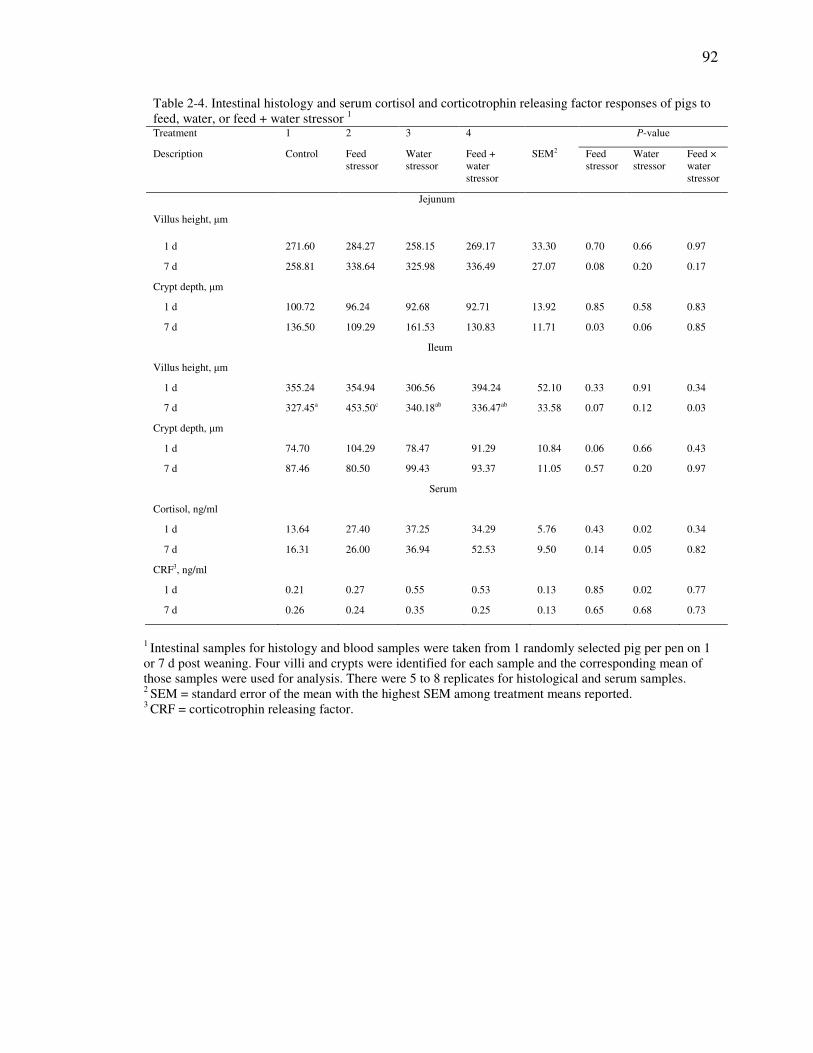
92
Table 2-4. Intestinal histology and serum cortisol and corticotrophin releasing factor responses of pigs to feed, water, or feed + water stressor 1 Treatment 1 2 3 4 P-value
Description Control Feed stressor
Water stressor
Feed + water stressor
SEM2 Feed stressor
Water stressor
Feed × water stressor
Jejunum
Villus height, µm
1 d 271.60 284.27 258.15 269.17 33.30 0.70 0.66 0.97
7 d 258.81 338.64 325.98 336.49 27.07 0.08 0.20 0.17
Crypt depth, µm
1 d 100.72 96.24 92.68 92.71 13.92 0.85 0.58 0.83
7 d 136.50 109.29 161.53 130.83 11.71 0.03 0.06 0.85
Ileum
Villus height, µm
1 d 355.24 354.94 306.56 394.24 52.10 0.33 0.91 0.34
7 d 327.45a 453.50c 340.18ab 336.47ab 33.58 0.07 0.12 0.03
Crypt depth, µm
1 d 74.70 104.29 78.47 91.29 10.84 0.06 0.66 0.43
7 d 87.46 80.50 99.43 93.37 11.05 0.57 0.20 0.97
Serum
Cortisol, ng/ml
1 d 13.64 27.40 37.25 34.29 5.76 0.43 0.02 0.34
7 d 16.31 26.00 36.94 52.53 9.50 0.14 0.05 0.82
CRF3, ng/ml
1 d 0.21 0.27 0.55 0.53 0.13 0.85 0.02 0.77
7 d 0.26 0.24 0.35 0.25 0.13 0.65 0.68 0.73
1 Intestinal samples for histology and blood samples were taken from 1 randomly selected pig per pen on 1 or 7 d post weaning. Four villi and crypts were identified for each sample and the corresponding mean of those samples were used for analysis. There were 5 to 8 replicates for histological and serum samples. 2 SEM = standard error of the mean with the highest SEM among treatment means reported. 3 CRF = corticotrophin releasing factor.

93
Table 2-5. Jejunal gene expression responses of pigs to feed, water, or feed + water stressor 1
Treatment 1 2 3 4 P-value
Description Control Feed stressor
Water stressor
Feed + water stressor
SEM2 Feed stressor
Water stressor
Feed × water stressor
MUC 2 (arbitrary units)
1d3 3.76 4.09 3.82 5.91 2.11 0.58 0.05 0.97
7 d 4.75 4.44 0.84 0.58 1.77 0.88 0.05 0.95
TNF-α (arbitrary units)
1 d 4.05a 0.45b 1.10b 1.31b 0.73 0.01 0.07 < 0.01
7 d 2.44 1.18 1.04 1.04 0.52 0.12 0.06 0.12
IL-6 (arbitrary units)
1 d3 2.58 0.44 5.73 2.70 2.09 0.07 0.26 0.73
7 d 13.08 8.56 10.01 6.23 5.66 0.94 0.23 0.39
CL-1 (arbitrary units)
1 d 1.35 0.89 1.35 2.24 1.06 0.81 0.46 0.46
7 d 0.67 0.14 0.41 0.49 0.15 0.13 0.70 0.07
OC (arbitrary units)
1 d 0.93 1.41 0.56 0.54 0.24 0.34 0.02 0.29
7 d 1.45 0.78 1.14 1.15 0.54 0.49 0.95 0.48
ZO-1 (arbitrary units)
1 d 1.30 1.81 1.26 1.07 0.32 0.60 0.21 0.27
7 d 2.87 1.99 1.99 2.27 0.97 0.71 0.72 0.48
1 Mucosa samples were taken from 1 randomly selected pig per pen for 4 to 7 replicates on 1 or 7 d post weaning. The housekeeping gene used was GAPDH. 2 SEM = standard error of the mean with the highest SEM among treatment means reported. 3P-value from transformed data using a log transformation.
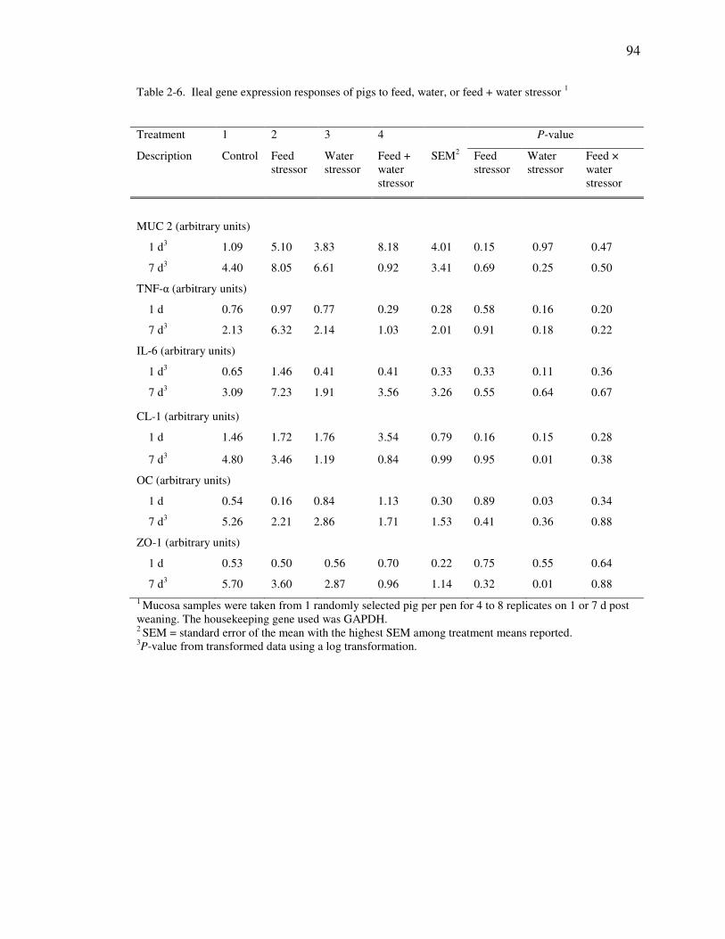
94
Table 2-6. Ileal gene expression responses of pigs to feed, water, or feed + water stressor 1
Treatment 1 2 3 4 P-value
Description Control Feed stressor
Water stressor
Feed + water stressor
SEM2 Feed stressor
Water stressor
Feed × water stressor
MUC 2 (arbitrary units)
1 d3 1.09 5.10 3.83 8.18 4.01 0.15 0.97 0.47
7 d3 4.40 8.05 6.61 0.92 3.41 0.69 0.25 0.50
TNF-α (arbitrary units)
1 d 0.76 0.97 0.77 0.29 0.28 0.58 0.16 0.20
7 d3 2.13 6.32 2.14 1.03 2.01 0.91 0.18 0.22
IL-6 (arbitrary units)
1 d3 0.65 1.46 0.41 0.41 0.33 0.33 0.11 0.36
7 d3 3.09 7.23 1.91 3.56 3.26 0.55 0.64 0.67
CL-1 (arbitrary units)
1 d 1.46 1.72 1.76 3.54 0.79 0.16 0.15 0.28
7 d3 4.80 3.46 1.19 0.84 0.99 0.95 0.01 0.38
OC (arbitrary units)
1 d 0.54 0.16 0.84 1.13 0.30 0.89 0.03 0.34
7 d3 5.26 2.21 2.86 1.71 1.53 0.41 0.36 0.88
ZO-1 (arbitrary units)
1 d 0.53 0.50 0.56 0.70 0.22 0.75 0.55 0.64
7 d3 5.70 3.60 2.87 0.96 1.14 0.32 0.01 0.88
1 Mucosa samples were taken from 1 randomly selected pig per pen for 4 to 8 replicates on 1 or 7 d post weaning. The housekeeping gene used was GAPDH. 2 SEM = standard error of the mean with the highest SEM among treatment means reported. 3P-value from transformed data using a log transformation.

95
Figure 2-1. Effect of acute weaning stressors on ileal mast cell density. Mast cell counts were performed for 6 pigs per treatment and are expressed as the number of toluidine-blue positive cells per mm2 tissue. On 1 d post-weaning P = 0.58, 0.18, and 0.30 for the effects of the feed stressor, water stressor, and feed × water interaction, respectively. On 7 d post-weaning P = 0.10, 0.95, and 0.53 for effects of the feed stressor, water stressor, and feed x water interaction, respectively.
100
150
200
250
300
350
400
d 1 d 7
cell
s/m
m2
Day post weaning
Control
Feed stressor
Water stressor
Feed + water
stressor

96
CHAPTER 3. IMPACT OF ACUTE FEED AND WATER DEPRIVATION AT WEANING AND SUBSEQUENT HEAT STRESS EVENT ON GROWTH PERFORMANCE, SERUM STRESS MARKERS AND ILEAL MUCOSA
CHARACTERISTICS IN NURSERY PIGS
3.1. Abstract
The current experiment was conducted to investigate the impact of a feed and
water deprivation event at weaning and subsequent heat stress event on growth
performance, serum stress markers, and ileal characteristics in nursery pigs. Mixed-sex
pigs were allotted on the basis of IBW (7.0 kg ± 0.89) in a RCBD with treatments in a
split-plot arrangement and consisting of the whole-plot factor of with or without a 24-h
feed + water deprivation event at weaning and the sub-plot factor of with or without a
cyclic 3-d heat stress event starting 27 d post-weaning. Growth performance was
measured throughout the experiment and on 1, 27, and 30 d post-weaning one pig from
each pen was selected, blood was collected for measurement of serum cortisol,
corticotrophin releasing factor (CRF), and endotoxins, and a segment of the mid ileum
was excised for morphological measurements. Furthermore, an ileal mucosal scraping
was taken and gene expression of mucin (MUC2), tumor necrosis factor alpha (TNF-α),
glucose transporter 2 (GLUT2), claudin 1 (CL-1), occludin (OC), and zonula occludens
1 (ZO-1) was measured by RT PCR. Feed + water deprivation reduced ADG (P < 0.01)
1 d post-weaning and body weights (P < 0.05) up to 27 d post-weaning. At 30 d

97
post-weaning, there was both a reduction due to heat stress and interaction between the
stress events on ADG and ADFI (P < 0.05). There was an increase (P < 0.05) in serum
CRF and endotoxins and a tendency for an increase (P = 0.09) in serum cortisol due to
the deprivation event 1 d post-weaning. There was a reduction in villous height to crypt
depth ratio (P = 0.05) due to the deprivation event 1 d post-weaning and an interaction
between the stress events (P = 0.05) for crypt depth on d 30 post-weaning. Gene
expression of GLUT 2 tended to decrease (P = 0.09), CL-1 tended to increase (P =
0.10), and OC decreased (P = 0.05) due to the deprivation event 1 d post-weaning.
Expression of the OC gene decreased (P < 0.05) due to the deprivation event 27 d post-
weaning and OC and ZO-1 gene expression tended to decrease (P = 0.07) due to the
heat stress event 30 d post-weaning. Results from the current study show a link between
feed and water deprivation at weaning and subsequent heat stress response with
implications on growth, serum stress markers, and ileal characteristics.
3.2. Introduction
During the nursery phase, pigs are exposed to weaning, environmental
temperature, and dietary stressors that are linked to reduced performance and health and
gastrointestinal (GI) dysfunction (Pluske et al., 1997; Lalles et al., 2004; Smith et al.
2010). Previous research shows a connection between feed and water deprivation,
markers of stress, and GI dysfunction in weaned pigs (Pluske et al., 1997; Hu et al.,
2013; Horn et al., 2014). Brooks et al. (2001) showed that approximately 50 and 10 %
of pigs do not consume feed for the first 24- and 48-h post-weaning. Low post-weaning

98
feed intake has been shown to cause villus atrophy and crypt hyperplasia that
corresponds to short- and long-term negative impacts on growth, gut function, and
disease susceptibility (Pluske et al., 1997; Lalles et al., 2004; Wijtten et al., 2011).
Water intake can be limited in weaned pigs due to transport time or weaning-associated
lethargy although limited information exists on the effect of dehydration on nursery pig
GI function. Our lab recently showed that a 24-h post-weaning water deprivation event
resulted in reduced growth performance, increased serum stress markers, and changes in
markers of mucosal integrity (Horn et al., 2014). Heat stress in pigs is associated with a
reduction in performance, changes in metabolism, and reduced gut integrity (Pearce et
al., 2013a,b). Briefly, heat stress reduces blood flow to GI tissues for body temperature
regulation, which causes a hypoxic environment in the gut and reduces mucosal
integrity (Yan et al., 2006; Lambert, 2009; Yu et al., 2010l; Pearce et al. 2013a). In a
production setting, pigs are commonly exposed to multiple concurrent or subsequent
stressors, therefore it is necessary to investigate stressor interactions (Hyun et al., 1998).
In grow-finish pigs Morrow-Tesch et al. (1994) and Hyun et al. (1998) showed that
concurrent heat and social stressors impacted growth performance additively although
little information exists on the impact of subsequent stressors on growth performance
during the nursery phase.
Several nursery pig studies show a link between individual stressors and gut
function (Pluske et al., 1997; Pearce et al., 2013a; Horn et al., 2014), however a dearth
of information exists exploring interactions between stressors during the nursery phase.
Therefore, our objective was to investigate the impact of a 24-h feed and water
deprivation event at weaning followed by a cyclic, 3-d heat stress event starting at 27 d

99
post-weaning. We hypothesized that the stress events would affect growth performance,
gut morphology, serum stress markers, and intestinal cytokine and tight junctions. To
achieve our objectives we measured growth performance, serum cortisol, CRF, and
endotoxins, ileal morphology, and ileal mucosa cytokine and tight junction gene
expression.
3.3. Materials and methods
All animal procedures were approved by the JBS United Animal Care and Use
Committee.
Animals and treatments
A total of 260 PIC 337 × C29 weaned, mixed-sex pigs (initial BW = 7.0 ± 0.89
kg, 19 d old) were used in a randomized complete block design with BW as the
blocking factor. The pigs were housed in pens (1.21 × 1.11 m) equipped with one
fixed-nipple drinker and one space dry-box feeder (4.87 × 1.52 × 0.30 m). Room
temperature was 34.4°C at weaning and reduced by 0.5°C each subsequent day of the
trial until the heat stress event. Treatments were in a 2 × 2 split-plot arrangement with
the whole-plot factors consisting of with or without a 24-h feed + water deprivation
event at weaning and the sub-plot factor of with or without a cyclic 3-d heat stress event
starting 27 d post-weaning. There were 22 whole-plot replicate pens per treatment and
11 sub-plot replicate pens per treatment with 5 to 6 mixed-sex pigs per pen. The
deprivation event was similar as described by Horn et al. (2014). Briefly, the
deprivation event consisted of withholding of feed and water for a period of 24-h

100
following weaning. Following the stressor, pigs were allowed free access to feed and
water and were fed a mash-based feeding program that met or exceeded NRC
requirements (NRC, 2012) and consisted of starter, transition and late nursery diets for
7, 7, and 21 d post weaning, respectively (Table 3-1). The heat stress event consisted of
a cyclic, 8-h increase in room temperature to 35˚C (61% relative humidity) starting
from 27 to 29 d post-weaning. Evening and overnight room temperatures were allowed
to drop down to 29˚C during the 3-d heat stress period. Room temperature was
maintained at 23˚C (55% relative humidity) for pigs not exposed to the heat stress event.
Rectal temperatures were recorded from the same 2 pigs per pen 24-h before the heat
stress event, daily during the heat stress event midpoint, and 24-h following the heat
stress event. Pigs and feeders were weighed on 1, 7, 14, 21, 27, 30, and 35 d post-
weaning to calculate ADG, ADFI, and G:F for the respective growth phases. On 1, 27,
and 30 d post-weaning, 1 pig per pen was randomly selected, blood was collected via
venipuncture into serum collection tubes (CORVAC serum separator tube, Tyco
Healthcare Group LP, Mansfield, MA, USA), and subsequently euthanized via CO2
asphyxiation. Immediately following euthanasia, 10-cm segments of the ileum were
excised for histological and gene expression measurements.
Serum stress markers
Blood was collected at approximately the same time each day to minimize the
effect of diurnal rhythms. Following blood collection serum was separated via
centrifugation (20 min, 10,000 × g at 4°C) and stored at -80°C until analysis. Serum
levels of CRF and cortisol were determined using commercial ELISA kits (CRF:
Phoenix Pharmaceuticals, Belmont, CA; cortisol: R&D systems, Minneapolis, MN).

101
Serum endotoxin concentrations were measured by chromogenic assay using the Pierce
LAL Chromogenic Endotoxin Kit (Fisher Scientific, Pittsburgh, PA). Briefly, the serum
samples were heat shocked for 15 min at 70˚C and diluted 100 fold in pyrogen-free
water. Serum endotoxins catalyze cleavage of p-nitroaniline (PNA) from Ac-Ile-Glu-
Ala-Arg-PNA, which forms a colormetric substrate that is measured at 410 nm.
Therefore, the amount of PNA detected by colormetric assay is proportional to the
amount of endoxotin present in the sample, which was interpolated from a standard
curve.
Histological measurements
Excised intestinal segments were flushed in ice-cold 10% phosphate-buffered
saline, stapled to a cardboard background, and fixed in 10% buffered formalin for
approximately 30 d. Samples were subsequently dehydrated with ethanol, cleared with
Sub-X® (Polysciences, Inc., Warrington, PA) and placed in paraffin (Polyfin paraffin,
Sigma Polysciences, St. Louis, MO). Segments were sliced (5 µm) and stained with
hematoxylin and eosin by the Purdue Histology and Phenotyping Laboratory (Purdue
University, West Lafayette, IN USA). Villus height and crypt depth were measured and
villus height to crypt depth ratio was calculated. Means from at least 4 villi per segment
were analyzed for differences. Villus length is defined as the length from the villus tip
to the valley between villi and crypt depth is defined as the length between the crypt
opening and base.
RNA isolation, cDNA Synthesis, and real-time PCR
Immediately after removal, intestinal segments were cut in half, exposing the
lumen, and mucosal contents were scraped with a metal spatula. Mucosal contents were
102
immediately placed in 5 mL of Trizol reagent (Invitrogen, Grand Island, NY) and stored
at -80°C until RNA isolation. Extraction of RNA was according to the Trizol protocol
(Invitrogen, Grand Island, NY). Reverse transcription was carried out using the
MMLV reverse transcription system of Promega and real-time PCR was performed on a
Bio-Rad iCyler using a reaction mix that consisted of 0.50 µg cDNA, 0.075 nmol of
each forward and reverse primers, SYBR Green Master (Roche, Basel, Switzerland),
and nuclease-free water (Ambion, Austin, TX) to reach a reaction volume of 20 µl per
well. Following a 5-min incubation period at 95ºC, reactions were cycled 50 times
using the following protocol: 10s at 95ºC, 20 s at 55ºC, and 72ºC for 30 s. Gene
expression of mucin (MUC2), glucose transporter 2 (GLUT2), tumor necrosis factor
alpha (TNF-α), claudin 1 (CL-1), occludin (OC), and zonula occludens 1 (ZO-1) were
measured using glyceraldehyde-3-phosphate dehydrogenase (GAPDH) as a
housekeeping control. Primers used are presented in Table 3-2.
Statistical analysis
Data was analyzed using the MIXED procedure of SAS (SAS Inst., Inc., Cary,
NC) appropriate for a split-plot design with the whole-plot factor of feed + water
deprivation event and sub-plot factor of heat stress event. Simple effect means were
separated using the PDIFF option in SAS where there was an interaction of whole- and
sub-plot factors. Significant difference was defined as P ≤ 0.05 and 0.05 ≤ P ≤ 0.10 was
considered a tendency.

103
3.4. Results
There were no treatment-specific instances of mortality or morbidity throughout
the trial. Furthermore, there was no treatment effect (P < 0.10) on daily scour scores.
Rectal temperatures were recorded 24-h before and after, and daily during the heat
stress event from two pigs per pen (data not shown). There was no effect of treatment
on rectal temperatures 24-h before or after the heat stress event. There was, on average,
a 0.35 ˚C daily increase (P < 0.05) in rectal temperature at the midpoint of the heat
stressor. Growth performance data are reported in Tables 3-3 and 3-4. All pigs lost
weight from 0 to 1 d post-weaning, although pigs exposed to the deprivation event were
0.23 kg lighter (P = 0.01) compared with non-stressed pigs. From 1 to 7 d post-
weaning, there was an 8.7% increase in ADG, but 9.1% decrease in ADFI (P < 0.05)
relative to non-stressed pigs due to the deprivation event. Bodyweights of feed + water-
deprived pigs continued to be lighter (by 0.60 kg) compared with non-stressed pigs (P =
0.03) on 27 d post-weaning. Immediately following the heat stress event (30 d post-
weaning) there was an effect of the sub-plot heat stress event and a stress event
interaction (P < 0.05) for ADG and ADFI and a tendency (P = 0.10) for FBW. Relative
to pigs exposed to no stress event, there was a 0.92, 0.65, and 1.92 kg decrease in FBW
at 30 d post-weaning for the heat stress only, feed + water deprivation only, and feed +
water stress and heat stress, respectively. There was no impact (P > 0.10) of stress
factors the week following the heat stress event (30 to 35 d post-weaning).
Cumulatively, there was a tendency for a feed + water deprivation and heat stress event

104
interaction (P = 0.10) for FBW and ADG and a heat stress event and stress event
interaction (P = 0.03) for ADFI.
Ileal morphology data are reported in Tables 3-3 and 3-4. The average villus
and crypt depth were 284 and 97 µm 1 d post-weaning, respectively. Villous height was
decreased (P = 0.03) by 27% but crypt depth was not impacted (P > 0.10) 1 d post-
weaning with feed+water deprivation. The average villus height and crypt depth were
354 and 220 µm and 323 and 180 µm on 27 and 30 d post-weaning, respectively. There
was no impact (P > 0.10) of treatment on ieal morphology 27 d post-weaning although
there was an interaction effect (P = 0.05) 30 d post-weaning. The interaction effect
shows that pigs exposed to no stress events had the deepest crypts.
Serum marker data are reported in Tables 3-5 and 3-6. Immediately following
the deprivation event (1 d post-weaning) there was a tendency (P = 0.09) for a 29 %
increase in serum cortisol and an 8.1 and 94 % increase (P < 0.05) in serum CRF and
endotoxins, respectively. Furthermore, on 27 d post-weaning there was a tendency (P =
0.07) for a 37% increase in serum endotoxins and on 30 d post-weaning, there was a
tendency (P = 0.08) for an increase in serum CRF due to the deprivation event.
Ileal mucosa gene expression analyses are presented in Tables 3-7 and 3-8.
Expression of MUC2, GLUT2, TNF-α, CL-1, OC, and ZO-1 genes were measured with
GAPDH as the housekeeping gene. There was no impact (P > 0.10) of treatment on
GADPH gene expression. On 1 d post-weaning there was a tendency for a decrease (P =
0.09) in GLUT2 gene expression, a tendency for an increase (P = 0.10) in CL-1 gene
expression, and a decrease (P = 0.05) in OC gene expression due to the deprivation

105
event. Furthermore, there was a decrease (P < 0.01) in OC gene expression due to the
deprivation event on 27 d post-weaning. On 30 d post-weaning, there was no impact of
the deprivation event on gene expression although there was a tendency for a decrease
(P = 0.07) in OC and ZO-1 gene expression due to the heat stress event.
3.5. Discussion
A relationship exists between stress events during the nursery period, short- and
long-term performance, and gastrointestinal function (Pluske et al., 1997; Lalles et al.,
2004; Smith et al., 2010). Within the first 24-h post-weaning it has been estimated that
approximately 50 % of pigs do not consume feed (Brooks et al., 2001), which is marked
by poor pig performance, increased disease susceptibility, and atrophy of intestinal
morphology (Pluske et al., 1997). Furthermore, dehydration is common during the
post-weaning period and in an experimental setting has been shown to be associated
with an increase in markers of stress and reduced gut integrity (Horn et al., 2014). The
impact of heat stress on feed intake and metabolism in growing pigs has been well
documented (Collin et al., 2001; Baumgard and Rhoads, 2011), however recent
literature shows heat stress also results in hypoxia-associated changes in GI integrity
and cellular absorptive properties (Yu et al., 2010; Pearce et al., 2013a; Pearce et al.,
2014). Previous research shows stressors are additive (Hyun et al., 1998) although there
is a dearth of information exploring the relationship between a heat stressor that is
subsequent to a post-weaning deprivation stressor in nursery pigs. Because stress
events commonly occur concurrent or subsequent to each other it is critical to

106
understand stressor interactions. Furthermore, an understanding of the impact of
common nursery pig stressors on performance and GI function will allow for
development of management and nutritional strategies to help mitigate the stress-
induced negative effects.
For the current experiment there was no mortality and fall-off pigs (visual
morbidity) was below 5%. Immediately following weaning all pigs lost weight
although the pigs exposed to the deprivation event had 4% lower BW compared to
control pigs. During the first 1 to 3 d following weaning it is common for nursery pigs
to lose weight and this period has been referred to as the “transient growth check”
period as reported by Pluske et al. (1997) and Lalles et al. (2004). During the period
following the deprivation event (2 to 7 d post-weaning) pigs exposed to the deprivation
event had improved gains although their BW remained 0.13 kg lighter compared to
control pigs. The improvement in gain during the immediate period following the
stressor is related to water gorging and is similar to a previous experiment reported by
our lab (Horn et al., 2014). However, at 27 d post-weaning the pigs exposed to the 24-h
post-weaning deprivation event remained 0.60 kg lighter compared to control pigs.
Following the 3-d, cyclic heat stress event there was a 16 and 20% reduction in ADG
and ADFI, respectively, which resulted in a 1.1 kg lighter pig. Furthermore, an
interaction existed that shows pigs exposed to both the deprivation and heat stress
events had lower ADFI compared to other treatment groups. The interaction carried
over cumulatively and shows the pigs exposed to both stress events have the lowest
overall gain and ADFI. It has been well documented that pigs are susceptible to heat
stress due to lack of sweat glands which corresponds to increased basal metabolic rate

107
and urination, and decreased feed intake (D’Allaire et al., 1996). Increased water losses
along with heat-induced increases in appetite-suppressing neuropeptides such as ghrelin
are largely responsible for attenuated pig performance (Pearce et al., 2014). There is
limited information on the impact a cyclic heat stress event in nursery pigs, therefore
comparing results from the current trial to other published studies is limited. Pearce et al.
(2013b) showed that in 46-kg pigs exposed to a constant, 24-h heat stress (35.5˚C) there
was a 1.6˚C increase in body temperature and a 5 and 53% reduction in BW and FI,
respectively. Kerr et al. (2005) reported a 23 and 14% reduction in gain and ADFI in
pigs exposed to a 36-d heat stressor (33˚C), whereas Hyun et al. (1998) reported
approximately a 10% reduction in ADG and ADFI during a 4-wk heat stressor (34˚C).
A dearth of information exists on the interactive effects of subsequent stressors in
nursery pigs although Hyun et al. (1998) reported that a stressor interaction existed in
pigs concurrently exposed to heat and social stress.
Poor feed intake during the immediate post-weaning period results in limited
energy for the rapidly developing gut which has been shown to reduce intestinal villus
height and GI function (Pluske et al., 1997). For the current experiment, there was a 27%
reduction in ileal villus height immediately following the deprivation event although
there was recovery in deprivation-induced morphological effects by 27 d post-weaning.
Previous studies reported little impact of nursery pig stress events on intestinal
morphology (Moeser et al., 2007) and it is not clear why there was such a drastic effect
of the deprivation event for the current study. Reduced villus height indicates reduced
potential for nutrient utilization during the first week post-weaning along with increased
susceptibility to enteric pathogens (Lalles et al., 2004) and further research is warranted

108
to better understand the practical implications of these results. There was no impact of
the heat stress event on ileal morphology following the 3-d event although there was a
stressor interaction that shows pigs not exposed to any stressor had the deepest crypts.
During heat stress, blood flow to GI tissues is limited, which limits oxygen and nutrient
delivery and results in limited gut tissue growth or atrophy (Yan et al., 2006; Pearce et
al. 2013a). Pearce et al. (2013a and 2014) showed that when growing pigs were
exposed to a constant heat stressor there was a reduction in villus height and an increase
in crypt depth within 24-h following initiation of the stressor. Conversely, Yu et al.
(2010) reported in pigs that morphological changes were not shown until 3 d into a
cyclic heat stressor. For the current experiment it is likely that there was no impact of
the heat stressor on ileal morphology due to its cyclic and limited nature. The stressor
interaction on crypt depth indicates increased cellular proliferation and further work
measuring molecular markers of proliferation and differentiation are needed to better
understand these results.
Previous studies have shown that serum CRF and cortisol are markers of nursery
pig stress and are linked to stress-induced changes in growth and gastrointestinal
function (Moeser et al. 2007; Smith et al., 2010; Horn et al., 2014). For the current
study there were 28 and 8% increases in serum cortisol and CRF immediately following
the post-weaning deprivation event. The data reported in the current study are
consistent with reports by Smith et al. (2010) and Horn et al. (2014). Stress-induced
changes in central and peripheral CRF release have been shown to increase intestinal
mast cell activation and recruitment and can be a contributing factor to inappropriate
intestinal secretion of TNF-α (Overman et al., 2012). Changes in mucosal immunity can

109
alter enterocyte tight junction protein structure and subsequently increase intestinal
permeability (Lambert, 2009). Glucocorticoids are a critical part of the stress-
adaptation response and drive stress-induced metabolic changes. There was no impact
of the heat stress event or a stress event interaction for the current study. Yu et al. (2010)
reported an increase in serum cortisol each day following a cyclic heat stressor using a
mini pig model. Compared to Yu et al. (2010), pigs in the current study were exposed to
a lower degree of heat stress (35˚C vs. 40˚C) for a longer duration (8 h vs. 5 h) which
could explain the lack of change in serum cortisol levels. Under normal conditions,
bacterial cell wall components, such as endotoxins, should be very low in the serum due
to properly functioning physical barriers. An increase in serum endotoxin level can lead
to endotoxemia and activation of immune and hepatic detoxification mechanisms (Hall
et al., 2001). Clinically, endotoxemia can manifest as increased body temperature,
reduced feed intake, lethargy, and in severe cases septic shock and death. Furthermore,
it has been well documented that changes in serum endotoxin level are related to
reduced intestinal integrity in rodents, chickens, pigs, and humans (Bouchama et al.,
1991; Hall et al., 2001; Cronje et al., 2007; Pearce et al., 2013a,b). For the current
study, there was a 16-fold increase in serum endotoxin levels due to water deprivation
event 1 d post-weaning. Furthermore, there was a 1.6-fold increase in serum endotoxins
due to the deprivation event 27 d post-weaning. The increase in serum endotoxins at 27
d post-weaning is not clear since it does not correlate with any changes in intestinal
integrity. It may be possible that the aforementioned changes are related to a stressor-
induced microbial population shift rather than changes in intestinal integrity and
warrants further investigation from a microbiology perspective. Previous research

110
shows an increase in serum endotoxin levels in growing pigs exposed to a constant heat
stress event are linked to increased intestinal permeability (Pearce et al., 2013a; Pearce
et al., 2014). For the current study there was no impact of the heat stress event or a
stress event interaction on serum endotoxin levels. The discrepancy between the current
study and previous reports is likely due to the duration and intensity of the heat stress
event.
Tight junction protein complexes form a selective pore between epithelial cells
and can be influenced by stress, cytokines, and proteolytic enzymes (Li et al., 2012).
Hu et al. (2013) demonstrated in pigs that changes in tight junction gene expression
were related to weaning stress-induced changes in gut permeability. Changes in tight
junction gene and protein expression are driven by myosin light chain kinase (MLCK)
actions on cytoskeletal proteins (Lambert, 2009). Serum stress mediators, cytokines,
and hypoxia have been shown to be inducers of MLCK and are related to loss of
intestinal integrity (Overman et al., 2012; Pearce et al., 2013a). For the current study
there was a decrease in gene expression of the tight junction protein OC immediately
following the deprivation stress event that was maintained up to 27 d post-weaning.
These results are consistent with those reported by our lab in a previous experiment
using a deprivation stressor (Horn et al., 2014) and by those reported by Hu et al. (2013)
when pigs were exposed to general weaning stress. Previous research shows a link
between inflammatory cytokines and changes in tight junction gene expression (Ma et
al., 2005) although similar results were not observed for the current trial. The
discrepancy between the current experiment and the literature may be due to sampling
time and stress event intensity. As previously mentioned, during heat stress changes in

111
blood flow lead to a hypoxic environment in the gut resulting in limited delivery of
energy and nutrients to enterocytes (Yan et al., 2006). Resulting consequences include
changes in tight junction structure and increased intestinal permeability. For the current
study we showed that at the end of the cyclic, 3-d heat stress event there was a decrease
in expression of tight junction genes OC and ZO-1. Pearce et al. (2013a) showed a
decrease in ZO-1 gene expression and an increase in OC gene expression at the end of a
constant, 3-d heat stressor in growing pigs. Furthermore, it has been reported that tight
junction gene expression patterns change throughout a heat stress event (Pearce et al.,
2013a). The discrepancy between OC gene expression results for the current study and
previous studies are likely related to sample timing. Mucins are a major component of
the mucus layer and serve an integral role in GI function and protection (Atuma et al.,
2001; Horn et al., 2008). For the current experiment there was no impact of the
deprivation stressor on MUC2 gene expression, which is consistent with results reported
by Horn et al. (2014). Limited information exists on the impact of heat stress on mucin
dynamics in pigs. Pearce et al. (2014) reported an increase in MUC2 gene expression
during a heat stressor and the authors suggested the aforementioned increase was a
protective mechanism. Further work is needed to understand gastrointestinal secretions
over the time course of a heat stressor. During psychological stress, glucocorticoids
have been shown to decrease GLUT2 activity and abundance (Shepherd et al., 2004).
Consistently, we observed a 2.7-fold decrease in GLUT2 gene expression immediately
following the deprivation event. Shepherd et al. (2004) suggested the decrease in
GLUT2 is a mechanism to prevent intestinal counteraction of the lytic actions of
cortisol during stress. Conversely, during a heat stress event an increase in intestinal

112
glucose transport is believed to be a protective mechanism to minimize cellular damage
(Kellet and Brot-Laroche, 2005). For the current study, there was no change in GLUT 2
gene expression due to the heat stress event which is consistent with results reported by
Pearce et al. (2013b). It is possible that increases in heat-stress associated glucose
uptake are mainly mediated by SGLT-1 due to the ability of the transporter to
cotransport water and Na along with glucose (Wright and Loo, 2006). There were no
stressor interactions observed for ileal mucosa gene expression markers which is likely
related to the limited and cyclic nature of the heat stress event and timing of sampling
for the current study.
A 24-h feed and water deprivation event at weaning has short- and long-term
implications on growth performance and results in a 0.60-kg lighter pig up to 27 d post-
weaning. During the heat stress event, weight gain and feed intake were reduced and a
feed + water deprivation by heat stressor interaction existed that showed pigs exposed
to both stress events subsequently had the poorest performance. Furthermore, the
deprivation event increased serum stress markers, negatively affected ileal morphology,
and altered ileal mucosa genes related to nutrient absorption and cellular integrity. The
heat stress event did not impact serum stress markers or ileal morphology, although
tight junction gene expression was altered. These results show that nursery pig
performance is impacted by subsequent stress events and therefore special focus should
be given to developing nutritional and management strategies that help mitigate stress
during the nursery phase.

113
3.6. Acknowledgements
The authors would like to express their appreciation to JBS United, Inc. and
Biomatrix International for their financial and technical support of this trial. Further
appreciation is given to the employees of Burton Russell Farm at JBS United.

114
3.7. References
Atuma, C., V. Strugala, A. Allen, and L. Holm. 2001. The adherent gastrointestinal
mucus gel layer: thickness and physical state in vivo. Am. J. Physiol. 280:
G922-929.
Baumgard, L.H., and R.P. Rhoads. 2011. Effects of environment on metabolism. In:
Collier, R.J., editor. Environmental Physiology: John Wiley and Sons, Inc.
Bouchama, A., R. Parhar, A. el-Yazigi, K. Sheth, and S. al-Sedairy. 1991. Endotoxemia
and release of tumor necrosis factor and interleukin 1 alpha in acute heatstroke. J.
Appl. Physiol. 70:2640–2644.
Brooks, P.H., C.A. Moran, J.D. Beal, V. Demeckova, and A. Campbell.
2001. Liquid feeding for the young piglet, in: Varley M.A., Wiseman J.
(Eds.), The weaner pig: nutrition and management, CAB International,
Wallingford, UK, pp. 153 -178.
Collin, A., J. van Milgen, and J. Le Dividich. 2001. Modeling the effect of high,
constant temperature on food intake in young growing pigs. J. Anim. Sci. 72:
519–529.
Cronje, P. B. 2007. Gut health, osmoregulation and resilience to heat stress in poultry.
Aust. Poult. Sci. Symp. 19: 9–13.
D’Allaire, S., R. Drolet, and D. Brodeur. 1996. Sow mortality associated with high
ambient temperatures. Can. Vet. J. 37: 237–239.

115
Hall, D. M., G. R. Buettner, L. W. Oberley, L. Xu, R. D. Matthes, and C. V. Gisolfi.
2001. Mechanisms of circulatory and intestinal barrier dysfunction during whole
body hyperthermia. Am. J. Physiol. Heart Circ. Physiol. 280:H509–H521.
Horn, N., F. Ruch, G. Miller, K. M. Ajuwon, and O. Adeola. 2014. Impact of acute
water and feed deprivation events on growth performance, intestinal
characteristics, and serum stress markers in weaned pigs. J. Anim. Sci. 92:
4407-4416.
Hu, C.H., K. Xiao, Z.S. Luan, and S. Jong. 2013. Early weaning increases intestinal
permeability, alters expression of cytokine and tight junction proteins, and activates
mitogen-activated protein kinases in pigs. J. Anim. Sci. 91: 1093-1101.
Hyun, Y., M. Ellis, G. Riskowski, and R.W. Johnson. 1998. Growth performance of pigs
subjected to multiple concurrent environmental stressors. J. Anim. Sci. 76: 721-
727.
Kellett, G.L. and E. Brot-Laroche. 2005. Apical GLUT2: A Major Pathway of
Intestinal Sugar Absorption. Diabetes. 54: 3056-3062.
Kerr, B.J., J. T. Yen, J. A. Nienaber, and R. A. Easter. 2003. Influences of dietary
protein level, amino acid supplementation and environmental temperature on
performance, body composition, organ weights and total heat production of
growing pigs. J. Anim. Sci. 81: 1998-2007.
Lalles, J.P., G. Boudry, C. Favier, N. LE floc’h, I. Luron, L. Montagne, I.P.
Oswald, S. Pie, C. Piel, and B. Seve. 2004. Gut function and
dysfunction in young pigs: physiology. Anim. Res. 53:301–316.

116
Lambert, G.P. 2009. Stress-induced gastrointestinal barrier dysfunction and
its inflammatory effects. J. Anim. Sci. 87 (E suppl.): E101-E108.
Li, X., S. Akhart, and M.A. Choudry. 2012. Alteration in intestine tight
junction protein phosphorylation and apoptosis is association with
increase in IL-18 levels following alcohol intoxication and burn injury.
Biochem. Biophys. Acta. 1822: 186-203.
Ma, T., M.A. Biovin, D. Ye, A. Pedram, and H.M. Said. 2005. Mechanism
of TNF-α modulation of Caco-2 intestinal epithelial tight junction barrier:
Role of myosin light-chain kinase protein expression. Am. J. Physiol.
288: G422-G430.
Moeser, A.J., C.V. Klok, K.A. Ryan, J.G. Wooten, and D. Little. 2007.
Stress signaling pathways activated by weaning mediate intestinal
dysfunction in the pig. Am. J. Physiol. Gastrointest. Liver Physiol. 292:
G173–G181.
Morrow-Tesch, J. L., J. J. McGlone, and J. L. Salak-Johnson. 1994. Heat and social
effects pig immune parameters. J. Anim. Sci. 72: 2599-2609.
NRC, 2012. Nutrient requirements of Swine. 11th rev ed. Natl. Acad. Press,
Washington, D.C.
Overman, E.L., J.E. Rivier, and A.J. Moeser. 2012. CRF Induces Intestinal
Epithelial Barrier Injury via the Release of Mast Cell Proteases and
TNF-a. PLOS ONE. 7(6): e39935.

117
Pearce, S.C., V. Mani, T. E. Weber, R. P. Rhoads, J. F. Patience, L. H. Baumgard, and
N. K. Gabler. 2013a. Heat stress and reduced plane of nutrition decreases
intestinal integrity and function in pigs. J. Anim. Sci. 91: 5183-5193.
Pearce, S.C., V. Mani, R. L. Boddicker, J. S. Johnson, T. E. Weber, J. W. Ross, R. P.
Rhoads, L. H. Baumgard, and N. K. Gabler. 2013b. Heat stress reduces
intestinal barrier integrity and favors intestinal glucose transport in growing pigs.
PLOS ONE. 8(8): e70215.
Pearce, S.C., M. V. Sanz-Fernandez, J. H. Hollis, L. H. Baumgard, and N. K. Gabler.
2014. Short-term exposure to heat stress attenuates appetite and intestinal
integrity in growing pigs. J. Anim. Sci. 92: 5444-5454.
Pluske, J.R., D.J. Hampson, and I.H. William. 1997. Factors influencing the
structure and function of the small intestine in the weaned pig: a review,
Livest. Prod. Sci. 51: 215–236.
Shepherd, E.J., P.A. Helliwell, O.J. Mace, E.L. Morgan, N. Patel, and G.L. Kellett.
2004. Stress and glucocorticoid inhibit apical GLUT2-trafficking and intestinal
glucose absorption in rat small intestine. J. Physiol. 561(1): 281-290.
Smith, F., J.E. Clark, B.L. Overman, C.C. Tozel, J.H. Huang, J.E. Rivier, A.T.
Blikslager, and A.J. Moeser. 2010. Early weaning stress impairs development of
mucosal barrier function in the porcine intestine. Am. J. Physiol. Gastrointest.
Liver Physiol. 298(3):G352-G363.
Wijtten, P.J., J. van der Meulen, and M.W. Verstegen. 2011. Intestinal barrier
function and absorption in pigs after weaning: a review. Br. J.
Nutr.105(7): 967-81.

118
Wright, E.M., and D.D. Loo. 2000. Coupling between Na+, sugar, and water transport
across the intestine. Ann. N.Y. Acad. Sci. 915: 54–66.
Yan, Y., Y. Zhao, H. Wang, and M. Fan. 2006. Pathophysiological factors underlying
heatstroke. Med. Hypotheses 67: 609–617.
Yang, P.C., S.H. He, and P.Y. Zheng. 2007. Investigation into the signal transduction
pathway via which heat stress impairs intestinal epithelial barrier function.
Gastroentero. 22: 1883-1831.
Yu, J., P. Yin, F. Liu, G. Cheng, K. Guo, A. Lu, X. Zhu, W. Luan, J. Xu. 2010. Effect
of heat stress on the porcine small intestine: A morphological and gene
expression study. Comp. Biochem. Physiol. A. 156: 119-128.

119
Table 3-1. Ingredient composition of diets
Item Phase 1 Phase 2 Phase 3
Ingredients, g/kg
Corn 450.5 522.8 690.1
Soybean meal 225.0 275.0 275.0
Whey permeate 200.0 100.0 -
Plasma 60.0 20.0 -
Fish meal 38.0 63.0 -
Monocalcium phosphate1 9.0 6.0 9.0
Limestone2 7.5 5.0 10.0
Salt 2.5 3.5 6.5
Vitamin premix3 1.0 1.0 1.0
Mineral premix4 0.8 0.8 0.8
Selenium premix5 0.5 0.5 0.5
L-Lys HCL 2.8 1.4 4.3
DL-Met 1.7 1.0 1.5
L-Thr 0.7 - 1.3
Total 1,000 1,000 1,000
Calculated nutrients and energy, as-fed basis
ME, kcal/kg 3317.6 3297.8 3260.6
CP, g/kg 225.0 234.0 186.0
Ca, g/kg 8.6 8.1 5.9
P, g/kg 8.0 7.4 5.3
Nonphytate P, g/kg 6.4 5.5 3.3
Ca:P 1.08 1.09 1.12
SID Lys, g/kg 13.9 13.0 12.1
1 Contained 17% Ca and 21.1 % P.
2 Contained 38% Ca.
3 Vitamin premix per kilogram of diet: vitamin A, 5,090 IU; vitamin D3 1,270 IU ; vitamin E 22 IU, vitamin K activity 7.2 mg; menadione, 2,412 µg; vitamin B12, 22.4 µg; riboflavin 8.11 mg; d-pantothenic acid 61.20; and niacin 106.59 mg.
4 Mineral premix supplied per kilogram of diet: Cu (as copper chloride), 18 mg; I (as ethylenediamine dihydroiodide) 0.95 mg; Fe (as iron carbonate) 334 mg; Mn (as manganese oxide) 60 mg; and zinc (as zinc oxide) 150 mg.
5 Supplied as 300 µg of Se per kilogram of diet.

120
Table 3-2. Primers used for RT-PCR
Gene1 Primer (5`-3`)
GAPDH (forward) GGGCATGAACCATGAGAAGT
GAPDH (reverse) TGTGGTCATGAGTCCTTCCA
MUC2 (forward) CAACGGCCTCTCCTTCTCTGT
MUC2 (reverse) GCCACACTGGCCCTTTGT
GLUT2 (forward) CTGCTTGGTCTATCTGCTGTG
GLUT2 (reverse) TTGATGCTTCTTCCCTTTCTTT
TNF-α (forward) ATGGGCCCCCAGAAGGAAGAG
TNF-α (reverse) GATGGCAGAGAGGAGGTTGAC
CL-1 (forward) TACTTTCCTGCTCCTGTC
CL-1 (reverse) AAGGCGTTAATGTCAATC
OC (forward) ATCAACAAAGGCAACTCT
OC (reverse) GCAGCAGCCATGTACTCT
ZO-1 (forward) GAGTTTGATAGTGGCGTT
ZO-1 (reverse) GTGGGAGGATGCTGTTGT
1GAPDH= glyceraldehyde-3-phosphate dehydrogenase, MUC2= mucin 2, GLUT2= glucose transporter 2, TNF-α= tumor necrosis factor α, CL-1= claudin 1, OC= occludin, and ZO-1= zonula occludens 1.

121
Table 3-3. Growth performance and ileal morphology responses of pigs exposed to a 24-h post-weaning feed and water deprivation event
1 SEM= standard error of the mean.
2 There were 20 to 22 replicates per treatment with 5 to 6 pigs per pen at the start of the trial.
3 There were 12 to 16 replicates per treatment.
4 villous height to crypt depth ratio.
Treatment
No feed and water deprivation
Feed and water deprivation
SEM1 P-value
Growth performance2
IBW, kg 6.99 6.95 0.19 0.32 0 to 1 d post-weaning FBW, kg 6.84 6.58 0.18 < 0.01 ADG, kg -0.16 -0.39 0.01 < 0.01 ADFI, kg 0.04 0.00 < 0.01 < 0.01 G:F, kg:kg -4.00 0.00 < 0.01 < 0.01 1 to 7 d post-weaning FBW, kg 8.14 8.01 0.19 0.01 ADG, kg 0.21 0.23 0.01 0.01 ADFI, kg 0.22 0.20 0.01 0.02 G:F, kg:kg 0.96 1.20 < 0.01 < 0.01 7 to 14 d post-weaning FBW, kg 10.26 10.02 0.23 0.07 ADG, kg 0.31 0.30 0.01 0.53 ADFI, kg 0.35 0.34 0.01 0.38 G:F, kg:kg 0.89 0.89 0.01 0.97 14 to 21 d post-weaning FBW, kg 13.61 13.13 0.27 0.01 ADG, kg 0.50 0.45 0.02 0.04 ADFI, kg 0.67 0.62 0.01 0.01 G:F, kg:kg 0.73 0.73 0.01 0.77 21 to 27 d post-weaning FBW, kg 17.30 16.71 0.31 0.03 ADG, kg 0.62 0.59 0.02 0.08 ADFI, kg 0.91 0.87 0.02 0.15 G:F, kg:kg 0.68 0.67 0.01 0.33 Ileal morphology3 1 d post-weaning Villous height, µm 328 241 31.67 0.03 Crypt depth, µm 93 101 6.80 0.38 VH:CD4 3.57 2.44 0.30 0.05 27 d post-weaning Villous height, µm 386 352 51.76 0.57 Crypt depth, µm 218 221 29.20 0.94 VH:CD4 1.80 1.75 0.30 0.92

122
122
122
Table 3-4. Growth performance and ileal histology responses of pigs exposed to a heat stress event subsequent to a post-weaning feed and water
deprivation event1
Treatment SEM2 P-value
Whole-plot factor
No feed or water deprivation
Feed and water deprivation
Whole-plot
Sub-plot
Interaction Whole-plot
Sub-plot
Interaction
Sub-plot factor
Heat stress
No heat stress
Heat stress
No heat stress
Growth performance3
27 to 30 d post-weaning FBW, kg 18.65 19.57 17.65 18.92 0.38 0.47 0.54 0.12 0.10 0.08 ADG, kg 0.56a 0.65ab 0.56a 0.69b 0.03 0.03 0.03 0.69 0.01 0.03 ADFI, kg 0.81a 0.98b 0.77a 0.99b 0.03 0.03 0.03 0.57 < 0.01 < 0.01 G:F, kg:kg 0.68 0.65 0.74 0.68 0.02 0.02 0.03 0.08 0.17 0.14 30 to 35 d post-weaning FBW, kg 21.54 22.77 21.08 22.32 0.41 0.52 0.56 0.38 0.16 0.11 ADG, kg 0.60 0.59 0.61 0.62 0.01 0.01 0.02 0.23 0.91 0.48 ADFI, kg 0.95 0.95 0.97 1.02 0.02 0.02 0.03 0.14 0.45 0.32 G:F, kg:kg 0.64 0.62 0.63 0.61 0.01 0.01 0.02 0.73 0.34 0.78 0 to 35 d post-weaning Gain, kg 15.27 15.53 14.10 15.36 0.32 0.36 0.44 0.14 0.16 0.10 ADG, kg 0.44 0.44 0.40 0.40 0.01 0.01 0.01 0.14 0.16 0.10 ADFI, kg 0.58ac 0.61c 0.56b 0.59ab 0.01 < 0.01 0.01 0.15 0.03 0.03 G:F, kg:kg 0.75 0.72 0.72 0.74 0.01 0.02 0.02 0.54 0.81 0.45 Ileal morphology4
30 d post-weaning Villous height, µm
318 329 309 336 18.10 17.22 27.33 0.93 0.43 0.87
Crypt depth, µm
156a 215b 179a 171a 11.58 10.79 15.35 0.44 0.14 0.05
VH:CD 5 2.11 1.59 1.76 1.97 0.13 0.13 0.19 0.88 0.51 0.19 1Difference in superscript denotes difference between simple means.
2SEM = standard error of the mean.
3There were 9 to 11 replicates per treatment with 4 to 6 pigs per pen.
4There were 6 to 8 replicates per treatment.
5 Villous height to crypt depth ra

123
Table 3-5. Serum measurements of pigs exposed to a 24-h post-weaning feed and water
deprivation event1
1 There were 9 to 12 replicates per treatment.
2 SEM= standard error of the mean.
3 CRF = corticotrophin releasing factor.
4 EU/mL = endotoxin units per mL.
Treatment
No feed and water deprivation
Feed and water deprivation
SEM2 P-value
1 d post-weaning Cortisol, ng/mL 40.53 57.46 6.32 0.09 CRF, ng/mL3 1.57 1.71 0.05 0.03 Endotoxins, EU/mL4 0.83 13.03 3.15 0.02 27 d post-weaning Cortisol, ng/mL 45.84 45.17 5.80 0.94 CRF, ng/mL3 2.04 2.04 0.02 0.77 Endotoxins, EU/mL4 37.04 58.68 8.81 0.07

124
122
124
Table 3-6. Serum measurements of pigs exposed to a heat stress event subsequent to a post-weaning feed and water deprivation event 1 Treatment SEM2
P-value
Whole-plot factor
No feed or water
deprivation
Feed and water deprivation
Whole-plot
Sub-plot
Interaction Whole-plot
Sub-plot
Interaction
Sub-plot factor Heat stress
No heat stress
Heat stress
No heat stress
30 d post-weaning
Cortisol, ng/mL
33.49 33.50 37.79 27.67 3.78 4.19 5.39 0.87 0.54 0.63
CRF, ng/mL3 1.64 1.66 1.68 1.74 0.02 0.02 0.03 0.08 0.26 0.51 Endotoxin, EU/mL4
7.04 14.53 13.56 15.51 3.49 3.32 5.47 0.38 0.25 0.53
1There were 4 to 8 replicates per treatment with 4 to 6 pigs per pen. 2SEM = standard error of the mean. 3 CRF= corticotrophin releasing factor. 4 EU/mL = endotoxin units per mL.

125
Table 3-7. Ileal mucosa gene expression of pigs exposed to a 24-h post-weaning feed and water
deprivation event1,2,3
Treatment
No feed and water deprivation
Feed and water deprivation
SEM4 P-value
1 d post-weaning
MUC25 0.33 1.79 0.70 0.24
GLUT25 3.20 1.18 0.95 0.09
TNF-α5 1.70 0.39 1.18 0.26
CL-1 1.56 1.92 0.82 0.10
OC5 3.06 0.72 0.71 0.05
ZO-15 1.67 1.54 0.82 0.95
27 d post- weaning
MUC25 2.80 2.15 1.81 0.14
GLUT2 1.71 0.99 0.48 0.33
TNF-α 3.04 0.84 0.55 0.22
CL-15 1.14 1.81 0.91 0.86
OC5 1.62 0.87 0.49 < 0.01
ZO-1 2.02 1.04 0.59 0.30 1 There were 4 to 8 replicates per treatment.
2 MUC2 = mucin; GLUT2 = glucose transporter 2; TNF-α = tumor necrosis factor alpha; CL-1 =
claudin 1; OC = occludin; ZO-1 = zonula occludens 1.
3Gene expression relative to housekeeping gene GAPDH.
4 SEM= standard error of the mean.
5 Data was transformed to meet model assumptions.

126
Table 3-8. Ileal mucosa gene expression of pigs exposed to a heat stress event subsequent to a
post-weaning feed and water deprivation event1,2,3
Treatment SEM4 P-value
Whole-plot factor
No feed or water deprivation
Feed and water deprivation
Whole-plot
Sub-plot
Inter-action
Whole-plot
Sub-plot
Interaction
Sub-plot factor
Heat stress
No heat stress
Heat stress
No heat stress
30 d post-weaning MUC2 1.89 2.08 1.12 2.04 0.41 0.37 0.59 0.46 0.37 0.62 GLUT25 1.04 1.43 1.14 1.24 0.21 0.23 0.33 0.63 0.22 0.50 TNF-α5 1.47 1.44 1.18 1.49 0.33 0.32 0.49 0.73 0.45 0.86 CL-1 1.94 3.16 3.39 2.78 0.71 0.76 1.09 0.66 0.78 0.79 OC 0.85 1.19 0.88 1.19 0.13 0.12 0.18 0.88 0.07 0.34 ZO-15 1.02 1.39 0.87 1.29 0.18 0.23 0.29 0.84 0.07 0.32
1 There were 4 to 8 replicates per treatment.
2 MUC2 = mucin; GLUT2 = glucose transporter 2; TNF-α = tumor necrosis factor alpha; CL-1 =
claudin 1; OC = occludin; ZO-2 = zonula occludens 2.
3Gene expression relative to housekeeping gene GAPDH.
4 SEM= standard error of the mean.
5 Data was transformed to meet model assumptions.

127
CHAPTER 4. GARLIC DIALLYL DISULFIDE AND DIALLYL TRISULFIDE MITIGATES EFFECTS OF PRO-OXIDANT INDUCED CELLULAR STRESS AND HAS IMMUNE MODULATORY FUNCTION IN LPS-STIMULATED
PORCINE EPITHELIAL CELLS
4.1. Abstract
The objective of the current study was to determine if garlic-derived diallyl
disulfide (DADS) and diallyl trisulfide (DATS) could mitigate oxidative and endotoxin
stress, using an intestinal porcine epithelial cell (IPEC-J2) model. The experiment was
arranged as a 2 × 2 × 2 factorial of DADS + DATS (0 or 18 µM), pro-oxidant stressor
(hydrogen peroxide at 0 or 100 µM), and endotoxin stressor (lipopolysaccharide (LPS)
at 0 or 10 µg/mL) with a minimum of 4 to 8 replicates per treatment per variable. Cells
were incubated with DADS + DATS for 18 h, LPS for 6 h, then with hydrogen peroxide
for 3 h. Gene expression was measured by RT-PCR for cytokines, interleukin 8 (IL-8)
and tumor necrosis factor alpha (TNF-α), and tight junction proteins, claudin 1 (CL-1),
occludin (OC), and zonula occludens 1 (ZO-1). Trans-epithelial electrical resistance
(TEER), antioxidant enzymes superoxide disumutase (SOD) and catalase, and apical
secretion of IL-8 protein into the incubation medium was also measured. There was an
increase (P < 0.01) in TNF-α and IL-8 gene expression due to LPS, although there was
no effect of hydrogen peroxide or DADS + DATS. Furthermore, there was a tendency
for an increase (P = 0.08) in ZO-1 gene expression due to DADS + DATS.

128
Treatment with DADS + DATS and hydrogen peroxide did not affect TEER although
there was a decrease (P = 0.02) in TEER with LPS incubation. Treatment of cells with
hydrogen peroxide reduced SOD and catalase activity (P < 0.01) which was restored
with pre-incubation of DADS + DATS (P < 0.10). There was an increase (P < 0.01) in
IL-8 secretion due to LPS which was further augmented (P < 0.01) by pre-incubation
with DADS + DATS. Based on the results from the current study, DADS + DATS can
ameliorate oxidative effects of hydrogen peroxide, as well as alter IL-8 secretion in
LPS-treated IPEC-J2 cells.

129
4.2. Introduction
Nursery pig gastrointestinal health and function can be influenced by several
factors including dietary components, environmental stress, and intestinal microbiology.
Recent literature shows weaning disrupts cellular oxidative status and leads to an
increase in markers of oxidative tissue damage in pigs (Yin et al., 2014). Furthermore,
pathogenic E.coli is a common diarrhea-causing agent in the post-weaned pig leading to
significant mortality, morbidity, and economic losses (Fairbrother et al., 2005).
Endotoxins, such as LPS, present on the outer membrane of gram negative E.coli elicit
inflammatory responses in gastrointestinal cells largely through activation of the NFκB
pathway in a TLR-dependent manner leading to changes in cellular oxidative status
(Burkey et al., 2007). Gastrointestinal dysfunction is linked to cellular oxidative
damage and inappropriate stimulation of immune mechanisms which translates into
long-lasting impacts on pig health, performance, and digestive function (Pluske et
al.,1997; Lambert, 2009; Smith et al., 2010). Therefore, nutritional strategies exists to
mitigate the aforementioned oxidation-associated maladies which include, but are not
limited to, dietary supplementation of plasma proteins, milk carbohydrates, prebiotics,
probiotics, acidifiers, antioxidant vitamins and enzymes, and botanicals.
Bioactive components of garlic have been shown to have hepatoprotective,
antioxidant, neuroprotective, immune modulatory properties, and growth-promoting
benefits (Amagase et al. 2001; Amagase, 2006). Following grinding of whole garlic,
downstream bioactive components such as DADS, DATS, and S-allyl cysteine (SAC)
are formed (Amagase, 2006). Aged garlic and SAC has have been shown to restore

130
antioxidant function and reduce lipid peroxidation (Yamasaki and Lau, 1997; Wei and
Lau, 1998) and garlic bioactive components, such as DADS and DATS, have been
reported to have radical scavenging properties (Lang et al., 2004), and may reduce
NFκB-induced production of inflammatory cytokine through inhibition of TLR-4
interaction with endotoxins (Zhou et al., 2011). In weaned pigs, improvements in
gastrointestinal morphology and performance and reduced incidence of diarrhea with
supplementation of crude garlic preparations have been shown (Tatara et al., 2008;
Huang et al., 2011). Furthermore, Liu et al. (2013 and 2014) showed in weaned pigs
that dietary supplementation of garlic bioactive components influence markers of innate
immune function and cellular oxidative status in the intestinal mucosa and alter
pathogenic E.coli-associated recruitment of innate immune cells to the ileum.
The IPEC-J2 cell line, derived from the jejunum of neonatal pigs, has been
shown to be morphologically similar to in-vivo cells and elicits a robust response to
endotoxin and oxidative stressors (Geens and Niewold, 2010; Paszti-Gere et al., 2012),
making this cell line an appropriate in-vitro model for studying epithelia cell response
to inflammatory and oxidant stress. We sought to determine the impact of a garlic-
derived DADS + DATS in oxidative- and endotoxin-stressed cells and hypothesized the
DADS + DATS would mitigate oxidative and endotoxin associated effects. To achieve
these objectives, TEER, antioxidant enzymes, cytokine, and tight junction gene
expression, and apical chemokine secretion in IPEC-J2 cells were measured in response
to treatment with DADS + DATS.
131
4.3. Materials and methods
Cell culture and reagents
Intestinal porcine epithelial cells (IPEC-J2) are derived from the jejunum of a
newborn, unsuckled pig (Paszti-Gere et al., 2012) and were kindly provided as a gift
from Dr. Nicholas Gabler (Iowa State University, Ames, IA). Cells were grown and
maintained in a humidified incubator (37 ˚C, 5 % CO2) in 25 cm2 flasks. Cells were
grown in Dulbecco’s Modified Eagle’s Medium (DMEM, Sigma-Aldrich, St. Louis,
MO) with 5% Fetal Bovine Serum (FBS, Sigma-Aldrich, St. Louis, MO), 10 µg/ml
insulin, 5.5 µg/ml transferrin, 5.0 ng/ml selenium (Sigma-Aldrich, St. Louis, MO), 5
ng/ml epidermal growth factor (Sigma-Aldrich, St. Louis, MO) and 100 IU/ml and 100
µg/ml penicillin and streptomycin, respectively (Sigma-Aldrich, St. Louis, MO)
(growing media). For the experiment, cells were maintained in growing media until
confluent and in differentiation media which was similar to growing media except
devoid of FBS. The garlic-derived DADS + DATS was obtained from Stanford
Chemicals (Irvine, Ca) and consisted of 45% DADS and DATS which was verified by
HPLC analysis at the Purdue University Metabolite Profiling Facility (West Lafayette,
IN). LPS used in the current experiment was derived from E. coli O55:B5 (Sigma-
Aldrich, St. Louis, MO). Immediately prior to experimental treatment the DADS +
DATS, hydrogen peroxide, and LPS were solubilized with DMEM media. All
treatments were administered on the cellular apical surface.
Experimental design

132
There were 8 treatments and 4 to 8 replicates per treatment arranged as a 2 × 2 ×
2 factorial for the factors of DADS + DATS (0 or 18 µM), oxidative stressor (hydrogen
peroxide at 0 or 100 µM), and endotoxin stressor (LPS at 0 or 10 µg/mL). Cells were
incubated with DADS + DATS for 18 h prior to a 3 or 6 h hydrogen peroxide or LPS
incubation, respectively. Pilot studies were conducted to identify the optimal treatment
level and duration of exposure for DADS + DATS, hydrogen peroxide, and LPS based
on lactate dehydrogenase release. Furthermore, lactate dehydrogenase release (Sigma-
Aldrich, St. Louis, MO) was used to measure cytotoxicity of experimental reagents for
the current experiment and was below 10%.
Measurement of gene expression by RT-PCR
As previously mentioned, cells were seeded into 24-well plates, differentiated,
and the experiment was conducted. Cells were lysed with 5 mL of Trizol reagent
(Invitrogen, Grand Island, NY) and stored at -80°C until RNA isolation. RNA was
extracted according to the Trizol protocol (Invitrogen, Grand Island, NY). Reverse
transcription was carried out using the MMLV reverse transcription system of Promega
and real-time PCR was performed on a Bio-Rad iCyler using a reaction mix that
consisted of 0.50 µg cDNA, 0.075 nmol of each forward and reverse primers, SYBR
Green Master (Roche, Basel, Switzerland), and nuclease-free water (Ambion, Austin,
TX) to reach a reaction volume of 20 µL per well. Porcine-specific primers are reported
in Supplemental Table 1. Gene expression was conducted for TNF-α, IL-8, CL-1, OC,
and ZO-1 with 18S used as a housekeeping gene for normalization. Primers used are
presented in Table 4-1.

133
Measurement of SOD and catalase activity
For SOD and catalase measurements, cells were seeded in 24-well plates
(Sigma-Aldrich, St. Louis, MO) at 105 cells and the experiment was conducted 7-d post
confluence. Superoxide dismutase activity was determined by the water-soluble
tetrazolium salt method (Sigma-Aldrich, St. Louis, MO) and catalase activity was
determined by colorimetric assay (Sigma-Aldrich, St. Louis, MO).
Measurement of TEER
Cells were seeded at 105 cells in culture media into Transwell-COL inserts with
a pore size of 0.40 µm (Sigma-Aldrich, St. Louis, MO). The experiment was conducted
14-d post confluence and TEER was measured before and after the experimental
treatment and expressed as percent change relative to initial value. Initial TEER
readings of 300 ohms/cm2 were detected before the experiment proceeded.
Measurement of cytokine secretion
Following the aforementioned experimental treatments, 100 µL of apical media
was collected and immediately frozen at -80 ˚C. Protein analysis was conducted for IL-
8 by sandwich enzyme immunoassay technique (R and D systems, Inc., Minneapolis,
MN).
Statistical analysis
Data was analyzed using the Mixed procedure of SAS as a factorial experiment
with main effects and subsequent interactions. If an interaction existed, simple effects
were separated using the Tukey’s mean separation test. Assumptions were validated
using the Shapiro-Wilks Test for normality and Bartlett’s-Levene’s test for equal

134
variances (Proc Univariate). Any data plus or minus three standard deviations from the
mean was considered an outlier and removed from analysis.
4.4. Results
Gene expression data are presented in Table 4-2. There was an increase in IL-8
and TNF-α gene expression due to LPS incubation (P < 0.01) but no impact of DADS +
DATS or hydrogen peroxide. Pre-incubation of cells with DADS + DATS tended to
increase (P = 0.08) ZO-1 gene expression although there were no other main effects.
There was a reduction in TEER due to LPS (Fig. 4-1, P = 0.02) but no impact of DADS
+ DATS or hydrogen peroxide (Fig. 4-1). Relative to control cells, the 6-h LPS
incubation reduced TEER values by approximately 9 percentage points.
There was an increase (P = 0.02) in SOD inhibition rate due to hydrogen
peroxide (Fig. 4-2) and a tendency (P = 0.08) for a DADS + DATS × hydrogen
peroxide interaction (Fig. 4-2). Relative to control cells there was a 3.7 fold increase in
SOD inhibition rate due to hydrogen peroxide and pre-incubation with DADS + DATS
mitigated approximately 68% of the hydrogen peroxide-associated increase in inhibition
rate. Similarly, there was a reduction in catalase activity due to hydrogen peroxide (Fig.
4-3, P < 0.01) which was restored with DADS + DATS pre-incubation (Fig. 4-3, P <
0.01). Relative to control cells there was a 54% reduction in catalase activity due to
hydrogen peroxide incubation and pre-incubation of cells with DADS + DATS allowed
for a 96% recovery of the hydrogen peroxide-associated reduction of catalase activity.

135
Apical IL-8 secretion is presented in Figure 4-4. There was an impact of DADS
+ DATS, hydrogen peroxide, and LPS on IL-8 secretion (P < 0.01). Furthermore, there
was a DADS + DATS and LPS interaction (P < 0.01) that shows augmentation of IL-8
secretion due to pre-incubation with DADS + DATS in LPS-treated cells. Specifically,
incubation with LPS increased apical IL-8 secretion by 4.60 fold whereas pre-
incubation of LPS-treated cells with DADS + DATS resulted in a 5.4 fold increase.
Additionally, a hydrogen peroxide and LPS interaction (P = 0.04) existed which shows
a decrease in IL-8 secretion with LPS and hydrogen peroxide incubation compared to
cells treated with LPS only.
4.5. Discussion
The gastrointestinal epithelial cell layer serves as a key communication
intermediate between intestinal luminal contents and physiological systems in addition
to nutrient absorption functions (Paszti-Gere et al., 2014). Luminal oxidants and
endotoxins are detected by epithelial cells and elicit a series of immune and protective
responses that can impact nutrient uptake and metabolism and immune status. Human
adenocarcinoma cells (Caco-2) are commonly used for in-vitro studies although
concerns still exist in regard to the ability of these cells cells to elicit a response to
luminal contents similar to that of in-vivo GI epithelial cells (Geens and Niewold, 2010).
Porcine-derived cells lines, such as IPEC-J2, were isolated from the pig small intestine,
are non-transformed, and have the ability to elicit a robust immunological response
making them a viable in-vitro model to study oxidative- and inflammatory-associated

136
cellular stressors (Geens and Niewold, 2010; Paszti-Gere et al., 2012). Bioactive
components in garlic, such as SAC, dially sulfides, and isolated proteins have been
shown to be anti-inflammatory, anti-oxidative, and immune modulatory (Borek, 2001;
Amagase, 2006). In-vitro studies show altered immune status and improved antioxidant
status in stressed cells (Yamasaki and Lau, 1997; Wei and Lau, 1998; Lang et al., 2004)
and in-vivo studies in pigs and rats show improved growth and intestinal function
(Tatara et al., 2008; Huang et al., 2011; Liu et al., 2013; Badr et al., 2014). Therefore,
the IPEC-J2 cell model was used to investigate the ability of garlic-derived DADS +
DATS to mitigate hydrogen peroxide oxidant stress and LPS-induced endotoxin stress
in the current study.
Oxidative stress occurs when cellular antioxidant systems do not sufficiently
mitigate cellular and extracellular oxidants, such as ROS and hydrogen peroxide,
leading to damage of cellular lipid membranes, protein, and DNA (Cai et al., 2013).
Reactive oxygen species and hydrogen peroxide are generated during aerobic
respiration and immune activation and from extracellular sources such as dietary
products and endotoxins present in the gut lumen (Paszti-Gere et al., 2010). Innate
antioxidant enzyme systems such as SOD, catalase, and the glutathione complex exist to
breakdown ROS and hydrogen peroxide to water and oxygen. For the current
experiment there was a reduction in activity of antioxidant enzymes SOD and catalase
due to hydrogen peroxide that was largely mitigated by pre-treatment with DADS +
DATS. Garlic bioactive components SAC, DADS, and DATS have been shown to have
radical scavenging properties (Borek, 2001) and Wei and Lau (1998) showed that a
garlic preparation mitigated hydrogen peroxide-induced increases in ROS and improved
137
antioxidant enzyme activity using a bovine endothelial cell model. Badr and Al-
Mulhim (2014) showed that in rats with indomethacin-induced gastric damage, there
were recoveries in SOD and glutathione activity and a reduction in malondialdhyde
with supplementation of 100 and 200 mg/kg garlic product. More recently, Kohda
(2013) showed that aged garlic acts directly to mitigate ROS and indirectly by
stimulating gene expression of cellular antioxidant systems in a nueroblastoma cell line.
A dearth of information exists on the impact of garlic products on porcine antioxidant
cellular functions. The antioxidant effects observed in the current experiment are likely
due to direct radical scavenging properties of diallyl sulfides present in high
concentrations (45%) in the garlic product used.
E.coli pathogenesis is common in nursery pigs and is responsible for
considerable post-weaning associated diarrhea, morbidity, and mortality (Fairbrother et
al., 2005). Endotoxins, such as LPS, present on the outer membrane of pathogenic
E.coli are responsible for TLR-dependent activation of immunological pathways.
Likewise, ROS from exogenous or endogenous sources serves as a key intermediate in
the oxidative-stress signaling mechanism for inflammation (Mittal et al., 2014). In the
current experiment there was an increase in IL-8 and TNF-α gene expression due to
LPS incubation and no effect of hydrogen peroxide or DADS + DATS. The effect of
LPS on IL-8 and TNF-α gene expression is similar as reported in other studies using
IPEC cells (Arce, 2010; Liu et al., 2012; Paszti-Gere et al., 2014). Paszti-Gere et al.
(2012) showed an increase in IL-8 and TNF-α gene expression due to 1 mM hydrogen
peroxide in IPEC-J2 cells. The discrepancy in results between the current experiment
and previous literature may be due to experimental methods including hydrogen

138
peroxide dose and incubation time. There was an increase in IL-8 secretion due to LPS
and DADS + DATS further modulated the LPS-induced effects as demonstrated by the
LPS × garlic interaction. The literature shows conflicting data on the effects of garlic
products on immune modulation. Kyo et al. (2001) showed aged-garlic products
modulate the immune system, enhance natural killer cell activity, and augment
concavalin A stimulation. Clemmens et al. (2010) showed proteins QR-1 and QR-2
isolated from raw garlic have mitogenic capabilities. A dearth of information exists on
immune modulatory properties of garlic in vivo although Jafari et al. (2012) showed
garlic extract enhanced complement activity in broiler chickens and Kuo et al. (2011)
showed that supplementation of 100 mg garlic product per kg body weight increased
nuetrophilia in endotoxin-injected rats. Conversely, Lang et al. (2004) showed that a
synthetic allicin product reduced inflammatory cytokine secretion in unstimulated and
TNF-α stimulated Caco-2 cells and Shih et al. (2010) showed that in a neutrophil-like
cell model there was reduced chemotactic response and cell migration due to a garlic
product. Furthermore, Lee et al. (2012) showed that dially sulfides decreased
neutrophil infiltration in endotoxin-exposed rats and Liu et al. (2013) showed a decrease
in neutrophils and macrophages in weaned pigs exposed to E. coli and supplemented
with a garlic product. Discrepancies exist in the literature in regards to the effects of
garlic products on chemokine dynamics which could be due to variation in bioactive
components, concentration of garlic product supplemented, and duration of
supplementation. Therefore, further research is warranted to better understand the
mechanisms of garlic bioactive components on immune dynamics.

139
Previous research shows that endotoxins and oxidative stress can lead to
changes in tight junction (TJ) protein structure and gene expression leading to GI
dysfunction (Seth et al., 2009; Qin et al., 2009). In the current experiment only LPS
decreased TEER but there was a tendency for DADS + DATS incubation to increase
Z0-1 gene expression. Paszti-Gere et al. (2012) showed no changes in TJ gene
expression or TEER due to hydrogen peroxide in IPEC-J2 cells. Changes in TEER due
to LPS in the current experiment are consistent as reported by Geens and Niewold
(2010). Recently, a mucosal gene expression profile study in pigs by Liu et al. (2014)
showed that supplementation of garlic product did not impact TJ gene expression and
largely impacted genes related to innate defense mechanisms. Park et al. (2011)
reported changes in metalloprotease and claudin gene expression due to dially sulfides
and Roselli et al. (2007) showed an improved in TEER due to synthetic allicin in E.
coli-infected IPEC-J2 cells that was not related to antimicrobial effects. The ZO
proteins are an intracellular component of the tight junction complex that are in contact
with actin cytoskeleton components (Nusrat et al., 2000; Gonzalez-Mariscal et al.,
2003). Previous research shows that inflammation-associated changes in myosin light
chain kinase phosphorylation leads to cytoskeleton-induced changes in tight junction
protein expression that is associated with loss of cellular integrity (Shen et al., 2006).
An increase in ZO-1 gene expression could translate into improved epithelial cell
integrity although the aforementioned gene expression change was not associated with a
change in TEER and needs further investigation before conclusions can be drawn.
In conclusion, supplementation of DADS + DATS mitigated hydrogen-peroxide
induced oxidant effects in IPEC-J2 cells and augmented secretion of IL-8 in LPS-

140
stimulated cells. Further investigation of the impact of garlic bioactive components on
immune modulation and innate immune function is needed and additional research is
needed to understand the functional impact of DADS + DATS on ZO-1 gene and
protein expression. Taken together, results from the current experiment show that
supplementation of DADS + DATS can improve mucosal antioxidant status and impact
IL-8 dynamics using an IPEC-J2 model.

141
4.6. Acknowledgments
We would like to thank Hui Yan (Purdue University, Animal Sciences) for his
assistance with cell culture, Dr. Arun Bhunia (Purdue University, Food Science) for his
assistance with laboratory analyses, and Aly Gautier (Purdue University, Animal
Sciences) for her assistance with gene expression analysis.

142
4.7. References
Amagase, H., B.L. Petesch, H. Matsuura, S. Kasuga, and Y. Itakura. 2001. Intake of
garlic and its bioactive components. J. Nutr. 131: 955S–962S.
Amagase, H. 2006. Clarifying the real bioactive constituents of garlic. J. Nutr. 136:
716S–725S.
Arce, C., M. Ramirez-Boo, C. Lucena, and J.J. Garrido. 2010. Innate immune activation
of swine intestinal epithelial cell lines (IPEC-J2 and IPI-2I) in response to LPS
from Salmonella typhimurium. Comp. Immunol. Microbiol. Infect. Dis. 33:161-
174.
Badr, G.M., and J.A. Al-Mulhim. 2014 The protective effect of aged garlic extract on
nonsteroidal anti-inflammatory drug-induced gastric inflammations in male
albino rats. Evid. Based Complement. Alternat. Med. 2014: 759642.
Borek, C. 2001. Antioxidant health effects of aged garlic extract. J. Nutr. 131: 1010S–
1015S.
Burkey, T.E., K.A. Skjolaas, S.S. Dritz, J.E. Minton. 2007. Expression of toll-like
receptors, interleukin 8, macrophage migration inhibitory factor, and
osteopontin in tissues from pigs challenged with Salmonella enterica serovar
Typhimurium or serovar Choleraesuis. Vet. Immunol. Immunopathol. 115:309-
319.

143
Cai, X., X. Chen, X. Wang, C. Xu, Q. Guo, L. Zhu, S. Zhu, and J. Xu. 2013. Pre-
protective effect of lipoic acid on injury induced by H2O2 in IPEC-J2 cells. Mol.
Cell Biochem. 378:73-81.
Clemmens, F., S.N. Pramod, and Y.P. Venkatesh. 2010. Identity of the
immunomodulatory proteins from garlic (Allium sativum) with the major garlic
lectins or agglutinins. Int. Immunopharmacol. 10: 316–324.
Fairbrother, J.M., E. Nadeau, and C.L. Gyles. 2005. Escherichia coli in postweaning
diarrhea in pigs: an update on bacterial types, pathogenesis, and prevention
strategies. Anim. Health Res. Rev. 6: 17–39.
Geens, M., and T.A. Niewold. 2010. Preliminary characterization of the transcriptional
response of the porcine intestinal cell line IPEC-J2 to enterotoxigenic
Escherichia coli, Escherichia coli, and E. coli lipopolysaccharide. Comp. Funct.
Genomics. 2010: 469583.
Gonzalez-Mariscal, L., A. Betanzos, N. Nava, and B.E. Jaramillo. 2003. Tight junction
proteins. Prog. Biophys. Mol. Biol. 1: 1-44.
Huang, R.H., X.S. Qiu, F.X. Shi, C.L. Hughes, Z.F. Lu, and W.Y. Zhu. 2011. Effects of
dietary allicin on health and growth performance of weanling piglets and
reduction in attractiveness of faeces to flies. Animal. 5: 304–311.
Jafari, R.A., M. Ghorbanpoor, and M. Makkei. 2012. Effect of garlic supplementation
on complement activity in broiler chickens. Comp. Clin. Pathol. 21:183–185.
144
Kohda, K., H. Goda, K. Itoh, K. Samejima, and T. Fukuuchi. 2013. Aged garlic extract
reduces ROS production and cell death induced by 6-hydroxydopamine through
activation of the Nrf2-ARE pathway in SH-SY5Y cells. J. Pharm. Pharmacol. 4:
31-40.
Kuo, C.H., S.H Lee, K. M. Chen, C.L. Lii, and C.T. Liu. 2011. Effect of garlic oil on
neutrophil infiltration in the small intestine of endotoxin-injected rats and its
association with levels of soluble and cellular adhesion molecules. J. Agric.
Food Chem. 59:7717-7725.
Kyo, E., N. Uda, S. Kasuga, and Y. Itakura. 2001. Immunomodulatory effects of aged
garlic extract. J. Nutr. 131:1075S–1079S.
Lang, A., M. Lahava, E. Sakhninia, I. Barshack , H.H. Fiddera, B. Avidana, E. Bardana,
R. Hershkovizc, S. Bar-Meira, andY. Chowersa. 2004. Allicin inhibits
spontaneous and TNF-α induced secretion of proinflammatory cytokines and
chemokines from intestinal epithelial cells. Clin. Nutr. 23: 1199–1208.
Lambert, G.P. 2009. Stress-induced gastrointestinal barrier dysfunction and
its inflammatory effects. J Anim Sci. 87 (E suppl.): E101-E108
Lee, J.S., E.G. Awki, S.J. Lee, D.D. Tassew, Y.B. Park, K.S. Park, M.K. Park, B. Kim,
and S.C. Park. 2012. Effect of Lactobacillus plantarum CJLP243 on the growth
performance and cytokine response of weaning pigs challenged with
enterotoxigenic Escherichia coli. J. Anim. Sci. 90: 3709-3717.

145
Liu, Y., M. Song, T.M. Che, D. Bravo, and J.E. Pettigrew. 2012. Anti-inflammatory
effects of several plant extracts on porcine alveolar macrophages in vitro. J.
Anim. Sci. 90: 2774-2783.
Liu, Y., M. Song, T.M. Che, A.S. Almeida, J.J. Lee, D. Bravo, C.W. Maddox, and J.E.
Pettigrew. 2013. Dietary plant extracts alleviate diarrhea and alter immune
responses of weaned pigs. J. Anim. Sci. 91:5294-5306.
Liu, Y., M. Song, D. Che, C. Bravo, C.W. Maddox, and J.E. Pettigrew. 2014. Effects of
capsicum oleoresin, garlic botanical, and turmeric oleoresin on gene expression
profile of ileal mucosa in weaned pigs. J Anim Sci. 92:3426-3440.
Mittal, M., M.R. Siddiqui, K. Tran, S.P. Reddy, and A.B. Malik. 2014. Reactive oxygen
species in inflammation and tissue injury. Antioxid. Redox Signal. 20: 1126–
1167.
Nusrat, A., J.R. Turner, and J.L. Madara. 2000. Molecular physiology and
pathophysiology of tight junctions. IV. Regulation of tight junctions by
extracellular stimuli: nutrients, cytokines, and immune cells. Am. J. Gastrointest.
Liver Physiol. 279:G851-G857.
Park, H.S., G.Y.Kim, I.W. Choi, N.D. Kim, H.J.Hwang, Y.W. Choi, and Y.H.Choi.
2011. Inhibition of matrix metalloproteinase activities and tightening of tight
junctions by diallyl disulfide in AGS human gastric carcinoma cells. J. Food Sci.
76: T105-T111.

146
Paszti-Gere, E., E. Csibrik-Nemeth, S. Krisztina, R. Csizinszky, J. Csaba, and P. Galfi.
2012. Acute Oxidative Stress Affects IL-8 and TNF-α Expression in IPEC-J2
Porcine Epithelial Cells. Inflammation. 35: 994-1004.
Paszti-Gere, E., G. Matis, O. Farkas, A. Kulcsar, O. Palocz, G. Csiko, Z. Neogrady, P.
Galfi. 2014. The effects of intestinal LPS exposure on inflammatory responses
in a porcine enterohepatic co-culture system. Inflammation. 37: 247-260.
Pluske, J.R., D.J. Hampson, and I.H. William. 1997. Factors influencing the
structure and function of the small intestine in the weaned pig: a review.
Livest. Prod. Sci. 51: 215–236.
Qin, H., Z. Zhang, X. Hang, and Y.L. Jiang. 2009. L. plantarum prevents
Enteroinvasive Escherichia coli-induced tight junction proteins changes in
intestinal epithelial cells. BMC Microbiol. 9:63-72.
Roselli, M., M.S. Britti, I. Lerou-Luron, H. Marfaing, W.Y. Zhu, E. Mengheri. 2007.
Effect of different plant extracts and natural substances (PENS) against
membrane damage induced by enterotoxigenic Escherichia coli K88 in pig
intestinal cells. Toxicol. in Vitro. 21: 224–229.
Seth. P., G. Samak, J.A. Shull, A. Seth, and R. Rao. 2009. Protein phosphatase 2A plays
a role in hydrogen peroxide-induced disruption of tight junctions in Caco-2 cell
monolayers. Biochem. J. 421:59-70.
Shen, L., E.D. Black, E.D. Witkowski, W.J. Lencer, V. Guerriero, E.E. Schneeberger,
and J.R. Turner. 2006. Myosin light chain phosphorylation regulates barrier
function by remodeling tight junction structure. J. Cell Sci. 119: 2095-2106.

147
Shih, P.C., C.H. Kuo, J.Y. Juang, C.H. Liu, L. Hsu, C.T. Liu. 2010. Effects of garlic oil
on the migration of neutrophil-like cell studied by using a chemotactic gradient
labchip. J Biomed. Biotech. 2010: 319059.
Smith, F., J.E. Clark, B.L. Overman, C.C. Tozel, J.H. Huang, J.E. Rivier, A.T.
Blikslager, and A.J. Moeser. 2010. Early weaning stress impairs development of
mucosal barrier function in the porcine intestine. Am J Physiol Gastrointest
Liver Physiol. 298(3):G352-G363
Tatara, M.R., E. Śliwa, K. Dudek, A. Gawron, T. Piersiak, P. Dobrowolski, J.
Mosiewicz, A.K. Siwicki, and T. Studziński. 2008. Aged garlic extract and
allicin improve performance and gastrointestinal tract development of piglets
reared on artificial sow. Ann Agric Environ Med. 15: 63–69.
Wei, Z., and B.H. Lau. 1998. Garlic inhibits free radical generation and augments
antioxidant enzyme activity in vascular endothelial cells. Nutr. Res. 18:61–70.
Yamasaki, T., and B.H. Lau. 1997. Garlic compounds protect vascular endothelial cells
from oxidant injury. Folia Pharmacol Jpn. 110: Suppl. 1:138–141.
Yin, J., M.M Wu, H. Xiao, W.K. Ren, J.L. Duan, G. Yang, T.J. Li, and Y.L. Yin. 2014.
Development of an antioxidant system after early weaning in piglets. J. Anim.
Sci. 92: 612-19.
Zhao, L., J.Y. Lee, and D.H. Hwang. 2011. Inhibition of pattern recognition receptor-
mediated inflammation by bioactive phytochemicals. Nutr. Rev. 69:310–332.

148
Table 4-1. Primers used for RT-PCR1
Gene Primer (5`-3`)
18S (forward) ATCCCTGAGAAGTTCCAGCA
18S (reverse) CCTCCTGGTGAGGTCGATGT
IL-8 (forward) TTTCTGCAG CTCTCTGTGAGG
IL-8 (reverse) CTGCTGTTGTTGTTGCTTCTC
TNF-α (forward) CGTCGCCGT TGTAGC
TNF-α (reverse) GCCCATCTGTCGGCACCACC
CL-1 (forward) AGAAGATGCGGATGGCTGTC
CL-1 (reverse) CCCAGAAGGCAGAGAGAAGC
OC (forward) TCCTGGGTGTGATGGTGTTC
OC (reverse) CGTAGAGTCCAGTCACCGCA
ZO-1 (forward) GAGTTTGATAGTGGCGTT
ZO-1 (reverse) GTGGGAGGATGCTGTTGT
1IL-8 = interleukin 8, TNF-α = tumor necrosis factor alpha, CL-1 = claudin 1; OC = occludin, and ZO-1 = zonula occludens 1.

149
Table 4-2. Main effects of garlic diallyl disulfide (DADS) and diallyl trisulfide (DATS), hydrogen peroxide, and LPS on cytokine and tight junction gene expression in IPEC-J2 cells1
1Expression of the following genes relative to house keeping gene 18S: IL-8 = interleukin 8, TNF-α = tumor necrosis factor alpha, CL-1 = claudin 1; OC = occludin, and ZO-1 = zonula occludens 1.
2SEM= standard error of the mean.
3 Data did not meet model assumption of normality, therefore P-values are from log-transformed data.
Gene IL-8 TNF-α CL-1 OC ZO-1
DADS + DATS level
No DADS + DATS 1.60 2.37
1.02
1.51
1.09
DADS + DATS 2.12 4.87
1.20
1.43
1.82
SEM2 0.35 1.02
0.17
0.25
0.27
Hydrogen peroxide level
No hydrogen peroxide 1.91 3.30
0.97
1.39
1.46
Hydrogen peroxide 1.85 3.94
1.25
1.56
1.45
SEM2 0.35 1.28
0.17
0.26
0.27
LPS level
No LPS 0.65 1.67
0.92
1.39
1.35
LPS 3.10 5.57
1.30
1.57
1.56
SEM2 0.35 1.02
0.18
0.26
0.27
P-values3
DADS + DATS 0.58 0.33
0.33
0.96
0.08
Hydrogen peroxide 0.55 0.70
0.19
0.29
0.99
LPS < 0.01 < 0.01
0.33
0.19
0.31

150
Figure 4-1. Main effects of garlic diallyl disulfide (DADS) and diallyl trisulfide (DATS), hydrogen peroxide, and LPS on trans-epithelial electrical resistance (TEER). P-values were 0.77, 0.22, and 0.01 for main effects of DADS + DATS, hydrogen peroxide, and LPS, respectively, with no significant interactions.
70
80
90
100
110
No
DADS +
DATS
DADS +
DATS
No
peroxide
Peroxide No LPS LPS
% c
han
ge
TEER
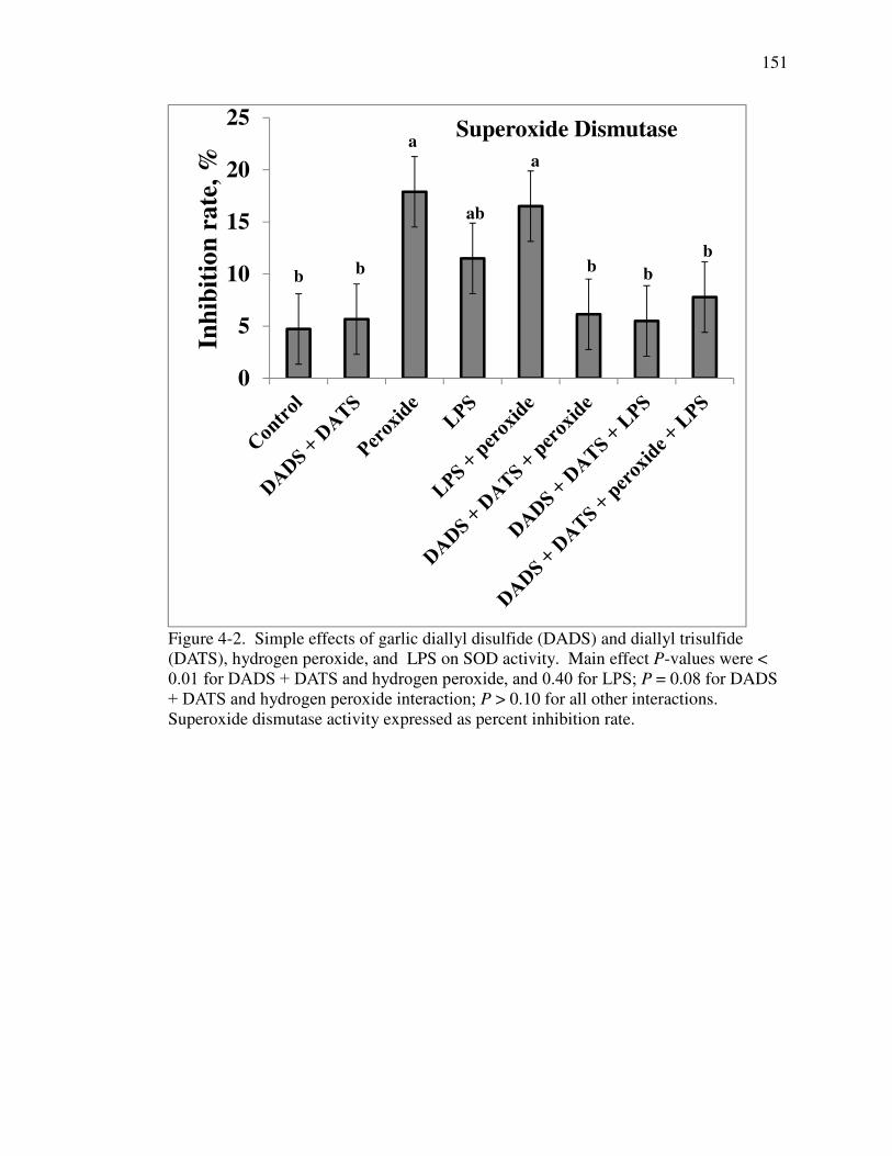
151
Figure 4-2. Simple effects of garlic diallyl disulfide (DADS) and diallyl trisulfide (DATS), hydrogen peroxide, and LPS on SOD activity. Main effect P-values were < 0.01 for DADS + DATS and hydrogen peroxide, and 0.40 for LPS; P = 0.08 for DADS + DATS and hydrogen peroxide interaction; P > 0.10 for all other interactions. Superoxide dismutase activity expressed as percent inhibition rate.
0
5
10
15
20
25
Inh
ibit
ion
ra
te, %
Superoxide Dismutase
bb
a
b
ab
a
b
b

152
Figure 4-3. Simple effects of garlic diallyl disulfide (DADS) and diallyl trisulfide (DATS), hydrogen peroxide, and LPS on catalase activity. P < 0.05 for main effects of DADS + DATS, hydrogen peroxide, and LPS, P < 0.01 for DADS + DATS and hydrogen peroxide interaction, hydrogen peroxide and LPS interaction, and DADS + DATS and hydrogen peroxide and LPS interaction, and P = 0.17 for DADS + DATS and LPS interaction. Catalase activity expressed as mMol H
20
2 degraded per mL per
min.
0
5
10
15
20
Act
ivit
y
Catalase
a
b
aa
b
a
b
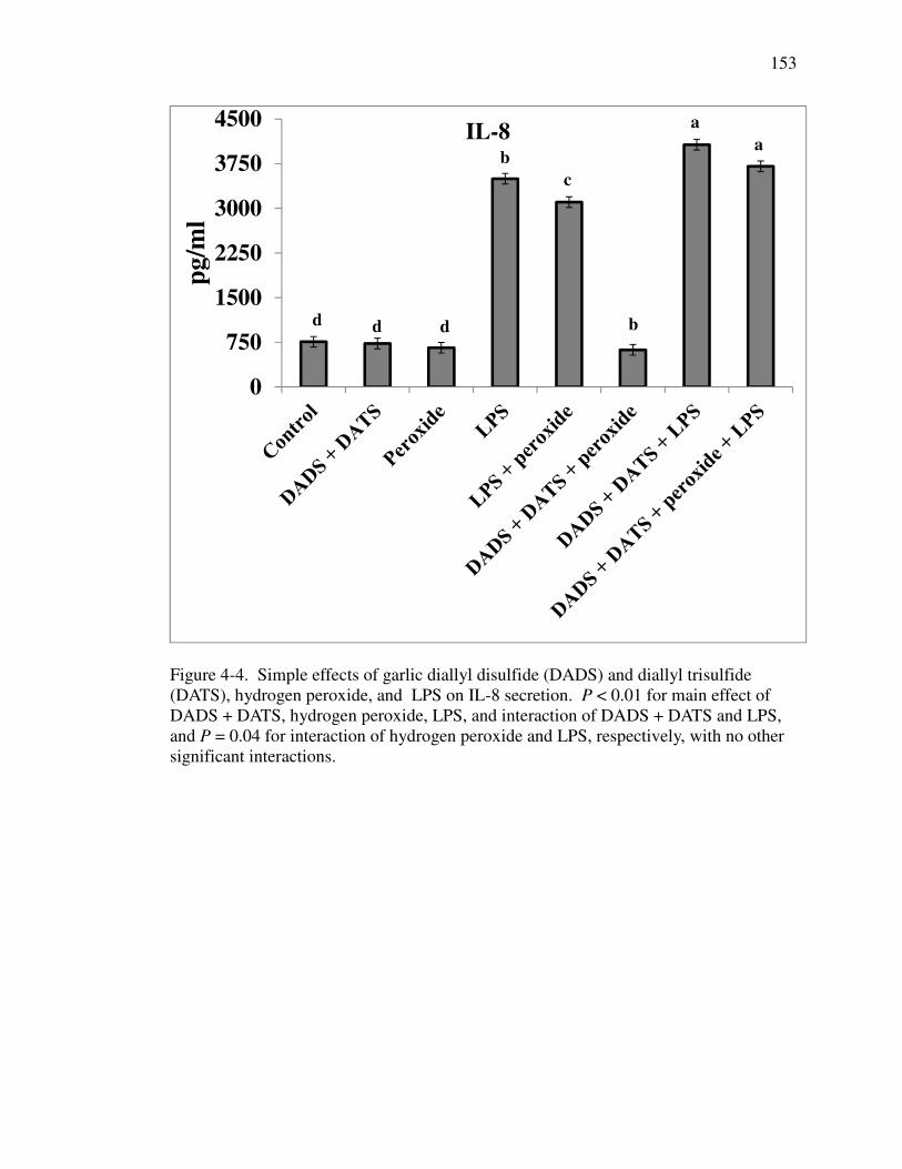
153
Figure 4-4. Simple effects of garlic diallyl disulfide (DADS) and diallyl trisulfide (DATS), hydrogen peroxide, and LPS on IL-8 secretion. P < 0.01 for main effect of DADS + DATS, hydrogen peroxide, LPS, and interaction of DADS + DATS and LPS, and P = 0.04 for interaction of hydrogen peroxide and LPS, respectively, with no other significant interactions.
0
750
1500
2250
3000
3750
4500
pg
/ml
IL-8
d d d b
b
c
a
a

154
CHAPTER 5. EXPRESSION OF CYTOKINE AND TIGHT JUNCTION GENES AND ILEAL MORPHOLOGY IN NURSERY PIGS IN RESPONSE TO GARLIC
DIALLYL DISULFIDE AND DIALLYL TRISULFIDE COMPOUNDS
5.1. Abstract
Bioactive components in garlic have anti-inflammatory, antioxidant, and
immune modulatory properties that may improve gastrointestinal function in pigs. The
current experiment was conducted to identify the optimal dosage of garlic-derived
bioactive components diallyl disulfide (DADS) and diallyl trisulfide (DATS) and to
evaluate the effect of graded doses of DADS and DATS on nursery pig performance
and gastrointestinal function. Eight replicate pens of barrows were allotted on the basis
of initial BW (12.45 ± 1.37 kg) in a RCBD to one of six treatments that consisted of
administration of 0, 0.45, 0.90, 1.80, 3.60, or 7.20 mg DADS + DATS per kg
bodyweight. The DADS + DATS was administered daily by oral gavage to overnight-
fasted pigs for a period of 6 d with soybean oil as the carrier and vehicle control.
Growth performance and daily stool consistency scores were recorded. At the end of
the experiment, pigs were euthanized and the mid ileum was excised for morphological
and gene expression measurements. Mucosal gene expression was conducted by RT
PCR for mucin (MUC2), interleukin 8 (IL-8), tumor necrosis factor (TNF-α), claudin 1
(CL-1), occludin (OC), and zonula occludens 1 (ZO-1). Supplementation with DADS +

155
DATS did not affect growth performance or stool consistency, although there was a
tendency (P = 0.06) for increased villous height. Expression of the IL-8 gene was
linearly increased (P = 0.01) and there was a tendency for a decrease (P = 0.07) in ZO-1
gene expression with garlic supplementation. There was no effect of treatment on
expression of MUC2, TNF-α, CL-1, and OC genes. Results from the current study
show that DADS + DATS supplementation improves ileal villus height and the broken-
line regression-derived adequate dose to optimize villus height response was 1.71 mg
DADS + DATS per kg BW. Furthermore, DADS + DATS supplementation impacts
IL-8 and ZO-1 gene expression, which may have implications on mucosal immune
function and cellular integrity.

156
5.2. Introduction
Nursery pig gastrointestinal health and function can be influenced by several
factors including dietary components, environmental stress, and intestinal microbiology.
Gastrointestinal dysfunction can lead to cellular oxidative damage and over-stimulation
of inflammatory pathways with long-lasting implications on pig health, performance,
and digestive function (Pluske et al., 1997; Lambert 2009; Smith et al., 2010).
Bioactive phytochemicals in garlic products have been shown to have hepatoprotective,
antioxidant, and immune enhancing properties. Following grinding of fresh garlic, the
bioactive phytochemical alliin is rapidly broken down to thiosulfinates, such as allicin,
that have been shown to be antimicrobial (Ankri and Mirelman, 1999; Amagase et al.,
2001; Amagase, 2006). Dially disulfide, DATS, and S-allyl cysteine (SAC) are
downstream bioactive components from extremely unstable and spontaneously
degrading garlic thiosulfinates (Amagase et al., 2001; Amagase, 2006). Meta-analysis
shows downstream bioactive components of garlic may mitigate oxidant-induced
cellular dysfunction and influence innate immune dynamics (Borek, 2001; Kyo et al.,
2001).
Previous research shows enhanced growth performance and intestinal function
and reduced morbidity with supplementation of crude preparations of garlic products to
young pigs (Tatara et al., 2008; Huang et al., 2011; Yan et al., 2012). The exact mode of
action of garlic bioactive components is not fully understood although in-vitro research
suggests that garlic-derived organosulfur compounds enhance intestinal cell integrity,
reduce cellular oxidative stress by mitigation of reactive oxygen species (ROS) and

157
increase antioxidant enzyme function (Yamasaki and Lau 1997; Wei and Lau 1998;
Roselli et al., 2007). There is a dearth of information in the literature on the impact of
garlic bioactive components DADS and DATS on nursery pig performance and
gastrointestinal health. We hypothesized that an adequate dose of orally administered
downstream bioactive components DADS and DATS would enhance ileal mucosal
morphology and integrity in pigs. To achieve our objectives we determined an effective
dose as well as the effect of various doses of DADS + DATS on growth performance,
ileal mucosal morphology, and cytokine and tight junction gene expression.
5.3. Materials and methods
All animal procedures were approved by the Purdue University Animal Care and Use
Committee.
Animals and treatments
A total of 48 PIC 337 × C29 barrows (initial BW = 12.45 ± 1.37 kg, 34 d old)
were allotted to treatment on the basis of initial BW in a randomized complete block
design. The barrows were individually housed in cement-floor pens containing one
nipple drinker and feed trough per pen. Room temperature was maintained at 24.4°C
and there was 14 h of light per day. Pigs were allowed a 6-d acclimation period prior to
initiation of experimental treatment. There were 6 treatments that consisted of daily
administration of 0, 0.45, 0.90, 1.80, 3.60, or 7.20 mg of garlic-derived DADS +
DATS per kg BW by oral gavage for a period of 6 d. There were 8 replicates per
treatment with 1 barrow per pen. The oral gavage was prepared fresh daily with

158
soybean oil as the carrier and vehicle control and administered at 0800 h to overnight-
fasted barrows. Barrows were allowed free access to water and were fed a mash-based
grower diet that met or exceeded NRC requirements (NRC, 2012; Table 5-1). The garlic
product was obtained from Stanford Chemicals (Irvine, Ca) and consisted of 22%
DADS and 23% DATS with the remaining 55% consisting of a methanol-based carrier.
Barrows and feeders were weighed at the start and end of the experiment to calculate
ADG, ADFI, and G:F. Scours were subjectively monitored by the same person twice
daily (0800 and 1600 h) and recorded corresponding to a scale of 1 = no scour, 2 =
moderate fecal looseness, or 3 = severe diarrhea. On d 6 of the experiment, barrows
were euthanized by intramuscular injection with ketamine (Fort Dodge Laboratories
Inc., Fort Dodge, IA), Telazol (Fort Dodge Laboratories), and xylazine (Bayer Corp.,
Shawnee Mission, KS) at doses of 4, 2, 2, and 2.2 mg/kg BW, respectively, followed by
asphyxiation with CO2. Immediately following euthanasia a 10-cm segment of the distal
ileum was excised for histological and gene expression measurements.
Histological measurements
Excised intestinal segments were flushed in ice-cold 10% phosphate-buffered
saline (VWR International, Radnor, PA), stapled to a cardboard background, and fixed
in 10% buffered formalin (VWR International, Radnor, PA) for approximately 30 d.
Samples were subsequently dehydrated with ethanol (VWR International, Radnor, PA),
cleared with Sub-X® (Polysciences, Inc., Warrington, PA) and placed in paraffin
(Polyfin paraffin, Sigma Polysciences, St. Louis, MO). Segments were sliced (5 µm)
and stained with hematoxylin and eosin by the Purdue Histology and Phenotyping
Laboratory (Purdue University, West Lafayette, IN USA). Villus height and crypt depth

159
were measured and villus height to crypt depth ratio was calculated. Means from at least
4 villi per segment were analyzed for differences. Villus length is defined as the length
from the villus tip to the valley between villi and crypt depth is defined as the length
between the crypt opening and base.
RNA isolation, cDNA Synthesis, and real-time PCR
Immediately after removal, intestinal segments were flushed with ice-cold PBS
(VWR International, Radnor, PA), cut in half exposing the lumen, and mucosal contents
were scraped with a metal spatula. Mucosal contents were immediately placed in 2 mL
of Trizol reagent (Invitrogen, Grand Island, NY) and stored at -80°C until RNA
isolation. RNA was extracted according to the Trizol protocol (Invitrogen, Grand
Island, NY). Reverse transcription was carried out using the MMLV reverse
transcription system of Promega (Promega, Madison, WI) and real-time PCR was
performed on a Bio-Rad iCyler (Bio-Rad Laboratories, Hercules, CA) using a reaction
mix that consisted of 0.50 µg cDNA, 0.075 nmol of each forward and reverse primers,
SYBR Green Master (Roche, Basel, Switzerland), and nuclease-free water (Ambion,
Austin, TX) to reach a reaction volume of 20 µl per well. Following a 5-min
incubation period at 95ºC, reactions were cycled 50 times using the following protocol:
10s at 95ºC, 20 s at 55ºC, and 72ºC for 30 s. Expression of mucin (MUC2), tumor
necrosis factor alpha (TNF-α), interleukin 8 (IL-8), claudin 1 (CL-1), occludin (OC),
and zonula occludens 1 (ZO-1) genes were measured using 18S as a housekeeping
control. Primers used are presented in Table 5-2.

160
Statistical analysis
Data were analyzed using the MIXED procedure of SAS (SAS Inst., Inc., Cary,
NC) with the fixed effect of treatment and the random effect of block. The following
statistical model was used in the analysis: Yij = µ + Wi + Bj + εij where Y is the
response criterion; µ is the overall mean; Wi is the effect of ith treatment (i = 1,2,3,
…,6); Bj is the effect of jth block (k = 1, 2, 3,… 8); and εij is the error term. If there was
an effect of treatment, single-degree-of-freedom linear and quadratic contrasts were
conducted with coefficients for unequally spaced levels that were derived using the IML
procedure of SAS. Incidence of diarrhea (scour scores) was analyzed as non-parametric
data using the NPAR1WAY procedure of SAS. Broken-line regression analysis was
conducted using the NLIN procedure of SAS as described by Robbins et al. (2006).
Significant difference was defined as P ≤ 0.05 and 0.05 ≤ P ≤ 0.10 was considered a
tendency. Model assumptions for normality and equal variances were validated by the
Shapiro-Wilks test (UNIVARIATE procedure of SAS) and Brown and Forsythe test
(GLM procedure of SAS), respectively.
5.4. Results
Pigs were healthy throughout the experiment and there were no incidences of
morbidity, feed refusal, or aversion to receipt of the DADS + DATS gavage. Growth
performance and stool consistency are reported in Table 5-3. There was no effect of
treatment on growth performance or stool consistency during the experiment (P > 0.05).
The average initial BW, final BW, and G:F were 12.45 ± 1.37 kg, 15.90 ± 1.82 kg, and

161
0.65 ± 0.17, respectively. Moderate stool looseness (scour score = 2) was reported
throughout the experiment and there were isolated incidences of severe diarrhea (scour
score = 3) observed that were not treatment specific and did not correspond with
changes in growth performance.
The average villus height and crypt depth were 350 ± 69.6 µm and 134 ± 40.5 µm,
respectively (Table 5-3). There was a tendency for an effect of treatment on villus
height (P = 0.06) but not crypt depth (P = 0.50). Pigs that received a daily oral gavage
of 0.90, 1.80, 3.60, or 7.20 mg DADS + DATS per kg BW had a 12, 17, 34, or 15 %
increase in villi length, respectively, in comparison with pigs receiving the vehicle
control gavage. The villus height plateaued at 381 µm which was with daily
supplementation of 1.71 mg DADS + DATS per kg BW (Figure 5-1).
Gene expression data are reported in Table 5-4. There was no effect of treatment on
housekeeping gene expression. Daily oral gavage of DADS + DATS linearly increased
(P = 0.01) IL-8 gene expression and there was a tendency for decreased ZO-1 (P = 0.07)
gene expression. In pigs that received daily oral gavage of 7.2 mg DADS + DATS per
kg BW, there was approximately a 100% increase in IL-8 gene expression and a 56 %
decrease in ZO-1 gene expression relative to pigs receiving the vehicle control gavage.
There was no impact (P > 0.10) of experimental treatment on MUC2, TNF-α, CL-1, and
OC gene expression. The expression of the ZO-1 gene plateaued at 0.90 arbitrary units
(AU) which was with daily supplementation of 0.90 mg DADS + DATS per kg BW
(Figure 5-2).

162
5.5. Discussion
During the nursery phase, pigs are exposed to various environmental, social, and
nutritional stress events that are linked to growth performance and gastrointestinal
function (Pluske et al., 1997; Lalles et al., 2007; Smith et al., 2010). A clear link has
been established between structure and function of the small intestine and pig growth
performance (Pluske et al., 1997) and nutritional and management strategies have been
shown to mitigate nursery pig gastrointestinal dysfunction (Lalles et al., 2007). The
aforementioned strategies include supplementation of specific ingredients including
plasma protein, milk carbohydrates, prebiotics, probiotics, and botanicals. Garlic
bioactive components have been shown to be antimicrobial (Ankri and Mirelman, 1999),
antioxidant (Borek, 2001), immune modulatory (Kyo et al., 2001), and anti-
inflammatory (Lang et al., 2004). Recent literature from in-vitro and rodent experiments
suggests supplementation of garlic may be a viable option to improve animal
performance and gastrointestinal function (Lang et al., 2004; Roselli et al., 2007; Badr
and Al-Mulhim 2014). In pigs, Tatara et al. (2008) and Huang et al. (2011) showed
supplementation of a crude garlic product improved performance and intestinal
morphology and Liu et al. (2013) showed garlic product supplementation reduced
E.coli-induced diarrhea and impacted innate immune parameters in sham- and E.coli-
infected animals. Therefore, the objective of the current experiment was to determine
an effective dose as well as the effect of graded doses of DADS + DATS on nursery pig
performance, stool consistency, and ileal mucosa gene expression of immune and
cellular integrity markers.

163
Previous research showed enhanced growth performance and intestinal function and
reduced morbidity with supplementation of garlic products to young pigs (Tatara et al.,
2008; Huang et al., 2011; Yan et al., 2012). Tatara et al. (2008) showed improved
growth performance in artificially-raised piglets that corresponded to greater villous
height and crypt depth throughout the small intestine and Huang et al. (2011) similarly
showed improved bodyweight gain and reduced diarrhea incidence in weaned pigs
supplemented with 0.10 to 0.25 g/kg garlic product in the feed. The exact mode of
action of garlic bioactive components is not fully understood although in-vitro research
suggests that garlic-derived organosulfur compounds reduce cellular oxidative stress by
mitigation of reactive oxygen species (ROS) and increase antioxidant enzyme function
(Yamasaki and Lau 1997; Wei and Lau, 1998). Furthermore, DADS and DATS have
been shown to have radical scavenging properties and reduce TNF-α- and hydrogen
peroxide-induced activation of NFκB and subsequent inflammatory cytokine release
(Lang et al., 2004; Zhou et al., 2011). Garlic bioactive components have been shown to
enhance cellular integrity in a porcine epithelial cell model exposed to E.coli (Roselli et
al., 2007) although a dearth of information exists on the effects of garlic on tight
junction proteins. Additionally, garlic components have been shown to influence both
immune modulation and suppression (Kyo et al., 2001). Liu et al. (2013) showed
reduced ileal infiltration of neutrophils and macrophages and serum inflammatory
cytokines in E.coli-infected nursery pigs fed a garlic product and more recently Lui et al.
(2014) showed by gene expression profile that nursery pigs fed a garlic product had
increased expression of genes associated with innate immune defense and oxidative

164
reduction and decreased expression of genes associated with antigen presentation and
iron binding.
The literature contains conflicting evidence for the effects of garlic products on
nursery and grow-finish pig growth performance. Huang et al. (2011) showed
supplementation of 0.10 to 0.25 g garlic oil per kg diet improved nursery pig growth
performance over a 28-d feeding period and Tatara et al. (2008) reported a 3 to 5 %
improvement in BW in young pigs fed diets supplemented with 1 to 2 mg/kg garlic or
allicin supplemented by oral gavage. Likewise, Yan et al. (2012) showed
supplementation of 2 to 4 g garlic product per kg diet improved growth performance
over an extended duration. Conversely, Chen et al. (2008) showed no effect of garlic
product supplementation in growing pigs and Holden (1998) showed garlic product
supplementation to grow-finish pigs impaired gain in a feed intake-dependent manner.
In the current study there was no impact of DADS + DATS supplementation on nursery
pig growth performance, and given the short duration of the study, this observation was
not unexpected. The feed efficiency values reported for the current experiment are
similar to values reported by Horn et al. (2014) and Cromwell et al. (2008) although
ADG and ADFI were slightly higher than previously reported by our lab using pigs
from the same genetic source which is likely due to the high health status and
experimental design (individually-penned pigs). A previous experiment by Huang et al.
(2011) showed the effects of garlic supplementation were not realized during the first
week of supplementation. Furthermore, most in-vivo and in-vitro studies demonstrating
garlic-product efficacy contain an experimental challenge event such as early weaning,
E.coli infection, or oxidative damage (Tatara et al., 2008; Badr and Al-Mulhim 2014;
165
Liu et al., 2014). Therefore, the lack of growth performance response due to DADS +
DATS for the current experiment is likely related to the health status of the pigs and
duration of supplementation. Previous studies in nursery pigs show that garlic product
supplementation may reduce pig morbidity by reducing incidence of diarrhea (Huang et
al., 2011; Liu et al. 2013). Huang et al. (2011) reported a linear decrease in overall
incidence of diarrhea with most drastic effects during the early period of
supplementation. Similarly, Liu et al. (2013) reported that supplementation of garlic
product to E.coli-infected or sham pigs reduced diarrhea score throughout the
experiment or during the first 5 d post weaning, respectively. It is hypothesized that
garlic products may act to reduce nursery pig post-weaning incidence of diarrhea
though enhancing innate immune function (Liu et al., 2014) or radical scavenging
properties of organo-sulfur bioactive components acting to reduce gastrointestinal
inflammation (Borek, 2001; Huang et al., 2011). Furthermore, Huang et al. (2011)
suggested that garlic products affect gastrointestinal motility and secretion though
agonistic effects on TRPA-1 receptors. For the current experiment there was no effect
of DADS + DATS supplementation on diarrhea incidence. Discrepancies between the
current study and previous literature are likely due to the age and health status of the
pigs.
A clear distinction should be made between the effects of fresh garlic and aged
garlic bioactive components on gastrointestinal function. Generally, fresh garlic
bioactive components, such as allicin, are associated with gastrointestinal damage
(Amagase et al., 2001) whereas evidence exists that aged garlic bioactive components
are associated with improved gastrointestinal function (Horie et al., 1997; Tatara et al.,

166
2008; Peinado et al., 2012). In artificially-reared neonatal pigs, supplementation with
garlic products improved intestinal morphology throughout the gastrointestinal tract
(Tatara et al., 2008). Conversely, garlic product supplementation had no impact on
intestinal morphology in E.coli-infected nursery pigs supplemented with garlic (Liu et
al., 2013). Discrepancies in the literature are likely due to differences in product
bioactive components, animal health status, and experimental design. For the current
pig experiment, villus height increased 11, 15, 25, or 13 % with supplementation oral
gavage of 0.90, 1.80, 3.60, and 7.20 mg DADS + DATS per kg BW, respectively
relative to pigs that did not receive the garlic product gavage. The broken-line
regression-derived adequate dose to optimize villus height response was 1.71 mg
DADS + DATS per kg BW, which for a 10- to 15-kg pig consuming 1.2 kg feed
translates to approximately 0.04 g DADS + DATS per kg feed. Ileal morphology data
reported in the current experiment are within range of values reported in the literature
for nursery pigs (Liu et al., 2008; Rubio et al., 2010). The exact mechanism of action of
garlic bioactive components on GI morphology is not fully understood, although it may
be related to microbial and immune actions. Tatara et al. (2008) showed a garlic
product modulated GI antimicrobial factors such as lysozyme and Peinado et al. (2012)
showed a garlic bioactive component reduced broiler chicken enteropathogen
colonization that was associated with improved gastrointestinal morphology. The
aforementioned studies suggest morphological benefits of garlic are related to microbial
modulation and innate immune function although further research is needed to better
understand the impact of garlic bioactive components on gastrointestinal microbial
ecology and enterocyte function.

167
Garlic products have been shown to be immune modulatory in in-vitro and in-vivo
research models (Kyo et al., 2001; Lang et al., 2004; Liu et al., 2014). Using a rodent
cell model, Kyo et al. (1997 and 2001) showed aged garlic decreased antigen-specific
histamine release from basophils and increased natural killer cell phagocytic activity
suggesting aged garlic has anti-allergic and anti-tumor properties, respectively.
Furthermore, Kyo et al. (2001) showed aged-garlic increased spleen cell proliferation
and augmented concavalin A stimulation further suggesting an immune stimulatory role
of garlic bioactive components. Likewise, Clemmens et al. (2010) identified two
distinct immunomodulatory proteins in an ex-vivo experiment using human immune
cells and Alma et al. (2014) showed increased urinary excretion of IL-12 and IFN-γ in
humans supplemented with a garlic product. Conversely, Lang et al. (2004) showed a
synthetic garlic product reduced spontaneous and TNF-α stimulated inflammatory
cytokine secretion in Caco-2 cells. Recent literature in pigs showed reduced ileal
macrophage and neutrophil infiltration in sham and E.coli-infected animals with garlic
product supplementation (Liu et al., 2013). Furthermore, Liu et al. (2014) showed that
supplementation of garlic to nursery pigs for 9 d increased genes associated with
defense response and complement components and decreased genes associated with
antigen presentation and processing. For the current experiment there was a linear
increase in IL-8 gene expression due to garlic supplementation. To the author’s
knowledge there are currently no published studies showing the impact of garlic
bioactive components on ileal IL-8 gene expression. Interleukin 8 is a chemokine that is
involved in recruitment and activation of innate immune cells such as neutrophils and
macrophages and a change in IL-8 gene expression may be associated with changes in

168
innate immune function. There was no impact of treatment on TNF-α gene expression,
which is consistent with the literature (Liu et al., 2014). Taken together, results from
the current study and previous in-vitro and in-vivo studies, suggest a complex
relationship between garlic bioactive components and immune modulation. Further
studies should investigate the impact of innate immune cell activation in intestinal
tissues along with measurement of innate-immune cell molecular markers to more
clearly understand the immunological effects of garlic product supplementation. One of
the major constituents of the mucus layer is mucin which plays a critical intestinal
barrier function (Montagne et al., 2004). For the current experiment there was no impact
of garlic supplementation on MUC2 gene expression, and this is consistent with results
reported by Liu et al. (2014) in nursery pigs. Tight junction proteins serve to control
epithelial cell permeability and integrity (Gonzalez-Mariscal et al., 2003). In the
current experiment there was no effect of treatment on CL or OC gene expression,
although there was a tendency for a decrease in ZO-1 gene expression as oral gavage
dose increased from 0 to 7.20 mg DADS + DATS per kg BW with a breakpoint at 0.90
mg DADS + DATS per kg BW which roughly translates to 0.02 g DADS + DATS per
kg feed for a 10- to 15-kg pig consuming 1.20 kg feed per day. Roselli et al. (2007)
showed enhanced membrane barrier function in E.coli-infected IPEC cells due to
supplementation with a synthetic garlic product that was not due to antimicrobial effects
and Park et al. (2011) showed DADS increased trans-epithelial electrical resistance,
decreased CL gene expression, and increased metalloprotease inhibitor gene expression.
In contrast, Liu et al. (2014) showed no impact of a garlic product on ileal mucosa tight
junction gene expression profile in nursery pigs. For the current study, it is possible that

169
the change in ZO-1 gene expression is related to increases in IL-8 chemokine gene
expression and innate immune function, although further research is needed to better
understand the relationship between immune modulation and intestinal barrier function.
Previous research shows that garlic product supplementation improves growth
performance and health (Tatara et al., 2008; Huang et al., 2011) which may be due to
immune modulatory (Kyo et al., 2001) and antioxidant functions (Borek, 2001) of
bioactive components. In the current study there was no impact of the garlic product on
pig growth which may be due to duration of feeding and pig health status. Oral gavage
of DADS + DATS to pigs influences ileal morphology, and IL-8 and ZO-1 gene
expression in nursery pigs. Further research is warranted to better understand the
relationship between changes in IL-8 and ZO-1 gene expression and innate immune
function in nursery pigs. Overall, results from the current study show that DADS +
DATS in garlic influences gastrointestinal dynamics and may be strategy to enhance
nursery pig health and digestive function. Furthermore, approximately 0.08 g DADS +
DATS per kg feed is adequate to improve ileal villus height in nursery pigs.
5.6. Acknowledgements
The authors would like to express their appreciation to Biomatrix and JBS
United for their financial and technical support of this trial. Further appreciation is
given to Jason Fields, Purdue Animal House, for support with pig husbandry. The
authors would also like to express appreciation to Aly Gautier and Daniel Miranda,
Purdue University Department of Animal Sciences, for their assistance with treatment

170
administration and Donna Brooks, Purdue University Veterinary School, for her
assistance with histological measurements.

171
5.7. References
Alma, E., A. Eken, H. Ercil, K. Yelsel, and N. Daglioglu. 2014. The effect of garlic
powder on human urinary cytokine excretion. Urology J. 11(1): 1309-1315.
Amagase, H., B.L. Petesch, H. Matsuura, S. Kasuga, and Y. Itakura. 2001. Intake of
garlic and its bioactive components. J. Nutr. 131: 955S–962S.
Amagase, H. 2006. Clarifying the real bioactive constituents of garlic. J. Nutr. 136: 716S–725S.
Ankri, S., and D. Mirelman. 1999. Antimicrobial properties of allicin from garlic.
Microb. Infect. 2: 125−129.
Badr, G.M., and J.A. Al-Mulhim. 2014. The Protective effect of aged garlic extract on
nonsteroidal anti-inflammatory drug-induced gastric inflammations in male
albino rats. Evid. Based Complement. Alternat. Med. 2014: 759642.
Borek, C. 2001. Antioxidant Health Effects of Aged Garlic Extract. J. Nutr. 131:
1010S–1015S.
Chen, Y.J., I.H. Kim, J.H. Cho, J.S. Yoo, Q. Wang, Y. Wang, and Y. Haung. 2008.
Evaluation of dietary L-carnitine or garlic powder on growth performance, dry
matter and nitrogen digestibilities, blood profiles and meat quality in finishing
pigs. Anim. Feed Sci. Tech. 141(1): 141-152.
Clemmens, F., S.N. Pramod, and Y.P. Venkatesh. 2010. Identity of the
immunomodulatory proteins from garlic (Allium sativum) with the major garlic
lectins or agglutinins. Int. Immunopharmacol.10: 316–324.

172
Cromwell, G.L., G.L. Allee, and D.C. Mahan. 2008. Assessment of lactose level in the
mid- to late- nursery phase on performance of weanling pigs. J. Anim. Sci. 86:
127-133.
Gonzalez-Mariscal, L., A. Betanzos, P. Nava, and B.E. Jaramillo. 2003. Tight junction
proteins. Progr. Biophys. Molec. Biol. 81: 1-44.
Horie, T., S. Awazu, Y. Itakura, and T. Fuwa. 1997. Alleviation by garlic of antitumor
drug-induced damage to the intestine. J. Nutr. 131(3s): 1071S-1074S.
Holden, P.J., J. McKean, and E. Franzenburg. 1998. Botanicals for pigs-garlic. Iowa
State University, Swine Research Report.
Horn, N., F. Ruch, G. Miller, K. M. Ajuwon, and O. Adeola. 2014. Impact of acute
water and feed deprivation events on growth performance, intestinal
characteristics, and serum stress markers in weaned pigs. J. Anim. Sci. 92:4407-
4416.
Huang, R.H., X.S. Qiu, F.X., Shi, C.L. Hughes, Z.F. Lu, and W.Y. Zhu. 2011. Effects
of dietary allicin on health and growth performance of weanling piglets and
reduction in attractiveness of faeces to flies. Animal. 5: 304–311.
Kyo, E., N. Uda, M. Kakimoto, K. Yokoyama, M. Ushijima, I. Sumioka, S. Kasuga,
and Y. Itakura, Y. 1997. Anti-allergic effects of aged garlic extract.
Phytomedicine.4: 335–340.
Kyo, E., N. Uda, S. Kasuga, and Y. Itakura. 2001. Immunomodulatory effects of aged
garlic extract. J. Nutr. 131:1075S–1079S.

173
Lalles J.P., P. Bosi, H. Smidt, and C.R. Stokes. 2007. Nutritional management of gut
health in pigs around weaning. Proc. Nutr. Soc. 66: 260–268.
Lambert, G.P. 2009. Stress-induced gastrointestinal barrier dysfunction and
its inflammatory effects. J. Anim. Sci. 87 (E suppl.) E101-E108.
Lang, A., M. Lahava, E. Sakhninia, I. Barshack, H.H. Fiddera, B. Avidana, E. Bardana,
R. Hershkovizc, S. Bar-Meira, and Y. Chowersa. 2004. Allicin inhibits
spontaneous and TNF-α induced secretion of proinflammatory cytokines and
chemokines from intestinal epithelial cells. Clin. Nutr. 23: 1199–1208.
Liu P., X.S. Piao, S.W. Kim, L. Wang, Y.B. Shen, H.S. Lee, and S.Y. Li. 2008. Effects
of chito-oligosaccharide supplementation on the growth performance, nutrient
digestibility, intestinal morphology, and fecal shedding of Escherichia coli and
Lactobacillus in weaning pigs. J. Anim. Sci. 86(10): 2609-2618.
Liu, Y., M. Song, T.M. Che, A.S. Almeida, J.J. Lee, D. Bravo, C.W. Maddox, and J.E.
Pettigrew. 2013. Dietary plant extracts alleviate diarrhea and alter immune
responses of weaned pigs. J. Anim. Sci. 91:5294-5306.
Liu, Y., M. Song, D. Che, D. Bravo, C.W. Maddox, and J.E. Pettigrew. 2014. Effects
of capsicum oleoresin, garlic botanical, and turmeric oleoresin on gene
expression profile of ileal mucosa in weaned pigs. J. Anim. Sci. 92:3426-3440.
Montagne, L., C. Piel, and J. P. Lalles. 2004. Effect of diet on mucin kinetics and
composition:nutrition and health implications. Nutr. Rev. 62:105–114.
NRC, 2012. Nutrient requirements of Swine. 11th rev ed. Natl. Acad. Press,
Washington, D.C.

174
Olukosi, O.A., and N. D. Dono. 2014. Modification of digesta pH and intestinal
morphology with the use of benzoic acid or phytobiotics and the effects on
broiler chicken growth performance and energy and nutrient utilization. J. Anim.
Sci. 92:3945–3953.
Park, H.S., G.Y. Kim, I.W.Choi, N.D. Kim, H.J. Hwang, Y.W. Choi, and Y.H. Choi.
2011. Inhibition of matrix metalloproteinase activities and tightening of tight
junctions by diallyl disulfide in AGS human gastric carcinoma cells. J. Food
Sci.76: T105-T111.
Peinado, M.J., R. Ruiz, A. Echavarri , and L. A. Rubio. 2012. Garlic derivative propyl
propane thiosulfonate is effective against broiler enteropathogens in vivo. Poult.
Sci. 91:2148–2157.
Pluske, J.R., D.J. Hampson, and I.H. William. 1997. Factors influencing the
structure and function of the small intestine in the weaned pig: a review,
Livest. Prod. Sci. 51, 215–236.
Robbins, K. R., A. M. Saxton, and L. L. Southern. 2006. Estimation of nutrient
requirements using broken-line regression analysis. J. Anim. Sci. 84:E155–E165.
Roselli, M., M.S. Britti, I. Lerou-Luron, H. Marfaing, W.Y. Zhu, and E. Mengheri.
2007. Effect of different plant extracts and natural substances (PENS) against
membrane damage induced by enterotoxigenic Escherichia coli K88 in pig
intestinal cells. Toxicol. in Vitro. 21: 224–229.
175
Rubio, L.A., R. Ruiz, M.J. Peinado, and A. Echavarri. 2010. Morphology and
enzymatic activity of the small intestinal mucosa of Iberian pigs as compared
with a lean pig strain. J. Anim. Sci. 88(11): 3590-3597.
SAS Institute. 2006. SAS/STAT User’s Guide. Release 9.1. SAS Institute Inc., Cary,
NC.
Smith, F., J.E. Clark, B.L. Overman, C.C. Tozel, J.H. Huang, J.E. Rivier, A.T.
Blikslager, and A.J. Moeser. 2010. Early weaning stress impairs development of
mucosal barrier function in the porcine intestine. Am. J. Physiol. Gastrointest.
Liver Physiol. 298(3):G352-G363.
Tatara, M.R., E. Śliwa, K. Dudek, A. Gawron, T. Piersiak, P. Dobrowolski , J.
Mosiewicz, A. K. Siwicki, and T. Studziński. 2008. Aged garlic extract and
allicin improve performance and gastrointestinal tract development of piglets
reared on artificial sow. Ann. Agric. Environ. Med. 15: 63–69.
Wei, Z., and B.H., Lau. 1998. Garlic inhibits free radical generation and augments
antioxidant enzyme activity in vascular endothelial cells. Nutr. Res. 18:61–70.
Yan, L., Q.W. Meng, and I.H. Kim. 2012. Effects of fermented garlic powder
supplementation on growth performance, nutrient digestibility, blood
characteristics, and meat quality in grow-finish pigs. Anim. Sci. J. 83: 411-417.
Yamasaki, T., and B.H. Lau. 1997. Garlic compounds protect vascular endothelial cells
from oxidant injury. Folia Pharmacol. Jpn.110 (Suppl.1):138–141.
Zhao, L., J.Y. Lee, and D.H. Hwang. 2011. Inhibition of pattern recognition receptor-
mediated inflammation by bioactive phytochemicals. Nutr. Rev. 69:310–320.
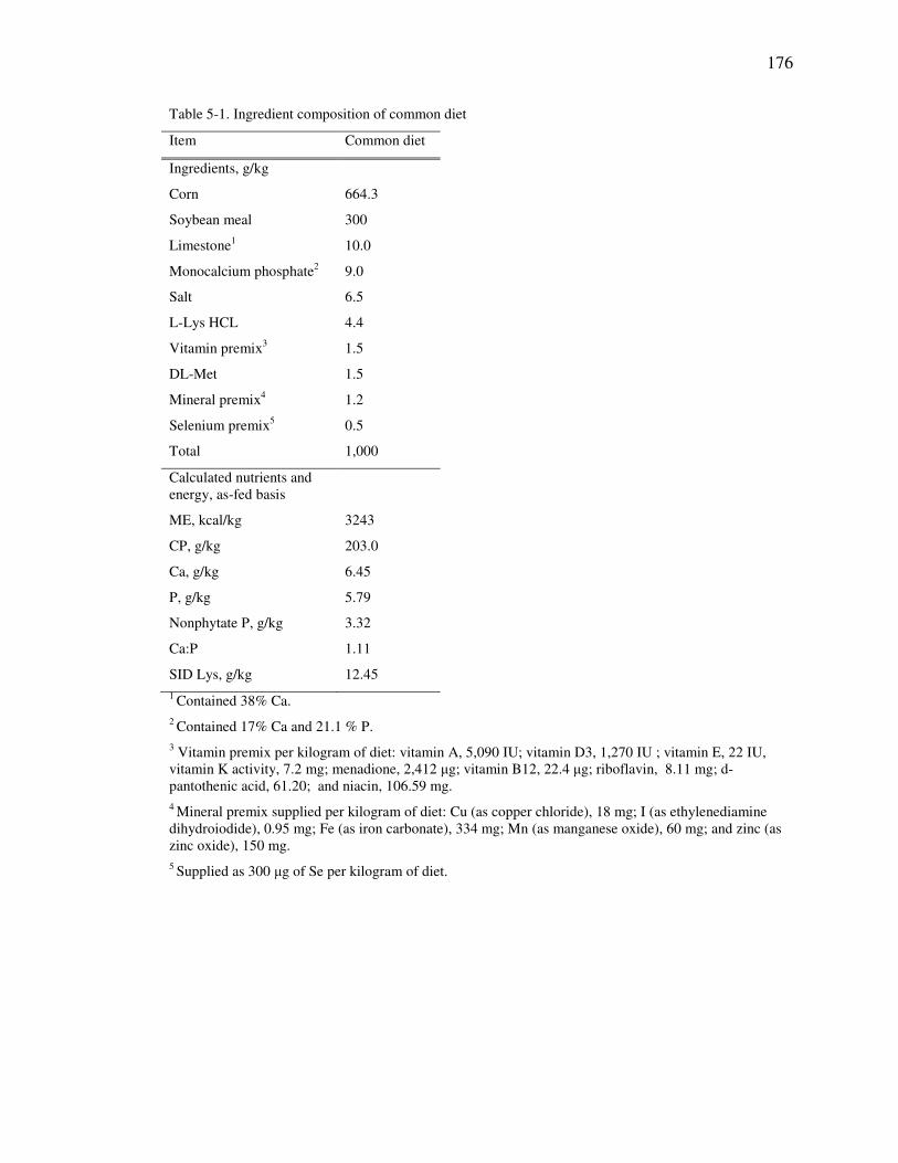
176
Table 5-1. Ingredient composition of common diet
Item Common diet
Ingredients, g/kg
Corn 664.3
Soybean meal 300
Limestone1 10.0
Monocalcium phosphate2 9.0
Salt 6.5
L-Lys HCL 4.4
Vitamin premix3 1.5
DL-Met 1.5
Mineral premix4 1.2
Selenium premix5 0.5
Total 1,000
Calculated nutrients and energy, as-fed basis
ME, kcal/kg 3243
CP, g/kg 203.0
Ca, g/kg 6.45
P, g/kg 5.79
Nonphytate P, g/kg 3.32
Ca:P 1.11
SID Lys, g/kg 12.45
1 Contained 38% Ca.
2 Contained 17% Ca and 21.1 % P.
3 Vitamin premix per kilogram of diet: vitamin A, 5,090 IU; vitamin D3, 1,270 IU ; vitamin E, 22 IU, vitamin K activity, 7.2 mg; menadione, 2,412 µg; vitamin B12, 22.4 µg; riboflavin, 8.11 mg; d-pantothenic acid, 61.20; and niacin, 106.59 mg.
4 Mineral premix supplied per kilogram of diet: Cu (as copper chloride), 18 mg; I (as ethylenediamine dihydroiodide), 0.95 mg; Fe (as iron carbonate), 334 mg; Mn (as manganese oxide), 60 mg; and zinc (as zinc oxide), 150 mg.
5 Supplied as 300 µg of Se per kilogram of diet.

177
Table 5-2. Primers used for RT-PCR1
Gene Primer (5`-3`)
18S (forward) ATCCCTGAGAAGTTCCAGCA
18S (reverse) CCTCCTGGTGAGGTCGATGT
MUC2 (forward) CAACGGCCTCTCCTTCTCTGT
MUC2 (reverse) GCCACACTGGCCCTTTGT
TNF-α (forward) CGTCGCCGT TGTAGC
TNF-α (reverse) GCCCATCTGTCGGCACCACC
IL-8 (forward) TTTCTGCAG CTCTCTGTGAGG
IL-8 (reverse) CTGCTGTTGTTGTTGCTTCTC
CL-1 (forward) AGAAGATGCGGATGGCTGTC
CL-1 (reverse) CCCAGAAGGCAGAGAGAAGC
OC (forward) TCCTGGGTGTGATGGTGTTC
OC (reverse) CGTAGAGTCCAGTCACCGCA
ZO-1 (forward) GAGTTTGATAGTGGCGTT
ZO-1 (reverse) GTGGGAGGATGCTGTTGT
1 MUC2 = mucin 2; TNF-α = tumor necrosis factor alpha; IL-8 = interleukin 8; CL-1 = claudin 1; OC = occluding; and ZO-1 = zonula occludens 1.

178
Table 5-3. Growth performance and ileal histology responses of pigs that received oral gavage of DADS
+ DATS 1
DADS + DATS orally administered, mg /kg BW2
0 0.45 0.90 1.80 3.60 7.20 SEM3 P-value
Item IBW, kg 12.5 12.4 12.5 12.4 12.5 12.4 0.50 0.99 FBW, kg 16.4 15.3 15.7 16.0 15.9 16.0 0.65 0.65 ADG, g 629 526 522 604 564 563 54.72 0.54 ADFI, g 955 814 800 907 1021 959 96.76 0.39 G:F, g:g 0.71 0.63 0.67 0.70 0.55 0.62 0.06 0.41 Scour scores4 1.51 1.63 1.56 1.50 1.86 1.74 0.14 0.55 Villus height, µm (VH)5 310 293 348 363 415 357 32.04 0.06 Crypt depth, µm (CD)5 129 141 133 123 170 126 20.46 0.50 VH:CD 2.53 2.24 2.86 3.26 2.80 2.98 0.45 0.58
1 Pigs were administered DADS + DATS by oral gavage daily with soybean oil as the carrier for 6 d. For growth performance data there were 6 to 8 replicate pens per treatment and for histology data there were 4 to 6 replicate pens per treatment with 1 pig per pen. Pigs were allowed a 6-d adaptation period prior to
the trial.
2 BW=body weight.
3 SEM = standard error of the mean. The highest SEM is reported.
4Analyzed as non-parametric data. Scour scores were recorded once daily on a scale from 1 to 3, with 1 = normal stools and 3 = severe diarrhea.
5 Distal ileum was sampled at the end of the trial for histological analysis.

179
Table 5-4. Ileal mucosal gene expression responses of pigs that received oral gavage of DADS + DATS 1
DADS + DATS orally administered, mg /kg BW2
0 0.45 0.90 1.80 3.60 7.20 SEM3 P-value
Item MUC2 1.72 1.19 1.21 1.33 1.41 1.41 0.42 0.95 IL-84,5 2.08 0.75 0.91 2.54 2.60 4.15 0.96 0.02 TNF-α 1.44 1.46 1.34 1.21 1.08 1.43 0.31 0.93 IL-64 1.38 1.59 1.46 1.85 1.75 1.07 0.76 0.87 CL-14 3.48 1.45 2.75 1.45 0.95 0.46 1.15 0.49 OC 1.89 0.81 1.41 1.19 1.01 1.18 0.36 0.27 ZO-1 1.78 0.87 1.33 0.92 1.02 0.78 0.25 0.07 1Pigs were administered DADS + DATS by oral gavage daily with soybean oil as the carrier for 6 d. There were 4 to 6 pigs per treatment. Pigs were allowed a 6-d adaptation period prior to the trial. BW= body weight, MUC2 = mucin 2, IL-8 = interleukin 8, TNF-α = tumor necrosis factor alpha, IL-6 = interleukin 6, CL-1 = claudin 1, OC = occludin, ZO-1 = zonula occludens 1. 18S was used as the housekeeping gene.
2 BW=body weight.
3 SEM = standard error of the mean. The highest SEM is reported.
4 P-values derived from log transformed data.
5 Linear effect at P < 0.01.

180
Figure 5-1. Fitted broken-line plot of villus height as a function of DADS + DATS
intake in nursery pigs. The adequate DADS + DATS intake determined was 1.71 ±
0.49 mg/kg BW (P = 0.10).
200
300
400
500
0 2 4 6 8
Vil
lus
hei
gh
t, µ
m
DADS + DATS, mg/kg BW

181
Figure 5-2. Fitted broken-line plot of ZO-1 gene expression as a function of DADS +
DATS intake in nursery pigs. The adequate DADS + DATS intake determined was
0.90 ± 0.06 mg/kg BW (P = 0.08).
0
1
2
3
0 2 4 6 8
ZO
-1 (
AU
)
DADS + DATS, mg/kg BW

182
CHAPTER 6. DETERMINATION OF THE ADEQUATE DOSE OF GARLIC DIALLYL DISULFIDE AND DIALLYL TRISULFIDE FOR EFFECTING
CHANGES IN GROWTH PERFORMANCE, NUTRIENT AND E DIGESTIBILITY, ILEAL CHARACTERISTICS, AND SERUM IMMUNE
PARAMETERS IN BROILER CHICKENS
6.1. Abstract
The objective of the current experiment was to determine the adequate dose and
impact of graded concentrations of garlic diallyl disulfide (DADS) and diallyl trisulfide
(DATS) on growth performance, total-tract nutrient and E digestibility, serum immune
parameters, and ileal characteristics in broiler chickens. At 28-d post-hatch, male broiler
chickens were allotted on the basis of initial bodyweight (BW) (1.34 ± 0.106 kg) in a
RCBD to one of six treatments that consisted of an oral gavage of 0, 0.45, 0.90, 1.80,
3.6, or 7.2 mg of DADS + DATS per kg BW with 8 replicate cages per treatment and 4
birds per cage. The DADS + DATS was administered to birds by daily oral gavage for a
period of 6 d. Growth performance was recorded and excreta were collected for analysis
of DM, N, and E digestibility and on the last day of the experiment, the median bird in
each cage was euthanized and the mid ileum was excised for morphological and gene
expression measurements and blood was collected for serum natural antibody and
complement assays. Body weight gain and villus height were linearly increased (P <
0.01) with oral gavage of DADS + DATS. There was a quadratic effect (P < 0.01) of

183
the oral gavage on digestibility of DM, N, and E that corresponded to an average
broken-line regression-derived adequate dose of 1.16 mg DADS + DATS per kg BW.
Supplementation of DADS + DATS by oral gavage had no impact on gene expression
markers although there was a tendency for an increase (P = 0.10) in serum natural
antibody activity due to treatment. Results from the current study indicate that
supplementation of a gavage containing DADS + DATS improves BW gain, ileal
morphology, and digestibility of DM, N, and E and affects serum immune parameters in
broiler chickens. The average broken-line regression-derived adequate dose to optimize
BW gain and villus height response was 2.51 mg DADS + DATS per kg BW.

184
6.2. Introduction
Broiler chickens are exposed to various dietary, environmental, and microbial
factors that can influence gastrointestinal function during their lifecycle.
Gastrointestinal dysfunction can lead to oxidative cellular damage and inappropriate
immune activation with long-lasting implications on health, performance, and digestive
function (Yegani and Korver, 2008). Bioactive phytochemicals in garlic have been
shown to contain antioxidant, immune modulatory, antimicrobial, and anti-parasitic
properties (Amagase et al., 2001; Amagase 2006). The intact garlic bulb contains
compartmentalization of bioactive AA precursors, such as alliin, and enzymes, such as
allinase. Following crushing or grinding of the garlic bulb, alliin comes in contact with
allinase and the unstable intermediate allicin is formed, which is rapidly and
spontaneously degraded to stable bioactive phytochemicals such as the oil-soluble
derivatives diallyl sulfide (DAS), DADS, and DATS and water-soluble derivatives such
as s-allyl-cys (SAC). Previous research using in-vitro and rodent models shows
bioactive garlic components may be able to improve gastrointestinal function by
mitigation of oxidative intermediates, and immune and microbial modulation (Borek,
2001; Kyo et al. 2001; Ramiah et al., 2014).
Variable results on the impact of garlic product supplementation to poultry have
been shown. Cross et al. (2011) showed garlic product supplementation improved
broiler chick growth performance for the first 7-d post hatch, although Olukosi and
Dono (2014) showed no improvement in broiler chicken performance with dietary
supplementation of a crude garlic preparation. Garlic-associated improvements in

185
growth performance may be due to enhanced gastrointestinal morphology (Tatara et al.,
2008) and nutrient digestibility (Yan et al., 2012). Additionally, garlic bioactive
components have been shown to modulate poultry immune function by influencing
complement activity (Jafari et al., 2010), increasing lymphocyte proliferation (Hanieh et
al., 2012), and reducing parasitic-associated intestinal inflammation (Kim et al., 2013)
which may have implications on broiler pathogen resistance, morbidity, mortality,
gastrointestinal function, and growth performance. However, a dearth of information
still exists on the impact of these garlic-derived compounds in poultry. Therefore, we
hypothesized that an adequate dose of orally administered downstream garlic bioactive
components DADS and DATS would enhance growth performance, ileal morphology,
and markers of cellular integrity and immune dynamics in broiler chickens. To achieve
our objectives, we determined the effective dose as well as the effect of various doses of
DADS + DATS on growth performance, total-tract digestibility of DM, N, and E, ileal
morphology, ileal mucosa cytokine and tight junction gene expression, and serum
natural antibody and complement activity.
6.3. Materials and methods
All procedures used in the current study were approved by the Purdue
University Animal Care and Use Committee.
Experimental design
Male broiler chicks (Ross 708) were purchased from a local hatchery on d of
hatch, weighed, and tagged for identification. Birds were housed in electrically-heated

186
battery cages (Alternative Design and Manufacturing and Supply, Inc., Siloam Springs,
AR) and allowed ad-libitum access to feed and water throughout the experiment.
Brooder temperature was kept at 35.5, 31, and 27, and 25 ˚C from 0 to 7 d, 8 to 14 d,
and 15 to 34 d post-hatch, respectively. Birds were allowed ad-libitum access to feed
and water throughout the experiment and a fed standard broiler starter and grower diets
that met or exceeded NRC requirements (NRC, 1994) from 0 to 21 d and 22 to 34 d
post-hatch, respectively. The broiler grower diet formula and specifications are
presented in Table 6-1. Chromic oxide was included in the grower diet (5 g/kg) and
served as an indigestible marker. At 28-d post-hatch, birds were allotted on the basis of
initial BW (1.34 kg ± 0.106) in a RCBD to one of six treatments that consisted of
administration of 0, 0.45, 0.90, 1.80, 3.60, or 7.20 mg DADS + DATS product per kg
bodyweight with 8 replicates per treatment and 4 birds per cage. Birds were
administered the freshly prepared DADS + DATS solution by daily oral gavage at
approximately the same time each day for a period of 6 d with soybean oil as the carrier
and vehicle control. The garlic-derived DADS + DATS product was obtained from
Stanford Chemicals (Irvine, Ca) and consisted of 45% DADS and DATS.
Sample collection
Body weight and feed intake were measured at the start and conclusion of the 6-
d experiment. On 32 d post-hatch excreta trays were emptied, cleaned and lined with
waxed paper. Excreta was collected on 33 and 34 d post-hatch, pooled by cage, and
stored at -20˚C until processed for analysis. At the end of the experiment the median
bird in each cage was selected and euthanized by CO2 asphyxiation. Blood was
collected for serum analysis and the distal ileum was excised and flushed with ice-cold

187
phosphate buffered saline (VWR International, Radnor, PA) for gene expression and
histology. The excised ileal segment was cut in half exposing the lumen, scraped with a
glass slide, and contents were immediately placed in 2 mL of Trizol reagent (Invitrogen,
Grand Island, NY) and stored at -80°C for real-time quantitative PCR analysis. For
histological measurements a 10-cm segment of the excised ileum was stapled to a
cardboard background and fixed in 10% buffered formalin (VWR International, Radnor,
PA) for approximately 30 d prior to processing.
Serum natural antibody and complement analysis
Serum was separated by centrifuging collected blood samples at 1,400 × g at
8˚C for 15 minutes and preserved at -20˚C until analysis. Extracted serum samples
were subsequently analyzed for natural antibody and complement activity by Cotter
Laboratories (Arlington, MA) as described by Cotter (2012). Briefly, the sera were
tested by microtiter analysis for capacity to agglutinate rabbit erythrocytes by two
agglutination types, strong (HA1) and weak (HA2) for natural antibody activity; and 2
degrees of lysis, complete (L100) and half (L50) and visually evaluated using end-point
dilutions for complement activity.
Chemical analysis of diet and excreta
Pooled excreta samples were dried in a forced-air oven for 1 wk at 56˚C and the
diet and excreta were subsequently ground to a homogenous mixture by passing through
a 0.50-mm screen (Retsch ZM 100, GmbH & Co. K.C., Haan, Germany). Dry matter
content of the diet and excreta were determined by overnight drying at 105˚C. Diet and
excreta gross energy was assessed by adiabatic bomb calorimetry (Model 1261, Parr
Instrument Co., Moline, IL, USA) using benzoic acid as the calibration standard and

188
nitrogen content was determined by the combustion method (Leco Model FP 2000,
Leco Corp., St. Joseph, MI, USA) using ethylenediaminetetraacetic acid as the standard.
Furthermore, diets and excreta were wet-ashed (nitric/perchloric wet ash, AOAC
Method 935.13A) and chromium concentration was determined by using a
spectrophotometer (Spectronic 21D, Milton Roy Co., Rochester, NY) according to
methods described by Fenton and Fenton (1979). Apparent total-tract digestibility was
calculated using the formula:
ND, % = 100 × [ 1 – (Cri/Cro × No/Ni)]
where ND represents percent nutrient digestibility; Ci and Co are concentration of
chromium in the diet or excreta, respectively; Ni and No are concentration of nutrient or
energy in the diet or excreta, respectively.
Histological measurements
Following tissue fixation, samples were dehydrated with ethanol (VWR
International, Radnor, PA), cleared with Sub-X® (Polysciences, Inc., Warrington, PA)
and placed in paraffin (Polyfin paraffin, Sigma Polysciences, St. Louis, MO). Segments
were sliced (5 µm) and stained with hematoxylin and eosin by the Purdue Histology and
Phenotyping Laboratory (Purdue University, West Lafayette, IN USA). Villus height
and crypt depth were measured and villus height to crypt depth ratio was calculated.
Means from at least 4 villi per segment were analyzed for differences. Villus length is
defined as the length from the villus tip to the valley between villi and crypt depth is
defined as the length between the crypt opening and base.

189
Ileal mucosa gene expression by quantitative real-time PCR
Expression of ileal mucosa MUC2, IL-8, TNF-α, IL-6, TLR-4, OC, CL-2, ZO-1,
and ZO-2 genes were determined using quantitative real-time PCR. Ribonucleic acid
was extracted according to the Trizol protocol (Invitrogen), and reverse transcription
was performed using the MMLV reverse transcription system of Promega (Promega,
Madison, WI, USA). Quantitative real-time PCR was performed on a Bio-Rad iCycler
(Bio-Rad Laboratories, Hercules, Ca, USA) using a reaction mix that consisted of 0.50
µg cDNA, 0.075 nmol each of forward and reverse primers, SYBR Green Master
(Roche, Basel, Switzerland), and nuclease-free water (Ambion, Austin, TX) to reach a
reaction volume of 20 µL per well. Following a 5-min incubation period at 95°C,
reactions were cycled 50 times using the following protocol: 10 s at 95°C, 20 s at 55°C,
and 30 s at 72°C. The standard curve was determined using pooled samples, and water
served as a negative control. All reactions were analyzed in duplicate and formation of a
single PCR product was confirmed by melting curves. Gene expression level was
calculated using the ∆∆Ct method (Livak and Schmittgen, 2001) and normalized using
glyceraldehyde phosphate dehydrogenase (GAPDH) expression. Primers used are
presented in Table 6-2.
Statistical analysis
Data was analyzed using the MIXED procedure of SAS (SAS Inst., Inc., Cary,
NC) with the fixed effect of treatment and the random effect of block. The following
statistical model was used in the analysis: Yij = µ + Wi + Bj + εij where Y is the
response criterion; µ is the overall mean; Wi is the effect of ith treatment (i = 1,2,3,
…,6); Bj is the effect of jth block (k = 1, 2, 3,… 8); and εij is the error term. If there was

190
an effect of treatment, single-degree-of-freedom linear and quadratic contrasts were
conducted with coefficients for unequally spaced levels generated using the IML
procedure of SAS. Broken-line regression analysis was conducted for BW gain, villus
height, and nutrient digestibility measurements using the NLIN procedure of SAS as
described by Robbins et al. (2006). Significant difference was defined as P ≤ 0.05; and
0.05 ≤ P ≤ 0.10 was considered a tendency. Model assumptions for normality and equal
variances were validated by the Shapiro-Wilks test (UNIVARIATE procedure of SAS)
and Brown and Forsythe test (GLM procedure of SAS), respectively.
6.4. Results
The garlic product was analyzed at the Purdue University Metabolite Profiling
Facility (West Lafayette, IN) by HPLC and the two bioactive components were
determined to be DADS (22%) and DATS (23%) with the remaining 55% of product
consisting of a methanol-based solvent.
There was no mortality or morbidity during the experiment and initial BW of
broiler chickens 28-d post-hatch was 1.34 ± 0.106 kg. Overall means for final body
weight, BW gain, ADG, ADFI, and G:F were 1.74 ± 0.102 kg, 399 ± 48.6 g, 66.6 ±
8.10 g, 79.4 ± 14.0 g, and 0.85 ± 0.14, respectively. Final body weight, BW gain, and
ADG were linearly increased (P < 0.01) due to treatment (Table 6-3). Relative to birds
receiving the vehicle control gavage, there was a 5.4, 6.9, 7.6, 8.5, and 17.8 % increase
in BW gain for broiler chickens receiving the oral gavage containing 0.45, 0.90, 1.80,
3.60, and 7.2 mg DADS + DATS per kg BW, respectively. The breakpoint for BW gain

191
was a DADS + DATS intake level of 3.48 mg per kg BW (Figure 6-1A). There was no
effect of DADS + DATS supplementation on ADFI or G:F. The overall means for villus
height, crypt depth, and villus height to crypt depth ratio (VH:CD) were 714 ± 107.7
µm, 290 ± 64.3 µm, and 2.59 ± 0.62. Villus height was increased linearly (P = 0.01)
with increasing concentrations of DADS + DATS (Table 6-3). Relative to birds
receiving the vehicle control gavage, there was a 6.6, 11.0, 20.4, 12.4, and 20.5 %
increase in villus height for birds receiving a gavage containing 0.45, 0.90, 1.80, 3.60,
and 7.20 mg DADS + DATS per kg BW, respectively. Furthermore, there was a
tendency (P = 0.07) for an increase in VH:CD due to treatment. The breakpoint for
villus height was a DADS + DATS intake level of 1.54 mg DADS + DATS per kg BW
(Figure 6-1 B). There was no effect (P > 0.10) of treatment on crypt depth.
The effect of oral administration of DADS + DATS on total-tract DM, N, E, and
ME digestibility is reported in Table 6-3. Overall means for DM, N, and E digestibility
(%) were 74.3 ± 3.9, 66.3 ± 4.4, and 75.0 ± 3.1, respectively. Furthermore, the overall
mean for ME was 3.08 ± 0.13 kcal/g. There was a quadratic effect of treatment (P <
0.01) on all variables measured. Relative to birds that received a gavage of the vehicle
control, there were 3.9, 9.4, 9.7, 9.4, and less than 1 % increases in DM digestibility
with an oral gavage of 0.45, 0.90, 1.80, 3.60, and 7.20 mg DADS + DATS per kg BW,
respectively. Likewise, there were 3.3, 7.4, 4.9, 7.7, and less than 1% increases in N
digestibility and 4.8, 6.0, 3.7, 6.7, and 1.3 % increases in E digestibility, relative to birds
that received an oral gavage of the vehicle control. Breakpoint analyses for total-tract
digestibility of DM, N, and E revealed adequacy at 1.02, 1.59, and 0.87 mg DADS +
DATS per kg BW, respectively (Figure 6-2).

192
There was no response in the expression of MUC2, IL-8, TNF-α, IL-6, TLR4,
OC, CL-1, ZO-1, ZO-2 genes to oral gavage with DADS + DATS (Table 6-4).
Expression of the housekeeping gene (GAPDH) was not affected oral gavage with
DADS + DATS.
Natural antibody and serum complement activity are reported in Table 6-4.
There was a tendency for a treatment effect (P = 0.10) on natural rabbit strong
agglutination (Rb HA1), which is an indicator of natural antibody production. Relative
to birds receiving the vehicle control gavage, there was a -4.7, 0, 8.8, 3.6, and 10%
change in natural antibody production with supplementation of an oral gavage
containing 0.45, 0.90, 1.80, 3.60, and 7.20 mg DADS + DATS per kg BW. Rabbit
complete and partial lysis titers (Rb L100 and Rb L50) represent the classical and
alternative complement pathways, respectively. There was no effect of treatment (P >
0.10) on serum complement markers.
6.5. Discussion
Nutritional strategies exist to optimize the broiler chicken gastrointestinal
environment allowing for improved efficiency and health and include dietary
supplementation of probiotics, prebiotics, organic acids, enzymes, polyunsaturated fatty
acids, and phytochemicals (Sugiharto, 2014). Phytochemicals present in garlic have
been shown to be antimicrobial (Ankri and Mirelman, 1999), antioxidant (Borek, 2001),
immune modulatory (Kyo et al., 2001), and anti-inflammatory (Lang et al., 2004). In
broiler chickens, Ramiah et al. (2014) and Peinado et al. (2012) showed improved

193
growth and intestinal morphology with supplementation of garlic bioactive components,
respectively, whereas Olukosi and Dono (2014) showed garlic product supplementation
improves utilization of E and N. Furthermore, several studies show garlic bioactive
components have immune modulatory functions in poultry and swine (Kim et al., 2013;
Liu et al., 2013 and 2014). Taken together, the aforementioned literature suggests that
garlic bioactive components may be a nutritional strategy to optimize broiler chicken
gastrointestinal function. The literature shows conflicting information for the effects of
garlic products on broiler chicken growth performance. Ramiah et al. (2014) showed a
periodical improvement in broiler growth performance when birds were fed diets
containing 0.50% garlic product, whereas Cross et al. (2011) showed a 6%
improvement in broiler BW gain only during the first 7 d post-hatch, and Olukosi and
Dono (2014) reported no change in broiler chicken growth performance with
supplementation of 10 g garlic meal/kg diet. For the current experiment there was a
linear improvement in BW gain with supplementation of increasing doses of the DADS
+ DATS-containing gavage. The growth performance values reported from the current
experiment are similar to values reported by Cross et al. (2011) and Ramiah et al.
(2014). The improvement in growth performance is perhaps related to improvements
in ileal morphology, nutrient digestibility, and change in immune status, as discussed
below. Generally, fresh garlic bioactive components, such as allicin, are associated
with gastrointestinal damage (Amagase et al., 2001), whereas evidence exists that aged
garlic bioactive components are associated with improved gastrointestinal function
(Horie et al., 1997, Tatara et al., 2008, and Peinado et al., 2012). Conflicting evidence
exists on the effects of garlic on gastrointestinal morphology in poultry. Olukosi and

194
Dono (2014) showed no impact of garlic meal on jejunal morphology, whereas Peinado
et al. (2012) showed increased ileal villus height and mucosa thickness in broiler
chickens fed a diet supplemented with the garlic bioactive component propane
thiosulfinate. Discrepancies in the literature are likely due to differences in product
bioactive components, animal health status, and experimental design. For the current
experiment there was a linear increase in villus height and a tendency for an increase in
villus height to crypt depth ratio with supplementation of an oral gavage containing
increasing doses of DADS + DATS. Ileal morphology data reported in the current
experiment are within range of values reported in the literature for 35-d-old broiler
chickens (Gao et al., 2008; Boroojeni et al., 2011). Improvements in villus height
suggest improved absorptive potential and gut secretory function. The mechanism by
which garlic bioactive components improve ileal morphology is not fully understood
although it could be related to changes in microbial ecology and mucosal immune
function. Peinado et al. (2012) showed that garlic-derived bioactive components
reduced gastrointestinal enteropathogen load and were associated with improved
gastrointestinal morphology and Olukosi and Dono (2014) showed supplementation of
garlic meal to broiler chickens altered gastrointestinal pH and fatty acid composition.
Furthermore, Tatara et al. (2008) showed that supplementation of a garlic product to
neonatal pigs resulted in increased serum antimicrobial factors such as lysozyme that
was associated with improved morphology throughout the small intestine. The average
broken-line regression-derived adequate dose to optimize BW gain and villus height
response was 2.51 mg DADS + DATS per kg BW, which for a 1.75-kg broiler chicken

195
consuming 80 g feed per day translates to approximately 0.06 g DADS + DATS per kg
feed.
Recent literature shows garlic product supplementation improves nutrient
retention and digestibility utilization in swine and poultry. Yan et al. (2012) showed an
improvement in DM and N in grow-finish pigs supplemented with a fermented garlic
product, and Olukosi and Dono (2014) reported garlic meal supplementation increased
AME, AMEn, N digestibility, and ileal digestible energy. Conversely, Cross et al.
(2011) showed no change in DM, OM, and N digestibility when broiler chickens were
fed a garlic product. In the current experiment, there was a quadratic effect of treatment
on digestibility of DM, N, and E. More specifically, nutrient digestibility increased with
supplementation of an oral gavage containing 0.45 to 3.6 mg DADS + DATS per kg
BW and then decreased with an oral gavage containing 7.20 mg DADS + DATS per kg
BW. The average broken-line regression-derived adequate dose to optimize total-tract
nutrient and E digestibility was 1.16 mg DADS + DATS per kg BW, which for a 1.75-
kg broiler chicken consuming 80 g feed per day translates to approximately 0.03 g
DADS + DATS per kg feed. Olukosi and Dono (2014) reported a probiotic-like effect
of garlic product supplementation that was associated with an increase in VFA
production and a decrease in cecal pH, and although Cross et al. (2011) did not report
any changes in nutrient digestibility with garlic product supplementation to broiler
chickens, they did report changes in cecal isovaleric and isobutyric acid concentrations
that are indicative of microbial modulation. The discrepancy between a quadratic effect
of treatment on nutrient digestibility and a linear effect of treatment on growth and ileal
morphology is not clear, although it suggests that DADS + DATS has a complex and

196
multi-faceted mode of action. Previous research shows that garlic product
supplementation can impact animal lumen microbial by-products, immune function, and
ileal morphology (Tatara et al., 2008; Cross et al., 2011; Kim et al., 2013). We
hypothesize that the bioactive garlic component-induced microbial and immune
changes are concentration specific; hence nutrient utilization and energy partitioning are
dependent on the concentration of the bioactive component fed and subsequent
physiological response. Further research is needed to investigate the aforementioned
hypothesis.
Garlic bioactive components have been demonstrated to be immune
modulatory using in-vivo and in-vitro research models (Kyo et al., 2001; Lang et al.,
2004; Hanieh et al., 2012; Kim et al., 2013; Liu et al., 2014). Kyo et al. (2001) showed
aged garlic was mitogenic and augmented concavalin A using a murine-cell model and
Clement et al. (2010) identified two distinct immune stimulatory proteins from garlic
using an ex-vivo human cell culture model. Similarly, using white leghorn lymphocytes
in-vitro, Hanieh et al. (2012) showed garlic bioactive components stimulate
proliferation of splenocytes and lymphocytes, increased gene expression of mitogenic
cytokines IL-2 and INF-γ, and increased killing activity of macrophages. Kim et al.
(2013) showed garlic-derivatives, propyl thiosulphinate and propyl thiosulphinate oxide
enhanced the immune response to an Eimeria infection in broiler chickens. For the
current experiment there was a tendency for an increase in Rb HA1 with increasing
supplementation of the DADS + DATS-containing gavage, which is indicative of an
increase in pentameric IgM concentrations (Cotter, 2012). A dearth of information
exists on the impact of garlic bioactive products on serum antibody dynamics, although

197
Kim et al. (2013) reported enhanced serum antibody levels in Eimeria-infected broiler
chickens. In broiler chickens Jafari et al. (2010) showed fresh-garlic components
influence serum complement activity and Liu et al. (2014) showed garlic product
supplementation increases genes related to complement function in the mucosa of
nursery pigs. In the current experiment there was no impact of treatment on serum
complement activity. The discrepancy between the current results and the literature
could be due to broiler health status, age, or product bioactivity. Conflicting evidence
exists on the impact of garlic bioactive components on cytokine gene and protein
expression. Lang et al. (2004) showed reduced expression and secretion of
inflammatory cytokines and chemokines in TNF-α stimulated and unstimulated Caco-2
cells and Liu et al. (2014) showed no impact of a garlic product on ileal mucosa
cytokine gene expression in nursery pigs. In the current study there was no impact of
treatment on mucosal gene expression of MUC2, IL-8, TNF-α, IL-6, and TLR4. The
tight junction protein complex comprises of intra- and extra-cellular proteins that form a
selective paracellular pore and serve to control epithelial cell integrity and permeability
(Gonzalez-Mariscal et al., 2003). Previous research by Park et al. (2011) shows DADS
increases trans-epithelial electrical resistance, decreases claudin gene expression, and
increases metalloprotease inhibitor gene expression. Furthermore, Roselli et al. (2007)
reported improved cellular integrity in E.coli-infected epithelial cells treatment with a
garlic product that was not related to an antimicrobial effect. In this study there was no
impact of treatment on tight junction gene expression which is consistent with recently-
published results investigating garlic product supplementation to nursery pigs (Liu et al.,
2014).

198
Results from the current study show that DADS + DATS supplementation
by daily oral gavage for a period of 6 d improves broiler chicken growth performance,
ileal morphology, and total-tract nutrient and E digestibility. There tends to be an
increase in serum pentameric IgM due to oral supplementation of a DADS + DATS-
containing gavage, which is indicative of improved humoral immune function. The
improvement in broiler performance, ileal morphology, and nutrient and E digestibility,
is not linked to changes in mucosal gene expression markers measured for the current
study. Additional studies are needed to understand the relationships among garlic
bioactive components, microbial ecology, and immune modulation. Overall, results
from the current study show that garlic-derived DADS + DATS influences broiler
performance and gastrointestinal function and that 0.06 g DADS + DATS per kg feed is
adequate to improve BW gain and ileal morphology, whereas 0.03 g DADS + DATS
per kg feed is adequate to improve apparent total-tract nutrient and E digestibility.
6.6. Acknowledgements
The authors would like to express their appreciation to Biomatrix and JBS
United for their financial and technical support of this trial. Further appreciation is
given to Jason Fields, Purdue Animal House, for support with chicken husbandry. The
authors would also like to express appreciation to Jamie Purrington and Catalina
Stefanello, Purdue University Department of Animal Sciences, for their assistance with
treatment administration, Donna Brooks, Purdue University Veterinary School, for her

199
assistance with histological measurements, Dr. Paul Cotter for his assistance with serum
analysis.

200
6.7. References
Amagase, H., B.L. Petesch, H. Matsuura, S. Kasuga, and Y. Itakura. 2001. Intake of
garlic and its bioactive components. J. Nutr. 131: 955S–962S.
Amagase, H. 2006. Clarifying the real bioactive constituents of garlic. J. Nutr. 136:
716S–725S.
Ankri, S., and D. Mirelman. 1999. Antimicrobial properties of allicin from garlic.
Microb. Infect. 2: 125−129.
Borek, C. 2001. Antioxidant health effects of aged garlic extract. J. Nutr. 131: 1010S–
1015S.
Boroojeni, F.G., A. H. Samie, M. A. Edriss, M. Khorvash, G. Sadeghi, A. Van Kessel,
and J. Zentek. 2011. Replacement of corn in the diet of broiler chickens using
foxtail millet produced by 2 different cultivation strategies Poult. Sci. 90: 2817-
2827.
Cross, D.E., R.M. Mcdevitt, and T. Acamovic. 2011. Herbs, thyme essential oil and
condensed tannin extracts as dietary supplements for broilers, and their effects
on performance, digestibility, volatile fatty acids and organoleptic properties. Br.
Poult. Sci. 52: 227-337.
Clement, F., S.N. Pramod, and Y.P. Venkatesh. 2010. Identity of the
immunomodulatory proteins from garlic (Allium sativum) with the major garlic
lectins or agglutinins. Int. Immunopharmacol.10: 316–324.

201
Cotter, P.F. 2012. The contrasting properties of 2 xenogenic erythrocyte-reactive natural
antibodies in commercial ducks. Poult. Sci. 91: 653-659.
Fenton, T. W., and M. Fenton. 1979. An improved procedure for the determination of
dietary chromic oxide in feed and feces. Can. J. Anim. Sci. 59:631–634.
Gao J., H. J. Zhang, S. H. Yu, S. G. Wu, I. Yoon, J. Quigley, Y. P. Gao, and G. H. Qi.
2008. Effects of yeast culture in broiler diets on performance and
immunomodulatory functions. Poult. Sci. 87: 1377-1384.
Gonzalez-Mariscal, L., A. Betanzos, P. Nava, and B.E. Jaramillo. 2003. Tight junction
proteins. Progr. Biophys. Molec. Biol. 81: 1-44.
Hanieh, H., K. Narabara, Y. Tanaka, Z. Gu, A. Abe, and Y. Kondo. 2012.
Immunomodulatory effects of Alliums and Ipomoea batata extracts on
lymphocytes and macrophages functions in White Leghorn chickens. Anim. Sci.
J. 83: 68-76.
Horie, T., S. Awazu, Y. Itakura, and T. Fuwa. 1997. Alleviation by garlic of antitumor
drug-induced damage to the intestine. J. Nutr. 131(3s): 1071S-1074S.
Jafari, R. A., M. Ghorbanpoor, and M. Makkei. 2012. Effect of garlic supplementation
on complement activity in broiler chickens. Comp. Clin. Pathol. 21: 183-185.
Kim, D., H.S. Lillehoj, S.H. Lee, E. P. Lillehoj, and D. Bravo. 2013. Improved
resistance to Eimeria acervulina infection in chickens due to dietary
supplementation with garlic metabolites. Br. J. Nutr. 109: 76-88.
Kyo, E., N. Uda, S. Kasuga, and Y. Itakura. 2001. Immunomodulatory effects of aged
garlic extract. J. Nutr. 131:1075S–1079S.

202
Lang, A., M. Lahava, E. Sakhninia, I. Barshack, H.H. Fiddera, B. Avidana, E. Bardana,
R. Hershkovizc, S. Bar-Meira, and Y. Chowersa. 2004. Allicin inhibits
spontaneous and TNF-α induced secretion of proinflammatory cytokines and
chemokines from intestinal epithelial cells. Clin. Nutr. 23: 1199–1208.
Liu, Y., M. Song, T.M. Che, A.S. Almeida, J.J. Lee, D. Bravo, C.W. Maddox, and J.E.
Pettigrew. 2013. Dietary plant extracts alleviate diarrhea and alter immune
responses of weaned pigs. J. Anim. Sci. 91:5294-5306.
Liu, Y., M. Song, D. Che, D. Bravo, C.W. Maddox, and J.E. Pettigrew. 2014. Effects
of capsicum oleoresin, garlic botanical, and turmeric oleoresin on gene
expression profile of ileal mucosa in weaned pigs. J. Anim. Sci. 92:3426-3440.
Livak, K. J., and D. Schmittgen. 2001. Analysis of relative gene expression data using
real-time quantitative PCR and the 2−∆∆Ct method. Methods. 25:402–408.
NRC. 1994. Nutrient Requirements of Poultry. 9th rev. ed. National Academy Press,
Washington, DC.
Olukosi, O.A., and N. D. Dono. 2014. Modification of digesta pH and intestinal
morphology with the use of benzoic acid or phytobiotics and the effects on
broiler chicken growth performance and energy and nutrient utilization. J. Anim.
Sci. 92:3945–3953.
Park, H.S., G.Y. Kim, I.W.Choi, N.D. Kim, H.J. Hwang, Y.W. Choi, and Y.H. Choi.
2011. Inhibition of matrix metalloproteinase activities and tightening of tight
junctions by diallyl disulfide in AGS human gastric carcinoma cells. J. Food
Sci.76: T105-T111.

203
Peinado, M.J., R. Ruiz, A. Echavarri, and L. A. Rubio. 2012. Garlic derivative propyl
propane thiosulfonate is effective against broiler enteropathogens in vivo. Poult.
Sci. 91:2148–2157.
Ramiah, S. K., I. Zulkifli, N. Asyikin, A. Rahim, M. Ebrahimi, and G. Y. Meng. 2014.
Effects of two herbal extracts an virginiamycin supplementation on growth
performance, intestinal microflora population and fatty acid composition in
broiler chickens. Austr. Asian J. Anim. Sci. 27: 375-382.
Robbins, K. R., A. M. Saxton, and L. L. Southern. 2006. Estimation of nutrient
requirements using broken-line regression analysis. J. Anim. Sci. 84:E155–E165.
Roselli, M., M.S. Britti, I. Lerou-Luron, H. Marfaing, W.Y. Zhu, and E. Mengheri.
2007. Effect of different plant extracts and natural substances (PENS) against
membrane damage induced by enterotoxigenic Escherichia coli K88 in pig
intestinal cells. Toxicol. in Vitro. 21: 224–229.
Sugiharto, S. 2014. Role of nutraceuticals in gut health and growth performance of
poultry. J. Saudi Soc. Agric. Sci. 2014.
SAS Institute. 2006. SAS/STAT User’s Guide. Release 9.1. SAS Institute Inc., Cary,
NC.
Tatara, M.R., E. Śliwa, K. Dudek, A. Gawron, T. Piersiak, P. Dobrowolski , J.
Mosiewicz, , A. K. Siwicki, and T. Studziński T. 2008. Aged garlic extract and
allicin improve performance and gastrointestinal tract development of piglets
reared on artificial sow. Ann. Agric. Environ. Med. 15: 63–69.

204
Yan, L., Q.W. Meng, and I.H. Kim. 2012. Effects of fermented garlic powder
supplementation on growth performance, nutrient digestibility, blood
characteristics, and meat quality in grow-finish pigs. Anim. Sci. J. 83: 411-417.
Yegani, M. and D. R. Korver. 2008. Factors affecting intestinal health in poultry. Poult.
Sci. 87: 2052-2063.

205
Table 6-1. Composition of common grower diet
Ingredient g/kg
Corn 572.0
Soybean meal (47% CP) 310.0
Soybean oil 50.0
Monocalcium phosphate 15.0
Limestone (38% Ca) 15.0
Salt 4.0
Vitamin-mineral premix1 3.0
DL-Methionine 3.0
Chromic oxide premix2 25.0
Threonine 1.0
Lysine 2.0
Total 1000.0
Nutrient content, formulated
Protein, g/kg 200
ME, kcal/kg 3252
Ca, g/kg 9.1
P, g/kg 6.7
Ca:P 1.3
Non-phytate P, g/kg 4.3 1Supplied the following per kg diet:Vitamin A, 5484 IU; vitamin D3, 2643 ICU; vitaminE,11 IU; menadione sodium bisulfite,4.38 mg; riboflavin, 5.49 mg; d-pantothenic acid, 11 mg; niacin, 44.1 mg; choline chloride, 771 mg; vitaminB12, 13.2 ug; biotin, 55.2 ug; thiamine mononitrate, 2.2 mg; folic acid, 990 ug; pyridoxine hydrochloride, 3.3 mg; I, 1.11 mg; Mn, 66.06 mg; Cu, 4.44 mg; Fe, 44.1 mg; Zn, 44.1 mg; Se, 300 ug. 220% Chromic oxide, 80% fine ground corn.

206
Table 6-2. Primer sequences (5’ to 3’) used in real-time PCR
Gene1 Forward primer Reverse primer
GAPDH TCCTAGGATACACAGAGGACCA CGGTTGCTATATCCAAACTCA
MUC2 GCTACAGGATCTGCCTTTGC AATGGGCCCTCTGAGTTTTT
IL-8 GCGGCCCCCACTGCAAGAAT TCACAGTGGTGCATCAGAATTGAGC
TNF-α AGATGGGAAGGGAATGAACC CTGGTTACAGGAAGGGCAAC
IL-6 GAATGTTTTAGTTCGGGCACA TTCCTAGAAGGAAATGAGAATGC
TLR-4 GTTCCTGCTGAAATCCCAAA TATGGATGTGGCACCTTGAA
OC TCATCGCCTCCATCGTCTAC TCTTACTGCGCCTCTTCTGG
CL-1 CTGATTGCTTCCAACCAG CAGGTCAAACAGAGGTACAAG
ZO-1 CTTCAGGTGTTTCTCTTCCTCCTC CTGTGGTTTCATGGCTGGATC
ZO-2 CGGCAGCTATCAGACCACTC CACAGACCAGCAAGCCTACAG
1 GAPDH = glyceraldehyde 3-phosphate dehydrogenase; MUC2 = mucin 2; IL-8 = interleukin 8; TNF-α = tumor necrosis factor alpha; IL-6 = interleukin 6; TLR4 = toll-like receptor 4; OC = occludin; CL-1 = claudin 1; ZO-1 = zonula occludens 1; and ZO-2 = zonula occludens 2.

207
Table 6-3. Growth performance, ileal morphology, and total-tract nutrient and energy digestibility of broiler chickens that received an oral gavage of graded levels of garlic diallyl disulfide (DADS) and diallyl trisulfide
(DATS)
P-value
DADS + DATS orally administered, mg /kg BW1
0 0.45 0.90 1.80 3.60 7.20 SEM2 Treatment Linear Quadratic
Growth performance
Initial weight, kg 1.34 1.35 1.33 1.36 1.34 1.33 0.039 0.67 - - Final weight, kg 1.71 1.73 1.74 1.74 1.74 1.79 0.161 0.03 < 0.01 0.87 Weight gain3, g 367 388 394 397 401 446 6.05 0.03 < 0.01 0.87 ADG3, g 61 65 66 66 67 74 2.67 0.03 < 0.01 0.86 ADFI3, g 72 79 81 76 83 85 5.11 0.40 - - G:F3 0.88 0.83 0.82 0.90 0.83 0.88 0.06 0.85 - - Ileal morphology
Villus height (VH), µm 632 677 710 794 722 795 39.1 0.03 0.01 0.22 Crypt depth (CD), µm 283 325 263 324 245 297 24.8 0.11 - - VH:CD4 2.40 2.12 2.61 2.56 3.02 2.71 0.26 0.07 - - Total-tract nutrient and energy digestibility
DM, % 71.8 74.7 76.0 74.2 76.4 72.2 0.97 0.04 0.42 < 0.01 N, % 63.6 65.8 68.7 66.9 68.9 63.6 1.40 0.04 0.49 < 0.01 E, % 72.0 75.6 76.6 74.8 77.2 73.0 0.92 <0.01 0.63 < 0.01 ME, kcal/g 2.96 3.10 3.14 3.07 3.17 3.00 0.04 <0.01 0.63 < 0.01
1 Birds were fed a common grower diet and received the garlic DADS + DATS by daily oral gavage
based on body weight (BW) for 6 d starting at 28 d post-hatch.
2 Standard error of the mean.
3 Data analyzed using initial body weight as a covariate.
4 Data did not meet model assumptions therefore the P-value is from log-transformed data.

208
Table 6-4. Ileal mucosal gene expression and serum natural antibody and complement profile of broiler chickens that received an oral gavage of graded levels of garlic diallyl disulfide (DADS) and diallyl
trisulfide (DATS)
DADS + DATS product orally administered, mg /kg BW1
0 0.45 0.90 1.80 3.60 7.20 SEM2 P-value
Ileal gene expresion3
MUC24 1.22 1.03 0.92 1.64 1.43 1.21 0.31 0.16 IL-84 1.34 1.37 1.95 1.40 1.02 1.11 0.52 0.82 TNF-α4 1.04 1.12 0.96 0.94 1.04 1.04 0.12 0.90 IL-6 1.32 1.29 2.62 1.06 1.39 3.19 1.12 0.32 TLR4 1.00 1.17 0.98 0.93 1.08 0.99 0.15 0.89 OC 1.15 1.32 1.01 1.08 1.24 0.79 0.21 0.45 CL-14 1.22 1.19 1.43 1.06 1.66 0.77 0.45 0.87 ZO-1 1.00 1.32 1.01 0.90 1.38 1.19 0.19 0.27 ZO-24 0.91 0.98 0.87 0.90 1.16 1.09 0.21 0.64
Natural antibody and serum complement profile5
Rb HA1 5.63 5.37 5.63 6.17 5.84 6.25 0.28 0.10 Rb HA2 6.88 8.00 7.38 7.12 7.45 7.13 0.74 0.85 Rb L100 1.25 2.13 2.13 1.33 1.86 1.85 0.36 0.25 Rb L50 3.50 4.00 4.25 4.17 3.86 4.50 0.38 0.37
1 Birds were fed a common grower diet and received the garlic DADS + DATS by daily oral gavage based on body weight (BW) for 6 d starting at 28 d post-hatch. 2 Standard error of the mean.
3 Expression of the following genes relative to housekeeping gene GAPDH: MUC2= mucin 2; IL-8= interleukin 8; TNF-α= tumor necrosis factor alpha; IL-6= interleukin 6; TLR4= toll-like receptor 4; OC=
occluden; CL-1= claudin 1; ZO-1= zonula occludens 1; ZO-2= zonula occludens 2.
4 Data did not meet model assumptions therefore P-values are from log-transformed data.
5 Analyzed as non-parametric data: HA1 = strong hemagglutination, HA2 = weak hemagglutination, L100 = complete lysis and L50 = incomplete lysis titers, representing the classical and alternative lytic pathways,
respectively.

209
Figure 6-1. Fitted broken-line plot of BW gain (A) and villus height (B) as a function of DADS + DATS
intake in broiler chickens. The adequate DADS + DATS intake determined was 3.48 ± 2.67 (P = 0.02)
and 1.54 ± 0.79 (P < 0.01) mg/kg BW for gain and villus height, respectively.
300
350
400
450
500
0 2 4 6 8
DADS + DATS, mg/kg BW
BW gain, g A
600
700
800
900
0 2 4 6 8
DADS + DATS, mg/kg BW
Villus height, µm B

210
Figure 6-2. Fitted broken-line plot of apparent total-tract digestibility of DM, N, and E as a function of DADS + DATS intake in broiler chickens. The adequate DADS + DATS intake determined was 1.02 ± 0.18(P = 0.05), 1.59 ± 0.57(P = 0.12), and 0.87± 0.04 (P < 0.01) mg/kg BW for DM, N, and E digestibility, respectively.
Total-tract nutrient and E digestibility

211
CHAPTER 7. ABILITY OF GARLIC-DERIVED DIALLY DISULFIDE AND DIALLYL TRISULFIDE SUPPLEMENTED BY ORAL GAVAGE TO
MITIGATE EFFECTS OF AN ACUTE POST-WEANING FEED AND WATER DEPRIVATION EVENT IN NURSERY PIGS
7.1. Abstract
During the post-weaning phase, nursery pigs encounter environmental and
dietary stressors that impact growth performance and intestinal function. Compounds in
garlic have been shown to contain anti-inflammatory, antioxidant, and immune
modulatory properties that may be able to mitigate the effects of nursery pig stressors.
The objective of the current experiment was to determine if oral gavage of garlic-
derived diallyl disulfide (DADS) and diallyl trisulfide (DATS) could mitigate the
effects of a 24-h post-weaning feed + water deprivation event in nursery pigs. Pigs (6.0
± 0.05 kg, 21 d old) were allotted to 4 treatments in a RCBD at weaning. There were 8
replicate pens per treatment with 6 mixed-sex pigs per pen. Treatments were arranged
as a 2 × 2 factorial and consisted of with or without a 24-h post-weaning feed + water
deprivation event and with or without an oral gavage containing 3.6 mg DADS + DATS
per kg BW. The DADS + DATS gavage was administered daily for a period of 6 d with
soybean oil as the carrier and vehicle control. Growth performance and morbidity were
recorded throughout the experiment and on 1, 6, and 21 d post-weaning 1 pig per pen
was selected, blood was collected for measurement of serum stress markers, and the

212
pig was euthanized. A segment of the mid ileum was subsequently excised for
morphological, and gene and protein expression measurements. Mucosal gene
expression was conducted by RT PCR for interleukin 8 (IL-8), tumor necrosis factor
(TNF-α), superoxide dismutase (SOD), glutathione peroxidase (GPX), claudin 1 (CL-1),
occludin (OC), and zonula occludens 1 (ZO-1). Furthermore, activity of mucosal SOD
was measured by colorimetric assay and ZO-1 protein expression was measured by
western blot analysis. Immediately following the feed + water deprivation event there
was a decrease (P < 0.01) in growth performance and an increase (P = 0.01) in serum
cortisol. There was no effect of the DADS + DATS-containing gavage on growth
performance or serum stress markers throughout the experiment. The feed + water
deprivation event tended (P = 0.10) to decrease ileal villus height and supplementation
of DADS + DATS by oral gavage increased (P = 0.03) villus height 1 d post-weaning.
Supplementation of DADS + DATS by oral gavage decreased (P = 0.03) and tended to
decrease (P = 0.08) gene expression of SOD on 6 and 21 d post-weaning, respectively.
Furthermore, at 1 d post-weaning, ileal mucosa SOD activity was decreased (P = 0.01)
by the feed + water deprivation and increased (P = 0.04) by oral supplementation of
DADS + DATS although there was no effect of treatment on SOD activity on 6 or 21 d
post-weaning. Expression of the tight junction genes ZO-1 and OC were reduced (P ≤
0.05) due to the feed + water deprivation event 1 d post-weaning. There was an effect
(P = 0.05) of feed + water deprivation and a tendency of an effect (P = 0.08) for an
interaction between the feed + water deprivation event and oral gavage of DADS +
DATS on 6 and 21 d post-weaning, respectively, on the expression of the tight junction
genes ZO-1 and OC. Results from the current study show that an acute feed + water

213
deprivation event can impact growth performance, intestinal characteristics, and
antioxidant status in nursery pigs, which can be partially mitigated by oral
supplementation of garlic compounds DADS + DATS.
7.2. Introduction
Following weaning, pigs are exposed to environmental and dietary stressors that
can limit digestive potential and ultimately growth performance. Post-weaning stress-
induced lethargy has been linked to poor feed and water consumption which
subsequently leads to atrophy of intestinal morphology and increased incidence of
mortality and morbidity (Pluske et al., 1997; Lambert 2009; Smith et al., 2010; Horn et
al., 2014). Furthermore, recent literature in pigs shows weaning leads to increased
oxidative stress which subsequently causes intestinal dysfunction (Zhu et al., 2012).
Nutritional strategies including dietary supplementation of plasma proteins, lactose,
probiotics, prebiotics, and plant-derived compounds exist to mitigate weaning-
associated gastrointestinal dysfunction (Lalles et al., 2007). Bioactive compounds in
garlic have been shown to contain antioxidant, antimicrobial, and immunomodulatory
properties (Amagase et al., 2001; Amagase, 2006). Furthermore, in-vitro studies show
garlic compounds mitigate oxidant-induced cellular dysfunction, modulate innate
immune function, and stimulate proliferation of lymphocytes. Recent studies show
improved growth performance and intestinal function with supplementation of crude
garlic preparations to young pigs although the mode of action is poorly understood
(Tatara et al., 2008; Huang et al., 2011). The mechanism of action for the ability of

214
garlic compounds to improve cellular oxidative status and improve animal
gastrointestinal characteristics has been partially attributed to the ability of some garlic
compounds, such as DADS and DATS, to have radical scavenging properties (Borek,
2001; Kyo et al., 2001). Limited information exists on the ability of garlic compounds
to reduce stress effects in monogastric species, although Prieto and Campo (2010)
showed that dietary supplementation of a garlic extract alleviated heat stress in white
leghorn chickens. For the current experiment, we hypothesized that garlic-derived
DADS + DATS would mitigate the effects of a post-weaning feed + water deprivation
event. To achieve our objectives we administered DADS + DATS by daily oral gavage
to pigs exposed to a 24-h post-weaning feed + water deprivation event and measured
growth performance, serum stress markers, ileal morphology, and gene and protein
expression markers of immune and antioxidant status and cellular integrity.
7.3. Materials and methods
All animal procedures were approved by the Purdue University Animal Care
and Use Committee.
Animals and treatments
A total of 192 crossbred (Hampshire × Duroc × Yorkshire × Landrace) mixed-
sex pigs (initial BW = 6.0 ± 0.05 kg, 21 d old) were allotted to treatment on the basis of
initial BW in a randomized complete block design. The pigs were housed in pens (1.83
× 2.44 m) with 1 cup nipple drinker and 1 feed trough. Room temperature was 34.4˚C
at weaning and reduced by 0.50˚C each subsequent day and there was 14 h of artificial
215
light per day. There were 4 treatments arranged as a 2 × 2 factorial that consisted of: 1)
no feed or water deprivation event and oral gavage of vehicle control, 2) feed and water
deprivation event and oral gavage of vehicle control, 3) no feed or water deprivation
event and oral gavage of DADS + DATS, and 4) feed and water deprivation event and
oral gavage of DADS + DATS. There were 8 replicates per treatment with 6 mixed-sex
pigs per pen and gender was balanced within replicate. The feed + water deprivation
event was administered at weaning for a period of 24 h. Following the deprivation event
pigs were allowed feed and water ad libitum and fed a 3-phase mash-based feeding
program that met or exceed NRC requirements (NRC, 2012; Table 7-1). The oral
gavage contained 3.6 mg DADS + DATS per kg BW, was prepared fresh daily with
soybean oil as the carrier and vehicle control, and administered daily to each pig at 0800
h for a period of 6 d post-weaning. The garlic product was obtained from Stanford
Chemicals (Irvine, Ca) and consisted of 22% DADS and 23% DATS with the remaining
55% consisting of a methanol-based carrier. Garlic compound activity was validated by
the Purdue University Metabolite Profiling Laboratory (Purdue University, West
Lafayette, IN). Pigs and feeders were weighed on 1, 6, and 21 d post-weaning to
calculate ADG, ADFI, and G:F. Scours were subjectively monitored by the same person
twice daily (0800 and 1600 h) and recorded corresponding to a scale of 1 = no scour, 2
= moderate fecal looseness, or 3 = severe diarrhea. Incidence of injectable medications
and visual morbidity (fall-off pigs) were also monitored and recorded. On 1, 6, and 21
d post-weaning the median pig in each pen was selected, blood was collected into serum
collection tubes (CORVAC, serum separator tube, Tyco Healthcare Group LP,
Mansfield, MA) and euthanized by intramuscular injection with ketamine (Fort Dodge
216
Laboratories Inc., Fort Dodge, IA), Telazol (Fort Dodge Laboratories), and xylazine
(Bayer Corp., Shawnee Mission, KS) at doses of 4, 2.2, and 2.2 mg/kg BW, respectively,
followed by asphyxiation with CO2. Immediately following euthanasia a 10-cm
segment of the distal ileum was excised for histological and gene and protein expression
measurements.
Histological measurements
Excised intestinal segments were flushed with ice-cold 10% phosphate-buffered
saline (VWR International, Radnor, PA), stapled to a cardboard background, and fixed
in 10% buffered formalin (VWR International, Radnor, PA) for approximately 30 d.
Samples were subsequently dehydrated with ethanol (VWR International, Radnor, PA),
cleared with Sub-X® (Polysciences, Inc., Warrington, PA) and placed in paraffin
(Polyfin paraffin, Sigma Polysciences, St. Louis, MO). Segments were sliced (5 µm)
and stained with hematoxylin and eosin by the Purdue Histology and Phenotyping
Laboratory (Purdue University, West Lafayette, IN USA). Villus height and crypt depth
were measured and villus height to crypt depth ratio was calculated. Means from at least
4 villi per segment were analyzed for differences. Villus length is defined as the length
from the villus tip to the valley between villi and crypt depth is defined as the length
between the crypt opening and base.
RNA isolation, cDNA Synthesis, and real-time PCR
Immediately after removal, intestinal segments were flushed with ice-cold PBS
(VWR International, Radnor, PA), cut in half exposing the lumen, and mucosal contents
were scraped with a metal spatula. Mucosal contents were immediately placed in 2 mL
of Trizol reagent (Invitrogen, Grand Island, NY) and stored at -80°C until RNA

217
isolation. Extraction of RNA was according to the Trizol protocol (Invitrogen, Grand
Island, NY). Reverse transcription was carried out using the MMLV reverse
transcription system of Promega (Promega, Madison, WI) and real-time PCR was
performed on a Bio-Rad iCyler (Bio-Rad Laboratories, Hercules, CA) using a reaction
mix that consisted of 0.50 µg cDNA, 0.075 nmol of each forward and reverse primers,
SYBR Green Master (Roche, Basel, Switzerland), and nuclease-free water (Ambion,
Austin, TX) to reach a reaction volume of 20 µl per well. Following a 5-min
incubation period at 95ºC, reactions were cycled 50 times using the following protocol:
10 S at 95ºC, 20 s at 55ºC, and 72ºC for 30 s. Expression of interleukin 8 (IL-8), tumor
necrosis factor alpha (TNF-α), superoxide disumutase (SOD), glutathione peroxidase
(GPX), claudin 1 (CL-1), occludin (OC), and zonula occludens 1 (ZO-1) genes were
measured using GAPDH as a housekeeping control.
Protein analysis
An additional mucosal scraping was taken from the ileum, immediately frozen
in liquid nitrogen, and stored at - 80 ˚C. Samples were thawed and protein was
extracted in RIPA lysis buffer (Sigma-Aldrich, St. Louis, MO), centrifuged at 3,300 ×
g for 15 min, and the supernatant was recovered for protein analysis. Protein
concentration was then determined by the bicinchonininc acid assay method (Pierce
Endogen, Rockford, IL). Superoxide dismutase activity was determined from the
protein lysate by the Dojindo’s highly water-soluble tetrazolium salt assay kit (Sigma-
Aldrich, St. Louis, MO) and expressed as SOD units per mg protein. For western blot
analysis, proteins were denatured at 95°C in loading buffer and separated on a 12.5%
SDS-polyacrylamide gel. Membranes were blotted with anti-ZO-1 (Cell Signaling,

218
Beverly, MA) overnight at 4˚C, and specific bands were detected with an alkaline
phosphatase-conjugated secondary antibody (Upstate, Lake Placid, NY). Visualized
bands were determined by autoradiographs prepared from gels, and signal intensity was
quantified using a Digital Science Imaging System (V. 2.0.1, Kodak, New Haven, CT).
Following blotting and visualization with the target antibody, membranes were stripped
using stripping buffer (Sigma-Aldrich, St. Louis, MO) and β-actin was visualized and
quantified using aforementioned procedures and used as a housekeeping protein.
Statistical analysis
Data were analyzed using the MIXED procedure of SAS (SAS Inst. Inc., Cary,
NC) with the fixed effects of feed + water deprivation event and DADS + DATS oral
gavage and the random effect of block. The following statistical model was used in the
analysis: Yijk = µ + Wi + Fj + (WF)ij + Bk + εijk, where Y is the response criterion, µ is
the overall mean, Wi is the effect of the ith feed + water deprivation event (i = 1, 2), Fj is
the effect of the jth effect of DADS + DATS gavage (j = 1, 2), Bk is the effect of kth
block (k = 1, 2, 3,…, 8), (WF)ij is the interaction effect between the ith feed + water
deprivation event and the jth DADS + DATS oral gavage, and εijk is the error term.
When an interaction effect existed, simple effect means were separated using the PDIFF
option in SAS. When there was no interaction effect the interaction term was combined
with the error term for statistical analysis. Significant difference was defined as P <
0.05, and 0.05 ≤ P ≤ 0.10 was considered a tendency. Model assumptions for normality
and equal variances were validated by the Shapiro-Wilks test (Proc Univariate) and
Brown-Forsythe test (Proc GLM), respectively.

219
7.4. Results
There were 4 instances of mortality during the trial that were not treatment
specific. Additionally, a moderate scour was evident for most pigs during the first week
post-weaning although there was no effect of treatment (data not shown). Subsequent to
the scour, there were a total of 19 and 45 instances of injectable medications and fall-off
pigs, respectively (Fig. 7-2). There was a numerical, but not statistical, increase in
medication injections and fall-off observations in pigs exposed to the deprivation event
which was mitigated by oral gavage of DADS + DATS. Growth performance is
presented in Table 7-1. There was a decrease (P < 0.01) in ADG, ADFI, and G:F
during the first phase post-weaning that resulted in a 0.23-kg lighter pig due to the
deprivation event. During the second phase (1 to 6 d post-weaning) there was an
increase (P < 0.05) in growth performance due to the previous deprivation event that
showed a 55, 30, and 48% increase in ADG, ADFI, and G:F, respectively relative to
pigs not exposed to the deprivation event. There was no effect of the oral DADS +
DATS-containing gavage during phases 1 or 2. During phase 3 (6 to 21 d post-weaning)
and cumulatively, there was no effect of treatment on growth performance. The
average FBW was 8.1 kg at 21 d post-weaning and the average cumulative ADG, ADFI,
and G:F for the 21-d trial were 0.14 kg, 0.24 kg, and 0.31, respectively.
On 1 d post-weaning, the average ileal villus height was 256 ± 34 µm and crypt
depth was 72 ± 4 µm (Fig.7- 2). The deprivation event tended (P = 0.10) to decrease
villus height. Additionally, the oral gavage of DADS + DATS increased (P = 0.03)
villus height and there was a tendency (P = 0.09) for a deprivation event and DADS +

220
DATS interaction that showed the oral gavage of DADS + DATS could partially
mitigate the effects of the deprivation event on ileal villus height. There was no effect
of the deprivation event on crypt depth 1 d post-weaning. On 6 d post-weaning the
average villus height and crypt depth were 228 and 40 µm, respectively. The was no
effect (P < 0.10) of the deprivation event or an interactive effect on ileal morphology 6
d post-weaning (data not shown). At the end of the 21-d trial average villus height and
crypt depth were 279 and 50 µm, respectively, and there was no effect of treatment on
ileal morphology (data not shown).
At 1 d post-weaning the average serum cortisol and CRF concentrations were
6.47 ± 0.06 and 0.85 ± 0.03 ng/mL, respectively (Table 7-3). Immediately following the
deprivation event, at 1 d post-weaning, there was a 3% increase (P = 0.01) in serum
cortisol and a tendency for an 8% increase (P = 0.09) in serum CRF. There was no
effect of the DADS + DATS-containing gavage or stressor interaction 1 d post-weaning.
On 6 and 21 d post-weaning the average serum cortisol concentrations were 6.42 ± 0.08
and 6.67 ± 0.05 ng/mL,respectively, and the average serum CRF concentrations were
0.62 ± 0.04 and 0.46 ± 0.02 ng/mL, respectively. There was no effect of treatment on
serum cortisol or CRF concentrations on 6 and 21 d post-weaning (Table 7-3).
Gene expression data are shown in Table 7-4. There was no effect of treatment
on expression of the housekeeping gene, GAPDH. Furthermore, there was no effect of
treatment on expression of IL-8, TNF-α, GPX, or CL-1 genes in the ileal mucosa on 1,
6, and 21 d post-weaning (data not shown). There was a decrease (P = 0.02) and a
tendency for a decrease (P = 0.08) in expression of the SOD gene in the ileal mucosa
due to the oral gavage of DADS + DATS on 6 and 21 d post-weaning, respectively, but

221
there was no treatment effect 1 d post-weaning. There was a decrease (P < 0.05) in
tight junction genes OC and ZO-1 in the ileal mucosa 1 d post-weaning due to the post-
weaning deprivation event. However, there was no effect of treatment on expression of
OC on 6 and 21 d post-weaning. At 6 and 21 d post-weaning there was an interaction
(P = 0.05) and a tendency for an interaction (P = 0.08), respectively, between the
deprivation event and the DADS + DATS-containing gavage for ZO-1 gene expression.
There was a decrease (P = 0.01) in SOD activity due to the deprivation event
and an increase (P = 0.04) in SOD activity due to supplementation of DADS + DATS
by oral gavage 1 d post-weaning (Fig. 7-3). There was no effect of treatment on ileal
mucosa SOD enzyme activity on 6 and 21 d post-weaning (data not shown).
Furthermore, there was no impact of treatment on ZO-1 protein expression (Fig. 7-4).
7.5. Discussion
A clear link has been established between weaning-associated stress events, feed
and water consumption, gastrointestinal function, and growth performance (Pluske et al.,
1997; Spreewenberg et al., 2001). Poor post-weaning feed consumption and
subsequent negative effects are of major concern to the U.S. swine industry and Brooks
et al. (2001) estimated that up to 10% of pigs do not consume feed for up to 48 h post-
weaning. Furthermore, poor water consumption in nursery pigs is common due to
transportation or weaning-associated lethargy and a recent study in our lab (Horn et al.,
2014) showed that a 24-h water deprivation event has long-lasting implications on
nursery pig performance and intestinal health. Therefore, it is necessary to evaluate
nutritional and management strategies to minimize stress effects in nursery pigs.

222
Compounds derived from garlic have been well documented to have antioxidant,
antimicrobial, and immune modulatory properties using in-vitro and rodent research
models (Amagase et al., 2001; Amagase, 2006). Recent literature in young pigs shows
that garlic compounds can reduce E.coli-associated diarrhea and improve intestinal
morphology (Tatara et al., 2008; Lui et al., 2013) although a dearth of information
exists on the mechanism of action of garlic-derived compounds. Therefore the objective
of the current trial was to determine if a post-weaning oral gavage of garlic-derived
compounds DADS + DATS could mitigate effects of an acute post-weaning feed +
water deprivation event.
For the current study, overall pig performance was within the expected range for
the Purdue University Research Farm although there was a high incidence of moderate
stool looseness during the week following weaning. Furthermore, following the week-
long incident of stool looseness there was a high degree of injectable medications given
and several pigs that exhibited morbidity. Although not statistical, it is noteworthy to
point out that pigs exposed to the deprivation event had a 6 and 3 times as much
injectable medications and instances of visual morbidity as pigs not exposed to the
deprivation event. Furthermore, when pigs exposed to the deprivation event were orally
gavaged with DADS + DATS, the number of injectable medications and fall-off pigs
were reduced to levels similar to pigs not exposed to the deprivation event. Limited
information exists on the impact of garlic compounds on pig morbidity although Huang
et al. (2010) showed a reduction in scour incidence in nursery pigs fed diets
supplemented with dietary crude garlic compounds and Liu et al. (2013) showed
dietary garlic product supplementation reduced diarrhea incidence in nursery pigs

223
exposed to E.coli. Huang et al. (2010) suggests that reduced nursery pig morbidity due
to dietary garlic supplementation is due to antimicrobial mechanisms as shown by
Roselli et al. (2007) and discussed by Ankri and Mirelman (1999). Furthermore,
Ramiah et al. (2014) showed a reduction in gastrointestinal enteropathogens in broilers
fed 0.50% crude garlic powder. Morbidity results from the current trial are noteworthy
and warrant further validation in a large-scale production trial. The post-weaning feed
+ water deprivation event resulted in a 0.23-kg lighter pig when compared to pigs not
exposed to the deprivation event 1 d post-weaning. Furthermore, there was a 55%
improvement in ADG 6 d post-weaning due to the deprivation event but no further
effects up to 21 d post-weaning. Deprivation event results are consistent with those
reported by our lab in a previous study using a similar experimental design (Horn et al.,
2014). In our previous study we showed that the post-deprivation event growth
improvement was related to a 48 h period of water gorging. Conversely, our previous
research shows the deprivation event has a negative impact on growth performance
during the late nursery phase (Horn et al., 2014). Discrepancies between the current trial
and previous research are likely due to differences in pig source and trial duration.
Daily oral gavage of garlic-derived DADS + DATS for 6 d post-weaning had no impact
on nursery pig growth performance. Conflicting evidence exists on the impact of garlic
compounds on pig growth performance. Tatara et al. (2008) showed up to a 5%
improvement in BW of pigs artificially reared with a milk replacer including garlic
compounds and Yan et al. (2014) showed an improvement in growth performance in
grow-finish pigs fed diets supplemented with dietary garlic. Conversely, Chen et al.
(2008) and Holden (1998) showed no effect of dietary garlic supplementation on pig

224
growth performance and Olukosi and Dono (2014) showed no effect of supplementation
of dietary crude garlic meal on broiler chicken performance. Discrepancies in the
literature are likely due to differences in animal health and garlic compound bioactive
components. It is possible for the current trial there was no impact of the DADS +
DATS-containing gavage on growth performance due to the short (6 d) duration.
Serum stress mediators have been shown to increase at weaning, especially due
to weaning stress events, and are related to poor intestinal function (Smith et al., 2010;
Wang et al., 2015). For the current trial, there was a 3 and 8 % increase in serum stress
markers cortisol and CRF 1 d post-weaning. This data, consistent with growth
performance results, shows that the 24-h post-weaning feed + water deprivation was
stressful. Furthermore, the data in the current report is consistent with data reported
from a similar experiment (Horn et al., 2014). Post-weaning perception of stress results
in an increase of the central stress mediator, CRF, which in turn triggers hypothalamic
release of ACTH, and then adrenal release of the glucocorticoid cortisol. Cortisol has
been well documented to cause stress-coping metabolic and behavioral changes
(Moeser et al., 2007). Furthermore, central and peripheral CRF release leads to reduced
gastrointestinal integrity and mucosal inflammatory response through stimulation and
recruitment of intestinal mast cells and secretion of inflammatory cytokines (Overman
et al., 2012). Recent literature shows that a spike in post-weaning CRF is associated
with villus atrophy and up to a 40% increase in intestinal permeability (Wang et al.,
2015). There was no impact of oral gavage of DADS + DATS on serum stress markers,
or any effect of treatment on serum stress markers on 6 and 21 d post-weaning. The
aforementioned is consistent with the growth performance results and is likely due to

225
the limited nature of the deprivation event and negative feedback mechanisms that exist
to limit long-term increases in cortisol and CRF.
Poor feed intake following weaning limits energy supply to gastrointestinal
tissues and leads to intestinal villus atrophy (Pluske et al., 1997). Conflicting results
exist on the impact of an acute post-weaning stress event on intestinal morphology.
Previous research from our lab (Horn et al., 2014) along with results reported by
Moeser et al. (2007) showed there is little impact of feed + water or early weaning
stress events on ileal morphology, respectively. However, Wang et al. (2015) showed a
42 % decrease in villus height which was attributed to weaning-associated stress. At 1 d
post-weaning in the current study pigs exposed to the deprivation event had a 29%
reduction in ileal villus height when compared to pigs not exposed to the deprivation
event. The aforementioned decrease in villus height shows that the deprivation event
has the potential to limit nutrient absorption and may compromise gut protective
function during the first week post-weaning. Oral gavage of DADS + DATS reversed
the impact of the deprivation event on ileal villus height 1 d post-weaning. Current
results show that pigs exposed to the deprivation event and receiving the oral gavage of
DADS + DATS had a 37% increase in villus height when compared to pigs exposed to
the deprivation event but not orally gavaged with DADS + DATS. Limiting and
conflicting evidence exists on the impact of garlic compounds on intestinal morphology.
Tatara et al. (2008) showed that supplementation of milk replacer with a garlic product
resulted in improved villus height and crypt depth in neonatal pigs whereas Lui et al.
(2013) showed no improvement in intestinal morphology in nursery pigs fed diets
supplemented with a dietary garlic product and challenged with pathogenic E.coli. The

226
exact mechanism of action of DADS + DATS on ileal morphology is not fully
understood although it is likely related to antimicrobial, immune modulatory, or
antioxidant properties of garlic compounds. Peinado et al. (2014) showed dietary
supplementation of the garlic compound propyl propane thiosulfinate reduced intestinal
enteropathogens in broilers, Ramiah et al. (2014) and Olukosi and Dono (2014) showed
alteration of intestinal VFA concentrations with dietary supplementation of crude garlic
preparations. Furthermore, dietary supplementation of a garlic product resulted in
increased intestinal innate immune secretions and affected genes related to immune
function in the ileal mucosa of nursery pigs (Tatara et al., 2008; Liu et al., 2014).
Post-weaning changes in markers of intestinal mucosa immune, antioxidant, and
cellular integrity status have been shown to be related to short- and long-term effects on
pig growth efficiency and health (Pie et al., 2005; Lambert, 2009; Zhu et al., 2012; Hu
et al., 2013). Specifically, changes in post-weaning expression of mucosal cytokine
gene expression have been shown to be induced by weaning stress and play a critical
role in regulating mucosal immune and inflammatory status (Hu et al., 2013). For the
current trial there was no impact of the feed + water deprivation event on expression of
cytokines TNF-α or IL-8 in the ileal mucosa which is consistent with results reported by
Horn et al. (2014) using a similar deprivation stress event. However, Hu et al. (2013)
reported an increase in mucosal cytokine genes due to early weaning stress from 3 to 7
d post-weaning in pigs. Because expression of cytokines is time-specific, it is likely
that there was no impact of the feed + water deprivation event due to sample timing.
Using in-vitro and rodent models, garlic compounds have been shown to be immune
modulatory (Kyo et al., 2001). For the current trial there was no impact of

227
administration of DADS + DATS by oral gavage on mucosa cytokine expression and
these results are consistent with a mucosal gene expression profile study reported by
Liu et al. (2014).
Under normal physiological conditions, cellular antioxidant enzyme systems,
such as SOD and GPX, exist to maintain cellular oxidative balance (Zhu et al., 2012).
Oxidative stress occurs when cellular antioxidant systems do not sufficiently mitigate
cellular and extracellular oxidants, such as ROS and hydrogen peroxide, leading to
damage of cellular lipid membranes, protein, and DNA (Cai et al., 2013). Recent
literature in pigs shows that weaning-associated stress events result in changes in
antioxidant status that can be ameliorated by supplementation of dietary antioxidants
(Zhu et al., 2012; Yin et al., 2014). For the current experiment there was a 40%
decrease in mucosal SOD activity immediately following the deprivation event,
suggesting that the post-weaning deprivation event induced a short-term period of
oxidative stress in nursery pigs. There was no impact of the feed + water deprivation
event on SOD or GPX enzyme activity on 6 and 21 d post-weaning and no impact of
the deprivation event on SOD or GPX gene expression at any time point. Consistent
with this observation, Yin et al. (2014) reported an early weaning stress event caused a
decrease in SOD activity 1 d post-weaning but did not impact GPX activity until 3 d
post-weaning. Furthermore, Yin et al. (2014) concluded that a poor connection exists
between ileal SOD gene expression and SOD enzyme activity due to exogenous factors
such as Cu and Zn status and post-translational protein modification. Borek (2001)
reported that garlic compounds have radical scavenging properties and potential to alter
cellular oxidative status. Wei and Lau (1998) showed that a garlic product was able to

228
mitigate hydrogen peroxide-induced oxidation and improved antioxidant enzyme
function using an endothelial cell model. More recently, Badr and Al-Mulhim (2014)
showed supplementation of 200 mg garlic product per kg diet was able to mitigate
indomethacin-induced gastric damage by improving SOD and GPX enzyme activity.
For the current experiment, there was an increase in SOD activity due to oral gavage of
DADS + DATS. This data shows that the oral gavage was able to improve SOD activity
in pigs not exposed to a deprivation event and was able to partially mitigate oxidant
effects of the deprivation event. Interestingly, there was a decrease in SOD gene
expression due to the DADS + DATS-containing gavage on 6 and 21 d post-weaning.
The reverse effect of the DADS + DATS gavage on gene expression versus enzyme
activity is not clear but may be due to negative feedback mechanisms related to anti-
peroxide or post-translational effects of DADS + DATS. Sadi et al. (2008) showed that
antioxidant-induced effects of vitamin C and lipoic acid on rat liver SOD activity were
poorly related to SOD gene expression and Pedraza-Chaverri et al. (2001) showed that a
decrease in catalase activity in rat tissue due to dietary supplementation of 2% garlic
was not related to garlic-induced changes in enzyme activity.
Tight junction proteins are critical for control of epithelial cell integrity
(Gonzalez-Mariscal et al., 2003). Weaning associated stress events have been shown to
induce changes in tight junction dynamics that are linked to poor gut function and
animal efficiency (Hu et al., 2013; Horn et al., 2014). Results from the current report
show that the deprivation event reduced expression of OC and ZO-1 tight junction
genes in the ileal mucosa 1 d post-weaning, but not on 6 and 21 d post-weaning. Hu et
al. (2013) showed that an early weaning event reduced OC and ZO-1 expression up to 7

229
d post-weaning and Horn et al. (2014) showed a feed + water deprivation event
impacted tight junction gene expression mainly during the first week post-weaning.
Limited information exists on the impact of garlic compounds on tight junction
dynamics in pigs. Roselli et al. (2007) showed that a garlic product was able to mitigate
the effects of E.coli on porcine cellular integrity and Park et al. (2011) showed DADS
increased trans-epithelial resistance and increased gene expression of metalloprotease
inhibitor protein. Conversely, Liu et al. (2014) reported no impact of a dietary garlic
supplementation on gene expression markers of cellular integrity in the ileal mucosa of
nursery pigs. There was a deprivation event by DADS + DATS interaction for ZO-1
gene expression on 6 and 21 d post-weaning in the current study that showed a reversal
effect of the deprivation event and oral gavage when in combination. Perhaps the
DADS + DATS-induced effects are related to microbial population shifts and further
work is needed investigating the impact of DADS + DATS on intestinal microbial
ecology and cellular integrity. Furthermore, the interactive effect suggests that DADS +
DATS may be initiating hormesis on cellular integrity as discussed by Mattson (2008)
and further work is needed investigating the impact of garlic compounds on adaptive
stress signaling pathways. The effect of DADS + DATS on ZO-1 gene expression did
not translate into detectable protein changes. The relationship between tight junction
gene and protein expression in pigs is poorly understood and further research is
warranted.
A 24-h feed + water deprivation event resulted in an increase in serum stress
markers, reduction in growth performance, and atrophy of ileal villi during the first
week post-weaning. Additionally, results from the current trial show the deprivation

230
event has negative implications on mucosal antioxidant status and cellular integrity as
measured by SOD activity and tight junction gene expression, respectively. Daily oral
gavage of garlic-derived DADS + DATS for 6 d post-weaning improved ileal villus
height and mucosal SOD activity and partially mitigated the effects of the post-weaning
deprivation event on ileal characteristics. Overall, results from the current trial show
that supplementation of garlic-derived compounds DADS and DATS by oral gavage
may be a viable strategy to improve post-weaning gastrointestinal dysfunction in
nursery pigs.
7.6. Acknowledgments
The authors would like to express their appreciation to Biomatrix International
and JBS United, Inc. for their financial and technical support of this trial. Further
appreciation is given to Richard Byrd, Purdue Animal Sciences Research and Education
Farm, for support with pig husbandry. The authors would also like to express
appreciation to Hui Yan, Purdue University Department of Animal Sciences, for his
assistance with western blot analysis, Donna Brooks, Purdue University Veterinary
School, for her assistance with histological measurements, and Bruce Cooper, Purdue
University Metabolite Profiling Laboratory, for his assistance with garlic compound
analysis.
231
7.7. References
Amagase, H., B.L. Petesch, H. Matsuura, S. Kasuga, and Y. Itakura. 2001. Intake of
garlic and its bioactive components. J. Nutr. 131: 955S–962S.
Amagase, H. 2006. Clarifying the real bioactive constituents of garlic. J. Nutr. 136: 716S–725S.
Ankri, S., and D. Mirelman. 1999. Antimicrobial properties of allicin from garlic.
Microb. Infect. 2: 125−129.
Badr, G.M., and J.A. Al-Mulhim. 2014. The protective effect of aged garlic extract on
nonsteroidal anti-inflammatory drug-induced gastric inflammations in male
albino rats. Evid. Based Complement. Alternat. Med. 2014: 759642.
Borek, C. 2001. Antioxidant health effects of aged garlic extract. J. Nutr. 131: 1010S–
1015S.
Brooks, P. H., C. A. Moran, J. D. Beal, V. Demeckova, and A. Campbell. 2001. Liquid
feeding for the young piglet. In: M. A. Varley, and J. Wiseman, editors, The
weaner pig: Nutrition and management. CAB Int., Wallingford, UK. p. 153–178.
Cai X, Chen X, Wang X, Xu C, Guo Q, Zhu L, Zhu S, Xu J. 2013. Pre-protective effect
of lipoic acid on injury induced by H2O2 in IPEC-J2 cells. Mol. Cell Biochem.
378:73-81.
Chen, Y.J., I.H. Kim, J.H. Cho, J.S. Yoo, Q. Wang, Y. Wang, and Y. Haung. 2008.
Evaluation of dietary L-carnitine or garlic powder on growth performance, dry
matter and nitrogen digestibilities, blood profiles and meat quality in finishing
pigs. Anim. Feed Sci. Tech. 141(1): 141-152.

232
Gonzalez-Mariscal, L., A. Betanzos, P. Nava, and B.E. Jaramillo. 2003. Tight junction
proteins. Progr. Biophys. Molec. Biol. 81: 1-44.
Holden, P.J., J. McKean, and E. Franzenburg. 1998. Botanicals for pigs-garlic. Iowa
State University, Swine Research Report.
Horn, N., F. Ruch, G. Miller, K. M. Ajuwon and O. Adeola. 2014. Impact of acute
water and feed deprivation events on growth performance, intestinal
characteristics, and serum stress markers in weaned pigs. J. Anim. Sci. 92:4407-
4416.
Hu, C. H., K. Xiao, Z. S. Luan, and S. Jong. 2013. Early weaning increases intestinal
permeability, alters expression of cytokine and tight junction proteins, and
activates mitogen-activated protein kinases in pigs. J. Anim. Sci. 91:1094–1101.
Huang, R.H., X.S. Qiu, F.X., Shi, C.L. Hughes, Z.F. Lu, and W.Y. Zhu. 2011. Effects
of dietary allicin on health and growth performance of weanling piglets and
reduction in attractiveness of faeces to flies. Animal. 5: 304–311.
Kyo, E., N. Uda, S. Kasuga, and Y. Itakura. 2001. Immunomodulatory effects of aged
garlic extract. J. Nutr. 131:1075S–1079S.
Lalles J.P., P. Bosi, H. Smidt, and C.R. Stokes. 2007. Nutritional management of gut
health in pigs around weaning. Proc. Nutr. Soc. 66: 260–268.
Lambert, G.P. 2009. Stress-induced gastrointestinal barrier dysfunction and
its inflammatory effects. J. Anim. Sci. 87 (E suppl.): E101-E108.

233
Liu, Y., M. Song, T.M. Che, A.S. Almeida, J.J. Lee, D. Bravo, C.W. Maddox, and J.E.
Pettigrew. 2013. Dietary plant extracts alleviate diarrhea and alter immune
responses of weaned pigs. J. Anim. Sci. 91:5294-5306.
Liu, Y., M. Song, D. Che, D. Bravo, C.W. Maddox, and J.E. Pettigrew. 2014. Effects
of capsicum oleoresin, garlic botanical, and turmeric oleoresin on gene
expression profile of ileal mucosa in weaned pigs. J. Anim. Sci. 92:3426-3440.
Mattson, M.P. 2008. Hormesis defined. Ageing Res. Rev. 7(1): 1-7.
Moeser, A. J., C. V. Klok, K. A. Ryan, J. G. Wooten, and D. Little. 2007. Stress
signaling pathways activated by weaning mediate intestinal dysfunction in the
pig. Am. J. Physiol. Gastrointest. Liver Physiol. 292:G173–G181.
NRC, 2012. Nutrient requirements of Swine. 11th rev ed. Natl. Acad. Press,
Washington, D.C.
Olukosi, O.A., and N. D. Dono. 2014. Modification of digesta pH and intestinal
morphology with the use of benzoic acid or phytobiotics and the effects on
broiler chicken growth performance and energy and nutrient utilization. J. Anim.
Sci. 92:3945–3953.
Overman, E. L., J. E. Rivier, and A. J. Moeser. 2012. CRF induces intestinal epithelial
barrier injury via the release of mast cell proteases and TNF-a. PLoS ONE
7(6):e39935.
Paszti-Gere, E., E. Csibrik-Nemeth, S. Krisztina, R. Csizinszky, J. Csaba, and P. Galfi.
2012. Acute oxidative stress affects IL-8 and TNF-α expression in IPEC-J2
porcine epithelial cells. Inflammation. 35: 994-1004.

234
Park, H.S., G.Y. Kim, I.W.Choi, N.D. Kim, H.J. Hwang, Y.W. Choi, and Y.H. Choi.
2011. Inhibition of matrix metalloproteinase activities and tightening of tight
junctions by diallyl disulfide in AGS human gastric carcinoma cells. J. Food
Sci.76: T105-T111.
Pedraza-Chaverrí, J., M. Granados-Silvestre, O. N. Medina-Campos, P. D. Maldonado,
I. M. Olivares-Corichi, and M. E. Ibarra-Rubio. 2001. Post-transcriptional
control of catalase expression in garlic-treated rats. Mol. Cell Biochem. 216: 9-
19.
Peinado, M.J., R. Ruiz, A. Echavarri, and L. A. Rubio. 2012. Garlic derivative propyl
propane thiosulfonate is effective against broiler enteropathogens in vivo. Poult.
Sci. 91:2148-2157.
Pie, S., J.P. Lalles, F. Blazy, J. Laffitte, B. Seve, and I.P. Oswalk. 2004. Weaning Is
associated with an upregulation of expression of inflammatory cytokines in the
intestine of piglets. J. Nutr. 134: 641-647.
Pluske, J.R., D.J. Hampson, and I.H. William. 1997. Factors influencing the
structure and function of the small intestine in the weaned pig: a
review. Livest. Prod. Sci. 51, 215–236.
Prieto, M.T. and J. L. Campo. 2010. Effect of heat and several additives
related to stress levels on fluctuating asymmetry,
heterophil:lymphocyte ratio, and tonic immobility duration in white
leghorn chicks. Poult. Sci. 89:2071-2077.
235
Ramiah, S. K., I. Zulkifli, N. Asyikin, A. Rahim, M. Ebrahimi, and G. Y.
Meng. 2014. Effects of two herbal extracts an virginiamycin
supplementation on growth performance, intestinal microflora
population and fatty acid composition in broiler chickens. Austr.
Asian J. Anim. Sci. 27: 375-382.
Roselli, M., M.S. Britti, I. Lerou-Luron, H. Marfaing, W.Y. Zhu, and E. Mengheri.
2007. Effect of different plant extracts and natural substances (PENS) against
membrane damage induced by enterotoxigenic Escherichia coli K88 in pig
intestinal cells. Toxicol. in Vitro. 21: 224–229.
SAS Institute. 2006. SAS/STAT User’s Guide. Release 9.1. SAS Institute Inc., Cary,
NC.
Sadi, G., O. Yılmaz, and T. Guray. 2008. Effect of vitamin C and lipoic acid on
streptozotocin-induced diabetes gene expression: mRNA and protein
expressions of Cu–Zn SOD and catalase. Mol. Cel Biochem. 309: 109-116.
Smith, F., J.E. Clark, B.L. Overman, C.C. Tozel, J.H. Huang, J.E. Rivier, A.T.
Blikslager, and A.J. Moeser. 2010. Early weaning stress impairs development of
mucosal barrier function in the porcine intestine. Am. J. Physiol. Gastrointest.
Liver Physiol. 298(3):G352-G363.
Spreeuwenberg, M. A. M., J. M. A. J. Verdonk, H. R. Gaskins, and M. W. A. Verstegen.
2001. Small intestine epithelial barrier function is compromised in pigs with low
feed intake at weaning. J. Nutr. 131:1520–1527.

236
Tatara, M.R., E. Śliwa, K. Dudek, A. Gawron, T. Piersiak, P. Dobrowolski , J.
Mosiewicz, , A. K. Siwicki, and T. Studziński T. 2008. Aged garlic extract and
allicin improve performance and gastrointestinal tract development of piglets
reared on artificial sow. Ann. Agric. Environ. Med. 15: 63–69.
Valko, M., D. Leibfritz, J. Moncol, M. T. Cronin, M. Mazur, and J. Telser. 2007. Free
radicals and antioxidants in normal physiological functions and human disease.
Int. J. Biochem. Cell Biol. 39:44–84.
Wang, H., C. Zhang, G.Wu, Y. Sun, B. Wang,B. He, Z. Dai, and Z. Wu. 2015.
Glutamine enhances tight junction protein expression and modulates
corticotropin-releasing factor signaling in the jejunum of weanling piglets. J.
Nutr. 145: 25-31.
Wei, Z., and B.H., Lau. 1998. Garlic inhibits free radical generation and augments
antioxidant enzyme activity in vascular endothelial cells. Nutr. Res. 18:61–70.
Yin, J., M. M. Wu, H. Xiao, W. K. Ren, J. L. Duan, G. Yang,T. J. Li, and Y. L. Yin.
2014. Development of an antioxidant system after early weaning in piglets. J.
Anim. Sci. 92: 612-619.
Zhu, L.H., K. L. Zhao, X. L. Chen, and J. X. Xu. 2012. Impact of weaning and an
antioxidant blend on intestinal barrier function and antioxidant status in pigs. J.
Anim. Sci. 90: 2581-2589.

237
Table 7- 1. Ingredient composition of diets
Item Phase 1 Phase 2 Phase 3
Ingredients, g/kg
Corn 450.5 522.8 690.1
Soybean meal 225.0 275.0 275.0
Whey permeate 200.0 100.0 -
Plasma 60.0 20.0 -
Fish meal 38.0 63.0 -
Monocalcium phosphate1 9.0 6.0 9.0
Limestone2 7.5 5.0 10.0
Salt 2.5 3.5 6.5
Vitamin premix3 1.0 1.0 1.0
Mineral premix4 0.8 0.8 0.8
Selenium premix5 0.5 0.5 0.5
L-Lys HCl 2.8 1.4 4.3
DL-Met 1.7 1.0 1.5
L-Thr 0.7 - 1.3
Total 1,000 1,000 1,000
Calculated nutrients and energy, as-fed basis
ME, kcal/kg 3317.6 3297.8 3260.6
CP, g/kg 225.0 234.0 186.0
Ca, g/kg 8.6 8.1 5.9
P, g/kg 8.0 7.4 5.3
Nonphytate P, g/kg 6.4 5.5 3.3
Ca:P 1.08 1.09 1.12
SID Lys, g/kg 13.9 13.0 12.1
1 Contained 17% Ca and 21.1 % P. 2 Contained 38% Ca. 3 Vitamin premix per kilogram of diet: vitamin A, 5,090 IU; vitamin D3 1,270 IU ; vitamin E 22 IU, vitamin K activity 7.2 mg; menadione, 2,412 µg; vitamin B12, 22.4 µg; riboflavin 8.11 mg; d-pantothenic acid 61.20; and niacin 106.59 mg. 4 Mineral premix supplied per kilogram of diet: Cu (as copper chloride), 18 mg; I (as ethylenediamine dihydroiodide) 0.95 mg; Fe (as iron carbonate) 334 mg; Mn (as manganese oxide) 60 mg; and zinc (as zinc oxide) 150 mg. 5 Supplied as 300 µg of Se per kilogram of diet.

238
Table 7-2. Main effects of feed + water deprivation and an oral gavage containing diallyl disulfide (DADS) and diallyl trisulfide (DATS) on growth performance of pigs 1,2
Item Post-Weaning Feed + Water Deprivation
DADS + DATS P-value
- + - + SEM3 Deprivation DADS + DATS
IBW, kg 6.01 5.97 6.09 5.89 0.05 0.28 < 0.01 Phase 1, 0 to 1 d post-weaning4
FBW, kg 5.83 5.60 5.73 5.70 0.02 < 0.01 0.24 ADG, kg -0.18 -0.42 -0.28 -0.32 0.02 < 0.01 0.26 ADFI, kg 0.04 0.00 0.03 0.02 < 0.01 < 0.01 0.30 G:F -4.19 0.00 -3.46 -4.92 0.86 < 0.01 0.30 Phase 2, 1 to 6 d post-weaning4
FBW, kg 5.93 6.00 6.00 5.94 0.06 0.44 0.54 ADG, kg 0.04 0.09 0.06 0.07 0.01 < 0.01 0.76 ADFI, kg 0.07 0.10 0.09 0.09 0.01 0.03 0.81 G:F 0.46 0.88 0.62 0.73 0.12 0.01 0.47 Phase 3, 6 to 21 d post-weaning4
FBW, kg 8.11 8.11 8.06 8.17 0.23 0.99 0.77 ADG, kg 0.34 0.32 0.32 0.33 0.03 0.66 0.70 ADFI, kg 0.24 0.24 0.24 0.24 0.01 0.93 0.66 G:F 0.62 0.58 0.60 0.60 0.03 0.41 0.94 Cumulative4
ADG, kg 0.23 0.23 0.22 0.24 0.03 0.99 0.62 ADFI, kg 0.18 0.19 0.18 0.19 0.01 0.71 0.66 G:F 0.54 0.55 0.55 0.55 0.03 0.94 0.99
1Pigs exposed to a 24-h post-weaning feed + water deprivation and administered a DADS + DATS-containing oral gavage daily from 0 to 6 d post-weaning. 2There were 8 replicates per treatment with 6 pigs per pen at the start of the trial. 3SEM = standard error of the mean. The higested SEM among treatment means used. 4Data analyzed using IBW as a covariate.

239
Table 7-3. Main effects of feed + water deprivation and an oral gavage containing diallyl disulfide (DADS) and diallyl trisulfide (DATS) on serum stress markers of pigs 1,2
Item Post-weaning feed + water deprivation
DADS + DATS
P-value
- + - + SEM3 Deprivation DADS + DATS
1 d post-weaning
Cortisol, ng/mL
6.35 6.55 6.47 6.42 0.06 0.01 0.49
CRF, ng/mL4
0.81 0.88 0.85 0.85 0.03 0.11 0.89
6 d post-weaning
Cortisol, ng/nL
6.42 6.42 6.40 6.44 0.08 0.93 0.68
CRF, ng/mL4
0.64 0.59 0.61 0.62 0.05 0.30 0.91
21 d post-weaning
Cortisol, ng/mL
6.86 6.66 6.67 6.66 0.04 0.66 0.88
CRF, ng/mL4
0.48 0.44 0.47 0.45 0.02 0.15 0.67
1Pigs exposed to a 24-h post-weaning feed + water deprivation and administered a DADS + DATS-containing oral gavage daily from 0 to 6 d post-weaning. 2There was a minimum of 7 replicates per treatment. 3SEM = standard error of the mean. The higested SEM among treatment means used. 4 CRF = corticotrophin releasing factor.

240
Table 7-4. Simple effects of feed + water deprivation and an oral gavage containing diallyl disulfide (DADS) and diallyl trisulfide (DATS) on ileal gene expression responses of pigs1,2,3
Item P-value
Post-weaning feed + water deprivation
- + - + SEM4 Deprivation DADS + DATS
Interaction
DADS + DATS
- - + +
SOD, d post-weaning
15 5.5 13.4 9.15 6.10 3.81 0.52 0.76 0.14
6 2.94 2.65 1.12 1.25 0.68 0.91 0.02 0.75
21 1.81 1.84 1.14 1.11 0.41 0.99 0.08 0.94
OC, d post-weaning
1 1.44 1.07 1.77 0.94 0.30 0.05 0.75 0.43
6 1.87 1.29 1.18 1.75 0.37 0.97 0.74 0.11
215 1.09 1.84 1.00 1.17 0.36 0.49 0.50 0.54
ZO-1, d post-weaning
15 1.34 1.11 1.41 1.19 0.25 0.02 0.67 0.97
6 2.06a 0.99b 0.90b 1.26ab 0.36 0.20 0.30 0.05
215 1.91a 0.93b 0.93b 1.37ab 0.42 0.47 0.48 0.08
1Pigs exposed to a 24-h post-weaning feed + water deprivation and administered a DADS + DATS-containing gavage daily from 0 to 6 d post-weaning. 2There were 5 to 8 replicates per treatment. 3 Gene expression of superoxide dismutase (SOD), occludin (OC), and zonula occludens 1 (ZO-1) expressed relative to housekeeping gene glyceraldehyde 3-phosphate dehydrogenase (GAPDH). 4SEM = standard error of the mean. The higested SEM among treatment means used. 5P-value from transformed data using a log transformation.

241
Figure 7-1. Simple effects of feed + water deprivation and an oral gavage containing diallyl disulfide (DADS) and diallyl trisulfide (DATS) on total instances of injectable medications and fall-off pig observations. Pigs exposed to a 24-h post-weaning feed + water deprivation and administered garlic-derived DADS + DATS by daily oral gavage from 0 to 6 d post-weaning. Analyzed as non-parametric data. There were 8 replicate pens per treatment with 6 pigs per pen at the start of the trial. P-values = 0.12 and 0.36 for medications and fall-offs, respectively
0
5
10
15
20
25
No deprivation Deprivation No deprivation +
DADS and
DATS
Deprivation +
DADS and
DATS
Tota
l in
stan
ces
Medications
Fall-offs

242
Figure 7-2. Simple effects of feed + water deprivation and an oral gavage containing diallyl disulfide (DADS) and diallyl trisulfide (DATS) on ileal morphology 1 d post-weaning. Pigs exposed to a 24-h post-weaning feed + water deprivation and administered garlic-derived DADS + DATS by daily oral gavage from 0 to 6 d post-weaning. There were 7 replicates per treatment. For villus height P = 0.10, 0.03, and 0.09 for main effects of deprivation, DADS + DATS gavage, and subsequent interaction, respectively. For crypt depth there was no effect of the deprivation event, DADS + DATS, or subsequent interaction.
0
100
200
300
No deprivation Deprivation No deprivation +
DADS and DATS
Deprivation +
DADS and DATS
µm
Villus
height
Crypt
depth

243
Figure 7-3. Simple effects of feed + water deprivation and an oral gavage containing diallyl disulfide (DADS) and diallyl trisulfide (DATS) on superoxide dismutase (SOD) activity in ileal mucosa 1 d post-weaning. Pigs exposed to a 24-h post-weaning feed + water deprivation and administered garlic-derived DADS + DATS by daily oral gavage from 0 to 6 d post-weaning. There was a minimum of 7 replicates per treatment. P-values = < 0.01, 0.04, and 0.51 for main effects of feed + water deprivation, DADS + DATS gavage, and subsequent interaction, respectively. One unit of activity is the amount of enzyme that inhibits the rate of superoxide formation reaction by 100%.
0
2.5
5
7.5
No deprivation Deprivation No deprivation +
DADS and DATS
Deprivation +
DADS and DATS
SO
D, U
/mg p
rote
in

244
0
0.5
1
1.5
2
2.5
3
1 6 21
ZO
-1/β
-act
in
Days post-weaning
No deprivation
Deprivation
No deprivation + DADS and
DATSDeprivation + DADS and DATS
ZO-1
Β-actin
Figure 7-4. Simple effects of feed + water deprivation and an oral gavage containing diallyl disulfide (DADS) and diallyl trisulfide (DATS) on ZO-1 protein in ileal mucosa. Pigs exposed to a 24-h post-weaning feed + water deprivation and administered garlic-derived DADS + DATS by daily oral gavage from 0 to 6 d post-weaning. There was a minimum of 4 replicates per treatment. There was no effect of the deprivation event, DADS + DATS gavage, or subsequent interaction at any point.

245
CHAPTER 8. SUMMARY
Pigs are exposed to various stress events throughout their production cycle that
have implications on gastrointestinal function. One of the most notable and unavoidable
stress events is weaning. Poor feed intake, lethargy, and diarrhea are common during the
post-weaning period which can lead to breakdown of the gut barrier and in turn causes
chronic inflammation, poor performance, and disease susceptibility. The mucosal
immune system of pigs between 18 to 28 d of age is immature and prone to over
activation by stress events, which exacerbates weaning-associated gut dysfunction.
Nutritional strategies exist to mitigate weaning-associated gut dysfunction and include
supplementation of highly-digestible carbohydrate and AA sources, probiotics, prebiotics,
and plant-derived compounds. Compounds derived from garlic have been shown to alter
microbial ecology and mucosal immune function and mitigate oxidative stress using in-
vitro or rodent models. The objectives of the studies presented within this dissertation
were to characterize the impact of a post-weaning deprivation event on nursery pig
growth and intestinal function and to determine the ability of garlic-derived bioactive
components diallyl disulfide (DADS) and diallyl trisulfide (DATS) to mitigate weaning-
associated gastrointestinal dysfunction.

246
The first experiment (Chapter II) was conducted to characterize the impact of a
feed + water deprivation on nursery pig growth and gut function and the second
experiment (Chapter III) was conducted to determine if an interaction exists between a
post-weaning deprivation event and subsequent heat stress event. In the first experiment
pigs were exposed to a 24-h feed, water, or feed + water event immediately post-weaning.
Growth performance was monitored up to 28 d post-weaning and intestinal
characteristics were measured during the first wk post-weaning. The water deprivation
event was more detrimental then the feed deprivation event and resulted in an
approximate 0.30 kg loss immediately following which was followed by a brief water-
gorging period resulting in improved performance. However, pigs exposed to the post-
weaning deprivation event had reduced performance in the mid- and late-nursery periods
which translated into a 0.46 kg lighter pig at 28 d post-weaning. These results clearly
show that acute post-weaning water deprivation has long-term implications on pig
performance. For the second experiment pigs were exposed to a 24-h post-weaning feed
+ water deprivation event followed by a 3-d cyclic heat stressor starting at 27 d post-
weaning. The heat stress event resulted in a 14% reduction in gain which was recovered
the week following. However, a post-weaning deprivation event by heat stress event
interaction existed that showed pigs exposed to both stress events had the poorest overall
ADG and ADFI when compared to pigs exposed to only one stress event or no stress
events. Furthermore, for both experiments, the post-weaning deprivation event
corresponded with a spike in serum CRF and changes in tight junction gene expression
during the first week post-weaning. These data show that post-weaning gastrointestinal
dysfunction may be related to neuroendocrine signaling and degradation of tight junction
247
proteins. However, the impact of deprivation stress on gut morphology and mucosal tight
junction gene expression was not consistent across experiments.
The third experiment (Chapter IV) was to identify the mode of action of garlic-
derived DADS and DATS using an IPEC-J2 cell model. Hydrogen peroxide (100 µM)
was used an oxidative stressor, whereas LPS (10 µg/mL) was used as an endotoxin
stressor. Epithelial cells were pre-incubated with DADS + DATS (18 µM) for 18 h and
hydrogen peroxide and LPS were then exposed to the cells for 6 and 3 h, respectively.
Treatment of IPEC-J2 cells with LPS induced an immunological response which showed
up-regulation of IL-8 and TNF-α genes and a decrease in trans-epithelial electrical
resistance (TEER). Furthermore, incubation with hydrogen peroxide decreased
antioxidant enzyme activity, but did not impact TEER. These results show that IPEC-J2
cells can be used to study LPS- and hydrogen peroxide-induced effects. Pre-incubation
of IPEC-J2 cells with DADS + DATS resulted in augmentation of LPS-stimulated IL-8
secretion. These results show that garlic-derived DADS + DATS has immune
modulatory properties. However, due to limitations surrounding cell culture research,
definitive conclusions cannot be made in regards to whether the immunological impact
would be “positive” or “negative” in-vivo. Pre-incubation of IPEC-J2 cells with DADS
+ DATS mitigated the oxidant effects of hydrogen peroxide as measured by SOD and
catalase activity. These results show that garlic-derived DADS + DATS has immune
modulatory and antioxidant properties.
The fourth and fifth experiments of this dissertation (Chapters V and VI) were
aimed to identify the optimal dose and effects of various doses of DADS + DATS
supplemented by oral gavage on swine and broiler chicken performance and gut function.

248
Treatments consisted of daily oral supplementation of 0, 0.45, 0.90, 1.80, 3.60, and 7.20
mg DADS + DATS per kg BW for a period of 6 d. In pigs, the oral gavage did not affect
growth performance; however there was an increase in ileal villus height due to the
DADS + DATS-containing gavage. Furthermore, there was a linear increase in IL-8 gene
expression and a decrease in gene expression of the tight junction gene ZO-1. The IL-8
results are consistent with the aforementioned in-vitro study and suggest that DADS +
DATS has immune modulatory properties that alter tight junction dynamics. The optimal
oral dosage of DADS + DATS to maximize villus height in pigs was determined to be
1.71 mg/kg BW. In broiler chickens oral supplementation of DADS + DATS linearly
improved BW gain and villous height. Total-tract digestibility of DM, N, and E were also
improved in a quadratic fashion. However, markers of immune and tight junction
function in the mucosa were not affected. The optimal oral dose of DADS + DATS to
maximize BW gain and villus height was 2.5 mg/kg BW whereas the optimal oral dose to
maximize total-tract nutrient and E digestibility was 1.16 mg/kg BW. These studies
show that oral supplementation of DADS + DATS impacts growth and gastrointestinal
function in pigs and poultry although species-specific differences exist.
The objective of the sixth experiment (Chapter VII) was to determine if
supplementation of DADS + DATS could mitigate the effects of a 24-h post-weaning
feed + water deprivation event on nursery pig performance and gastrointestinal function.
The DADS + DATS (3.6 mg/kg BW) was administered by daily oral gavage starting at
weaning for a period of 6 d. The 24-h post-weaning deprivation event impacted growth
performance similar to previous experiments. However, in the final experiment the feed
+ water deprivation event resulted in atrophy of ileal morphology which was not
249
consistent with earlier experiments. The discrepancy is likely due to differences in pig
health and management between studies (JBS United vs. Purdue University pigs). Pigs
from experiment six had lower weaning weights and higher incidence of post-weaning
diarrhea when compared to pigs from previous experiments. The oral gavage of DADS
+ DATS did not affect growth performance; however it did improve ileal morphology 1 d
post-weaning in all pigs. The weaning deprivation resulted in decreased activity of the
antioxidant enzyme SOD which was mitigated by oral supplementation of DADS +
DATS. These results show that the oral gavage of garlic-derived DADS + DATS can
mitigate feed + water deprivation associated effects on ileal morphology and oxidative
status.
In conclusion, the results from this dissertation show that a post-weaning feed +
water deprivation event has short- and long-term implications on growth performance
and gastrointestinal architecture and function and stressor interactions exist. Furthermore,
garlic-derived DADS and DATS has antioxidant, immune modulatory, and gut-
enhancing properties. When supplemented by oral gavage DADS + DATS can partially
mitigate the effects of a post-weaning deprivation event. Further research is needed
investigating the mode of action of DADS and DATS. Specifically, an understanding of
how DADS + DATS influences NFκB pathway activation, up-regulation of antioxidant
response elements, and intestinal microflora would give insight into the mode of action of
the garlic-derived compounds. Also, better understanding of the relationship between
tight junction gene and protein expression and gastrointestinal permeability is needed.
Further research investigating the impact of garlic-derived compounds on microbial
ecology and mucosal immunity (using immune-challenged models) may provide insight

250
on the mode of action and why species-specific differences exist. Lastly, validation of
DADS + DATS effects on growth, nutrient and E digestibility, and gastrointestinal
function by dietary supplementation are needed before these results can be practically
applie

1
APPENDICES

251
Appendix A Propagating IPEC-J2 cells1
Resurrecting cells
1. Thaw cells in a 37˚C water bath
2. Transfer contents into a conical tube and suspend in 10 mL of growing media
3. Pellet cells by centrifugation
4. Re-suspend cells in 20 mL of fresh growing media and plate cells
5. Do not disturb cells for 1 to 2 d.
Initial cell maintenance
1. Grow cells in growing media
2. Replace media every 2 d
3. Split cells at 90% confluence
Splitting of cells
1. Wash cells with 10 mL PBS
2. Add 10 mL of 1X trypsin for 15 min until cells become detached
3. Add an equal amount of growing media to stop trypsin reaction
4. Centrifuge and discard supernatant
5. Re-suspend pellet in growing media by gentle rocking
6. Plate cells
Maintaining cells
1. Feed cells growing media until 100% confluence
2. Change out growing media every 2 d
3. Feed cells differentiation media starting at 100% confluence
4. Change out differentiation media every 3 to 4 d
5. Conduct experiment 7 to 14 d post confluence
Preserving cells
1. Conduct steps 1,2, and 3 under splitting of cells
2. Re-suspend pellet in 20% FBS and 5% DMSO
a. 1 ml FBS/DMSO mixture per 2 × 10⁶ cells
b. Mix suspension gently and aliquot to freezer vials
c. Freeze at -80˚C overnight then move to liquid N for long-term storage
1All liquids should be sterile and all steps should be conducted under aseptic conditions

252
Appendix B IPEC-J2 cell media recipes1
IPEC-J2 growing media recipe
1. Glucose-free DMEM/Ham’s F12 (1:1) (add 3.70 g sodium bicarbonate and adjust
to pH 7.2)
2. 5% FBS
3. Penicillin/Streptomycin (100 IU/mL and 100 µg/mL, respectively)
4. 10 µg/mL insulin, 5.5 µg/mL transferrin, and 5 ng/mL Se
5. 5 ng/mL epidermal growth factor
IPEC differentiation media recipe
1. Make IPEC Growing Media minus FBS
1Media should be mixed and maintained under sterile conditions.

253
Appendix C Western Blot Protocol
Antibody used:
Rabbit Anti-ZO-1, Gene Accession number: 097758, P3944, Q07157
Protocol:
Protein Extraction
1. Prepare RIPA lysis buffer mix
1x RIPA lysis buffer 500 µL/sample
Protease inhibitor 10 µl/ml (Store at -20 ℃)
Phosphatase inhibitor 3 10 µl/ml (Store at 4 ℃)
Na3VO4 (100 uM) 10 µl/ml (Store at -20 ℃)
PMSF (10 mg/ml) 10 µl/ml (Store at -20 ℃)
2. Add 500 µl RIPA lysis mix to each tube
3. Homogenize tissue samples
4. Centrifuge (4 ℃) at 10,000 rpm for 10 min
5. Collect supernatant into fresh tubes and store at -80 ℃
6. Estimate protein content by BCA analysis

254
10% Acrylamide Gel preparation
Resolving gel (10%)
2 gel
Stack gel (3.75%) 2 gel
Di H2O 9.9 ml
Di H2O 3.64 ml
30% acrylamide 8.33 ml
30% acrylamide 626 ul
1.5 M Tris-HCL (pH=8.8)
6.3 ml
1.0 M Tris-HCL (pH=6.8)
626 ul
10% SDS 250 ul
10% SDS 50 ul
10% APS 250 ul
10% APS 50 ul
TEMED 10 ul
TEMED 5 ul
1. Make Resolving gel solution
2. Pool the gel and add 0.01% SDS on top
3. Wait for coagulation
4. Make Stack gel solution
5. Pool Stack gel solution and add comb
6. Wait for coagulation
7. Add a little water on the gel if comb was removed and store at 4 ℃
Prepare sample mix, Run Gel and Membrane transfer
1. Add protein, lysis buffer, and loading buffer to each well
2. Incubate at 95℃ for 5 minutes then store on ice
3. Prepare electrophoresis instrument and clean well
4. Load 3 µl marker and 50 µl sample mix
5. Run gel at 150V for 80 miutes
Membrane transfer, Stain
1. Prepare 1x transferring buffer
2. Prepare and soak nitrocellulose membranes and blot papers

255
3. After running gel, soak the gel in transferring buffer for 10 minutes
4. Transfer protein from the gel to the membrane with transfer instrument (15 V, 45
minutes)
5. Stain membrane with Ponceau for few seconds
6. Use d H2O water to wash Ponceau, then use 1x TBST to wash membrane on the swing
table for 5 min for 4 times.
Western blotting
1. Blot with 5% BSA for 30 minutes
2. Blot with primary antibody overnight in the refridgerator
3. Pour back primary antibody, and wash membranes 4 times
4. Blot secondary antibody at room temperature for 1 hour
5. Pour out secondary antibody, and wash membranes 4 times
6. Add HRP substrate Luminal reagent for 5 minutes
7. Dry membrane
8. Put membrane between plastic membranes, and stabilize in Cassette
9. Expose and develop film
Washing and next Blot
1. Wash membranes 2 times
2. Add Blot stripping buffer for 30 minutes
3. Wash membranes 4 times.
4. Return to Western Blot steps.

13
VITA

\
VITA
Nathan Horn is from Brownsburg, IN. He graduated from Brownsburg High
School in 2001 (Brownsburg, IN) and earned a Bachelor’s degree from the College of
Agriculture at Purdue University in 2005 with a major in Animal Science and minor in
Biological Sciences. He continued his education at Purdue University and earned a
Master’s degree in Monogastric Nutrition in 2008. He worked as a research associated at
JBS United, Inc. from 2008 to 2012 with a focus on nursery pig nutrition and livestock
mycotoxicosis. During the winter of 2012 he returned to Purdue University and will earn
a Ph.D. in Monogastric Nutrition in December of 2015. Nathan has accepted a scientist
position at JBS United, Inc. (Sheridan, IN) following graduation.
256