investigation into the migration of leishmania within
TRANSCRIPT

A thesis submitted in partial fulfilment of the requirements for the degree of
MSc (by Research) in Biomedical Sciences
January 2019
Lancaster University Division of Biology and Life Sciences
Faculty of Health and Medicine
Yasmine Precious Kumordzi
INVESTIGATION INTO THE MIGRATION OF LEISHMANIA WITHIN PHLEBOTOMINE
SAND FLIES

Yasmine Precious Kumordzi
1
DECLARATION
I declare that the submitted work presented in this thesis is my own work and has not
previously been submitted for a degree at this university or any other institution.

Yasmine Precious Kumordzi
2
ACKNOWLEDGMENT
First and foremost, I would like to thank Dr Rod Dillon and Dr Alexandre Benedetto,
my Masters of Research supervisors for giving me this opportunity to work with their
group and for their guidance and support during this work. I would like to thank Dr
Rod Dillon, for his patience and useful discussions throughout this project. I would
also like to thank Dr Alexandre Benedetto for teaching me about microfluidics. I am
grateful to all the members of Dr Rod Dillon’s group especially Dr Raquel Vionette
and Ms Monica Staniek, for teaching me everything I know about sandfly rearing and
dissection.

Yasmine Precious Kumordzi
3
TABLE OF CONTENTS
DECLARATION ......................................................................................................... 1
ACKNOWLEDGMENT ............................................................................................... 2
TABLE OF CONTENTS ............................................................................................... 3
List of Figures ......................................................................................................... 6
List of Tables ........................................................................................................... 8
List of Images ......................................................................................................... 8
List of Abbreviations ............................................................................................... 9
Abstract ................................................................................................................ 11
CHAPTER ONE: GENERAL INTRODUCTION ............................................................. 13
1.1 Leishmaniasis ................................................................................................... 13 1.1.1 Epidemiology .................................................................................................................. 13 1.1.2 Clinical Presentation ....................................................................................................... 14 1.1.3 Causative Agent and Vector ........................................................................................... 18 1.1.4 Interventions .................................................................................................................. 18
1.2 Sandfly – The vector ......................................................................................... 21 1.2.1 Life cycle ......................................................................................................................... 22 1.2.2 Structure ......................................................................................................................... 23 1.2.3 Feeding ........................................................................................................................... 26 1.2.4 Microbiota ...................................................................................................................... 30
1.3 Leishmania protozoa - The parasite ................................................................. 35 1.3.1 Flagellum ........................................................................................................................ 36 1.3.2 Kinetoplast ..................................................................................................................... 38 1.3.3 Plasma membrane .......................................................................................................... 43
CHAPTER TWO: LITERATURE REVIEW .................................................................... 49
2.1 Leishmania Sand fly Interactions ...................................................................... 49 2.1.1 Metacyclogenesis of suprapylaria Leishmania ............................................................... 49 2.1.2 Developmental cycles in the sand fly ............................................................................. 53 2.1.3 Leishmania manipulation of the Phlebotomine host interaction ................................... 55 2.1.4 Important features of Leishmania for transmission ...................................................... 56 2.1.5 Transmission ................................................................................................................... 58
2.2 Chemotaxis ...................................................................................................... 59 2.2.1 Leishmania chemotaxic assay methods ......................................................................... 62 2.2.2 Microfluidics for studying Leishmania migration ........................................................... 68
2.3 Aims of the project ........................................................................................... 70
CHAPTER THREE: MATERIALS AND METHODS ....................................................... 72
3.1 General Methods ............................................................................................. 72 3.1.1 Insect rearing .................................................................................................................. 72 3.1.2 Parasites ......................................................................................................................... 73 3.1.3 Preparation of culture medium ...................................................................................... 73 3.1.4 Growth of parasites ........................................................................................................ 74

Yasmine Precious Kumordzi
4
3.2 Part A: Leishmania morphological studies of Leishmania tarentolae and Leishmania mexicana in axenic culture ......................................................................... 74
3.2.1 Slide preparation ............................................................................................................ 74 3.2.2 Morphological configuration and classification of promastigotes ................................. 75
3.3 Part B: Capillary assays of Leishmania challenged with an array of chemical compounds ................................................................................................................... 75
3.3.1 Preparation of Washing and Incubation Solution (WIS) buffer and promastigote suspension .................................................................................................................................... 75 3.3.2 Preparation of capillary tube solution ............................................................................ 76 3.3.3 Collecting results ........................................................................................................... 77 3.3.4 Experimental conditions ................................................................................................ 78
3.4 Part C: Development of a microfluidic method to analysing Leishmania chemotaxis ................................................................................................................... 79
3.4.1 Microfabrication ............................................................................................................ 79 3.4.2 Cell migration in microfluidic device .............................................................................. 80
3.5 Part D: Experimental infections of the Lutzomyia longipalpis with Leishmania tarentolae and Crithidia fasiculata, and Aedes aegypti with Crithidia fasiculata ........... 81
3.5.1 Heat treatment of sheep blood ..................................................................................... 81 3.5.2 Infection protocol .......................................................................................................... 81 3.5.3 Dissection ...................................................................................................................... 82
CHAPTER FOUR: RESULTS ...................................................................................... 83
4.1 Leishmania mexicana morphometric analysis in vitro ...................................... 83
4.2 Leishmania tarentolae morphometric analysis in vitro ..................................... 88 4.2.1 Bulgtomonad promastigotes .......................................................................................... 94 4.2.2 Kinetoplast Nucleus swapping promastigotes ............................................................... 96
4.3 Analysing chemotaxic migration ....................................................................... 98 4.3.1 Capillary assay ................................................................................................................ 98 4.3.2 Capillary assay morphometrics .................................................................................... 100
4.4 Microfluidic Chip design ................................................................................ 113 4.4.1 Design and operations of the microdevice ................................................................... 113
................................................................................................................................... 114 4.4.2 Initial testing of microfluidic device ............................................................................. 116
4.5 Preliminary study of hypopylarian status of L. tarentolae .............................. 116
CHAPTER FIVE: DISCUSSION ................................................................................ 118
5.1 Is Leishmania tarentolae hypopylarian? ......................................................... 118
5.2 Morphometric analysis and comparison between L. mexicana and L. tarentolae 119
5.2.1 Leishmania mexicana ................................................................................................... 120 5.2.2 Leishmania tarentolae .................................................................................................. 121
5.3 Traditional Capillary Chemotaxic Assays ......................................................... 122
5.4 Leishmania promastigote taxis ....................................................................... 124 5.4.1 Migration of Leishmania within the sand fly alimentary canal .................................... 125 5.4.2 Morphology of migrated promastigotes ...................................................................... 127
5.5 Understanding the taxis of hypopylarian and suprapylarian and parasites. .... 129
5.6 Development of a microfluidic device for the screening of chemoattractants . 132

Yasmine Precious Kumordzi
5
5.7 Future ............................................................................................................ 135
CHAPTER SIX: CONCLUSION ................................................................................ 137
REFERENCES ....................................................................................................... 139
APPENDICES ....................................................................................................... 161

Yasmine Precious Kumordzi
6
List of Figures
Figure 1. Leishmaniasis life cycle
Figure 2. Reported distribution of cutaneous (A) and visceral (B) leishmaniasis in the New World.
Figure 3. Reported distribution of cutaneous (A) and visceral (B) leishmaniasis in the Old World
Figure 4. Sandfly life cycle
Figure 5. The anatomy of the alimentary canal of sandflies
Figure 6. Illustration of the 3 cell types in the Malpighian tubules (MT)
Figure 7. The digestion of the blood meal within the sandfly midgut
Figure 8. The anatomy of Lu. longipalpis gut and the pH of the midgut during the first 10h (a) and 24
hours (b) after blood ingestion
Figure 9. Network analysis showing the shared bacteria species between sandflies species
Figure 10. Lutzomyia longipalpis gut microbiota
Figure 11. The two main morphological forms of Leishmania spp
Figure 12. Schematic representation of the main intracellular organelles from Leishmania
promastigote (left) and amastigote (right) forms
Figure 13. Kinetoplast repositioning in relation to other organelles during the life cycle of a flagellate
Figure 14. The cell cycle of promastigote L. mexicana by light microscopy
Figure 15. Illustrations showing the properties of each cell measurement
Figure 16. Illustration of promastigote morphologies categorisation
Figure 17. A electron micrograph through the flagellar pocket of a kinetoplastid
Figure 18. Metacyclogenesis of suprapylarian Leishmania
Figure 19. Diagrammatic representation of Leishmania development sections within a sandfly
Figure 20. Schematic diagram of LPGs from representative procyclic and metacyclic Leishmania
species
Figure 21. Apparatus used in early chemotaxis assay. A) Illustration of the chemotaxis assay set up of
Adler B) Illustration of a Boyden chamber assay
Figure 22.Illustration of the experimental set up used by Oliveira et al, 2000
Figure 23. Illustration of the alimentary canal of the sandfly showing areas containing sugars (yellow)
and urea (red)
Figure 24. Diagrammatic representation of experimental apparatus used for capillary assay
Figure 25. Development of a microfluidic device for Leishmania taxis assay
Figure 26: Growth curve of Leishmania mexicana in vitro
Figure 27: Scatter plot showing L. mexicana cell body length against cell body width
Figure 28: Scatter plot showing L. mexicana cell body length against cell flagella length measured

Yasmine Precious Kumordzi
7
Figure 29: Scatter plot showing L. mexicana cell body width against cell flagella length measured
Figure 30: Scatter plot showing L. mexicana cell body length against distance from kinetoplast to
nucleus
Figure 31: L. mexicana morphological changes of promastigotes in culture
Figure 32: Growth curve of Leishmania tarentolae in vitro
Figure 33: Scatter plot showing L. tarentolae cell body length against cell body width
Figure 34: Scatter plot showing L. tarentolae cell body length against cell flagella length measured
Figure 35: Scatter plot showing L. tarentolae a cell body width against cell flagella length measured
Figure 36: Scatter plot showing L. tarentolae a cell body length against distance from kinetoplast to
nucleus
Figure 37: L. tarentolae morphological changes of promastigotes in culture
Figure 38: Morphology criteria for L. tarentolae Bulgtomonad promastigotes
Figure 39: Movement of Leishmania promastigotes in the presence of potential chemoeffectors
Figure 40a: The percentage of migrated categorised promastigotes of day 3 and 5 old culture of L.
mexicana towards potential chemoeffectors of fructose 0.5M
Figure 40b: The percentage of migrated categorised promastigotes of day 3 and 5 old culture of L.
tarentolae towards potential chemoeffectors of fructose 0.5M
Figure 41a: The percentage of migrated categorised promastigotes of day 3 and 5 old culture of L.
mexicana towards potential chemoeffectors of sucrose 0.5M.
Figure 41b: The percentage of migrated categorised promastigotes of day 3 and 5 old culture of L.
tarentolae towards potential chemoeffectors of sucrose 0.5M.
Figure 42a: The percentage of migrated categorised promastigotes of day 3 and 5 old culture of L.
mexicana towards potential chemoeffectors of mannose 0.5M.
Figure 42b: The percentage of migrated categorised promastigotes of day 3 and 5 old culture of L.
tarentolae towards potential chemoeffectors of mannose 0.5M
Figure 43a: The percentage of migrated categorised promastigotes of day 3 and 5 old culture of L.
mexicana towards potential chemoeffectors of urea 0.5M.
Figure 44a: The percentage of migrated categorised promastigotes of day 3 and 5 old culture of L.
tarentolae towards potential chemoeffectors of urea0.5M.
Figure 45a: The percentage of migrated categorised promastigotes of day 3 and 5 old culture of L.
mexicana towards potential chemoeffectors of crude PSG
Figure 45b: The percentage of migrated categorised promastigotes of day 3 and 5 old culture of L.
tarentolae towards potential chemoeffectors of crude PSG
Figure 46: Movement of Leishmania tarentolae promastigotes in the presence of chemoeffectors
Figure 47: Movement of Leishmania mexicana promastigotes in the presence of chemoeffectors
Figure 48: Microfluidic device design
Figure 50: Schematic of the integrated microfluidic device for cell-based High content screening

Yasmine Precious Kumordzi
8
List of Tables
Table 1. Transporters in Leishmania species and the nutrients they provide
Table 2. All the agents that have been showed to have positive chemotaxic responses from literature
Table 3. Summary of test substances and concentrations as used for the capillary assay.
List of Images
Image 1: Image shows Bulgtomonad promastigotes from Leishmania tarentolae axenic culture day 4
Image 2: Image shows kinetoplast and nucleus swapping promastigotes from Leishmania tarentolae
axenic culture day 7
Image 3: Image shows L. tarentolae promastigotes found in the hindgut on day 10 of infection in Lu.
longipalpis

Yasmine Precious Kumordzi
9
List of Abbreviations
A Anterior
AIs Autoinducers
ABC transporters ATP-binding cassette transporters
BSA Bovine serum albumin
CL Cutaneous leishmaniasis
CPV Cytoplasmic polyhedrosis virus
DMSO Dimethylsulfoxide
DALYs Disability-adjusted life years
DAPI 4′,6-diamidino-2-phenylindole
DNA Deoxyribonucleic acid
DKN Distance from kinetoplast to nucleus
DDT Dichlorodiphenyltricholoroethana
E. coli Escherichia coli
ELISA Enzyme-linked immunosorbent assay
FPPG Filamentous proteophosphoglycan
F Flagella
Fe2+ Ferrous iron
Fe3+ Ferric iron
GLUT Glucose transporter
G Phase Growth phase
IFT Intraflagellar transport
K Kinetoplast
kDNA Kinetoplast DNA
L. brazilensis Leishmania braziliensis
L. panamensis Leishmania panamensis
L. guyanensis Leishmania guyanensis
L. infantum Leishmania infantum
L. donovani Leishmania donovani
L. chagasi Leishmania chagasi
L. tropica Leishmania tropica
L. mexicana Leishmania mexicana
Leishmania spp Leishmania species
LPG Lipophosphoglycans

Yasmine Precious Kumordzi
10
Lu. longipalpis Lutzomyia longipalpis
LPRV1 Lutzomyia Piaui reovirus 1
LPRV2 Lutzomyia Piaui reovirus 2
LPNV Lutzomyia Piaui nodavirus
mL Milliliter
MCI Migrated Chemotaxic Index
MAP kinases Mitogen-activated protein kinases
N Nucleus
Nd:YAG Neodymium-doped yttrium aluminium garnet
NTD Neglected tropical disease
NADPH Nicotinamide adenine dinucleotide phosphate oxidase
PCR Polymerase chain reaction
PM Peritrophic membrane
PV Parasitophorous vacuole
PFR Paraflagellar rod
SFSV Sand fly fever Sicilian Viruses
S Phase Synthesis phase
sAP Filamentous acid phosphatase
TOSV Toscano viruses
TSLM Time of straight line movement
VL Visceral leishmaniasis
WIS Washing and incubating solution
WHO World Health Organisation

Yasmine Precious Kumordzi
11
Abstract Protozoan parasites of the genus Leishmania are the causative agents of a wide
spectrum of diseases from self-healing cutaneous leishmaniasis to visceral
leishmaniasis. The parasites undergo a complex life cycle including motile and non-
motile cell types within the insect vector and vertebrate host. Within the insect vector,
promastigotes generally migrate anteriorly along the gut as they undergo
morphological changes from procyclic to nectomonad and later to metacyclic form of
promastigotes. In order for the insect vector to transmit infective stage Leishmania
promastigotes to the mammalian host via a blood feed, metacyclic promastigotes need
to be located within the foregut. The study of the elicitors of migration within the sand
fly alimentary canal have to date been fragmentary with no exploration of the different
promastigote forms and the effects of the vast array of potential chemoeffectors
present. Two Leishmania species were selected based on their migration properties in
the sand fly gut. This study focussed on understanding the chemotaxis of different
morphotypes of posterior migrating reptilian- pathogenic Leishmania tarentolae
compared to the anterior migrating human pathogenic Leishmania mexicana within
the biochemical gradients of the sand fly alimentary canal.
This study explored the movement of both L. mexicana and L. tarentolae
promastigotes towards a gradient of urea that may be found emitting from Malpighian
tubules in the hindgut, the novel morphologies of L. tarentolae, the migration of
procyclics, neptomonads, leptomonads and metacyclics, and the development of a
novel microfluidic device for the study of chemotaxis in Leishmania.
The results from the chemotaxic assays suggested that the migration of promastigotes
occurred through the attraction towards cues such as the urea gradient from the
Malpighian tubules and hindgut, and the sugars gradient from the diverticulum. These
assays showed that L. tarentolae had a significantly higher attraction to urea and L.
mexicana to sugars; confirming the species-specific differences between suprapylarian
and hypopylarian parasites. Using different populations of L. mexicana and L.
tarentolae promastigotes, a significant difference in migration between population
based on age was observed. The results also suggested that a population rich in
leptomonads and nectomonads had a higher migration and therefore a higher
attraction towards the chemical cues. The results shed light on parasite migration that

Yasmine Precious Kumordzi
12
is dependent on the developmental stage of promastigotes as well as the species-
specific cues. The role that the cues play in determining which Leishmania species
can be transmitted via the bite of a sandfly are discussed.

Yasmine Precious Kumordzi
13
CHAPTER ONE: GENERAL INTRODUCTION
1.1 Leishmaniasis
Leishmaniases are a group of vector-borne (Gillespie et al, 2016) neglected
tropical disease (NTD) with a wide geographical distribution globally (Spotin et al,
2015; Alvar et al, 2012) and great impact in magnitude of morbidity and mortality
(Alvar et al, 2012). It is transmitted by the bite of an infected sandfly with the
protozoan parasite Leishmania (Figure 1).
Figure 1. Leishmania life cycle within a sandfly. Amastigotes are taken up in a bloodmeal by female sandflies. Within the midgut, the amastigotes transform into the procyclic promastigotes which can further replicate into more procyclics or differentiate into nectomonad promastigotes which migrate anteriorly and replicate into leptomonad promastigotes and further into haptomonad or metacyclic promastigotes. Image from Bates, 2018
1.1.1 Epidemiology
Leishmaniasis is endemic in 88 countries (Alvar et al, 2013; Alawieh et al,
2014) and is prevalent in areas in tropical and subtropical regions, and the
Mediterranean Basin (Alawieh et al, 2014; Gillespie et al, 2016). It has an estimated
incidence of 1.6 million new cases annually (Rezvan, Nourain and Navard, 2017);
causing over 50 thousand deaths annually (Gillespie et al, 2016; Rezvan, Nourain
and Navard, 2017), 3.3 million disability-adjusted life years (DALYs) lost annually
(Gillespie et al, 2016) and 350 million people at risk of infection worldwide (Rezvan,
Nourain and Navard, 2017). These factors have led to leishmaniasis to be considered
a public health problem worldwide (Costa et al, 2013) and categorized as a class I

Yasmine Precious Kumordzi
14
disease (emerging and uncontrolled) by the World Health Organisation (WHO)
(Rezvan, Nourain and Navard, 2017).
Leishmaniasis is known to have strong associations with poverty (Alvar et al,
2013) and environmental changes (Rezvan, Nourain and Navard, 2017), leading to
the burden of this NTD falling disproportionally on the poorest global population.
Poverty is associated with poor nutrition, housing conditions and sanitation, as well as
migration; all of which brings nonimmune hosts into close proximity to domestic
animals (potential reservoir), other infected persons and sandflies. This in addition to
the lack of access to healthcare, delays in diagnosis and treatment increases the risk of
disease progression, leading to an increase of the clinical manifested disease (Alvar et
al, 2013). The costly diagnosis and treatment of leishmaniasis leads to further
hardship for the families involved, reinforcing the cycle of the disease and poverty. In
the poverty stricken prevalent areas, periodic epidemics are known to occur (Ethiopia
(2005, 2006), Kenya (2008) and Sudan (2009-2011) (Gillespie et al, 2016).
Epidemics have also emerged due to conflicts and war where public health has
broken down and housing conditions have fallen, leading to the proximity to untreated
patients decreasing and the migration of immunologically naive migrants from
nonendemic to endemic areas (Alvar et al, 2013; Alawieh et al, 2014). An example
of this was seen in 2013 when an outbreak of Leishmaniasis in Lebanon occurred
following the migration of Syrian refugees from endemic Syria (Alawieh et al, 2014).
The high burden of leishmaniasis is linked to the great impact in magnitude of
morbidity and mortality that it has (Alvar et al, 2013). However, with symptomatic
cases taking months to show clinical manifestations after exposure (Alawieh et al,
2014) representing 5-16% of all cases and the lack of proper reporting, the actual
burden of leishmaniasis could be exceeding its estimations.
1.1.2 Clinical Presentation
The various species of Leishmania parasites and the wide distribution of insect
vectors allows for numerous interplay leading to various clinical manifestations of the

Yasmine Precious Kumordzi
15
disease (Rezvan, Nourain and Navard, 2017). The clinical manifestations have been
classified into three (3) main forms of leishmaniasis dependent on characteristics and
are later further divided; cutaneous leishmaniasis (CL), visceral leishmaniasis (VL)
and mucocutaneous leishmaniasis (ML) (Alawieh et al, 2014).
Cutaneous Leishmaniasis
CL is typically not life threatening and presents as changes in the skin’s
appearance, such as papules that may ulcerate forming lesions at the site of bite
(Bañuls et al, 2011) and multiple nodules. This form of leishmaniasis is caused by
Leishmania amazonensis, Leishmania mexicana, Leishmania braziliensis, Leishmania
panamesis, Leishmania peruviana and Leishmania guayanensis (New World CL)
(Figure 2A), Leishmania infantum, Leishmania chagasi (Mediterranean and Caspian
Sea regions) and Leishmania major, Leishmania tropica, Leishmania aethiopica (Old
World CL) (Figure 3A) (Rezvan, Nourain and Navard, 2017). These ulcers heal
spontaneously after 2-10 months dependent on lesion severity, unless a secondary
infection occurs at the site of lesion. CL is further subdivided to Anthroponotic CL,
Zoonotic CL, and Diffuse CL making CL the most complex form to diagnose
(Bañuls et al, 2011).
Mucocutaneous Leishmaniasis
ML occurs following chronic CL (Reithinger et al, 2010) in a metastatic
manner, where CL lesions act as a primary site and dissemination occurs through the
lymphatic system or blood vessels (Bañuls et al, 2011). This usually causes
destruction to the oronasal and pharyngeal cavities in 90% of cases (Bañuls et al,
2011), causing eating and breathing problems (Rezvan, Nourain and Navard, 2017).
ML infections leave life-long disfiguring scars leading to social stigma and in the
worst cases leads to mortality. This form of leishmaniasis is mostly caused by L.
brazilensis, however rare forms have been associated with L. panamensis, L.
guyanensis in the New World and occasionally L. infantum and Leishmania donovani
(Rezvan, Nourain and Navard, 2017; Bañuls et al, 2011).

Yasmine Precious Kumordzi
16
Visceral Leishmaniasis
VL is the most severe form of leishmaniasis affecting internal organs, leading
to systemic infection (Rezvan, Nourain and Navard, 2017) and high fatality (Alvar
et al, 2013; Gillespie et al, 2016). It is also known as kala-azar, black fever and
Dumdum (Rezvan, Nourain and Navard, 2017). This form of leishmaniasis is
caused by L. donovani complex which is composed of 3 species; L. donovani, L.
infantum and L. chagasi, however the composition of complex has been challenged
(Mauricio et al, 1999; Rezvan, Nourain and Navard, 2017; Lukes et al, 2007;
Bañuls et al, 2011). The distribution of VL in the Old World and New World is
similar to that of CL (Figure 2B & 3B). In VL, the symptomps vary in severity from
fever, skin pigmentation, anaemia, hepatosplenomegaly and a depressed immune
response.
Clinical presentation does not always occur. Over 90% of L. donovani and L.
infantum human infections are asymptomatic (Rezvan, Nourain and Navard, 2017).
This leads to asymptomatic carriers such as dogs in Brazil acting as the ideal
reservoirs as they escape culling programmes becoming vital in infection propagation
(Bañuls et al, 2011).
Figure 2. Reported distribution of cutaneous (A) and visceral (B) leishmaniasis in the New World. Evidence consensus for presence of the disease ranging from green (complete consensus on the absence: −100%) to purple (complete consensus on the presence of disease: +100%); blue spots indicate occurrence points or centroids of occurrences within small polygons. Image adapted from Pigott et al, 2014

Yasmine Precious Kumordzi
17
Figure 3. Reported distribution of cutaneous (A) and visceral (B) leishmaniasis in the Old World. Evidence consensus for presence of the disease ranging from green (complete consensus on the absence: −100%) to purple (complete consensus on the presence of disease: +100%); blue spots indicate occurrence points or centroids of occurrences within small polygons. Image adapted from Pigott et al, 2014

Yasmine Precious Kumordzi
18
1.1.3 Causative Agent and Vector
The causative agent of leishmaniasis is the protozoan parasite of the genus
Leishmania (Gillespie et al, 2016), a kinetoplastid (Spotin et al, 2015) which is
transmitted by the bite of an infected female phlebotomine sandfly (Alawieh et al,
2014), its insect vector. There are over 20 Leishmania species that are pathogenic to
humans (Gillespie et al, 2016; Rezvan, Nourain and Navard, 2017). The correlation
between the occurrence of human leishmaniasis and high rates of infected dogs shows
the important role dogs play as reservoirs to maintain the propagation of parasites and
transmission (Costa et al, 2013; Gillespie et al, 2016). The presence of reservoir
hosts depends on the type of Leishmania species (Alawieh et al, 2014).
Anthroponotic species such as L. tropica in Turkey (Zeyrek et al, 2007) are restricted
to human hosts therefore the human population is used as the main reservoir for
infection (Reithinger et al, 2010), whereas zoonotic species such as L. mexicana in
Brazil (Pimentel et al, 2015) have animal hosts (Alawieh et al, 2014). Although
Leishmania species such as L. tropica are known to be anthroponotic in areas such as
Kabul in Afghanistan (Reithinger et al, 2010) and Sanliurfa Province in Turkey
(Zeyrek et al, 2007), these species can be zoonotic in other areas such as in central
and northern Isreal where Rock Hyraxes are reservoirs (Talmi-Frank et al, 2010),
suggesting that anthroponoticity is dependent on area.
1.1.4 Interventions
Controlling the disease is dependent on early diagnosis and treatment
(Rezvan, Nourain and Navard, 2017). Diagnosis has advanced over the recent years,
however there is a lack of a ‘gold standard’ test in place for effective control and
eradication (Rezvan, Nourain and Navard, 2017). Diagnosis is based on clinical
criteria manifested in humans, histopathology of lesions, detection and isolation of
parasites from the lesions which can be done by microscopy or culture methods,
employment of soluble Leishmania protein in enzyme-linked immunosorbent assay
(ELISA) method tests, and the analysis of the small subunit ribosomal RNA genes
employing the polymerase chain reaction (PCR) (Rezvan and Moafi, 2015). Within
diagnosis, the identification of species is necessary for the appropriate treatment and
Yasmine Precious Kumordzi
19
control of the disease within the community. Using microscopic diagnosis,
leishmanial species cannot be distinguished from one another due to their similar
morphologies. Therefore, techniques have been employed for the confirmation of
species including isozyme analysis and molecular techniques as the kinetoplast DNA
is unique to each Leishmania species (Rezvan and Moafi, 2015).
With the knowledge of the specific causative Leishmania species, the relevant
environmental control and treatment can be employed. There are a few approved drug
treatments, however no prophylactic drug is available for visceral and cutaneous
leishmaniasis due to the biology of the Leishmania parasites in the human body. The
key aspects to the biology of the Leishmania parasites that affect the development of
drugs includes the location of the intracellular form of parasite, the varying regional
species and the relationship of the parasite to the host immunity resulting in different
results (Croft and Olliaro, 2011).
Chemotherapy (Gillespie et al, 2016; Horn and Duraisingh, 2014) is the key
treatment for all three (3) clinical manifestations of leishmaniasis. As chemotherapy is
expensive and requires a long and complicated treatment regime over a period of time,
it is not a treatment available to the majority of people affected by the disease. The
only preventative methods currently used widely are vector and reservoir control
when infection is anthroponotic. This includes the use of methods such as limiting
exposure to the vector and reservoir through control: use of insect repellent, culling of
infected dogs (Costa et al, 2013), use of insecticide-impregnated collars (Reithinger
et al, 2004) and use of impregnated bed nets (Gillespie et al, 2016; Alawieh et al,
2014). Despite the implementation of these control mechanisms, a high incidence of
leishmaniasis remains in many focal areas such as Latin American countries (Costa et
al, 2013) and India (Gomes et al, 2017). In Brazil, culling of dogs is the main strategy
used however is ineffective (Costa et al, 2013) due to culling programmes not being
continuous due to a lack of a structured surveillance system, financial problems and
the insensitivity of diagnostic testing (Courtenay et al, 2002). This causes the
continuous cycle of disease. In India, insect repellent containing the neurotoxin
dichlorodiphenyltricholoroethane (DDT) is used as the main strategy for the control of
the insect vector, however with continuous use the emergence of DDT resistance has
resulted in this strategy being ineffective (Gomes et al, 2017).

Yasmine Precious Kumordzi
20
Due to the magnitude in mortality and morbidity, an effective preventative
measure such as vaccination for leishmaniasis is the most appropriate (Rezvan,
Nourain and Navard, 2017). Vaccines elicit long lasting immunity which would be
ideal in controlling or eliminating leishmaniasis (Gillespie et al, 2016; Alawieh et al,
2014) in a cost-effective manner. The argument for vaccine development is that
Leishmania immunity is present in the majority of people who recover from
leishmaniasis. The basis of this was used in the ancient practice of ‘leishmanization’
where an immunized individual used a thorn to introduce live parasite to another
(Gillespie et al, 2016). Currently there is no licensed vaccine available against human
leishmaniasis (Gillespie et al, 2016), however there are a number of candidates in
various pre-clinical stages in development, such as LEISH-F2 and F3 based on
Leishmania antigen epitopes (Rezvan and Moafi, 2015; Gillespie et al, 2016).
With the interplay between distribution of Leishmania species, sandfly species,
leishmaniasis disease, control programmes in place, asymptomatic and
immunosuppressed persons, and diagnostic tools available, the full understanding of
all areas of this NTD is crucial in understanding the disease.

Yasmine Precious Kumordzi
21
1.2 Sandfly – The vector
There are over 800 recognized sand-fly species, which are divided into two (2)
main classifications of 464 New World species and 375 Old World species and can be
further subdivided (Akhoundi et al, 2016). Of these over 800 species, only 98 have
been proven or suspected to transmit Leishmania parasites to humans (Maroli et al,
2012; Ready, 2013): 42 Phlebotomus species (Old World) and 56 Lutzomyia species
(New World) (Maroli et al, 2012). Unlike nonhematophagous male sand flies
(Lestinova et al, 2017), female Lutzomyia and Phlebotomus sand flies (Diptera:
Psychodidae: Phlebotominae) are hematophagous insects (Telleria et al, 2010). This
makes them important in the transmission of Leishmania parasites (Lantova and
Volf, 2014). They are often considered to be the only natural vectors of Leishmania,
however midges (Diptera: Ceratopogonidae) have also been shown to play a role in
transmission of leishmanial parasites (Kwakye-Nuako et al, 2015; Dougall et al,
2011). Midges are vectors in areas with limited or absent phlebotomine sand flies
such as in north Australia (Dougall et al, 2011) and in the Volta region of Ghana
(Kwakye-Nnako et al, 2015). Leishmania parasites are coinoculated with saliva to
the vertebrate host during the process of blood feeding (Lestinova et al, 2017), this
makes the act of blood feeding imperative for the successful transmission of
Leishmania parasites for disease in humans.

Yasmine Precious Kumordzi
22
1.2.1 Life cycle
Figure 4. Sandfly life cycle. Adapted from European Center for Disease Prevention and Control: Phlebotomine sand flies- Factsheet for experts (https://ecdc.europa.eu/en/disease-vectors/facts/phlebotomine-sand-flies) and Lutzomyia longipalpis image by Ray Wilson.
The sandfly life cycle has four (4) distinct life stages: egg, larva, pupa and
adult fly (Figure 4). Eggs are produced from the adult female sandfly and develop
through 4 instars where they scavenge before developing into the pupal stage. The
adult sandfly emerges from the pupa, mates and the cycle continues. Aside from
mating, feeding is crucial for the development of eggs by the female sandfly therefore
only females blood feed. However, autogenous species of sandfly are known (Chelbi,
Kaabi and Zhioua, 2007). If the sandfly survives oviposition, a new gonotrophic
cycle requires another blood feed hence the transmission of Leishmania parasites
(Peter J Myler and Nicolas Fasel, 2008).
Yasmine Precious Kumordzi
23
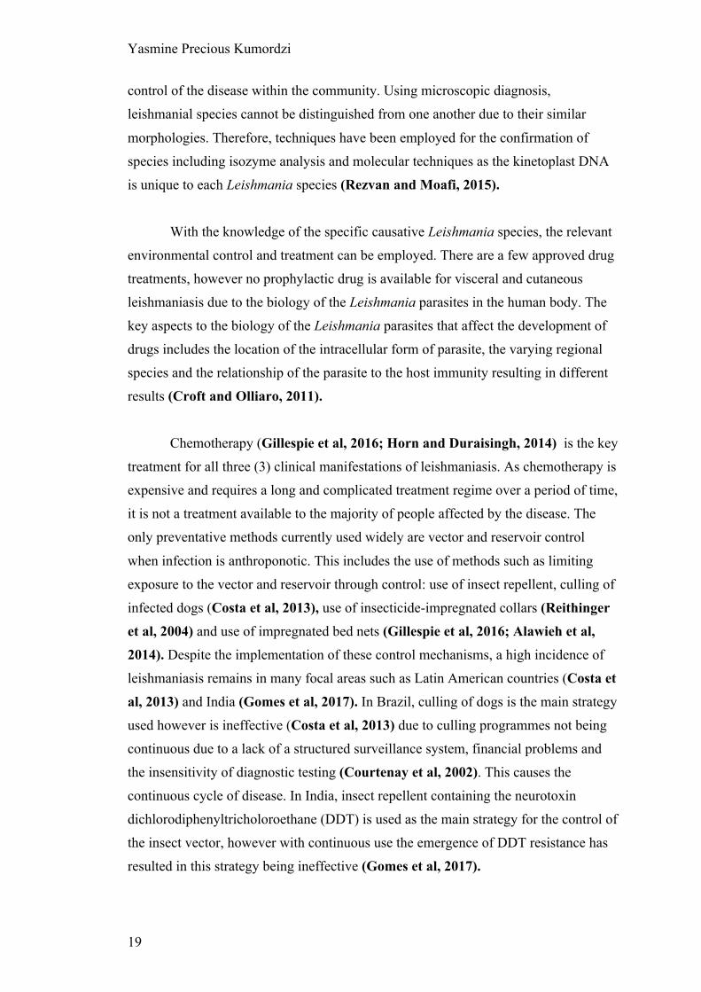
1.2.2 Structure
Compared to other vector groups, the biology of sandflies is poorly known
(Manson, Cooke and Zumla, 2009). Sandfly gut has a slightly more complex
structure in which it has compartmentalized regions where specific activities occur
(Figure 5). This allows for each region to have a unique function and
microenvironment. For the development of Leishmania and the factors that may affect
migration, focus is given to the gut, malpighian tubules and the ventral diverticulum
(crop).
Figure 5. The anatomy of the alimentary canal of sandflies, showing the compartmentalized regions of the gut (midgut, hindgut and foregut), ventral diverticulum (crop), and malpighian tubules. Ilustration from Mansoon, Cooke and Zumla, 2009.
Similar to humans, the alimentary canal represents the passage from the mouth
to anus functioning primarily to convert food into absorbable particles by the actions
of enzymes and muscular movements. The alimentary tube is segmented into 3
primary regions; the foregut, midgut and hindgut which are histologically distinct in
terms of their purpose (Richards and Davies, 1977).
Feeding is essential for the survival of the sandfly in addition to the mode of
transmission in which disease is spread. The cibarial muscle contraction provides the
suction that pulls food in fluid form into the pharynx before passing into the
abdominal midgut or crop (Schlein, Jacobson and Messer, 1992). The crop is a
foregut organ (Stoffolano and Haselton, 2013) used as a reservoir for carbohydrates.

Yasmine Precious Kumordzi
24
With a series of pumps and sphincters the flow of fluid into the crop and fluid into the
midgut is regulated dependent on hydrostatic pressure (Thomson, 1975). The rate of
crop emptying of fluid to the midgut for digestion is based on the metabolic rate of the
sandfly, temperature (Moloo and Kutuza, 1971), growth of the peritrophic membrane
(PM) (Harmsen, 1973) and composition of the haemolymph (Stoffolano and
Haselton, 2013).
The two main segments for parasite development is the midgut and hindgut
(Figure 5). The midgut is composed of two segments; the narrow anterior (thoracic)
midgut which follows from the foregut and the wide posterior (abdominal) midgut
(Adler and Theodor, 1929). This area is lined with a layer of microvillar epithelium
which has a number of functions; it secretes the PM following a blood meal (Rudin
and Hecker, 1982), secretes and produces the digestive enzymes required, and
absorbs nutrients for transport following the digestion process (Soares and Turco,
2003). Most of the enzyme produced are proteinases such as trypsins which are more
active in the gut alkaline environment (Figure 8). Enzyme levels increases following
the bloodmeal and decreases as digestion declines, proportional to the level of protein
found in the midgut from the blood (Lehane, 2005). The hindgut is cuticle lined
similarly to the foregut. The pylorus leads directly from the midgut and contains rows
of posteriorly-directed protrusions possibly to aid the removal of remnants from
digestion (Warburg, 2008).
The malpighian tubules are located between the midgut and the hindgut;
playing an important role in insects as the primary excretory system (Ramsay, 1951)
and perform osmo-regulation (Littau and Smith, 1960). They are composed of
bundles of tubes made up of 3 different types of cells (Figure 6) (Littau and Smith,
1960), the epithelial cells at the distal regions are brush bordered whilst honeycombed
at the proximal region of the tubules (Littau and Smith, 1960). These narrow tubes
infiltrate the haemocoel containing hemolymph from which waste is collected into the
distals of the malpighian tubes. Towards the proximal regions, water is reabsorbed
back into the haemocoel. The nitrogenous waste remaining in the lumen is converted
to urea and later uric acid crystals which is eliminated into the hindgut as excreta.

Yasmine Precious Kumordzi
25
Figure 6. Illustration of the 3 cell types in the Malpighian tubules (MT) from I at the distal region in contact with the haemolymph within the haemocoel surrounding the midgut, to IV at the proximal region associated to the hindgut. Region I contain MT I cells have a loosely packed microvilli brush boarder up to 3ul in length and 0.1 - 0.15ul in diameter in contact with the haemolymph with a dense population of granules. Region II and IV contain MT II and IV cells respectively which are structurally similar with numerous randomly distributed mitochondria and shallow infoldings. Region III contain MT III cells containing numerous granular vesicles used for the main role of excretion. Image from (Littau and Smith, 1960)

Yasmine Precious Kumordzi
26
1.2.3 Feeding
Both male and female sandflies require a regular source of carbohydrates for
energy acquired from honeydew excreted on plants and plant sap as a ‘sugar meal’
(Schlein and Jacobson, 1999). Female sandflies require an additional source of
protein to support the development of eggs which they acquire from the ‘blood meal’
(Ready, 1979) known as gonotrophic concordance (Lehane, 2005). Due to the
specific need of female sand flies to acquire a ‘blood meal’, Leishmania parasites can
pass to and from the sand fly during a blood feed making them an ideal vector
(Telleria et al, 2010). The proboscis is used for both feeds, however the blood and
sugar have separate destinations due to batch digestion. The blood meal travels
through the stomodeal valve and is kept within the midgut where a PM, a sac like
structure is secreted by the midgut epithelium within the first couple of hours (Dillon
et al, 2006). The sugar meal begins by travelling down the stomodeal valve into the
gut, however is quickly diverted into the crop (Tang and Ward, 1998). This
separation is important as the sugar meal within the crop may contain proteinases that
can inhibit blood digestion (Stoffolano and Haselton, 2013).
Sugar meal
The sugar meal is the most important for the survival of the sand fly; in the
wild sand flies feed on plants as their source of sugars. Whilst feeding, sandflies can
adopt a ‘sugar feeding mode’ where they have raised palps (Tang and Ward, 1998).
This sugar meal is kept completely separate from the blood meal in the crop.
Carbohydrate digestion is initiated here due to the presence of salivary glands
enzymes before it is slowly released into the gut where digestion by (alpha)-
glucosidases continues. This gradual release is possibly to avoid significant body fluid
osmolarity fluctuations (Stoffolano and Haselton, 2013). The pH of solely sugar fed
sandflies has a slightly acidic pH of 6 which is the optimum pH for (alpha)-
glucosidase activity. This enzyme is membrane bound and involved in the breakdown
of disaccharides to simple sugars for digestion by hydrolyzing the terminal non-
reducing 1-4 bonds.

Yasmine Precious Kumordzi
27
Blood meal
The blood meal ingested is contained in the midgut surrounded by the PM,
confining the early stage development of Leishmania to within the PM (Pruzinova et
al, 2015). For the initial infection of the sandfly, amastigote forms of Leishmania are
ingested within the blood meal. These parasites multiply and morphologically
transform for the establishment of infection. Before the establishment of infection in
the midgut, the parasites encounter 3 main handicaps (Pruzinova et al, 2015; Telleria
et al, 2010; Shaden Kamhawi, 2006); enzymatic activities, midgut peristalsis and the
PM.
The first hindrance is the activities of digestive enzymes, particularly the
activity of trypsin. Trypsin is the most abundant digestive enzymes within the gut of
hematophagous insects, confined to the midgut lumen (Dillon and Lane, 1993;
Telleria et al, 2010). There are other enzymes that affect Leishmania in the midgut
such as aminopeptidase found in the midgut wall (Dillon and Lane, 1993). These
enzymes are produced by the midgut epithelial cells post blood-meal with distinct
peak times dependent on sand fly species (Dillon and Lane, 1993). Along with
digesting the blood serum, the activities of these midgut proteases select for
‘compatible’ Leishmania to sand fly species combination (Pruzinova et al, 2015).
This is done by the natural vector parasite Leishmania species having the ability to
modify the midgut environment to favour its development by interfering with trypsin
production and subsequently pH and enzyme efficiency (Santos et al, 2014). The
survival to this proteolytic attack is the first essential step for the parasite development
and the establishment of infection within the vector (Pimenta et al, 1997).
The second hindrance is the type I PM formed in response to blood feeding
(Lehane, 1997). Following a blood meal, the PM is developed rapidly and fully
formed by 6-24 hours post blood-meal dependent on sand fly species (Pruzinova et
al, 2015). The PM acts as a physical barrier for the protection of the midgut
epithelium to damage from pathogens found in the midgut lumen (Lehane, 1997) and
conversely protects the Leishmania parasites by compartmentalizing them from the
hydrolytic activities of the midgut (Pruzinova et al, 2015; Secundino et al, 2005).
This remains intact until digestion finishes and the disintegration of the PM occurs.
The absence of PM is associated to the loss of midgut infections due to the lethal

Yasmine Precious Kumordzi
28
conditions of the sand fly midgut (Pimenta et al, 1997). However, this prevents the
escape of the parasites into the ectoperitrophic space and as the remnants of the blood
meal is defecated following digestion can lead to the loss of parasites. As
intraperitrophic Leishmania parasites are not able to traverse the PM prior to its
disintegration (Sádlová and Volf, 2009), escape from the blood meal occurs in the
period between PM disintegration and defecation. L. major infections showed sandfly
derived chitinases disintegrate the PM from the posterior end, therefore escaping the
PM to the ectoperitrophic space requires the high densities of parasite found at the
anterior area of the PM to migrate to the posterior end (Sádlová and Volf, 2009). This
however may be species-specific.
The third hindrance is the action of midgut peristalsis (antegrade)- the motor
pattern of the midgut to propel contents in the direction towards the anus for excretion
of the blood meal remains following digestion (Figure 7) (Shaden Kamhawi, 2006).
Here parasites are exposed to possibly being expelled if they have not escaped the
PM. Therefore strategies have been employed by Leishmania parasite to slow down
excretion such as the secretion of a myoinhibitory neuropeptide relaxing the midgut
(Vaidyanathan, 2004). This leads to the sandfly being less efficient in expelling the
blood meal remains, increasing the time period in which parasite can escape the PM.
Following the escape from the PM, parasites can still be removed from the
midgut by peristalsis and defecation; they therefore need to colonize and attach to the
midgut epithelium to prevent this.

Yasmine Precious Kumordzi
29
Figure 7. The digestion of the blood meal within the sandfly midgut. A-D shows the formation of the PM in S. schwetzi (A) and P. papatasi (B), P. orientalis (C) and in P. argentipes (D). The arrow indicates the thin PM separating the blood bolus (BB) from the midgut epithelium (ME). Following digestion, the remnants remain within the PM through its disintegration. E shows the PM intact with no leak of remnants into the ME. F-H shows degradation occurring in the PM causing a leak of remnants into the ME. Image from (Pruzinova et al, 2015).
From the changes of diet, the physiology of the midgut of female sandflies
modifies to support the digestion of both sugars and blood. These modifications
requires the changes of enzymes as well as the changes of pH needed for the
appropriate activities (Figure 8) (Santos et al, 2008). Following the blood meal, the
slow release of sugars from the crop to the midgut is interrupted by the presence of the
blood meal and the modifications (such as changes in enzymes) present for the
digestion of blood. These changes: presence of enzymes such as trypsin and
chymotrypsin along with the change of acidic pH 6 to alkaline pH 7-8 (Figure 8)
allows for protein digestion and potentially favours Leishmania development within
the gut before an acidic environment is reintroduced post-blood meal.

Yasmine Precious Kumordzi
30
Figure 8. The anatomy of Lu. longipalpis gut and the pH of the midgut during the first 10h (a) and 24 hours (b) after blood ingestion shows the returning of an acidic pH from alkaline following the digestion of the blood meal. The thoracic midgut (TM) and diverticulum (D) filled with sugar solution shows an acidic pH (5.5-6). (AM) abdominal midgut. Image from (Santos et al, 2008).
The initial development of Leishmania parasites within the gut plays within
the fine balance of pH favouring it’s development along with the hindrances of the
presence of the enzymes, midgut peristalsis and the PM development.
1.2.4 Microbiota
Within the gut, Leishmania parasites join the symbiotic resident microbial
community within the alimentary canal of the sandfly (Pumpuni et al, 1996). The
microbiota is said to have a major role influencing the induction, maturation and
function of the host immune system (Telleria et al, 2018) along with the development
of Leishmania parasites (Fraihi et al, 2017); making the interactions between the
parasite and sandfly microbiota important in understanding the migration and
transmission of Leishmania. The life cycle of the sandfly (Figure 4) reflects where
resident and pathogenic microbes found in the sandfly’s microbiota originates from.
This includes disease causing bacteria (Herrer and Christensen, 1975) and viruses
(Depaquit et al, 2010).

Yasmine Precious Kumordzi
31
There is little known about viruses found in sandflies (Depaquit et al, 2010)
however, there are a few phleboviruses transmitted by sandflies that cause disease in
humans such as Toscano viruses (TOSV) (Depaquit et al, 2010) and Sand fly fever
Sicilian Viruses (SFSV) (Ayhan et al, 2017). There have been fewer reports of
sandflies with the presence of a virus and a Leishmania infection. A study using
Phlebotomus papatasi infected with cytoplasmic polyhedrosis virus (CPV) showed a
resistance to the Leishmania infection, possibly due to the pathological modifications
by the virus preventing the attachment of Leishmania to the epithelium and the early
exposure to the digestive enzymes found in the gut during blood digestion (Warburg
and Ostrovska, 1987). Considering other studies of infections of Leishmania
parasites with various viruses (Faucher et al, 2014, Ergunay et al, 2014) it can be
determined that there is a complex relationship between the two which differs
between Leishmania species and specific viruses. Apart from human pathogenic
viruses, there are novel viruses of Lu. longipalpis; Lutzomyia Piaui reovirus 1
(LPRV1), Lutzomyia Piaui reovirus 2 (LPRV2), Lutzomyia Piaui nodavirus (LPNV)
(Aguiar et al, 2015).
The life cycle (Figure 4) shows the egg stage and larvae stage from which
adult sand flies emerge. Their resident microbiota originates from the diverse and
undefined environments in which each stage develop and the food they ingest. This
creates a complex network of overlapping bacteria found within the gut of sandflies
species which can be seen in Figure 9. This can be dependent on developmental stage
and country of origin (Telleria et al, 2018).

Yasmine Precious Kumordzi
32
Figure 9. Network analysis showing the shared bacteria species between sandflies species. Phlebotomus sand flies are identified by squares surrounded by green and bacteria found in Lutzomyia sand flies identified with squares surrounded by blue. Coloured circles represent bacteria species that are shared between sand flies species. White circles represent bacteria species that are unique to each of the sand flies species and are listed inside large rectangles. Image from Fraihi et al, 2017
Ingested food can influence the gut microbiota in both larvae (Vivero et al,
2016) and adult sandflies (Oliveira et al, 2000). The microbial diversity of adult
female sandflies changes with blood feeding (Figure 10) with the resident microbiota
present in non-blood fed females altering when sandflies are blood fed and return
following blood digestion and excreta removed (Endris et al, 1982, Monteiro et al,
2016, Kelly et al, 2017, Pires et al 2017). After a blood meal, bacterial diversity
decreases and overall bacteria numbers increase due to the nutrient-rich environment
provided by the blood (Volf, Kiewegova and Nemec, 2002). This shows how
substantial a role feeding has on the microbiota of sandflies as Leishmania
development primarily occurs during blood digestion when the resident microbiota of
the gut differs as seen in Figure 10.

Yasmine Precious Kumordzi
33
Figure 10. Lutzomyia longipalpis gut microbiota. Network analysis showing bacteria found in Lu. longipalpis dependent on feeding conditions. Image from Telleria et al, 2018
There is a stark difference in bacteria found in ‘blood and infected flies’ and in
‘blood fed flies only’, however as the data used has been acquired from different
publications it represents the wide range of variation of gut microbiota and reflects the
conditions of diet and habitat of sandflies globally (Figure 10) (Mukhopadhyay et al,

Yasmine Precious Kumordzi
34
2012). Bacterial diversity of Leishmania infected midguts decreases over time post
infection to an Acetobacteraceae family rich microbiota (Kelly et al, 2017). The
largest evidence suggesting that the interaction between the microbiota of the sandfly
gut and Leishmania parasite is important was showed by Kelly et al; antibiotic
suppression of gut microbiota resulted in the arrest of parasitic replication and
development (metacyclogenesis) without affecting the health of both the sandfly and
the Leishmania present in the gut showing the huge influence of the microbiota to
infection establishment.
The bacteria present in the sandfly gut have not been further
compartmentalized into foregut, midgut and hindgut. However, it is understood that
quorum sensing will occur in the midgut between bacteria (Fuqua, Winans and
Greenberg, 1994). During quorum sensing, extracellular signalling molecules are
produced to be detected by other bacteria known as autoinducers (AIs). These AIs
diffuse producing concentration gradients which can be detected by bacteria via
receptors present on their surface. It may be possible for Leishmania parasites to also
recognise these molecules, producing a response to their presence.
Yasmine Precious Kumordzi
35
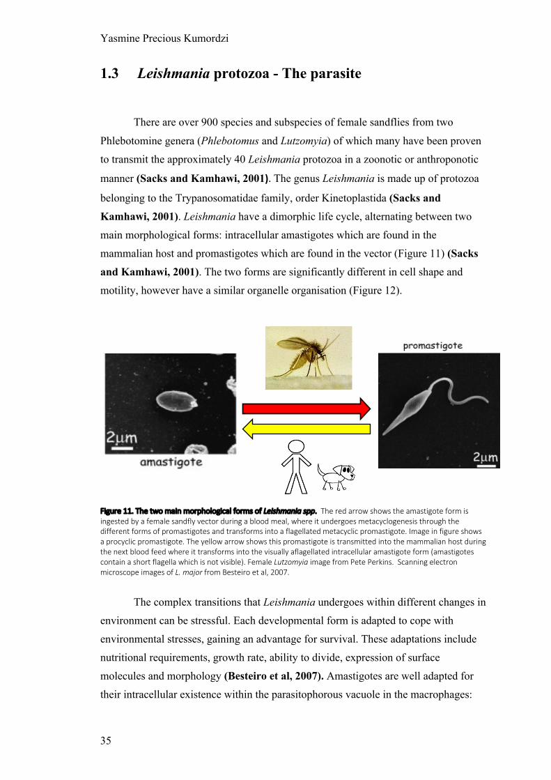
1.3 Leishmania protozoa - The parasite
There are over 900 species and subspecies of female sandflies from two
Phlebotomine genera (Phlebotomus and Lutzomyia) of which many have been proven
to transmit the approximately 40 Leishmania protozoa in a zoonotic or anthroponotic
manner (Sacks and Kamhawi, 2001). The genus Leishmania is made up of protozoa
belonging to the Trypanosomatidae family, order Kinetoplastida (Sacks and
Kamhawi, 2001). Leishmania have a dimorphic life cycle, alternating between two
main morphological forms: intracellular amastigotes which are found in the
mammalian host and promastigotes which are found in the vector (Figure 11) (Sacks
and Kamhawi, 2001). The two forms are significantly different in cell shape and
motility, however have a similar organelle organisation (Figure 12).
Figure 11. The two main morphological forms of Leishmania spp. The red arrow shows the amastigote form is ingested by a female sandfly vector during a blood meal, where it undergoes metacyclogenesis through the different forms of promastigotes and transforms into a flagellated metacyclic promastigote. Image in figure shows a procyclic promastigote. The yellow arrow shows this promastigote is transmitted into the mammalian host during the next blood feed where it transforms into the visually aflagellated intracellular amastigote form (amastigotes contain a short flagella which is not visible). Female Lutzomyia image from Pete Perkins. Scanning electron microscope images of L. major from Besteiro et al, 2007.
The complex transitions that Leishmania undergoes within different changes in
environment can be stressful. Each developmental form is adapted to cope with
environmental stresses, gaining an advantage for survival. These adaptations include
nutritional requirements, growth rate, ability to divide, expression of surface
molecules and morphology (Besteiro et al, 2007). Amastigotes are well adapted for
their intracellular existence within the parasitophorous vacuole in the macrophages:

Yasmine Precious Kumordzi
36
non-motile as migration is not required, reduced size to exist within a small enclosed
space, flagellum length is reduced significantly that it does not emerge from the
flagellar pocket (de Souza and Souto-Padron, 1980), are acidophiles due to the
acidic environment (Antoine et al, 1990) and have a different energy metabolism than
promastigotes (Coombs et al, 1982). Interestingly, from all promastigote forms,
metacyclic promastigotes are pre-adapted for the mammalian survival, expressing
stage-specific surface molecules and complement-resistant (Besteiro et al, 2007).
This shows that adaptation occurs not only for the environment they are within but for
the next anticipated change in environment.
The morphology of a protozoan is one way to cope with the environmental
stresses they may encounter, gaining a competitive advantage for survival. Therefore,
observation of the morphology of the cell and their ultrastructures is fundamental to
understanding the structure and behaviour of the specific protozoa. The main
organelles here are the flagellum, kinetoplast and cell membrane.
1.3.1 Flagellum
The immobilisation occurring in the absence of an external flagellum in
amastigote forms demonstrates that flagellum is the essential sole means of motility
therefore is the key player to migration (Landfear et al, 2001). This shows diversity
within a singular organism to adapt structure and therefore function for a distinct life
cycle stage (Wheeler, Gluenz and Gull, 2015). L. mexicana can form two distinct
flagellum forms; a canonical 9+2 axoneme restricted to motile promastigotes and a 9v
axeneme (9+0 axeneme with a collapsed radial symmetry with irregular inward
migration of other doublets) restricted to immobile amastigotes used for sensing and
signalling (Silverman and Leroux, 2009, Singla and Reiter, 2006). The 9+2
axoneme form is present in Chlamydomonas reinhardtii a biflagellated green algae
that uses its flagella for motility and cell-cell recognition in mating (Silflow and
Lefebvre, 2001), and the 9+0 axoneme form is present in primary mammalian cilia
acting as immobile sensory antennae to coordinate cellular signalling pathways (Satir
and Christensen, 2008).

Yasmine Precious Kumordzi
37
Figure 12. Schematic representation of the main intracellular organelles from Leshmania promastigote (left) and amastigote (right) forms. The flagellum pocket represents the anterior end of the cell and migration occurs in a forward motion.Images adapted from Besteiro et al, 2007

Yasmine Precious Kumordzi
38
The ability to transition between the amastigote and promastigote life stages
within a change of environmental factors such as pH and temperature, shows that this
transition also occurs with the flagellum axoneme to remodel between the 9+2 and
9+0 forms (Wheeler, Gluenz and Gull, 2015). This malleability allows for
adaptation needed in different environments; motile parasite transmission or immobile
within a macrophage. Ciliogenesis is the process by which the flagellum is built,
intraflagellar transport (IFT) increases the flagellar length from the basal body
(Witman, 2003). IFT decreases during the transition of the 9+2 form to the 9+0
axoneme structure, and the requirement of paraflagellar rod (PFR) (Langousis and
Hill, 2014) for normal motility suggests that both IFT and PFR play a key role in the
flagellum shortening and loss of central pair. For motility, axonemal dynenin motors
which are attached to the A-tubule of each outer doublet microtubule undergoes
structural changes powered ATP-dependently. This causes reversible attachment to B-
tubule of the neighbouring doublet, sliding of doublets and resistance causing
bending, known as the ‘sliding filament’ mechanism (Satir, 1968) causing movement
by wave-like beating of the flagellum. The flagellum pocket is an invaginated site
specialized for endocytosis, making it a portal for host-parasite interactions; relaying
information about the microenvironment to allow changes in the parasite to reflect the
demands of its environment (Landfear and Ignatushchenko, 2001).
1.3.2 Kinetoplast
As a flagellated protozoa with the presence of a kinetoplast in the
mitochondrion, Leishmania spp is a kinetoplastid (Simpson, 1968). Other parasites in
this category such as Trapanosoma cruzi and Trypanosoma brucei responsible for
serious human diseases such as Chagas disease and African sleeping sickness
respectively, share the commonality of kinetoplast DNA (kDNA) described by Trager
as ‘a small spherical or rod-shaped structure lying just posterior to the basal body of
the flagellum’ (Trager, 1965). Kinetoplastids have similar genomic organization and
cellular structures, undergoing morphological changes during the progression of their
life cycle and having 6,000 orthologs (common ancestral genes) out of 8,000 genes
(Stuart et al, 2008) in their genome in common. They however have very distinct
properties such as their insect vector and the resulting human disease. These

Yasmine Precious Kumordzi
39
commonalities, allows for the advanced knowledge of kDNA in Trypanosomes spp to
support and build on that of Leishmania spp.
Along with cell morphological changes observed in development, the
kinetoplast is useful for pinpointing the progression within the life cycle of
kinetoplastids. It is therefore used to determine differentiated forms due to its
positional movement relative to other organelles; particularly the nucleus during cell
progression. The morphologic forms of flagellates are defined by the position of the
flagellum pocket, nucleus and kinetoplast which varies along the anterior/posterior
axis of the cell body. As the kDNA is always posterior to the flagellar pocket and
remains closely connected to the basal body (Vargas-Parada, 2010), this morphology
can simply be defined as the kDNA location relative to the nucleus. This simplistic
categorization results in 4 categories where the position of the kinetoplast changes
relative to the nucleus; trypomastigote, epimastigote and promastigote having a
long/slender body with a protruding flagellum, and amastigote having a round/oval
shaped with no protruding flagellum (Figure 13). The flagellum emergence dictates
the anterior end of the cell body. In trypomastigotes, the kDNA and flagellar pocket is
located at the posterior end of the parasite relative to the nucleus. Epimastigotes have
a close centralized kDNA, anterior to the nucleus; and due to the flagellum spanning
along the cell body an undulating membrane is formed in a similar manner to that in
trypomastigotes. In Trypanosomes spp., the posterior flagellum emerges from the
flagellar pocket however is not free as it attaches to the cell body creating
morphological forms (trypomastigotes and epimastigotes) with undulating membrane.
Promastigotes have the kDNA located furthest anterior to the nucleus resulting in the
flagellum being completely free and without an undulating membrane.
The morphologies present for each flagellate life cycle are restricted and
dependent on specific species; Trypanosoma brucei only having trypomastigote and
epimastigote forms (Field and Carrington, 2009) and L. mexicana only having
amastigote and promastigote forms.

Yasmine Precious Kumordzi
40
Figure 13. Kinetoplast repositioning in relation to other organelles during the life cycle of a flagellate. FP flagellar pocket, kDNA, kinetoplast DNA. Image from Field and Carrington, 2009.
With the kinetoplast location being one of the key observations to pinpoint
progression of life cycle, understanding the kinetoplast movement is important. The
kinetoplast is attached to the basal body which is adjacent to the flagellar pocket; the
flagellum is formed by extension from the basal body (Wheeler, Gluenz and Gull,
2015). This relation between the three different components (nucleus, flagellum
pocket and kDNA) results in differences between the length of unattached flagellum
at the anterior end of the cell affecting the motility of the cell.
1.3.2.1 Cell cycle
Within an asynchronous population of Leishmania promastigotes, dividing cells can
be identified through morphological changes by phase contrast microscopy (Ambit et
al, 2011). With the use of DAPI (4′,6-diamidino-2-phenylindole) staining the nucleus
(N) and the kinetoplast (K) with the visualisation of the cell body and the flagellum,
the cell cycle position of the individual cell can be given. A cell in G1 phase has the

Yasmine Precious Kumordzi
41
configuration of 1N1K1F and a cell in cytokinesis has the configuration 2N2K2F
configuration.
Figure 14. The cell cycle of promastigote L. mexicana by light microscopy. Micrographs of major cell cycle stages; cells were ordered based on number of kinetoplasts (K), nuclei (N) and flagella (F). The kinetoplast and nucleus are indicated in (A). Arrow in D shows the emergence of the new short flagellum. The scale bar represents 5 µm. Image from Wheeler, Gluenz and Gull 2010
During the cell cycle, the Leishmania cell initially grows in length with the DNA
content remaining constant (Figure 14 A). DNA synthesis begins with the duplication
of DNA content while cell length remains constant (S phase) (Figure 14 B and C).
Finally, the cell length reduces (G2 phase and mitosis) (Figure 14 D and E) and
cytokinesis divides (Figure 14 E and F) the cell from daughter cell, returning to the
start of the cell cycle (Wheeler, Gluenz and Gull 2010).

Yasmine Precious Kumordzi
42
Figure 15. Illustrations showing the properties of each cell measurement: cell body length and width, kinetoplast and nucleus DAPI intensity, flagellum length, kinetoplast-anterior (K-A) length, nucleus-kinetoplast (N-K) length, and nucleus-anterior (N-A) length. Image from Wheeler, Gluenz and Gull 2010 The kinetoplast of L. mexicana is positioned at a constant distance of approximately
2.5 µm from the anterior end of the cell (Wheeler, Gluenz and Gull 2010). Nuclear
position within the cell varies but has a relationship with the cell length defined as n ≈
2.5 + 0.2l where n = anterior–nucleus distance and l = cell length in micrometres
(Figure 15) (Wheeler, Gluenz and Gull 2010).
1.3.2.2 Morphometrics
There are different morphological changes that promastigotes undergo during
metacyclogenesis from the initial procyclic promastigote to the metacyclic
promastigotes. These developmental forms are defined according to their cell body
length, cell body width and flagellum length (Figure 16) (Rogers et al, 2002):

Yasmine Precious Kumordzi
43
Figure 16. Illustration of promastigote morphologies categorisation. L. mexicana morphological categorization (Rogers et al, 2002)
1.3.3 Plasma membrane
The contiguous surface membrane (Figure 17) of kinetoplastid protozoa are
divided: the flagellar membrane, the flagellar pocket and the pellicular plasma
membrane each unique to each other (Landfear and Ignatushchenko, 2001).
The flagella pocket is the deep invagination at the base of the flagellum,
responsible for uptake of large nutrients by endocytosis and secretion of proteins. L-
haemoglobin is seen to be internalised from the flagella pocket membrane (Landfear
and Ignatushchenko, 2001). Filamentous acid phosphatase (sAP) and filamentous
proteophosphoglycan (fPPG) is secreted into the extracellular medium here.
The flagellar membrane covers the flagellum. Specialised membrane proteins
are found in the flagella membrane and serve in sensing and signalling such as
LmjAQP1 (Figarella et al, 2007), ISO1 (Snapp and Landfear, 1999), LmGT1

Yasmine Precious Kumordzi
44
(Burchmore et al, 2003), receptor-adenylate cyclases (Sanchez et al, 1995).
LmjAQP1 is a aquaglyceroporin channel involved in detection of extracellular
osmotic gradients and osmotaxis (Figarella et al, 2007). LmGT1 is a glucose
transporter also found in flagella pocket membrane. Receptor-adenylate cyclases have
been expressed and may function as an adenylate cyclase (Sanchez et al, 1995). ISO1
is also found in the flagella pocket (Landfear and Ignatushchenko, 2001). This
shows the flagellum pocket and flagellum membrane allows for the detection of
signals through the membrane proteins located here.
The pellicular plasma membrane covers the entire of the cell surface and is
covered with densely packed microtubules and glycolipid lipophosphoglycan (LPG)
coat and contains many permeases for nutrient uptake (Landfear and
Ignatushchenko, 2001), such as LmGT2 and LmGT3.
Figure 17. A electron micrograph through the flagellar pocket of a kinetoplastid (T. brucei) showing the contiguous surface membrane. Image from Landfear and Ignatushchenko, 2001.
Kinetoplast DNA
Cytoplasmic vesicle

Yasmine Precious Kumordzi
45
Nutrition of all protozoa is holozoic, therefore they require organic materials
(Johnson, 1941). Parasitic protozoa such as Leishmania, acquire nutrients from the
hosts by using transport proteins located on their plasma membrane known as
permeases. Due to the intracellular parasitism nature of the life cycle, there are
obstacles that the parasite needs to overcome to acquire the nutrients required for
growth (Landfear, 2011). These are described by Landfear as competition with the
sandfly and mammalian host for essential nutrients required and the distinct
environmental stresses in the sandfly and the macrophages. These distinct
environments lead to stresses such pH, temperature and available nutrients found in
each of the host. These can be solved by the parasite having an nutrient uptake system
which can be modified to accommodate the alternate environment each host;
alteration in the uptake in accordance to available nutrient and developmental stage of
parasite.
Amastigotes remain within the parasitophorous vacuole (PV) in the
macrophage of the mammalian host, there are two main ways in which amastigotes
may obtain nutrients; vesicular transport and transporters. Vesicular transport from the
macrophage plasma membrane of phagolysosomal degradation products present in the
PV lumen or from the host cell cytosol. Nutrients such as hexoses, purines, iron and
polyamines are imported through transporters on the cell surface membrane (Table 1)
into the amastigote by transporters located in the parasite plasma membrane
(Landfear, 2011).

Yasmine Precious Kumordzi
46
Table 1. Transporters in Leishmania species and the nutrients they provide. FM flagellar membrane PPM Pellicular plasma membrane FPM flagella pocket membrane (Landfear, 2011).
Hexose transporters
Hexose sugars include glucose, fructose, galactose and mannose. Plant nectar
is an important part of sandfly diets. Therefore these sugars are readily found in high
concentrations within the thoracic midgut of the sandfly following a sugar meal
(Schlein, 1986). Hexose transporter genes LmxGT1, LmxGT2 and LmxGT3 are
encoded in L. mexicana genome by single copy genes that are clustered together at a
single locus (Burchmore and Landfear, 1998) and homologous to mammalian
glucose transporter (GLUT) family. There is a significant downregulation of these
genes in amastigotes, reflecting the lower glucose concentration within the
macrophage (Cairns, Collard and Landfear, 1989). Glucose is metabolised by
promastigotes as a source of energy that is readily available, using the glycosome
organelle to contain and metabolise the glucose (Opperdoes, 1987).
Nutrient (s) Parasite Transporters (location) Hexoses L. mexicana LmxGT1 (FM, FPM)
LmxGT2 (PPM)
LmxGT3 (PPM)
Purines L. donovani
L.major
LdNT1 to LdNT4
LmaNT1 to LmaNT4
Iron L. amazonensis LIT1
Polyamines L. major LmaPOT1
Amino acids L. donovani LdAAP3
Polyols L. donovani LdMIT
Folates Leishmania species FT1 to FT14
Nucleotide sugars L. donovani
L. major
LPG2
LPG5
Phospholipids L. donovani
LdMT
Water, glycerol, osmolytes L. major LMAQP1
Cations L. amazonensis Lmaa1

Yasmine Precious Kumordzi
47
LmGT1, LmGT2 and LmGT3 proteins transport glucose, fructose, galactose
and mannose, and LmGT2 and LmGT3 transport ribose (Naula et al, 2010). The
LmGT1 membrane protein acts as a glucose sensor, null mutants of LmGT1 growth is
rapid without entering stationary phase, therefore may ensure that parasites know
when glucose has been exhausted (Burchmore et al, 2003).
Purine transporters
Leishmania lack a de novo biosynthetic pathway to synthesize purines,
therefore as an essential nutrient these need to be salvaged from host (Hammond and
Gutteridge, 1984). In this process preformed purine nucleosides or nucleobases are
salvaged and transported into the parasite, followed by interconversion or metabolism
to phosphorylated nucleotides which can be used (Landfear, 2011). Identified
nucleoside transporters for specific purine includes; LdNT1 (substrates adenosine and
the pyrimidine nucleosides) (Vansudevan et al, 1998), LdNT2 (substrates inosine,
guanosine, xanthosine) (Carter et al, 2000), LmaNT3 and LmaNT4 (the substrates
hypoxanthine, xanthine, adenine, and guanine). These purine transporters are active
proton-coupled symporters therefore can use their high affinity to compete with host
tissues effectively. Interestingly, NT3 and NT4 have differing transport optimum pH.
NT3 has an optimum neutral whilst NT4 has an optimum acidic pH. This suggests that
NT3 is designed to function optimally in the insect vector whilst NT4 is designed to
function optimally in the phagolysosome (Ortiz et al, 2009). Low levels of purines
are detected by Leishmania and responded to by the upregulation of mRNA encoding
purine transporters for the salvaging (Carter et al, 2010).
Iron transporters
The phagolysosome is a kept free of iron by a Fe2+ pump, therefore
Leishmania are in an area lacking iron. This is done as Fe2+ is toxic in high levels,
however iron is taken up by Leishmania in the form of Fe2+ (Wilson et al, 2002).
Holotransferrin, a complex of two Fe3+ ions and transferrins protein is however pesent
in phagolysosome. The iron in these complexes are utilised by Leishmania however
needs to be reduced prior to internalization (Wilson et al, 1994). A membrane-bound
NADPH-dependent iron reductase converts Fe3+ bound to protein to Fe2+ at the
extracellular surface of the parasite (Wilson et al, 2002); Leishmania iron transporter

Yasmine Precious Kumordzi
48
LIT1-1 and LIT1-2. These are however only expressed in environment lacking iron,
therefore not expressed in promastigotes (Landfear, 2011) and could possibly be a
trigger for the differentiation of amastigotes (Mittra et al, 2013).
Amino Acid transporters
Amino acids have multiple functions and this is showed by amino acid
transporters representing the largest families of permeases (except for ABC
transporters) encoded within the genome, with about 35 in Leishmania major (Aslett
et al, 2009). AAP3 arginine carrier from L. donovani (LdAAP3) (Shaked-Mishan et
al, 2006) transport arginine an essential amino acid in protein synthesis and used a s a
precursor for polyamines synthesis, and LdAAP7 transport lysine (Inbar et al, 2010).

Yasmine Precious Kumordzi
49
CHAPTER TWO: LITERATURE REVIEW
2.1 Leishmania Sand fly Interactions
Leishmania parasites need sand flies to complete their life cycle and to
propagate (Kamhawi, 2006). Parasites are transmitted by the bite of an infected insect
vector. To achieve the status of being a vector, there are criteria that need to be
fulfilled. This includes the vector showing anthropophilia, the vector being found in
nature infected with the same Leishmania species as found within human and
reservoir infections in the same area, the vector fully supporting the development of
parasite after the digestion of the blood meal and the ability to transmit the parasite by
a bite (Killick-Kendrick, 1990). Therefore, parasite vector interactions important for
the possibility of transmission includes the ability to survive defecation by attaching
to the midgut epithelium, and the migration of infective forms of the parasites to the
stomodeal valve and anterior midgut (Maia and Depaquit, 2016).
2.1.1 Metacyclogenesis of suprapylaria Leishmania
Within the sand fly, Leishmania parasites undergo a complex development
process that occurs exclusively within the alimentary canal. This begins in the midgut
where macrophages infected with amastigotes are introduced to the digestive tract
with the ingested bloodmeal which goes through the digestion process. During the
digestion, parasites multiply and their morphology changes from the amastigote form
to the flagellated promastigote motile from. The blood meal arrival to the midgut
induces the production of the peritrophic matrix (PM), therefore Leishmania present
in the blood meal is further confined within the PM. To establish infection, surviving
parasites need to escape defecation and successfully attach to the midgut epithelium
(Pruzinova et al, 2015). This process from introduction to the digestive tract of the
sandfly to the establishment of infection in the midgut has many hindrances that need
to be overcome for establishment of infection. These includes successful development
and morphology changes of parasite, surviving digestion, escaping from the PM and
successful attachment to the epithelium before defecation occurs.
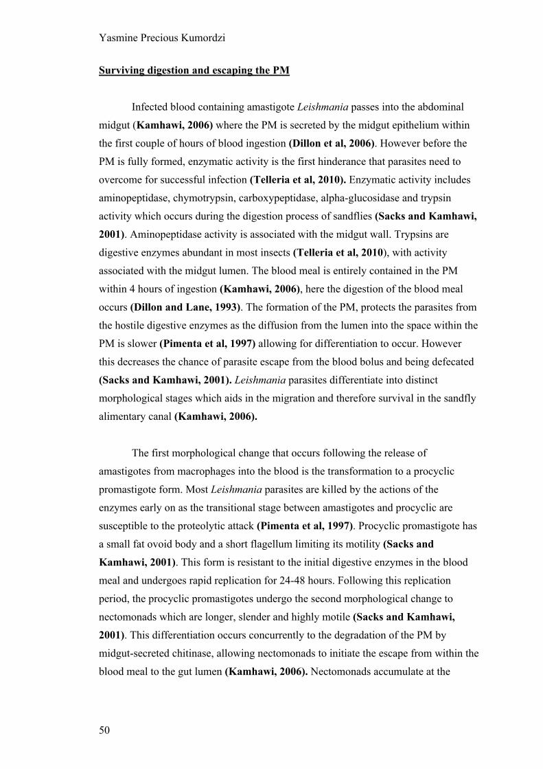
Yasmine Precious Kumordzi
50
Surviving digestion and escaping the PM
Infected blood containing amastigote Leishmania passes into the abdominal
midgut (Kamhawi, 2006) where the PM is secreted by the midgut epithelium within
the first couple of hours of blood ingestion (Dillon et al, 2006). However before the
PM is fully formed, enzymatic activity is the first hinderance that parasites need to
overcome for successful infection (Telleria et al, 2010). Enzymatic activity includes
aminopeptidase, chymotrypsin, carboxypeptidase, alpha-glucosidase and trypsin
activity which occurs during the digestion process of sandflies (Sacks and Kamhawi,
2001). Aminopeptidase activity is associated with the midgut wall. Trypsins are
digestive enzymes abundant in most insects (Telleria et al, 2010), with activity
associated with the midgut lumen. The blood meal is entirely contained in the PM
within 4 hours of ingestion (Kamhawi, 2006), here the digestion of the blood meal
occurs (Dillon and Lane, 1993). The formation of the PM, protects the parasites from
the hostile digestive enzymes as the diffusion from the lumen into the space within the
PM is slower (Pimenta et al, 1997) allowing for differentiation to occur. However
this decreases the chance of parasite escape from the blood bolus and being defecated
(Sacks and Kamhawi, 2001). Leishmania parasites differentiate into distinct
morphological stages which aids in the migration and therefore survival in the sandfly
alimentary canal (Kamhawi, 2006).
The first morphological change that occurs following the release of
amastigotes from macrophages into the blood is the transformation to a procyclic
promastigote form. Most Leishmania parasites are killed by the actions of the
enzymes early on as the transitional stage between amastigotes and procyclic are
susceptible to the proteolytic attack (Pimenta et al, 1997). Procyclic promastigote has
a small fat ovoid body and a short flagellum limiting its motility (Sacks and
Kamhawi, 2001). This form is resistant to the initial digestive enzymes in the blood
meal and undergoes rapid replication for 24-48 hours. Following this replication
period, the procyclic promastigotes undergo the second morphological change to
nectomonads which are longer, slender and highly motile (Sacks and Kamhawi,
2001). This differentiation occurs concurrently to the degradation of the PM by
midgut-secreted chitinase, allowing nectomonads to initiate the escape from within the
blood meal to the gut lumen (Kamhawi, 2006). Nectomonads accumulate at the

Yasmine Precious Kumordzi
51
anterior of the blood bolus as the PM degradation initiates from the anterior (Sacks
and Kamhawi, 2001).
Attachment to the matrix wall- hindrance
Within the gut lumen, nectomonads may attach to the midgut epithelial wall
using LPG on the surface of their flagellum to the glycoconjugates on the microvilli in
the midgut; this allows parasites to escape defecation. Attachment of Leishmania
nectomonads to the microvilli on the surface of the midgut epithelium is important for
successful development of parasites within the sandfly midgut (Dillon et al, 2006).
These nectomonads also initiate migration towards the anterior midgut and
some nectomonads undergo another differentiation into leptomonads, a shorter form
of nectomonads. Due to migration and rapid replication, both nectomonad and
leptomonad forms are found to colonize the thoracic midgut where they undergo the
transformation into metacyclic and haptomonad promastigotes (Rogers et al, 2002).
Within the stomodeal valve, leptomonads secrete parasite secreting gel (PSG) which
contains a large number of metacyclic promastigote (Stierhof et al, 1999) and the
leptomonads that are continuously replicating and differentiating into metacyclics.
Metacyclic promastigotes have slender bodies and the longest flagellum advancing
their motility and haptomonad promastigotes bind to the cuticle-lined surface of the
valve via the insertion of the flagellum into the hemi-desmosome-like structures. The
metacyclic promastigote form is known widely as the infective stage parasite
(Kamhawi, 2006). This whole process which is dependent on the Leishmania species,
roughly takes 6-9 (Kamhawi, 2006) days

Yasmine Precious Kumordzi
52
Figure 18. Metacyclogenesis of suprapylarian Leishmania. The development of Leishmania in a permissive vector in a time dependent manner, showing the morphological changes that occurs from procyclics to haptomonads within the sandfly midgut. A) Procyclic promastigotes transform to nectomonad promastigotes which migrate to the anterior of the blood bolus as PM disintergration initiates at the anterior. B) Nectomonads escape from the blood bolus. Digested blood begins to be excreted. Nectomonads attach to the midgut epithelium and transform to leptomonads which rapidly replicate. C) Some nectomonads remain attached to the midgut. Leptomonads migrate to the thoriac midgut where they secrete PSG and transform to motile metacyclics and haptomonads. The PSG damages the cardia and some parasites escape to the foregut. Image from Sacks and Kamhawi, 2001.
A
B
C

Yasmine Precious Kumordzi
53
2.1.2 Developmental cycles in the sand fly
Metacyclogenesis within sand flies can vary depending on the Leishmania
species. However, in literature, most of the illustrated metacyclogenesis is based on
suprapylarian Leishmania species such as L. mexicana, L. amazonensis and L.
chagasi. Through the section of gut in which development occurs, Leishmania
species can be divided into 3 groups as suggested by Lainson et al. (Lainson et al,
1979): hypopylaria, peripylaria and suprapylaria (Figure 19).
Hypopylarian species develop in the hindgut and are assigned the subgenus
Sauroleishmania such as L. tarentolae (Lainson et al, 1979). These species are not
transmissible to mammalian hosts but have a lizard host and natural vector
Sergentomyia sandfly. There is little known about the transmission of
Sauroleishmania, however it is hypothesized that reptiles get infected by consuming
infected sandflies (Wilson and Southgate, 1979). Possible reasons for the inability of
transmission has been associated to the thicker PM formed around the BB by
Sergentomyia compared to Lutzomyia and Phlebotumus sandflies. This does not
support anterior migration or metacyclogenesis as parasites cannot escape the thicker
PM and development is therefore arrested at procyclic promastigotes (Lawyer et al,
1990). However when a L. tarentolae, a hypopylarian species develops within
Lutzomyia longipalpis, development is seen and attachment to the hindgut epithelial is
viewed however anterior migration does not occur and parasite is lost (Dillon and Liu,
unpublished work). Possible reasoning for this includes the inability of metacyclic
lipophosphoglycans (LPG) to reattach, flagellum attachment overcome due to
abrasion from excreta released from the malpighian tubules and midgut peristalsis,
and the possible lack of positive chemoeffectors.
Suprapylaria develop in the midgut and attachment of Leishmania
nectomonads occurs in the abdominal midgut. Suprapylaria species are assigned the
subgenus Leishmania such as L. mexicana. Peripylorian parasites initially develop in
the hindgut where their nectomonads may attach and later migrate anteriorly.
Peripyloria species are assigned the subgenus Viannia such as L. braziliensis. Their
ability to migrate further without expulsion similar to hypopylaria species is due a
modification in metacyclic LPG, enabling reattachment (Soares et al, 2005). Both

Yasmine Precious Kumordzi
54
suprapylaria and peripylaria migrate to invade the foregut and are therefore able to be
transmitted for disease causing.
Figure 19. Diagrammatic representation of Leishmania development sections within a sandfly as proposed by Lainson and Shaw (1979). Structural features of the sand fly includes the proboscis (pr), stomodeal valve (sv), cardia (c), thoracic midgut (tm), abdominal midgut (am), malpighian tubules (mt), pylorus (py) and rectum (r). Figure shows the sections of Hypopylaria (development confined to the hindgut, with the parasites not able to be transmitted due to loss during defecation), Suprapylaria (development confined to the midgut and foregut, with the parasites able to be transmitted) and Peripylaria (development occurs throughout the gut and the parasites can be transmitted). Black shows the sections of Leishmania development, with the yellow arrows showing the migration of parasite following metacyclogenesis . Image from Kaufer et al, 2017.
Yasmine Precious Kumordzi
55
2.1.3 Leishmania manipulation of the Phlebotomine host interaction
Some species have been shown to manipulate the action of their sandfly vector
in order to benefit their survival (Kamhawi, 2006). Defecation is the main approach
by which sandflies potentially expel parasites. Defeating this is therefore one of the
ways in which Leishmania manipulates its interaction with sandflies to enhance
survival. The PM containing the blood undergoes degradation by chitinases before
expulsion occurs. Leishmania is shown to secrete chitinases to encourage earlier
degradation of the PM to allow nectomonads to escape into the lumen (Ramalho-
Ortigão et al, 2005). Genes coding secreted chitinases has been identified in many
Leishmania species with L. mexicana showing an overexpression of LmexCht1 (Joshi
et al, 2005), therefore nectomonads escape the PM quicker, a higher parasite load
occuring and infection is established faster (Rogers et al, 2008). This chitinase
activity may also aid in transmission by damaging the chitin-covered stomodeal valve
(Rogers et al, 2008). L. major secretes a myoinhibitory neuropeptide that successfully
inhibits midgut peristalsis, therefore sandfly parasite loss through defecation is
reduced (Vaidyanathan, 2004, Vaidyanathan, 2005).
Secretion of phosphoglycans by leptomonads causes the build-up of PSG, a
filamentous proteophosphoglycan (fPPG)-rich gel that accumulates in the anterior
midgut and stomodeal valve leading to blockage in these parts (Sacks and Kamhawi,
2001). fPPG is expelled from promastigote flagellar pockets, and can be found in the
center of rosettes in culture where flagella aggregate (Stierhof et al, 1999). This
manipulation causes a change in the nature of the feeding of the sandfly, multiple
feeding due to incomplete feeding, and the regurgitation of promastigotes to aid
sandfly feeding (Rogers et al, 2002, Rogers et al, 2004). This along with the
production of chitinases by haptomonads for valve degeneration (Schlein, Jacobson
and Messer, 1992) impairs feeding for increased efficiency in transmission of
metacyclic promastigotes during feeding (Rogers et al, 2004, Kamhawi, 2006). This
supports for efficient transmission.
Leishmania parasites interfere with digestive enzyme activities. Promastigotes inhibit
proteolytic activity in the midgut (Borovsky and Schlein, 1987) and amastigotes
cause delays in trypsin and aminopeptidase activity (Dillon and Lane, 1993).
Yasmine Precious Kumordzi
56
2.1.4 Important features of Leishmania for transmission
The main feature of Leishmania parasites that allows for transmissibility is the
flagellum but the surface receptors and LPGs play a role in transmission.
LPGs are the most abundant glycoconjugates of promastigotes expressed on
the entire surface of promastigotes (Sacks and Kamhawi, 2001), playing a large role
in adhesion to the midgut epithelium via galectin on the sand fly following escape of
nectomonads. They play a role in enabling competent Leishmania species able to
develop within Phlebotomus species, known as ‘permissive’ and ‘restricted’ vectors
(Kamhawi et al, 2000). Permissive vectors allow the survival and/or development of
several Leishmania spp within them, whereas restricted vectors allow the survival of
specific Leishmania spp. Change occurs in the LPG during differentiation of the non-
infective promastigote form to the infective promastigote form. All LPGs are
composed of a conserved glycan core region of Gal(α1,6) Gal(α1,3) Galf(β1,3)
[Glc(α1)-PO4] Man(α1,3) Man(α1,4)-GlcN(α1) linked to a 1-O-alkyl-2-lyso-
phosphatidylinositol anchor, and a conserved (Gal(β1,4)Man(α1)-PO4 backbone of
repeat units and the oligosaccharide cap with a branching variable sugar which is
responsible for the polymorphism amongst Leishmania species (Soares and Turco,
2003).
Metacyclogenesis results in increasingly more sugars getting masked and LPG
elongating during promastigote development (Soares et al, 2002). In suprapylaria
species which develop in the midgut, metacyclic LPG does not bind to the midgut
microvilli (Soares et al, 2002) due to structural changes caused by metacyclogenesis.
This permits the release of infective stage promastigotes during transmission
(Pimenta et al, 1992). Compared to suprapylaria species, peripylaria species differ in
midgut attachment (Soares et al, 2005); peripylaria metacyclics make less LPG than
procyclics in which glucose residues are added. This is possibly due to their
development in the hindgut therefore to avoid defecation, they migrate to the midgut
to reattach before migrating further anterior. There are however no studies on LPGs
in hypopylarian Leishmania species which develop in the hindgut.

Yasmine Precious Kumordzi
57
Figure 20. Schematic diagram of LPGs from representative procyclic and metacyclic Leishmania species. The structure of the glycan core is Gal(α1,6)Gal(α1,3)Galf(α1,3)[Glc(α1-PO4)-6]-Man(α1,3)Man(α1,4)GlcN(α1,6) linked to the 1-O-alkyl-2-lyso-phosphatidylinositol anchor. The repeat units are 6-Gal(β1,4)Man(α1)-PO4. The diagram shows the result of metacyclogenesis on LPG- elongation and downregulation of glucose resulting in the loss of epitope for binding to midgut epithelium. Arabinosyl (Ara). Image from Turco and Sacks, 2003.
Flagellum is important for motility for escape, allowing the parasite to propel
forwards. Due to the position of the flagellum at the anterior of the promastigote, it is
the first structure of the promastigote to explore the environment it is moving towards.
This allows the opportunity for flagellum to be the main sensory organ through the
mitogen-activated protein (MAP) kinases pathway (Rotureau et al, 2009). The length
of flagellum continuously increases throughout metacyclogenesis. The length of
Leishmania has been showed to be important in the adhesion to the midgut, with
shorter flagellated promastigotes being more readily excreted due to not binding to the
matrix epithelium following escaping the PM (Cuvillier et al, 2003).

Yasmine Precious Kumordzi
58
2.1.5 Transmission
Metacyclogenesis of Leishmania ends with highly infective metacyclic
promastigotes within the cardia of the sandfly. As pool feeders, sandflies cut the skin
of their host damaging dermal capillaries to create a pool of blood on the surface of
the skin which they can suck into the pharnyx and is later diverted into the midgut
through the one way cibarial valve (Rogers, 2012). This presence of PSG
accumulation and pathology caused by the accumulation such as the damage to the
cibarial valve affects the manner in which the sandfly feeds, infected sandflies have
multiple feeding attempts by increasing probing, feeding time increases and
incomplete blood meal usually occurs (Beach, Leeuwenburg and Kiilu 1985)
showing that there is difficulty in feeding. This enhances the chances of transmission
during feeding (Killick-Kendrick et al, 1977).
During the blood meal of an infected sandfly, metacyclic promastigotes can be
transmitted to the mammalian host. The mechanism falls within two hypothesis: the
regurgitation hypothesis and the inoculation hypothesis. The inoculation model
proposed that only promastigotes in the proboscis are involved in transmission due to
infection occurring when sandflies were observed to probe skin however no blood
feeding taking place (Killick-Kendrick et al, 1977, Beach, Leeuwenburg and Kiilu,
1985). The regurgitation model was initially reported by Short and Swaminath 1928.
This proposed that the physical obstruction caused by PSG along with the pathology
caused by chitinase damage causes a regurgitation of the PSG including promastigotes
into the pool of blood before feeding can occur (Schlein, Jacobson and Messer,
1992). Efficient transmission requires the movement of promastigotes to the anterior of the
sandfly midgut (Leslie, Barrett and Burchmore, 2002), however the exact stimulus
that allows this process to occus is not well established. Killick-Kendrick
hypothesized the sugar gradient created by the sugar released from the crop provided
an stimulus for migration (Killick-Kendrick, 1978). This has later been supported by
chemotaxic assays.

Yasmine Precious Kumordzi
59
2.2 Chemotaxis
Chemotaxis is the migration of cells directionally when exposed to gradients
of chemoeffector molecules (Englert, Manson and Jayaraman, 2009). Naturally
there will be multiple chemoeffectors in an area, creating multiple coexisting
overlapping gradients of specific chemical sensors in which chemotaxis occurs.
For microbes, it is advantageous to move away from unfavourable conditions
towards favourable environments such as those containing a source of food (Adler,
1966), making chemotaxis important for survival. Cells detect gradients of chemicals
in two ways; directly measuring spatial gradients across the cell or indirectly
temporally sensing gradients while motile. Motile bacteria randomly change direction
regularly however with the detection of a gradient, this behaviour pauses allowing the
bacteria to modify its movement dependent on how favourable the gradient is
(Armitage, 1992).
The first step of chemotaxis is the detection of gradient. Understanding of the
detection method of the molecules used for chemotaxis to occur is thought to be
through chemoreceptors; chemoeffector molecules binding to the receptors found on
the cell surface or the detection of their metabolites (Adler, 1969). Ligand molecules
bind to their specific membrane chemoreceptors, activating them and triggering signal
transduction pathways downstream.
Chemotaxis has been largely studied in Escherichia coli by Adler (Adler,
1972), acting as the basis for most of the chemotaxis work on other microbes
(Keymer et al, 2006). Like E. coli, motile microbes can be attracted or repelled by
specific chemical stimuli (Adler, 1973) – positive and negative chemotaxis
respectively. Assays describing measuring this chemotaxis were initially discovered in
the 1880’s by Pfeffer and Englemann (Adler, 1966) and continue to be improved
upon.
Chemotaxis studies require a way to deliver chemicals to cells in a controlled
gradient, for cells to be able to sense and direct their movement according. Earlier
work presented a variety of techniques. The first described basic chemotaxic assay is

Yasmine Precious Kumordzi
60
the Quantitative Pfeffer method (Adler, 1973) which involves placing motile E. coli
at one end of a capillary tube containing a source of energy and oxygen. This allows
the migration of E. coli into the tube resulting in two visible bands; the first containing
E.coli that has consumed all the sugars and the second band migrating for the
remainder oxygen (Adler, 1966). This therefore includes the overlap of osmotaxis and
chemotaxis. This method was slightly altered by Adler and Armitage separately
resulting in different chemotaxis assays. Adler’s method for measuring chemotaxis
(Adler, 1973) was based on using a capillary tube with a small chamber. A U-shaped
tube was bent from a capillary tube, placed on a glass slide and sealed using a cover
slip forming the chamber in which 0.2ml of bacterial suspension was added as seen in
Figure 21. Another capillary tube was sealed at one end and used to contain the
attractant. This was inserted into the chamber containing bacterial suspension. After 1
hour incubation, contents of the capillary tube were removed and plated for colony
count the following day (Adler, 1973). Armitage developed a method similar to the
Boyden Chamber assay (Boyden, 1962), by using blind well chemotactic chambers,
quantifying the motile bacteria that migrated from the bacterial suspension well
through a polycarbonate membrane (2 μm) into the chemotactic chamber (Figure 21).
The migrated bacteria were collected and counted using a Coulter counter (Armitage,
Josey and Smith, 1977).
Some assays included the use of agar-plates. One such assay uses the PP
chamber where there are 2 parallel channels and a connecting channel between the
parallel channels with small containers on each side; one will be filled with the cell
suspension and the other with the chemoattractant (Köhidai, 1995)

Yasmine Precious Kumordzi
61
Figure 21. Apparatus used in early chemotaxis assay. A) Illustration of the chemotaxis assay set up of Adler. Comprising of a bent capillary tube to a U shape enclosed between a glass slide and a coverslip. This forms a compartment for the bacterial suspension. A second capillary tube is sealed and placed in a solution with chemotaxic agent which fills the capillary tube by capillary action. This is placed in the bacterial suspension and diffuses into the suspension to form a gradient to attract bacteria into the capillary tube containing attractant. Counts are done to final the number of migrated cells. (Image from Adler, 1973). B) Illustration of a Boyden chamber assay (Image from Lautenschlaeger, 2011) The insert containing cells is lowered into a chamber containing the chemotaxic agent. These two solutions are separated in their respective compartments by the presence of a porous membrane from which chemoattractant molecules diffuse through forming a gradient to attract the cells to migrate to the area of higher chemotaxic agent. The cells migrated into the chemotaxis agent compartment are counted.

Yasmine Precious Kumordzi
62
The conditions in these primitive chemotaxis assays does not allow
chemotaxis to be considered solo. In Adler’s assay, the result included the effect of
disturbing E. coli prior to plating, the conditions of growth may alter the final counts
that are obtained and the processing allows for a lot more human error to affect the
result. In Armitage’s assay, the vessel was completely sealed allowing outside
influences such as oxygen and evaporation to be less of a limiting factor compared to
PP chamber assays and Adler’s assay. The polycarbonate membrane was meant to
allow a slower diffusion of chemical however this is not the case. The membrane may
have affected migration of bacteria. PP chamber agar assay would have allowed the
diffusion of chemoattractant however bacteria that needed fluid to migrate within
would be limited due to evaporation and the absorption of fluid by agar. These main
disadvantages make these methods highly insensitive.
2.2.1 Leishmania chemotaxic assay methods
Due to the importance of Leishmania migration in the transmission of
leishmaniasis, chemotaxic studies have been employed for a better understanding of
potential chemoeffector molecules that act within the sandfly gut.
Bray (Bray, 1983) was the first to study chemotaxis with promastigote
Leishmania forms. He used an insensitive method (Oliveira et al, 2000) which was
composed of a disposable syringe filled with promastigote suspension covered at the
bottom with 1.2 um pore size Millipore filter and immersed in a solution of
chemotaxic fluid (Oliveira et al, 2000). This method was recognised as insensitive by
Oliveira et al who developed an assay based on Adler’s 1973 bacterial chemotaxic
method. The similarities were the use of capillary tubes for experimental set up and
the use of a defined media which was first done by Adler. Capillary tubes were filled
with the defined media washing and incubating solution (WIS) with 0.004% enriched
bovine serum albumin (BSA) containing the test chemotaxic agent and 0.2% agarose.
Before the solution solidified, modelling clay was used to push the solution to 1cm of
the end of the tube and this was covered with Parafilm. To form a gradient, the 1cm
unfilled end was filled with WIS. 18 capillary tubes prepared in this manner were
placed in a petri dish, positioning them in place using corrugated plastic support. 50ml
of WIS was placed in the petri dish for 30 minutes to form a gradient within the 1cm

Yasmine Precious Kumordzi
63
of the capillary tubes; the tubes were removed and the WIS in the petri dish was
replaced with a Leishmania promastigote solution of 5x10⁵ cells ml-1. The support and
capillary tubes were replaced back in the petri dish horizontally for an incubation of 1
hour after which the capillary tubes were removed. Migrated cells were removed from
the WIS 1cm open end and collected for counting using a haemocytometer. This used
L. chagasi and L. amazonensis promastigotes which both showed a chemotaxic
response to all the carbohydrates tested (glucose, fructose, sucrose, raffinose,
mannose, galactose, maltose and melibiose) however the absence of a control
substance which was not attractive to the promastigotes suggests the inclusion of
osmotaxic movement resulting in an overall positive chemotaxis result (Leslie,
Barrett and Burchmore, 2002). There was a significant higher chemotaxic response
in stationary phase promastigotes, this was associated with the greater mobility of
metacyclic forms that are in higher numbers in stationary phase growth in culture
(Oliveira et al, 2000).
Figure 22. Illustration of the experimental set up used by Oliveira et al, 2000. Image from Oliveira et al, 2000
Parafilm
Modelling clay WIS 0.2% agarose
Plastic support
Petri dish Leishmania promastigotes suspension
Weight

Yasmine Precious Kumordzi
64
The capillary assay from Oliveira et al, 2000 was further developed by Leslie,
Barrett and Burchmore, 2002 who establish a reviewed method using a similar
experimental set up. The same WIS was used by Leslie, Barrett and Burchmore
which was used to fill the capillary tubes. WIS with 0.1% agarose and test chemotaxic
agent was used to fill the tubes in a similar manner as Oliveira et al, 2000. These
capillary tubes were vertically placed in bijou tubes containing late-log L. mexicana
promastigotes 2.5x10⁷ cells ml-1. This was incubated at room temperature for 1 hour
and cells were collected from the 1cm open end and counted with a haemocytometer.
This included a control using a capillary tube with no chemotaxic agent included
(Leslie, Barrett and Burchmore, 2002). Similar to Oliveira et al, 2000, all tested
chemotaxic agents showed a chemotaxic response compared to the control.
Interestingly, fixed cells were also used in this assay with the test chemotaxic agent of
D-glucose. This showed a very low level migration into the capillary tube showing
that passive movement into the capillary tubes was not an limiting factor in this assay
and the result was from promastigotes actively migrating into the tubes (Leslie,
Barrett and Burchmore, 2002). However, the idea that motility was based on an
osmotic gradient was confirmed in this as in the absence of an osmotic gradient by
assaying promastigotes in NaCl no significant movement into the capillary tube
occurred (Leslie, Barrett and Burchmore, 2002).
This revelation by Leslie, Barrett and Burchmore, 2002 brought about a
study by Barros et al, 2006 which investigated the chemotaxic and osmotaxic
responses in promastigotes Leishmania. In this study, the experimental set up used L.
amazonensis in WIS to measure ‘time of straight line movement’ (TSLM). A mixture
of promastigotes in WIS added to WIS containing the chemotaxic agent was mixed
and placed on a slide under a coverslip before promastigotes were observed under a
microscope. This showed how long promastigotes cultured to the stationary phase
travelled in a straight line before chaotic angular motion (tumbling) occurs when
added to a new environment which included the testing chemotaxic agent (Barros et
al, 2006). Although stated that this method allowed chemotaxic responses to be
distinguished from osmotaxic responses, the results from this method can only be used
to conclude on the length of time it takes for ‘adaptation’ to occur within a new
environment of different concentration of testing chemotaxic agents as the experiment
occurs in a zero gradient environment. Adaptation is the process by which cells
particularly bacteria detect a difference in environment using a temporal sensing

Yasmine Precious Kumordzi
65
mechanism representing a form of memory (Macnab and Koshland, 1972), therefore
TSLM records how long it takes for the cell to stop responding as it has realised the
signal as constant.
Relatively recently, deYsasa Pozzo publish two studies utilizing optical
tweezers to measure the directional forces exerted on L. amazonensis under a glucose
gradient (deYsasa Pozzo et al, 2007, deYsasa Pozzo et al, 2009). The glucose
chemical gradient was created using two chambers connected by a duct small enough
for the gradient to be kept constant for over ten hours (deYsasa Pozzo et al, 2007).
Individual Leishmania were attached to a 9 μm diameter bead connected to a Nd:YAG
laser which recorded optical forces and therefore the forces of the flagellum. This
study showed that unlike bacteria which using straight line movement and tumbling to
detect gradients, Leishmania uses circular movements and tumbling to detect
gradients (deYsasa Pozzo et al, 2009). With a higher concentration of glucose,
Leishmania have a clearer sense of direction and move with greater force.
All methods used to study Leishmania taxis to date show that there is recorded
taxis which includes both chemotaxis and osmotaxis however the effect of osmotaxis
is shown to be tiny (Barros et al 2006). Earlier studies required a higher
concentration of test chemotaxic agents to show results making it insensitive
(Oliveira et al, 2000, deYsasa Pozzo et al, 2009, Leslie, Barrett and Burchmore,
2002, Díaz et al, 2011). deYsasa Pozzo’s method used real time observation of the
forces exerted and directionality of individual parasites (deYsasa Pozzo et al, 2009)
whilst allowing direct observation of the behaviour, however did not have a control.
As the capillary assay used by Oliveira and Leslie were easiest and cheapest
methods for chemotaxis in Leishmania, Díaz explored standardizing this method and
found 30 minutes incubation and strict osmolarity of solutions were imperative
parameters to record chemotactic rather than chemokinetic responses of the
Leishmania promastigotes (Díaz et al, 2011). Díaz went further to show how
chemotaxis may be useful in vivo by showing that poly-lysine-methotrexate-conjugate
have some chemotaxtic response, particularly methotrexate conjugates with a terminal
serine amino acid. This may therefore improve drug-targeting towards the Leishmania
parasites (Díaz et al, 2013).
Yasmine Precious Kumordzi
66
With the requirement of a more sensitive methodology, it is apparent from the
literature (Table 2) that away from sugars, other avenues have not been explored as
chemotaxic agents. The environmental habitat of Leishmania promastigote within the
sandfly is primarily the midgut which has a differing physiochemical nature
dependent on the anterior or posterior locations. The migration of some Leishmania
species towards the anterior region where a sugar gradient emanates from the crop can
be explained as osmotaxtic and chemotaxtic responses such as that recorded in vitro (
Oliveira et al, 2000, deYsasa Pozzo et al, 2009, Leslie, Barrett and Burchmore, 2002,
Díaz et al, 2011). However, unlike Leishmania promastigotes that migrate anteriorly,
some Leishmania species escape the PM, attach to the epithelium of the gut and
migrate posteriorly therefore not being disease causing species. This brings up
questions about what other chemoeffectors particularly chemoattractants could be
acting at the posterior of the sandfly alimentary canal.
The importance of motility as well as the presence of chemical sensors
forming gradients for chemotaxis allows us to understand that specific chemical
stimuli might play a role in the migration of Leishmania parasites towards the foregut
and not towards the hindgut (Figure 23). Studying chemotaxis quantitatively acts as a
method to measuring how effective specific chemicals are as chemoattractants,
however the cheaper methods employed are not effective as they do not offer a stable
environment away from outside influences and require a high concentration of
attractant for chemotaxis to be detected making it insensitive (Oliveira et al, 2000).
Hence, a more sensitive methodology is required to study Leishmania chemotaxis.

Yasmine Precious Kumordzi
67
Table 2. All the agents that have been showed to have positive chemotaxic responses from literature.
Tested
agent
Study Leishmania
spp. D-Glucose Oliveira et al, 2000 deYsasa Pozzo et al, 2009
Leslie, Barrett and Burchmore, 2002
Díaz et al, 2011
L. chagasi
L. amazonensis
L.mexicana
Fructose Oliveira et al, 2000 Díaz et al, 2011
Leslie, Barrett and Burchmore, 2002
L. chagasi
L. amazonensis
L.mexicana
Sucrose Oliveira et al, 2000 Barros et al, 2006 L. chagasi
L. amazonensis
Raffinose Oliveira et al, 2000 L. chagasi
L. amazonensis
Mannose Oliveira et al, 2000
Leslie, Barrett and Burchmore 2002
L. chagasi
L. amazonensis
L. mexicana
Galactose Oliveira et al, 2000 L. chagasi
L. amazonensis
Maltose Oliveira et al, 2000 L. chagasi
L. amazonensis
Melibiose Oliveira et al, 2000 L. chagasi
L. amazonensis
L-Glucose Leslie, Barrett and Burchmore, 2002 L. mexicana
Inositol Leslie, Barrett and Burchmore, 2002 L. mexicana
Guanosine Barros et al, 2006 L. amazonensis
Glycine Barros et al, 2006 L. amazonensis
NaCl Leslie, Barrett and Burchmore, 2002
Barros et al, 2006
L. amazonensis
Mannitol Barros et al, 2006 L. amazonensis

Yasmine Precious Kumordzi
68
Figure 23. Illustration of the alimentary canal of the sandfly showing areas containing sugars (yellow) and urea (red). The crop contains sugars (yellow) which are slowly released into the abdominal midgut for digestion. As sugars are digested, the gradient of sugar will be continuous yet descending to the hindgut . The malpighian tubules process excreta from the haemolyph which is passed into the hindgut as urate crystals (red). This excreta is removed from the rectum however the presence of urate crystals in the hindgut will allow some to diffuse and be present in the posterior hindgut. With this diffusion gradient, it can be assumed that the posterior midgut will have a similar concentration of urea and sugars. Adapted from Schlein, 1993.
2.2.2 Microfluidics for studying Leishmania migration
The most-studied model for chemotaxis E.coli (Berg, 2004), has been used as
a basis to develop methods of chemotaxis studies for Leishmania parasites.
Manipulation to result in gradient development constraint in a controlled area with
limited flow has been shown in traditional chemotaxis assays such as capillary assays
however, precision and reliability in technique is poor.
For gradient development and investigating gradient sensing in vivo,
microfluidic technology has developed to be an essential technique as it meets all the
criteria for precise manipulation (Lin and Levchenko, 2015). Microfluidic devices
consist of small chips in which microchannels form precise yet complex structured
patterns appropriate for the controlled manipulation of biochemicals and cells for
gradient development and gradient sensing (Whitesides et al, 2001). Advances in the
application of microfluidic systems have allowed the improvements in studying
microbes within a defined environment. This allows for a micrometre scale chemical

Yasmine Precious Kumordzi
69
gradients to be created by flow or simple diffusion within a common channel known
as flow based microfluidic gradient generators and diffusion based microfluidic
gradient generators respectively, in which taxis can be studied. Flow based
microfluidic gradient generators use two channels with different concentrations
merging into a central channel (Brody and Yager, 1997) or branching network of
serpentine channels (Jeon et al, 2000) for the diffusion of biochemical between the
streams as they flow together into one channel (Figure 24(A)). Diffusion based
microfluidic gradient generators use passive diffusion between a source and a sink to
generate a gradient (Figure 24(B)).
Figure 24. Microfluidic gradient generation designs. (A) Flow based and (B) Diffusion based microfluidic gradient generators. The green colour represents the distribution of the biochemical used in each device. Image from (Lin and Levchenko, 2015)

Yasmine Precious Kumordzi
70
Compared to traditional chemotaxis assays, microfluidics allows a high level
of control and reproducibility of flows and concentration gradients of volumes similar
to that of the microenvironments of the microbes (Seymour et al, 2008) with bulk
forces like gravity, buoyancy and momentum having little effect on flow . The
transparent chip is also advantageous for easy visualisation using microscopy
allowing real time assessment.
Microfluidic chemotaxis assays were proposed as sensitive alternatives for
Leishmania parasites (Díaz et al, 2011) and have been used to explore the swimming
dynamics of Trypanosoma brucei (Uppaluri et al, 2012). This showed that by using
microfluidic tools, live parasites can be subjected to flow within a defined
environment for a range of applications such as chemotaxis, whilst meeting the
sensitivity levels that is not possible with the traditional chemotaxic assays.
2.3 Aims of the project
For successful parasite transmission from insect vector to mammalian host,
the flagellated promastigote forms of the parasite need to migrate from the midgut of
the phlebotomine sand fly to the anterior part of the digestive tract. This involves
chemotaxis toward nutritional/chemical cues. There are a number of unanswered
questions regarding the exact chemical nature of these gradients, how they are
established, and the parasitic forms able to detect them.
It was decided to use suprapylarian Leishmania mexicana compared to
hypopylarian Leishmania tarentolae to find out if they differed in their chemotaxic
properties.
To address these issues, this project aimed to:
(1) Produce a morphometric analysis (based on cell body length, width, flagellum
length, distance kinetoplast- nucleus) of Leishmania tarentolae and Leishmania
mexicana growth in culture conditions, which can be used to identify the forms of
promastigotes found in an axenic culture.
Yasmine Precious Kumordzi
71
(2) Perform a pilot chemical screen (sugars, urea, and salts) using two approaches to
measure Leishmania chemotaxis. The first experimental approach involves an
established capillary assay based on a single endpoint measurement. The second novel
approach (in development) would use a microfluidic imaging setup to monitor in real-
time individual parasite migration towards the chemical cue.
Results from this study should: (1) establish new technologies to finely measure and
screen Leishmania behaviour such as chemotaxic migration, and (2) provide further
insight into the chemicals/metabolites Leishmania migratory forms respond to, which
could lead to effective chemical strategies to reduce the Leishmania vector-host
transmission rate.

Yasmine Precious Kumordzi
72
CHAPTER THREE: MATERIALS AND METHODS
3.1 General Methods
The present project consists of four parts. A) Leishmania morphometric
studies of Leishmania tarentolae and Leishmania mexicana in culture. B) Capillary
assays studies of both L. tarentolae and L. mexicana involving the examination of
different chemical stimuli. C) The development of a microfluidic method to analyse
Leishmania chemotaxis in a confined environment. D) Experimental infections of the
sandfly Lutzomyia longipalpis with L. tarentolae and Crithidia fasiculata for the
examination of PSG formation in the foregut, midgut and hindgut.
3.1.1 Insect rearing
All sand fly experiments used Lutzomyia longipalpis reared in a closed
laboratory colony that was established (1980’s) from individuals caught in Jacobina,
Bahia-Brazil and maintained in Lancaster University according to standard laboratory
conditions (Modi,1997). The sand flies were kept within incubators (LMS Cool
Incubators) maintained at 26 ± 2ºC under an 8 hours light/16 hours dark photoperiod
and humid conditions of > 80%. All sand flies were offered autoclaved 70% v/v
sucrose solution in cotton wool and females fed on sheep blood via a Hemotek
membrane feeder (Discovery Workshops-UK) at 37ºC using chicken skin. Freezer
stored adult chicken skin was prepared by thawing, followed by washing with 70%
w/v ethanol and washed with deionized filtered water. Excess fat was removed from
the inner surface of the skin using a single edged razor blade and the intact skin was
used as the membrane for the Hemotek.
All mosquito experiments used adult Aedes aegypti reared in a closed laboratory
colony maintained in Lancaster University. For general rearing, mosquitoes were
maintained at 26°C, 84% relative humidity, under a 12 hr light and 12 hr dark cycle.
Yasmine Precious Kumordzi
73
All adult mosquitos were offered autoclaved 10% v/v sucrose solution soaked filter
paper and blood fed weekly on anesthetized mice.
3.1.2 Parasites
Leishmania mexicana (WHO strain MNYX/BZ/62/M379 promastigotes,
Leishmania tarentolae (Green Florescent Protein, GFP), Leishmania mexicana
(DSRed), Leishmania tarentolae LV101 promastigotes and Crithidia fasiculata
promastigotes were maintained at 26ºC temperature in culture medium prepared as
described below.
3.1.3 Preparation of culture medium
The culture medium used was dependent on the nutritional requirements of the
parasites.
Leishmania tarentolae and Leishmania mexicana were cultured in medium
199 with Hank’s Salts with HEPES, L-glutamine and 1.4 g/L NaHCO3 (Lonza Cat
Number BE12-118F) supplemented with 1x BME vitamins (Sigma Cat Number B
6891), foetal bovine serum (FBS, Hyclone Foetal Bovine Serum SLS Cat number
HYC85) heat inactivated at 56ºC for 30 minutes and filtered using a 0.2 µm single-use
filter unit and 25 µg/ml gentamycin sulphate (Sigma G1272 10 mg/ml stock). The
final concentration of FBS was 10% and 20% for Leishmania tarentolae and
Leishmania mexicana respectively.
Crithidia fasiculata was cultured in Warrens medium comprised of brain heart
infusion broth (OXOID CM1135), hemin solution (50x stock solution prepared by Dr
Micheal Ginger and stored at 4ºC) and 5% FBS (Hyclone Foetal Bovine Serum SLS
Cat number HYC85) heat inactivated at 56ºC.

Yasmine Precious Kumordzi
74
3.1.4 Growth of parasites
Promastigotes in culture were grown using the appropriate culture medium as
described above and maintained at culture densities between 1x10⁵ and 1x10⁷ cell/ml-1
by repeated sub-passaging into fresh medium when cultures reached a late-log phase.
Passage number remained under 30 for experiments.
The typical cell culture density of each was determined by haemocytometer
counting every 24 hours to produce a growth curve. Parasites were prepared for
haemocytometer counting using a 1:1 ratio of 2% paraformaldehyde to promastigote
medium before counting under a light microscope (MICROTEC LM-1) at 40x
magnification.
3.2 Part A: Leishmania morphological studies of Leishmania tarentolae and Leishmania mexicana in axenic culture
3.2.1 Slide preparation
Slides with Leishmania promastigotes from the axenic culture was prepared at
different time points (approximately 24 hours apart) following sub-passage. A volume
of 500μl of culture was centrifuge washed 3 times at 2,000rcf for 5 minutes with
sterile phosphate buffer solution (PBS, Sigma 79382). 100μl of solution containing
washed promastigotes was smeared on a glass slide, air-dried and fixed with cold
methanol. These fixed slides were Giemsa stained using10% Giemsa solution (Sigma
G5637) for 5 minutes before being washed with deionised water and air-dried. For the
visualisation of the kinetoplast and nucleus, approximately 300ul DAPI stock solution
(VECTASHIELD Antifade Mounting Medium with DAPI, H-1200) was added
directly on the Giemsa stained slides. A glass coverslip was used to distribute the dye
evenly on the slide and absorbent tissue paper was used to remove excess dye around
the coverslip before the edges was sealed with nail polish. The slides were examined
under a light microscope at 40x and photomicrographs were taken.

Yasmine Precious Kumordzi
75
3.2.2 Morphological configuration and classification of promastigotes
For the classification and quantification of morphological configurations using
the Image-J software (https://imagej.nih.gov/ij/), approximately 100 randomly
selected promastigotes from each time point were measured in 3 independent
experiments using the following parameters: body length, flagellar length, body width,
and the distance between the kinetoplast and nucleus (position of the kinetoplast in
relation to the nucleus was also noted). The data for each promastigote was used to
categorise each as: procyclic, leptomonad, nectomonad and metacyclic as described
by Rogers et al. (2002) and Čiháková and Volf (1997).
3.3 Part B: Capillary assays of Leishmania challenged with an array of chemical compounds
75mm long glass capillary tubes (Richardsons of Leicester ltd, C1330)
containing chemical stimuli immersed in a suspension of Leishmania were used to
produce a concentration gradient contained within the central cavity of the tube
(method modified from Oliveira et al, 2000). Following an incubation period of 1
hour, migrated promastigotes were collected from the capillary tube for taxic
responses of Leishmania to the chemical stimulus to be determined.
3.3.1 Preparation of Washing and Incubation Solution (WIS) buffer and promastigote suspension
Washing and incubation solution (WIS) buffer was prepared according to
Oliveira et al, (2000) with slight alteration comprised of 30mM sodium ß-
glycerophosphate, 87mM NaCl, 27mM KCl, 2mM CaCl22H20, 2mM MgCl2 and
0.004% FBS with the final solution lowered to pH 6.8 with HCl using a pH meter
(Hanna instruments HI 2210). WIS buffer was used for both the promastigote
suspension and the capillary tube solution to ensure the only differential factor for
taxis to occur was the specific chemical stimuli.

Yasmine Precious Kumordzi
76
Leishmania promastigotes from early log phase and late log phase were used
to prepare promastigote suspensions from which taxis will occur. Prior to the
experiment, a cell count was performed from culture and the volume of cells for 1x10⁷
cells/ml was centrifuged at 1000g for 5 minutes before washing with WIS buffer 3
times. This promastigote suspension of 1x10⁷ cells/ml in WIS buffer was kept in 26ºC
for approximately 30 minutes before aliquoted in 1ml in bijou tubes for capillary
assays.
3.3.2 Preparation of capillary tube solution
To the WIS buffer containing 0.004% FBS, agarose (Sigma A9539) was added
making the capillary tube solution of 1% agarose. For each experimental condition,
specific concentration of substance to be tested was added making the final specific
testing substance capillary solution. For controlled experiments, no testing substance
was added. The final solution was microwaved to dissolve the agarose and used to fill
the capillary tube by capillary action 1cm from the end of the tube. Before the agarose
set, modelling clay was added to end of the capillary tube containing the testing
substance to prevent evaporation. This is showed in diagrammatic form in Figure 24.
An ultra-fine gel loading-tip pipette was used to fill the remaining 1cm open end with
WIS buffer with 0.004% FBS avoiding air bubbles. This allowed for the diffusion of
testing substance within the capillary tube into the WIS buffer. Capillary tubes were
suspended in the bijou tubes containing 1ml of 1x10⁷ promastigote suspension. Each
bijou tube contained 1 capillary tube and was incubated in a 26ºC incubator for 30
minutes.

Yasmine Precious Kumordzi
77
3.3.3 Collecting results
Figure 24. Diagrammatic representation of experimental apparatus used for capillary assay. A glass capillary tube containing WIS buffer with 1% agarose and test substance was placed in a bijou tube containing promastigotes and incubated for 30 minutes.
Following the incubation period of 30 minutes, the capillary tubes were
carefully removed from the bijou tube and an ultra-fine gel loading tipped pipette was
used to extract 5μl of suspension from the open end of each of the capillary tube. The
collected suspension was used for cell counts and slide preparation as explained above
(2.2.1). Cell counts using a haemocytometer were performed to calculate the number
of migrated cells as cells per ml. Slides were prepared to analyse the population of
migrated cells in more detail; the morphology of migrated promastigotes were
analysed as above (2.2.2) to classify them as procyclic, leptomonad, nectomonad and
metacyclic accordingly.
The results obtained from each repeat were used to produce a mean result of
number of migrated Leishmania tarentolae and Leishmania mexicana and the
classification of migrated population for each test substance after 30 minutes
incubation period. A chemotaxis index (CI) (equation modified from Oliveira et al. )

Yasmine Precious Kumordzi
78
was calculated for all results with a positive score indicating attraction towards the test
substance and a negative index evidence of repulsion.
MigrationChemotaxisIndex(MCI)
= (Numberofcellsmigratedintest– Numberofcellsmigratedincontrol
Totalnumberofcellsperml)
3.3.4 Experimental conditions
Substances that were tested were used in specific amounts for the known
concentration of substance in the capillary tubes to be calculated and listed in Table 3.
L-glutamine, L-leucine, L-methionine, L-cysteine, glycine, L-tyrosine, L-histidine,
Uric acid and L-aspartic acid were trialled however required a large volume of
Dimethylsulfoxide (DMSO) for solubilisation.
Test substances Concentrations tested
D-glucose (AnalaR) 1.0M 0.5M 0.1M
Sucrose (Fisher) 0.5M
Urea (Electran) 1.0M 0.5M 0.1M
NaCl (Fisher) 0.5M
Mannose (Sigma) 0.5M
D-sorbitol (Sigma) 0.5M
Fructose (Sigma) 0.5M
Promastigote secretory gel
(PSG)
Copper Sulphate (Sigma) 0.05M
Table 3. Summary of test substances and concentrations as used for the capillary assay.
The PSG used was collected from Lutzomyia longipalpis infection with Leishmania
mexicana from the foregut and kept in -4°C in 10μl PBS. For use in the capillary
assay, this was homogenised with a pestle before being centrifuged at 3000g for 5
minutes. The supernatant was removed without disturbing the pellet and used to
prepare the capillary tube. For the control, capillary tubes containing no substances
was used, therefore only 1% agarose, WIS buffer and 0.004% FBS remained in the
capillary tube.

Yasmine Precious Kumordzi
79
3.4 Part C: Development of a microfluidic method to analysing Leishmania chemotaxis
3.4.1 Microfabrication
Computer-aided design of microfluidic chip geometries was done using the
AutoCAD 2009 software (Auto-desk). The master fabrication was kindly prepared at
the University College London (UCL) generating the master mould containing the
central culture chamber and the testing substances channels. For the microfluidic
device fabrication at Lancaster University, the mould was cleaned and plasma
activated, immersed in 1% tri-decafluoro-1,1,2,2-tetrahydrooctyl-1-
dimethylchlorosilane (13F; MCC) in toluene for 30 min, washed, dried, and baked at
55°C for 15 min.
Polydimethyl methylhydrogen siloxane (PDMS) casts were prepared by
mixing 1:10 parts of curing agent to base Sylgard 184 elastomer (Dow Corning
Corp.). The PDMS mix was degassed and poured onto the 13F-coated patterned wafer
and cured at 55°C overnight to harden. The release of the mould obtained a replica of
the micro-channels on the PDMS block. Individual devices were cut and stored
covered, feature-side up. For the bonding of the microfluidic device to a glass
substrate, individual PDMS casts were exposed to plasma for 30s on both side before
gentle pressure was applied to bond the device to the glass. The assembled chips were
baked for 15 min at 75°C for irreversible bonding.

Yasmine Precious Kumordzi
80
3.4.2 Cell migration in microfluidic device
The test substances were introduced to the peripheral chambers and
promastigotes in WIS solution with 0.004% FBS were introduced in the central
chamber (Figure 25). Once saturated with cells, the fluid streams adjusted allowing
for a linear gradient of substance spanning from the peripheral chamber to the central
cells. Upon sensing the substances, cells migrated into transversal channels where
they were imaged using fluorescence time-lapsed microscopy effected by a scanning
laser confocal microscope.
Figure 25: Development of a microfluidic device for Leishmania taxis assay. Top view of the radial chip, showing 12 inlets along the outer edge and a singular central reservoir for cell culture.

Yasmine Precious Kumordzi
81
3.5 Part D: Experimental infections of the Lutzomyia longipalpis with Leishmania tarentolae and Crithidia fasiculata, and Aedes aegypti with Crithidia fasiculata
3.5.1 Heat treatment of sheep blood
Sheep blood was heat-treated as follows: 5 ml of sheep blood was centrifuged
at 3000g for 5 minutes. Serum was removed and incubated at 56 ºC for 45 minutes in
a water bath. PBS of the same volume as serum removed was added to red cells and
centrifuge washed 3 times at 4 ºC. Following all washing and given a suitable time to
cool down, PBS supernatant was removed and serum was returned to the pellet of red
cells for resuspension. This blood was used for the infection protocol.
3.5.2 Infection protocol
Parasites used for fly infections were cultivated as above (3.1.4). Counts were
used to determine the volume of culture to wash and pellet to result in 2.0 x 10⁷ cells
per ml in 2 ml of either decomplemented sheep as above (3.5.1) blood for L. mexicana
and L. tarentolae, and Warrens medium or 5% autoclaved sucrose solution for C.
fasiculata. Viability of parasites were checked before and after infection under
microscope.
Lutzomyia longipalpis
4-5 day old female Lu. longipalpis were infected with L. tarentolae, L.
mexicana and C. fasiculata. For L. tarentolae and L. mexicana infections, sandflies
were fed using infected decomplemented sheep blood containing washed parasites
whilst C. fasiculata infections used Warrens medium containing washed parasites. For
infection, Lu. longipalpis fed on the infected blood or medium using the Hemotek and
chicken skin membrane feeding method held at 37 ºC.

Yasmine Precious Kumordzi
82
Aedes aegypti with Crithidia fasiculata
4-5 day old female A. aegypti were infected with L. tarentolae and C.
fasiculata. A. aegypti was infected with C.fasiculata and L. tarentolae in a 5% sucrose
solution soaked in cotton wool. Feeding with capillary tubes and on a glass slide were
also trialled.
3.5.3 Dissection
After infecting, visibly fed females were separated to ensure that all dissected
insects had ingested parasites during infection. Due to all females normally feeding
on a sugar meal, it was assumed that all uninfected insects had fed on sucrose.
To verify successful infections, fed females were dissected in the days
following their infection to observe the presence of live parasites within the gut: 1
day, 3 days, 5 days and 7 days post infection . The sandflies and mosquitoes for
dissection were collected in a solution with 15ml PBS and one drop of detergent
Tween 80 (Sigma) preventing insects using surface tension for escape; solution was
kept on ice. Singularly, flies were collected with a tweezer and placed on a glass slide
containing a drop of sterile PBS for dissection in which the full gut was removed.
Tweezers and needles used in dissections were cleaned with 70% ethanol between
each insect dissection to prevent anomalous PSG detection.
The results of the dissection were processed in different ways. The whole gut
was examined under a light microscope to determine the location of the infection. The
whole gut was later separated into hindgut, midgut, foregut or kept as a whole gut.
Roughly 20 insects were dissected for each condition, with separated gut sections kept
in 10μl sterile PBS (pH 7.2) on ice before homogenisation. For the later visualisation
of parasites, each section was homogenised with a pestle before being used to prepare
slides. After air drying, these slides were later fixed and stained for analysis as above
(3.2.1). For the analysis of PSG, the section of gut homogenate was centrifuged at
1300g for 10 minutes before the supernatant was removed without disturbing the
pellet and placed in a fresh microcentrifuge tube for storage in a freezer for further
analysis.

Yasmine Precious Kumordzi
83
CHAPTER FOUR: RESULTS
4.1 Leishmania mexicana morphometric analysis in vitro
Prior to the establishment of the development profile of L. mexicana
promastigotes in culture, a growth curve was constructed to examine the growth rate
of the parasite in vitro. Growth of parasites in 199 medium with 20% FBS as stated in
the Materials and Methods section was monitored over 168.5 hours with cell density
calculated using a haemocytometer approximately every 24 hours (27.5, 51.5, 74.5,
101.5, 120.5, 149.0 and 168.5 hours). An average of 10 counts for each time point was
used for average results plotted to give the lag, exponential and stationary phases of
parasite growth within the environmental conditions mentioned.
Figure 26: Growth curve of Leishmania mexicana in vitro. Graph shows the parasite density (y axis) over time (x axis) in 199 medium culture containing 20% FBS and BME vitamins. 0 hours represents the initial density of parasite immediately following passage of late log stage parasites into a new culture flask. Parasite growth was followed for 168.5 hours with the final population density of 6.0 x 10⁶ cells ml-1. Red line shows the standard error. Data presented was from 3 repeated experiments ( n=3).
10000
100000
1000000
10000000
100000000
0 20 40 60 80 100 120 140 160 180
Num
ber o
f Par
asite
s per
ml
Time (Hours)
Growth curve of Leishmania mexicana in vitro

Yasmine Precious Kumordzi
84
Hours 0 - approximately 25 shows the lag phase with no increase in number of
parasites, hours 25- 100 shows an exponential increase in the population density
representing the exponential phase of the parasite growth and hours 100-
approximately 105 shows a plateau in number of parasites in which cell division and
cell death rate is equivalent representing the stationary phase (Figure 26). Following
this at 105 hours, there is a steady decline in the number of parasites representing the
death phase.
To generate the developmental profile (Figures 27 to 31) of L. mexicana,
parameters were measured of cells collected daily at the time points (27.5, 51.5, 74.5,
101.5, 120.5, 149.0 and 168.5 hours) from the axenic culture growth for 5 days. The
development profile of L. mexicana promastigotes used the measured parameters of
body length (BL), body width (BW), and flagellum length (FL). However an
additional parameter of distance from kinetoplast (K) to nucleus (N) was calculated
from only 1N1K cells (2N1K and 2N2K cells were not included in this data) to
establish its changes during the development of promastigotes. Using these parameters
stated, the morphology of parasite in vitro is presented graphically for the variability
between parameters to be visualised (Figure 27, 28, 29 and 30).
Figure 27: Scatter plot showing L. mexicana cell body length (x axis) against cell body width (y axis) measured in μm. Each point represents a singular cell, with the date collected from culture represented by the colour of dot shown in legend. Day 1 n=47, Day 2 n=42, Day 3 n=54, Day 4 n=55, Day 5 n=88. Data presented was from 3 repeated experiments.
0
0.5
1
1.5
2
2.5
3
3.5
4
4.5
0 2 4 6 8 10 12 14 16 18
Body
Wid
th (μ
m)
Body Length (μm)
L. mexicana Body Length vs Body Width
Day 1
Day 2
Day 3
Day 4
Day 5

Yasmine Precious Kumordzi
85
Figure 28: Scatter plot showing L. mexicana cell body length (x axis) against cell flagella length (y axis) measured in μm. Each point represents a singular cell, with the date collected from culture represented by the colour of dot shown in legend. Day 1 n=47, Day 2 n=42, Day 3 n=54, Day 4 n=55, Day 5 n=88. Data presented was from 3 repeated experiments.
Figure 29: Scatter plot showing L. mexicana cell body width (x axis) against cell flagella length (y axis) measured in μm. Each point represents a singular cell, with the date collected from culture represented by the colour of dot shown in legend. Day 1 n=47, Day 2 n=42, Day 3 n=54, Day 4 n=55, Day 5 n=88. Data presented was from 3 repeated experiments.
0
5
10
15
20
25
30
0 2 4 6 8 10 12 14 16 18
Flag
ella
r Len
gth
(μm
)
Body Length (μm)
L. mexicana Body Length vs Flagellar Length
Day 1
Day 2
Day 3
Day 4
Day 5
0
5
10
15
20
25
30
0 0.5 1 1.5 2 2.5 3 3.5 4 4.5
Flag
ella
r Len
gth
(μm
)
Body Width (μm)
L. mexicana Flagellar Length vs Body Width
Day 1
Day 2
Day 3
Day 4
Day 5

Yasmine Precious Kumordzi
86
Figure 30: Scatter plot showing L. mexicana cell body length (x axis) against distance from kinetoplast to nucleus (y axis) measured in μm. Each point represents a singular cell, with the date collected from culture represented by the colour of dot shown in legend. Day 1 n=47, Day 2 n=42, Day 3 n=54, Day 4 n=55, Day 5 n=88. Data presented was from 3 repeated experiments. Figure 27 shows the distribution of cell body length against cell body width of L.
mexicana promastigotes is variable. Visualising groupings as days collected from the
axenic culture, day 1 body length ranges between 5.022 μm and 12.185 μm with the
body width ranging between 0.93 μm and 2.982 μm. Day 2 length ranges increases to
between 2.773 μm and 12.749 μm with the body width reducing to ranging between
0.615 μm and 2.432 μm. This continues to day 3, with the length range increasing to
between 5.381 μm and 15.209 μm, and the body width decreasing to range between
0.347 μm and 1.648 μm. However in day 4, the length range decreases to between
5.954 μm and 12.474 μm with the body width further reducing to range between 0.483
μm and 1.534 μm. Day 5 length ranges increases as normal to between 2.39 μm and
13.749 μm with the body width increasing to range between 0.683 μm and 1.768 μm.
These changes in body length and body width are small; however when all the cells
are visualised as a whole, a potential trend can be seen in the data. With the increase
in cell body length (BL), there is a decrease in the body width (BW).
0
2
4
6
8
10
12
14
16
0 2 4 6 8 10 12 14 16 18
DKN
(μm
)
Body Length (μm)
L. mexicana Body Length vs DKN
Day 1
Day 2
Day 3
Day 4
Day 5
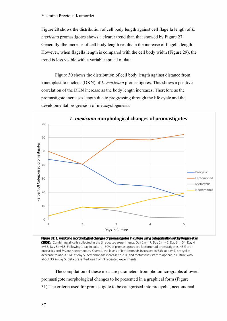
Yasmine Precious Kumordzi
87
Figure 28 shows the distribution of cell body length against cell flagella length of L.
mexicana promastigotes shows a clearer trend than that showed by Figure 27.
Generally, the increase of cell body length results in the increase of flagella length.
However, when flagella length is compared with the cell body width (Figure 29), the
trend is less visible with a variable spread of data.
Figure 30 shows the distribution of cell body length against distance from
kinetoplast to nucleus (DKN) of L. mexicana promastigotes. This shows a positive
correlation of the DKN increase as the body length increases. Therefore as the
promastigote increases length due to progressing through the life cycle and the
developmental progression of metacyclogenesis.
Figure 31: L. mexicana morphological changes of promastigotes in culture using categorization set by Rogers et al. (2002). Combining all cells collected in the 3 repeated experiments, Day 1 n=47, Day 2 n=42, Day 3 n=54, Day 4 n=55, Day 5 n=88. Following 1 day in culture, 50% of promastigotes are leptomonad promastigotes, 45% are procyclics and 5% are nectomonads. Overall, the levels of leptomonads increases to 63% at day 5, procyclics decrease to about 16% at day 5, nectomonads increase to 20% and metacyclics start to appear in culture with about 3% in day 5. Data presented was from 3 repeated experiments.
The compilation of these measure parameters from photomicrographs allowed
promastigote morphological changes to be presented in a graphical form (Figure
31).The criteria used for promastigote to be categorised into procyclic, nectomonad,
0
10
20
30
40
50
60
70
1 2 3 4 5
Perc
ent O
f Cat
egor
ised
prom
astig
otes
Days In Culture
L. mexicana morphological changes of promastigotes
Procyclic
Leptomonad
Metacyclic
Nectomonad

Yasmine Precious Kumordzi
88
leptomonad, haptomonad and metacyclic promastigotes was dependent on
morphology criteria (Figure 16). For L. mexicana, criteria set by Rogers et al, (2002)
was used. This gave a graph that shows the changes in promastigote forms over 5
days from culture (Figure 31). Day 1 represented 1 day following passage and
contained 50% leptomonad promastigotes, 5% nectomonad promastigotes and 45%
procyclic promastigotes. Procyclic promastigotes have a body length between 6.5 to
11.5 μm, a flagellum length less than the body length and the body width variable.
This form of promastigotes decreased over the days to 16% in day 5. Leptomonad
promastigotes have a body length between 6.5 to 11.5 μm with the flagella greater
than body length. This form of promastigotes decreased to 40% in day 2, then rapidly
increased to 59% in day 3 before steadily increasing over the days to 63% in day 5.
Nectomonad promastigotes have a body length greater than 12 μm. This form of
promastigotes increased over the days to 20% in day 5. Recovering metacyclics
showed the onset of metacyclogenesis. This occurred after day 2, however metacyclic
promastigotes did not dominate the culture in day 5 as presumed, with only 3% of
cells in day 5 being categorised as metacyclic promastigotes.
4.2 Leishmania tarentolae morphometric analysis in vitro
Prior to the establishment of the development profile of L. tarentolae
promastigotes in culture, a growth curve was constructed to examine the growth rate
of the parasite in vitro. Growth of parasites in 199 medium with 10% FBS as stated in
the Materials and Methods section was monitored over 175.5 hours with cell density
calculated using a haemocytometer approximately every 24 hours (33.3, 56.0, 76.5,
101.0, 124.0, 151.5 and 175.5 hours). An average of 10 counts for each time points
was used for average results plotted to give the lag, exponential and stationary phases
of parasite growth within the environmental conditions mentioned.
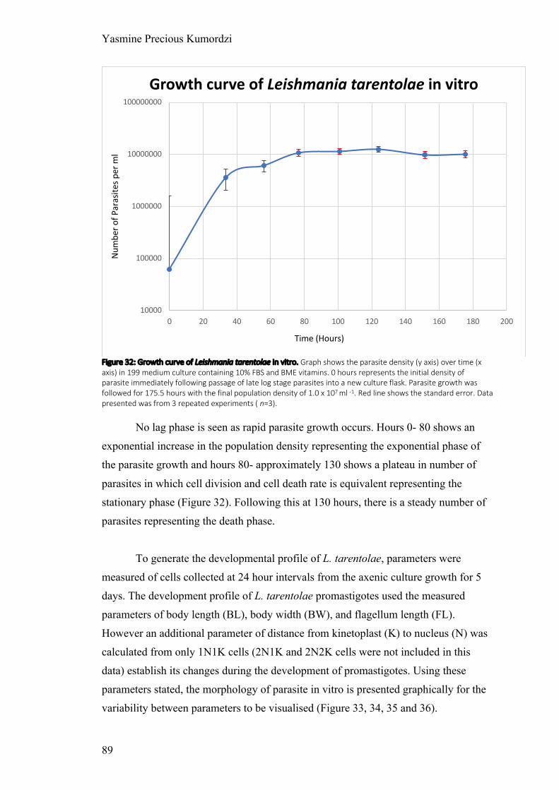
Yasmine Precious Kumordzi
89
Figure 32: Growth curve of Leishmania tarentolae in vitro. Graph shows the parasite density (y axis) over time (x axis) in 199 medium culture containing 10% FBS and BME vitamins. 0 hours represents the initial density of parasite immediately following passage of late log stage parasites into a new culture flask. Parasite growth was followed for 175.5 hours with the final population density of 1.0 x 10⁷ ml -1. Red line shows the standard error. Data presented was from 3 repeated experiments ( n=3).
No lag phase is seen as rapid parasite growth occurs. Hours 0- 80 shows an
exponential increase in the population density representing the exponential phase of
the parasite growth and hours 80- approximately 130 shows a plateau in number of
parasites in which cell division and cell death rate is equivalent representing the
stationary phase (Figure 32). Following this at 130 hours, there is a steady number of
parasites representing the death phase.
To generate the developmental profile of L. tarentolae, parameters were
measured of cells collected at 24 hour intervals from the axenic culture growth for 5
days. The development profile of L. tarentolae promastigotes used the measured
parameters of body length (BL), body width (BW), and flagellum length (FL).
However an additional parameter of distance from kinetoplast (K) to nucleus (N) was
calculated from only 1N1K cells (2N1K and 2N2K cells were not included in this
data) establish its changes during the development of promastigotes. Using these
parameters stated, the morphology of parasite in vitro is presented graphically for the
variability between parameters to be visualised (Figure 33, 34, 35 and 36).
10000
100000
1000000
10000000
100000000
0 20 40 60 80 100 120 140 160 180 200
Num
ber o
f Par
asite
s per
ml
Time (Hours)
Growth curve of Leishmania tarentolae in vitro
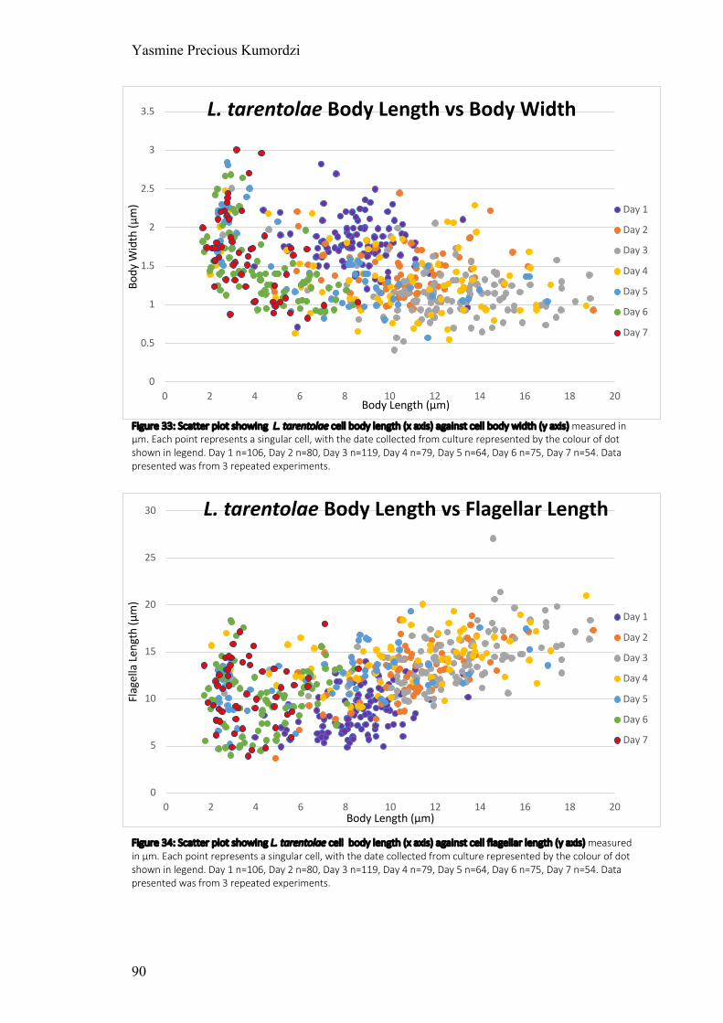
Yasmine Precious Kumordzi
90
Figure 33: Scatter plot showing L. tarentolae cell body length (x axis) against cell body width (y axis) measured in μm. Each point represents a singular cell, with the date collected from culture represented by the colour of dot shown in legend. Day 1 n=106, Day 2 n=80, Day 3 n=119, Day 4 n=79, Day 5 n=64, Day 6 n=75, Day 7 n=54. Data presented was from 3 repeated experiments.
Figure 34: Scatter plot showing L. tarentolae cell body length (x axis) against cell flagellar length (y axis) measured in μm. Each point represents a singular cell, with the date collected from culture represented by the colour of dot shown in legend. Day 1 n=106, Day 2 n=80, Day 3 n=119, Day 4 n=79, Day 5 n=64, Day 6 n=75, Day 7 n=54. Data presented was from 3 repeated experiments.
0
0.5
1
1.5
2
2.5
3
3.5
0 2 4 6 8 10 12 14 16 18 20
Body
Wid
th (μ
m)
Body Length (μm)
L. tarentolae Body Length vs Body Width
Day 1
Day 2
Day 3
Day 4
Day 5
Day 6
Day 7
0
5
10
15
20
25
30
0 2 4 6 8 10 12 14 16 18 20
Flag
ella
Len
gth
(μm
)
Body Length (μm)
L. tarentolae Body Length vs Flagellar Length
Day 1
Day 2
Day 3
Day 4
Day 5
Day 6
Day 7

Yasmine Precious Kumordzi
91
Figure 35: Scatter plot showing L. tarentolae cell body width (x axis) against cell flagellar length (y axis) measured in μm. Each point represents a singular cell, with the date collected from culture represented by the colour of dot shown in legend. Day 1 n=106, Day 2 n=80, Day 3 n=119, Day 4 n=79, Day 5 n=64, Day 6 n=75, Day 7 n=54. Data presented was from 3 repeated experiments.
Figure 36: Scatter plot showing L. tarentolae cell body length (x axis) against distance from kinetoplast to nucleus (y axis) measured in μm. Each point represents a singular cell, with the date collected from culture represented by the colour of dot shown in legend. Day 1 n=106, Day 2 n=80, Day 3 n=119, Day 4 n=79, Day 5 n=64, Day 6 n=75, Day 7 n=54. Data presented was from 3 repeated experiments.
0
5
10
15
20
25
30
0 0.5 1 1.5 2 2.5 3 3.5
Flag
ella
Len
gth
(μm
)
Body Width (μm)
L. tarentolae Flagellar Length vs Body Width
Day 1
Day 2
Day 3
Day 4
Day 5
Day 6
Day 7
0
1
2
3
4
5
6
7
8
9
10
0 2 4 6 8 10 12 14 16 18 20
DKN
(μm
)
Body Length (μm)
L. tarentolae Body Length vs DKN
Day 1
Day 2
Day 3
Day 4
Day 5
Day 6
Day 7

Yasmine Precious Kumordzi
92
Figure 33 shows the distribution of cell body length against cell body width of
L. tarentolae promastigotes is variable. Visualising groupings as days collected from
the axenic culture, day 1 body length ranges between 5.136 μm and 13.466 μm with
the body width ranging between 0.711 μm and 2.5 μm. Day 2 length ranges increases
to between 6.375 μm and 16.152 μm with the body width ranging between 0.925 μm
and 2.45 μm. This continues to day 3, with the length range increasing to between
2.583 μm and 18.913 μm, and the body width ranging between 0.44 μm and 2.508
μm. However in day 4, the length range decreases to between 2.491 μm and 16.496
μm with the body width reducing to range between 0.55 μm and 2.481 μm. Day 5
length ranges increases as normal to between 2.361 μm and 17.007 μm with the body
width increasing to range between 0.575 μm and 2.809 μm. Day 6 length ranges
decreases rapidly to between 1.69 μm and 7.725 μm with the body width increasing to
range between 0.911 μm and 2.688 μm. Day 7 length ranges increases as normal to
between 1.692 μm and 8.574 μm with the body width increasing to range between
0.828μm and 2.962 μm. These changes in body length and body width are small;
however when all the cells are visualised as a whole, a slight trend can be seen in the
data. With the increase in cell body length (BL), there is a decrease in the body width
(BW) similar to L. mexicana (Figure 27).
Figure 34 shows the distribution of cell body length against cell flagellar
length of L. tarentolae promastigotes shows a clearer trend than that showed by
Figure 33. Generally, the increase of cell body length results in the increase of
flagellar length. However, when flagellar length is compared with the cell body width
(Figure 35), the trend is less visible with a variable spread of data.
Figure 36 shows the distribution of cell body length against distance from
kinetoplast to nucleus (DKN) of L. tarentolae promastigotes. This shows a positive
correlation of the DKN increase as the body length increases. Therefore as the
promastigote generally increases length due to progressing through the life cycle and
the developmental progression of metacyclogenesis similar to that of L. mexicana
(Figure 30).

Yasmine Precious Kumordzi
93
Figure 37: L. tarentolae morphological changes of promastigotes in culture using categorization set by Rogers et al. (2002). Combining all cells collected in the 3 repeated experiments, Day 1 n=106, Day 2 n=80, Day 3 n=119, Day 4 n=79, Day 5 n=64, Day 6 n=75, Day 7 n=54. Following 1 day in culture, 55% of promastigotes are procyclics, 42% leptomonads, 2% nectomonads and 1% metacyclics. Procyclics decreased significantly over the days to 0 found after 7 days. Nectomonads increased rapidly to 68% in day 3 to further decreased to 0 at day 6. Leptomonads increased to 75% in day 5 and later decreased steadily to 28% in day 7. Metacyclics increase steadily to 5% in day 5 and rapidly increase significantly to 72% in day 7. Data presented was from 3 repeated experiments.
The compilation of these measure parameters from photomicrographs allows
promastigote morphological changes to be presented in a graphical form (Figure
37).The criteria used for different promastigote stages was based on morphology
criteria (Figure 16). To assign promastigote stages in both Leishmania species
measured (L. mexicana and L. tarentolae), criteria set by Rogers et al, (2002)
developed for L. mexicana was used. This gave a graph that shows the changes in
promastigote forms over 7 days from culture (Figure 37). Day 1 represents 1 day
following passage and contains 55% procyclics, 42% leptomonads, 2% nectomonads
and 1% metacyclics. The percentage of promastigotes categorised as procyclics
decreases significantly to 13% in day 2 and then remained steadily low for the
remainder of the growth in culture. Nectomonads on the other hand increase rapidly to
68% in day 3, later decreasing rapidly to 0% in day 6 and remaining at that level.
0
10
20
30
40
50
60
70
80
1 2 3 4 5 6 7
Perc
ent O
f Cat
egor
ised
Days In Culture
L. tarentolae morphological changes of promastigotes
Procyclic
Leptomonad
Metacyclic
Nectomonad
Procyclic
Leptomonad
Metacyclic
Nectomonad

Yasmine Precious Kumordzi
94
Leptomonads increase on day 2 to 53%, decreased to 28% in day 3 before increasing
again to 75% in day 5 and decreasing again. Metacyclics increased steadily to 5% in
day 5 and rapidly increased to 72% in day 7.
The photomicrographs from which these morphometric analyses were
collected from showed some unfamiliar morphologies that could not be explained or
properly categorised using the criteria from Rogers et al (2002).
4.2.1 Bulgtomonad promastigotes
These were first described by Dillon and Liu (unpublished) in which a new
criteria was suggested taking these promastigotes into consideration. Bulgtomonads
were described as having a body width between 2.3-3.5 μm with the body length
roughly 15 μm.
Cells that fit the visual description of bulgtomonads promastigotes were found
in the axenic culture of L. tarentolae on day 4 (Image 1). These cells made up 20.24%
of cells found in day 4. The bulgtomonad promastigotes all had their nucleus
contained within the ‘bulge’ of the cell with the kinetoplast adjacent to the flagella.
11% of promastigotes that visually fit the criteria for bulgtomonad promastigotes did
fit within the described morphology of bulgtomonad promastigotes by Dillon and Liu.
Due to the low proportion of cells fitting the criteria, the cells that visually fit the
description of bulgtomonads from day 4 L. tarentolae in culture have been used to
give a revised average morphological criteria for bulgtomonad promastigotes (Figure
38).

Yasmine Precious Kumordzi
95
Image 1: Image shows Bulgtomonad promastigotes from Leishmania tarentolae axenic culture day 4. Left images were stained with Giemsa and right images were stained with DAPI. The thick yellow arrow points to the nucleus DNA whilst the thin green arrow points to the kinetoplast DNA. Red circles showing regions of visual bultomonad promastigotes.

Yasmine Precious Kumordzi
96
Figure 38: Morphology criteria for L. tarentoloae Bulgtomonad promastigotes. Body width across bulge of around 1.88μm, distance from nucleus to kinetoplast (DKN) of 2.24μm and cell body length of 9.44μm.
4.2.2 Kinetoplast Nucleus swapping promastigotes
Both the nucleus and kinetoplast is seen under DAPI staining, with the
kinetoplast DNA being smaller and brighter when compared to the nucleus DNA. The
kinetoplast is seen to be anterior to the nucleus in most promastigotes, however in
some promastigotes seen in the death phase of L. tarentolae the seems to be a change
in its location (Image 2 and 3). These cells were spherical in cell body shape and had a
flagellum significantly longer than its cell body. Taking into account all cells that
presented this morphology, this cell type was categorized; cell body length was
roughly 2 μm, cell body width was roughly 1.7 μm, flagellum length was roughly
10.842 and DKN was roughly 0.7 μm.

Yasmine Precious Kumordzi
97
Image 2: Image shows kinetoplast and nucleus swapping promastigotes from Leishmania tarentolae axenic culture day 7. Left images were stained with Giemsa and right images were stained with DAPI. The promastigotes of interest are circled, with green arrows showing the kDNA.

Yasmine Precious Kumordzi
98
4.3 Analysing chemotaxic migration
Chemotaxic assay mentioned in Materials and Methods 3.3 was used to
produce all the data. Capillary assay was set up using PSG collected in a L. mexicana
infection with Lu. longipalpis, 0.5M glucose, 0.5M fructose, 0.5M urea, 0.5M
sorbitol, 0.5M mannose, 0.5M sucrose and 0.05M copper sulphate. Copper sulphate
resulted in some migrated cells, however all cells were lysed and therefore could not
be used for further analysis.
4.3.1 Capillary assay
Figure 39 shows the migration chemotaxis index (MCI); a positive index
showed attraction towards the chemoeffector whereas a negative index shows
repulsion. Visualising the graph, generally all chemoeffectors have a positive index
for L. mexicana and L. tarentolae for both days (day 3 and day 5). PSG shows positive
chemotaxic index throughout; with L. mexicana day 5 having the highest MCI of
2.428, L. mexicana day 3 having an MCI of 2.285, L. tarentolae day 5 having an MCI
of 0.804 and L. tarentolae day 3 having an MCI of 0.333. The data shows that L.
mexicana is more attracted to PSG compared to L. tarentolae, with forms found in the
late stationary phase/death phase showing higher attraction. Glucose shows a positive
MCI to all L. mexicana and L. tarentolae day 5 promastigotes. L. tarentolae day 3
show a slight negative MCI of 0.752. L. mexicana and L. tarentolae day 5
promastigotes show similar MCIs in fructose between 0.4 to 0.8 and glucose of
roughly 1.65 (Figure 39). L. mexicana day 3 have the highest MCI to both sugars,
followed by L. mexicana day 5. Using a 5% two-tailed Mann Whitney U-test
(appendix II) for L. mexicana day 3 and day 5 glucose recorded cell population of
migrated cells result, the p value of 0.006233 was calculated showing that the result
was highly significant. L. mexicana and L. tarentolae day 5 have MCIs below 1.0
towards urea, however L. tarentolae day 3 show a high MCI of 2.820. Using a 5%
two-tailed Mann Whitney U-test (appendix II) for L. tarentolae day 3 and day 5 urea
results, the p value of 0.000532 showing that the result was highly significant.
Sorbitol and mannose generally have a low MCI between -1.0 and 1.0, however L.
mexicana day 3 have a MCI of 1.678 towards mannose. Sucrose generally has
attractiveness similar to glucose and fructose, with L. mexicana day 3 having a MCI
of 6.0.

Yasmine Precious Kumordzi
99
Overall, L. mexicana day 3 show a stronger attraction to when compared to L.
mexicana day 5 towards the sugars glucose, fructose, mannose and sucrose. L.
tarentolae day 3 show great attraction towards urea, with others not having much of
an attraction.
Figure 39: Movement of Leishmania promastigotes in the presence of potential chemoeffectors. Graph shows the migration chemotaxis index (mentioned in Materials and Methods) of 3 (Exponential growth phase) and 5 (Stationary growth phase) day old culture of L. mexicana and L. tarentolae towards potential chemoeffectors of PSG, glucose 0.5M, fructose 0.5M, urea 0.5M, sorbitol 0.5M, mannose 0.5M and sucrose 0.5M. Raw data in appendix IV. The date collected from culture represented by the colour of bars shown in legend. Data presented for glucose, fructose, sorbitol, mannose, urea and sucrose was from 3 repeated experiments. Data for PSG was from 1 experiment consisting of 5 capillary tubes for each condition. Standard error showed by error bars.
-2 -1 0 1 2 3 4 5 6 7 8
sucrose
mannose
sorbitol
urea
fructose
glucose
PSG
Movement index of Leishmania promastigotes in the presence of potential chemoeffectors
L. mexicana day 5 L. mexicana day 3
L. tarentolae day 5 L. tarentolae day 3
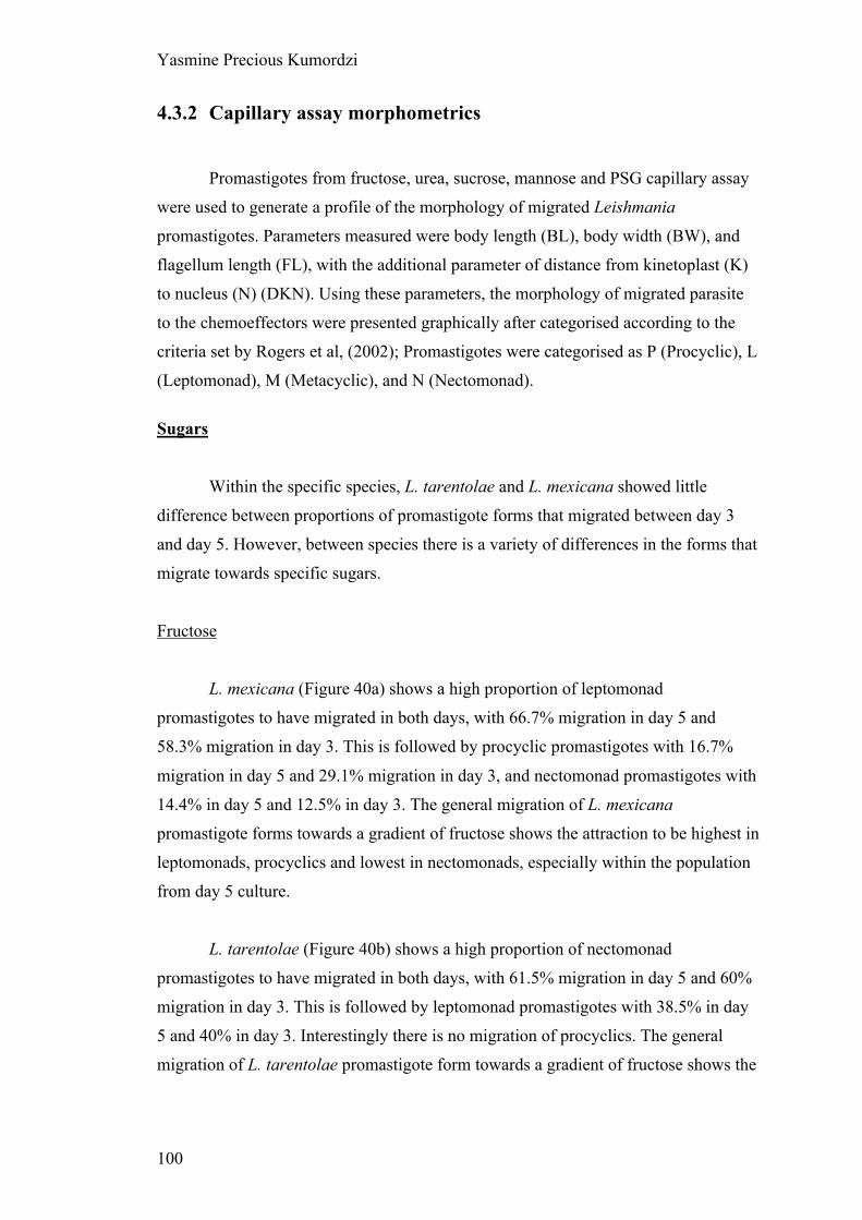
Yasmine Precious Kumordzi
100
4.3.2 Capillary assay morphometrics
Promastigotes from fructose, urea, sucrose, mannose and PSG capillary assay
were used to generate a profile of the morphology of migrated Leishmania
promastigotes. Parameters measured were body length (BL), body width (BW), and
flagellum length (FL), with the additional parameter of distance from kinetoplast (K)
to nucleus (N) (DKN). Using these parameters, the morphology of migrated parasite
to the chemoeffectors were presented graphically after categorised according to the
criteria set by Rogers et al, (2002); Promastigotes were categorised as P (Procyclic), L
(Leptomonad), M (Metacyclic), and N (Nectomonad). Sugars
Within the specific species, L. tarentolae and L. mexicana showed little
difference between proportions of promastigote forms that migrated between day 3
and day 5. However, between species there is a variety of differences in the forms that
migrate towards specific sugars.
Fructose
L. mexicana (Figure 40a) shows a high proportion of leptomonad
promastigotes to have migrated in both days, with 66.7% migration in day 5 and
58.3% migration in day 3. This is followed by procyclic promastigotes with 16.7%
migration in day 5 and 29.1% migration in day 3, and nectomonad promastigotes with
14.4% in day 5 and 12.5% in day 3. The general migration of L. mexicana
promastigote forms towards a gradient of fructose shows the attraction to be highest in
leptomonads, procyclics and lowest in nectomonads, especially within the population
from day 5 culture.
L. tarentolae (Figure 40b) shows a high proportion of nectomonad
promastigotes to have migrated in both days, with 61.5% migration in day 5 and 60%
migration in day 3. This is followed by leptomonad promastigotes with 38.5% in day
5 and 40% in day 3. Interestingly there is no migration of procyclics. The general
migration of L. tarentolae promastigote form towards a gradient of fructose shows the

Yasmine Precious Kumordzi
101
attraction to be highest with nectomonads followed by leptomonads, however
procyclic forms are showed to have no migration towards fructose.
The biggest difference in migration towards a gradient of fructose between L.
tarentolae and L. mexicana is the absence of any migrated procyclic promastigote
forms in L. tarentolae, and the promastigote form with highest attraction nectomonads
in L. tarentolae and leptomonads in L. mexicana.
Sucrose
L. mexicana (Figure 41a) shows a high proportion of leptomonad
promastigotes to have migrated in both days, with 59.6% migration in day 5 and
55.0% migration in day 3. This is followed by procyclic promastigotes with 23.1%
migration in day 5 and 30.0% migration in day 3, and nectomonad promastigotes with
15.4% in day 5 and 10.0% in day 3. The general migration of L. mexicana
promastigote forms towards a gradient of sucrose shows the attraction to be highest in
leptomonads, procyclics and lowest in nectomonads, especially within the population
from day 5 culture.
L. tarentolae (Figure 41b) shows a high proportion of nectomonad
promastigotes to have migrated in both days, with 59.0% migration in day 5 and
45.5% migration in day 3. This is followed by 29.5% migration of leptomonad
promastigotes in day 5 and 36.4% migration of procyclics in day 3, then a migration
of 9.8% in procyclic promastigotes day 5 and 13.6% migration of leptomonads in day
3.
The biggest difference in migration towards a gradient of sucrose between L.
tarentolae and L. mexicana is the procyclic form with highest attraction being
nectomonads in L. tarentolae and leptomonads in L. mexicana.
Mannose
L. mexicana (Figure 42a) shows a high proportion of leptomonad
promastigotes to have migrated in both days, with 61.9% migration in day 5 and
45.5% migration in day 3. This is followed by procyclic promastigotes with 30.2%

Yasmine Precious Kumordzi
102
migration in day 5 and 40.9% migration in day 3, and nectomonad promastigotes with
7.9% in day 5 and 13.6% in day 3. The general migration of L. mexicana promastigote
forms towards a gradient of mannose shows the attraction to be highest in
leptomonads, procyclics and lowest in nectomonads, especially within the population
from day 5 culture.
L. tarentolae (Figure 42b) shows a high proportion of nectomonad and
leptomonad promastigotes to have migrated in both days. However, the promastigote
with highest migration differs between age of culture used. Day 5 culture shows a
nectomonad migration of 68.2% being the highest migration whereas day 3 culture
shows the highest migration to be 50.0% in leptomonads. This is followed by 31.8%
leptomonad migration in day 5 culture and 42.9% nectomonad migration in day 3
culture. There is no other forms of promastigotes that are found to migrate in day 5
culture however day 3 show a low migration of 7.1% in procyclics.
The biggest difference in migration towards a gradient of mannose is the
promastigote form with highest attraction being leptomonads in L. mexicana and
differing between leptomonads in day 3 and nectomonads in day 5 L. tarentolae. The
is also no found procyclic migration in day 5 tarentolae.
Between sugars there are similarities as well as a few differences. L. mexicana
shows the highest migration for both day 3 and day 5 from leptomonad promastigotes,
followed by procyclic promastigotes and later nectomonad promastigotes. There are
very few migrated metacyclic promastigotes found; sucrose had 5% migration found
in day 3 and 1.9% found in day 5, mannose had no metacyclics in either days and
fructose had no metacyclics found in day 3 and 2.4% migration found in day 5. L.
tarentolae shows a different migrated promastigote form profile when compared to L.
mexicana. L. tarentolae from day 5 shows the highest migration from nectomonads,
followed by leptomonads and then procyclics. However in response to sucrose
mannose and fructose, none of the procyclics or metacyclics from day 5 culture
migrate. L. tarentolae from day 3 show more of a variation in the forms that migrate;
sucrose presents a high migration of nectomonads, followed by procyclics and lowest
with leptomonads, mannose presents a high migration of leptomonads, followed by
nectomonads and lowest with procyclic, whilst fructose presents a high migration of
nectomonads followed by leptomonad with no migration of procyclics.

Yasmine Precious Kumordzi
103
Figure 40a: The percentage of migrated categorised promastigotes of day 3 and 5 old culture of L. mexicana towards potential chemoeffectors of fructose 0.5M. Promastigotes were categorised as P (Procyclic), L (Leptomonad), M (Metacyclic), and N (Nectomonad). Raw data in appendix IV. The date collected from culture represented by the colour of bars shown in legend. Data presented was from 3 repeated experiments.
Figure 40b:The percentage of migrated categorised promastigotes of day 3 and 5 old culture of L. tarentolae towards potential chemoeffectors of fructose 0.5M. Promastigotes were categorised as P (Procyclic), L
-10
0
10
20
30
40
50
60
70
80
90
100
M Fructose Day 3 M Fructose Day 5
Perc
enta
ge o
f mig
rate
d pr
omas
tigot
es fo
rms
Migrated forms of Leishmania mexicana promastigotes towards fructose
P L M N
-10
0
10
20
30
40
50
60
70
80
90
100
DAY 3 fructose DAY 5 fructose
Perc
enta
ge o
f mig
rate
d pr
omas
tigot
es fo
rms
Migrated forms of Leishmania tarentolae promastigotes towards fructose
P L M N

Yasmine Precious Kumordzi
104
(Leptomonad), M (Metacyclic), and N (Nectomonad). Raw data in appendix IV. The date collected from culture represented by the colour of bars shown in legend. Data presented was from 3 repeated experiments.
Figure 41a: The percentage of migrated categorised promastigotes of day 3 and 5 old culture of L. mexicana towards potential chemoeffectors of sucrose 0.5M. Promastigotes were categorised as P (Procyclic), L (Leptomonad), M (Metacyclic), and N (Nectomonad). Raw data in appendix IV. The date collected from culture represented by the colour of bars shown in legend. Data presented was from 3 repeated experiments.
Figure 41b: The percentage of migrated categorised promastigotes of day 3 and 5 old culture of L. tarentolae towards potential chemoeffectors of sucrose 0.5M. Promastigotes were categorised as P (Procyclic), L (Leptomonad), M (Metacyclic), and N (Nectomonad). Raw data in appendix IV. The date collected from culture represented by the colour of bars shown in legend. Data presented was from 3 repeated experiments.
-10.0
0.0
10.0
20.0
30.0
40.0
50.0
60.0
70.0
80.0
90.0
100.0
M Sucrose Day 3 M Sucrose Day 5
Perc
enta
ge o
f mig
rate
d pr
omas
tigot
es fo
rms
Migrated forms of Leishmania mexicana promastigotes towards sucrose
P L M N
-10
0
10
20
30
40
50
60
70
80
90
100
DAY 3 sucrose DAY 5 sucrose
Perc
enta
ge o
f mig
rate
d pr
omas
tigot
es fo
rms
Migrated forms of Leishmania tarentolae promastigotes towards sucrose
P L M N

Yasmine Precious Kumordzi
105
Figure 42a: The percentage of migrated categorised promastigotes of day 3 and 5 old culture of L. mexicana towards potential chemoeffectors of mannose 0.5M. Promastigotes were categorised as P (Procyclic), L (Leptomonad), M (Metacyclic), and N (Nectomonad). Raw data in appendix IV. The date collected from culture represented by the colour of bars shown in legend. Data presented was from 3 repeated experiments. .
Figure 42b: The percentage of migrated categorised promastigotes of day 3 and 5 old culture of L. tarentolae towards potential chemoeffectors of mannose 0.5M. Promastigotes were categorised as P (Procyclic), L (Leptomonad), M (Metacyclic), and N (Nectomonad). Raw data in appendix IV. The date collected from culture represented by the colour of bars shown in legend. Data presented was from 3 repeated experiments.
-10
0
10
20
30
40
50
60
70
80
90
100
M Mannose day 3 M Mannose day 5
Perc
enta
ge o
f mig
rate
d pr
omas
tigot
es fo
rms
Migrated forms of Leishmania mexicana promastigotes towards mannose
P L M N
-10
0
10
20
30
40
50
60
70
80
90
100
DAY 3 Mannose DAY 5 Mannose
Perc
enta
ge o
f mig
rate
d pr
omas
tigot
es fo
rms
Migrated forms of Leishmania tarentolae promastigotes towards mannose
P L M N

Yasmine Precious Kumordzi
106
Urea
Within the specific species, L. tarentolae and L. mexicana showed little
difference between proportions of promastigote forms that migrated between day 3
and day 5. However, between species there is a variety of differences in the forms that
migrate towards urea.
L. mexicana (Figure 44) shows a high proportion of leptomonad promastigotes
to have migrated in both days, with 52.9% migration in day 5 and 35.7% migration in
day 3. This is followed by nectomonad promastigotes with 29.4% migration in day 5
and 28.6% migration in day 3, procyclic promastigotes with 11.8% in day 5 and
21.4% in day 3 and metacyclic promastigotes with 5.9% in day 5 and 14.3% in day 3.
The general migration of L. mexicana promastigote forms towards a gradient of urea
shows the attraction to be highest in leptomonads, followed by nectomonads,
procyclics and lowest migration in metacyclics.
L. tarentolae (Figure 44) shows a high proportion of leptomonad and
Nectomonad promastigotes migrated in both days. Day 5 L. tarentolae shows
nectomonad promastigote to have the highest migration of 56.5% whilst day 3 shows
leptomonad promastigote with 60.0% to have the highest migration. This is followed
by 39.1% migration of leptomonad in day 5 and 33.3% migration of nectomonads in
day 3 cultures. Procyclics shows a very small migration of 6.6% in day 3 and 4.3 in
day 5.
The biggest difference in migration towards a gradient of urea between L.
tarentolae and L. mexicana is the absence of L. tarentolae metacyclic forms in
migration. However, generally similar forms of promastigotes migrate; leptomonad
and nectomonads. Promastigote Secreting Gel (PSG) Unlike the other chemoeffectors tested, PSG showed the widest variation.
L. mexicana day 3 showed procyclics as the most migrated form of 50.0%,
followed by leptomonads at 42.9% and nectomonads at 7.14%. L. mexicana day 5
showed leptomonads as the most migrated form of 56.0%, followed by nectomonads

Yasmine Precious Kumordzi
107
at 24.0%, procyclics at 16.0% and metacyclics at 4.0%. Generally, L. mexicana had
procyclics and leptomonads form of promastigotes migrate towards PSG (Figure 45a)
L. tarentolae day 3 showed nectomonads as the most migrated form of 75.0%,
followed by leptomonads at 18.75% and procyclics at 6.25%. L. tarentolae day 5
showed leptomonads as the most migrated form of 81.8%, followed by procyclics and
nectomonads both at 9%. On both days no metacyclics were found to have migrated
(Figure 45b)

Yasmine Precious Kumordzi
108
Figure 43a: The percentage of migrated categorised promastigotes of day 3 and 5 old culture of L. mexicana towards potential chemoeffectors of urea 0.5M. Promastigotes were categorised as P (Procyclic), L (Leptomonad), M (Metacyclic), and N (Nectomonad). Raw data in appendix IV. The date collected from culture represented by the colour of bars shown in legend. Data presented was from 3 repeated experiments.
Figure 44a: The percentage of migrated categorised promastigotes of day 3 and 5 old culture of L. tarentolae towards potential chemoeffectors of urea 0.5M. Promastigotes were categorised as P (Procyclic), L (Leptomonad), M (Metacyclic), and N (Nectomonad). Raw data in appendix IV. The date collected from culture represented by the colour of bars shown in legend. Data presented was from 3 repeated experiments.
-10
0
10
20
30
40
50
60
70
80
90
100
M Urea Day 3 M Urea Day 5
Perc
enta
ge o
f mig
rate
d pr
omas
tigot
es fo
rms
Migrated forms of Leishmania mexicana promastigotes towards urea
P L M N
-10
0
10
20
30
40
50
60
70
80
90
100
DAY 3 Urea DAY 5 Urea
Perc
enta
ge o
f mig
rate
d pr
omas
tigot
es fo
rms
Migrated forms of Leishmania tarentolae promastigotes towards urea
P L M N

Yasmine Precious Kumordzi
109
Figure 45a: The percentage of migrated categorised promastigotes of day 3 and 5 old culture of L. mexicana towards potential chemoeffectors of crude PSG Promastigotes were categorised as P (Procyclic), L (Leptomonad), M (Metacyclic), and N (Nectomonad). Raw data in appendix IV. The date collected from culture represented by the colour of bars shown in legend. Data presented was from 1 experiment.
Figure 45b: The percentage of migrated categorised promastigotes of day 3 and 5 old culture of L. tarentolae towards potential chemoeffectors of crude PSG Promastigotes were categorised as P (Procyclic), L (Leptomonad), M (Metacyclic), and N (Nectomonad). Raw data in appendix IV. The date collected from culture represented by the colour of bars shown in legend. Data presented was from 1 experiment.
-30
-20
-10
0
10
20
30
40
50
60
70
80
90
100
110
120
Day 3 PSG Day 5 PSGPerc
enta
ge o
f mig
rate
d pr
omas
tigot
es fo
rms
Migrated forms of Leishmania tarentolae promastigotes towards PSG
P L M N
-30
-20
-10
0
10
20
30
40
50
60
70
80
90
100
110
120
M psg day 3 M psg day 5Perc
enta
ge o
f mig
rate
d pr
omas
tigot
es fo
rms
Migrated forms of Leishmania mexicana promastigotes towards PSG
P L M N

Yasmine Precious Kumordzi
110
Figure 46: Overall movement of Leishmania tarentolae promastigotes in the presence of chemoeffectors. Graph shows the percentage of migrated categorised promastigotes of day 3 and 5 old culture of L. tarentolae towards potential chemoeffectors of PSG, glucose 0.5M, fructose 0.5M, urea 0.5M, sorbitol 0.5M, mannose 0.5M and sucrose 0.5M. Promastigotes were categorised as P (Procyclic), L (Leptomonad), M (Metacyclic), and N (Nectomonad). Raw data in appendix IV. The date collected from culture represented by the colour of bars shown in legend.
-20.0
0.0
20.0
40.0
60.0
80.0
100.0
P L M N
Perc
enta
ge o
f mig
rate
d pr
omas
tigot
es
Categorised promastigote
Movement of Leishmania tarentolae promastigotes that migrate towards chemoeffectors
DAY 3 Urea
DAY 5 Urea
DAY 3 Mannose
DAY 5 Mannose
DAY 3 sucrose
DAY 5 sucrose
DAY 3 fructose
DAY 5 fructose
Day 3 PSG
Day 5 PSG

Yasmine Precious Kumordzi
111
Figure 46 shows the types of L. tarentolae promastigotes that migrate towards
specific chemoeffectors. Metacyclic promastigotes show the least migration, however
the number of metacyclic promastigotes found in day 5 and day 3 culture is very low
in L. tarentolae. Procyclic promastigotes is the second least migrated form, with less
than 10% of migrated cells towards chemoeffectors falling in this category. With 3
day old L. tarentolae, 35% of procyclic promastigotes migrate towards sucrose.
Leptomonad and nectomonad promastigotes are the most migrated types of L.
tarentolae. Interestingly when using 5 day old L. tarentolae towards PSG, over 80%
of migrated cells are leptomonads and under 10% being nectomonads. This shows
that L. tarentolae nectomonads and leptomonads (particularly nectomonads) play the
major role in migration towards chemoeffectors and not metacyclics.
Figure 47 shows the types of L. mexicana promastigotes that migrate towards
specific chemoeffectors. Similar to L. tarentolae, metacyclic promastigotes show the
least migration. Nectomonads show the second lowest migration, with high
nectomonad promastigotes migration between 20% and 30% towards urea and PSG.
More procyclic promastigotes migrate in L. mexicana compared to L. tarentolae, with
migration of procyclics being over 10%. The highest migration of procyclic
promastigotes is towards PSG when using 3 day old promastigotes of 50%, second
highest is migration towards mannose of 41%. Leptomonad promastigotes are the
highest migrated form of promastigotes, showing migration towards all
chemoeffectors. This shows that L. tarentolae procyclics and leptomonads play the
major role in migration towards chemoeffectors and not metacyclics as suggested in
literature.

Yasmine Precious Kumordzi
112
Figure 47: Overall movement of Leishmania mexicana promastigotes in the presence of chemoeffectors. Graph shows the percentage of migrated categorised promastigotes of day 3 and 5 old culture of L. mexicana towards potential chemoeffectors of PSG, glucose 0.5M, fructose 0.5M, urea 0.5M, sorbitol 0.5M, mannose 0.5M and sucrose 0.5M. Promastigotes were categorised as P (Procyclic), L (Leptomonad), M (Metacyclic), and N (Nectomonad). Raw data in appendix IV. The date collected from culture represented by the colour of bars shown in legend.
-20
0
20
40
60
80
100
P L M N
Perc
enta
ge o
f mig
rate
d pr
omas
tigot
es
Categorised promastigote
Movement of Leishmania mexicana promastigotes that migrates towards chemoeffectors
M Fructose Day 3
M Fructose Day 5
M Sucrose Day 3
M Sucrose Day 5
M Mannose day 3
M Mannose day 5
Urea Day 3
Urea Day 5
M psg day 3
M psg day 5

Yasmine Precious Kumordzi
113
4.4 Microfluidic Chip design
4.4.1 Design and operations of the microdevice
To explore the various migration of Leishmania to various gradients of
chemoeffectors in a controlled and stable manner, we developed a microfluidic
device made up of optically transparent PDMS containing one layer as showed in
Figure 48 (A,B). The device generated consists of a chip with a diameter of 7 cm.
The chip has a 12 chambers along the extremity of the chip with channels
connecting them to the central reservoir chamber for parasite culture. This
generates a concentration gradient from the chamber at the extremity containing
test chemoeffector to the central reservoir chamber containing cells collected from
parasite culture. The chambers had a height of 200μm with connecting channels
20μm in height. This type of microfluidic gradient generator has a couple of potential
problems such as cross flow occurring between the sink and source (Paliwal et al,
2007). This problem was addressed in the design by increasing fluidic resistance
between the sink and source by decreasing the height of a part of the connecting
channel from 20μm to 6μm for 250μm of the length of channel Figure 48(C,D).
Before the assay, fluorescent Leishmania parasites obtained from Dr Paul
Bates and Dr Hector Diaz-Albiter were washed with PBS and resuspended in WIS
0.004% FBS. A known volume of parasites was loaded into the central reservoir
chamber and potential chemoeffectors such as glucose in WIS 0.004% FBS 0.5M
were used loaded into the chambers at the extremities before they were covered with a
glass cover slip. Static diffusion based microfluidic gradient generator utilises
diffusion between the two chambers to generate gradients across the channel in which
can be detected by the parasites for migration to occur.
A confocal laser scanning microscope was used for live imaging of the
migrating Leishmania parasites. Following the loading of both chambers, a glass
coverslip was used to cover the chip, completing the device. Images were taken at a
regular interval of 30 seconds of the whole device for an hour and was later stitched to
form a video in which migration can be viewed.

Yasmine Precious Kumordzi
114
Figure 48 (A,B): Microfluidic device design. Top view of the radial chip, showing 12 chambers along the outer edge and a singular central reservoir for the cell culture. The complete design is shown (B) with detailed region shown (A)

Yasmine Precious Kumordzi
115
Figure 48(C,D): Microfluidic device design. Schematic illustration of the of the microfluidic chip. From a single inlet, four branching channels subject the flow into microchannels (C) with dimensions of channels and wells differing in height (D).

Yasmine Precious Kumordzi
116
4.4.2 Initial testing of microfluidic device
Initial testing using Leishmania parasites in a WIS 0.004% FBS solution
placed in the central chamber and WIS 0.004% FBS in the chambers at the extremities
showed that the size of the channels and basic design of the device allowed for good
stable movement of promastigote Leishmania parasites within the channels when
viewed under an inverted microscope at x40. However, testing under the correct
conditions for microfluidic studies showed outside influence on the migration and
fluid diffusion. Placing the cover slip on the loaded chip resulted in some movement
of liquid and after about 15 minutes the chip began to dry, this affects both diffusion
and the migration of promastigotes. Due to these problems, the experiment was not
pursued further.
4.5 Preliminary study of hypopylarian status of L. tarentolae
4-5 day old Lu. longipalpis and Aedes. aegypti were infected with Leishmania
parasites and Crithidia fasiculata to study the establishment and development of
successful infections. This was done by using dissections of the full gut and
visualisation of the foregut, midgut and hindgut separately to understand the infection.
Lu. longipalpis was infected separately with L. tarentolae, L. mexicana and C.
fasiculata infections and monitored throughout by analysing the gut throughout the
infection. A successful L. mexicana infection was established within the first
experiment, giving rise to the PSG plug used for the chemotaxis assay. In this initial
infection, a suprapylarian infection was seen. Unfortunately L. mexicana did not
establish infection with the second experiment. Successful L. tarentolae infections
were established in roughly 60% of sandflies. The parasites within these infections
were contained within the hindgut and posterior midgut with heavy infection
contained within the hindgut. Attachment to hindgut epithelium was not visualised. A
loss of infection occurred 5-10 days following the infection, with Leishmania
parasites seen within the rectum of the sandfly. It can therefore be assumed that L.
tarentolae establishes a hypopylarian infection in Lu. Longipalpis which is lost

Yasmine Precious Kumordzi
117
through excretion. C. fasiculata infections within the sandfly established similar to L.
tarentolae in a hypopylarian mode.
A. aegypti mosquito infected with L. tarentolae and C. fasiculata was not the
main aspect of this study therefore only one preliminary set of experiment was done.
16% of fed mosquitoes had the presence of parasites in the first day of dissection (day
3). This establishment of infection was quickly lost after day 3 and the presence of
parasites were loosely within the midgut and hindgut similar to L. tarentolae infection
in Lu. Longipalpis.

Yasmine Precious Kumordzi
118
CHAPTER FIVE: DISCUSSION
5.1 Is Leishmania tarentolae hypopylarian?
Leptomonad promastigotes are responsible for the production of promastigote
secretory gel (PSG) in suprapylarian parasites (Rogers et al, 2002). This
developmental stage of promastigote is the precursor of metacyclic promastigotes, the
infective stage of promastigote. PSG is known to have a large role in transmission as
it is produced at the site of metacyclogenesis. However, this has to date only been
investigated in suprapylarian Leishmania spp. infections such as L. mexicana. From
the knowledge of developmental and migration changes of suprapylarian Leishmania
spp leading to transmission, it is understood that PSG gel plug occurs in the cardia and
thoracic midgut. The gel contains predominantly leptomonads from which metacyclic
promastigotes are differentiated from. This accumulation of PSG not only allows for
metacyclic promastigotes to be produced in an environment promoting transmission
during the next blood feed but distorts the stomodeal valve for the regurgitation
process to occur for successful blood meal intake and infection. As reported (Vionette,
Ginger and Dillon unpublish data) L. tarentolae establishes in a hypopylarian manner
in Lutzomyia longipalpis, successive developmental stages occur within the posterior
gut. The reservoir of leptomonads within a successful infection would therefore be
located in the hindgut and it was hypothesised that PSG production might occur in
this location. Any PSG produced in the hindgut might have a role in preventing
premature loss of parasites in the excreta due to partial blockage of the hindgut lumen
(Vionette, Ginger and Dillon unpublished). Diaz-Albiter et al, 2018 showed that
unlike reported by Vionette, Ginger and Dillon (unpublish data), L. tarentolae-GFP
infected Lu. longipalpis had a distribution that was diffuse with no specific areas of
binding to the gut (Diaz-Albiter et al, 2018). Due to the differentiation of leptomonad
promastigotes to metacyclic promastigotes being the last stage in metacyclogenesis
and the manner in which hypopylarian infection occurs, the discovery of PSG in
regions of the gut can be used to determine the location of leptomonads and
metacyclics.

Yasmine Precious Kumordzi
119
A preliminary experiment performed at the end of this study used sectional gut
dissection of successfully infected Lu. longipalpis to image the morphology of L.
tarentolae and detect PSG. The intention was to monitor changes that occur in L.
tarentolae infection and confirm the manner of infection that occurs with L.
tarentolae. The samples of morphology and PSG were not able to be analysed within
the time frame of this study but visualisation of the full gut and gut sections (foregut,
midgut and hindgut) from infection showed an hypopylarian infection with
leptomonad-type morphology of parasites within the posterior midgut and hindgut.
Based purely on visualisation of the infected gut, it can be confirmed that L.
tarentolae is a hypopylarian Leishmania species however no visual PSG formation
was seen. This could be due to continuous removal of produced PSG, a low-density
infection preventing large amounts of PSG to be produced or alternatively lack of
PSG formation by these parasites in the hindgut. Unlike in the foregut where the
cardia stops this from occurring, midgut peristalsis and the muscles around the pylorus
act to push excreta out for excretion. Further analysis of samples produced would give
more insight into the manner of L. tarentolae infection.
5.2 Morphometric analysis and comparison between L. mexicana and L. tarentolae
The morphometric analysis follows the development of two species of Leishmania; L.
mexicana a suprapylarian parasite and L. tarentolae a hypopylarian parasite, with the
results showing the multiplicative promastigotes within the axenic culture and the
sequence of morphology changes that occurs in culture. These changes mimic the
morphological transformations that occurs within the sand fly during the basic process
of metacyclogenesis. There are many published morphometric analysis of L. mexicana
parasite (Bates et al, 1994) however although a good model kinetoplastid,
morphometrics of L. tarentolae has not been explored well enough to be published.
Comparisons of these two species has provided a morphometric analysis of the
metacyclogenesis development that occurs in an axenic culture; cell body length,
width, flagellum length and distance between the kinetoplast and nucleus. However
using morphology particularly flagellum length to interpret the form of promastigote
using Bates et al, 1994 does not take into account the fact that the flagellum length is

Yasmine Precious Kumordzi
120
continuously growing throughout numerous cell cycles (Wheeler, Gluenz and Gull
2010). Therefore many promastigotes that fall within the leptomonad criteria
according to morphology may actually be procyclics. Only promastigotes within the
multiplicative promastigote stages were found. This was largely due to the continuous
passaging and the conditions that the promastigotes were kept in in terms of
temperature and pH.
5.2.1 Leishmania mexicana
The morphologies associated with L. mexicana promastigotes within the
sandfly and axenic culture (Rogers et al, 2002) have all been found. The changes in
morphology could be seen to dramatically change throughout development on a daily
basis, however using the morphological category stated by Rogers et al, 2002 only the
four main developmental forms (procyclic, nectomonad, leptomonad and metacyclic
promastigote) were found in culture. These followed a similar pattern of development
initiating with high procyclic promastigotes which reduces overtime, an increase of
leptomonads and nectomonads, and the emergence of metacyclic promastigotes.
However, monitoring of 5 days did not yield a high percentage of metacyclic
promastigotes. This was surprising as Rogers et al, 1993 showed an exponential
increase of metacyclic L. mexicana in an axenic culture with 20% of total cells in
culture being metacyclics at 4 days following passage and roughly 100% of cells in
culture 6 days following passage being metacyclics. This differed significantly from
the present data produced, with the only difference in methodology being the pH of
culture, media used and the passage number of culture. Bates and Tetley, 1993
reported similar data, explaining that the production of significantly pure cultures of
metacyclics is due to the change of pH to a more acidic 5.5 compared to cultivation in
an axenic pH 7 culture used which produced a morphologically mixed population.
Looking at the promastigote morphologies present in the first few days
(roughly day 3) post infection within a sand fly (Rogers et al, 1993), the population is
very mixed containing a very low level of procyclic promastigotes, metacyclics and
amastigotes, with higher numbers of nectomonad and leptomonad promastigotes. This
made stages of promastigotes found in day 5 representative of day 3 post infection of
Yasmine Precious Kumordzi
121
the sandfly. The promastigotes in this stage are representative of the developmental
stage of promastigotes in which they begin the escape and taxis towards the foregut to
be the first found in thoracic midgut (Rogers et al, 1993). Another realisation from
the data produced was the amount of time in which metacyclics took to develop.
There is a possibility that with a high number of passages, promastigotes struggle to
meet their original developmental cycle, taking significantly longer to develop to the
metacyclic stage and therefore were not seen within the 7 days given.
5.2.2 Leishmania tarentolae
With the lack understanding of a complete developmental cycle of L.
tarentolae in an axenic culture, it was important that this Leishmania species was fully
explored to understand the changes they undergo in culture when compared to L.
mexicana taxis.
The first to be explored was the similarity in the parameters between L.
mexicana and L. tarentolae. These parameters (Body length vs Body Width, Body
Length vs Flagellum Length, Flagellum Length vs Body Width and Body Length vs
Distance between Kinetoplast and Nucleus (DKN)) was similar showing that the
development of L. tarentolae was very similar to L. mexicana in terms of the
progression of morphometrics (Wheeler, Gluenz and Gull 2010) and the
developmental forms that can be found in an axenic culture. This was as body length
increased body width decreased, as body length increased flagellum length increased,
as body length increased DKN increased, and body width and flagellum length have
few correlations. These correlations are stronger with the L. tarentolae data due to the
increase of dataset and following the development over a longer period.
Following morphology criteria set by Rogers et al, 2002, the general
morphological changes in axenic culture were similar to that of L. mexicana studied
in sandfly infection (Rogers et al, 2002). Day 3 and day 5 were good examples of the
developmental stages found in day 3 and day 4 post-infection in L. mexicana
respectively. Similar to L. mexicana, these stages represent the stages in which escape
from the PM and initiation of taxis occurred. Surprisingly two unpublished
morphologies were found; bulgetomonad and kinetoplast nucleus swapping
promastigotes.

Yasmine Precious Kumordzi
122
An unpublished report by Dillon and Liu, 2014 set a new criterion for L.
tarentolae in which the new potential morphology of L. tarentolae promastigote was
found. This described bulgetomonad morphology was visually found in the axenic
culture emerging from about day 4 making up 20% of all analysed cells. These cells
have a ‘bulge’ in the cell with the kinetoplast directly adjacent to the flagellum.
Although, visually fitting the description stated by Dillon and Liu, only 11% fitted this
in morphology parameters and therefore a revised criterion has been set here of a
bulge width of roughly 1.88 um, nucleus within the bulge and the DKN of roughly
2.14um.
Wheeler, Gluenz and Gull 2010 showed DAPI stained promastigotes labelling
the nucleus and kinetoplast, with the kinetoplast labelled stronger than nucleus. This
difference in intensity was used to distinguish between the kinetoplast and the
nucleus. Unlike all other L. mexicana and early development L. tarentolae
promastigotes, some L. tarentolae showed cells with the less intense DAPI stained
organelle closest to the flagellum. These cells were seen in the death phase of culture.
The cell shape was spherical with a longer flagellum to cell width length. This
suggested that during the death phase, some promastigotes undergo changes that
causes the kinetoplast and nucleus to exchange locations. These cells labelled
‘Kinetoplast nucleus swapping promastigotes’ have not been reported as yet. Due to
the location within the growth stage of the cells, it can be assumed that with L.
tarentolae promastigote a swapping of the kinetoplast and nucleus or the significant
shrinkage of nucleus precedes the death of the cell.
5.3 Traditional Capillary Chemotaxic Assays
Traditional capillary assays have been used in many forms for the study of
chemotaxis through the delivery of chemicals that can be detected by cells in a
controlled gradient manner (Adler, 1973). This technique however has many
disadvantages in terms of having influences from outside condition such Brownian

Yasmine Precious Kumordzi
123
movement and dissolved oxygen changes within solution. Brownian movements
affect the movement of the cells within solution and has an effect on the overall
migratory potential of the cells. An outside influence that also affects migration within
a traditional capillary assay is the amount of dissolved oxygen within the solution, this
was seen in earlier assays tested (results not included) using a modified PP chamber.
Promastigotes in this assay migrated and accumulated around the channels where the
dissolved oxygen was high therefore results using this PP chamber assay did not
reflex chemotaxis. Both forms of chemotaxic assay used do not distinguish between
osmotaxis and chemotaxis in the proposed assays as there was no salt usage to ensure
there was no osmosis occurring within the microfluidic device or the capillary tubes.
The use of the traditional capillary assay as used by Oliveira et al and Barros et
al allowed for comparisons between methods and results. However, there was large
discrepancy between each. Leslie, Barrett and Burchmore, 2002 used late-log L.
mexicana, Oliveira et al, 2000 used L. chagasi and L. amazonensis at log phase and
stationary phase, Barros et al, 2006 used L. amazonensis at day 6 and deYsasa Pozzo
et al 2009 used L. amazonensis. As the developmental form of promastigotes was not
investigated prior to experiment and knowledge of only the non-specific growth stage,
it is not possible to predict with certainty what morphological forms of promastigotes
were used in each of these experiments.
To investigate promastigote migration in the hours following PM
disintegration, it is important to investigate using the chemotaxic capillary or
microfluidic device using a promastigote population of nectomonad, leptomonad and
mostly procyclic. Population of these promastigotes was collected for use from both
L. tarentolae and L. mexicana grown in culture and determined using morphometric
analysis as stated by Rogers et al, 2002.
An improvement to the methodology carried out in previous promastigote
chemotaxis assays (Oliveira et al, 2000, deYsasa Pozzo et al, 2009, Leslie, Barrett
and Burchmore, 2002, Díaz et al, 2011) was the collection of cells following
migration, and analysing the forms of migratory cells. This not only allowed the
determination of the chemotaxic agent but also the determination of the
developmental morphological form of promastigotes that was attracted to it.

Yasmine Precious Kumordzi
124
5.4 Leishmania promastigote taxis
The main part of the method during the experiment was the collection of the
migratory cells. These samples were used to determine the number and morphology of
promastigotes that had migrated from the solution into the capillary tubes containing
the testing chemotaxic agent. From previous chemotaxic assays (Oliveira et al, 2000,
deYsasa Pozzo et al, 2009, Leslie, Barrett and Burchmore, 2002, Díaz et al, 2011),
everything tested showed a level of taxis towards the test chemoeffectors, however no
substance has been used successfully to deter the migration of cells towards it. Copper
sulphate is known to be lethal to cells at a high dose (Davies, 1978). This was
therefore used assuming it would be show negative chemotaxis or a negative
chemotaxis index.
Biochemical gradients have been suggested to be used as a mechanism for
signalling to Leishmania promastigotes within the sandfly alimentary canal (Killick-
Kendrick, 1978). This mechanism takes advantage of the putative sugar
concentration that exists from the crop as sugars are released slowly into the midgut
for digestion. This began the numerous adaptations from Bray’s initial chemotaxic
assays in 1983 (Bray, 1983) to the more specialised optical tweezers assay by
deYsasa Pozzo. These have all focussed on sugar concentrations and the movement of
promastigotes towards a sugar gradient without the presence of a control
chemoeffector that showed a negative taxis. No presence of a chemical that deters the
migration insinuates a lack of chemotaxic movement but osmotaxic movement. This
brings the question, what other chemoeffector are promastigotes attracted to and what
type of taxis occurs in the sandfly. The experiment designed to answer this was to
perform a pilot chemical screen using more chemoeffectors and to collect the
migrated promastigotes to determine which morphological population was attracted to
each chemical.
Similar to previous assays, the general movement index of Leishmania
promastigotes in the presence of potential chemoeffectors all showed a movement
toward all chemicals of interest (Figure 39) including copper sulphate. The
promastigotes recovered following migration towards copper sulphate microscopically

Yasmine Precious Kumordzi
125
showed cell lysis, with the cell membrane ruptured. This therefore was not
quantifiable and was removed from results reported.
Interestingly, based on the counted number of migrated promastigotes L.
tarentolae showed a negative movement index towards glucose and mannose with the
L. tarentolae day 3 population and mannose with the L. tarentolae day 5 population
(Figure 39). However only the negative movement index towards glucose in the L.
tarentolae day 3 population is significantly relevant. Generally, L. mexicana showed a
high positive movement index towards PSG, and the sugars tested whereas L.
tarentolae showed a lower positive with a few negative movement indexes towards
sugars tested and PSG. Urea showed an opposite outcome when compared to L
mexicana and L tarentolae movement index with sugars, with a generally low positive
movement index and high positive movement index respectively.
Using chi-squared test on a contingency table with SPSS containing all the raw
data from the migration assay, there was showed to be no association between
promastigote morphology and the chemical cue that migrated towards in L. mexicana
(Appendix IIIa). This was however the opposite in L. tarentolae (Appendix IIIb)
where there was a strong association suggesting that specific L. tarentolae
promastigote forms migrate towards certain chemical cues.
5.4.1 Migration of Leishmania within the sand fly alimentary canal
Sand flies predominantly feed on plant-derived fluids which are composed of
sugars. This ‘sugar meal’ is stored in the ventral diverticulum (crop) of the sand fly
separate from the abdominal midgut which in an infected sand fly will contain the
‘blood meal’ encapsulated by the PM. Following digestion and the escape of early
developmental stage promastigotes such as nectomonads and procyclics, nectomonads
attach to the midgut epithelium which is seen in both L. tarentolae (Dillon and Liu,
unpublished) and L. mexicana. Therefore migration following this escape and
attachment dictates transmissibility. Sugars from the crop are slowly released into the
midgut for carbohydrate digestion, this generates a sugar gradient from the crop,
cardia, thoracic midgut and finally the abdominal midgut. This gradient has been used

Yasmine Precious Kumordzi
126
continuously to support chemotaxis in Leishmania promastigotes, however the use of
L. tarentolae in this study and its attraction to all sugars used suggests another source
of gradient which they might have a higher affinity to.
The malpighian tubules are located posterior to the abdominal midgut,
excreting their product into the pylorus ready for excretion by the sandfly. The osmo-
regulation function of the tubules leads to the proximal regions to have a high
concentration similar to the high concentration found in the crop of sugars. The
nitrogenous waste of uric acid crystals have a high level of urea. This led to our
hypothesis that there is a gradient generated from the pylorus and malpighian tubules
towards the abdominal midgut of urea, illustrated in Figure 23 that may be more
attractive to hypopylaria Leishmania species such as L. tarentolae leading to proximal
migration. This hypothesis was tested using urea as a chemoeffector within the
capillary tubes to analyse the migration of both L. mexicana and L. tarentolae. Urea
showed a movement index of below 1 when using a population of L. mexicana at day
3 from culture and day 5 from culture as well as a population of L. tarentolae at day 5
from culture. Compared to the movement index showed when using PSG, glucose,
fructose and sucrose, this was significantly lower attraction. The day 5 L. tarentolae
population however showed a movement index of 2.8; the maximum attraction seen
from L. tarentolae towards any of the tested chemoeffectors. This suggests that
compared to sugars such as glucose, fructose, sorbitol, mannose and sucrose, L.
tarentolae is more attracted to urea and therefore could be the reason to why it
migrated towards the urea gradient and therefore pylorus which contains the highest
concentration of urea. L. mexicana on the other hand had an attraction towards all
tested chemoeffectors including the urea. The attraction to sugars however was
significantly stronger showing the population from 3 days in culture having the
strongest attraction.
The differential attraction between Leishmania spp. along with the differential
attraction towards each chemoeffector dependent on Leishmania spp. shows that
migration of Leishmania spp. within a sand fly was an interplay between osmotaxis
and chemotaxis. Osmotaxis showed by the migration generally towards any sample
with higher concentration including copper sulphate which leads to the death of
promastigotes. Chemotaxis is showed by the differential attraction towards
chemoeffectors.

Yasmine Precious Kumordzi
127
Looking at the populations of Leishmania used, L. mexicana showed initial
attraction to sugars such as glucose, fructose and sucrose but later the attraction to
PSG increases as the promastigotes develop and begin to change to the transmissible
form of metacyclic promastigotes. L. tarentolae on the other hand begin with the high
attraction to urea and later the attraction to sucrose is initiated. This could possibly
suggest the manner in which peripylaria Leishmania spp. migrate within the sandfly
alimentary canal. The question of attachment comes to play here. LPG’s present on
the surface of promastigotes, allowing the binding to the midgut epithelium. However
it is known that procyclic and metacyclic LPG differ, with metacyclics having fewer
LPGs when compared to procyclics (Soares et al, 2002) with the level of LPG
decreasing through metacyclogenesis. With the development and migration of
peripylarian Leishmania spp. possibly reaching the metacyclic developmental stage
within the hindgut, the attraction to sugars could initiate the migrate to the foregut
hence why Diaz-Albiter et al, 2018 viewed L. tarentolae-GFP infection as diffuse
with no specific areas of binding. The action of the midgut peristalsis and excretion
might deter this forward migration leading to no or few L. tarentolae found in the
foregut and the loss of infection.
5.4.2 Morphology of migrated promastigotes
The study is the first to differentiate chemotaxis of Leishmania based on their
morphology. When looking at migration, understanding the types of morphological
types of promastigotes migrating towards each chemoeffector is important to further
understand the sequence in development and migration that occurs in the sandfly. This
is well explained in suprapylarian Leishmania species such as L. mexicana infections
(Figure 18) where metacyclogenesis is initiated in the midgut from the transformation
of amastigotes to procyclics and later nectomonads which attach to the midgut
epithelium to escape the blood bolus. Nectomonads initiate the migration towards the
anterior midgut where they transform into leptomonads and haptomonads. As
Nectomonad promastigotes initiate the migration towards the anterior midgut (Rogers
et al, 2002), it was hypothesised that this form of promastigotes are the most involved
in migration.

Yasmine Precious Kumordzi
128
L. mexicana promastigotes (Figure 47) showed leptomonads to be the highest
morphological population that were collected from the migrated samples in fructose,
sucrose, mannose, urea and PSG, accounting for 50.6% of all migrated forms
(Appendix III). The differentiation into the leptomonad form of promastigote occurs
at the thoracic midgut (Sunter and Gull, 2017). However from infection studies
(Rogers et al, 2002) leptomonads were found in the anterior and posterior midgut. As
they do not attach to the midgut epithelium, they are able to migrate. The finding that
migration of leptomonads is potentially greater than that of nectomonads towards the
chemoattractants is surprising however shows that metacyclogenesis and development
might not entirely be limited to a specific compartment within the sandfly alimentary
canal. The migration of metacyclics is very limited however this is attributed to the
low amounts of metacyclics found in the culture at both day 3 and day 5 populations.
The high attraction of L. tarentolae promastigotes to urea suggests that the
migration of promastigotes is generally towards the gradient of urea from the uric
crystals stored in the pylorus, the data from the types of migrated promastigotes was
quite similar to that found in L. mexicana. L. tarentolae promastigotes (Figure 46)
showed nectomonads to be the highest morphological population to be collected from
the migrated samples in the sugars (fructose, sucrose and mannose). The day 3
population showed a high level of the migrated promastigotes were leptomonads
whereas in day 5, the highest level of promastigotes that migrated were nectomonads.
Nectomonads have been suggested to be the morphology of promastigote to initiate
migration. This is due to the location in which specific morphologies of Leishmania
are found within the gut during the development of infection, from the abdominal
midgut to the thoracic midgut.
Leptomonads are found in both the posterior abdominal midgut and the
anterior abdominal midgut (Rogers et al, 2002). This morphology of promastigote
need to migrate to differentiate into metacyclic promastigotes in the thoracic midgut
and foregut where metacyclics are required. The seen step by step development in
specific regions of the gut could possibly be driven by the increased attraction of
promastigotes as they develop to ensure the final infectious metacyclic promastigote is
in the cardia gut region.

Yasmine Precious Kumordzi
129
Therefore compared to nectomonad which are seen in higher levels in the
thoracic midgut, leptomonads have a stronger attraction to the sugar cue from the
diverticulum for migration to ensure high levels of metacyclic promastigotes are
found in the cardia. This supports the assumption that nectomonads are the first to
initiate migration however leptomonads react to the sugar cue stronger (Appendix
IIIa) to migrate possibly further and faster.
5.5 Understanding the taxis of hypopylarian and suprapylarian and parasites.
This study has brought about a lot of new understanding of the less studied
Leishmania species, L. tarentolae. Previous understanding of sand fly infection has
mostly been based upon the infection of L. mexicana, a well-studied Leishmania
species which establishes a suprapylarian infection. This mode of infection leads to
the migration and development of promastigotes towards the foregut where
metacyclogenesis ends with the production of metacyclic promastigotes. This form of
promastigotes produce PSG which aids in their accumulation at the foregut; this build-
up of PSG causes the damage of the cibarial valve. Suprapylarian infection not only
causes the escape of parasites into the foregut but the need for the infected sandfly to
regurgitate the parasites prior to feeding. The main events allowing the establishment
and successful transmission of infective parasites are : i) the delays of excretion
(Vaidyanathan, 2005), ii) the escape from the PM (Joshi et al, 2005), iii) the
migration of Leishmania parasites within the sandfly alimentary canal (Killick-
Kendrick, 1978), and iv) the attachment to the midgut epithelium (Warburg, Tesh
and McMahon-Pratt, 1989).
The initial activity that occurs from the arrival of the infected blood meal is the
production of the PM compartmentalising the blood bolus from the midgut lumen.
Digestion occurs within the PM, which remains intact until digestion finishes. To
enhance the number of parasites that can escape from the blood bolus, excretion is
delayed. The delays of excretion occurs through the secretion of myoinhibitory
neuropeptide. This was shown in L. major to act on the sandfly by slowing down the
action of midgut peristalsis by relaxing the midgut (Vaidyanathan, 2005). Escaping
Yasmine Precious Kumordzi
130
the PM has to be accomplished by any Leishmania spp. to establish infection within
the sandfly, unrelated to the location of infection. Chitinases (Joshi et al, 2005) are
produced by parasites to encourage early escape from the blood bolus by degrading
the PM. This allows nectomonads to escape the blood bolus and escape the actions of
midgut peristalsis and excretion by attachment to the midgut epithelium.
The mode of infection of L. tarentolae parasites has remained unconfirmed
due to conflicting data, however visualising an L. tarentolae infection within
Lutzomyia longipalpis has confirmed a hypopylarian infection. The established
hypopylarian infection does not permit the transmission of parasites during blood feed
This could be due to many reasons. The decreased number of successfully infected
sandflies seen with L. tarentolae when compared to L. mexicana in Lu. longipalpis
confirms that successful development and escape from within the blood bolus is
challenging for L. tarentolae compared to L. mexicana. Unlike in L. infantum, L.
panamensis, L. mexicana, L. braziliensis, L. major, L. donovani, L. mexicana and L.
guyanensis, no chitinase protein has been found in L. tarentolae to encourage the
breakdown of the PM (Data from UniProt, Appendix I). However due to the
breakdown of the PM following digestion, a complete loss of all L. tarentolae
infection does not occur. This along with visualising infections within sandflies
confirms that establishment of infection is possible within a sandfly.
Unpublished work by Dillon and Liu showed attachment to the hindgut
epithelium where parasites developed (Image 3). This all shows that some of the
events required for successful transmission are fulfilled: escaped excretion, escaped
from the PM enclosing the blood bolus and attached to the epithelium. As a
hypopylarian infection, no migration towards the foregut was observed. Therefore this
brings up questions about the migration of Leishmania parasites within the alimentary
canal of the sandfly.

Yasmine Precious Kumordzi
131
Image 3: Image shows L. tarentolae promastigotes found in the hindgut on day 10 of infection in Lu. longipalpis. Red arrow shows the parasite attached to the cuticular surface of the hindgut. (Dillon and Liu, unpublished)
Capillary assays showed the migration of both L. tarentolae and L. mexicana
promastigotes which were described as leptomonads by Rogers et al. This confirms
that migration is part of the events within the alimentary canal of the sandfly. L.
tarentolae and L. mexicana have different gradient cues which they migrate towards.
Following escape from the PM and attachment to the epithelium wall; hindgut
epithelium for L. tarentolae as described by Dillon and Liu and midgut epithelium for
L. mexicana, migration via the gradient cues determines the location in which
metacyclogenesis ceases. This location contains metacyclic promastigotes with
modified LPGs which do not attach to the epithelium. For L. mexicana promastigotes,
migration towards the cue of sugars results in metacyclogenesis in the anterior midgut
and foregut. Metacyclic promastigotes therefore accumulate, produce PSG and
chitinase for the blockage and damage of the cibarial valve, and the loss of epithelium
attachment due to LPG modifications. This all leads to the transmission of L.
mexicana metacyclic promastigotes. For L. tarentolae on the other hand, migration
towards the cue of urea may result in metacyclogenesis in the hindgut. It can be
proposed that metacyclic promastigotes are therefore not able to escape excretion as

Yasmine Precious Kumordzi
132
attachment does not occur due to modifications of the LPG resulting in the loss of
infection and the absence of PSG at high levels in the hindgut.
5.6 Development of a microfluidic device for the screening of chemoattractants
The main aim of the project was to develop a microfluidic device capable of precise
chemical gradient control to mimic microenvironment in vitro with the sandfly
associated with promastigote migration. Promastigotes are reported to migrate
successfully towards a glucose gradient within the sandfly alimentary canal during
metacyclogenesis in order to aggregate at the foregut ready for infection to occur
during the next blood feed. This influence of glucose is therefore very important,
however has not been measured well as the methodology was not able to be fully
developed in the time available. Microfluidic devices allows concentrations to be
controlled to study migration responses as a function of concentration. This alternative
methodology to traditional chemotaxis capillary assay is said to be more sensitive,
therefore to measure this a microfluidic device was developed along with traditional
chemotaxis assays particularly capillary assays being explored to see how much
information can be obtained from these avenues of chemotaxic assays.
The design of device had several requirements; there were a number of
possible chemoeffectors found by the preliminary studies using capillary assays in
which would be advantageous to test simultaneously. Using a hypopylarian L.
tarentolae and suprapylarian L. mexicana, being able to use the same chip was
important for repeatability. A transparent chip which would allow good optical
transparency for parasite visualisation; therefore using the right material for chip
fabrication was vital. PDMS was therefore chosen as it met the criteria of
transparency, it bonded strongly with glass and had an easier fabrication procedure for
complicated design. The design developed for the purpose of visualising the migration
of Leishmania to a chemical gradient is illustrated in Figure 42. This design aimed to
achieve a device in which the promastigotes within the central reservoir had a
relatively low shear stress on cell activity and maintain a stable concentration of the
inputted chemoeffectors. Here diffusion occurs from one chamber to another via a

Yasmine Precious Kumordzi
133
channel therefore occurs by passive diffusion in static microfluidic gradient
generators, with each chamber containing potential chemoeffectors to the central
chamber representing the spatial distribution of a potential biochemical factor of
interest from source to sink respectively of the device (Lin and Levchenko, 2015).
Although flow based gradient generators are generally aimed for chemotaxic assays,
diffusion based microfluidic gradient generators have recently been showed to be
successful as chemotaxis assays (Skoge et al, 2010, Smith et al, 2015), showing a
trend towards simple designs in which complex fluid automation is not required (Lin
and Levchenko, 2015).
This type of diffusion is advantageous in this type of chemotaxic assay as
Leishmania parasites have a low adhesiveness, are in suspension, not naturally
exposed to shear force and reside in a flow free environment within the insect;
therefore using a flow based gradient generator offers a flow rate not experienced by
parasites within the insect vector as well as introducing a mechanical input which can
influence cell behaviour in the assay (Walker et al, 2005, Polacheck, Charest and
Kamm, 2011). Another advantage which aids in the preparation for visualisation is the
substantial amount of time for a steady gradient to be achieved.
The vast majority of microfluidic design have focused on varying a single
biochemical cue, however promastigotes in the sandfly alimentary canal constantly
receive inputs from a variety of sources. Therefore to address this, the design of the
device allowed for numerous chemical gradients to be created towards one central
reservoir of Leishmania allowing the parasite to react and migrate according to the
most attractive chemoeffector similar to within the alimentary canal of the sandfly
where many chemoeffector cues are present.
The design of device solved the problems which were predicted such as such
as cross flow by decreasing the height of a part of the connecting channel. It also
allowed reproducibility and repetition on experiment by using a stable and strong
material that was easy to fabricate, image through and cheap to produce for future
work. This device however has further improvements discovered after testing. The
design of the device had the correct proportions of channels for the migration of
Leishmania parasites and the gradient development through passive diffusion as static

Yasmine Precious Kumordzi
134
microfluidic gradient generators towards the central reservoir for an intergrated device
for high content screening.
However, during the testing of the chip, problems were encountered such as
the movement of liquid following the placement of a cover slip and the drying out of
the liquid within the chip. This caused problems for viewing the real chemotaxic
migration of the promastigotes as these problems would have affected them. A design
consideration therefore which is highly recommended for the future work is
redesigning the current design in the fabrication to include ports to form convection
units and a completely sealed microfluidic chamber where evaporation causing drying
out might be reduced significantly. Inlet and outlet ports will allow fluids to travel
from an external source such as a syringe pump into the device whilst maintaining the
pressure in the device. In this redesign, each of the current ‘inlets’ seen in Figure 25
will be converted into a convection unit with each unit acting as a source of a given
solute balancing the pressure between each unit. This redesign will not be able to hold
a stable gradient over a long period of time, therefore sinks may have to be placed
between sources. This redesign is based on devices produced for cell-on-a-chip
technology such as the device designed by Ye et al, 2007 (Figure 50).
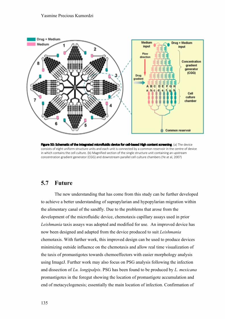
Yasmine Precious Kumordzi
135
Figure 50: Schematic of the integrated microfluidic device for cell-based High content screening. (a) The device consists of eight uniform structure units and each unit is connected by a common reservoir in the centre of device in which contains the cell culture. (b) Magnified section of the single structure unit containing an upstream concentration gradient generator (CGG) and downstream parallel cell culture chambers (Ye at al, 2007)
5.7 Future
The new understanding that has come from this study can be further developed
to achieve a better understanding of suprapylarian and hypopylarian migration within
the alimentary canal of the sandfly. Due to the problems that arose from the
development of the microfluidic device, chemotaxis capillary assays used in prior
Leishmania taxis assays was adopted and modified for use. An improved device has
now been designed and adapted from the device produced to suit Leishmania
chemotaxis. With further work, this improved design can be used to produce devices
minimizing outside influence on the chemotaxis and allow real time visualization of
the taxis of promastigotes towards chemoeffectors with easier morphology analysis
using ImageJ. Further work may also focus on PSG analysis following the infection
and dissection of Lu. longipalpis. PSG has been found to be produced by L. mexicana
promastigotes in the foregut showing the location of promastigote accumulation and
end of metacyclogenesis; essentially the main location of infection. Confirmation of

Yasmine Precious Kumordzi
136
PSG production in the hindgut of an L. tarentolae infection would be a highly
interesting discovery; it would suggest that PSG has different roles for Leishmania
with different modes of infection. The study so far has taken into account the
gradients that were predicted to be formed at the hindgut originating from the
malpighian tubules, and the foregut originating from the crop, however many more
gradients will be present in the alimentary canal which could act upon the migration
of promastigotes. Other potential chemoeffectors can be based upon many organs,
microbial communities etc. therefore for a greater understanding emphasis should be
put in the study on other potential chemoeffectors.

Yasmine Precious Kumordzi
137
CHAPTER SIX: CONCLUSION
Leishmania parasites cause the diseases leishmaniasis, a neglected tropical
disease with a wide geographical distribution globally. It is transmitted via the bite of
an infected sand fly. An important aspect to understanding transmission of infective
stage promastigotes is understanding what differs between the distinct infection of
hypopylarian and the forward migrating suprapylarian Leishmania species within a
sandfly. This work demonstrated a pilot screen of chemoeffectors and the
development of a novel technique for a Leishmania real time chemotaxis assay to
detect chemoeffectors that Leishmania promastigotes migrate towards. The
development of a microfluidic device for better chemotaxic responses and precise
control of gradient without outside influence needs future input into the device
redesign. This thesis focused on studying L. tarentolae a hypopylarian and L.
mexicana a suprapylarian. This gives the ability to compare results and understand the
differences between each mode of infection and transmission.
Leishmania promastigotes undergo several morphological changes that have
been well studied in L. mexicana, giving morphological categories according to
criteria. L. mexicana fitted this criteria very well, however L. tarentolae showed
challenges as two new morphologies were found. These findings of new morphology
were named bulgetomonad and kinetoplast nucleus swapping promastigotes. This
focus on morphometry showed the stage of promastigotes in an axenic culture that
was required to replicate the stages of promastigotes in which escaped the PM. This
stage of culture was used for further capillary assay as it represented the stage that
initiates the migration within the alimentary canal. The migration of promastigotes
within the alimentary canal has always been associated with the gradient of sugars
emitted from the crop during the slow release of sugars to the midgut for digestion.
However this positive taxis did not answer the question of migration of hypopylarian
Leishmania species as they develop and migrate towards the hindgut. Using capillary
assays and urea as a potential chemoeffector showed that compared to sugars, L.
tarentolae had greater attraction to urea hence the hindgut migration and inability to
be transmitted via the bite of an infected sandfly. Interestingly, analysis of migrated

Yasmine Precious Kumordzi
138
promastigotes showed a higher migration of leptomonad promastigotes. This
questions whether or not nectomonads are the primary migratory promastigote
morphological stage within a sandfly. As a pilot screen of potential chemoeffectors ,
this study brings about a lot of new understanding of the less studied species L.
tarentolae.

Yasmine Precious Kumordzi
139
REFERENCES
Adler, J. (1966). Chemotaxis in Bacteria. Science, 153(3737), pp.708-716.
Adler, J. (1969). Chemoreceptors in Bacteria. Science, 166(3913), pp.1588-1597.
Adler, J. (1973). A Method for Measuring Chemotaxis and Use of the Method to
Determine Optimum Conditions for Chemotaxis by Escherichia coli. Journal of
General Microbiology, 74(1), pp.77-91.
Adler, S. and Theodor, O. (1929). Observations on leishmania ceramodactyli.
N.SP. Transactions of the Royal Society of Tropical Medicine and Hygiene, 22(4),
pp.343-356.
Aguiar, E., Olmo, R., Paro, S., Ferreira, F., de Faria, I., Todjro, Y., Lobo, F., Kroon,
E., Meignin, C., Gatherer, D., Imler, J. and Marques, J. (2015). Sequence-independent
characterization of viruses based on the pattern of viral small RNAs produced by the
host. Nucleic Acids Research, 43(13), pp.6191-6206.
Akhoundi, M., Kuhls, K., Cannet, A., Votýpka, J., Marty, P., Delaunay, P. and
Sereno, D. (2016). A Historical Overview of the Classification, Evolution, and
Dispersion of Leishmania Parasites and Sandflies. PLOS Neglected Tropical Diseases,
10(3), p.e0004349.
Alawieh, A., Musharrafieh, U., Jaber, A., Berry, A., Ghosn, N. and Bizri, A. (2014).
Revisiting leishmaniasis in the time of war: the Syrian conflict and the Lebanese
outbreak. International Journal of Infectious Diseases, 29, pp.115-119.
Alvar, J., Croft, S., Kaye, P., Khamesipour, A., Sundar, S. and Reed, S. (2013). Case
study for a vaccine against leishmaniasis. Vaccine, 31, pp.B244-B249.
Alvar, J., Yactayo, S. and Bern, C. (2006). Leishmaniasis and poverty. Trends in
Parasitology, 22(12), pp.552-557.

Yasmine Precious Kumordzi
140
Ambit, A., Woods, K., Cull, B., Coombs, G. and Mottram, J. (2011). Morphological
Events during the Cell Cycle of Leishmania major. Eukaryotic Cell, 10(11), pp.1429-
1438.
Antoine JC, Prina E, Jouanne C, Bongrand P. Parasitophorous vacuoles of Leishmania
amazonensis-infected macrophages maintain an acidic pH. Infect Immun.
1990;58(3):779-87.
Armitage, J. (1992). Bacterial motility and chemotaxis. Sci Prog, 76(301-302 Pt 3-4),
pp.451-77.
Armitage, J., Josey, D. and Smith, D. (1977). A Simple, Quantitative Method for
Measuring Chemotaxis and Motility in Bacteria. Journal of General Microbiology,
102(1), pp.199-202.
Aslett, M., Aurrecoechea, C., Berriman, M., Brestelli, J., Brunk, B., Carrington, M.,
Depledge, D., Fischer, S., Gajria, B., Gao, X., Gardner, M., Gingle, A., Grant, G.,
Harb, O., Heiges, M., Hertz-Fowler, C., Houston, R., Innamorato, F., Iodice, J.,
Kissinger, J., Kraemer, E., Li, W., Logan, F., Miller, J., Mitra, S., Myler, P., Nayak,
V., Pennington, C., Phan, I., Pinney, D., Ramasamy, G., Rogers, M., Roos, D., Ross,
C., Sivam, D., Smith, D., Srinivasamoorthy, G., Stoeckert, C., Subramanian, S.,
Thibodeau, R., Tivey, A., Treatman, C., Velarde, G. and Wang, H. (2009).
TriTrypDB: a functional genomic resource for the Trypanosomatidae. Nucleic Acids
Research, 38(suppl_1), pp.D457-D462.
Ayhan, N., Sherifi, K., Taraku, A., Bërxholi, K. and Charrel, R. (2017). High Rates of
Neutralizing Antibodies to Toscana and Sandfly Fever Sicilian Viruses in Livestock,
Kosovo. Emerging Infectious Diseases, 23(6), pp.989-992.
Bañuls, A., Bastien, P., Pomares, C., Arevalo, J., Fisa, R. and Hide, M. (2011).
Clinical pleiomorphism in human leishmaniases, with special mention of
asymptomatic infection. Clinical Microbiology and Infection, 17(10), pp.1451-1461.
Barros, V., Oliveira, J., Melo, M. and Gontijo, N. (2006). Leishmania amazonensis:
Chemotaxic and osmotaxic responses in promastigotes and their probable role in

Yasmine Precious Kumordzi
141
development in the phlebotomine gut. Experimental Parasitology, 112(3), pp.152-
157.
Bates, P. (1994). Complete developmental cycle of Leishmania mexicana in axenic
culture. Parasitology, 108(01), p.1.
Bates, P. and Tetley, L. (1993). Leishmania mexicana: Induction of Metacyclogenesis
by Cultivation of Promastigotes at Acidic pH. Experimental Parasitology, 76(4),
pp.412-423.
Beach, R., Leeuwenburg, J. and Kiilu, G. (1985). Modification of Sand Fly Biting
Behavior by Leishmania Leads to Increased Parasite Transmission. The American
Journal of Tropical Medicine and Hygiene, 34(2), pp.278-282.
Berg, H. (2004). E. coli in Motion. Biological and Medical Physics, Biomedical
Engineering.
Besteiro, S., Williams, R., Coombs, G. and Mottram, J. (2007). Protein turnover and
differentiation in Leishmania. International Journal for Parasitology, 37(10),
pp.1063-1075.
Borovsky, D. and Schlein, Y. (1987). Trypsin and chymotrypsin-Iike enzymes of the
sandfly Phlebotomus papatasi infected with Leishmania and their possible role in
vector competence. Medical and Veterinary Entomology, 1(3), pp.235-242.
Boyden, S. (1962). The chemotactic effect of mixtures of antibodies and antigen on
polymorphonuclear leucocytes. Journal of Experimental Medicine, 115(3), pp.453-
466.
BRAY, R. (1983). Leishmania: Chemotaxic Responses of Promastigotes and
Macrophages In Vitro1. The Journal of Protozoology, 30(2), pp.322-329.
Brody, J. and Yager, P. (1997). Diffusion-based extraction in a microfabricated
device. Sensors and Actuators A: Physical, 58(1), pp.13-18.

Yasmine Precious Kumordzi
142
Burchmore, R. and Landfear, S. (1998). Differential Regulation of Multiple Glucose
Transporter Genes in Leishmania mexicana. Journal of Biological Chemistry,
273(44), pp.29118-29126.
Burchmore, R., Rodriguez-Contreras, D., McBride, K., Barrett, M., Modi, G., Sacks,
D. and Landfear, S. (2003). Genetic characterization of glucose transporter function in
Leishmania mexicana. Proceedings of the National Academy of Sciences, 100(7),
pp.3901-3906.
Cairns, B., Collard, M. and Landfear, S. (1989). Developmentally regulated gene from
Leishmania encodes a putative membrane transport protein. Proceedings of the
National Academy of Sciences, 86(20), pp.7682-7686.
Carter, N., Drew, M., Sanchez, M., Vasudevan, G., Landfear, S. and Ullman, B.
(2000). Cloning of a Novel Inosine-Guanosine Transporter Gene from Leishmania
donovani by Functional Rescue of a Transport-deficient Mutant. Journal of Biological
Chemistry, 275(27), pp.20935-20941.
Carter, N., Yates, P., Gessford, S., Galagan, S., Landfear, S. and Ullman, B. (2010).
Adaptive responses to purine starvation in Leishmania donovani. Molecular
Microbiology, p.92-107.
Čiháková, J. and Volf, P. (1997). Development of different Leishmania major strains
in the vector sandflies Phlebotomus papatasi and P. duboscqi. Annals of Tropical
Medicine & Parasitology, 91(3), pp.267-279.
Coombs, G., Craft, J. and Hart, D. (1982). A comparative study of Leishmania
mexicana amastigotes and promastigotes, enzyme activities and subcellular
locations. Molecular and Biochemical Parasitology, 5(3), pp.199-211.
Costa, D., Codeço, C., Silva, M. and Werneck, G. (2013). Culling Dogs in Scenarios
of Imperfect Control: Realistic Impact on the Prevalence of Canine Visceral
Leishmaniasis. PLoS Neglected Tropical Diseases, 7(8), p.e2355.
Courtenay, O., Quinnell, R., Garcez, L., Shaw, J. and Dye, C. (2002). Infectiousness
in a Cohort of Brazilian Dogs: Why Culling Fails to Control Visceral Leishmaniasis in

Yasmine Precious Kumordzi
143
Areas of High Transmission. The Journal of Infectious Diseases, 186(9), pp.1314-
1320.
Croft, S. and Olliaro, P. (2011). Leishmaniasis chemotherapy—challenges and
opportunities. Clinical Microbiology and Infection, 17(10), pp.1478-1483.
Cuvillier, A., Miranda, J., Ambit, A., Barral, A. and Merlin, G. (2003). Abortive
infection of Lutzomyia longipalpis insect vectors by aflagellated LdARL-3A-Q70L
overexpressing Leishmania amazonensis parasites. Cellular Microbiology, 5(10),
pp.717-728.
Davies, A.G. (1978). Pollution studies with marine plankton. II. Heavy metals. Adv.
Mar. Biol., 15: 381-508.
de Souza, W., and Souto-Padron, T. (1980). The Paraxial Structure of the Flagellum
of Trypanosomatidae. The Journal of Parasitology, 66(2), p.229.
de Ysasa Pozzo, L., Fontes, A., de Thomaz, A., Barbosa, L., Ayres, D., Giorgio, S.
and Cesar, C. (2007). Leishmania amazonensis chemotaxis under glucose gradient
studied by the strength and directionality of forces measured with optical
tweezers. Imaging, Manipulation, and Analysis of Biomolecules, Cells, and Tissues V.
Depaquit, J., Grandadam, M., Fouque, F., Andry, P. and Peyrefitte, C. (2010).
Arthropod-borne viruses transmitted by Phlebotomine sandflies in Europe: a
review. Eurosurveillance, 15(10), pii:19507.
Diaz-Albiter, H., Regnault, C., Alpizar-Sosa, E., McGuinness, D., Barrett, M. and
Dillon, R. (2018). Non-invasive visualisation and identification of fluorescent
Leishmania tarentolae in infected sand flies. Wellcome Open Research, 3, p.160.
Díaz, E., Köhidai, L., Ríos, A., Vanegas, O., Ponte-Sucre, A. (2011). Ensayos de
quimiotaxis in vitro en Leishmania sp. Evaluación de la técnica de los capilares- dos
cámaras en promastigotes. Rev. Fac. Farm. (UCV) 74, 31–40.

Yasmine Precious Kumordzi
144
Díaz, E., Köhidai, L., Ríos, A., Vanegas, O., Silva, A., Szabó, R., Mező, G., Hudecz,
F. and Ponte-Sucre, A. (2013). Leishmania braziliensis: Cytotoxic, cytostatic and
chemotactic effects of poly-lysine–methotrexate-conjugates. Experimental
Parasitology, 135(1), pp.134-141.
Dillon, R. and Lane, P. (1993). Bloodmeal digestion in the midgut of Phlebotomus
papatasi and Phlebotomus langeroni. Medical and Veterinary Entomology, 7(3),
pp.225-232.
Dillon, R. and Lane, R. (1993). Influence of Leishmania infection on blood-meal
digestion in the sandflies Phlebotomus papatasi and P. langeroni. Parasitology
Research, 79(6), pp.492-496.
Dillon, R., Ivens, A., Churcher, C., Holroyd, N., Quail, M., Rogers, M., Soares, M.,
Bonaldo, M., Casavant, T., Lehane, M. and Bates, P. (2006). Analysis of ESTs from
Lutzomyia longipalpis sand flies and their contribution toward understanding the
insect–parasite relationship. Genomics, 88(6), pp.831-840.
Docampo, R., de Souza, W., Miranda, K., Rohloff, P. and Moreno, S. (2005).
Acidocalcisomes - conserved from bacteria to man. Nature Reviews Microbiology,
3(3), pp.251-261.
Dougall, A., Alexander, B., Holt, D., Harris, T., Sultan, A., Bates, P., Rose, K. and
Walton, S. (2011). Evidence incriminating midges (Diptera: Ceratopogonidae) as
potential vectors of Leishmania in Australia. International Journal for Parasitology,
41(5), pp.571-579.
Englert, D., Manson, M. and Jayaraman, A. (2009). Flow-Based Microfluidic Device
for Quantifying Bacterial Chemotaxis in Stable, Competing Gradients. Applied and
Environmental Microbiology, 75(13), pp.4557-4564.
Ergunay, K., Kasap, O., Orsten, S., Oter, K., Gunay, F., Yoldar, A., Dincer, E., Alten,
B. and Ozkul, A. (2014). Phlebovirus and Leishmania detection in sandflies from
eastern Thrace and northern Cyprus. Parasites & Vectors, 7(1), p.575.

Yasmine Precious Kumordzi
145
Faucher, B., Piarroux, R., Mary, C., Bichaud, L., Charrel, R., Izri, A. and de
Lamballerie, X. (2014). Presence of sandflies infected with Leishmania infantum and
Massilia virus in the Marseille urban area. Clinical Microbiology and Infection, 20(5),
pp.O340-O343.
Field, M. and Carrington, M. (2009). The trypanosome flagellar pocket. Nature
Reviews Microbiology, 7(11), pp.775-786.
Figarella, K., Uzcategui, N., Zhou, Y., LeFurgey, A., Ouellette, M., Bhattacharjee, H.
and Mukhopadhyay, R. (2007). Biochemical characterization of Leishmania major
aquaglyceroporin LmAQP1: possible role in volume regulation and
osmotaxis. Molecular Microbiology, 65(4), pp.1006-1017.
Fraihi, W., Fares, W., Perrin, P., Dorkeld, F., Sereno, D., Barhoumi, W., Sbissi, I.,
Cherni, S., Chelbi, I., Durvasula, R., Ramalho-Ortigao, M., Gtari, M. and Zhioua, E.
(2017). An integrated overview of the midgut bacterial flora composition of
Phlebotomus perniciosus, a vector of zoonotic visceral leishmaniasis in the Western
Mediterranean Basin. PLOS Neglected Tropical Diseases, 11(3), p.e0005484.
Fuqua, W., Winans, S. and Greenberg, E. (1994). Quorum sensing in bacteria: the
LuxR-LuxI family of cell density-responsive transcriptional regulators. Journal of
Bacteriology, 176(2), pp.269-275.
Gillespie, P., Beaumier, C., Strych, U., Hayward, T., Hotez, P. and Bottazzi, M.
(2016). Status of vaccine research and development of vaccines for
leishmaniasis. Vaccine, 34(26), pp.2992-2995.
Gomes, B., Purkait, B., Deb, R., Rama, A., Singh, R., Foster, G., Coleman, M.,
Kumar, V., Paine, M., Das, P. and Weetman, D. (2017). Knockdown resistance
mutations predict DDT resistance and pyrethroid tolerance in the visceral
leishmaniasis vector Phlebotomus argentipes. PLOS Neglected Tropical Diseases,
11(4), p.e0005504.
Hammond, D. and Gutteridge, W. (1984). Purine and pyrimidine metabolism in the
trypanosomatidae. Molecular and Biochemical Parasitology, 13(3), pp.243-261.
Yasmine Precious Kumordzi
146
Harmsen, R. (1973). The nature of the establishment barrier for Trypanosoma brucei
in the gut of Glossina pallidipes. Transactions of the Royal Society of Tropical
Medicine and Hygiene, 67(3), pp.364-373.
Herrer, A. and Christensen, H. (1975). Implication of Phlebotomus sand flies as
vectors of bartonellosis and leishmaniasis as early as 1764. Science, 190(4210),
pp.154-155.
Horn, D. and Duraisingh, M. (2014). Antiparasitic Chemotherapy: From Genomes to
Mechanisms. Annual Review of Pharmacology and Toxicology, 54(1), pp.71-94.
Inbar, E., Canepa, G., Carrillo, C., Glaser, F., Suter Grotemeyer, M., Rentsch, D.,
Zilberstein, D. and Pereira, C. (2010). Lysine transporters in human trypanosomatid
pathogens. Amino Acids, 42(1), pp.347-360.
Jeon, N., Dertinger, S., Chiu, D., Choi, I., Stroock, A. and Whitesides, G. (2000).
Generation of Solution and Surface Gradients Using Microfluidic Systems. Langmuir,
16(22), pp.8311-8316.
Johnson, W. (1941). Nutrition in the Protozoa. The Quarterly Review of Biology,
16(3), pp.336-348.
Joshi, M., Rogers, M., Shakarian, A., Yamage, M., Al-Harthi, S., Bates, P. and
Dwyer, D. (2004). Molecular Characterization, Expression, and in Vivo Analysis of
LmexCht1. Journal of Biological Chemistry, 280(5), pp.3847-3861.
Kamhawi, S. (2000). Protection Against Cutaneous Leishmaniasis Resulting from
Bites of Uninfected Sand Flies. Science, 290(5495), pp.1351-1354.
Kamhawi, S. (2006). Phlebotomine sand flies and Leishmania parasites: friends or
foes?. Trends in Parasitology, 22(9), pp.439-445.
Kaufer, A., Ellis, J., Stark, D. and Barratt, J. (2017). The evolution of trypanosomatid
taxonomy. Parasites & Vectors, 10(1).
Yasmine Precious Kumordzi
147
Kelly, P., Bahr, S., Serafim, T., Ajami, N., Petrosino, J., Meneses, C., Kirby, J.,
Valenzuela, J., Kamhawi, S. and Wilson, M. (2017). The Gut Microbiome of the
Vector Lutzomyia longipalpis Is Essential for Survival of Leishmania infantum. mBio,
8(1).
Keymer, J., Endres, R., Skoge, M., Meir, Y. and Wingreen, N. (2006). Chemosensing
in Escherichia coli: Two regimes of two-state receptors. Proceedings of the National
Academy of Sciences, 103(6), pp.1786-1791.
Killick-Kendrick, R. (1978). Recent advances and outstanding problems in the
biology of phlebotomine sandflies. A review. Acta Trop, 35(4), pp.297-313.
Killick-Kendrick, R. (1990). Phlebotomine vectors of the leishmaniases: a
review. Medical and Veterinary Entomology, 4(1), pp.1-24.
Killick-Kendrick, R., Leaney, A., Ready, P. and Molyneux, D. (1977). Leishmania in
phlebotomid sandflies - IV. The transmission of Leishmania mexicana amazonensis to
hamsters by the bite of experimentally infected Lutzomyia longipalpis. Proceedings of
the Royal Society of London. Series B. Biological Sciences, 196(1122), pp.105-115.
Köhidai, L. (1995). Method for determination of chemoattraction in Tetrahymena
pyriformis. Current Microbiology, 30(4), pp.251-253.
Kwakye-Nuako, G., Mosore, M., Duplessis, C., Bates, M., Puplampu, N., Mensah-
Attipoe, I., Desewu, K., Afegbe, G., Asmah, R., Jamjoom, M., Ayeh-Kumi, P.,
Boakye, D. and Bates, P. (2015). First isolation of a new species of Leishmania
responsible for human cutaneous leishmaniasis in Ghana and classification in the
Leishmania enriettii complex. International Journal for Parasitology, 45(11), pp.679-
684.
Lainson, R., Ready, P. and Shaw, J. (1979). Leishmania in phlebotomid sandflies. VII.
On the taxonomic status of Leishmania peruviana , causative agent of Peruvian ‘uta’,
as indicated by its development in the sandfly, Lutzomyia longipalpis. Proceedings of
the Royal Society of London. Series B. Biological Sciences, 206(1164), pp.307-318.

Yasmine Precious Kumordzi
148
Landfear, S. (2011). Nutrient Transport and Pathogenesis in Selected Parasitic
Protozoa. Eukaryotic Cell, 10(4), pp.483-493.
Landfear, S. and Ignatushchenko, M. (2001). The flagellum and flagellar pocket of
trypanosomatids. Molecular and Biochemical Parasitology, 115(1), pp.1-17.
Langousis, G. and Hill, K. (2014). Motility and more: the flagellum of Trypanosoma
brucei. Nature Reviews Microbiology, 12(7), pp.505-518.
Lantova, L. and Volf, P. (2014). Mosquito and sand fly gregarines of the genus
Ascogregarina and Psychodiella (Apicomplexa: Eugregarinorida, Aseptatorina) –
Overview of their taxonomy, life cycle, host specificity and pathogenicity. Infection,
Genetics and Evolution, 28, pp.616-627.
Lautenschlaeger, F. (2011). Cell compliance: cytoskeletal origin and importance for
cellular function. (Doctoral thesis). https://doi.org/10.17863/CAM.16575
Lawyer, P., Githure, J., Roberts, C., Anjili, C., Mebrahtu, Y., Ngumbi, P., Koech, D.
and Odongo, S. (1990). Development of Leishmania major in Phlebotomus duboscqi
and Sergentomyia schwetzi (Diptera: Psychodidae). The American Journal of Tropical
Medicine and Hygiene, 43(1), pp.31-43.
Lehane, M. (1997). Peritrophic matrix structure and function. Annual Review of
Entomology, 42(1), pp.525-550.
Lehane, M. (2005). The biology of blood-sucking in insects. Cambridge: Cambridge
University Press, p.96.
Leslie, G., Barrett, M. and Burchmore, R. (2002). Leishmania mexicana:
promastigotes migrate through osmotic gradients. Experimental Parasitology, 102(2),
pp.117-120.

Yasmine Precious Kumordzi
149
Lestinova, T., Rohousova, I., Sima, M., de Oliveira, C. and Volf, P. (2017). Insights
into the sand fly saliva: Blood-feeding and immune interactions between sand flies,
hosts, and Leishmania. PLOS Neglected Tropical Diseases, 11(7), p.e0005600.
Lin, B. and Levchenko, A. (2015). Spatial Manipulation with Microfluidics. Frontiers
in Bioengineering and Biotechnology, 3.
Lukes, J., Mauricio, I., Schonian, G., Dujardin, J., Soteriadou, K., Dedet, J., Kuhls, K.,
Tintaya, K., Jirku, M., Chocholova, E., Haralambous, C., Pratlong, F., Obornik, M.,
Horak, A., Ayala, F. and Miles, M. (2007). Evolutionary and geographical history of
the Leishmania donovani complex with a revision of current taxonomy. Proceedings
of the National Academy of Sciences, 104(22), pp.9375-9380.
Macnab, R. and Koshland, D. (1972). The Gradient-Sensing Mechanism in Bacterial
Chemotaxis. Proceedings of the National Academy of Sciences, 69(9), pp.2509-2512.
Maia, C. and Depaquit, J. (2016). Can Sergentomyia (Diptera, Psychodidae) play a
role in the transmission of mammal-infecting Leishmania?. Parasite, 23, p.55.
Manson, P., Cooke, G. and Zumla, A. (2009). Manson's tropical diseases.
[Edinburgh]: Saunders Elsevier, p.1715.
Maroli, M., Feliciangeli, M., Bichaud, L., Charrel, R. and Gradoni, L. (2012).
Phlebotomine sandflies and the spreading of leishmaniases and other diseases of
public health concern. Medical and Veterinary Entomology, 27(2), pp.123-147.
Mauricio, I., Howard, M., Stothard, J. and Miles, M. (1999). Genomic diversity in the
Leishmania donovani complex. Parasitology, 119(3), pp.237-246.
Mittra, B., Cortez, M., Haydock, A., Ramasamy, G., Myler, P. and Andrews, N.
(2013). Iron uptake controls the generation of Leishmania infective forms through
regulation of ROS levels. The Journal of General Physiology, 141(3), pp.17-17.

Yasmine Precious Kumordzi
150
Modi G.B. (1997) Care and maintenance of phlebotomine sandfly colonies. In:
Crampton J.M., Beard C.B., Louis C. (eds) The Molecular Biology of Insect
Disease Vectors. Springer, Dordrecht
Moloo, S. and Kutuza, S. (1971). Feeding and crop emptying in Glossina brevipalpis
Newstead. Transactions of the Royal Society of Tropical Medicine and Hygiene,
65(2), pp.221-222.
Monteiro, C., Villegas, L., Campolina, T., Pires, A., Miranda, J., Pimenta, P. and
Secundino, N. (2016). Bacterial diversity of the American sand fly Lutzomyia
intermedia using high-throughput metagenomic sequencing. Parasites & Vectors,
9(1).
Mukhopadhyay, J., Braig, H., Rowton, E. and Ghosh, K. (2012). Naturally Occurring
Culturable Aerobic Gut Flora of Adult Phlebotomus papatasi, Vector of Leishmania
major in the Old World. PLoS ONE, 7(5), p.e35748.
Myler, P. and Fasel, N. (2008). Leishmania: After the Genome. Norfolk: Caister
academic Press, p.209.
Naula, C., Logan, F., Wong, P., Barrett, M. and Burchmore, R. (2010). A Glucose
Transporter Can Mediate Ribose Uptake. Journal of Biological Chemistry, 285(39),
pp.29721-29728.
Oliveira, F., Rowton, E., Aslan, H., Gomes, R., Castrovinci, P., Alvarenga, P.,
Abdeladhim, M., Teixeira, C., Meneses, C., Kleeman, L., Guimarães-Costa, A.,
Rowland, T., Gilmore, D., Doumbia, S., Reed, S., Lawyer, P., Andersen, J., Kamhawi,
S. and Valenzuela, J. (2015). A sand fly salivary protein vaccine shows efficacy
against vector-transmitted cutaneous leishmaniasis in nonhuman primates. Science
Translational Medicine, 7(290), pp.290ra90-290ra90.
Oliveira, J.S., Melo, M.N., Gontijo, N.F. (2000). A sensitive method for assaying
chemotactic responses of Leishmania promastigotes. Experimental Parasitology 96,
187–189.

Yasmine Precious Kumordzi
151
Oliveira, S., Moraes, B., Gonçalves, C., Giordano-Dias, C., d'Almeida, J., Asensi, M.,
Mello, R. and Brazil, R. (2000). Prevalência da microbiota no trato digestivo de
fêmeas de Lutzomyia longipalpis (Lutz & Neiva, 1912) (Diptera: Psychodidae)
provenientes do campo. Revista da Sociedade Brasileira de Medicina Tropical, 33(3),
pp.319-322.
Opperdoes, F. (1987). Compartmentation Of Carbohydrate Metabolism In
Trypanosomes. Annual Review of Microbiology, 41(1), pp.127-151.
Ortiz, D., Sanchez, M., Koch, H., Larsson, H. and Landfear, S. (2009). An Acid-
activated Nucleobase Transporter from Leishmania major. Journal of Biological
Chemistry, 284(24), pp.16164-16169.
Paliwal, S., Iglesias, P., Campbell, K., Hilioti, Z., Groisman, A. and Levchenko, A.
(2007). MAPK-mediated bimodal gene expression and adaptive gradient sensing in
yeast. Nature, 446(7131), pp.46-51.
Pigott, D. M., Bhatt, S., Golding, N., Duda, K. A., Battle, K. E., Brady, O. J., Messina,
J. P., Balard, Y., Bastien, P., Pratlong, F., Brownstein, J. S., Freifeld, C. C., Mekaru,
S. R., Gething, P. W., George, D. B., Myers, M. F., Reithinger, R., Hay, S. I. (2014).
Global distribution maps of the leishmaniases. eLife, 3, e02851.
doi:10.7554/eLife.02851
Pires, A., Villegas, L., Campolina, T., Orfanó, A., Pimenta, P. and Secundino, N.
(2017). Bacterial diversity of wild-caught Lutzomyia longipalpis (a vector of zoonotic
visceral leishmaniasis in Brazil) under distinct physiological conditions by
metagenomics analysis. Parasites & Vectors, 10(1).
Pimenta, P., Modi, G., Pereira, S., Shahabuddin, M. and Sacks, D. (1997). A novel
role for the peritrophic matrix in protecting Leishmania from the hydrolytic activities
of the sand fly midgut. Parasitology, 115(4), pp.359-369.
Pimenta, P., Turco, S., McConville, M., Lawyer, P., Perkins, P. and Sacks, D. (1992).
Stage-specific adhesion of Leishmania promastigotes to the sandfly midgut. Science,
256(5065), pp.1812-1815.

Yasmine Precious Kumordzi
152
Pimentel, D., Ramos, R., Santana, M., Maia, C., Carvalho, G., Silva, H. and Alves, L.
(2015). Prevalence of zoonotic visceral leishmaniasis in dogs in an endemic area of
Brazil. Revista da Sociedade Brasileira de Medicina Tropical, 48(4), pp.491-493.
Polacheck, W., Charest, J. and Kamm, R. (2011). Interstitial flow influences direction
of tumor cell migration through competing mechanisms. Proceedings of the National
Academy of Sciences, 108(27), pp.11115-11120.
Pozzo, L.Y., Fontes, A., de Thomaz, A.A., Santos, B.B., Farias, P., Ayres, D.C.,
Giorgio, S., Cesar, C.L., 2009. Studying taxis in real time using optical tweezers:
applications for Leishmania amazonensis parasites. Micron 40, 617–620.
Pruzinova, K., Sadlova, J., Seblova, V., Homola, M., Votypka, J. and Volf, P. (2015).
Comparison of Bloodmeal Digestion and the Peritrophic Matrix in Four Sand Fly
Species Differing in Susceptibility to Leishmania donovani. PLOS ONE, 10(6),
p.e0128203.
Pumpuni, C., Kent, M., Davis, J., Beier, J. and Demaio, J. (1996). Bacterial
Population Dynamics in Three Anopheline Species: The Impact on Plasmodium
Sporogonic Development. The American Journal of Tropical Medicine and Hygiene,
54(2), pp.214-218.
Ramalho-Ortigão, J., Kamhawi, S., Joshi, M., Reynoso, D., Lawyer, P., Dwyer, D.,
Sacks, D. and Valenzuela, J. (2005). Characterization of a blood activated chitinolytic
system in the midgut of the sand fly vectors Lutzomyia longipalpis and Phlebotomus
papatasi. Insect Molecular Biology, 14(6), pp.703-712.
Ramalho-Ortigao, M. (2010). Sand Fly-Leishmania Interactions: Long Relationships
are Not Necessarily Easy. The Open Parasitology Journal, 4(1), pp.195-204.
Ramsay, J. (1951). Osmotic Regulation in Mosquito Larvae: The Role of the
Malpighian Tubules. Journal of Experimental Biology, 28, pp.62-73.

Yasmine Precious Kumordzi
153
Ready, P. (1979). Factors Affecting Egg Production of Laboratory-Bred Lutzomyia
Longipalpis (Diptera: Psychodidae)1. Journal of Medical Entomology, 16(5), pp.413-
423.
Ready, P. (2013). Biology of Phlebotomine Sand Flies as Vectors of Disease
Agents. Annual Review of Entomology, 58(1), pp.227-250.
Reithinger, R., Coleman, P., Alexander, B., Vieira, E., Assis, G. and Davies, C.
(2004). Are insecticide-impregnated dog collars a feasible alternative to dog culling as
a strategy for controlling canine visceral leishmaniasis in Brazil?. International
Journal for Parasitology, 34(1), pp.55-62.
Reithinger, R., Mohsen, M. and Leslie, T. (2010). Risk Factors for Anthroponotic
Cutaneous Leishmaniasis at the Household Level in Kabul, Afghanistan. PLoS
Neglected Tropical Diseases, 4(3), p.e639.
Rezvan, H. and Moafi, M. (2015). An overview on Leishmania vaccines: A narrative
review article. Veterinary Research Forum, 6(1), pp.1-7.
Rezvan, H., Nourian, A. and Navard, S. (2017). An Overview on Leishmania
Diagnosis. JoMMID, 5(1 and 21), pp.1-11.
RG Endris et al. 1982 Mosquitos News 42: 400-407
Richards O.W., Davies R.G. (1977) The Alimentary Canal, Nutrition and
Digestion. In: IMMS’ General Textbook of Entomology. Springer, Dordrecht
Rogers, M. (2012). The Role of Leishmania Proteophosphoglycans in Sand Fly
Transmission and Infection of the Mammalian Host. Frontiers in Microbiology, 3.
Rogers, M., Chance, M. and Bates, P. (2002). The role of promastigote secretory gel
in the origin and transmission of the infective stage of Leishmania mexicana by the
sandfly Lutzomyia longipalpis. Parasitology, 124(05).

Yasmine Precious Kumordzi
154
Rogers, M., Hajmová, M., Joshi, M., Sadlova, J., Dwyer, D., Volf, P. and Bates, P.
(2008). Leishmania chitinase facilitates colonization of sand fly vectors and enhances
transmission to mice. Cellular Microbiology, 10(6), pp.1363-1372.
Rogers, M., Ilg, T., Nikolaev, A., Ferguson, M. and Bates, P. (2004). Transmission of
cutaneous leishmaniasis by sand flies is enhanced by regurgitation of fPPG. Nature,
430(6998), pp.463-467.
Rotureau, B., Morales, M., Bastin, P. and Späth, G. (2009). The flagellum-mitogen-
activated protein kinase connection in Trypanosomatids: a key sensory role in parasite
signalling and development?. Cellular Microbiology, 11(5), pp.710-718.
Rudin, W. and Hecker, H. (1982). Functional morphology of the midgut of a sandfly
as compared to other hematophagous nematocera. Tissue and Cell, 14(4), pp.751-758.
Sacks, D. and Kamhawi, S. (2001). Molecular Aspects of Parasite-Vector and Vector-
Host Interactions in Leishmaniasis. Annual Review of Microbiology, 55(1), pp.453-
483.
Sádlová, J. and Volf, P. (2009). Peritrophic matrix of Phlebotomus duboscqi and its
kinetics during Leishmania major development. Cell and Tissue Research, 337(2),
pp.313-325.
Sales, K., Costa, P., de Morais, R., Otranto, D., Brandão-Filho, S., Cavalcanti, M. and
Dantas-Torres, F. (2015). Identification of phlebotomine sand fly blood meals by real-
time PCR. Parasites & Vectors, 8(1).
Sanchez, M., Zeoli, D., Klamo, E., Kavanaugh, M. and Landfear, S. (1995). A Family
of Putative Receptor-Adenylate Cyclases from Leishmania donovani. Journal of
Biological Chemistry, 270(29), pp.17551-17558.
Santos, V., Araujo, R., Machado, L., Pereira, M. and Gontijo, N. (2008). The
physiology of the midgut of Lutzomyia longipalpis (Lutz and Neiva 1912): pH in
different physiological conditions and mechanisms involved in its control. Journal of
Experimental Biology, 211(17), pp.2792-2798.
Yasmine Precious Kumordzi
155
Santos, V., Vale, V., Silva, S., Nascimento, A., Saab, N., Soares, R., Michalick, M.,
Araujo, R., Pereira, M., Fujiwara, R. and Gontijo, N. (2014). Host Modulation by a
Parasite: How Leishmania infantum Modifies the Intestinal Environment of
Lutzomyia longipalpis to Favor Its Development. PLoS ONE, 9(11), p.e111241.
Satir, P. (1968). STUDIES ON CILIA: III. Further Studies on the Cilium Tip and a
"Sliding Filament" Model of Ciliary Motility. The Journal of Cell Biology, 39(1),
pp.77-94.
Satir, P. and Christensen, S. (2008). Structure and function of mammalian
cilia. Histochemistry and Cell Biology, 129(6), pp.687-693.
Schlein, Y. (1986). Sandfly diet and Leishmania. Parasitology Today, 2(6), pp.175-
177.
Schlein, Y. and Jacobson, R. (1999). Sugar meals and longevity of the sandfly
Phlebotomus papatasi in an arid focus of Leishmania major in the Jordan
Valley. Medical and Veterinary Entomology, 13(1), pp.65-71.
Schlein, Y., Jacobson, R. and Messer, G. (1992). Leishmania infections damage the
feeding mechanism of the sandfly vector and implement parasite transmission by
bite. Proceedings of the National Academy of Sciences, 89(20), pp.9944-9948.
Secundino, N., Eger-Mangrich, I., Braga, E., Santoro, M. and Pimenta, P. (2005).
Lutzomyia longipalpis Peritrophic Matrix: Formation, Structure, and Chemical
Composition. Journal of Medical Entomology, 42(6), pp.928–938.
Sevá, A., Ovallos, F., Amaku, M., Carrillo, E., Moreno, J., Galati, E., Lopes, E.,
Soares, R. and Ferreira, F. (2016). Correction: Canine-Based Strategies for Prevention
and Control of Visceral Leishmaniasis in Brazil. PLOS ONE, 11(9), p.e0162854.
Seymour, J., Ahmed, T., Marcos and Stocker, R. (2008). A microfluidic chemotaxis
assay to study microbial behavior in diffusing nutrient patches. Limnology and
Oceanography: Methods, 6(9), pp.477-488.

Yasmine Precious Kumordzi
156
Shaked-Mishan, P., Suter-Grotemeyer, M., Yoel-Almagor, T., Holland, N.,
Zilberstein, D. and Rentsch, D. (2006). A novel high-affinity arginine transporter from
the human parasitic protozoan Leishmania donovani. Molecular Microbiology, 60(1),
pp.30-38.
Sharifi, I., Fekri, A., Aflatonian, M., Khamesipour, A., Nadim, A., Mousavi, M.,
Momeni, A., Dowlati, Y., Godal, T., Zicker, F., Smith, P. and Modabber, F. (1998).
Randomised vaccine trial of single dose of killed Leishmania major plus BCG against
anthroponotic cutaneous leishmaniasis in Bam, Iran. The Lancet, 351(9115), pp.1540-
1543.
Silflow, C. and Lefebvre, P. (2001). Assembly and Motility of Eukaryotic Cilia and
Flagella. Lessons from Chlamydomonas reinhardtii. PLANT PHYSIOLOGY, 127(4),
pp.1500-1507.
Silverman, M. and Leroux, M. (2009). Intraflagellar transport and the generation of
dynamic, structurally and functionally diverse cilia. Trends in Cell Biology, 19(7),
pp.306-316.
Simpson, L. (1968). Effect of acriflavine on the kinetoplast of Leishmania tarentolae:
Mode of Action and Physiological Correlates of the Loss of Kinetoplast DNA. The
Journal of Cell Biology, 37(3), pp.660-682.
Singla, V. and Reiter, J. (2006). The Primary Cilium as the Cell's Antenna: Signaling
at a Sensory Organelle. Science, 313(5787), pp.629-633.
Skoge, M., Adler, M., Groisman, A., Levine, H., Loomis, W. and Rappel, W. (2010).
Gradient sensing in defined chemotactic fields. Integrative Biology, 2(11-12), p.659.
Smith, C., Chaichana, K., Lee, Y., Lin, B., Stanko, K., O'Donnell, T., Gupta, S., Shah,
S., Wang, J., Wijesekera, O., Delannoy, M., Levchenko, A. and Quiñones-Hinojosa,
A. (2015). Pre-Exposure of Human Adipose Mesenchymal Stem Cells to Soluble
Factors Enhances Their Homing to Brain Cancer. STEM CELLS Translational
Medicine, 4(3), pp.239-251.

Yasmine Precious Kumordzi
157
Smith, D. and Littau, V. (1960). CELLULAR SPECIALIZATION IN THE
EXCRETORY EPITHELIA OF AN INSECT, Macrosteles fascifrons STAL
(HOMOPTERA). The Journal of Cell Biology, 8(1), pp.103-133.
Snapp, E. and Landfear, S. (1999). Characterization of a Targeting Motif for a
Flagellar Membrane Protein in Leishmania enriettii. Journal of Biological Chemistry,
274(41), pp.29543-29548.
Soares, R. and Turco, S. (2003). Erratum to ''Lutzomyia longipalpis (Diptera:
Psychodidae: Phlebotominae): a review''. Anais da Academia Brasileira de Ciências,
75(4), pp.441-441.
Soares, R., Cardoso, T., Barron, T., Araújo, M., Pimenta, P. and Turco, S. (2005).
Leishmania braziliensis: a novel mechanism in the lipophosphoglycan regulation
during metacyclogenesis. International Journal for Parasitology, 35(3), pp.245-253.
Soares, R., Macedo, M., Ropert, C., Gontijo, N., Almeida, I., Gazzinelli, R., Pimenta,
P. and Turco, S. (2002). Leishmania chagasi: lipophosphoglycan characterization and
binding to the midgut of the sand fly vector Lutzomyia longipalpis. Molecular and
Biochemical Parasitology, 121(2), pp.213-224.
Spotin, A., Rouhani, S., Ghaemmaghami, P., Haghighi, A., Zolfaghari, M.,
Amirkhani, A., Farahmand, M., Bordbar, A. and Parvizi, P. (2015). Different
Morphologies of Leishmania major Amastigotes with No Molecular Diversity in a
Neglected Endemic Area of Zoonotic Cutaneous Leishmaniasis in Iran. Iranian
Biomedical Journal, 19(3), pp.149–159.
Stierhof, Y., Bates, P., Jacobson, R., Rogers, M., Schlein, Y., Handman, E. and Ilg, T.
(1999). Filamentous proteophosphoglycan secreted by Leishmania promastigotes
forms gel-like three-dimensional networks that obstruct the digestive tract of infected
sandfly vectors. European Journal of Cell Biology, 78(10), pp.675-689.
Stoffolano, J. and Haselton, A. (2013). The Adult Dipteran Crop: A Unique and
Overlooked Organ. Annual Review of Entomology, 58(1), pp.205-225.

Yasmine Precious Kumordzi
158
Stuart, K., Brun, R., Croft, S., Fairlamb, A., Gürtler, R., McKerrow, J., Reed, S. and
Tarleton, R. (2008). Kinetoplastids: related protozoan pathogens, different
diseases. Journal of Clinical Investigation, 118(4), pp.1301-1310.
Sunter, J. and Gull, K. (2019). Shape, form, function and Leishmania pathogenicity:
from textbook descriptions to biological understanding. Open Biol, 7(9)pii: 170165
Talmi-Frank, D., King, R., Warburg, A., Peleg, O., Nasereddin, A., Svobodova, M.,
Jaffe, C. and Baneth, G. (2010). Leishmania tropica in Rock Hyraxes (Procavia
capensis) in a Focus of Human Cutaneous Leishmaniasis. The American Journal of
Tropical Medicine and Hygiene, 82(5), pp.814-818.
TANG, Y. and WARD, R. (1998). Sugar feeding and fluid destination control in the
phlebotomine sandfly Lutzomyia longipalpis (Diptera: Psychodidae). Medical and
Veterinary Entomology, 12(1), pp.13-19.
Telleria, E., Araújo, A., Secundino, N., d'Avila-Levy, C. and Traub-Csekö, Y. (2010).
Trypsin-Like Serine Proteases in Lutzomyia longipalpis – Expression, Activity and
Possible Modulation by Leishmania infantum chagasi. PLoS ONE, 5(5), p.e10697.
Telleria, E., Martins-da-Silva, A., Tempone, A. and Traub-Csekö, Y. (2018).
Leishmania, microbiota and sand fly immunity. Parasitology, 145(10), pp.1336-1353.
Thomson, A. (1975). Synchronization of function in the foregut of the blowfly
phormia regina (Dipteris: Calliphoridae) during the crop-emptying process. The
Canadian Entomologist, 107(11), pp.1193-1198.
Trager, W. (1965). The Kinetoplast and Differentiation in Certain Parasitic
Protozoa. The American Naturalist, 99(907), pp.255-266.
Turco, S. and Sacks, D. (2003). Control of Leishmania–Sand Fly Interactions by
Polymorphisms in Lipophosphoglycan Structure. Recognition of Carbohydrates in
Biological Systems, Part B: Specific Applications, pp.377-381.
Yasmine Precious Kumordzi
159
Uppaluri, S., Heddergott, N., Stellamanns, E., Herminghaus, S., Zöttl, A., Stark, H.,
Engstler, M. and Pfohl, T. (2012). Flow Loading Induces Oscillatory Trajectories in a
Bloodstream Parasite. Biophysical Journal, 103(6), pp.1162-1169.
Vaidyanathan, R. (2004). Leishmania parasites (Kinetoplastida: Trypanosomatidae)
reversibly inhibit visceral muscle contractions in hemimetabolous and holometabolous
insects. Journal of Invertebrate Pathology, 87(2-3), pp.123-128.
Vaidyanathan, R. (2005). Isolation of a Myoinhibitory Peptide from Leishmania
major (Kinetoplastida: Trypanosomatidae) and Its Function in the Vector Sand
FlyPhlebotomus papatasi(Diptera: Psychodidae). Journal of Medical Entomology,
42(2), pp.142-152.
Vargas-Parada, L. (2010) Kinetoplastids and Their Networks of Interlocked
DNA. Nature Education 3(9):63
Vasudevan, G., Carter, N., Drew, M., Beverley, S., Sanchez, M., Seyfang, A., Ullman,
B. and Landfear, S. (1998). Cloning of Leishmania nucleoside transporter genes by
rescue of a transport-deficient mutant. Proceedings of the National Academy of
Sciences, 95(17), pp.9873-9878.
Vivero, R., Jaramillo, N., Cadavid-Restrepo, G., Soto, S. and Herrera, C. (2016).
Structural differences in gut bacteria communities in developmental stages of natural
populations of Lutzomyia evansi from Colombia's Caribbean coast. Parasites &
Vectors, 9(1).
Volf, P., Kiewegova, A. and Nemec, A. (2002). Bacterial colonisation in the gut of
Phlebotomus duboscqi (Diptera: Psychodidae): transtadial passage and the role of
female diet. Folia Parasitologica, 49(1), pp.73-77.
Walker, G., Sai, J., Richmond, A., Stremler, M., Chung, C. and Wikswo, J. (2005).
Effects of flow and diffusion on chemotaxis studies in a microfabricated gradient
generator. Lab on a Chip, 5(6), p.611.

Yasmine Precious Kumordzi
160
Warburg, A. (2008). The structure of the female sand fly (Phlebotomus papatasi)
alimentary canal. Transactions of the Royal Society of Tropical Medicine and
Hygiene, 102(2), pp.161-166.
Warburg, A. and Ostrovska, K. (1987). Cytoplasmic Polyhedrosis Viruses in
Phlebotomus papatasi Inhibit Development of Leishmania major. The Journal of
Parasitology, 73(3), p.578.
Warburg, A., Tesh, R. and McMahon-Pratt, D. (1989). Studies on the Attachment of
Leishmania Flagella to Sand Fly Midgut Epithelium. The Journal of Protozoology,
36(6), pp.613-617.
Wheeler, R., Gluenz, E. and Gull, K. (2010). The cell cycle of Leishmania:
morphogenetic events and their implications for parasite biology. Molecular
Microbiology, 79(3), pp.647-662.
Wheeler, R., Gluenz, E. and Gull, K. (2015). Basal body multipotency and axonemal
remodelling are two pathways to a 9+0 flagellum. Nature Communications, 6(1).
Whitesides, G., Ostuni, E., Takayama, S., Jiang, X. and Ingber, D. (2001). Soft
Lithography in Biology and Biochemistry. Annual Review of Biomedical Engineering,
3(1), pp.335-373.
Wilson V.C.L.C., Southgate B.A. Lizard Leishmania. In: Lumsden W.H.R., Evans
D.A., editors. vol. 2. Academic Press; London: 1979. pp. 241–268.
Wilson, M. E., Vorhies, R. W., Andersen, K. A., & Britigan, B. E. (1994). Acquisition
of iron from transferrin and lactoferrin by the protozoan Leishmania
chagasi. Infection and immunity, 62(8), 3262-9.
Wilson, M., Lewis, T., Miller, M., McCormick, M. and Britigan, B. (2002).
Leishmania chagasi: uptake of iron bound to lactoferrin or transferrin requires an iron
reductase. Experimental Parasitology, 100(3), pp.196-207.

Yasmine Precious Kumordzi
161
Witman, G. (2003). Cell Motility: Deaf Drosophila Keep the Beat. Current Biology,
13(20), pp.R796-R798.
Ye, N., Qin, J., Shi, W., Liu, X. and Lin, B. (2007). Cell-based high content screening
using an integrated microfluidic device. Lab on a Chip, 7(12), p.1696.
Zeyrek, F., Korkmaz, M. and Ozbel, Y. (2007). Serodiagnosis of Anthroponotic
Cutaneous Leishmaniasis (ACL) Caused by Leishmania tropica in Sanliurfa Province,
Turkey, Where ACL Is Highly Endemic. Clinical and Vaccine Immunology, 14(11),
pp.1409-1415.
Zhioua, E., Kaabi, B. and Chelbi, I. (2007). Entomological investigations following
the spread of visceral leishmaniasis in Tunisia. Journal of Vector Ecology, 32(2),
pp.371-375.
APPENDICES

Yasmine Precious Kumordzi
162
Appendix I: Table showing the chitinases found in all Leishmania species. Data from
UniProt
Appendix II

Yasmine Precious Kumordzi
163
Null hypothesis- there is no significant difference between the two migrated populations (group 1 and group 2). Appendix II (a): L. tarentolae day 3 Urea (group 1) and L. tarentolae day 5 Urea (group 2) migration assay used for the Mann Whitney U-test. For 5% two-tailed level, Critical value of U (from tables) is 15. As calculated U < U critical, we reject the Null Hypothesis therefore the result is significant. z critical (5%, two-tailed) is 1.959964. p value is 0.000532. Appendix II (b): L. mexicana day 3 Glucose (group 1) and L. mexicana day 5 Glucose (group 2) migration assay used for the Mann Whitney U-test For 5% two-tailed level, Critical value of U (from tables) is 20 As calculated U < U critical, we reject the Null Hypothesis therefore the result is significant. z critical (5%, two-tailed) is 1.959964. p value is 0.006233. Appendix III
Ho: The data shows an association between promastigote form and the chemical cue it is migrating towards.
Ha: The data shows NO association between promastigote form and the chemical cue it is migrating towards.
The significance level is 0.05. A P-value measures the strength of evidence in support of a Ho. If the P-value is less than the significant level, we reject the null hypothesis.

Yasmine Precious Kumordzi
164
Appendix III (a): L. mexicana chi-squared test on a contingency table with SPSS
Due to over 20% having expected count less than 5, therefore the expection has been violated. Therefore instead of using the Pearson Chi-Square, the likelihood ratio is to be used. Results show 23.546 statistic, 16 degrees of freedom (df) and the significance value of 0.1. 0.1 > Level of significance 0.05, therefore accept the the null hypothesis that there is no association between the type of promastigote morphology and the specific chemical cue migrated towards.

Yasmine Precious Kumordzi
165

Yasmine Precious Kumordzi
166
Appendix III (b): L. tarentolae chi-squared test on a contingency table with SPSS
Due to over 20% having expected count less than 5, therefore the expection has been violated. Therefore instead of using the Pearson Chi-Square, the likelihood ratio is to be used. Results show 38.956 statistic, 16 degrees of freedom (df) and the significance value of 0.001. 0.1 > Level of significance 0.05, therefore reject the null hypothesis that there is no association between the type of promastigote morphology and the specific chemical cue migrated towards.

Yasmine Precious Kumordzi
167
Appendix IV: Raw data used for growth curve, promastigote migration, morphological classification and SPSS in CD labelled (MRES RAW RESULTS CD)