investigatingtheroleofguanosineon human neuroblastoma cell
TRANSCRIPT

Investigating the Role of Guanosine onHuman Neuroblastoma CellDifferentiation and the UnderlyingMolecular MechanismsNatale Belluardo1†, Giuseppa Mudò1, Valentina Di Liberto1, Monica Frinchi 1,Daniele F. Condorelli 2, Ugo Traversa3, Francisco Ciruela4,5, Renata Ciccarelli 6,7,Patrizia Di Iorio6,7 and Patricia Giuliani 6,7*
1Department of Biomedicine, Neuroscience and Advanced Diagnostic, University of Palermo, Palermo, Italy, 2Department ofBiomedical and Biotechnological Sciences, Section of Medical Biochemistry, University of Catania, Catania, Italy, 3Department ofLife Sciences, University of Trieste, Trieste, Italy, 4Pharmacology Unit, Department of Pathology and Experimental Therapeutics,Faculty of Medicine and Health Sciences, Institute of Neurosciences, University of Barcelona, L’Hospitalet de Llobregat, Spain,5Neuropharmacology and Pain Group, Neuroscience Program, Institut d’Investigació Biomèdica de Bellvitge, IDIBELL,L’Hospitalet de Llobregat, Spain, 6Department of Medical, Oral and Biotechnological Sciences, “G. D’Annunzio” University ofChieti-Pescara, Chieti, Italy, 7Center for Advanced Studies and Technology, CAST, “G. D’Annunzio”University Foundation, Chieti,Italy
Neuroblastoma arises from neural crest cell precursors failing to complete the process ofdifferentiation. Thus, agents helping tumor cells to differentiate into normal cells canrepresent a valid therapeutic strategy. Here, we evaluated whether guanosine (GUO), anatural purine nucleoside, which is able to induce differentiation of many cell types, maycause the differentiation of human neuroblastoma SH-SY5Y cells and the molecularmechanisms involved. We found that GUO, added to the cell culture medium,promoted neuron-like cell differentiation in a time- and concentration-dependentmanner. This effect was mainly due to an extracellular GUO action since nucleosidetransporter inhibitors reduced but not abolished it. Importantly, GUO-mediated neuron-likecell differentiation was independent of adenosine receptor activation as it was not alteredby the blockade of these receptors. Noteworthy, the neuritogenic activity of GUO was notaffected by blocking the phosphoinositide 3-kinase pathway, while it was reduced byinhibitors of protein kinase C or soluble guanylate cyclase. Furthermore, the inhibitor of theenzyme heme oxygenase-1 but not that of nitric oxide synthase reduced GUO-inducedneurite outgrowth. Interestingly, we found that GUO was largely metabolized into guanineby the purine nucleoside phosphorylase (PNP) enzyme released from cells. Takentogether, our results suggest that GUO, promoting neuroblastoma cell differentiation,may represent a potential therapeutic agent; however, due to its spontaneous extracellularmetabolism, the role played by the GUO-PNP-guanine system needs to be furtherinvestigated.
Keywords: SH-SY5Ydifferentiation, neuroblastoma, guanosine, purine nucleoside phosphorylase, guanine, proteinkinase C, guanylate cyclase, heme oxygenase
Edited by:Manuela Marcoli,
University of Genoa, Italy
Reviewed by:Ko Fujimori,
Osaka University of PharmaceuticalSciences, JapanN. Hale Saybasılı,
Bogaziçi University, TurkeyCarla Cicala,
University of Naples Federico II, Italy
*Correspondence:Patricia Giuliani
†Deceased
Specialty section:This article was submitted to
Experimental Pharmacology and DrugDiscovery,
a section of the journalFrontiers in Pharmacology
Received: 26 January 2021Accepted: 19 March 2021Published: 27 April 2021
Citation:Belluardo N, Mudò G, Di Liberto V,
Frinchi M, Condorelli DF, Traversa U,Ciruela F, Ciccarelli R, Di Iorio P and
Giuliani P (2021) Investigating the Roleof Guanosine on Human
Neuroblastoma Cell Differentiation andthe Underlying Molecular Mechanisms.
Front. Pharmacol. 12:658806.doi: 10.3389/fphar.2021.658806
Frontiers in Pharmacology | www.frontiersin.org April 2021 | Volume 12 | Article 6588061
BRIEF RESEARCH REPORTpublished: 27 April 2021
doi: 10.3389/fphar.2021.658806
INTRODUCTION
Neuroblastoma (NB) is the most common extracranial solidtumor of childhood accounting for approximately 10% of allpediatric cancers, with most cases diagnosed before 5 years of age.NB prognosis and clinical course are extremely variable anddepend on the patient’s age, tumor stage, and location. Thus,patients can be classified into three pretreatment risk groups (low,intermediate, and high risk) with different outcomes rangingfrom “very good” in the low-risk group, with the possibility ofspontaneous regression, to “poor” outcome in the high-risk groupwith reduced chances of survival (Whittle et al., 2017; Newmanet al., 2019).
Treatment is tailored according to the risk assignment. Avery intensive approach is used for high-risk patients, withtreatment options including chemotherapy, surgical resection,high-dose chemotherapy with autologous stem cell rescue,radiotherapy, immunotherapy, and differentiating therapy(Smith and Foster, 2018). Interestingly, this last approach isbased on the knowledge that NB derives from neural crest cellprecursors failing to differentiate, thus remaining blocked atan undifferentiated stage. Therefore, agents able to induce celldifferentiation are an attractive therapeutic approach.However, few agents are available to this aim, and retinoicacid (RA) is the most commonly used but, unfortunately,resistance to this agent is frequent (Reynolds et al., 2003).This highlights the need of developing new potentialdifferentiating agents.
Guanine-based purines are a group of naturally occurringpurines including guanosine mono-, di-, and tri-phosphatenucleotides (GMP, GDP, and GTP, respectively), thenucleoside guanosine (GUO), and the nucleobase guanine(GUA). In addition to well-known intracellular roles(i.e., regulation of G-protein activity linked to metabotropicreceptors; formation of the second messenger cyclicGMP–cGMP-), growing evidence pointed out importantextracellular effects of GTP and GUO, suggesting that theymaybe considered as neuromodulatory signaling agentsregulating many different physiological functions at both thecentral and peripheral nervous system (Di Liberto et al., 2016;Tasca et al., 2018). Among these, it is potentially relevant to NBmanagement the GUO capacity to induce the differentiation ofseveral cell types, as demonstrated by in vivo and in vitro studies.Indeed, GUO treatment stimulated neurogenesis in a rat model ofParkinson’s disease (Su et al., 2009), and its chronicadministration increased differentiated neurons in mousehippocampal dentate gyrus (Bettio et al., 2016). Again, inhippocampal, cerebellar, and pheochromocytoma (PC12) cellcultures, GUO stimulated neurite outgrowth, and in PC12cells, this effect was enhanced by the copresence of nervegrowth factor (Gysbers and Rathbone, 1996; Böcklinger et al.,2004; Bau et al., 2005). GUO also promoted B16F10 melanomacell differentiation and inhibited cell motility, leading to adecreased melanoma malignancy (Naliwaiko et al., 2008).Noteworthy, Guarnieri et al. (2009) reported that extracellularGUO induced a mature neuronal phenotype in NB cells, but thesignaling pathways involved have not been investigated.
Altogether, these findings represented the natural premise thatprompted us to further investigate the differentiation effect ofGUO on a human NB cell line, SH-SY5Y, trying to identify somepathways involved in this potential effect. In parallel, weevaluated the activity of purine nucleoside phosphorylase(PNP), the enzyme involved in GUO conversion into GUA,since it is expressed in almost all tissues (Moriwaki et al.,1999) and might deeply affect the activity of GUO on cells. Inour opinion, results obtained in this study could open the way toidentify new therapeutic agents to implement the differentiation-based therapies in NB.
MATERIALS AND METHODS
Materials and ChemicalsThe human SH-SY5Y cell line was purchased from EuropeanCollection of Authenticated Cell Cultures (Salisbury,United Kingdom); NG-nitro-L-arginine methyl ester(L-NAME), GF109203X, dipyridamole, and LY294002 fromTocris (Milan, Italy); and all other drugs, antibodies, andreagents were obtained from Sigma-Aldrich unless otherwisestated.
Cell Culture and TreatmentSH-SY5Y cells were cultured in Dulbecco’s Modified EagleMedium (DMEM)/F12 medium supplemented with 2 mML-glutamine and 1% penicillin/streptomycin and differentconcentrations of inactive fetal bovine serum (FBS, 0–10%)and maintained in a humidified atmosphere of 5% CO2 at37°C. For evaluating the neuritogenic effect of GUO, cells weretreated with various concentrations (GUO 1–300 µM) fordifferent times (24–96 h). Medium containing GUO waschanged every 48 h. When used, the inhibitors of nucleosidetransporters, propentofylline 100 µM, S-(4-nitrobenzyl)-6-thioinosine (NBTI) 10 μM, and dipyridamole 10 μM wereadded to the medium 1 h before and during 48 h GUOtreatment. In some experiments, cells were treated 30 minbefore and during all GUO treatment, with the A1 adenosinereceptor antagonist (1,3-dipropyl-8-cyclopentylxanthine-DPCPX-100 nM), the A2A receptor antagonist ([4-(2-[7-amino-2-{2-furyl}{1,2,4}triazolo{2,3a}{1,3,5}triazin-5-ylamino]ethylphenol]-ZM241385–50 nM), or with the selective inhibitorsof 1) phosphatidyl inositol-3-kinase (PI3K) (LY294002, 25 µM),2) protein kinase C (PKC) (GF109203X, 1 µM), 3) solubleguanylate cyclase (sGC) (oxadiazolo[4,3-a]quinoxalin-1-one–OQD-, 10 µM), 4) nitric oxide synthase (NOS) (L-NAME,5 µM), or 5) heme oxygenase (HO) (zinc protoporphyrin IX-ZnPP-1 µM).
Neurite Outgrowth AssayTo evaluate the effect of test compounds on neurite outgrowth, atthe end of each experiment, cells were photographed using anAxiocam MRC 5 camera connected to an upright Zeiss Axiovert200 microscope equipped with an objective 20X Plan-Apo/0.75NA, total magnification 200x (Zeiss, Jena, Germany). Pictureswere taken from 3 to 5 independent microscopic fields from at
Frontiers in Pharmacology | www.frontiersin.org April 2021 | Volume 12 | Article 6588062
Belluardo et al. Guanosine-Induced Neuroblastoma Cell Differentiation
least 3 independent experiments under blind conditions. Imageswere acquired using the AxioVision software in bright fieldsettings (Zeiss). For each experiment, at least 100 cells werecounted and the number of neuron-like differentiated cells,defined as cells with one or more neurites longer than thediameter of the cell body, was determined to evaluate thepercentage of neurite-bearing cells.
ImmunocytochemistrySH-SY5Y cells were seeded in a six-well culture plate containingrectangular glass coverslips and exposed to different substances.Briefly, at the indicated time, cells were fixed in 2%paraformaldehyde for 30 min and washed with phosphate-buffered saline. Then, cells were permeabilized with Triton X-100 for 5 min and subsequently blocked with a solutioncontaining 1% serum bovine albumin and 5% normal goatserum (1 h, 37°C). Cells were incubated at 4°C overnight withprimary antibodies: mouse anti-β-tubulin antibody (1:250),mouse anti-MAP2 antibody (1:250), or mouse anti-NeuNantibody (1:100). Cells were then washed and stained with thesecondary antibody (1 h, 37°C). Coverslips were rinsed andmounted with DABCO-glycerol. The observations were madewith a fluorescence microscope Nikon Eclipse E800 and imagesacquired by a DMX 1200 photo camera.
Measurement of Extracellular Guanosineand Guanine Levels and Enzyme ActivityAssayTo evaluate the concentration of extracellular GUO and GUA, atthe indicated time, an aliquot of the medium was taken andimmediately heat-inactivated for 5 min at 70°C to prevent anyfurther metabolic degradation. After centrifugation, thesupernatant was filtered through 0.2 µm filters (Millipore,Vindrome, Italy) and analyzed by HPLC as reported below.
Samples containing PNP were obtained as described byGiuliani et al. (2017). Briefly, SH-SY5Y cells were incubated inserum-free medium without or with 100 µM GUO. After 6 and24 h, the medium was taken and the enzyme present wasconcentrated using Amicon Ultra 2 ml filters (cutoff 10 K,Merck Millipore), while cells were scraped in lysis buffer(5 mM HEPES pH 8.5, 2 mM EDTA, and protease inhibitorcocktail) and sonicated to obtain cytosolic extracts. Proteincontent was quantified using a colorimetric protein assay kit(Bio-Rad, Segrate, Italy). PNP activity was evaluated bymeasuring the transformation of GUO, the enzyme’s substrate,into GUA by HPLC analysis as previously reported (Giulianiet al., 2016). Briefly, the enzymatic reaction occurs in HEPES(50 mM; pH 7.0) containing 50 mM inorganic phosphate plus analiquot of the concentrated medium or a cytosolic sample as asource of PNP. 100 µMGUOwas then added and the mixture wasincubated by shaking at 37°C for 15 min. In the experimentsaimed to evaluate the specificity of the enzyme assay, the PNPinhibitor, forodesine 1 µM (D.B.A, Segrate, Italy), was added tothe reaction mixture before GUO. The reaction was stopped byheating the mixture at 70°C for 5 min. After centrifugation, thesupernatant was filtrated before HPLC analysis.
The HPLC (Agilent 1,100 Series, Waldbronn, Germany) wasequipped with a thermostated column compartment, a diodearray detector, and a fluorescence detector. The separation wasachieved by a Phenomenex Kinetex pentafluorophenyl analyticalcolumn (Phenomenex INC., Bologna, Italy) kept at 35°C andapplying a 15-min nonlinear gradient with a flow rate of 1 ml/min(for further details see Giuliani et al., 2016). The fluorescentsGUO and GUA were monitored at an excitation wavelength of260 nm and an emission wavelength of 375 nm. PNP activity wasexpressed as milli-international units (mIU) being 1 IU ofenzyme the amount of PNP that catalyzes the conversion of1 μmol of substrate per min.
Statistical AnalysisData were analyzed by one-way or two-way ANOVA followed byTukey’s or Dunnett’s post hoc test for multiple comparisonsusing GraphPad Prism software. All data were expressed asmean ± SEM of at least three independent experiments induplicate or triplicate. p values <0.05 were consideredstatistically significant.
RESULTS
Guanosine Induces Differentiation ofHuman Neuroblastoma SH-SY5Y CellsAlthough serum present in the culture medium provides optimalconditions for cell growth, it can interfere with the differentiationprocess, thus differentiation is usually performed by reducingserum concentrations (Brunner et al., 2010; Magalingam et al.,2020). However, serum deprivation can cause cell death (Braunet al., 2011; Rashid and Coombs, 2019). Therefore, we firstevaluated the lowest serum concentration that did not affectSH-SY5Y cell survival. To this aim, cell viability was assessed byMTT assay in cultures incubated for 24 h up to 96 h with amedium containing different serum concentrations (0–10%). Asdetailed in the Supplements, either serum-free medium ormedium containing 0.5% serum decreased cellular viabilityafter 48 h, while the presence of 1% FBS did not modify it.Conversely, medium containing 5 or 10% FBS caused a time-and concentration-dependent proliferative effect(Supplementary Figure S1). Thus, we performed thesubsequent experiments using a culture medium containing1% serum.
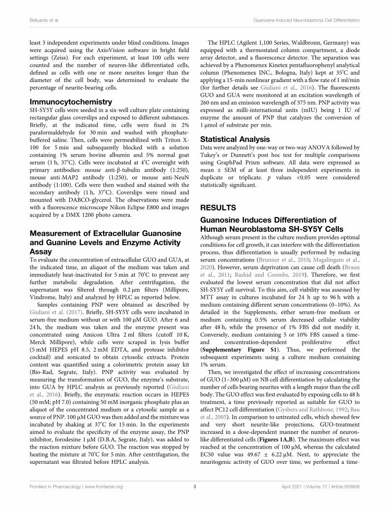
Then, we investigated the effect of increasing concentrationsof GUO (1–300 µM) on NB cell differentiation by calculating thenumber of cells bearing neurites with a length major than the cellbody. The GUO effect was first evaluated by exposing cells to 48 htreatment, a time previously reported as suitable for GUO toaffect PC12 cell differentiation (Gysbers and Rathbone, 1992; Bauet al., 2005). In comparison to untreated cells, which showed fewand very short neurite-like projections, GUO-treatmentincreased in a dose-dependent manner the number of neuron-like differentiated cells (Figures 1A,B). The maximum effect wasreached at the concentration of 100 μM, whereas the calculatedEC50 value was 49.67 ± 6.22 µM. Next, to appreciate theneuritogenic activity of GUO over time, we performed a time-
Frontiers in Pharmacology | www.frontiersin.org April 2021 | Volume 12 | Article 6588063
Belluardo et al. Guanosine-Induced Neuroblastoma Cell Differentiation
course study using 50 and 100 µM GUO, able to cause half andmaximal differentiation effects, respectively. In untreatedcultures, the percentage of neuron-like cells was quiteconstant, varying from 10 to 20% throughout the observationperiod. As expected, differentiation was greater in cells treatedwith 100 µM GUO than in those exposed to 50 μM, although inboth cases, the maximal effect was reached after 48–72 htreatment (Figure 1C).
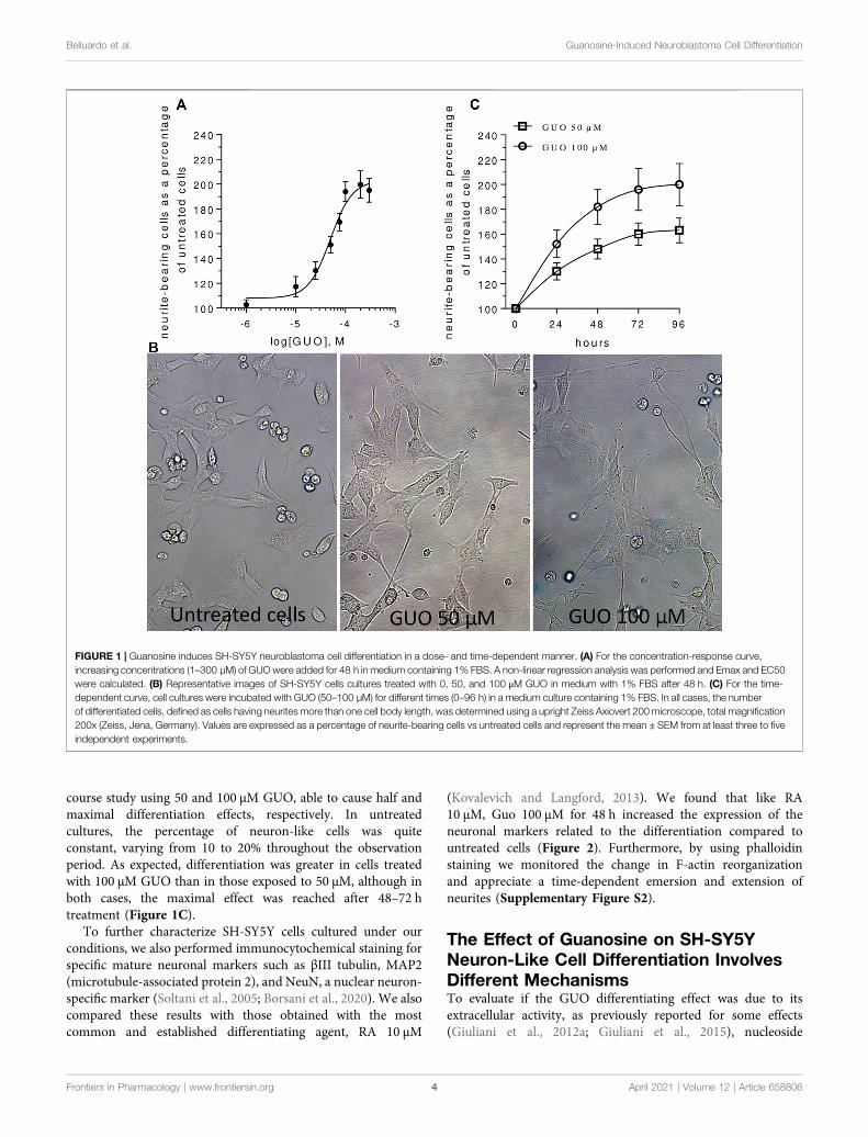
To further characterize SH-SY5Y cells cultured under ourconditions, we also performed immunocytochemical staining forspecific mature neuronal markers such as βIII tubulin, MAP2(microtubule-associated protein 2), and NeuN, a nuclear neuron-specific marker (Soltani et al., 2005; Borsani et al., 2020). We alsocompared these results with those obtained with the mostcommon and established differentiating agent, RA 10 µM
(Kovalevich and Langford, 2013). We found that like RA10 μM, Guo 100 µM for 48 h increased the expression of theneuronal markers related to the differentiation compared tountreated cells (Figure 2). Furthermore, by using phalloidinstaining we monitored the change in F-actin reorganizationand appreciate a time-dependent emersion and extension ofneurites (Supplementary Figure S2).
The Effect of Guanosine on SH-SY5YNeuron-Like Cell Differentiation InvolvesDifferent MechanismsTo evaluate if the GUO differentiating effect was due to itsextracellular activity, as previously reported for some effects(Giuliani et al., 2012a; Giuliani et al., 2015), nucleoside
FIGURE 1 | Guanosine induces SH-SY5Y neuroblastoma cell differentiation in a dose- and time-dependent manner. (A) For the concentration-response curve,increasing concentrations (1–300 µM) of GUOwere added for 48 h in medium containing 1% FBS. A non-linear regression analysis was performed and Emax and EC50were calculated. (B) Representative images of SH-SY5Y cells cultures treated with 0, 50, and 100 μM GUO in medium with 1% FBS after 48 h. (C) For the time-dependent curve, cell cultures were incubated with GUO (50–100 µM) for different times (0–96 h) in a medium culture containing 1% FBS. In all cases, the numberof differentiated cells, defined as cells having neurites more than one cell body length, was determined using a upright Zeiss Axiovert 200 microscope, total magnification200x (Zeiss, Jena, Germany). Values are expressed as a percentage of neurite-bearing cells vs untreated cells and represent the mean ± SEM from at least three to fiveindependent experiments.
Frontiers in Pharmacology | www.frontiersin.org April 2021 | Volume 12 | Article 6588064
Belluardo et al. Guanosine-Induced Neuroblastoma Cell Differentiation
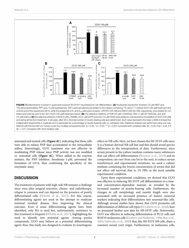
transporter blockers (10 µM NBTI plus 100 µM propentofylline-PPF- and 10 µM dipyridamole-DYP-) were added 1h before andduring GUO treatment (100 μM, 48 h). The uptake inhibitorsproduced only a partial reduction of about 20% of the GUO-induced effect (Figure 3A), without affecting untreated celldifferentiation when administered alone (data not shown).Since GUO effects might be due to adenosine receptoractivation (Dal-Cim et al., 2012), we next assessed adenosinereceptors involvement by adding selective antagonists for A1
(DPCPX, 100 nM), or A2A receptors (ZM241385, 50 nM) tocell cultures treated with GUO 100 µM for 48 h. NeitherDPCPX nor ZM241385 altered the ability of GUO to enhanceSH-SY5Y cell differentiation (Figure 3A), as well as neither bythemselves, affected the behavior of untreated cells (data notshown).
Finally, we investigated some possible signaling pathwaysinvolved in GUO-induced neuron-like cell differentiation. Asknown, pathways such as that of PI3K, PKC, and cGMP play akey role in many physiological functions including celldifferentiation (Heikkilä et al., 1993; Kimura et al., 1994; Bauet al., 2005). Thus, we analyzed their participation in the GUOeffect, adding selective inhibitors of PKC (GF1094002, 1 µM), orPI3K (LY294002, 25 µM), or sGC (ODQ, 10 µM) to the culturemedium treated with GUO 100 µM for 48 h. The PKC and sGCinhibitors reduced GUO-induced differentiation by about 24 and31% respectively, while the PI3K inhibitor did not affect it(Figure 3B). Since either HO-1 or NOS can stimulate sGC(Cary and Marletta, 2001), to further investigate the upstreampathways involved in cGMP formation, cells were exposed, toL-NAME (5 µM) or ZnPP (1 µM), the selective inhibitors of NOS
or HO activity, respectively. As shown in Figure 3C, the HOinhibitor, but not the NOS inhibitor, significantly reduced theGUO effect on neuron-like cell differentiation.
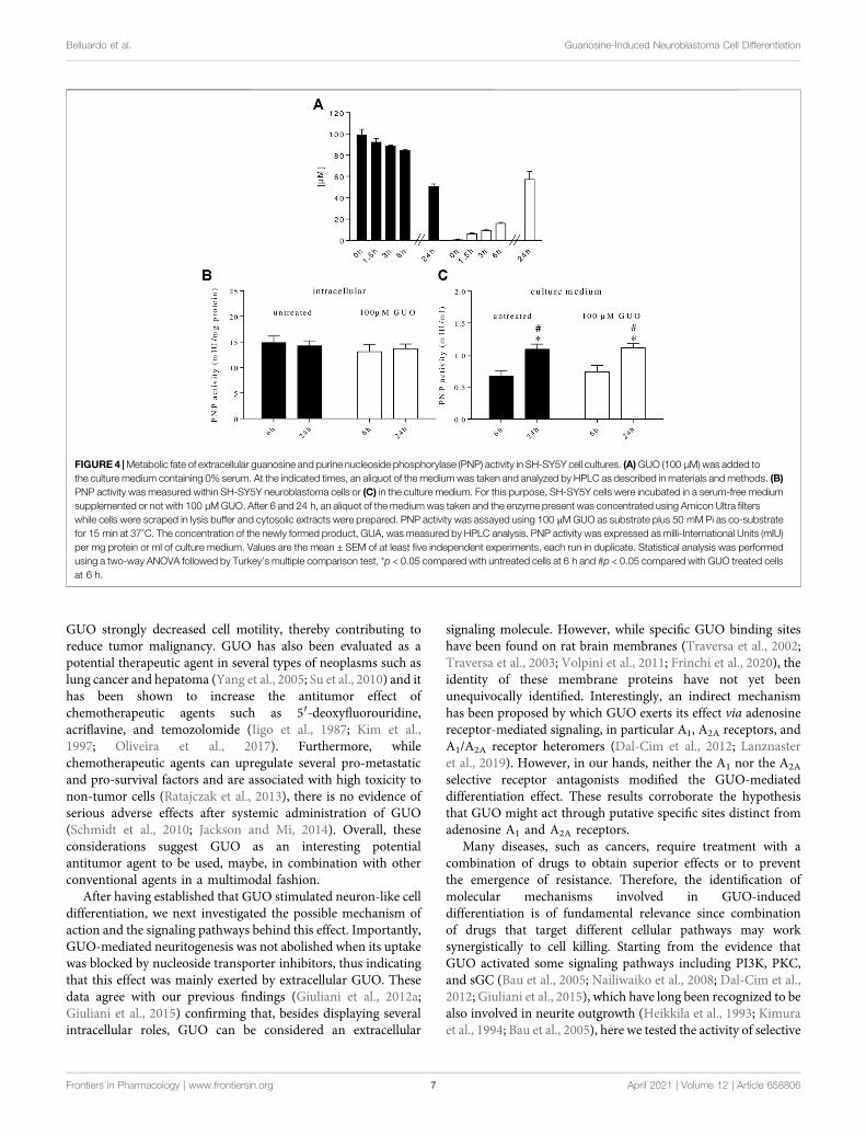
Exogenous Guanosine Added to SH-SY5YCell Cultures was Metabolized to GuanineIt is known that GUO is intracellularly metabolized to GUA byPNP enzyme. We recently found that PNP is also present inhuman plasma (Giuliani et al., 2016) and that rat C6 glioma cells,astrocytes, and microglial cells (Giuliani et al., 2017; Peña-Altamira et al., 2018), can release PNP in the extracellularmilieu. Thus, to better analyze the impact of GUO on celldifferentiation, we examined the metabolic fate of GUO in ourcultures. By HPLC analysis, we found that GUO (100 µM) addedto the cell cultures was no longer present extracellularly after 48 h,while the concentration of GUA, its direct metabolite, was 112 ±18 µM. To avoid interference with serum, in which PNP ispresent, a serum-free medium was used and cell cultures weretreated with 100 µM GUO for 24 h, a time period in which cellviability was not modified by serum absence (SupplementaryFigure S1). In this condition, exogenous GUO levels decreasedover time, while increasing concentrations of GUA concurrentlyappeared in the medium (Figure 4A).
We also evaluated PNP activity inside and outside cells. At thetwo time periods considered (6 and 24 h), in both untreated andGUO-treated cells, the intracellular PNP activity was constantover time (Figure 4B) and was always higher than theextracellular one (Figure 4C). On the contrary, outside thecells, the enzyme activity was higher after 24 h either in
FIGURE 2 | Guanosine increases the expression of specific mature neuronal markers in SH-SY5Y neuroblastoma cells. SH-SY5Y cells were cultured for 48 hwithout treatment (a) or in the presence of 100 µMGUO (b) or 10 µM RA (c). βIII-Tubulin and MAP2 filaments are highlighted in green while nuclei are stained in blue withDAPI. For NeuN expression, on the left side of the image nuclei are stained in green with NeuN while on the right side of the image nuclei are stained in blue with DAPI.Images are representative of one of three independent experiments.
Frontiers in Pharmacology | www.frontiersin.org April 2021 | Volume 12 | Article 6588065
Belluardo et al. Guanosine-Induced Neuroblastoma Cell Differentiation
untreated and treated cells (Figure 4C), indicating that these cellswere able to release PNP that accumulated in the extracellularmilieu. Interestingly, GUO treatment was not effective inmodulating PNP release since PNP activity was not modifiedvs. untreated cells (Figure 4C). When added to the reactionmixture, the PNP inhibitor, forodesine 1 μM, prevented theformation of GUA, thus confirming the specificity of theenzymatic assay.
DISCUSSION
The treatment of patients with high-risk NB remains a challengesince even after surgical resection, chemo- and radiotherapy,relapse is common and can depend on the presence of poorlydifferentiated cells (Whittle et al., 2017). For this reason,differentiating agents are used in the attempt to eradicateminimal residual disease, thus improving the clinicaloutcome. Even if some differentiating agents have beenidentified, only RA is used but, unfortunately, resistance tothis treatment is frequent (Whittle et al., 2017), highlighting theneed to identify new potential agents. Among purinecompounds, GUO may behave as a potential differentiatingagent; thus, this study was designed to evaluate its neuritogenic
effect on NB cells. Here, we have chosen the SH-SY5Y cells sinceit is a human-derived NB cell line and this should avoid speciesdifferences in the interpretation of data. Furthermore, sinceserum present in the culture medium contains many substancesthat can affect cell differentiation (Brunner et al., 2010) and itscomposition can vary from one lot to the next, to reduce seruminterferences and experimental variations, we used a culturemedium containing the lowest concentration of serum that didnot affect cell survival, that is, 1% FBS, as the most suitableexperimental condition.
Upon these experimental conditions, we showed that GUOwas effective in inducing SH-SY5Y cell differentiation in a time-and concentration-dependent manner, as revealed by theincreased number of neurite-bearing cells. Furthermore, thechanges in cell morphology and neurite length were alsoassociated with an increased presence of mature neuronalmarkers indicating their differentiation into neuronal-like cells.Although several studies have shown that GUO promotes celldifferentiation of different cell types (Di Liberto et al., 2016), herewe presented further new data for SH-SY5Y cells. Noteworthy,GUO was effective in inducing differentiation of PC12 cells andB16F10 melanoma cells (Gysbers and Rathbone, 1996; Bau et al.,2005; Naliwaiko et al., 2008) that, like SH-SY5Y NB cells, share acommon neural crest origin. Furthermore, in melanoma cells,
FIGURE 3 | Mechanisms involved in guanosine-induced SH-SY5Y neuroblastoma cell differentiation. (A) Nucleoside transporter blockers (10 µM NBTI plus100 µM propentofylline–PPF-plus 10 µM dypiridamole -DIP-) were simultaneously added to the medium containing 1% serum 1 h before GUO (100 µM) treatment anduntil the end of the experiment (48 h), while the antagonist of A1 and A2A adenosine receptor, DPCPX (100 nM) and ZM241385 (50 nM), respectively, were added 30 minbefore and until the end of the 48 h GUO (100 µM) treatment period. (B) The selective inhibitors of PI3K (25 µM LY294002), PKC (1 µM GF109203X), and sGC(10 µMOQD) or (C) the selective inhibitors of NOS (5 μML-NAME), HO (1 µM ZnPP) and sGC (10 µMOQD) were added to cultures before the addition of GUO (100 µM)and during all the GUO treatment. In all cases, after 48 h, the total number of neurite-bearing cells was determined. Each value represents the mean ± SEM of at least fiveindependent experiments in duplicate and is expressed as a percentage of neurite-bearing cells vs. untreated cells. Statistical analysis was performed using one-wayANOVAwith the Dunnett’s or Turkey’s post-hocmultiple comparisons test; *p < 0.05, **p < 0.02, ****p < 0.001 compared with untreated cells; #p < 0.05, # #p < 0.02, # ##p < 0.01 compared with GUO treated cells.
Frontiers in Pharmacology | www.frontiersin.org April 2021 | Volume 12 | Article 6588066
Belluardo et al. Guanosine-Induced Neuroblastoma Cell Differentiation
GUO strongly decreased cell motility, thereby contributing toreduce tumor malignancy. GUO has also been evaluated as apotential therapeutic agent in several types of neoplasms such aslung cancer and hepatoma (Yang et al., 2005; Su et al., 2010) and ithas been shown to increase the antitumor effect ofchemotherapeutic agents such as 5′-deoxyfluorouridine,acriflavine, and temozolomide (Iigo et al., 1987; Kim et al.,1997; Oliveira et al., 2017). Furthermore, whilechemotherapeutic agents can upregulate several pro-metastaticand pro-survival factors and are associated with high toxicity tonon-tumor cells (Ratajczak et al., 2013), there is no evidence ofserious adverse effects after systemic administration of GUO(Schmidt et al., 2010; Jackson and Mi, 2014). Overall, theseconsiderations suggest GUO as an interesting potentialantitumor agent to be used, maybe, in combination with otherconventional agents in a multimodal fashion.
After having established that GUO stimulated neuron-like celldifferentiation, we next investigated the possible mechanism ofaction and the signaling pathways behind this effect. Importantly,GUO-mediated neuritogenesis was not abolished when its uptakewas blocked by nucleoside transporter inhibitors, thus indicatingthat this effect was mainly exerted by extracellular GUO. Thesedata agree with our previous findings (Giuliani et al., 2012a;Giuliani et al., 2015) confirming that, besides displaying severalintracellular roles, GUO can be considered an extracellular
signaling molecule. However, while specific GUO binding siteshave been found on rat brain membranes (Traversa et al., 2002;Traversa et al., 2003; Volpini et al., 2011; Frinchi et al., 2020), theidentity of these membrane proteins have not yet beenunequivocally identified. Interestingly, an indirect mechanismhas been proposed by which GUO exerts its effect via adenosinereceptor-mediated signaling, in particular A1, A2A receptors, andA1/A2A receptor heteromers (Dal-Cim et al., 2012; Lanznasteret al., 2019). However, in our hands, neither the A1 nor the A2A
selective receptor antagonists modified the GUO-mediateddifferentiation effect. These results corroborate the hypothesisthat GUO might act through putative specific sites distinct fromadenosine A1 and A2A receptors.
Many diseases, such as cancers, require treatment with acombination of drugs to obtain superior effects or to preventthe emergence of resistance. Therefore, the identification ofmolecular mechanisms involved in GUO-induceddifferentiation is of fundamental relevance since combinationof drugs that target different cellular pathways may worksynergistically to cell killing. Starting from the evidence thatGUO activated some signaling pathways including PI3K, PKC,and sGC (Bau et al., 2005; Nailiwaiko et al., 2008; Dal-Cim et al.,2012; Giuliani et al., 2015), which have long been recognized to bealso involved in neurite outgrowth (Heikkila et al., 1993; Kimuraet al., 1994; Bau et al., 2005), here we tested the activity of selective
FIGURE 4 |Metabolic fate of extracellular guanosine and purine nucleoside phosphorylase (PNP) activity in SH-SY5Y cell cultures. (A)GUO (100 µM) was added tothe culture medium containing 0% serum. At the indicated times, an aliquot of the medium was taken and analyzed by HPLC as described in materials and methods. (B)PNP activity was measured within SH-SY5Y neuroblastoma cells or (C) in the culture medium. For this purpose, SH-SY5Y cells were incubated in a serum-free mediumsupplemented or not with 100 µMGUO. After 6 and 24 h, an aliquot of the medium was taken and the enzyme present was concentrated using Amicon Ultra filterswhile cells were scraped in lysis buffer and cytosolic extracts were prepared. PNP activity was assayed using 100 μMGUO as substrate plus 50 mM Pi as co-substratefor 15 min at 37°C. The concentration of the newly formed product, GUA, was measured by HPLC analysis. PNP activity was expressed as milli-International Units (mIU)per mg protein or ml of culture medium. Values are the mean ± SEM of at least five independent experiments, each run in duplicate. Statistical analysis was performedusing a two-way ANOVA followed by Turkey’s multiple comparison test, *p < 0.05 compared with untreated cells at 6 h and #p < 0.05 compared with GUO treated cellsat 6 h.
Frontiers in Pharmacology | www.frontiersin.org April 2021 | Volume 12 | Article 6588067
Belluardo et al. Guanosine-Induced Neuroblastoma Cell Differentiation
inhibitors of those signals on GUO-mediated neurite outgrowth.While the PI3K pathway was not involved, PKC and sGCsignaling pathways are required for the GUO effect since theirinhibition strongly attenuated differentiation. However, sinceneither PKC nor sGC inhibitor completely abolished GUO-effect, we believe that GUO-induced differentiation requiresthe activation of further signal transductions, such as a cyclicadenosine monophosphate pathway, as reported in other cells(Gysbers and Rathbone, 1996). This aspect needs to be stillinvestigated in SH-SY5Y cells.
Noteworthy, since GUO signal is also linked to the activationof enzymes such as HO or NOS, leading to increased productionof CO or NO, respectively, which in turn stimulate sGC togenerate cGMP (Cary and Marletta, 2001; Cary et al., 2006;Ryter et al., 2006), we investigated whether HO or NOS wereinvolved in the GUO-induced effect. Our data demonstrated thatHO or sGC inhibitors decreased the GUO effect while the NOSinhibitor was ineffective. These data corroborate those observedin PC12 cells (Bau et al., 2005) and strengthen the importance ofthe HO/sGC pathway in GUO-induced differentiation.
Finally, we could not overlook that cell purine homeostasis isensured by a complex network of enzymes, localized both intra-and extracellularly, and by membrane transporters, and thatenzymes controlling purine metabolism play a key role inregulating the biological effects of extracellular purines(Volonté and D’Ambrosi, 2009). Thus, since GUO was addedto the culture medium for a long time, we studied its metabolicfate in the extracellular medium. Indeed, after 48 h exogenouslyadministered, GUOwas no longer present in the culture medium,whereas we detected only GUA. Exogenous GUO could be takenup into the cells, transformed into GUA, and then releasedoutside the cells. Indeed, we found a strong PNP activityinside SH-SY5Y cells. Nonetheless, there is also the possibilitythat GUO could be metabolized extracellularly. Unlikeectonucleotidases (that convert nucleotides into nucleosides),the presence of extracellular enzymes metabolizing purinenucleosides and nucleobases is still a matter of debate.However, since no GUO kinase exists in mammals (Ipata,2001), the first step in the metabolism of exogenous GUOshould be its transformation into GUA by extracellular PNP.Noteworthy, the PNP presence in the culture medium was notdue to cell death since cell viability was not modified as evaluatedby MTT test, and PNP activity increased along time withoutmodification upon GUO treatment. Overall, these datacorroborate those already found in rat glioma C6 cells and inrat astrocytes and microglial cells (Giuliani et al., 2017; Peña-Altamira et al., 2018) and further support the existence of aconstitutive release of PNP from cells that, in these experiments,was unaffected by GUO treatment. Data on GUO metabolic fateraise some questions. Indeed, recent studies highlighted biologicaleffects also for GUA (Giuliani et al., 2012b; Zuccarini et al., 2018),although some signaling pathways involved in the GUO pro-differentiating effect are peculiar of GUO rather than GUA (e.g.,HO and PKC) (Zuccarini et al., 2018). In this perspective, it isnoteworthy that Garozzo et al. (2010) found that GUA, morethan GUO, can exert antiproliferation effects in human gliomacell lines; however, this effect was mainly due to an intracellular
effect, while in the present study, the GUO effect was mainlyextracellularly mediated since nucleoside transporterinhibitors did not abolish it. The evaluation of a potentialGUA role in NB cell differentiation is currently being tested,and some preliminary pilot experiments conducted usingforodesine, to inhibit the degradation of exogenous addedGUO, did not seem to modify GUO-induced differentiation,but further experiments will be addressed to unravel theinterplay between GUO, PNP, and GUA to identify newtherapeutic targets.
In conclusion, findings from this study demonstrate that GUOis effective in inducing NB cell differentiation, activating a processin which some molecular mechanisms have in part beenidentified (such as PKC, HO, and sGC cascades). Indeed, theseresults open a new perspective for NB treatment, thus furtherinvestigation on the role of GUA and the functioning of thecomplex guanine-based purine signaling in NB celldifferentiation might yield relevant implications for NBtherapeutic purposes.
DATA AVAILABILITY STATEMENT
The datasets presented in this article are not readily availablebecause the raw data supporting the conclusions of the articlewill be made available by the authors, upon reasonable request,to any qualified researcher. Requests to access the datasetsshould be directed to Patricia Giuliani, [email protected].
AUTHOR CONTRIBUTIONS
All the authors listed have made a substantial, direct, andintellectual contribution to the work, and approved it forpublication. All the authors want to dedicate this work inmemory of Professor Natale Belluardo who left us so suddenly.
FUNDING
This work was supported by the University of Chieti-Pescara withfunds (n. AT Giuliani 2019) and equipment to carry on thestudies.
ACKNOWLEDGMENTS
The authors would like to acknowledge Dr Simone Guarnieri forhis excellent technical assistance.
SUPPLEMENTARY MATERIAL
The SupplementaryMaterial for this article can be found online at:https://www.frontiersin.org/articles/10.3389/fphar.2021.658806/full#supplementary-material
Frontiers in Pharmacology | www.frontiersin.org April 2021 | Volume 12 | Article 6588068
Belluardo et al. Guanosine-Induced Neuroblastoma Cell Differentiation
REFERENCES
Böcklinger, K., Tomaselli, B., Heftberger, V., Podhraski, V., Bandtlow, C., andBaier-Bitterlich, G. (2004). Purine nucleosides support the neurite outgrowth ofprimary rat cerebellar granule cells after hypoxia. Eur. J. Cel. Biol. 83 (2), 51–54.doi:10.1078/0171-9335-00362
Bau, C., Middlemiss, P. J., Hindley, S., Jiang, S., Ciccarelli, R., Caciagli, F., et al.(2005). Guanosine stimulates neurite outgrowth in PC12 cells via activation ofheme oxygenase and cyclic GMP. Purinergic Signal 1 (2), 161–172. doi:10.1007/s11302-005-6214-0
Bettio, L. E. B., Neis, V. B., Pazini, F. L., Brocardo, P. S., Patten, A. R., Gil-Mohapel,J., et al. (2016). The antidepressant-like effect of chronic guanosine treatment isassociated with increased hippocampal neuronal differentiation. Eur.J. Neurosci. 43 (8), 1006–1015. doi:10.1111/ejn.13172
Borsani, E., Buffoli, B., Bonazza, V., Brunelli, G., Monini, L., Inchingolo, F., et al.(2020). In vitro effects of concentrated growth factors (CGF) on human SH-SY5Y neuronal cells. Eur. Rev. Med. Pharmacol. Sci. 24 (1), 304–314. doi:10.26355/eurrev_202001_19927
Braun, F., Bertin-Ciftci, J., Gallouet, A. S., Millour, J., and Juin, P. (2011). Serum-nutrient starvation induces cell death mediated by bax and puma that iscounteracted by p21 and unmasked by bcl-xL inhibition. PLoS One 6 (8),e23577. doi:10.1371/journal.pone.0023577
Brunner, D., Frank, J., Appl, H., Schöffl, H., Pfaller, W., and Gstraunthaler, G.(2010). Serum-free cell culture: the serum-free media interactive onlinedatabase. ALTEX 27 (1), 53–62. doi:10.14573/altex.2010.1.53
Cary, S. P. L., and Marletta, M. A. (2001). The case of CO signaling: why the jury isstill out. J. Clin. Invest. 107 (9), 1071–1073. doi:10.1172/JCI12823
Cary, S. P. L., Winger, J. A., Derbyshire, E. R., and Marletta, M. A. (2006). Nitricoxide signaling: no longer simply on or off. Trends Biochem. Sci. 31 (4),231–239. doi:10.1016/j.tibs.2006.02.003
Dal-Cim, T., Molz, S., Egea, J., Parada, E., Romero, A., Budni, J., et al. (2012).Guanosine protects human neuroblastoma SH-SY5Y cells againstmitochondrial oxidative stress by inducing heme oxigenase-1 via PI3K/Akt/GSK-3β pathway. Neurochem. Int. 61, 397–404. doi:10.1007/s11302-019-09679-w
Di Liberto, V., Mudò, G., Garozzo, R., Frinchi, M., Fernandez-Dueñas, V., Di Iorio,P., et al. (2016). The guanine-based purinergic system: the tale of an orphanneuromodulation. Front. Pharmacol. 7, 158. doi:10.3389/fphar.2016.00158
Frinchi, M., Verdi, V., Plescia, F., Ciruela, F., Grillo, M., Garozzo, R., et al. (2020).Guanosine-mediated anxiolytic-like effect: interplay with adenosine A1 and A2A
receptors. Int. J. Mol. Sci. 21 (23), 9281. doi:10.3390/ijms21239281Garozzo, R., Sortino, M. A., Vancheri, C., and Condorelli, D. F. (2010).
Antiproliferative effects induced by guanine-based purines requirehypoxanthine-guanine phosphoribosyltransferase activity. Biol. Chem. 391,1079–1089. doi:10.1515/BC.2010.106
Giuliani, P., Romano, S., Ballerini, P., Ciccarelli, R., Petragnani, N., Cicchitti, S.,et al. (2012a). Protective activity of guanosine in an in vitro model ofParkinson’s disease. Panminerva Med. 54, 43–51.
Giuliani, P., Buccella, S., Ballerini, P., Ciccarelli, R., D’Alimonte, I., Cicchitti, S.,et al. (2012b). Guanine-based purines modulate the effect of L-NAME onlearning and memory in rats. Panminerva Med. 54, 53–58.
Giuliani, P., Ballerini, P., Buccella, S., Ciccarelli, R., Rathbone, M. P., Romano, S.,et al. (2015). Guanosine protects glial cells against 6-hydroxydopamine toxicity.Adv. Exp. Med. Biol. 837, 23–33. doi:10.1007/5584-2014-73
Giuliani, P., Zuccarini, M., Buccella, S., Peña-Altamira, L. E., Polazzi, E., Virgili, M.,et al. (2017). Evidence for purine nucleoside phosphorylase (PNP) release fromrat C6 glioma cells. J. Neurochem. 141 (2), 208–221. doi:10.1111/jnc.14004
Giuliani, P., Zuccarini, M., Buccella, S., Rossini, M., D’Alimonte, I., Ciccarelli, R.,et al. (2016). Development of a new HPLCmethod using fluorescence detectionwithout derivatization for determining purine nucleoside phosphorylaseactivity in human plasma. J. Chromatogr. B. Analyt. Technol. Biomed. LifeSci. 1009-1010, 114–121. doi:10.1016/j.jchromb.2015.12.012
Guarnieri, S., Pilla, R., Morabito, C., Sacchetti, S., Mancinelli, R., Fanò, G., et al.(2009). Extracellular guanosine and GTP promote expression of differentiationmarkers and induce S-phase cell-cycle arrest in human SH-SY5Yneuroblastoma cells. Int. J. Dev. Neurosci. 27 (2), 135–147. doi:10.1016/j.ijdevneu.2008.11.007
Gysbers, J. W., and Rathbone, M. P. (1992). Guanosine enhances NGF-stimulatedneurite outgrowth in PC12 cells. Neuroreport 3 (11), 997–1000. doi:10.1097/00001756-199211000-00013
Gysbers, J. W., and Rathbone, M. P. (1996). Neurite outgrowth in PC12 cells isenhanced by guanosine through both cAMP-dependent and -independentmechanisms. Neurosci. Lett. 220 (3), 175–178. doi:10.1016/s0304-3940(96)13253-5
Heikkilä, J., Jalava, A., and Eriksson, K. (1993). The selective protein kinase Cinhibitor GF 109203X inhibits phorbol ester-induced morphological andfunctional differentiation of SH-SY5Y human neuroblastoma cells. Biochem.Biophysical. Res. Commun. 197 (3), 1185–1193. doi:10.1006/bbrc.1993.2602
Iigo, M., Miwa, M., Ishitsuka, H., and Nitta, K. (1987) Potentiation of thechemotherapeutic action of 5′-deoxy-5-fluorouridine in combination withguanosine and related compounds. Cancer Chemother. Pharmacol. 19,61–64. doi:10.1007/bf00296258
Ipata, P. L. (2011). Origin, utilization, and recycling of nucleosides in the centralnervous system. Adv. Physiol. Educ. 35 (1), 92–94. doi:10.1152/advan.00068.2011
Jackson, E. K., and Mi, Z. (2014). The guanosine-adenosine interaction exists invivo. J. Pharmacol. Exp. Ther. 350 (3), 719–726. doi:10.1124/jpet.114.216978
Kim, S. G., Kim, C. W., Ahn, E. T., Lee, K. Y., Hong, E. K., Yoo, B. I., et al. (1997).Enhanced anti-tumour effects of acriflavine in combination with guanosine inmice. J. Pharm. Pharmacol. 49, 216–222. doi:10.1111/j.2042-7158.1997.tb06783.x
Kimura, K., Hattori, S., Kabuyama, Y., Shizawa, Y., Takayanagi, J., Nakamura, S.,et al. (1994). Neurite outgrowth of PC12 cells is suppressed by wortmannin, aspecific inhibitor of phosphatidylinositol 3-kinase. J. Biol. Chem. 269 (29),18961–18967. doi:10.1016/s0021-9258(17)32260-3
Kovalevich, J., and Langford, D. (2013). Considerations for the use of SH-SY5Yneuroblastoma cells in neurobiology. Methods Mol. Biol. 1078, 9–21. doi:10.1007/978-1-62703-640-5_2
Lanznaster, D., Massari, C. M., Marková, V., Simková, T., Duroux, R., Jacobson, K.A., et al. (2019). Adenosine A1-A2A receptor-receptor interaction: contributionto guanosine-mediated effects. Cells 8, 1630. doi:10.3390/cells8121630
Magalingam, K. B., Radhakrishnan, A. K., Somanath, S. D., Md, S., andHaleagrahara, N. (2020). Influence of serum concentration in retinoicacid and phorbol ester induced differentiation of SH-SY5Y humanneuroblastoma cell line. Mol. Biol. Rep. 47 (11), 8775–8788. doi:10.1007/s11033-020-05925-2
Moriwaki, Y., Yamamoto, T., and Higashino, K. (1999). Enzymes involved inpurine metabolism—a review of histochemical localization and functionalimplications. Histol. Histopathol. 14 (4), 1321–1340. doi:10.14670/HH-14.1321
Naliwaiko, K., Luvizon, A. C., Donatti, L., Chammas, R., Mercadante, A. F., Zanata,S. M., et al. (2008). Guanosine promotes B16F10 melanoma cell differentiationthrough PKC-ERK 1/2 pathway. Chemico-Biol. Interact. 173 (2), 122–128.doi:10.1016/j.cbi.2008.03.010
Newman, E. A., Abdessalam, S., Aldrink, J. H., Austin, M., Heaton, T. E., Bruny, J.,et al. (2019). Update on neuroblastoma. J. Pediatr. Surg. 54 (3), 383–389. doi:10.1016/j.jpedsurg.2018.09.004
Oliveira, K. A., Dal-Cim, T. A., Lopes, F. G., Nedel, C. B., and Tasca, C. I. (2017).Guanosine promotes cytotoxicity via adenosine receptors and inducesapoptosis in temozolomide-treated A172 glioma cells. Purinergic Signal 13,305–318. doi:10.1007/s11302-017-9562-7
Peña-Altamira, L. E., Polazzi, E., Giuliani, P., Beraudi, A., Massenzio, F., Mengoni,I., et al. (2018). Release of soluble and vesicular purine nucleosidephosphorylase from rat astrocytes and microglia induced by pro-inflammatory stimulation with extracellular ATP via P2X7 receptors.Neurochem. Int. 115, 37–49. doi:10.1016/j.neuint.2017.10.010
Rashid, M. U., and Coombs, K. M. (2019). Serum-reduced media impacts on cellviability and protein expression in human lung epithelial cells. J. Cel. Physiol.234 (6),7718–7724. doi:10.1002/jcp.27890
Ratajczak, M. Z., Jadczyk, T., Schneider, G., Kakar, S. S., and Kucia, M. (2013).Induction of a tumor-metastasis-receptive microenvironment as an unwantedand underestimated side effect of treatment by chemotherapy or radiotherapy.J. Ovarian Res. 6, 95. doi:10.1186/1757-2215-6-95
Reynolds, C. P., Matthay, K. K., Villablanca, J. G., andMaurer, B. J. (2003). Retinoidtherapy of high-risk neuroblastoma. Cancer Lett. 197 (1-2), 185–192. doi:10.1016/s0304-3835(03)00108-3
Frontiers in Pharmacology | www.frontiersin.org April 2021 | Volume 12 | Article 6588069
Belluardo et al. Guanosine-Induced Neuroblastoma Cell Differentiation
Ryter, S. W., Alam, J., and Choi, A. M. K. (2006) Heme oxygenase-1/carbonmonoxide: from basic science to therapeutic applications. Physiol. Rev. 86 (2),583–650. doi:10.1152/physrev.00011.2005
Schmidt, A. P., Paniz, L., Schallenberger, C., Böhmer, A. E., Wofchuk, S. T.,Elisabetsky, E., et al. (2010). Guanosine prevents thermal hyperalgesia in a ratmodel of peripheral mononeuropathy. J. Pain 11, 131–141. doi:10.1016/j.jpain.2009.06.010
Smith, V., and Foster, J. (2018). High-risk neuroblastoma treatment review.Children 5 (9), 114. doi:10.3390/children5090114
Soltani, M. H., Pichardo, R., Song, Z., Sangha, N., Camacho, F., Satyamoorthy, K.,et al. (2005). Microtubule-associated protein 2, a marker of neuronaldifferentiation, induces mitotic defects, inhibits growth of melanoma cells,and predicts metastatic potential of cutaneous melanoma. Am. J. Pathol. 166(6), 1841–1850. doi:10.1016/S0002-9440(10)62493-5
Su, C., Picard, P., Rathbone, M. P., and Jiang, S. (2010). Guanosine-induceddecrease in side population of lung cancer cells: lack of correlation with ABCG2expression. J. Biol. Regul. Homeost. Agents 24, 19–25.
Su, C., Elfeki, N., Ballerini, P., D’Alimonte, I., Bau, C., Ciccarelli, R., et al. (2009).Guanosine improves motor behavior, reduces apoptosis, and stimulatesneurogenesis in rats with parkinsonism. J. Neurosci. Res. 87 (3), 617–625.doi:10.1002/jnr.21883
Tasca, C. I., Lanznaster, D., Oliveira, K. A., Fernández-Dueñas, V., andCiruela, F. (2018). Neuromodulatory effects of guanine-based purines inhealth and disease. Front. Cel. Neurosci. 12, 376. doi:10.3389/fncel.2018.00376
Traversa, U., Bombi, G., Di Iorio, P., Ciccarelli, R., Werstiuk, E. S., and Rathbone,M. P. (2002). Specific [3 H]-guanosine binding sites in rat brain membranes. Br.J. Pharmacol. 135, 969–976. doi:10.1038/sj.bjp.0704542
Traversa, U., Bombi, G., Camaioni, E., Macchiarulo, A., Costantino, G., Palmieri,C., et al. (2003). Rat brain guanosine binding site. Bioorg. Med. Chem. 11,5417–5425. doi:10.1016/j.bmc.2003.09.043
Volonté, C., and D’Ambrosi, N. (2009) Membrane compartments and purinergicsignalling: the purinome, a complex interplay among ligands, degradingenzymes, receptors and transporters. FEBS J. 276, 318–329. doi:10.1111/j.1742-4658.2008.06793.x
Volpini, R., Marucci, G., Buccioni, M., Dal Ben, D., Lambertucci, C., Lammi, C.,et al. , (2011). Evidence for the existence of a specific G protein-coupled receptoractivated by guanosine. ChemMedChem 6, 1074–1080. doi:10.1002/cmdc.201100100
Whittle, S. B., Smith, V., Doherty, E., Zhao, S., McCarty, S., and Zage, P. E. (2017).Overview and recent advances in the treatment of neuroblastoma. Expert Rev.Anticancer Ther. 17 (4), 369–386. doi:10.1080/14737140.2017.1285230
Yang, S. C., Chiu, C. L., Huang, C. C., and Chen, J. R. (2005). Apoptosis induced bynucleosides in the human hepatoma HepG2. World J. Gastroenterol. 11,6381–6384. doi:10.3748/wjg.v11.i40.6381
Zuccarini, M., Giuliani, P., Frinchi, M., Mudò, G., Serio, R. M., Belluardo, N., et al.(2018). Uncovering the signaling pathway behind extracellular guanine-induced activation of NO system: new perspectives in memory-relateddisorders. Front. Pharmacol. 9, 110. doi:10.3389/fphar.2018.00110
Conflict of Interest: The authors declare that the research was conducted in theabsence of any commercial or financial relationships that could be construed as apotential conflict of interest.
Copyright © 2021 Belluardo, Mudò, Di Liberto, Frinchi, Condorelli, Traversa,Ciruela, Ciccarelli, Di Iorio and Giuliani. This is an open-access articledistributed under the terms of the Creative Commons Attribution License (CCBY). The use, distribution or reproduction in other forums is permitted, provided theoriginal author(s) and the copyright owner(s) are credited and that the originalpublication in this journal is cited, in accordance with accepted academic practice.No use, distribution or reproduction is permitted which does not comply withthese terms.
Frontiers in Pharmacology | www.frontiersin.org April 2021 | Volume 12 | Article 65880610
Belluardo et al. Guanosine-Induced Neuroblastoma Cell Differentiation