investigating the root causes of multiple sclerosis using systems
TRANSCRIPT

Oral presentation at the System Dynamics Society conference at St. Gallen, Switzerland, July 23-26th 2012
! "!
Investigating the root causes of multiple sclerosis using systems analysis and dynamics, developing an integrated
treatment strategy
Thomas Ragnar Wood1, Ulrik Sverdrup2, Kristín Vala Ragnarsdóttir3, Harald U. Sverdrup4
1Guy's Hospital and St Thomas' Hospital, Great Maze Pond, London SE1 9RT, Great Britain 2Physics Engineering, LTH, Lund University, SE-221 00 Lund, Sweden, 3Earth Sciences, University of Iceland, 101 IS-Reykjavik, Iceland, 4Chemical Engineering, Lund University, Box 124, SE-221 00 Lund, Sweden
Abstract By applying the methods of systems analysis to the problem of what causes multiple sclerosis, we have been able to show that it is a systemic illness, with multiple interacting causes and mechanisms. The results were obtained by synthesizing state-of-the-art research on parts into a whole systemic picture of the whole. These main components make up the illness system:
1. The root cause: A Permeable blood-brain barrier (BBB) and transmission of provocative
substances, antibodies and leucocytes into the brain. This may be caused by chronic cerebro-spinal vascular insufficiency (CCSVI, and associated microclotting due to stagnant cerebro-venous blood flow), latent infections that cause a Th1-dominated immune response and release of matrix metalloproteases (MMPs) which can alter the BBB, or a general pro-inflammatory state created due to dietary factors.
2. Triggers are antibodies to myelin and oligodendrocytes from i. Infections by certain bacteria and viruses;
ii. Peripheral allergenic attacks iii. Myelin and oligodendrocyte debris in the blood stream
3. Reinforcing loops: a. Feedback system involving iron in the autocatalytic Fenton cycle and production of
oxidants, prominently peroxynitrite, reinforcing the increased permeability of the BBB; keeping the BBB compromised
b. Autoimmune feedback loop involving immune system and myelin sheath breakdown, leading to damages to the BBB, by oxidative denaturalization of the myelin surfaces.
c. Oxidative stresses, leading to scaring at damaged sites and impairment of the ability to repair myelin sheaths, making the physical damage permanent
The partial mechanisms were linked together into one large integrated model leading to the disease. Multiple sclerosis has no single cause, but a number of interacting of causes in a feedback system. The model is capable of describing the untreated progress of the illness as it has been observed. Multiple sclerosis is initiated by an opening of the blood-brain-barrier, combined with provocative substances in the blood, which initiate a vicious cycle of oxidative stresses and immune attack. The conceptual model was used to investigate the dynamics of the illness and understand its initiation and subsequent illness progression qualitatively. With the model we can start to analyse how treatment for multiple sclerosis must be approached in a multi-disciplinary way in order to be successful. The conceptual model was developed into a systems dynamics model in the STELLA environment. The simulation model is capable of reconstructing the basic outline of the illness, with the onset at 25 years of age, and the transition to secondary progressive disease at 45-55 years of age. This appears to confirm the validity of the fundamental features of our model.
Introduction
Multiple sclerosis (MS) is a chronic illness, characterised by immune attack on the myelin sheath of white matter in the central nervous system. The patient initially experiences attacks of paraesthesia, weakness, visual or bowel and bladder problems. These may initially resolve, but over time general health decreases alongside permanent disabilities, which often leave MS sufferers wheelchair-bound. Though rarely directly fatal, overall the life expectancy of MS patients is reduced by 5-10 years. On autopsy and in MRI gadolinium contrasted scans, lesions in the grey and white matter can be seen, where the nerve tissue of the brain has been destroyed. The atrophy is largely irreversible, but the

Oral presentation at the System Dynamics Society conference at St. Gallen, Switzerland, July 23-26th 2012
! #!
brain has a certain redundancy, and the critical functions can initially be compensated for. This will be experienced as a remission. Multiple sclerosis is an ailment without a cure and generally poor prognosis for the patient. Though some current treatments may reduce the severity of an attack, nothing as yet can slow the overall progression of the disease. The illness strikes more women than men, the ratio being close to 2:1 (Phadke 1987). We classify multiple sclerosis into 5 major types (El-Moslimany and Lublin 2008, Andersen 2008, Polman and van Osten 2008):
1. Clinically isolated syndrome. 2. Relapsing-remitting multiple sclerosis 3. Secondary progressive multiple sclerosis 4. Primary progressive multiple sclerosis 5. Progressive relapsing multiple sclerosis
In most cases, MS affects both the brain and the spinal cord. However, in approximately 20% of the cases it affects the spinal cord only. The general state-of-the-art picture of what multiple sclerosis represents is described by Kesselring (2008), Andersen (2008) and Dean and Kurtzke (1971). Until now multiple sclerosis has been an enigma - the visible symptoms are well described, but they are not well understood. Background and earlier work
Research into the cause of multiple sclerosis has gone on for decades, turning up few explanations for the aetiology of the disease (e.g. Allen 1981, Barnett and Sutton 2006). The condition was first properly described by Charcot (1868); however, it has probably existed as a disease since well before that time. One of the possible reasons for the lack of significant progress was the focus on the hypothesis that there was one single causal factor. Some recent efforts stand out as important milestones on the road to discovering the root cause of multiple sclerosis:
1. The observation of constricted blood vessels for the return venous blood from the brain and how this causes small lesions that allow blood and pathogenic material to leak into the brain through the compromised blood-brain barrier (Zamboni et al., 2003, 2005, 2006, 2007, 2008, 2009a,b,c, 2010, Francesci 2009, Zamboni 2009, Khan et al., 2010, Lee et al., 2009).
2. The discovery that multiple sclerosis is connected to accumulation of iron in the brain, creating large internal oxidative stresses in the brain (Hooper et al., 1998, 2000, Szabo et al., 2007, Szabo 2003, Gilun-Sherki et al., 2002).
3. The finding that multiple sclerosis not only affects white matter brain, but that it causes grey-matter necrosis and that this is an initially inflammatory process, proportional to the degree of micro-vascularisation of the grey-matter tissue.
Scope and objective
The objective of this study was to understand the different components influencing multiple sclerosis; its initiation, first as attacks, and the mechanisms which potentiate the disease towards its progressive phase. Finally, we use knowledge of the parts to derive an integrated model for MS that explains the underlying causalities of the illness, as well as exploring possible treatment strategies that can be derived from the systemic model. Materials and methods
The basic methodology applied was systems analysis of existing data - where cause and effect were systematically mapped in large system diagrams, recording causal chains in the systems and with emphasis on finding important feedback loops that are present (Haraldsson and Sverdrup 2004, Ahn et al., 2006a,b, Varkey et al., 2009). This we did in order to be able to investigate the behaviour of the whole integrated system, but varying the main drivers and studying the responses in the main outputs. This takes the expression of causal loop diagrams, which are powerful tools for problem solving in complex systems. In this we work with defining the goal, identifying the gap between the present status of the system and the goal, and developing a strategy for how the gap will be crossed. This

Oral presentation at the System Dynamics Society conference at St. Gallen, Switzerland, July 23-26th 2012
! $!
implies detecting the possibilities for changing the causal links, affect key driving factors, essential triggers, and important self-sustained feedback loops in the system. Figure 1 shows how systems analysis starts as simple mind-mapping, where a number of possible parameters or system parts are just “thrown up” on a board. They are then sorted in order to find their place within the system, and whether they are significant contributors to the overall picture. Some of the factors will have to be disassembled into subsystems, and many factors may be eliminated as a part of the systems analysis process. This is the first step towards establishing causal loop diagrams.
The different phases of a systems-thinking-based research process as was implemented here has been shown on the right (Sverdrup et al. 2012). Figure 2 shows an outline of how the stakeholder participation process is operated. A minimum number of 3 consecutive workshops, and often up to 5-10 workshops, are required to achieve good results (Sverdrup et al. 2012).
Excitotoxins
Environmental toxins
Food allergies and BBB damage
Microbial and viralinitiation
Immune system
Oxidative stresses
Food allergiesand
autoimmune triggers
Vascularproblems
Nervedamage
Figure 1. The systems analysis starts as simple mind-mapping, where a number of possible parameters or system parts are evaluated (left). The process goes through four phases.
Figure 2. Outline of how the stakeholder participation process is operated. From 2009 to 2011, we conducted a large literature survey, reading and mapping the contents of about 700 scientific articles (Sverdrup et al. 2012). That study provided us with a large number of causal loop diagrams and a systemic insight into MS as a system. The study, a 180 page booklet published as a Lund University report, constitutes what the authors call a “knowledgebook” - the scientific notes, methodology, description and activities log of our compilation as a part of the research process. For those interested in our detail, we would refer to that report. The knowledge book is a living document, and it steadily updated with everything new insight learned. The current article is drawn from this compilation and has been significantly further reworked.

Oral presentation at the System Dynamics Society conference at St. Gallen, Switzerland, July 23-26th 2012
! %!
Theory The generally accepted hypothesis for multiple sclerosis is that it is caused by a permeable blood-
brain-barrier (BBB), which is instrumental in causing attacks of the immune system against the central nervous system (CNS). We reviewed all of the proposed causal factors and assessed their validity. All this was synthesised into one single holistic multiple sclerosis aetiology as is shown below. Based on our systems analysis, an integrated picture of a systemic disease has emerged. Many components appear to be interacting, and it becomes evident that there is no single cause of MS - there are many causes, and they are connected in a system. The simple model we have derived, has four main components:
1. Leakage across the blood-brain barrier. Pathogens, antigens and blood can pass into the brain, leading to direct immune attack and iron deposition.
2. Infections which produce antibodies that cross-reaction with myelin in the CNS via molecular mimicry
3. Oxidative stresses caused by iron deposition into the brain and immune-mediated CNS inflammation. These both weaken the blood-brain barrier from within and keep it compromised – this produces a reinforcing feedback loop
4. An autoimmune feedback loop that once triggered, becomes self-sustaining, driving white matter scarring, oligodendrocyte necrosis and blood-brain barrier leakage.
Multiple sclerosis appears to be triggered when leaky vessels open a pathway for material
normally barred by the blood-brain barrier, to enter the CNS. This allows red and white blood cells, as well as antibodies, to transgress the blood-brain barrier. Diagnostically, this is seen as iron accumulation in the brain from red blood cell break-down, always in connection with lesions. Other pathologies may also lead to iron deposits in the brain, but multiple sclerosis is only caused when there are conditions that allow the oxidant cycles to start producing large amounts of oxidants. Antibodies entering the central nerve system may “mark” the myelin sheath for immune system attack as it has similar a localised molecular structure to the bacteria that the antibodies were intended for (Tivana et al. 1999, Ebringer et al. 2003, 2008, 2010). Myelin fragments then trigger further responses and an autoimmune feedback loop is established. The regenerative mechanisms strive to repair the damage however, the accumulated iron leads to elevated levels of Fe2+, resulting in oxidative stress, production of free-radicalr and reduction in the efficiency of the regenerative mechanism. Thus, re-myelination is prevented and multiple sclerosis has developed. At least three mechanisms appear to be associated with increasing permeability of the blood-brain barrier, any of which might trigger entry into the MS cycle in a susceptible individual:
1. Latent infections, often with associated Th1-dominant immune response 2. A diet-related pro-inflammatory state, and direct effect of dietary breakdown products having
a direct effect on gut and BBB permeability 3. Chronic Cerebro-Spinal Venous Insufficiency (CCSVI)
Chronic and latent infections and associated molecular mimicry Ebringer et al. (2005a,b, 2007), Gilden (2005), Grönning et al. (1993), Hughes et al. (2003), Panitch (1994) and Bennet et al. (2008) showed that antibodies from certain bacteria are often associated with multiple sclerosis. Ebringer and his colleagues among others, hypothesised that this could be one of the triggering factors for the disease. Others have worked on the Epstein Barr Herpes virus, the human herpes virus 6 and other viral components that have similar association patterns (Siegel 1997, Sola et al., 1993, Vaughan et al., 1996). It seems like this antibody affiliation is a plausible hypothesis with respect to epidemiological studies conducted in the Færøyar and Iceland concerning external contagious agents in multiple sclerosis (Bray et al., 1992, Dean and Kurtzke 1971, Ebers et al., 1986, Kurtzke 1983, 1993, Kurtzke and Hyllerstedt 1988, Kurtzke et al., 1979, Poses and Hibberd 1988, Bennet et al., 2008).

Oral presentation at the System Dynamics Society conference at St. Gallen, Switzerland, July 23-26th 2012
! &!
The role of dairy products and gluten According to our findings by far the most successful treatments for multiple sclerosis are specially designed diets. The common denominator of most of these diets, are the avoidance of dairy products, and to eliminate gluten. This is sometimes based on immunological reasoning, often from observed effects of experimental diets over long times (Swank and Goodwin 2003).
The elimination of dairy from the diet may in many cases lead to improvements that are orders of magnitude larger than anything the traditional medicine can accomplish (Wills and Unsworth 2002, Ventura et al. 1991, Volta et al. 2006, Volta and Georgio 2010). Research is needed to establish the effect of food in general. Milk and gluten give rise to several constituents that may interfere with different physiological mechanisms: 1. Small, dispersed fat particles are created through the homogenization process with ultrasonic
treatment, where the fat pearls in the milk are crushed to fragments by ultrasonic treatment. This creates fat particle with sizes below membrane size of the gastrointestinal tract, and they can thus pass into the bloodstream. Once in the blood-stream, they may get into the brain if the blood-brain barrier has been compromised.
2. Milk proteins: a. Casein is the protein of cheese, and it also contains bovine growth hormones. The proteins
may cause allergic reactions, and in the continuation lead to allergic or immune mechanisms compromising the intestinal gut lining membranes.
b. Gluten. There is often a reaction to gluten similar in effect to that of milk protein, where the intestinal tract may become inflamed and through that process become more permeable, letting certain substances through (Volta and de Giorgio 2010).
c. Legume proteins have been suspected to be involved in protein mimicry. Many empirical diets for multiple sclerosis eliminate these with good results.
d. Once an allergic reaction has been produced with release of histamines in significant amounts, the immune system is stimulated to increased alert.
3. Lactose. Some people are lactose intolerant and cannot properly metabolize it. This may cause an allergic reaction as the substance is not properly metabolized. The role of denaturalized sugars remain unresearched
!
Figure 3. Causal loop diagram for the mechanisms causing a permeable brain-blood-barrier is shown to the left. The emphasis of the diagram to the right is to show the triggering effect by different types of infection from both bacteria and different types of viruses.
Oral presentation at the System Dynamics Society conference at St. Gallen, Switzerland, July 23-26th 2012
! '!
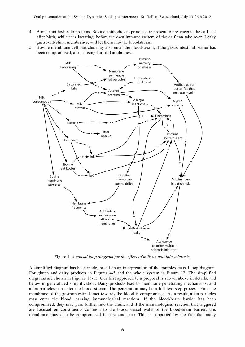
4. Bovine antibodies to proteins. Bovine antibodies to proteins are present to pre-vaccine the calf just after birth, while it is lactating, before the own immune system of the calf can take over. Leaky gastro-intestinal membranes, will let them into the bloodstream.
5. Bovine membrane cell particles may also enter the bloodstream, if the gastrointestinal barrier has been compromised, also causing harmful antibodies.
Milkconsumption
Saturatedfats
Myelinmimicry
Lactase
Hormones
Iron uptake
MilkProcessing
Membranepermeable
fat particles
Histamines
+ -
+
+
+
+
Immunomimicry
on myelin
Antibodies forbutter fat thatemulate myelin
Autoimmuneinitiation risk
Allergicreactions
Immunesystem alert
IgE
IgX
+ +
+
+
+
+
+
+
++
Fermentationtreatment
?
+
+
Milkprotein
+ +
Alteredproteins +
+
Intestinemembrane
permeability
Blood-Brain-Barrierleaks
Assistanceto other multiplesclerosis initiators
+
Membranefragments
Antibodiesand immuneattack on
membranes
Bovineantibodies
Bovinemembraneparticles
+
+
+
++
+
+
+
+
+ -B
+
+
Figure 4. A causal loop diagram for the effect of milk on multiple sclerosis.
A simplified diagram has been made, based on an interpretation of the complex causal loop diagram. For gluten and dairy products in Figures 4-5 and the whole system in Figure 12. The simplified diagrams are shown in Figures 13-15. Our first approach to a proposal is shown above in details, and below in generalized simplification: Dairy products lead to membrane penetrating mechanisms, and alien particles can enter the blood stream. The penetration may be a full two step process: First the membrane of the gastrointestinal tract towards the blood is compromised. As a result, alien particles may enter the blood, causing immunological reactions. If the blood-brain barrier has been compromised, they may pass further into the brain, and if the immunological reaction that triggered are focused on constituents common to the blood vessel walls of the blood-brain barrier, this membrane may also be compromised in a second step. This is supported by the fact that many

Oral presentation at the System Dynamics Society conference at St. Gallen, Switzerland, July 23-26th 2012
! (!
multiple sclerosis patients have been shown to have symptoms of a leaky gut (Samarkos and Vaiopoulos 2005, Salvemini et al. 2006). This in turn opens the door for viral triggers and blood entry to the brain, that results in oxidative stresses and peroxynitrite production, leading to the traditional multiple sclerosis aetiology. Immune systems responses to membrane fragments and degenerated tissue in the blood stream, may lead to initiation of autoimmune responses that leads to myelin destruction. This represents a known multiple sclerosis initiation sequence. !-casein (and milk in general) as a co-causative agent for multiple sclerosis Incidence of multiple sclerosis largely follows worldwide trends in dairy intake, and cell-mediated immunity against milk proteins is increased in multiple sclerosis patients (Lindeberg 2010). Immunoglobulin domains of antibodies specific for myelin oligodendrocyte glycoprotein, as found in patients with multiple sclerosis, have been found to cross-react with butyrophilin, a protein found in cow’s milk (Guggenmos et al. 2004). Butyrophilin is the major protein associated with fat droplets in milk, and when injected into rodents, produces the multiple sclerosis-like experimental autoimmune encephalitis (Winer et al. 2001). This is one of the main processes by which it is thought that molecular mimicry might have a role in the aetiology of multiple sclerosis. A study has also showed that patients with multiple sclerosis have higher antibody responses to both myelin oligodendrocyte glycoprotein and butyrophilin (Kennel De March et al. 2003). Also implicated is the chief milk protein, casein, and its breakdown proteins, particularly the !-caso-morphins (BCMs). !-casein is one of the four most common casein variants, with !-caseins A1 and A2 being the most prevalent. Caso-morphins are inactive within the native protein, but released during digestion. Once, absorbed, these proteins have been shown to have opioid-like activity within the central nerve system. BCM-7 is also associated with a number of psychiatric disorders (including schizophrenia and autism), as well as diseases associated with generalised inflammatory processes and autoimmunity (ischemic heart disease and diabetes) (Kamiski et al. 2007). Although no studies appear to have looked at the link between BCM-7 and multiple sclerosis, it could be implicated in a generalised inflammatory response, leading to increased permeability of the brain-blood-barrier. BCM-7 has been shown to be a direct releaser of histamine from peripheral leukocytes when injected into healthy volunteers (Kurek et al. 1992). Digestion of !-casein-a1 do cause production of !-caso-morfine, cause non-specific histamine release, activates mast cell granulations which will release MMP, the opening substance for the intercellular junctions in the blood-brain-barrier. Many authors have discussed the importance of gluten for multiple sclerosis (Hadjivassiliou et al. 1996, 2004, 2006, 2008, 2010, Gobbi et al. 1992. Volta et al. 2002, Lock et al. 2005, Volta et al. 2006, Cervio et al. 2007, Swank et al., 1952, 1983, Swank and Pullen 1983, Swank and Dugan 1987, 1990, Swank 1950, 1961, 1970, 1991, Swank, and Goodwin 2003, Swank and Hain 1952, MacDougall 1980, Shor et al. 2009, Hernandez-Lahoz et al. 2009, Ferro et al. 2008, Reichelt and Jensen 2004, Reynolds 1992). Several of these, but especially Swank and his associates have through a large database, built up on thousands of patients from 1948 to 2010, of empirical experiences been able to demonstrate the importance of removing gluten from the diet for multiple sclerosis patients. The mechanisms of gluten Up to 10% of the general population are suspected to have some degree of gluten intolerance (Sapone et al., 2011). Unlike wheat allergy, there appears to be a spectrum of diseases that arise due to gluten intake, but appear to not be due to immunological mechanisms. The symptoms may overlap with those seen in celiac disease, but are wide-ranging and can even be purely neurological (Hadjivassiliou et al., 2010). The commonest manifestation is due to cerebellar involvement, leading to “gluten ataxia”. However, patients may also present with mixed sensory-motor peripheral neuropathies or central nervous system white matter encephalopathy. All of these patients tend to have antibodies to Tissue Transglutaminase-6 (TGA) and gliding (AGA), but only a third have any evidence of enteropathy on biopsy. Gluten encephalopathy is associated with white matter lesions on MRI that are suggestive of a vascular aetiology. Initiating a gluten-free diet aides resolution of symptoms, but the lesions do not regress. There are also cases of patients with celiac disease presenting a multiple sclerosis-like illness and white-matter lesions identical to those seen in multiple sclerosis (Pengiran Tengah et al. 2004).
Oral presentation at the System Dynamics Society conference at St. Gallen, Switzerland, July 23-26th 2012
! )!
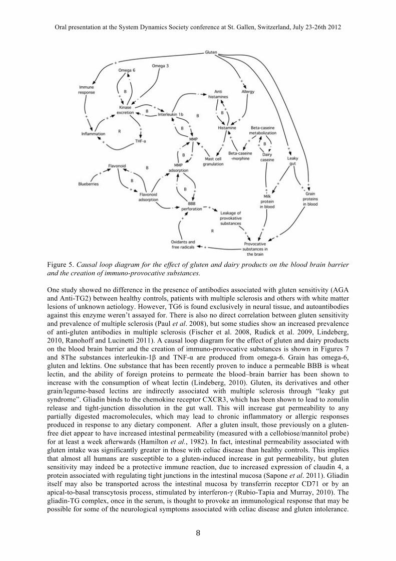
Figure 5. Causal loop diagram for the effect of gluten and dairy products on the blood brain barrier and the creation of immuno-provocative substances. One study showed no difference in the presence of antibodies associated with gluten sensitivity (AGA and Anti-TG2) between healthy controls, patients with multiple sclerosis and others with white matter lesions of unknown aetiology. However, TG6 is found exclusively in neural tissue, and autoantibodies against this enzyme weren’t assayed for. There is also no direct correlation between gluten sensitivity and prevalence of multiple sclerosis (Paul et al. 2008), but some studies show an increased prevalence of anti-gluten antibodies in multiple sclerosis (Fischer et al. 2008, Rudick et al. 2009, Lindeberg, 2010, Ranohoff and Lucinetti 2011). A causal loop diagram for the effect of gluten and dairy products on the blood brain barrier and the creation of immuno-provocative substances is shown in Figures 7 and 8The substances interleukin-1! and TNF-" are produced from omega-6. Grain has omega-6, gluten and lektins. One substance that has been recently proven to induce a permeable BBB is wheat lectin, and the ability of foreign proteins to permeate the blood–brain barrier has been shown to increase with the consumption of wheat lectin (Lindeberg, 2010). Gluten, its derivatives and other grain/legume-based lectins are indirectly associated with multiple sclerosis through “leaky gut syndrome”. Gliadin binds to the chemokine receptor CXCR3, which has been shown to lead to zonulin release and tight-junction dissolution in the gut wall. This will increase gut permeability to any partially digested macromolecules, which may lead to chronic inflammatory or allergic responses produced in response to any dietary component. After a gluten insult, those previously on a gluten-free diet appear to have increased intestinal permeability (measured with a cellobiose/mannitol probe) for at least a week afterwards (Hamilton et al., 1982). In fact, intestinal permeability associated with gluten intake was significantly greater in those with celiac disease than healthy controls. This implies that almost all humans are susceptible to a gluten-induced increase in gut permeability, but gluten sensitivity may indeed be a protective immune reaction, due to increased expression of claudin 4, a protein associated with regulating tight junctions in the intestinal mucosa (Sapone et al. 2011). Gliadin itself may also be transported across the intestinal mucosa by transferrin receptor CD71 or by an apical-to-basal transcytosis process, stimulated by interferon-# (Rubio-Tapia and Murray, 2010). The gliadin-TG complex, once in the serum, is thought to provoke an immunological response that may be possible for some of the neurological symptoms associated with celiac disease and gluten intolerance.
Oral presentation at the System Dynamics Society conference at St. Gallen, Switzerland, July 23-26th 2012
! *!
Gluten contains the protein gliadin that may act as a mimic for myelin proteins. There are clinical treatment records and anecdotic information available showing that diet over long time does have a positive effect on the venous valve insufficiency (CCSVI), however, this has not been subject to scientific medical study yet. For a possible mechanism, a causal loop diagram was developed, this is shown in Figure 5. CCSVI; The breakthrough of Zamboni et al. Dr. Paolo Zamboni, professor of angio-vascular medicine, at the University of Ferrara in Italy (Zamboni et al., 2003, 2005, 2006a,b, 2007, 2008, 2009a,b,c,d,e, 2010, Francesci et al., 2009, Zamboni 2009, Khan et al., 2010) was the first to fully understand the significance of the vascular observations as far back as in the 1930’s (Putnam 1935, 1937, 1939) and again in the 1950’s: (Fog 1948, 1951, 1964, 1965, Macchi 1954, Swank and Hain 1952, Lewis and Swank 1953), and again in the 1980’s (Schelling 1984, 1986, Swank et al. 1983). Dr. Zamboni and his research team suddenly realized that the venous constrictions leading to blood vessel leaks and breaching of the blood-brain-barrier could be the cause and not the result of multiple sclerosis. Zamboni et al. (2008, 2009) calls this chronic cerebro-spinal venous insufficiency, abbreviated CCSVI. He and his team could show in clinical experiments that the constrictions in vessels draining the brain and the spinal cord probably were related to the development of multiple sclerosis. A number of studies have demonstrated that the constrictions come before the multiple sclerosis (Bowman 1947, Adams 1988, 1989). We observe that all the veins that drain the central nervous system, may suffer from chronic cerebro-spinal venous insufficiency and this causes: The pair of internal jugular veins, the pair of vertebral veins, obstructions in the brachiocephalic vein (part of the clava), normally by wall thickening or single obstructive flaps, single filaments or obstructive wall membranes and the azygos system draining the spinal cord.
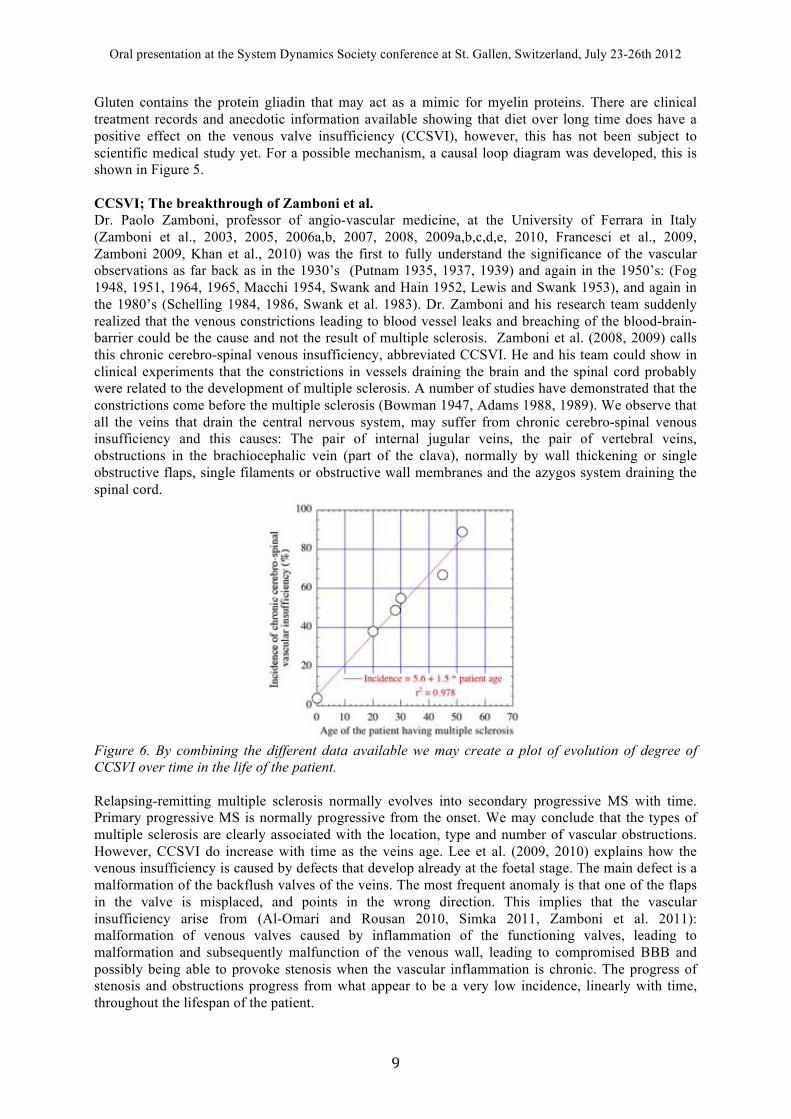
Figure 6. By combining the different data available we may create a plot of evolution of degree of CCSVI over time in the life of the patient. Relapsing-remitting multiple sclerosis normally evolves into secondary progressive MS with time. Primary progressive MS is normally progressive from the onset. We may conclude that the types of multiple sclerosis are clearly associated with the location, type and number of vascular obstructions. However, CCSVI do increase with time as the veins age. Lee et al. (2009, 2010) explains how the venous insufficiency is caused by defects that develop already at the foetal stage. The main defect is a malformation of the backflush valves of the veins. The most frequent anomaly is that one of the flaps in the valve is misplaced, and points in the wrong direction. This implies that the vascular insufficiency arise from (Al-Omari and Rousan 2010, Simka 2011, Zamboni et al. 2011): malformation of venous valves caused by inflammation of the functioning valves, leading to malformation and subsequently malfunction of the venous wall, leading to compromised BBB and possibly being able to provoke stenosis when the vascular inflammation is chronic. The progress of stenosis and obstructions progress from what appear to be a very low incidence, linearly with time, throughout the lifespan of the patient.
Oral presentation at the System Dynamics Society conference at St. Gallen, Switzerland, July 23-26th 2012
! "+!
Provocativeexposure
Valve surfacespecificantigen
Activatedimmune system
Antigensattached to
venous valves
Valvedegeneration
Antigenesattached to
venous walls
Venous walldegeneration
Venousinsufficiency
Venousbackflush
Blood-brain-Barrierpermeability
Hemosiderindeposits
Oxygendeficiency
Oxidantcascade
Demyelinationcascade
Cerebral temperaturecontrol insufficiency
++
+
+
+
+
+
+
+
+
+
+
++
++
+
+
+
+
+
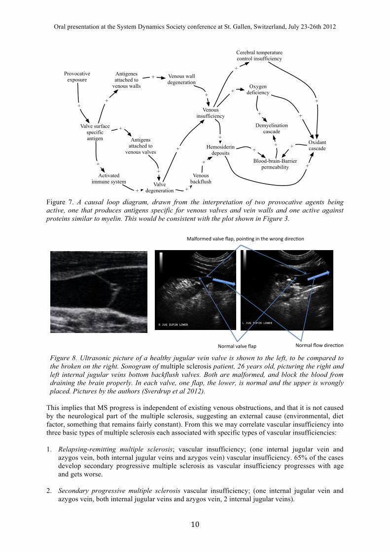
Figure 7. A causal loop diagram, drawn from the interpretation of two provocative agents being active, one that produces antigens specific for venous valves and vein walls and one active against proteins similar to myelin. This would be consistent with the plot shown in Figure 3.
!"#$%&'(%&()'*%+'
,%&-"#$).'(%&()'*%+/'+"01213'01'45)'6#"13'.0#)72"1'
!"#$%&'*"6'.0#)72"1'
Figure 8. Ultrasonic picture of a healthy jugular vein valve is shown to the left, to be compared to the broken on the right. Sonogram of multiple sclerosis patient, 26 years old, picturing the right and left internal jugular veins bottom backflush valves. Both are malformed, and block the blood from draining the brain properly. In each valve, one flap, the lower, is normal and the upper is wrongly placed. Pictures by the authors (Sverdrup et al 2012).
This implies that MS progress is independent of existing venous obstructions, and that it is not caused by the neurological part of the multiple sclerosis, suggesting an external cause (environmental, diet factor, something that remains fairly constant). From this we may correlate vascular insufficiency into three basic types of multiple sclerosis each associated with specific types of vascular insufficiencies: 1. Relapsing-remitting multiple sclerosis; vascular insufficiency; (one internal jugular vein and
azygos vein, both internal jugular veins and azygos vein) vascular insufficiency. 65% of the cases develop secondary progressive multiple sclerosis as vascular insufficiency progresses with age and gets worse.
2. Secondary progressive multiple sclerosis vascular insufficiency; (one internal jugular vein and azygos vein, both internal jugular veins and azygos vein, 2 internal jugular veins).

Oral presentation at the System Dynamics Society conference at St. Gallen, Switzerland, July 23-26th 2012
! ""!
3. Primary progressive multiple sclerosis vascular insufficiency; (azygos vein and azygos tributary
veins) only oriented around the spinal cord and its associated blood supply. Jugulars and vertebral veins are competent. The patient stays cognitively normal, but mobility is gradually lost.
4. Progressive relapsing multiple sclerosis vascular insufficiency; with complex and multiple
vascular insufficiency, affecting the brain, spinal cord and renal system (azygos vein and azygos tributary veins and both internal jugular vein and both internal vertebral veins, Illiac vein, renal vein). Quickly leads to central nerve system breakdown and demise.
Thus we can establish a relationship between the types of vascular insufficiency and different types of multiple sclerosis, remitting relapsing, secondary progressive, primary progressive. Vascular flow rates and their ramifications Constriction in the blood vessels that drain the spinal cord and brain leads to reflux of blood, that in turn cause minor leaks into the brain. The draining vessels are located far from the heart in the vascular system, and thus have low pressure gradients to operate under, taking support also from the pressure-variations caused by breathing (Zamboni 2010). Stenosis of the veins impedes blood flow, implying that less blood can return from the brain. In a recent study Zamboni et al. (2010) compared the venous status of 109 persons diagnosed with multiple sclerosis, and 177 persons determined to be without the illness. Of those with multiple sclerosis, Zamboni’s team could find venous constrictions in all of them (100%). In figure 8 from a multiple sclerosis patient, 26 years old, the right and left internal jugular veins bottom backflush valves are malformed, and block the blood from draining the brain. In each valve, one flap, the lower, is normal and the upper is wrongly placed. Their results show that 97% of the patients have malformed valves, constricted veins or collapsed or missing jugular veins. The interpretation of MRI scans, combined with Doppler ultrasound scans for checking actual blood flow in the detected sections, are central to the diagnostics of multiple sclerosis (Zamboni et al., 2009, 2010).
Figure 9. Ultrasonic Doppler measurements on disturbed flow through the right jugular vein with malformed backflush valves is shown. The measurements were made in erect and supine position, when the jugular veins carry 70% of the total brain blood drainage. The right jugular vein has clear backflushes, amounting to about 30% of normal flow. The scale of flow velocity (m/s). (Image by the authors at the Essential Clinic, Glasgow). Flow measurements and the ramifications of restricted blood flow through the brain Flow through the veins draining the head can be assessed using ultrasound techniques in non-invasive examinations. These examinations show that the blood flow can be significantly reduced. Figure 9 shows ultrasound Doppler measurements on flow through the left and right jugular vein with malformed backflush valves and significant flow impediment (Sverdrup et al. 2012). The measurements were made on a young man in erect position, when the vertebrate veins carry 70% of the brain blood drainage as compared to the flow through the jugular veins in the supine position when the situation is reversed (Weir 2010). Those with multiple sclerosis caused by vascular insufficiency, show symptoms of oxygen deficiency to the brain, similar to altitude sickness. Dizziness, confusion, problems with concentrations, lowered cognitive abilities, is all results of lack of oxygen supply to the brain. Bastianello et al. (2011) analysed the clinical data of 710 multiple sclerosis patients attending six centres carrying out liberation treatments. All patients had been submitted to venous Doppler sonography and diagnosed as having or not having CCSVI according to the criteria of Zamboni et al.

Oral presentation at the System Dynamics Society conference at St. Gallen, Switzerland, July 23-26th 2012
! "#!
(2009). CCSVI was diagnosed in 86% of the patients on the average, but the frequency varied greatly between the treatment centres. It is possible that these malformed veins are the result of an immune attack on the valves in the cerebral venous system, which causes chronic inflammation and wall scarring. In a recent review of the role of venous reflux, Simka (2009) stated: "It is hypothesized that pathological refluxing venous flow in the cerebral and spinal veins increases the expression of adhesion molecules, particularly ICAM-1, by the cerebrovascular endothelium" (Simka 2011). Along these lines, Ono et al. (1998) and Bergan et al. (2007, 2008) demonstrated, by occluding a major vein in laboratory rats, that the number of leukocytes migrating across the vessel wall increased progressively during occlusion. Multiple micro-haemorrhages occurred upstream of the occlusion (creating holes in the of the blood-brain barrier; these are usually 20–30 µm in diameter, but some breaches in the blood-brain-barrier are as large as 200 µm, real holes visible to the naked eye). "The venular occlusion experiments showed that reduced flow can rapidly set in motion an inflammatory cascade, including hallmarks like leukocyte adhesion to the endothelium, migration into the interstitium, free radical production and parenchymal cell death that begins soon after occlusion..."
Venous valve malfunction
Minor leaksin microvessels
in the brainIron
accumulation
Local pressure shocks
Bloodentry tothe brain
+
+
++
+
Oxidative stress, free radicals
+
Stagnant bloodin the small
vessel
Lining inflammation
Electromagneticradiation
+
+
+
+
Leaky gut problem
alien substancesin blood
Immune reactionto alien substances
Stimulation of histamines and MMP
+
+
+
+
++
Unsuitablediet
+
Valve inflammation
Antibody valvesurface adhesion
Immuneresponse
Antibody to valvesurface protein
molecular mimicry
+
+
+
+
+
+ R3
R1
Antibodyproduction
++
+
R2
+
R2
Figure 10. Causal loop diagram for the effect of vascular insufficiency. The emphasis of this diagram is to show how the venous vessels become leaky and open a door through the blood-brain barrier. Figure 10 shows a causal loop diagram for the effect of vascular insufficiency. Figure 3 shows some of the mechanisms that actually open the blood-brain-barrier. Figure 4 shows the causal loop diagram for the activation process for T-cells outside the central nervous system (CNS) and the role of molecular mimicry initiated by viral and bacterial infections. Key is the MMP compound that opens the tight joints in the blood-brain-barrier, histamine also can do that, it holds a key role not recognized for what it does. Figure 11 shows how CCSVI may unbalance the system within a multiple slerosis0sueceptible individual. The implications we have drawn from reading the literature is that either bacteria and/or virus may deliver the triggering protein material, but that it a virus is needed to incorporate it into a T-cell to be smuggled across the blood-brain barrier. Without CCSVI and
Oral presentation at the System Dynamics Society conference at St. Gallen, Switzerland, July 23-26th 2012
! "$!
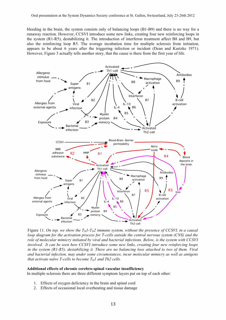
bleeding in the brain, the system consists only of balancing loops (B1-B9) and there is no way for a runaway reaction. However, CCSVI introduce some new links, creating four new reinforcing loops in the system (R1-R5), destabilizing it. The introduction of interferon treatment affect B8 and B9, but also the reinforcing loop R5. The average incubation time for multiple sclerosis from initiation, appears to be about 6 years after the triggering infection or incident (Dean and Kurtzke 1971). However, Figure 3 actually tells another story, that the cause is there from the first year of life.
Additional effects of chronic cerebro-spinal vascular insufficiency In multiple sclerosis there are three different symptom layers put on top of each other:
1. Effects of oxygen deficiency in the brain and spinal cord 2. Effects of occasional local overheating and tissue damage
Viralinfection
Bacterialinfection
Myelinproteinmimicry
Activated Th1-cell
ActivatedTh2-cell
IL-4IL-10
Superantigens
Allergies from external agents
Allergenicstimulus
from food
+
--+
+
+
+
++
+
++
Exposure
+
+
-
-
B3B4
B5
B2
B1Macrophageactivation
B-cellactivation
Interferon
+ Antibodies-
+
+
- -
+
+
+
B8
B7
B9
B6
Viralinfection
Bacterialinfection
Myelinproteinmimicry
Activated Th1-cell
ActivatedTh2-cell
IL-4IL-10
BBBadhesion substance
Superantigens
Allergies from external agents
Allergenicstimulus
from food
+
+
--+
+
+
+
++
+
++
Exposure
+
+
-
-
B3B4
B5
B2
B1
MMP
+
Iron
Nitricoxide
Blooddeposits inthe brain
+
-
Macrophageactivation
B-cellactivation
Interferon
+ Antibodies-
+
+
- -
+-
+
++
B8
B7
B9
B6
+
+
CCSVI Blood-Brain -Barrierpermeability
++
+
+R1R2
R3
R4
R5
Figure 11. On top, we show the TH1-TH2 immune system, without the presence of CCSVI, in a causal loop diagram for the activation process for T-cells outside the central nervous system (CNS) and the role of molecular mimicry initiated by viral and bacterial infections. Below, is the system with CCSVI involved. It can be seen how CCSVI introduce some new links, creating four new reinforcing loops in the system (R1-R5), destabilizing it. There are no balancing loos attached to two of them. Viral and bacterial infection, may under some circumstances, incur molecular mimicry as well as antigens that activate naïve T-cells to become TH1 and Th2 cells.

Oral presentation at the System Dynamics Society conference at St. Gallen, Switzerland, July 23-26th 2012
! "%!
3. Nerve damage in the brain and spinal cord Figure 13 summarizes the coupled effects of oxidants and oxidant stresses on nerve tissues, after Gilun-Sherki (2004) and Foster (2006), and interpreted by the authors. Oxygen depravation has turned out to be one of the main causes for several of the early symptoms in multiple sclerosis (Matute and Perez-Cerda 2005, Trapp and Stys 2009, Juurlink 1997, Keyser et al 2008, Zamboni et al 2009). Overheating of the brain seems to interact with both the immune attacks as well as the effects of oxygen depravation (Gupta et al 2002, Lavinio et al., 2007, White et al 1996). Oxidant stresses reworked into causal loop diagrams
After entry of iron to the brain, a production of free peroxynitrite starts after the local reservoirs of antioxidants have been depleted. This affects the autoimmune cycles and the scarification of damaged myelin tissues. It also starts a process, where the blood-brain barriers now becomes punctured from the brain side and out towards the blood in the body. The blood-brain barrier has is severely compromised through this process. Haider et al. (2011) confirms the presence of oxidized DNA and oxidized lipids in the mitochondria of astrocytes and in particular oligodendrocytes in multiple sclerosis patients, but not in controls. Oxidized DNA of oligodendrocytes correlated well with active ongoing inflammation and active lesions. Inflammation is not one single phenomenon, but there is a connection between inflammation, caused by microorganisms, and the subsequent immune system attack against the lesions. This stands in a communicating system with the regeneration, where attack and damage leads to activation of regenerative processes. According to a strictly immunological explanation of multiple sclerosis, the inflammatory process is caused by T-cells, a type of lymphocyte. Lymphocytes are cells that play an important role in the body's defences (Compston and Coles 2002, 2008). In multiple sclerosis, T-cells gain entry into the brain via the blood–brain barrier (Figures 4 and 7). Evidence from animal models also point to a role of B cells in addition to T-cells in development of the disease. The T-cells recognize myelin as a foreign substance, it becomes marked as alien by attached antibodies, and the immune systems attack it as if it were an invading virus. This triggers the inflammatory processes, stimulating other immune cells and soluble factors like cytokines and antibodies (Figure 9). Antibodies and potentially, denaturalization of the protein surfaces by oxidants and metals, cause the sheath to appear as alien. Free radicals promote the inflammation and retard the myelin sheath regeneration process. Several factors affect free radicals and the oxidants that are their precursors. Vitamin A, C, E, K, and D are all oxidant scavengers. Both NO and Fe2+/Fe3+ are involved in generation of peroxides, superoxide, peroxynitrite and free radicals (the Fenton reactions), all highly toxic to any living cells. In multiple sclerosis, oxygen deprivation is not very pronounced, but rather local and occurs through localized blood vessel lesions, and the damage associated with the iron accumulation. Free radicals are known to create blood vessel ulcers (Simka and Rybak 2008), and most probably establish a reinforcing link, keeping the pathway through the blood-brain barrier permanently or intermittently open, suggesting an explanation of the remitting/permanent types of the illness. Blood deposited in the brain generates NO* upon decomposition and leads to the deposits of iron in the brain. This is under reducing conditions transformed to free iron ions (Fe2+). These auto-catalyse degeneration of vitamin C, catecholamines and Vitamin E and may overcome the protection system against free radicals locally. When this happens, the metabolites will contain superoxides, further aggravating damages (Coyle and Puttefarcken 1993). The free radicals and Fe3+ in the brain interferes with the regenerative function of the oligodendrocytes. It is also suspected that they may have a role in changing harmless antibodies to a kind that initiate Th1 cell attack on the myelin sheaths of the brain. Peroxynitrite and superoxide Peroxynitrite is a very damaging oxidant, perhaps the most important in multiple sclerosis as well as other illnesses of similar nature (Pacher et al. 2006). It is formed very rapidly from superoxide and NO (Gilun-Sherki et al. 2002). NO is produced in the body at many sites, and serves as a signal substance in many instances. It is thus always present and can do significant damage to membranes in the body. Superoxide and hydrogen peroxide is regularly formed in mitochondria and during the normal operation of many cells, normally these are neutralised by metal-superoxide dismutase and the hydrogen peroxide by catalase. Nitric oxide (NO) in the system is important for this, as well as the
Oral presentation at the System Dynamics Society conference at St. Gallen, Switzerland, July 23-26th 2012
! "&!
stimulus from excess Ca2+. Superoxide is not supposed to be forming in amounts that will allow it to float freely around, but when the local superoxide dismutase capacity is overwhelmed, then peroxynitrite will be the result. Peroxynitrite is destroyed in a first order reaction with respect to glutathione peroxidase. And the oxidised glutathione peroxidase is regenerated at the cost of 2 glutathione (Stepien et al. 2000, Salvemini et al. 2006, Virag et al. 2003, Pacher et al. 2007, Szabo et al. 2007, Szabo 2003, Toncev et al. 2002, Druclovic et al. 2001, Spitsin et al. 2001, 2000, Hooper et al. 2000: Nelson et al. 2002), and selenium is required in the process. Low selenium status will thus affect the antioxidant status of a person. Vitamin E and C form an important function as scavengers of oxidants. They are not always catalytic like superoxide dismutase, glutathione peroxidase or katalase, but still important. Thus, a potentially powerful detoxification mechanism is in place to take care of oxidants in the body, and under normal conditions, this is very effective. However, through blood vessel leaks, conditions arise where the antioxidants locally become exhausted, leaving residual free radicals. In the micro-clotting that occurs in smaller and capillary blood vessels of those with multiple sclerosis (Swank 1952, Lewis and Swank 1953, Fog 1951, 1964, 1965, Swank et al. 1983, Zamboni et al. 2010), conditions will arise where peroxynitrite will be produced. The endogenous antioxidant enzymes like katalase, glutathione peroxidase and super oxide dismutase then have supply restrictions at the incidence site. Then peroxynitrite will cause increased permeability of the blood-brain barrier, and keep it permeable as long as the conditions are such that the peroxynitrite cannot be cleared.
BBB
BBB
Foreignmyelin-emulating
antibodies
CD4-T-Cellmodified
CD8-T-Cellmodified
B-Cellmodified
CD8-T-Cellmodified
CD4-T-Cellmodified
B-Cellmodified
Permeablebloodbrain
barrier
+
+
+
+
+
+
+
+
+
Release ofmyelin-specific
antibodies
Myelin markedby antibodies
Attack onmyelin
C5d-9
Myelinsheath
Myelindebris Macrofages
mobilized
+
+
+
+
+
+
+
+
-
+
+
+
Remyelination
-
+
Oligodendrocytes
Attack onoligodendrocytes
+
+
Oligodendrocytedebris
FasL/Fas
TNF-a
Interferon
Th1
Th2
InterLeukin
+
+
++
+
-
Fe
+
Oxidants
+
++
-
Blooddecomposition
+
+
+
+
+
Constrictedvessels
MMPMetallo-
proteinases
+ +
+
+
+
Surfacedenaturalization
Scarification
-
++
+
Myelinantibodies
++
Strongelectromagnetic
fields
Pollutants+
+
Inflammation of vessel wall
brain side
+
Oligocyteprecursors
-
Nitricoxide+
+
Apoptosis
+
+
+
+
-
Acineto-bacter
Pseudomonas
Epstein-Barrherpesvirus
Human Herpes Virus-6
T-Cell modificationmechanism
++
+
+
+
+
+
Yet unidentifiedviral vectors
+
+
-
+
Mercuryin the brain
Lead
-
+
-
Mercury
-
+
+
Inflammation of vessel wall
blood side
+
+
Metallo-proteinases inside BBB
+
-
+
Aspartame
Glutamate
Exitotoxins
-
+
++
+
+
+
-
-
+
- ++
+
--
-
-
-
Oligoclonalbands in
electrophoresisanalysis of blood
Oxidants inthe bloodstream
Greymatternecrosis
Visiblelesions onMRI scans
Transfer of oreignmyelin-emulating
antibodies
+
+
+
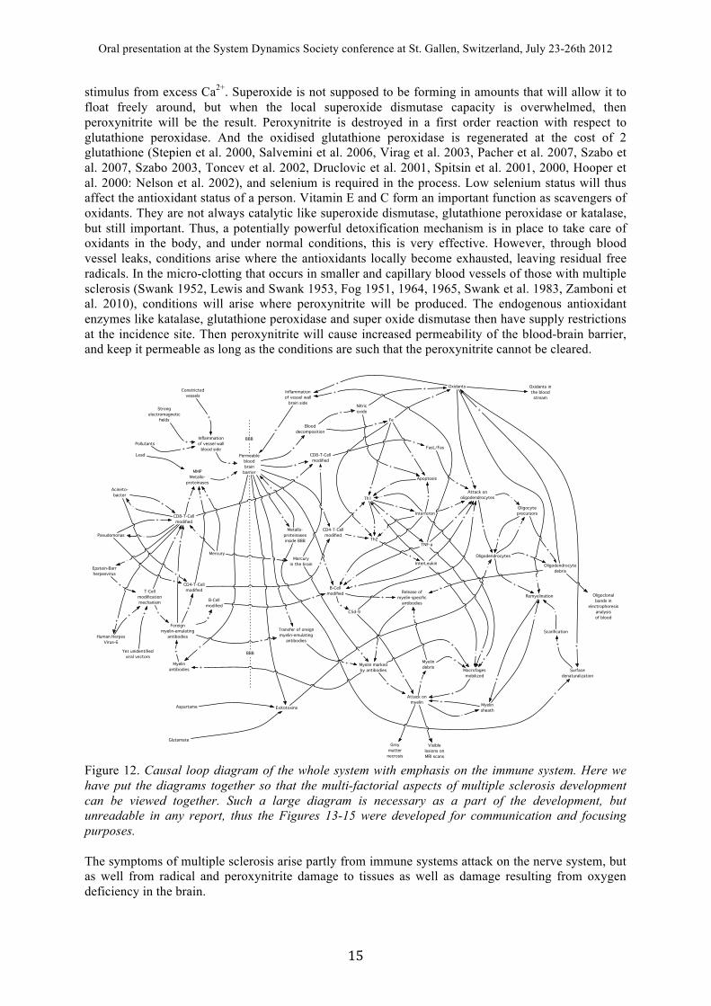
Figure 12. Causal loop diagram of the whole system with emphasis on the immune system. Here we have put the diagrams together so that the multi-factorial aspects of multiple sclerosis development can be viewed together. Such a large diagram is necessary as a part of the development, but unreadable in any report, thus the Figures 13-15 were developed for communication and focusing purposes. The symptoms of multiple sclerosis arise partly from immune systems attack on the nerve system, but as well from radical and peroxynitrite damage to tissues as well as damage resulting from oxygen deficiency in the brain.
Oral presentation at the System Dynamics Society conference at St. Gallen, Switzerland, July 23-26th 2012
! "'!
Summarizing root causes of multiple sclerosis In Figures 13-15 we show the causal loop diagram for the whole system of multiple sclerosis. Here we have put the diagrams together so that the multi-factorial aspects of multiple sclerosis development can be viewed together. We have simplified the large causal loop diagram (Figure 12) with all the details, down to a simple diagram (Figure 14) showing the most important loops and their interconnections. From this diagram, we can see the initiating functions, the compromising of the blood-brain barrier, the starting of the oxidative stresses, interacting with the autoimmune reaction to protein mimicry, promoting scarification and attacking the regenerative function. We can see that the medical doctor treating a patient will see the symptoms of 3 ailments overlaid on each other:
1. The effect of oxygen deficiency in the brain and spine leading to depression, cognitive deficiency and confusion
2. The effect of brain overheating leading to the classical symptoms of heat-stroke. Eventually, this may lead to permanent tissue damage
3. The effect of damage to neural tissue in the brain and spine leading to immobility, failure of inner organs, cognitive problems, impaired eyesight etc…
Multiple sclerosis has no simple cause-effect relationships that traditional methods will find. Multiple sclerosis is a whole system disease and must be seen as a system for any progress to be made. We can see that there are three important reinforcing feedback loops in the multiple sclerosis system that must be terminated in the treatment systemic process:
1. A feedback loop between minor vessel leaks and free radicals. 2. A feedback loop between immune system attacks, further production of iron and oxidants,
triggering autoimmune response 3. A feedback loop between wrongly triggered antibodies and myelin damage from leukocyte
attacks that produce more debris that further triggers attacks. All feedback loops must be closed in order for treatment to be effective. The integrated systems picture for multiple sclerosis with three different symptom group outputs is shown in Figure 16. This causal loop diagram was instrumental in developing a treatment strategy for turning the illness off, avoiding the internal self-reinitiating mechanisms. Figure 16 summarizes the treatment strategy as superimposed on the systems diagram for multiple sclerosis. The systems dynamics model Based on an integrated conceptual model for multiple sclerosis, a computer model was developed. The Integrated Multiple Sclerosis simulation model was validated against patient datasets. The numerical simulations model is based on mapping the whole structure of the disease with systems analysis. The causal loop diagrams form the basis of the design for the model. The model was programmed in the modelling environment STELLA, and parameterized on literature data and patient data (Figure 15). The conceptual model is expressed as causal loop diagrams, flow charts and inter-parameter quantifications were used to create the model. Examples of model outputs are shown in Figure 16. The model was used to simulate the progress of the disease with time for different potential treatments, including state-of-the-art medical approaches. This development work is still going on. We may from the modelling draw some important conclusions: The model seems to be able to predict the general outline of the disease as it is observed, a relapsing remitting behaviour between the onset and when it transforms into a progressive type. The remitting-relapsing multiple sclerosis with attacks are reproduced. The use of a multi-component treatment strategy seems to be able to cause significant improvements, if the underlying vascular problems can be improved. There is a shut-down sequence that must be followed, if not, the feedback loops will restart the illness. The model stresses the superiority of combined strategies, including diet, over single-component treatments. In the vascular system, we generate the flow constrictions, that create pressure shocks in the system, reverse flows and precipitations of minor clots in the fine vessels, that in turn leads to BBB permeability.
Oral presentation at the System Dynamics Society conference at St. Gallen, Switzerland, July 23-26th 2012
! "(!
Minor lesionsin microvessels
in the brain
Bloodentry tothe brain
Antibody and modified T-cell entry to the brain
Immune attac onmyelin sheaths
+
+
+
Myelin-activeantibodies
+
+
MyelinSheathdamage
Oligodendrocytes
Damagesignals
+-
+
B
+
+
-
R-
B
Fe(III)
+
Oxidative stress, free radicals
+
Scarification
-
+
Myelinfragments
+
+
Blooddecomposition
NO
+ +
Oligodendrocyteprecursors
-+
-
B
Peroxynitrite
Superoxide
Peroxide
Fe(II)
+
+
++++
+
Fentonreactions
Antioxidants-
-
--
+
-
+
+
+
+
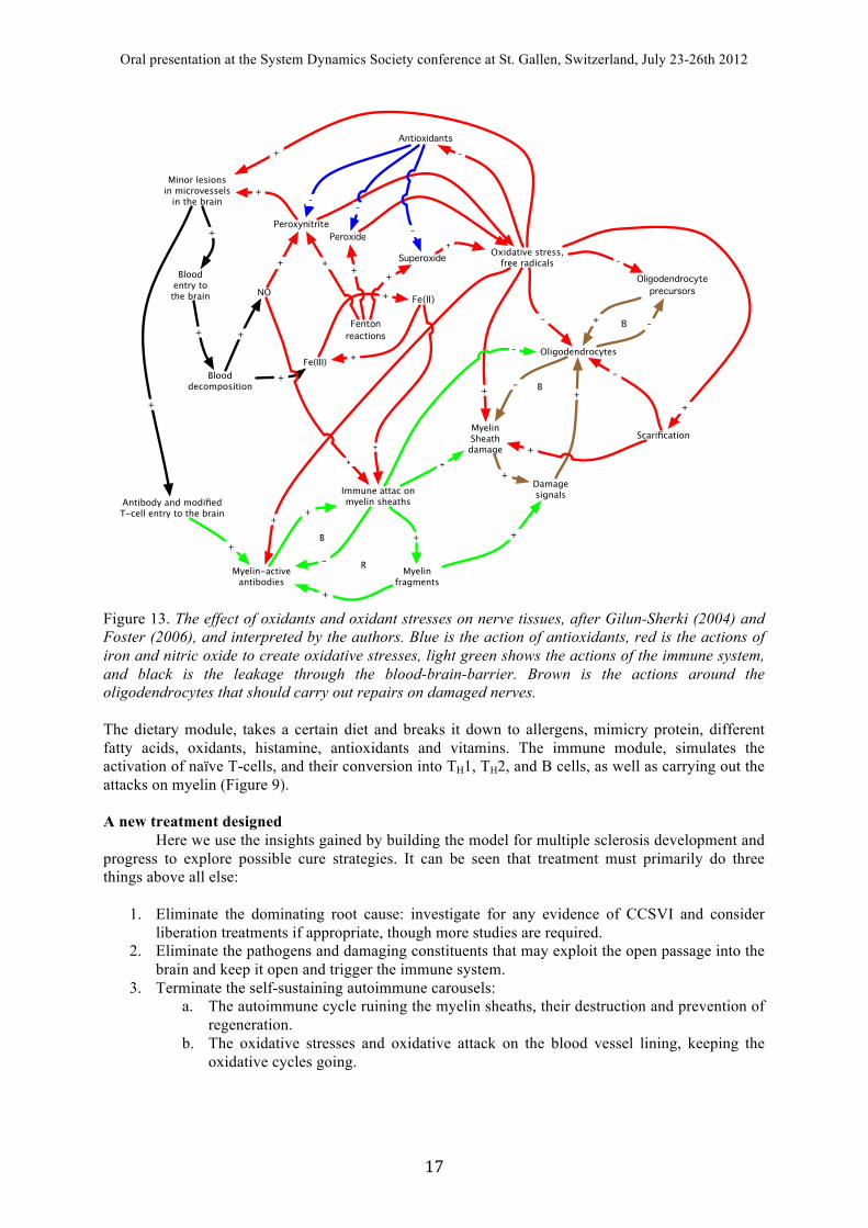
Figure 13. The effect of oxidants and oxidant stresses on nerve tissues, after Gilun-Sherki (2004) and Foster (2006), and interpreted by the authors. Blue is the action of antioxidants, red is the actions of iron and nitric oxide to create oxidative stresses, light green shows the actions of the immune system, and black is the leakage through the blood-brain-barrier. Brown is the actions around the oligodendrocytes that should carry out repairs on damaged nerves. The dietary module, takes a certain diet and breaks it down to allergens, mimicry protein, different fatty acids, oxidants, histamine, antioxidants and vitamins. The immune module, simulates the activation of naïve T-cells, and their conversion into TH1, TH2, and B cells, as well as carrying out the attacks on myelin (Figure 9). A new treatment designed
Here we use the insights gained by building the model for multiple sclerosis development and progress to explore possible cure strategies. It can be seen that treatment must primarily do three things above all else:
1. Eliminate the dominating root cause: investigate for any evidence of CCSVI and consider
liberation treatments if appropriate, though more studies are required. 2. Eliminate the pathogens and damaging constituents that may exploit the open passage into the
brain and keep it open and trigger the immune system. 3. Terminate the self-sustaining autoimmune carousels:
a. The autoimmune cycle ruining the myelin sheaths, their destruction and prevention of regeneration.
b. The oxidative stresses and oxidative attack on the blood vessel lining, keeping the oxidative cycles going.
Oral presentation at the System Dynamics Society conference at St. Gallen, Switzerland, July 23-26th 2012
! ")!
The strategy developed below has this aim, i.e. to stop the starting mechanisms, stop the self-perpetuating processes and to make regeneration of damaged nerve tissue possible. the patient can return to the same behaviour as before the onset of multiple sclerosis. He must most probably stay away from those components of the diet that caused the illness forever, and failure to do so may reintroduce the illness again.
Vascularinsufficiency
Blood flowinsufficiency through the
brain
CoolingInsufficient
oxygensupply
Cognitiveproblems
Depressions
Oxidantcycle
Immunereaction
Regenerativefunction
Nervedamage
-
+
+
+
+
++
-+
Compromizedblood-brain
barrier
+
Molecularprecursorsin blood
+
-
+
++
+ ++
Brainoverheating
-
R4B
R1
Cellularenergydeficit
Tissueinflammation
++
++
++ +
+
+
-
+
Food components
Proteinmimicry
+
+
Gutleakage
Antibodies
+
+
+
Infectousagents
+
+
+
Allergies
+
+
R3
R2R5
Body controlproblems
+
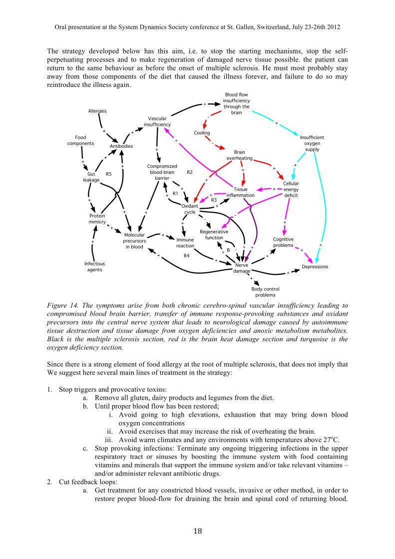
Figure 14. The symptoms arise from both chronic cerebro-spinal vascular insufficiency leading to compromised blood brain barrier, transfer of immune response-provoking substances and oxidant precursors into the central nerve system that leads to neurological damage caused by autoimmune tissue destruction and tissue damage from oxygen deficiencies and anoxic metabolism metabolites. Black is the multiple sclerosis section, red is the brain heat damage section and turquoise is the oxygen deficiency section. Since there is a strong element of food allergy at the root of multiple sclerosis, that does not imply that We suggest here several main lines of treatment in the strategy: 1. Stop triggers and provocative toxins:
a. Remove all gluten, dairy products and legumes from the diet. b. Until proper blood flow has been restored;
i. Avoid going to high elevations, exhaustion that may bring down blood oxygen concentrations
ii. Avoid exercises that may increase the risk of overheating the brain. iii. Avoid warm climates and any environments with temperatures above 27oC.
c. Stop provoking infections: Terminate any ongoing triggering infections in the upper respiratory tract or sinuses by boosting the immune system with food containing vitamins and minerals that support the immune system and/or take relevant vitamins – and/or administer relevant antibiotic drugs.
2. Cut feedback loops: a. Get treatment for any constricted blood vessels, invasive or other method, in order to
restore proper blood-flow for draining the brain and spinal cord of returning blood.
Oral presentation at the System Dynamics Society conference at St. Gallen, Switzerland, July 23-26th 2012
! "*!
This implies invasive treatment to eliminate chronic cerebro-spinal vascular insufficiency (CCSVI).
b. Elimination of free radicals through vitamin and antioxidant therapy in order to prevent weakening of vessel walls, and disruption of antibody programming. Strengthening the myelin sheath repair mechanisms with essential nutrients like omega 3 for healing
c. Immuno-regulating medication to break off the vicious autoimmune circle acting on oligodendrocytes and myelin sheaths, for example with interferon-! type short term intensive treatment.
3. Rebuild damaged structures: a. Strengthening of regenerative mechanisms (oligodendrocytes), through the necessary
resources demanded (e.g. omega 3 and?) as well as growth factors that can be included in the diet.
b. Prevent further BBB opening substances by filling the BBB membranes with flavonoids that prevent adsorption of foreign substances. The flavonoids from blueberries, blackberries and blackcurrants are most effective.
4. Remove risk factors a. Avoid all high histamine diets (Certain types of wine, fermented foods, old fish etc) b. Avoid heavy metal exposure of any kind c. Eliminate any unnecessary microwave radiation as it is suspected to be able to
compromise the blood-brain barrier and smaller venous blood vessels. Other treatment is only supportive until this has been achieved. For further recovery, removal of accumulated iron from the brain will be important, elimination of radicals through restoration of vitamin status, supply of proteins and optimal fats, restoring the regenerative capability, and elimination of trigger microorganisms in order to prevent relapses. Discussions Many articles focus on narrow aspects of multiple sclerosis and go in-depth on that, but seem to loose the overall understanding. However, that does not lead to any systemic overview, and thus such a methodology cannot find a cure. Only with people like Foster, Schelling, Simak, Zivadinov or Zamboni was a more systemic approach possible. However, none of these distinguished scientists are yet trained in systems analysis, they still haven’t learned those tools sufficiently well and thus do not fully manage to build up a complete system where it can be said to constitute a model. For that, a transdisciplinary systems approach and proper training over several years will be needed. Since multiple sclerosis appears to be a multi-agent system, there will not be any single-cause type of evidence available. From the derived causal loop diagrams, handbooks in nutritional therapy were used to look for points of treatment entry to the system (Cordain 2006a,b,c, Cordain et al. 2002, 2003, 2005, Eaton et al. 2007, 2009, Ahn et al. 2006a,b).

Oral presentation at the System Dynamics Society conference at St. Gallen, Switzerland, July 23-26th 2012
! #+!
Blood vesselinsufficiency
Permeableblood-brain
barrier Oxidativestresses
Transmissionof immunologicallyactive substances
Triggering of immune system
Myelindamage
Oligodendrocytemediated myelin
regeneration
Infections
Antioxidants
+
+
-
-
-
-+
+
+
+
-R2R1
R4
R3
+
+
+
Environmentalchemistry factors
+
Oxidant precursor food additives
+
++
Essentialcomponents
+
B3
B1
R6
- B4
Allergenes
Histamines
+
+
+
+
+
Unsuitablefood
+
R5
Overheating
Oxygendeficiency
+
+
++
Hyperthermiasymptoms
+
+
Neurological andimmunological symptoms
+
Antibodies
+
+
+
++
+
Hypoxiasymptoms
+
B2
+
+
Food
Figure 15. Integrated systems picture for multiple sclerosis with three different symptom group outputs. This causal loop diagram was instrumental in developing a treatment strategy for turning the illness off, avoiding the internal self-reinitiating mechanisms. R1-R5 are reinforcing loops, B1-B4 are balancing loops.
Blood vesselinsufficiency
Permeableblood-brain
barrier Oxidativestresses
Transmissionof immunologicallyactive substances
Triggering of immune system
Myelindamage
Oligodendrocytemediated myelin
regeneration
Infections
Vascularsurgery
ProtectivedietAntioxidants
Interferon
Antibiotictreatment
Prophylacticdiet
-
+
+
-
-
-
-+
+
+
-
+
-
-
-
R2R1
R4
R3
+
+
+
+
Environmentalchemistry factors
+
Environmentalprotectionpolicies
-
Oxidant precursor food additives
+
Foodsafety
regulations
Consumeravoidance
-
-
++
Behavioralcaution
-
Essentialcomponents
+
BB
B
+
-
BAllergenes
Histamines
-
+
+
+
++
Unsuitablefood
-
+
R5
Overheating
Oxygendeficiency
+
+
++
Behavioradjustment
-
Hyperthermiasymptoms
+
+
Neurological andimmunological
symptoms
+
Antibodies
+
+
+
+
+
+
Hypoxiasymptoms
Vacciniummyrtillus
-
Figure 16. A simplified causal loop diagram for MS, depicting the treatment strategies as well as the superimposed symptom assemblies.
Oral presentation at the System Dynamics Society conference at St. Gallen, Switzerland, July 23-26th 2012
! #"!
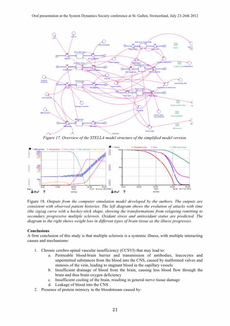
Figure 17. Overview of the STELLA model structure of the simplified model version.
Figure 18. Outputs from the computer simulation model developed by the authors. The outputs are consistent with observed patient histories. The left diagram shows the evolution of attacks with time (the zigzag curve with a hockey-stick shape, showing the transformations from relapsing remitting to secondary progressive multiple sclerosis. Oxidant stress and antioxidant status are predicted. The diagram to the right shows weight loss in different types of brain tissue as the illness progresses. Conclusions A firm conclusion of this study is that multiple sclerosis is a systemic illness, with multiple interacting causes and mechanisms:
1. Chronic cerebro-spinal vascular insufficiency (CCSVI) that may lead to: a. Permeable blood-brain barrier and transmission of antibodies, leucocytes and
unpermitted substances from the blood into the CNS, caused by malformed valves and stenosis of the vein, leading to stagnant blood in the capillary vessels
b. Insufficient drainage of blood from the brain, causing less blood flow through the brain and thus brain oxygen deficiency
c. Insufficient cooling of the brain, resulting in general nerve tissue damage d. Leakage of blood into the CNS
2. Presence of protein mimicry in the bloodstream caused by:

Oral presentation at the System Dynamics Society conference at St. Gallen, Switzerland, July 23-26th 2012
! ##!
a. Infections from certain bacteria and viruses that leak through the BBB to trigger immune response against nerve tissue
b. Leakage of pro-inflammatory substances in the bloodstream originating from food, (gluten, dairy, legumes) into the central nerve system through the compromised brain-blood barrier
3. Feedback system involving iron from blood decomposition, acting in the autocatalytic Fenton cycle and causing production of oxidants
a. Reinforcing the increased permeability of the blood-brain barrier from the inside, as well as causing general tissue damage
b. Oxidative damage to the oligodendrocytes and the myelin repair function 4. Feedback loop involving immune system activation, attacks on the myelin sheaths, and myelin
sheath breakdown, further triggered by more myelin debris
The symptoms of multiple sclerosis develop as a result of gradual destruction of the central nerve system, through immune system action and the effects of oxygen depravation and reoccurring deficient cooling of the brain. The treatment should be designed at targeting all causes listed above simultaneously, stopping the triggers, such as the cause for blood vessel leaks and targeting the triggering infections priming the immune system, as well as stopping self-propelling vicious circles, (immune and autocatalytic oxidant stress cycles) as well as strengthening the regeneration capability. Multiple sclerosis is not a single cause illness, but a whole integrated system of causes and effects. The systems overview is crucial for understanding the illness and for developing a treatment strategy for it. Our results show that no single treatment will do, a series of measures are necessary to stop the re-initiation of the inflammations, stop the precursors of free radicals, stop the autoimmune response, repair the compromised blood-brain barrier, all at once. And they will have to come in the right order, or the illness will self-reinitiate. Multiple sclerosis is a systemic illness, with multiple interacting causes and mechanisms:
1. Leaky blood vessels leading to permeable blood-brain barrier and transmission of antibodies
and leucocytes into the brain 2. This triggers infections from certain bacteria and viruses 3. A feedback system evolves involving iron in the autocatalytic Fenton cycle and production of
oxidants, reinforcing the increased permeability of the blood-brain barrier 4. Immune feedback loop develops involving immune system and myelin sheath breakdown,
leading to brain side damages to the blood-brain barrier, driven by oxidative denaturalization of the myelin surfaces.
The treatment is designed at targeting all 4 causes at once, stopping the triggers including the cause for blood vessel leaks and targeting the triggering infections, as well as stopping autoimmune self-propelling vicious circles, and as strengthening the regeneration capability. Figure 13, explains why the whole sickness takes time to progress and move in discrete steps. A cure must start with elimination of the initiators of the strongest causal factor. For multiple sclerosis this is leakage of blood and immuno-active substances across the blood-brain barrier that starts iron deposits in the brain. At intervals, the immune system will overcome the damage that is caused and initiated by blood vessel leakage and infective triggers. However, since the drivers are persistent, they will re-initiate the system, leading to the remitting-relapse type of illness. After some time, the damage will be so large that the immune system capacity is overrun and the multiple sclerosis progress becomes persistent. Our model is able to describe the evolution of multiple sclerosis in a patient, including the transitions and the timing of these. The details of the illness in the vascular and immune system are well modelled. The model is useful for investigating treatments for finding the cure for multiple sclerosis. Modelling work is ongoing and we expect to be able to report more at a later point. References Aubin ML, Leriche H, Aboulker J, Ernest C, Ecoiffier J, Metzger J. 1976, Cavo-spinal phlebography
in myelopathies of venous origin. Application of the method in 115 cases. Acta Radiol Suppl. 347:403-13.

Oral presentation at the System Dynamics Society conference at St. Gallen, Switzerland, July 23-26th 2012
! #$!
Adams CW 1988 Perivascular iron deposition and other vascular damage in multiple sclerosis. Journal of Neurology, Neurosurgery and Psychiatry 51:260–265
Ahn AC, Tewari M, Poon CS, Phillips RS 2006 The clinical applications of a systems approach. PLoS Med 3:e209. DOI: 10.1371/journal. pmed.0030209
Akassouglou, K., Adams, R.A., Bauer, J., Mercado, P., Tseveleki, V., Lassman, H., Probert, L. Strickland, S., 2004. Fibrin depletion decreases inflammation and delays the onset of demyelination in a tumour necrosis factor transgenic mouse model for multiple sclerosis. PNAS 109: 6698-6708
Allen IV. 1981 The pathology of multiple sclerosis hypotheses. Neuropathol Appl Neurobiol 7:169-200 Journal of Cerebral Blood Flow & Metabolism
Al-Omari, MH, Rousan LA, 2010 Internal jugular vein morphology and hemodynamics in patients with multiple sclerosis. International Angiology, 29:115-120
Ascherio, A, Munger, AL; 2007 Environmental Risk Factors for Multiple Sclerosis. Part I: The Role of Infection. Ann Neurol 61:288–299. Part II: Non-infectious factors. Ann. Neurol. 61:504–513
Agius, L. 2006. Multiple sclerosis is a neurodegeneration specifically targeting oligodendrocytes and myelin sheaths. Int. Jour. Molec. Med. and Adv. Sci. 2:23-29.
Bansil, S., Cook, SD. and Rohowsky-Kochan, C., 1995 Multiple Sclerosis: immune mechanism and update on current therapies. Annals of Neurology 37:S87-S101.
Barnett MH, Sutton I 2006 The pathology of multiple sclerosis: a paradigm shift. Curr Opin Neurolog. 19:242–247
Bartolomei, F. Salvi, R. Galeotti, E. Salviato, M. Alcanterini, E. Menegatti, M. Mascalchi, P. Zamboni P, 2010. Hemodynamic patterns of chronic cerebrospinal venous insufficiency in multiple sclerosis. Correlation with symptoms at onset and clinical course International Angiology, 29:183-188
Bray, PF., Luka J., 1992 Antibodies against Epstein-Barr nuclear antigen (EBNA) in multiple sclerosis CSF, and two pentapeptide sequence identities between EBNA and myelin basic protein, Neurology, 42:1798-1804.
Besler, HT, Comoglu, S, Okcu, Z. 2002. Serum Levels of Antioxidant Vitamins and Lipid Peroxidation in Multiple Sclerosis. Nutritional Neuroscience, 5:215–220
Bowman, T. 1944. A supravital analysis of disorders in the cerebral vascular permeability in man I. Acta Medica Scandinavica 118:1986-1890
Bowman, T. 1947. A supravital analysis of disorders in the cerebral vascular permeability in man II. Two cases of multiple sclerosis. Acta Psyciatrica et neurologica 46:58-71
Bennet, JL, Yu, X., Gilden, DH, Burgoon, MP, Owens, GP, 2008. Infectous agents and multipe sclerosis. In: Raine, CS., McFarland, HF, Hohlfeld, R., (Eds.) Multiple sclerosis; A comprehensive text. Pages 226-236. Saunders, Elsevier, London.
Bramow S, Frischer JM, Lassmann H, Koch-Henriksen N, Lucchinetti CF, Sorensen PS, Laursen H. 2010. Demyelination versus remyelination in progressive multiple sclerosis. Brain.; 133:2983-2998
Broadwell, R., Balin, BJ, Salcman, M. 1988. Transcytotic pathway for blood-borne protein through the blood-brain barrier. Proc. Nati.Acad. Sci. USA Vol. 85, pp. 632-636, January 1988 Neurobiology
Bunyan RF, Popescu BF, Carter JL, Caselli RJ, Parisi JE, Lucchinetti CF. 2011. Childhood-onset multiple sclerosis with progressive dementia and pathological cortical demyelination. Arch Neurol.; 68:525-528.
Carter, JL., 1995, Disease modifying therapies in multiple sclerosis. CNS Drugs, 3:99-114. Charcot J 1868 Histologie de la sclerose en plaques. Gazette des Hopitaux, Paris 41:554–555 Chari DM 2007 Remyelination in multiple sclerosis. International Review of Neurobiology 79:589–
620 Challoner PB Challoner PB, Smith K, Parker JD, MacLeod DL, Coulter. SN, Rose TM, Schultz ER,
Epstein LG., 1995 Plaque-associated expression of human herpes virus 6 in multiple sclerosis. Proceedings of the National Academy of Sciences of the United States of America, 92:7440-7444.
Chandaran, S, Compston, A. 2005, Neural stem cells as a potential source of oligodendrocytes for myelin repair. Journal of the Neurological Sciences 233:179 – 181
Oral presentation at the System Dynamics Society conference at St. Gallen, Switzerland, July 23-26th 2012
! #%!
Chapman, M. Hughes, L., Wilson, C, Namnyak, S, Thompson, E. Giovanni, G. 2005. No Evidence for Production of Intrathecal Immunoglobulin G against Acinetobacter or Pseudomonas in Multiple Sclerosis European Neurology. 53:27-31
Chang, A., Tourtelotte, WW., Rudick, R., Trapp, BD., 2002. Premyelinating oligodendrocytes in chronic lesions of multiple sclerosis. New England Journal of Medicine, 346:165-171
Correale J, Farez M, 2007, Association Between Parasite Infection and Immune Responses in Multiple Sclerosis Ann Neurol 61:97–108
Cordain L. 2006 Implications of Plio-Pleistocene Hominin Diets for Modern Humans. In: Early Hominin Diets: The Known, the Unknown, and the Unknowable. Ungar, P (Ed.), Oxford University Press, Oxford, 363-383
Correale, J. Farez, M. 2007. Association Between Parasite Infection and Immune Responses in Multiple Sclerosis. Ann. Neurol. 61:97–108
Cudrici, C, Niculescu, T., Nicilescu, F., Shil, M. L., Rus, H. 2006 Oligodendrocyte cell death in pathogenesis of multiple sclerosis: Protection of oligodendrocytes from apoptosis by complement. Journal of Rehabilitation research and development 43:123-132.
Corcione, A., Casazza, S., Ferreti, E., Giunti, D., Zappia, E., Pistorio, A., Gambini, C., Mancardi, GL, Ucelli, A. Pistoia, V. 2004 Recapitulation of B cell differentiation in the central nervous system of patients with multiple sclerosis. Proceedings of the National Academy of Sciences of the United States of America 101:11064-11069.
Craelius W, Migdal MW, Luessenhop CP, Sugar A, Mihalakis I. 1982 Iron deposits surrounding multiple sclerosis plaques. Arch. Pathol. Lab. Med.; 106:397-399.
Davison, Humphrey, Livesedge et al. 1975 Multiple Sclerosis Research. Elsevier Scientific Publishing New York,
Dean, G., Kurtzke, JF. 1971. On the risk of multiple sclerosis according to age at migration to South Africa. British Medical Journal, 3:725-729.
Dyment DA, Ebers GC, Sadovnick AD 2004. Genetics of multiple sclerosis Lancet Neuroogyl 3:104–110
Dolic, K., Weinstock-Guttman, B., Marr, K., Valnarov, V., Carl, E., Hagemeier, J., Brooks, C., Kilanowski, C., Hojnacki, D., Ramanathan, M., Zivadinov R., 2011. Risk Factors for Chronic Cerebrospinal Venous Insufficiency (CCSVI) in a Large Cohort of Volunteers. PLoS ONE 6:11:e28062:1-5
Eltzschig, H. K., Carmeliet, P., 2011. Hypoxia and Inflammation. New Engl J. Med. 364:656-65. Embry AF, 2010 Integrating CCSVI and CNS autoimmunity in a disease model for MS. International
Angiology, 29/2 Ebers, GC, Bulman, DE., Sadovnick, AD. Paty, DW. 1986. A population-based study of multiple
sclerosis in twins. New England Journal of Medicine, 25:1638-1642. Ebringer, A, Hughes, L, Rashid, T, Wilson, C, 2005. Acinetobacter immune responses in multiple
sclerosis. Arch. Neurol. 62:33-36 Ebringer A, Rashid T, Wilson C. 2005, Bovine spongiform encephalopathy, multiple sclerosis, and
Creutzfeldt-Jakob disease are probably autoimmune diseases evoked by Acinetobacter bacteria. Annales of the New York Academy of Science 1050:417-428.
Fabis MJ., Scott GS., Kean RB., Koprowski H., Hooper DC. 2007 Loss of blood-brain barrier integrity in the spinal cord is common to experimental allergic encephalomyelitis in knockout mouse models. Proceedings of the National Academy of Sciences of the United States of America. 104:5656-61
Fantelli, Mitsumoto, Sebek 1978. Multiple Sclerosis and Malabsorption Lancet, 1039-1040 Fisher E, Lee JC, Nakamura K, Rudick RA. 2008 Gray matter atrophy in multiple sclerosis: a
longitudinal study. Ann Neurol. 64:255-265. Franceschi C 2009 The unsolved puzzle of multiple sclerosis and venous function. Journal of
Neurology, Neurosurgery and Psychiatry 80:358 Fog. T. 1948: Rygmarvens patologiske anatomi ved dissemineret sclerose og dissemineret
encephalomyelitis. Disputats. Ejnar Munksgaard. Copenhagen. Fog, T. 1951. On the pathogenesis of multiple sclerosis. Acta Psyciatrica et neurobiological 74:22-31 Fog, T 1964. On the vessel-plaque relationships in the brain in multiple sclerosis. Acta Neurol. Scand
40:9-15

Oral presentation at the System Dynamics Society conference at St. Gallen, Switzerland, July 23-26th 2012
! #&!
Fog, T. 1965. The topography of plaques in multiple sclerosis, with special reference to cerebral plaques. Acta Neurol. Scand 41, suppl 15:1
Fog, M, 1966, The Shetland-Orkney-Faroe Project. Acta Neurol. Scand., 42, Suppl., 6-8 Forrester, J. 1971 World dynamics. Pegasus Communications, Waltham MA Foster, H., and Hoffer, A. 2004 The two faces of L-DOPA: Benefits and adverse side effects in the
treatment of Encephalitis lethargica, Parkinson’s disease, multiple sclerosis and amyotrophic lateral sclerosis. Medical Hypotheses, 62:177-181.
Foster, HD, 2006 What really causes multiple sclerosis? A free copy of this book is available at www.hdfoster.com.
Fox, R, Bethoux, F. Goldman, M, Cohen, J. 2006 Multiple sclerosis: Advances in understanding, diagnosing, and treating the underlying disease. Cleveland Clinic. Jour. of Medicine. 73:91-102
Freed DLJ, 1999, Do dietary lectins cause disease? British Medical Journal 318:1023-1024. Gay, D, Esiri, M. 2008. Blood-brain barriers damage in acute multiple sclerosis plaques in an
immunocytological study. Electromagn Biol Med. 27:215-29. Graham, J. 1989, 1993, Multiple Sclerosis, a self-help guide to its management. Healing Arts Press.
One Park Street, Rochester, Vermont 05767. Contact MSRC 4a Chapel Hill, Stansted Essex, UK CM24 8AG.
Gilden DH. 2005. Infectious causes of multiple sclerosis. The Lancet Neurology 4:195–202 Grønning M., Riise, T., Kvåle G, Albrektsen G. Midgard, R., Nylandet H., 1993 Infections in
Childhood Adolescence in Multiple Sclerosis, Neuroepidemiology, 12:61-69. Goldberg, P., Fleming, M.C., and Picard, E.H. 1986. Multiple sclerosis: Decreased relapse rate
through dietary supplementation with calcium, magnesium and vitamin D. Medical Hypothesis, 21:193-200.
Goodman, S., Gulick, E. 2008 Dietary Practices of People With Multiple Sclerosis. Int J MS Care. 10:47–57.
Goodin DS and Kappos, L 2008. Treatment of multiple sclerosis with disease-modifying therapies. In: Raine, C. S., McFarland, H F., Hohlfeld, R., (Eds.) Multiple sclerosis; A comprehensive text. Pages 315-332. Saunders, Elsevier, London.
Gupta, AK; Al-Rawi PG, Hutchinson PJ, Kirkpatrick PJ, 2002. Effect of hypothermia on brain tissue oxygenation in patients with severe head injury. British Journal of Anaesthesia 88:188-192
Gregory, AM, Hayflick, SJ, 2004. Neurodegeneration with brain iron accumulation. Orphanet Encyclopedia.
Guggenmos J, Schubart AS, Ogg S, Andersson M, Olsson T, Mather IH, Linington C. Antibody cross-reactivity between myelin oligodendrocyte glycoprotein and the milk protein butyrophilin in multiple sclerosis. J Immunol. 2004 172:661-668.
Haacke, E. M., 2011, Chronic Cerebral Spinal Venous Insufficiency in Multiple Sclerosis. Expert Rev Neurother. 11:5-9
Haider, L., Fischer, M. T., Frischer, JM Bauer, J., Höfftberger, R., Botond, G Esterbauer, H Binder, CJ Witztum, JL., Hans Lassmann H.. 2011. Oxidative damage in multiple sclerosis lesions. Brain 134; 1914–1924
Hadjivassiliou M, Sanders DS, Grünewald RA, Woodroofe N, Boscolo S, Aeschlimann D. 2010 The neurology of gluten sensitivity. Lancet Neurol; 9:330–342.
Haraldsson, HV and Sverdrup HU 2004. Finding Simplicity in complexity in biogeochemical modelling. In: Wainwright, J and Mulligan, M. (Eds.) Environmental Modelling: A practical approach, 211-223. J. Wiley and Sons Ltd., Chichester.
Haraldsson, H, Sverdrup, H, Belyazid, S., Holmqvist, J., Gramstad, R. 2007. The tyranny of small steps: A reoccurring behaviour in management. Journal of Systems Research and Behavioral Science, 24:1-19
Hayes, CE. 2000 Vitamin D: A natural inhibitor of multiple sclerosis. The Proceedings of the Nutrition Society, 59:531-535.
Hernández-Lahoz C, Rodríguez S, Tuñón A, Saiz A, Santamarta E, Rodrigo L. 2009 Sustained clinical remission in a patient with remittent-recurrent multiple sclerosis and celiac disease gluten-free diet for 6 years. Neurologia. 24:213-215.
Huges, LE, Smith, PA, Bonell, G, Natt, R. S, Wilson C, Tashid T, Amor S, Thompson E J, Croker J. Ebringer A. 2003, Cross-reactivity between related sequences found in Acinetobacter sp.,
Oral presentation at the System Dynamics Society conference at St. Gallen, Switzerland, July 23-26th 2012
! #'!
Pseudomonas aeruginosa, myelin basic protein and myelin oligodendrocyte glycoprotein in multiple sclerosis. Journal of Neuroimmunology 144:105-115
Ho, P., Ortiz, D., Rogers, E., Shea, T., 2002. Multiple aspects of homocysteine neurotoxicity: Glutamate excitotoxicity, kinease hyperactivation and DNA damage. Journal of Neuroscience research 70:694-702
Jonez, HD. 1953 The allergic aspect of multiple sclerosis. Calif. Med. 79:376–380 Juurlink, AHJ, 1998. The multiple sclerosis lesion: initiated by a localized hypoperfusion in a central
nervous system where mechanisms allowing leukocyte infiltration are readily upregulated Medical Hypotheses 51:299-303
Kamiñski, Cieœliñska, Kostyra. 2007 Polymorphism of bovine ! casein and its potential effect on human health. J Appl Genet 48, , :189–198
Kaufman, MD, 1998. Do Microbes with Peptides Mimicking Myelin Cause Multiple Sclerosis if the T Cell Response to their Unique Peptides is Limited? J. Theor. Biol. 193:691–708
Kesselring, J. 2008. History of multiple sclerosis. In: Raine, C. S., McFarland, H F., Hohlfeld, R., (Eds.) Multiple sclerosis; A comprehensive text. Pages 1-9. Saunders, Elsevier, London.
de Keyser J, Steen, C, Mostert, JP, Koch, MW, 2008 Hypoperfusion of the cerebral white matter in multiple sclerosis: possible mechanisms and pathophysiological significance. Blood Flow and Metabolism 28:1645–1651
Ke Y, Qian Z M 2003 Iron misregulation in the brain: a primary cause of neurodegenerative disorders. Lancet Neurology 2:246–253
Kermode AG, Thompson AJ, Tofts PD, Macmanus G, Kendall BE, Kingsley DPE, Mosley IF, Rudge P, Mcdonald WI 1990 Breakdown of the blood brain barrier precedes symptoms and other MRI signs of new lesions in multiple sclerosis: pathogenetic and clinical implications. Brain 113:1477–1489
Kremenchutzky M., Kinkel, P., 2010. The Efficacy of Interferon-! in Delaying Conversion to Clinically Definite Multiple Sclerosis Int J MS Care. 12:42–50
Kennel De March A, De Bouwerie M, Kolopp-Sarda MN, Faure GC, Béné MC, Bernard CC. Anti-myelin oligodendrocyte glycoprotein B-cell responses in multiple sclerosis. J Neuroimmunol. 2003 135:117-125.
Kurek M, Przybilla B, Hermann K, Ring J. A naturally occurring opioid peptide from cow's milk, !-casomorphine-7, is a direct histamine releaser in man. Int. Arch. Allergy Immunol. 1992; 97:115-120.
Kurtzke, JF., 1993 Epidemiologic Evidence for Multiple Sclerosis as an Infection, Clinical Microbiology Reviews, 6:382-427.
Kurtzke, JF. 1983. Epidemiology of multiple sclerosis. In: JF. Hallpike, CWM. Adams, and WW. Tourtellotte (Eds.), Multiple sclerosis: Pathology, diagnosis and management 47-95. Baltimore, MD: Williams and Wilkins.
Kurtzke, JF., and Hyllested, K. 1988. Validity of the epidemics of multiple sclerosis in the Faeroe Islands. Neuroepidemiology, 7:190-227.
Lane, RJ, Cuzzilla, ML, McMahon, CG, 2003. Intermediate to long term results of repairing incompetent multiple deep venous valves using external valvular stenting. ANZ. J. 73; 267-274
Landtblom, A, Flodin, U, Söderfelt, B., Wolfson, C, Axelson, O. 1996 Organic solvents and multiple sclerosis, The state of the current evidence. Epidemology 7:429-433
Lassmann, H, Bruck, W, Lucchinetti, C, 2001. Heterogeneity of multiple sclerosis pathogenesis, implications for for diagnosis and therapy. Trend Molec. Med. 7:115-121
Lee, BB, Laredo J, Neville R. 2010 Embryological background of truncular venous malformation in the extracranial venous pathways as the cause of chronic cerebrospinal venous insufficiency. Int. Angiol. 29:95-108
Leriche H, Aubin ML, Aboulker J. 1976. Cavo-spinal phlebography in myelopathies. Stenoses of internal jugular and azygos veins, venous compressions and thromboses. Acta Radiol Suppl. 347:415-7.
Ludyga T, Kazibudzki M, Simka M, et al. 2020 Endovascular treatment for chronic cerebrospinal venous insufficiency: is the procedure safe? Phlebology. 25:286-295.
Macchi, G. 1954 Pathology of bloodvessels in multiple sclerosis. Journal of Neuropathology and experimental neurology 13:378-384

Oral presentation at the System Dynamics Society conference at St. Gallen, Switzerland, July 23-26th 2012
! #(!
Magana SM, Keegan BM, Weinshenker BG, Erickson BJ, Pittock SJ, Lennon VA, Rodriguez M, Thomsen K, Weigand S, Mandrekar J, Linbo L, Lucchinetti CF. 2011. Beneficial plasma exchange response in central nervous system inflammatory demyelination. Arch Neurol; 68:870-878
Magana S, Weigand S, Thomsen K, Mandrekar J, Lucchinetti C, Keegan M. 2010. Duration Following Severe Attacks of CNS Demyelinating Disease to Plasma Exchange Initiation: How Long is Too Long? Neurology; 74 (Suppl 2):A370.
Malagoni, AM; Geleotti, R, Menegatti, E, Manfredini, F, Basaglia N, Salvi F, Zamboni, P, 2010. Is chronic fatigue the symptom of venous insufficiency associated with multiple sclerosis? A longitudinal pilot study. Int Angiol 29:176-182
Malarkey WB, Robles TF, Shaffer VA, Kiecolt-Glaser JK. 2006. Postive behaviros during marital conflict: influences on stress hormones. Journal of Social and Personal Relationships. 305-325.
Malarkey WB, Lucin K, Sanders V, Jones T, Popovich P. 2007. Impaired antibody synthesis after spinal cord injury is level-dependent and is due to sympathetic nervous system dysregulation. Experimental Neurology.
Malosse, D., Perron, H., Sasco, A. 1992 Correlation between milk and dairy product consumption and multiple sclerosis prevalence: A worldwide study. Neuroepidemiology, 11:304-312.
Mandato K, Hegener P, Siskin GP, et al. 2011. Safety of outpatient endovascular treatment of the IJ and azygos veins for chronic cerebrospinal venous insufficiency (CCSVI) in multiple sclerosis. J Vasc Interv Radiol. 22:S4.
Marrie RA 2004. Environmental risk factors in multiple sclerosis aetiology. Lancet Neurology 3: 709–18.
Marrie, RA, Rudick, R., Horwitz, R., Cutter, G., Tyry, T., Campagnolo, Vollmer, D. 2010. Vascular comorbidity is associated with more rapid disability progression in multiple sclerosis. Neurology 74:1041–1047
Mayer, JD. 1981. Geographic clues about multiple sclerosis. Annals of the Association of American Geographers, 71:28-39.
Minagar A and Alexander JS 2003 Blood-brain barrier disruption in multiple sclerosis. Multiple Sclerosis 9:540-549
Minagar, A., Jy, W., Jiminez, J.J., Alexamder, J.S., 2006, Multiple sclerosis as a vascular disease. Neurological Research 28: 230-235
Mungo, KL., Zhang, SM., O’Reilly, E., Hernan, MA., Olek, MJ., Willett, WC., Ascherio, A. 2004. Vitamin D intake and incidence of multiple sclerosis. Neurology, 62:60-65.
MacDougall, R., 1980, My fight against multiple sclerosis. Regenics Inc. Rt. 10, 2660 Toby Road, Mansfield Ohio 44903, Telephone 419-756-2994
Martin JM, Trink, B, Danemann, D, Dorsch, HM, Robinson B, 1991, Milk proteins in the aetiology of insulin-dependent diabetes mellitus (IDDM). Annals of Medicine, 23:447-452.
McLeod JG., Hammond SR., Hallpike JF., 1994, Epidemiology of multiple sclerosis in Australia. With NSW and SA survey results. Med. J. Aust., 160:117-122
El-Moslimany, H., Miller, AE, 2008. Escape therapies and management of multiple sclerosis. In: Raine, CS., McFarland, HF., Hohlfeld, R., (Eds.) Multiple sclerosis; A comprehensive text. 226-236. Saunders, Elsevier, London.
Oksenberg, JR, Hauser, SL. 2008. Genetics of multiple sclerosis. In: Raine, C. S., McFarland, H F., Hohlfeld, R., (Eds.) Multiple sclerosis; A comprehensive text. Pages 214-225. Saunders, Elsevier, London.
van Oosten, BW., Truyen, L., Barkhof, F. Polman, CH., 1995, Multiple sclerosis therapy, a practical guide. Drugs, 49:200-212.
Oro, AS., Guarino, TJ. Driver, R. Steinman, L. Umetsu, DT. 1996, Regulation of disease susceptibility: decreased prevalence of IgE-mediated allergic disease in patients with multiple sclerosis. J. Allergy Clin. Immunology, 97:1402-1408.
Oteiza, PI, Erlejman, AG, Verstraten, SV, Keen, CL, Fraga, CG, 2005 Flavonoid membrane interactions: A protective role. Clinical & Developmental Immunology 12:19–25
Pacher, P, Beckman, JS, Liaudet, L. 2007 Nitric oxide and peroxynitrite in health and disease. Physiol Rev. 87:315-424.

Oral presentation at the System Dynamics Society conference at St. Gallen, Switzerland, July 23-26th 2012
! #)!
Padke, JG. 1986 Survival pattern and cause of death in patients with multiple sclerosis: results from an epidemiological survey in north-east Scotland. Journal of neurology, neurosurgery and psychiatry 50:523-531.
Paul, F., Pfueller, C. F. Wuerfel, J. T. et al. Celiac antibodies in the diagnostic workup of white matter lesions. Neurology 2008; 71;223
Pender, MP, 2005 Oligodendrocyte Apoptosis Before Immune Attack In Multiple Sclerosis? Annals of Neurology, 57: 158
Pengiran Tengah CD, Lock; RJ, Unsworth, DJ, Wills, AJ; Multiple sclerosis and occult gluten sensitivity. Neurology 2004; 62:2326 –2327
Petrov I, Grozdinski L, Kaninski G, Iliev N, Iloska M, Radev A. 2011. Safety profile of endovascular treatment for chronic cerebrospinal venous insufficiency in patients with multiple sclerosis. J Endovasc Ther. 2011 18:314-23.
Polidoro, G, Ilio, C, Ardini, A, La Rovere, G, Frederici, G. 1984 Superoxide dismutase, reduced GSH and TBA-reactive products in erythrocytes of patients with multiple sclerosis. International Journal of Biochemistry 16:505-509
Putnam TJ. 1935. Studies in multiple sclerosis: encephalitis and sclerotic plaques produced by venular obstruction. Arch. of Neurol. and Psychiatry. 33:929-940.
Putnam T.J. 1937 Lesions of encephalomyelitis and multiple sclerosis. Venous trombosis as the primary alteration. Journal of the American Medical Assosciation 108:1477-1483
Putnam TJ 1939 Evidences of vascular occlusion in multiples clerosis and encephalomyelitis. Plasmati, P, Pastorelli, F, Fini, N, Salvi, F, Galeotti, R, Zamboni P 2010 Chronic cerebrospinal venous
insufficiency: report of transcranial magnetic stimulation follow-up study in a patient with multiple sclerosis. International Angiology, 29:189-192
Racke, MK., Gold, R. 2008. Novel and promising therapeutic strategies in multiple sclerosis. Raine, CS., McFarland, HF., Hohlfeld, R., (Eds.) 2008 Multiple sclerosis; A comprehensive text.
Saunders, Elsevier, London. Rappe, I. 2003 Structure and function of the brain-blood barrier. Science Newsletter 3:15-17 Rathbun S. W., and Kirkpatrick. A. C., 2007. Treatment of Chronic Venous Insufficiency. Current
Treatment Options in Cardiovascular Medicine 2007, 9:115–126 Reichelt KL, Jensen D. 2004 IgA antibodies against gliadin and gluten in multiple sclerosis. Acta
Neurol Scand;110:239-241 van Rensburg SJ, Johann VZ, Dinie H, Willie D, Jacobus H, Felix P, Rajiv E 2004 Biochemical model
for inflammation of the brain: the effect of iron and transferrin on monocytes and lipid peroxidation. Metab. Brain Dis. 19:97–112
Riise, T., Grønning M, Klauber MR, Barrett-Connor E, Nyland H Albrektsen. G, 1991 Clustering of Residence of Multiple Sclerosis Patients at Age 13 to 20 Years in Hordaland, Norway, American Journal of Epidemiology, 133:932-939.
Richert, J. MacFarlin, D., Bellini, W. 1983 Cloned measles virus-specific T lymphocytes from a twin with multiple sclerosis. Proceedings of the National Academy of Sciences of the United States of America 80:555-559
Robert, AM. et al., 1977 Action of anthocyanosides of vaccinium myrtillis on the permeability of the blood-brain barrier. Journal of Medicine, 8:321-332
Rosin, C., Colombo, S., Calver, AA., Bates, TE., Skaper, SD. 2005. Dopamine D2 and D3 receptor agonists limit oligodendrocyte injury caused by glutamate oxidative stress and oxygen/glucose deprivation. Glia, 52:336-343.
Rudick RA, Lee JC, Nakamura K, Fisher E. 2009 Gray matter atrophy correlates with MS disability progression measured with MSFC but not EDSS. J Neurol Sci. 282:106-111
Salvemini D, Doyle TM, Cuzzocrea S. 2006. Superoxide, peroxynitrite and oxidative/nitrative stress in inflammation. Biochemical Society Transactions 34:965-970
Sapolsky RM, 2001. Cellular defenses against excitotoxic insults. Journal of Neurochemistry, 76:1601-1611
Sharief MK, Noori MA, Ciardi M, Cirelli A, Thompson EJ. Increased levels of circulating ICAM-1 in serum and cerebrospinal fluid of patients with active multiple sclerosis. Correlation with TNF-" and blood-brain barrier damage. J. Neuroimmunol. 1993 43:15-21.
Schelling, F. 1984. Do venous refluxes hurt the brain in MS? The fundamental problem. In: Gonsette,
Oral presentation at the System Dynamics Society conference at St. Gallen, Switzerland, July 23-26th 2012
! #*!
RE and Delmotte P (eds); Immunological and clinical aspects of multiple sclerosis. Lancaster MTP Press 433-440
Schelling, F. 1986 Damaging venous reflux into the scull or spine, Relevance to multiple sclerosis. Medical Hypothesises 21:141-148
Schwarz, S, Leweling, H. 2005. Multiple sclerosis and nutrition. Mult. Scler. 11:24-42 Sepulcre, J., Sastre-Garriga, J., Cercignani, M.; Ingle, G.T., Miller, D.H., Thompson, AJ, 2006,
Regional Gray Matter Atrophy in Early Primary Progressive Multiple Sclerosis. A Voxel-Based Morphometry Study. Arch. Neurol. 63:1175-1181
Sheremata, WA, Jy, W., Horstmann, LL, Ahn, YS, Alexander, S., Minagar, A. 2008. Evidence of platelet activation in multiple sclerosis. Jour. Neuroinflam. 5:1-6,
Shor DB, Barzilai O, Ram M, Izhaky D, Porat-Katz BS, Chapman J, Blank M, Anaya JM, Shoenfeld Y. 2009 Gluten sensitivity in multiple sclerosis: experimental myth or clinical truth? Ann N Y Acad Sci.;1173:343-349
Simka M, Rybak Z, 2008 Hypothetical molecular mechanisms by which local iron overload facilitates the development of venous leg ulcers and multiple sclerosis lesions. Medical Hypotheses 71:293–297
Simka M. 2009. Blood brain barrierc ompromise with endothelial inflammation may lead to autoimmune loss of myelin during multiple sclerosis. Curr. Neurovasc. Res. 6, 132–139
Singh, RP, Sharad, S, Kapur, S., 2004. Free Radicals and Oxidative Stress in Neurodegenerative Diseases: Relevance of Dietary Antioxidants. JIACM 5:218-225
Singh, AV. and Zamboni, P 2009. Anomalous venous blood flow and iron deposits in mutiple sclerosis. Journal of Cerebral blood flow and metabolism 2009:1-12
Siskin, G, Mandato, K., Englander, M., 2011. Chronic Cerebrospinal Venous Insufficiency in Multiple Sclerosis Patients Endovascular today, July issue 32-32
Sola P. et al., 1993 Human herpesvirus 6 and multiple sclerosis: survey of anti-HHV-6 antibodies by immunofluorescence analysis and of viral sequences by polymerase chain reaction. Journal of Neurology, Neurosurgery and Psychiatry, 56:917-9.
Sospedra M., Martin, R. 2008. Immunology of multiple sclerosis. In: Raine, C. S., McFarland, H F., Hohlfeld, R., (Eds.) Multiple sclerosis; A comprehensive text. Pages 192-213. Saunders, Elsevier, London.
Steinmann, L, Martin, R., Bernard, C., Conlon, P., Oksenberg, JR, 2002. Multiple sclerosis: Deeper Understanding of Its Pathogenesis Reveals New Targets for Therapy. Annu. Rev. Neurosci. 25:491–505
Svenson, LW, Woodhead, SE. Platt, GH, 1994, Regional Variations in the Prevalence Rates of Multiple Sclerosis in the Province of Alberta, Canada. Neuroepidemiology, 13:8-13.
Sverdrup, HU., Ragnarsdóttir, KV., Wood, TR., Sverdrup, U., 2012. Investigating the root causes of multiple sclerosis using systems analysis and systems dynamics, used for developing an integrated treatment strategy. Reports in ecology and environmental engineering 2012:1. 175pp, ISSN 1104-2877, ISRN LUTKDD/TKKT-3019-SE
Sverdrup, H, Warfvinge, P, Sverdrup, C. 1990 A model for mercury exposure due to metallic mercury spills. In: D. Walkinshaw, (Ed.), Indoor Air ’90, V:225–230. Canada Mortage and Housing Corporation, 1990
Sverdrup, H, 2001 Setting critical limits for mercury, cadmium and lead to be used in calculation of critical loads for different receptors. In: Report from the Ad Hoc Expert meeting in Bratislava, Slovakia, 93-100, Conclusion from the Ad Hoc Expert meeting on setting critical limits for heavy metals. 109-125, J. Curlik, P. Sefcik, Z. Viechova, (Eds.). Published by the Slovak Ministry of Environment, Bratislava, 2001
Sverdrup, H, Haraldsson, H, Koca, D., Belyazid, S., 2012. System Thinking, System Analysis and System Dynamics: Modelling procedures for communicating insight and understanding by using adaptive learning in engineering for sustainability Liber Förlag, Stockholm. 310pp. (Printed September 2012)
Swank RL et al, 1952, Multiple Sclerosis in Rural Norway. Its geographic and occupational incidence in relation to nutrition. New England Journal of Medicine, 246:721-728.
Swank RL. Pullen, MH. 1977 The multiple sclerosis diet book. Garden City, New York Doubleday and Co.
Oral presentation at the System Dynamics Society conference at St. Gallen, Switzerland, July 23-26th 2012
! $+!
Swank, RL, and Dugan 1987 The multiple sclerosis diet book. A low-fat diet for the treatment of MS. Doubleday, New York
Swank, RL, and Goodwin, J. 2003. Review of MS patient survival on a Swank low saturated fat diet. Nutrition, 19:161-162.
Swank, RL, 1970. Twenty years on a lowfat diet. Archives of Neurology 23:460-474 Swank, RL, Roth, JG, Woody, DC 1983. Cerebral blood flow and red cell delivery in normal subjects
and in multiple sclerosis. Neurological Research 5:37-59 Swank, RL, 1950 Multiple sclerosis, a correlation of its incidence with dietary fat. New England
Journal of Medicine 246:721-728 Szabo, C 2003, Multiple pathways of peroxynitrite cytotoxicity. Toxicology letters 140-141:105-112 Tengah, P. Lock, RJ, Unsworth, J, Wills, AJ 2004 Multiple sclerosis and occult gluten sensitivity.
Neurology 62:-2326-2327 Todrich B, Pasquini JM, Garcia CI, Paez PM, Connor JR 2009 Oligodendrocytes and myelination: the
role of iron. Glia 57:467–478 Toshniwal, PK, Zarling EJ 1992 Evidence for increased lipid peroxidation in multiple sclerosis.
Neurochemistry Research 17:205-207 Trapp, BD, and Stys, PK. 2009 Virtual hypoxia and chronic necrosis of demyelinated axons in
multiple sclerosis Lancet Neurol 8: 280–291 Varkey, P; Karlapudi, S; Rose, S; Nelson, R; Warner, M, 2009. A Systems Approach for
Implementing Practice-Based Learning and Improvement and Systems-Based Practice in Graduate Medical Education. Academic Medicine: 84:335-339
Vaughan JH, Riise T, Rhodes GH, Nguyen MD, Barrett-Connor E, Nyland H, 1996 An Epstein-Barr virus-related cross reactivity autoimmune response in multiple sclerosis in Norway, Journal of Neuroimmunology, 69:95-102.
Vellinga MM, Oude Engberink RD, Seewann A, Pouwels PJ, Wattjes MP, van der Pol SM, Pering C, Polman CH, de Vries HE, Geurts JJ, Barkhof F 2008. Pluriformity of inflammation in multiple sclerosis shown by ultra-small iron oxide particle enhancement. Brain 131:800–807
Volta U, de Giorgio R 2010. Gluten sensitivity: an emerging issue behind neurological impairment? The Lancet Neurology 9:233-235
Volta U, De Giorgio R, Petrolini N, 2002. Clinical findings and anti-neuronal antibodies in coeliac disease with neurological disorders. Scand J Gastroenterol; 37: 1276–81.
de Vries, J. 1985. Multiple sclerosis. Mainstream Publishing Company, Edinburg. Walton JC, Kaufmann JC 1984, Iron deposits and multiple sclerosis. Archives of pathology
laboratory medicine: 108: 755-756 Weir, B., 2010. Multiple Sclerosis - A Vascular Aetiology? Can. J. Neurol. Sci. 2010; 37: 745-757 White, KD, Scoones, DJ, Newman PK 1996. Hypothermia in multiple sclerosis Journal of Neurology,
Neurosurgery and Psychiatry 61:369-375 Wucherpfennig, KW, 1995, Molecular mimicry in T cell-mediated autoimmunity: viral peptides
activate human T cell clones specific for myelin basic protein. Cell, 80:695-705. Wolswijk, G. 1997 Oligodendrocyte Precursor Cells in Chronic Multiple Sclerosis Lesions Multiple
Sclerosis 3:168-169 Zhang X, Markovic-Plese, S 2010: Interferon-! inhibits the Th17 cell-mediated autoimmune response
in patients with relapsing-remitting multiple sclerosis. Clinical Neurology and Neurosurgery 112:641-645
Zamboni P 2006 Iron-dependent inflammation in venous disease and proposed parallels in multiple sclerosis. Journal of the Royal Society of Medicine 99:589–593
Zamboni P, Menegatti E, Bartolomei I, Galeotti R, Malagoni AM, Tacconi G, Salvi F 2007 Intracranial venous haemodynamics in multiple sclerosis. Current Neurovascular Research 4:252–258
Zamboni P, Galeotti R, Menegatti E, Malagoni AM, Tacconi G, Dall’Ara S, Bartolomei I, Salvi F 2009b Chronic cerebrospinal venous insufficiency in patients with multiple sclerosis. J Neurol Neurosurg Psychiatry 80:392–339
Zamboni, P, Galeotti R, Menegatti E, Malagoni AM, Mascoli F, Dall’Ara S, Bartolomei I, Salvi F, 2009d Rationale and preliminary results of endovascular treatment of multiple sclerosis, the
Oral presentation at the System Dynamics Society conference at St. Gallen, Switzerland, July 23-26th 2012
! $"!
liberation procedure. In. Greenhalgh RH, (Ed) Vascular and endovascular controversies update. Pages 71-79. Biba Publishing, London
Zamboni, P. Galeotti, R., Menegatti, E. Malagoni, AM, Gianesini, S., Bartolomei, I., Mascoli, F., Salvi, F. 2009e. A prospective open-label study of endovascular treatment of chronic cerebrospinal venous insufficiency. J Vasc Surg 50:1348-1358.
Zamboni, P, Menegatti, E, Weinstock-Guttman, B, Schirda, C, Cox, JL, Malagoni, AM, Hojnacki, D, Cheryl Kennedy, C, Ellen Carl, E,. Dwyer, MG, Bergsland, N, Galeotti, R, Hussein, S, Bartolomei, I, Salvi, F, Zivadinov, R. 2009f. The severity of chronic cerebrospinal venous insufficiency in patients with multiple sclerosis is related to altered cerebrospinal fluid dynamics Functional Neurology 24:133-138
Zamboni, P. 2010 Chronic cerebrospinal venous insufficiency. International Angiology, 29:91-92 Zivadinov, R, Weinstock-Guttman, B, Pirko, I, 2011. Iron deposition and inflammation in multiple
sclerosis. Which one comes first? BMC Neuroscience 12: doi: 10.1186/1471-2202-12-60 Zuolo, M. 2008. Chronic Cerebrospinal Venous Insufficiency in Patients with Multiple Sclerosis: An
Experimental Study. University of Ferrara, School of Medicine, Department of Surgical, Anesthesiological and Radiological Sciences - General Surgery Dissertation Academic Year: 2007-2008.