investigating the hepatoprotective effects of limb remote
TRANSCRIPT

Investigating the hepatoprotective effects of limb remote ischemic perconditioning
Ph.D. Thesis
Zoltán Czigány M.D.
Doctoral School of Clinical Medicine
Semmelweis University
Supervisor: Attila Szijártó, M.D., Ph.D. Official reviewers: Norbert Németh, M.D., Ph.D. Levente Kiss, M.D., Ph.D. Head of the Final Examination Committee: Zoltán Máthé, M.D., Ph.D. Members of the Final Examination Committee: Andrea Ferencz, M.D., Ph.D. Kristóf Dede, M.D., Ph.D.
Budapest
2016
2
Introduction Over the past few decades there has been a progressive decrease in
both morbidity and mortality following major liver resections, liver transplantations, and other liver surgical procedures involving vessel occlusions. Mortality rate, associated with major resections, has been decreased by half as compared with the 1990s and is currently between 3-7%, depending on the study conducted.
It should be noted, however, that major liver resections performed in patients with chronic liver diseases, cirrhosis, or severe co-morbidities are still carrying a significant mortality risk (up to 16%). Duration of liver exclusion is one of the most significant determining factors affecting mortality within a homogenous patient population, requiring major liver surgeries, therefore numerous surgical and pharmacological methods have been developed to reduce liver ischemia-reperfusion (IR) injury.
Classic local surgical conditioning techniques, such as ischemic preconditioning and postconditioning, have proved useful in reducing the degree of IR injury in many organs, including the liver. A new theory regarding surgical conditioning techniques emerged in 1993: the concept of remote organ conditioning. The essence of this technique lays in the observation that target organ protection can be achieved by brief IR cycles applied to a distant organ.
Remote ischemic perconditioning (RIPER) refers to the application of brief, remote ischemic and reperfusion cycles instituted after the induction of sustained target organ ischemia, but before reperfusion.
This novel technique is capable of reducing myocardial or cerebral IR damage according to various experimental and clinical studies. Perconditioning has proved to be effective in various cases, however, the underlying mechanisms behind the protective effect have not been explored sufficiently to date. In the present doctoral work we designed two experimental studies to analyse the effects of RIPER in a rat model of liver IR injury and, furthermore, to investigate the role of neural elements in transferring the protective signals, between remote and target organs.
3
Objectives The main objective of our studies was to investigate the effects of a novel approach on the ischemic reperfusion injury of the rat liver. Following the initial studies on the protective effects of remote ischemic perconditioning we aimed to reveal whether neural elements are participating in the RIPER induced protection. In the present Doctoral Thesis we were looking for answers for the
following questions:
1. Is our rat model of hepatic ischemia-reperfusion injury and remote
ischemic perconditioning suitable and feasible to test the effects of
remote ischemic perconditioning?
2. Can the applied remote ischemic perconditioning protocol exert
any effects
a, on liver tissue injury?
b, on systemic hemodynamics and liver as well as lower
limb microcirculation?
c, on redox-homeostasis and systemic inflammation?
3. Is left femoral artery perconditioning also able to exert
hepatoprotection in our model, and if yes, does remote organ
denervation has any effect on the perconditioning induced
hepatoprotection?
4. Is the “tile-based” automated histological image analysis feasible
within a real experimental setting?

4
Materials and methods Experimental protocols were reviewed and approved by the
Institutional Animal Care and Use Committee of the Semmelweis University and were in accordance with Government Decree 40/2013. (II. 14.).
Male Wistar rats weighing 200-250 g were used (Semmelweis University Central Animal Facility, Budapest, Hungary) during the studies (Sn=114). The animals were held under standard animal care conditions.
Experimental procedures were performed in general anesthesia using ketamine (Calypsol®) and xylazine (Xylasin®). Surgical procedures
After i.p. anesthesia induction the right jugular vein (for anesthesia) and the right carotid artery (hemodynamic measurements) were cannulated using 22-gauge polyethylene catheters.
Laparotomy was performed via a median incision, and the liver was freed from its ligaments. Complete ischemia of the median and left lateral lobes was achieved by clamping of the hepatobiliary pedicle using an atraumatic microvascular clamp for 60 min. In the model, developed by our team, we induced ischemia in approximately two-thirds of the liver, whereas leaving an uninterrupted blood supply to the right lateral and caudate lobes. During IR periods the abdomen was covered with a plastic wrap to prevent fluid loss through evaporation.
Shunting lobes (right lateral and caudate lobes) were removed (30% liver resection) during the last 10 min of liver ischemia in each group, via pedical ligation method using a 4-0 silk thread. At the end of ischemia the vascular clip was removed and liver reperfusion was allowed.
Studies and experimental groups Study I.
Animals (Sn=72) were randomly allocated into three experimental groups: Sham, IR, IR-RIPER (n=24/group). Following 20 min of recovery period in anesthesia, all groups were subjected to 60 min of partial liver ischemia or corresponding observation period for the sham group, followed
5
by 1, 6 or 24 h of reperfusion. RIPER was achieved via exclusion of the infrarenal aorta (four cycles of 5 min of ischemia and 5 min of reperfusion during the last 40 min of liver ischemia). During the first 60 min of reperfusion, animals were continuously observed, blood pressure and liver microcirculation were monitored. After 1, 6 or 24 h of reperfusion, all animals were sacrificed in anesthesia (n=8/time point) for tissue and blood sampling.
Study II. Animals (Sn=42) were randomly allocated into six experimental groups: Sham, Sham-N, IR, IR-N, IR-RIPER, IR-RIPER-N (n=7/group). All animals underwent either left femoral and sciatic nerve resection (NR) or only preparation of the left femoral and sciatic anatomical structures. Subsequently, a recovery time of 20 min was allowed before ischemia induction. All groups were subjected to 60 min of partial liver ischemia or corresponding observation period for the Sham and Sham-N groups, followed by 24 h of reperfusion. RIPER was achieved via left femoral artery clamping (four cycles of 5 min of ischemia (I) and 5 min of reperfusion (R) during the last 40 min of liver ischemia). During the first 60 min of reperfusion, the animals were continuously observed, blood pressure and liver microcirculation were monitored. After 24 h of reperfusion all animals were sacrificed in anesthesia for tissue and blood sampling. Assessment of systemic hemodynamics and microcirculation
For hemodynamic and microcirculation analyses, a 125-min period was measured after laparotomy and 20 min of recovery (5 min of pre-ischemic baseline, 60 min of ischemia, and 60 min of reperfusion). Hemodynamics
Blood pressure was measured by an invasive blood pressure monitoring system (Kent Scientific Corporation, Torrington, CT). Study I: For comparison between groups, the average of the reperfusion mean arterial pressure (MAP) was calculated for the last 20 min reperfusion plateau of the graph of each animal. Study II: Most critical time points of reperfusion were evaluated and compared in Study II. (Reperfusion 0 min, 30 min, 60 min).

6
Microcirculation Microcirculation was measured using a laser Doppler monitor and
a surface probe (DRT4 device with DP1T surface probe; Moor Instruments Ltd, London, UK).
Laser Doppler flowmeter (LDF) probe was placed on a fixed location of the liver’s left lateral lobe and held in place. In Study I. a second surface probe was placed on the left femoral biceps muscle, to assess alterations in lower limb microcirculation during liver IR and perconditioning. Mathematical correction of data was performed during the evaluations. Integral of the reperfusion segment of the graphs (reperfusion area: RA) and maximal plateau (MP) of the reperfusion section of the graphs were introduced to characterise microcirculation.
Light microscopy and automated image analysis Histological analysis – Study I.
Histological samples were harvested from identical sites. The excised liver was embedded in paraffin. Slides, 3 µm thick, were stained with hematoxylin and eosin. The examining pathologist was not informed of the applied treatment or grouping. Slides were subjected to semiquantitative histological analysis as described by Suzuki et al. Sinusoidal congestion, hepatocyte necrosis, and ballooning degeneration were individually graded from 0-4. To simplify this complex scoring a total score, the sum of the aforementioned individual parameters with a maximum of 12 points/animal, was introduced. Ten random fields were evaluated per section. Histological analysis – Study II.
Following the same sample preparation procedure as described above, slides were scanned using Panoramic 250 Flash whole-slide scanner system (3DHISTECH Ltd, Budapest, Hungary). Two whole slide images were analyzed from each animal. Necrosis quantification was performed with an experimental automated image analysis software, provided by our collaboration partner (Fraunhofer Mevis Institute, Bremen, Germany)
After training of the software, slides of the present study were analysed using automatic analysis function. Results of quantification were revised, and further pathologic alterations were revealed in a blinded
7
fashion by two independent investigators, including a senior pathologist. Area of liver necrosis is given in percentage (necrosis%=necrotic area/[necrotic area+viable area] * 100%).
Biochemical examination
At the end of experiments, blood samples were collected via right ventricular puncture and then centrifuged (3000 rpm for 2x10 min, at room temperature). Samples were snap frozen in liquid nitrogen and stored at -80°C until analysis. Plasma aspartate aminotransferase (AST) alanine aminotransferase (ALT) and total bilirubin (tBi) levels were analyzed. Measurements were performed using automated clinical chemistry analyzer (Beckman Coulter AU480/2011; Beckman Coulter Inc, Brea, CA). Redox-state measurements
Residual liver tissue was homogenized at 4°C using Potter-Elvehjem homogenizer. Protein content of each sample was standardized (10 mg/mL) according to the method of Lowry et al. Measurement of tissue free radicals and antioxidant capacity
Chemiluminescence assay was adopted to measure tissue reactive oxygen species content. The analysis was performed according to the method of Blazovics et al. Measurements were carried out using Lumat LB 9051 luminometer (Berthold Technologies GmbH, Bad Wildbad, Germany). Light intensity, which is directly proportional to the concentration of free-radical agents in the sample, was given in relative light unit percentage compared with the background.
For analysis of antioxidant capacity three further spectrophotometric measurement protocols were implemented using Hitachi U-2000 instrument (Hitachi High- Technologies Corporation, Tokyo, Japan). We determined global reducing power of samples expressed in ascorbic acid equivalent (mgAA/mL). Hydrogen (H-) donating ability reflects the non-protein-related antioxidant capacity of the samples, which was measured according to the method of Blois modified by Blazovics et al. Results are given in inhibition percentage. Thiol (SH-) groups were measured via the approach of Sedlak and Lindsay. The results show the protein-bound reducing power of the samples in micromoles per liter.
8
Serum TNF-a levels Serum tumor necrosis factor alpha (TNF-a) levels were measured
using commercial enzyme-linked immunosorbent assay kits according to manufacturer’s guidelines (Quantikine Rat TNF-a Immunoassay kit; R&D Systems Inc, Mineapolis, MN). All samples were tested in duplicate. Concentrations were calculated from a standard curve and expressed in picogram per milliliter (pg/mL).
Statistical analysis
Values were expressed as mean ± standard deviation. Statistical analysis was performed using one-way analysis of variance (ANOVA) and Scheffe’s post-hoc correction. For analysis of histological scores Kruskal-Wallis H test was applied. Differences were considered significant when p < 0.05. Calculations were made with IBM SPSS Statistics 20 Software (IBM Corporation, Armonk, NY, USA). Results - Study I. Assessment of systemic hemodynamics and microcirculation Hemodynamics
There was no significant disparity between the pre-ischemic baseline MAP of the animals. Notable MAP reduction was observed in both of the IR injured groups at the onset of reperfusion. In regard to the late reperfusion, where the mean arterial pressure reached a plateau phase (last 20 minutes of the 60 minutes long reperfusion period), a significant difference was detected between the Sham and IR-RIPER groups compared to the IR group (Sham vs. IR: 80.6±1.9 mmHg vs. 67.8±7.6 mmHg, p=0.026; IR-RIPER vs. IR: 80.9±6.2 mmHg vs. 67.8±7.6 mmHg, p=0.012). Microciculation
Liver microcirculation of the Sham group stayed around the pre-ischemic baseline during the experiment. Significant improvement of reperfusion area and maximal plateau was observed in the IR-RIPER group compared with the IR group (RA: p=0.005; MP: p=0.0002).

9
Regarding the reperfusion microcirculation of the lower limb, the MP value of the IR-RIPER group was significantly higher compared to the IR group (MP: p=0.038). Histological analysis
No significant alterations could be observed in the Sham groups. After one hour of reperfusion an intense sinusoidal congestion was visible in the IR1hour group, in contrast, an expressly favorable histological state was detected in the IR+RIPER1hour group. After 6 hours of reperfusion there were several confluent centrilobular necrotic zones on the sections of the IR6hours group, but in the IR+RIPER6hours group only a few small necrotic lobules were visible. On the slides of animals subjected to 24 hours of reperfusion pronounced necrotic zones were observed in the IR24hours group. Similar pathological alterations were detected in the IR+RIPER24hours group; nevertheless the overall damage was significantly fewer in this treated group than in the IR24hours group. Accordingly, the total score was significantly lower after each examined reperfusion time point in the IR-RIPER group compared with the IR group.
Biochemical examination
AST and ALT levels were significantly lower in the IR-RIPER group compared with the IR group after each examined reperfusion time point, indicating less hepatocellular damage in the perconditioned group. Redox state measurements
The chemiluminescent intensity of the liver homogenate significantly decreased in the IR-RIPER group compared with the IR group after 6 and 24 hours, but not after 1 hour of reperfusion (IR-RIPER6hours vs. IR6hours p=0.046; IR-RIPER24hours vs. IR24hours p=0.049).
Reducing power, referring to liver tissue global antioxidant capacity, was preserved in the IR-RIPER24hours group after 24 hours of reperfusion, as compared with the IR24hours group (IR-RIPER24hours vs. IR24hours p=0.025). Significantly higher levels of protein related (SH-groups) and protein independent (H-donating ability) antioxidants were detected in

10
the IR-RIPER group compared with the IR group, with exception of the 1h reperfusion group measurements (Table 1.).
Serum TNF-alpha levels
Sandwich ELISA, performed for TNF-alpha detection, showed prominent differences. Significant reduction was observed in serum TNF-alpha levels in the IR-RIPER1hour group compared with the IR1hour group after 1 hour of liver reperfusion (p<0.001), and there was also significant disparity between the two IR injured groups (IR-RIPER6hours vs. IR6hours) after 6 hours of reperfusion (p<0.05). At 24 hours of reperfusion we found dramatically reduced TNF-alpha levels in all experimental groups.
TABLE 1. Redox homeostasis data
Sham IR IR+RIPER ROS (RLU%) 1 h 0.05±0.02 0.14±0.02 a 0.16±0.05 a
6 h 123.8±16.6 162.3±10.9 b 133.5±11.0 c 24 h 137.3±23.6 182.0±14.0 a 141.7±18.7 c
Reducing Power (µgAA/ml) 1 h 144.9±39.0 106.4±14.2 a 116.6±17.5
6 h 102.9±5.4 76.4±8.0 a 86.4±4.6 a 24 h 90.6±6.8 55.2±11.8 b 76.1±11.9 c
SH-groups (µmol/L) 1 h 815.9±138.2 493.4±100.4 b 622.7±40.4 a 6 h 456.1±97.7 249.7±82.6 a 455.1±101.4 c 24 h 523.1±25.4 349.2±54.9 a 499.9±112.4 c
H-donating ability (inhibition%) 1 h 84.6±8.8 68.8±10.3 84.1±11.8
6 h 43.6±3.0 29.5±4.8 a 41.8±6.4 c 24 h 46.4±19.7 16.4±7.3 a 37.9±5.1 c
ap<0.05 vs. Sham; bp<0.01 vs. Sham; cp<0.05 vs. IR; (mean±standard deviation, n=8/group)

11
Results – Study II. Assessment of systemic hemodynamics and microcirculation Hemodynamics
In the pre-ischemic baseline MAP and the ischemic phase did not differ significantly between the experimental groups. After reperfusion, a severe drop (~35-40 mmHg) in blood pressure was observed in each IR injured experimental group (p<0.001 IR, IR-N, IR-RIPER, IR-RIPER-N vs. Sham and Sham-N) without any conspicuous differences between groups. During the course of reperfusion MAP improved in the four IR injured groups, did not reach however the values of the Sham or Sham-N animals during the 60 minutes observation period. Thirty minutes after start of reperfusion, MAP of the perconditioned animals (IR-RIPER group) was significantly higher (p<0.05) compared with the IR and IR-N groups.
After 60 minutes of reperfusion MAP values of IR-RIPER animals showed significant elevation in relation to the other three IR injured groups (p<0.05 IR-RIPER vs. IR-RIPER-N; p<0.01 IR-RIPER vs. IR and IR-N). Microcirculation
During the experiments no significant differences were found between the microcirculation of the sham operated groups (Sham, Sham-N). Significantly reduced reperfusion microcirculation could be observed in all four IR injured groups compared with the sham operated groups. Nevertheless, perconditioning treatment was able to significantly improve the reperfusion area and maximal plateau (p=0.034, p=0.041; IR-RIPER vs. IR, respectively) compared with the IR group. This improvement was completely abolished in the IR-RIPER-N group, namely the reperfusion microcirculation of these animals stayed around the values of the control groups. No significant differences were detected between IR and IR-N and IR-RIPER-N groups. Histological analysis
Notable pathological alterations were not observable on the slides of the sham operated animals (Sham and Sham-N groups). Perconditioning

12
reduced the extent of liver tissue necrosis compared with the IR and IR-N groups (p<0.01), according to the necrosis morphometry. Nerve resection before perconditioning treatment (IR-RIPER-N group) resulted in a significantly higher damage than in the IR-RIPER group (p<0.01 IR-RIPER vs. IR-RIPER-N). No significant differences were detected between this group (IR-RIPER-N group) and the IR or IR-N groups.
Biochemical examination
Serum transaminase levels were prominently elevated after 24 hours of reperfusion in the IR injured groups. Regarding ALT, significantly increased levels (p<0.01) were detected in the IR, IR-N, and IR-RIPER-N groups compared with the Sham and Sham-N groups. In the RIPER group serum ALT elevation was significantly milder than in the other three IR injured groups (p<0.05 IR-RIPER vs. IR, IR-N, IR-RIPER-N).
Characteristics of AST release were different. In case of aspartate aminotransferase, a significant elevation (p<0.01) was revealed in all four IR injured groups compared with the Sham and Sham-N groups, however, the perconditioning treatment had no significant effect on AST levels.
No severe alterations were observed concerning the tBi levels in any of the experimental groups following 60 min ischemia and 24 hours of reperfusion. Significant elevation of tBi can be seen in the IR, IR-N, and IR-RIPER-N groups but not in the IR-RIPER group when compared to the Sham operated groups.
Redox state measurements
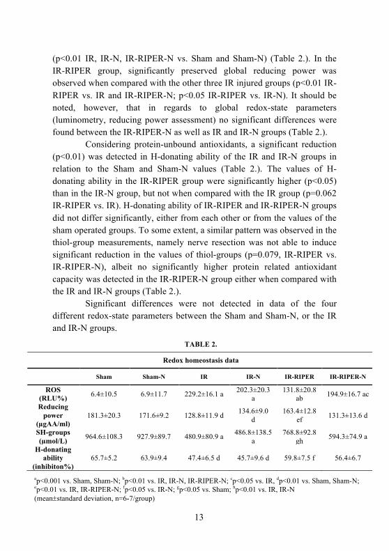
Chemiluminescent intensity - thus tissue free-radical content – was significantly elevated after 24 hours observation period in all IR injured groups when compared with the sham groups (p<0.001 IR, IR-N, IR-RIPER, IR-RIPER-N vs. Sham and Sham-N), nonetheless perconditioning treatment potently reduced the level of free-radicals in liver homogenate in relation to the three other IR injured groups (p<0.01 IR-RIPER vs. IR, IR-N, IR-RIPER-N) (Table 2.).
A more complex pattern was observed regarding the different parameters of tissue antioxidant capacity. Global reducing power of the IR, IR-N, and IR-RIPER-N groups was significantly reduced in comparison with the values assessed in the samples of the sham-operated animals
13
(p<0.01 IR, IR-N, IR-RIPER-N vs. Sham and Sham-N) (Table 2.). In the IR-RIPER group, significantly preserved global reducing power was observed when compared with the other three IR injured groups (p<0.01 IR-RIPER vs. IR and IR-RIPER-N; p<0.05 IR-RIPER vs. IR-N). It should be noted, however, that in regards to global redox-state parameters (luminometry, reducing power assessment) no significant differences were found between the IR-RIPER-N as well as IR and IR-N groups (Table 2.).
Considering protein-unbound antioxidants, a significant reduction (p<0.01) was detected in H-donating ability of the IR and IR-N groups in relation to the Sham and Sham-N values (Table 2.). The values of H-donating ability in the IR-RIPER group were significantly higher (p<0.05) than in the IR-N group, but not when compared with the IR group (p=0.062 IR-RIPER vs. IR). H-donating ability of IR-RIPER and IR-RIPER-N groups did not differ significantly, either from each other or from the values of the sham operated groups. To some extent, a similar pattern was observed in the thiol-group measurements, namely nerve resection was not able to induce significant reduction in the values of thiol-groups (p=0.079, IR-RIPER vs. IR-RIPER-N), albeit no significantly higher protein related antioxidant capacity was detected in the IR-RIPER-N group either when compared with the IR and IR-N groups (Table 2.). Significant differences were not detected in data of the four different redox-state parameters between the Sham and Sham-N, or the IR and IR-N groups.
TABLE 2.
Redox homeostasis data
Sham Sham-N IR IR-N IR-RIPER IR-RIPER-N
ROS (RLU%) 6.4±10.5 6.9±11.7 229.2±16.1 a 202.3±20.3
a 131.8±20.8
ab 194.9±16.7 ac
Reducing power
(µgAA/ml) 181.3±20.3 171.6±9.2 128.8±11.9 d 134.6±9.0
d 163.4±12.8
ef 131.3±13.6 d
SH-groups (µmol/L) 964.6±108.3 927.9±89.7 480.9±80.9 a 486.8±138.5
a 768.8±92.8
gh 594.3±74.9 a
H-donating ability
(inhibiton%) 65.7±5.2 63.9±9.4 47.4±6.5 d 45.7±9.6 d 59.8±7.5 f 56.4±6.7
ap<0.001 vs. Sham, Sham-N; bp<0.01 vs. IR, IR-N, IR-RIPER-N; cp<0.05 vs. IR, dp<0.01 vs. Sham, Sham-N; ep<0.01 vs. IR, IR-RIPER-N; fp<0.05 vs. IR-N; gp<0.05 vs. Sham; hp<0.01 vs. IR, IR-N (mean±standard deviation, n=6-7/group)

14
Conclusions
1. Sixty minutes of 70% partial liver ischemia in rats is a feasible experimental model to investigate the consequences and characteristics of liver IR injury and the effects of remote ischemic perconditioning. In this model of moderate liver ischemic-reperfusion injury, application of percoditioning resulted in significant alterations in several parameters.
2. a, After 60 minutes of partial liver ischemia remote ischemic perconditioning, applied in 4 cycles of 5 minutes of ischemia and 5 minutes of reperfusion, could potently mitigate liver injury, according to the post-reperfusion levels of serum transaminases and based on the semiquantitative histological scoring. b, Perconditioning positively influenced systemic hemodynamics (mean arterial pressure) and microcirculation (liver and lower limb) of the animals, manifested in significant differences between reperfusion macro- and microcirculation values of treated and non-treated animals. c, Significantly lower tissue free-radical levels and preserved antioxidant capacity were observed with the use of perconditioning after 6 and 24 hours of reperfusion. Parallel, conspicuous reduction was detected in serum values of the pro-inflammatory cytokine, TNF-a, during the early phase of reperfusion.
3. In the second study, left femoral artery perconditioning could also exert potent protection against the deleterious effects of liver exclusion and reperfusion. We could observe significant alteration between treated and non-treated groups in tissue damage (ALT, necrosis%), liver microcirculation and systemic hemodynamics as well as in liver tissue redox-homeostasis. Perconditioning induced hepatoprotection was abolished by denervation of the vascular bed, where conditioning was applied.
15
4. Automated histological image analysis is an accurate and useful tool for evaluation of the histological samples of a real experiment. Teaching of the algorithm can be achieved easily, analysis of the slides is fast and reliable.
Bibliography of the candidate`s publications
Articles providing the basis of the doctoral thesis:
1. Czigány Z, Turóczi Zs, Bulhardt O, Hegedüs V, Lotz G, Rakonczay Z, Balla Z, Harsányi L, Szijártó A. (2012) Távoli szervi kondicionálás: rövid távú hepatoprotektív hatások patkánymodellben. [Remote ischemic perconditioning: short term effects on rat liver ischemic-reperfusion injury.] Orvosi Hetilap [Hungarian Medical Journal], 153 (40): 1579-1587. 2. Czigány Z, Turóczi Zs, Onódy P, Harsányi L, Hegedüs V, Szijártó A. (2013) Remote ischemic perconditioning protects the liver from ischemia-reperfusion injury. Journal of Surgical Research, 185: 605-613. 3. Czigány Z, Turóczi Zs, Kleiner D, Lotz G, Homeyer A, Harsányi L, Szijártó A. (2015) Neural elements behind the hepatoprotection of remote perconditioning. Journal of Surgical Research, 193: 642-651.
Further articles:
1. Szijártó A, Czigány Z, Turóczi Zs, Harsányi L. (2012) Remote ischemic perconditioning – a simple, low risk method to decrease ischemic-reperfusion injury: Models, protocols, and the mechanistic background. Journal of Surgical Research, 178 (2): 797-806. 2. Turóczi Zs, Fülöp A, Czigány Z, Varga G, Rosero O, Tőkés T, Kaszaki J, Lotz G, Harsányi L, Szijártó A. (2015) Improvements of small intestinal microcirculation by postconditioning after lower limb ischemia. Microvascular Research, 98: 119-125. 3. Fülöp A, Budai A, Czigány Z, Lotz G, Dezső K, Paku S, Harsányi L, Szijártó A. (2015) Alterations in hepatic lobar function in regenerating rat liver. Journal of Surgical Research, 197 (2): 307-317.

16
4. Czigány Z, Iwasaki J, Yagi S, Nagai K, Szijártó A, Uemoto S, Tolba RH. (2015) Improving research practice in rat orthotopic and partial orthotopic liver transplantation: a review, recommendation and publication guide. Invited Review. European Surgical Research, 55: 119-138.
5. Lauber DT, Tihanyi DK, Czigány Z, Fülöp A, Budai A, Drozgyik D, Lotz
G, Harsányi L, Szijártó A. (2016) Liver regeneration after different degrees of portal vein ligation. Journal of Surgical Research, 203 (2): 451-458.