investigating cognitive neuroplasticity in single cases...
TRANSCRIPT
Neurocase (2002) Vol. 8, pp. 355–368
Investigating Cognitive Neuroplasticity in Single Cases:Lessons Learned from Applying Functional NeuroimagingTechniques to the Traditional Neuropsychological CaseStudy Framework
S. G. Romero, C. F. Manly and J. Grafman
Cognitive Neuroscience Section, National Institute of Neurological Disorders and Stroke, National Institutes of Health, Bethesda,Maryland, USA
Abstract
We summarize two case studies as a context for discussing the use of neuroimaging as a convergent methodology inthe study of neuroplasticity in single subjects. Throughout this paper we argue for a different approach for includingneuroimaging in these types of study. Previous case studies of neuroplasticity in patients (ours as well as othersreported elsewhere) have added neuroimaging to the traditional neuropsychological framework of comparing patientresults with matched control groups, and synthesized results through descriptions of anatomical and behavioraldissociations. This type of approach is referred to as the comparison approach. We advocate a different approach thatbuilds on findings from previous behavioral skill learning research. Specifically, we propose adding neuroimagingthroughout learning or recovery of the ability of interest and making inferences from systematic changes in activationtopography and intensity that occur within the context of predicted behavioral changes. We dub this approach theonline approach. This approach should allow future investigators to circumvent many of the interpretation pitfalls thatare common in comparison studies.
Introduction
Since the ground-breaking work with monkeys demonstratingadaptation to lesions in the central and peripheral nervoussystems (Merzenich et al., 1983), there has been a substantialgain in our understanding of the mechanisms underlyingneuroplasticity in humans (Chollett et al., 1991; Weiller et al.,1993; Grafman and Christen, 1999). Much of this work hasbeen enabled by modern functional neuroimaging techniques.Although functional neuroimaging studies offer someadvantages over the use of behavioral methods in traditionalneuropsychological studies (Humphreys and Price, 2001),there are important issues in design and interpretation thatmust be considered when using functional neuroimagingtechniques.
The advantages of neuroimaging include the convergentevidence it provides concerning which brain areas areinvolved in spared or recovered cognitive processing. It mayalso allow for the tracking of functional changes during therecovery of, or compensation for, damaged processes. Thereare, however, unique challenges in using functional neuro-imaging techniques in single case studies.
Correspondence to: J. Grafman, Cognitive Neuroscience Section, National Institute of Neurological Disorders and Stroke, National Institutes of Health,Building 10, Room 5c205, 10 Center Drive, MSC 1440, Bethesda, MD 20892-1440, USA. Tel: �1 301 496 0220; Fax: �1 301 480 2909;e-mail: [email protected]
This paper will address these issues and offer somesuggestions for optimizing functional neuroimaging investi-gations of neuroplasticity in single cases. First, we outlineresults from two of our recent case studies. Then, usingexamples from our experience, we discuss in detail theissues that should be considered when using functionalneuroimaging. Throughout this discussion we will developand present an optimal approach for combining functionalneuroimaging with behavioral measures in case studies.
The standard practice in neuropsychological studies(without functional neuroimaging) is to assess behavioralperformance and anatomy separately and later synthesizethese results through structural descriptions of spared ordamaged tissue, comparisons with control groups, andbehavioral dissociations. For example, an earlier study ofpatient GK (who is presented below) assessed multiplelanguage functions following a massive left hemispherestroke and interpreted the observed partial abilities in termsof spared right hemisphere processing (Rapcsak et al., 1991).Note that this is a reasonable approach in the absence offunctional neuroimaging data.
356 S. G. Romero, C. F. Manly and J. Grafman
Functional neuroimaging techniques, however, wouldallow Rapcsak et al.’s interpretation to be empirically testedprovided standard assumptions are met regarding inferencesfrom functional neuroimaging results. These assumptionsinclude the fact that most techniques measure blood flow asa correlate of neuronal firing, and that the authors use propertask design and statistical analysis. Although these issues areimportant to any functional neuroimaging study, they areperipheral to the present topic. Thus, they will not bediscussed further here except to mention that they alsomust be considered when designing functional neuroimagingstudies and making inferences regarding the resulting patternsof activation.
There are two ways to incorporate functional neuroimaginginto case studies, especially in studies of neuroplasticity. Thefirst is to add it to the traditional case study framework. Inthis case, comparisons are made with control participants,and activation differences in the single case are interpretedthrough comparison to a specific group of controls or toprevious neuroimaging studies with unimpaired participants.We refer to this approach as the comparison approach.
The other approach, which we will discuss more fully, werefer to as the online approach. Although the online approachis not always feasible, we will argue that when possible, itis the preferred approach for neuroplasticity studies of singlecases. In the online approach, neuroplasticity is assessedduring learning or practice of the ability in question. In casestudies, this means that progress is assessed across therelearning, recovery, or reacquisition of a damaged or defi-cient skill or task, both at the behavioral and neuronal levels.We will further argue that the online approach circumventsmany of the inferential ambiguities of a comparison design.It also capitalizes on the extensive amount of behavioralresearch on skill learning and the general principles thatoperate across different tasks and skills.
Basic behavioral principles of skill acquisition
The online design assumes that any relearning that occursafter brain injury is the product of general behavioral andneural principles that also apply to learning in unimpairedparticipants. In this section we will focus our comments ona few key findings from the past 20 years of behavioralstudies delineating the mechanisms of skill acquisition, andoutline the main theories of skill learning which are basedon these findings. In the next section we focus on the typesof neuroplasticity that have been observed from studies oflearning and recovery of function.
There are several prevalent findings in the skill acquisitionliterature. First, response times decrease with practice acrossa wide range of tasks, from cigar rolling to memory retrieval(Crossman, 1959). Regardless of the task, this speed-up canbe well characterized by a general class of mathematicalfunctions called power functions with a single fitted parameter[see Newell and Rosenbloom (1981) for an extensive discus-sion of the power law].
The second finding is that most acquired skills show goodlong-term retention (Proctor and Dutta, 1995). Usually thisretention is demonstrated by some measure of savings (e.g.response time speed-up) that indicates that the originallearning and the relearning during a retention test do notoccur at the same rate. After training it is rare for there notto be some evidence of savings from training over the initiallearning session.
Another finding that is well documented in the skillliterature is that different forms of training can producedrastically different outcomes regarding the acquisition andretention of skills. In general, it has been found that factorswhich promote faster acquisition can sometimes interferewith long-term retention. Similarly, factors which optimizelong-term retention of a skill do not expedite initial learningof a skill [see Schmidt and Bjork (1992) and Healy and Bourne(1995) for extensive discussions concerning these factors].
One final finding worth mentioning concerns the specificityof training that can be expected. In general, practice-relatedlearning has been shown to be very specific to the items andtasks that are trained (Healy and Bourne, 1995). Transfer oftraining is rare and only occurs in very specific cases (Proctorand Dutta, 1995). These findings will be of great importancewhen designing online studies for the recovery of function.
Following early work by Bryan and Harter (1899) andlater work by Fitts (1964), many of the modern theories of skillacquisition explain speed-up in processing as a progressionthrough different learning stages. They differ, however, inwhat stages they propose, and we present three main theoriesto illustrate the breadth of theoretical positions which havebeen adopted. It should also be noted here that none of theserepresents a comprehensive theory of skill acquisition norhave all the factors and mechanisms involved in skill acquisi-tion been fully worked out. Instead these theories should beviewed as a guide for designing online patient studies.
The ACT theory (Anderson, 1983, 1992) proposes atransition from a declarative form of knowledge to a proced-ural form. Declarative knowledge is ‘what’ knowledge;procedural knowledge is ‘how to’ knowledge. In thedeclarative stage, knowledge consists of facts that must berehearsed, including situational characteristics and instruc-tions. During practice, the learner develops task-specificprocedures which do not require the active maintenance ofdeclarative knowledge. Performance continues to improve asthese procedures are refined or tuned to the practiced context.The ACT theory assumes that performance is subsumedby a single strategy or type of processing that becomesmore refined.
In contrast, Logan (1988) and Rickard (1997) havesuggested that sometimes acquisition involves a strategicswitch from one type of processing to another. These theoriessuggest that initially, a multi-step algorithm is used to performa task. With practice, direct links to memory are developedand eventually expert performance relies on memory retrieval.Logan assumes that both processes begin in parallel and thefaster one produces the response, whereas Rickard assumes
Neuroimaging of single cases 357
that the two strategies must be processed sequentially.Because initially there is no memory representation for thetask, the longer algorithm determines performance. As thetask becomes more practiced and memory traces get stronger,performance will rely mostly on the faster memory retrievalprocess. Logan also assumes that each trial results in aseparate or independent record in memory. Rickard assumesthat a single representation or record is built up in memoryfor each item and is appended during practice.
Taken together, these results from behavioral studies ofskill learning suggest that several considerations are necessarywhen designing online neuroimaging studies with singlecases. First, the skill and tasks in question must be analyzedto assess to what extent strategy shifts can be expected, and,second, to what extent strategies may be processed in parallel.Third, the design of a proposed training regimen should beconsistent with the goals and predicted outcomes of the study.Recovered functioning would be of only limited value if itwas not long lasting. We now consider previous functionalneuroimaging investigations of neuroplasticity.
Neuroimaging studies of neuroplasticity
In general, there are four forms of neuroplasticity thathave been observed. These four forms are referred to ashomologous area adaptation, cross-modal reassignment, mapexpansion and compensatory masquerade (Grafman andLitvan, 1999). Homologous area adaptation refers to theemergence of a new cognitive process in a homologousregion in the opposite hemisphere. Cross-modal reassignmentrefers to the reassignment of areas usually receiving inputfrom one sensory modality to process information fromanother sensory modality. Map expansion refers to the growthof a functional area either with training or following theloss of input to an adjacent region. Finally, compensatorymasquerade refers to the phenomenon by which a cognitiveprocess appears to be normal or recovered due to a shift inprocessing to an atypical strategy.
Two types of previous neuroimaging study have providedsome of the evidence for these forms of neuroplasticity.One type has focused on skill learning using unimpairedvolunteers, and most of these have studied visuomotor skilllearning. The second has focused on the recovery of functionin patients with developmental or acquired lesions. In general,studies of skill learning demonstrate that training or practiceleads to map expansion (Elbert et al., 1995; Schlaug et al.,1995), with an immediate effect on cortical activation that isspecific to the trained items (Karni et al., 1995). Thenas participants become expert, fewer resources are needed(Pascual-Leone et al., 1995) and experts exhibit more compactactivation than novices (Krings et al., 2000).
Investigations of functional recovery have shown thatdeafferented cortical areas are recruited for cross-modalreassignment (Sadato et al., 1996; Bounomano andMerzenich, 1998). Conversely, it has also been shown thatfor a particular function (e.g. language processing), atypical,
auxiliary cortical areas are recruited when there is a lesionto a typical processing area (Chollet et al., 1991; Weilleret al., 1993; Nudo and Miliken, 1996). It is unclear whatrole these auxiliary areas play in the recovery of function.These areas might be involved in a compensatory masqueradeor they could be involved in a simple homologous areaadaptation (a new cortical area supporting the same cognitivestrategy).
In language, for example, it is generally believed thatunimpaired (right-handed) participants only use left hemi-sphere areas for certain forms of language processing [butsee Binder (1997) and Weiller et al. (1997) for conflictingneuroimaging results]. However, patients with left hemispherelesions in language areas seem to activate homologous righthemisphere areas to an extent that is correlated with damagein the left hemisphere (Karbe et al., 1998). A couple ofneuroimaging studies have also demonstrated perilesionalleft hemisphere activations (Heiss et al., 1993; Price et al.,1995). What is not clear is how novel these supplementalareas of activation are in the patients. In other words,is the right hemisphere processing simply reactivation ofsymmetrical representations inhibited in unimpaired subjectsduring lateralization, or are these areas recruited and usedby patients alone as a function of a newly emerging processingin the right hemisphere (Weiller, 1998)? Notably, thesedifferences could just be due to cognitive strategy differencesbetween impaired and unimpaired groups (i.e. compensatorymasquerade). Similarly, it is not clear to what extent activationin perilesional areas reflects processing that is contributingto performance. Perilesional activations could just indicateattempted processing in partially functioning cortical areasthat are no longer necessary or sufficient for the performanceof a task. These uncertainties are directly related to the useof the comparison approach in these studies.
Two cases
The following cases are discussed with an emphasis on whatguided our interpretation of the results. Our summaries ofthese cases are intended to provide a basis for the theoreticaldiscussion of planning future case studies involving func-tional neuroimaging and neuroplasticity. Separate reports ofthe specific studies have been submitted independently forpublication (Basso et al., submitted; Romero et al., sub-mitted).
Case GK
GK, a 65-year-old male, was right handed prior to his strokes.He suffered two strokes, a massive left hemisphere stroke atthe age of 45 years and a smaller ischemic lesion in the rightfrontal operculum at the age of 61 years. GK receivedextensive language training after the first stroke and pro-gressed from global aphasia to resembling a Broca’s typeaphasic. His second stroke left his language abilities stable(Rapcsak et al., 1991). GK has a right visual field hemianopia

358 S. G. Romero, C. F. Manly and J. Grafman
Table 1. Summary of GK’s results on the Psycholinguistic Assessment ofLanguage Processing in Aphasia (PALPA) and the Boston Naming Test
PALPA items19 Letter identification 100%21 Letter discrimination 100%23 Spoken–written letter matching 24/26 (92%)24 Visual lexical decision with ‘illegal’ pseudowords 60/60 (100%)25 Lexical decision imageability and frequency 112/120 (93%)26 Visual lexical decision and morphology 45/60 (75%)27 Visual lexical decision and spelling–sound regularity 59/60 (98%)29 Letter length reading 24/24 (100%)30 Syllable length reading 16/18 (89%)31 Word reading
Low imageability and low frequency 15/20 (75%)Low imageability and high frequency 17/20 (85%)High imageability and low frequency 19/20 (95%)High imageability and high frequency 19/20 (95%)
33 Grammatical class reading 25/40 (62.5%)36 Pseudoword reading 0/24 (0%)47 Spoken word–picture matching 39/40 (97%)48 Written word–picture matching 38/40 (95%)51 Word semantic association
Low imageability 8/15 (53%)High imageability 11/15 (73%)
52 Spoken word–written word matching 23/30 (77%)
Boston Naming TestTotal correct 33Self correct 3No response 9Total errors 18
Semantic errors 15Neologisms/jargon 0Phonological errors 3
and right-sided hemiplegia. Table 1 presents the resultsof our assessment of GK’s language abilities with thePsycholinguistic Assessment of Language Processing inAphasia (PALPA; Kay et al., 1992) and the Boston NamingTest (Kaplan et al., 1978). GK was completely unable toread non-words and had trouble reading real words thatwere low frequency and low imageability. Overall GK’sperformance was consistent with a classification of deepdyslexia.
As alluded to above, we wanted to test the hypothesisthat GK’s recovered language abilities were the product ofpurely right hemisphere processing. Thus, we studied GK’srecovered language abilities using a comparison approach.
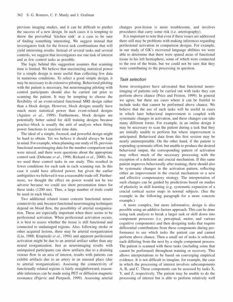
We initially assessed the extent of GK’s structural braindamage with a high-resolution (1.5 mm/slice) magneticresonance imaging (MRI) scan. Although the damage toGK’s left hemisphere was extensive, there were remainingislands of tissue with normal signal intensity in the lefthemisphere. These areas were present in the perirolandic andsensorimotor cortex and also in the basal frontal lobe, uncusand globus pallidus. A portion of the anterior limb of theleft internal capsule also showed comparable signal intensityto the internal capsule in the right and seemed to connectthe thalamus with the spared left perirolandic region. A high-resolution T1-weighted structural MRI of GK’s brain isdisplayed in Fig. 1. We assessed GK’s metabolic activitywith a resting (i.e. no sensory stimulation) positron emissiontomography (PET) scan. In addition to the majority of the
Fig. 1. Orthogonal view using neurological orientation (i.e. left is left) ofGK’s magnetic resonance image, with two axial slices. Relative to the centerof the volume, the slices are 6.6 mm to the right and 5.6 mm anterior. Theleft axial slice is 20.2 mm inferior and the right axial slice is 3.0 mm inferior.The lesion encompasses the majority of the left hemisphere, although thereis some spared tissue in the basal frontal lobe, uncus, globus pallidus, and aperirolandic region connected to the thalamus by a spared portion of theanterior limb of the left internal capsule.
right hemisphere, this study demonstrated normal metabolicactivity in the spared left basal frontal and perirolandicregions.
In the functional MRI study of GK’s language abilities weused three tasks, single word reading (either words orpseudowords), lexical decision, and a visual detection task.Data were collected from GK and three age-, gender-, andeducation-matched controls. We used block design functionalMRI in which the lexical decision and word reading taskswere compared with the visual detection task in separateruns. Runs with the reading task used words or pseudowordsbut not both in the same run. In each run, the reading taskwas alternated with the visual detection task, such that therewere three blocks of each task per run. We point out for laterdiscussion that GK could not read any pseudowords in ourpilot testing.
Discriminative responses during scanning sessions werenot possible because verbal responses produce movementartifacts and GK could not press a button with his right handand the use of a button box (in the left hand) was also notpossible. Instead, GK and the controls were instructed to dothe tasks in their heads (i.e. read words, decide if a real wordwas presented or detect the visual stimulus), and then pressthe button in their left hand when they were done in eachtrial. This enabled us to collect response time data. We alsocollected data on the tasks outside the scanner, using verbalresponses and the same response windows as during thescanning session so we could assess accuracy.
Neuroimaging of single cases 359
A custom gradient echo-planar imaging sequence (field ofview � 24, acquisition matrix � 64 � 64, repetition time �3000 ms, echo time � 40 ms, flip angle � 90°) was used tocollect the functional volumes. Each volume consisted of 18axial slices, 6 mm thick. Four dummy volumes were acquiredat the beginning of each new run in order to reach a steady-state magnetization. A structural T1-weighted brain MRI wasalso collected for each subject and was used to overlay thestatistical activation maps for anatomical identification.
The analysis was performed with SPM96 (WellcomeDepartment of Cognitive Neurology, London, UK). Theimages were realigned to the first volume of the first run.The controls’ images were normalized into the referencesystem of Talairach and Tournoux (1988). Normalization ofGK’s images into this standard space could not be performeddue to the size of the lesion involved and the presence ofperilesional tissue. Images collected during the two runs thatincluded each type of reading task (either word or pseudowordreading) were analyzed separately. All images collectedduring the two runs in which the lexical decision taskwas alternated with the detection task were also analyzedseparately. All activation differences are reported relative tothe detection task. We applied ANCOVA global normalizationto remove global signal differences among runs. For thecontrol group analysis, we combined the data for all threecontrols. Contrasts were specified as subtractions. Becausewe expected a priori to find left inferior frontal gyrus andleft inferior temporal gyrus activation in the control group, wetested this hypothesis by thresholding the resulting statisticalmaps with a Z-value � 3.09 (P � 0.001). Cluster analysiswas performed only for GK with a test both for spatial extentand Z level with a threshold of P � 0.05.
In the scanner, the controls were slower at reading wordsor making lexical decisions than in the visual detection task.The controls were also slower at reading pseudowords thanwords. Additionally, the controls’ accuracy was above 95%for all tasks (measured outside the scanner). In the scanner,GK was slower for word reading than for lexical decision,but was as fast in the lexical decision task as in the visualdetection task. Outside the scanner, GK was unable to readany pseudowords but was 93% accurate in reading wordsand 95% accurate in making lexical decisions.
Activation was found in the left inferior temporal gyruswhen the controls were reading words relative to the detectiontask and there was activity near Broca’s region when thecontrols read pseudowords relative to the detection task. Forthe lexical decision task (compared with the detection task),the controls exhibited activation in the bilateral inferiorfrontal gyrus, and in the left orbitofrontal and occipitalregions as well as the right cerebellum. No reliable activationwas detected when GK was reading pseudowords relativeto the detection task, but reliable activation was found inthe right hemisphere for (real) word reading relative to thedetection task. Specifically, activation was found in the rightmiddle temporal gyrus, motor and pre-motor areas, andright cerebellum. Additional activation for GK (during word
reading relative to the detection task) was also found inresidual tissue in the left inferior frontal lobe. For lexicaldecision, GK exhibited significant activation in the rightinferior frontal gyrus. In concordance with other neuro-imaging studies (e.g. Binder, 1997), the matched controlsexhibited reliable activation mainly in the left inferior tem-poral gyrus for reading real words in relation to the controltask and also near Broca’s area in the left hemisphere whenreading only pseudowords in relation to the control task. Forlexical decision (relative to the control task), the controlsexhibited activation in bilateral inferior frontal gyri. Directcomparisons between GK and the controls were not possiblebecause GK’s images could not be normalized. These differ-ences in patterns of activation suggest that GK’s recoveredlanguage abilities are due to plastic neuronal changes in theright hemisphere.
Our study of GK was more informative than traditionalneuropsychological case studies because of the addition offunctional neuroimaging data. GK’s results generally supportthe hypothesis that his recovered language abilities are theproduct of right hemisphere processing enabled either throughhomologous area adaptation or compensatory masquerade.We cannot, however, differentiate between the homologousarea adaptation and compensatory masquerade hypothesesbecause we cannot be definitive regarding GK’s cognitivestrategies in these tasks. Similarly, we cannot rule out thepossibility of perilesional contributions, especially involvingthe basal frontal and perirolandic regions. These twodifficulties are directly related to the comparison approachused for this study. It is important to note that even ifactivation changes are due to a strategy shift, this may be atypical compensatory change in the recovery of cognitivefunction. Just as a strategy shift might be typical in theacquisition of a cognitive skill for unimpaired participants,patients may need to relearn skills by starting again with aresource-intensive strategy and then switch to a more efficientstrategy. Conversely, patients may just depend on a resource-intensive strategy that may or may not have been used duringthe original learning of the skill in question. We will discussthis issue in more depth below.
Case JS
In our study of this patient we were interested in investigatingthe possible cognitive and neural etiology of developmentaldyscalculia [see Romero et al. (submitted) for a full reporton this study]. At initial presentation, JS was an 18-year-oldright-handed male with no neurological history, except adiagnosis of developmental dyscalculia in elementary school.During his initial evaluation, JS demonstrated a normalfull-scale IQ with substantial scatter: above average scoreson all of the non-numerical subtests and very poor perform-ance on the arithmetic and coding subtests. On the Key MathDiagnostic Arithmetic Test (Connolly et al., 1976), JS scoredat the 79th percentile for understanding basic arithmeticconcepts, at the 42nd percentile for using numbers in applied

360 S. G. Romero, C. F. Manly and J. Grafman
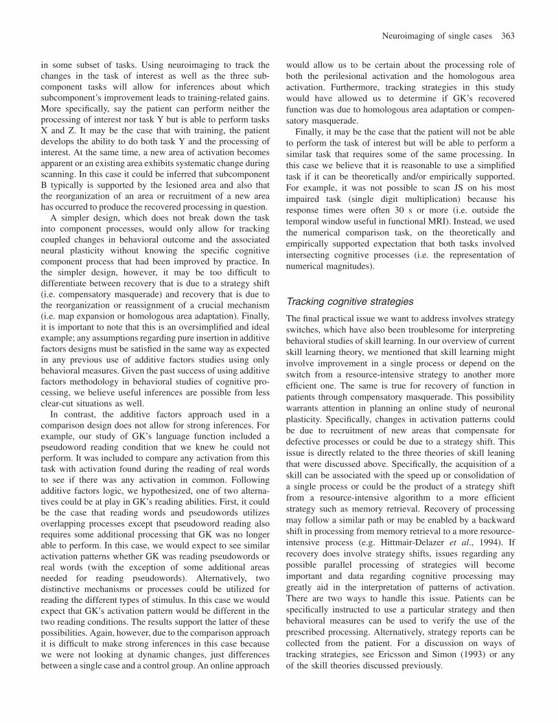
Fig. 2. Top-left presents a T1-weighted sagittal scout image of JS’s anatomical magnetic resonance image. The white line indicates the slice used for the color-scale spectroscopy data in the lower panel. The white boxes in the top-right panel indicate the placement of the regions of interest in the posterior temporoparietallobes for analysis of the spectroscopic data. The bottom panel presents color maps for the levels of N-acetyl-aspartate (NAA), choline (Cho) and creatine (Cre).Regions of low metabolite signal intensity are represented in green and blue. Regions of high metabolite signal intensity are represented in yellow and red. Thearrows indicate areas of decreased Cho and Cre in the left posterior temporoparietal lobe. There was also a mild (non-significant) focal decrease in NAA.
settings (e.g. making change), but lower than the firstpercentile for computational ability (i.e. addition, subtraction,multiplication and division). In contrast, JS scored above the95th percentile on the Raven Progressive Matrices test ofspatial reasoning (Raven, 1958). Further pilot testing revealedthat JS had great difficulty in single digit multiplication tasks:verification (e.g. 4 � 7 � 21 true or false?) and production(e.g. 4 � 7 � ?). JS needed to use a paper and pencil forthe majority of the problems in both tasks. Thus, his meanresponse times ranged from 7.1 to 11.3 s and 6.9 to 12.7 sfor verification and production, respectively. JS was 87%accurate for verification and 73% accurate in the productiontask. JS did perform relatively normally in a pilot numericalcomparison task (e.g. select the larger number: 54, 59). Inthis task, JS exhibited the numerical distance effect inresponse times, such that he was reliably faster in choosingthe larger of the two numbers when the numerical distancebetween them was larger, and slower when the two presentednumbers were numerically closer together.
Based on a previous study of patients with developmentaldyslexia (Rae et al., 1998), we first neurologically assessedJS with structural MRI and magnetic resonance spectroscopy(MRS). Although the structural MRI was independently readby two experienced neuroradiologists as normal, the MRSscans demonstrated a focal deficiency in the left inferior
parietal lobe (see Fig. 2) of three metabolites that arecorrelated with neuronal density (Levy et al., 1999). Thisfinding is entirely consistent with earlier lesion studiesand with functional neuroimaging studies with unimpairedparticipants that implicate the left parietal lobe in numericalprocessing (Grafman et al., 1982; Grafman and Rickard,1996).
The MRS finding suggested two possible explanations forJS’s numerical difficulties, one involving plasticity. Theycould be due to: (1) a typical network for number processingthat is missing a particular mechanism in the inferior parietalareas; or (2) the migration of these processes to atypicalcortical areas or the recruitment of new cortical areas follow-ing damage to the typical processing area (i.e. homologousarea adaptation or map expansion). Thus, we wanted to usefunctional MRI to assess the location of numerical processingin JS’s brain. We chose to use the numerical comparison taskfor this part of the study. Based on previous research andtheoretical models regarding number processing, we expectedthat the brain areas used for the numerical comparison taskwould also be used for single digit arithmetic (McCloskeyet al., 1985; Viscuso et al., 1989; McCloskey andLindemann, 1992).
We used a comparison approach for this experiment bycollecting data from JS and five age- and education-matched
Neuroimaging of single cases 361
control participants. Brain activation associated with numberprocessing was assessed in all participants by contrastingbrain activation on the number comparison task with thatelicited by three different control tasks in a block designfunctional MRI study. We used three control tasks becauseprevious functional neuroimaging studies of the task hadyielded variable findings that may have been attributable tothe different control tasks used between the studies (Dehaeneet al., 1996; Chochon et al., 1999; Rickard et al., 2000). Inone control task, font comparison, the participants werepresented with the same numbers as those used in the numbercomparison task and the task was just to decide if the twonumbers were presented in the same font (e.g. 53 54). Inthe second control task, the detect ones task, the participantswere presented with a string of four numbers and had toindicate if the string contained the number 1 (e.g. 7813). Inthe final control task, the participants were presented withtwo pairs of symbols (e.g. ❉✴ ❄✵) and had to indicate ifthe two pairs were the same (i.e. the same symbols in thesame order). These three control tasks were alternated twiceper functional run in a pseudo-randomized manner. The samefunctional and structural scanning sequences as those usedfor GK were used in this study.
All data pre-processing and statistical analysis was per-formed using SPM96. Each participant’s data were pre-processed as follows. First, all the functional images fromevery run were realigned to the first image acquired duringthe first run. Then, the high-resolution anatomical image foreach participant was spatially normalized to the Talairachatlas (Talairach and Tournoux, 1988) using only affinetransformations, and these normalization parameters wereapplied to all of the realigned functional images. Spatialsmoothing was applied to the realigned, normalized functionalimages with a three- dimensional, 10 mm full width at halfmaximum Gaussian kernel.
The statistical analysis was first performed separately foreach individual and then again on the group data for thecontrols. For each analysis, temporal smoothing and meanmagnetic resonance signal normalization (ANCOVA) wereapplied. For the group analysis we used a voxelwise signi-ficance threshold of 0.001 corrected for multiple comparisonsto P � 0.05 with the standard SPM96 spatial extent correction.For the single subject analyses a voxelwise threshold of0.005 was corrected for multiple comparisons to P � 0.1(spatial extent correction) due to the lower degrees of freedomavailable in these analyses. Contrasts were defined as simplesubtractions comparing the number comparison task witheach of the three control conditions.
In the group analysis of the controls there were two mainareas of activation: (1) a bilateral parietal region includingthe left superior parietal lobe, left precuneus and rightprecuneus extending from Talairach z � 48 to Talairachz � 72; and (2) an area in the left medial frontal gyrus.Similar to these findings, the single subject analysis for JSresulted in significant activation in the left superior parietallobe extending from z � 60 to z � 76 as well as activation
in the right paracentral lobule directly bordering on theprecuneus extending from z � 48 to z � 76. The comparisonof the peak activation results between the control participantsand JS suggests that the number comparison process for bothis located in the same parietal location. However, if wecompare the spread of activation, JS did not exhibit activationin the more inferior areas of the parietal lobe but the controlsdid. Furthermore, JS exhibited activation in the more superiorareas of the parietal lobe but the controls did not. Althoughsuch a subtle shift cannot be quantified statistically, thesedescriptive results suggest a possible shift in JS’s numbercomparison processing to more superior areas away from theareas that the MRS scan indicated as being deficient. Thisevidence supports the inference that map expansion hasoccurred in JS.
Discussion
The two cases discussed above have shed light on the issuesinvolved in adding functional neuroimaging as a convergentmethod in the study of neuroplasticity in single cases. In thepast, neuropsychological case studies have only includedbehavioral assessments and, when possible, structuralimaging. Recently, however, it has been possible to addfunctional neuroimaging to these studies, but this techniquehas been utilized within the traditional framework of com-paring patients with controls. These studies can be moreinformative than studies including only behavioral methods,but we believe that there is a better way to incorporatefunctional neuroimaging into studies of recovery of function.Due to convergent neuroimaging and behavioral findings,our studies of GK and JS were slightly more informativethan they would have been had we used only behavioralmethods. As we have pointed out, however, these studies arenot without some ambiguity in the interpretation of thefunctional neuroimaging results that are directly tied to theuse of the traditional comparison approach. Specifically, GK’srecovered language processing could be the product of eitherhomologous area adaptation or compensatory masquerade,or some combination of the two that might also includeprocessing by perilesional areas. Similarly, it is difficult toquantify a spatial shift of processing in JS with the comparisonapproach. We will now turn our attention to discussing themethodological issues involved in using functional neuro-imaging in patient studies based on our particular experiencewith these two patients as well as reports in the literature,striving along the way to point out many of the advantagesto using an online approach for future studies.
Practical issues to be considered
Statistical power and task selection should be the firstconsiderations in any neuroimaging design with a singlecase. There are a couple of reasons to consider them. Becausethe field of human brain mapping is relatively new, thecognitive process of interest may not have been targeted in
362 S. G. Romero, C. F. Manly and J. Grafman
previous imaging studies, and it can be difficult to predictthe success of a new design. In such cases it is tempting tothrow the proverbial ‘kitchen sink’ at a case to be sureof finding something interesting. We suggest instead thatinvestigators look for the fewest task combinations that willyield interesting results. Instead of several tasks and severalcontrols, we suggest that investigators use one task of interestand as few control tasks as possible.
The logic behind this suggestion assumes that scanningtime is limited. We believe that maximizing statistical powerfor a simple design is more useful than collecting few datain numerous conditions. To select a good simple design, itmay be necessary to do extensive piloting. Behavioral pilotingwith the patient is necessary, but neuroimaging piloting withcontrol participants should also be carried out prior toscanning the patient. It may be tempting to choose theflexibility of an event-related functional MRI design ratherthan a block design. However, block designs usually havemuch more statistical power than event-related designs(Aguirre et al., 1999). Furthermore, block designs arepotentially better suited for skill training designs becausepractice–block is usually the unit of analysis when fittingpower functions to reaction time data.
The ideal of a simple, focused, and powerful design mightbe hard to obtain. Yet we believe it should always be keptin mind. For example, when planning our study of JS, previousfunctional neuroimaging data for the number comparison taskwere mixed, and there were questions about the appropriatecontrol task (Deheane et al., 1996; Rickard et al., 2000). So,we used three control tasks in our study. This resulted infewer conditions for each task in each scanning run. In thiscase it could have affected power, but given the earlierambiguities we believed it was a reasonable trade-off. Further-more, we thought the effect on power would not be tooadverse because we could use short presentation times forthese tasks (1200 ms). Thus, a large number of trials couldbe used in each block.
Two additional related issues concern functional neuro-connectivity and, because functional neuroimaging techniquesdepend on blood flow, the possibility of arterial reorganiza-tion. These are especially important when there seems to beperilesional activation. When perilesional activation occurs,it is best to assess whether the tissue is still functional andconnected to undamaged regions. Also, following stroke orother acquired lesions, there may be arterial reorganization(Liu, 1988; Krupinski et al., 1994) and apparent perilesionalactivation might be due to an arterial artifact rather than anyneural reorganization. Just as neuroimaging results withunimpaired participants can show artifacts due to a drainingvenous flow in an area of interest, results with patients canexhibit artifacts due to an artery in an unusual place (dueto arterial reorganization). Assessing the connectivity offunctionally related regions is fairly straightforward; reason-able inferences can be made using PET or diffusion magneticresonance (Pajevic and Pierpaoli, 1999). Assessing arterial
changes post-lesion is more troublesome, and involvesprocedures that carry some risk (i.e. arteriography).
It is important to note that even if these issues are addressedthere still may be problems with making inferences regardingperilesional activation in comparison designs. For example,in our study of GK’s recovered language abilities we wereable to determine that there were spared areas of functionaltissue in his left hemisphere, some of which were connectedto the rest of the brain, but we could not be sure that theywere contributing to the processing in question.
Task selection
Some investigators have advocated that functional neuro-imaging of patients only be carried out with tasks they canperform above chance (Price and Friston, 1999). In generalwe agree, but there are cases where it can be fruitful toinclude tasks that cannot be performed above chance. Webelieve that the use of such tasks will be useful in casesin which later behavioral improvement is coupled withsystematic changes in activation, and these changes can takemany different forms. For example, in an online design itmay be necessary to scan the patient during a task that theyare initially unable to perform but where improvement isanticipated. Behavioral data from this first session may benearly uninterpretable. On the other hand, if the patient isexpending systematic effort, but unable to produce the desiredbehavioral output, the corresponding pattern of activationmay reflect much of the necessary processing with theexception of a deficient and crucial mechanism. If this samepatient improves behaviorally after training, there should alsobe systematic changes in the activation pattern, reflectingeither an improvement in the crucial mechanism or a newand effective compensatory strategy. The interpretation ofsuch changes can be guided by predictions based on studiesof plasticity in skill learning (e.g. systematic expansion of acrucial cortical sector map) in normal subjects. (See theexample in the following paragraph for a more concreteexample.)
A more complex, but more informative, design is alsopossible using an additive factors approach. This can be doneusing task analysis to break a larger task or skill down intocomponent processes (i.e. perceptual, motor, and variouscognitive components) and then designing tasks that requiredifferential contributions from these components during per-formance to see which tasks the patient can and cannotperform above chance. Then a small set of tasks is selected,each differing from the next by a single component process.The patient is scanned with these tasks (including some thatcannot be performed) throughout training or recovery. Thisallows interpretations to be based on converging empiricalevidence. It is not difficult to imagine, for example, the casein which the processing of interest involves subcomponentsA, B, and C. These components can be assessed by tasks X,Y, and Z, respectively. The patient may be unable to do theprocessing of interest but is able to perform relatively well
Neuroimaging of single cases 363
in some subset of tasks. Using neuroimaging to track thechanges in the task of interest as well as the three sub-component tasks will allow for inferences about whichsubcomponent’s improvement leads to training-related gains.More specifically, say the patient can perform neither theprocessing of interest nor task Y but is able to perform tasksX and Z. It may be the case that with training, the patientdevelops the ability to do both task Y and the processing ofinterest. At the same time, a new area of activation becomesapparent or an existing area exhibits systematic change duringscanning. In this case it could be inferred that subcomponentB typically is supported by the lesioned area and also thatthe reorganization of an area or recruitment of a new areahas occurred to produce the recovered processing in question.
A simpler design, which does not break down the taskinto component processes, would only allow for trackingcoupled changes in behavioral outcome and the associatedneural plasticity without knowing the specific cognitivecomponent process that had been improved by practice. Inthe simpler design, however, it may be too difficult todifferentiate between recovery that is due to a strategy shift(i.e. compensatory masquerade) and recovery that is due tothe reorganization or reassignment of a crucial mechanism(i.e. map expansion or homologous area adaptation). Finally,it is important to note that this is an oversimplified and idealexample; any assumptions regarding pure insertion in additivefactors designs must be satisfied in the same way as expectedin any previous use of additive factors studies using onlybehavioral measures. Given the past success of using additivefactors methodology in behavioral studies of cognitive pro-cessing, we believe useful inferences are possible from lessclear-cut situations as well.
In contrast, the additive factors approach used in acomparison design does not allow for strong inferences. Forexample, our study of GK’s language function included apseudoword reading condition that we knew he could notperform. It was included to compare any activation from thistask with activation found during the reading of real wordsto see if there was any activation in common. Followingadditive factors logic, we hypothesized, one of two alterna-tives could be at play in GK’s reading abilities. First, it couldbe the case that reading words and pseudowords utilizesoverlapping processes except that pseudoword reading alsorequires some additional processing that GK was no longerable to perform. In this case, we would expect to see similaractivation patterns whether GK was reading pseudowords orreal words (with the exception of some additional areasneeded for reading pseudowords). Alternatively, twodistinctive mechanisms or processes could be utilized forreading the different types of stimulus. In this case we wouldexpect that GK’s activation pattern would be different in thetwo reading conditions. The results support the latter of thesepossibilities. Again, however, due to the comparison approachit is difficult to make strong inferences in this case becausewe were not looking at dynamic changes, just differencesbetween a single case and a control group. An online approach
would allow us to be certain about the processing role ofboth the perilesional activation and the homologous areaactivation. Furthermore, tracking strategies in this studywould have allowed us to determine if GK’s recoveredfunction was due to homologous area adaptation or compen-satory masquerade.
Finally, it may be the case that the patient will not be ableto perform the task of interest but will be able to perform asimilar task that requires some of the same processing. Inthis case we believe that it is reasonable to use a simplifiedtask if it can be theoretically and/or empirically supported.For example, it was not possible to scan JS on his mostimpaired task (single digit multiplication) because hisresponse times were often 30 s or more (i.e. outside thetemporal window useful in functional MRI). Instead, we usedthe numerical comparison task, on the theoretically andempirically supported expectation that both tasks involvedintersecting cognitive processes (i.e. the representation ofnumerical magnitudes).
Tracking cognitive strategies
The final practical issue we want to address involves strategyswitches, which have also been troublesome for interpretingbehavioral studies of skill learning. In our overview of currentskill learning theory, we mentioned that skill learning mightinvolve improvement in a single process or depend on theswitch from a resource-intensive strategy to another moreefficient one. The same is true for recovery of function inpatients through compensatory masquerade. This possibilitywarrants attention in planning an online study of neuronalplasticity. Specifically, changes in activation patterns couldbe due to recruitment of new areas that compensate fordefective processes or could be due to a strategy shift. Thisissue is directly related to the three theories of skill leaningthat were discussed above. Specifically, the acquisition of askill can be associated with the speed up or consolidation ofa single process or could be the product of a strategy shiftfrom a resource-intensive algorithm to a more efficientstrategy such as memory retrieval. Recovery of processingmay follow a similar path or may be enabled by a backwardshift in processing from memory retrieval to a more resource-intensive process (e.g. Hittmair-Delazer et al., 1994). Ifrecovery does involve strategy shifts, issues regarding anypossible parallel processing of strategies will becomeimportant and data regarding cognitive processing maygreatly aid in the interpretation of patterns of activation.There are two ways to handle this issue. Patients can bespecifically instructed to use a particular strategy and thenbehavioral measures can be used to verify the use of theprescribed processing. Alternatively, strategy reports can becollected from the patient. For a discussion on ways oftracking strategies, see Ericsson and Simon (1993) or anyof the skill theories discussed previously.
364 S. G. Romero, C. F. Manly and J. Grafman
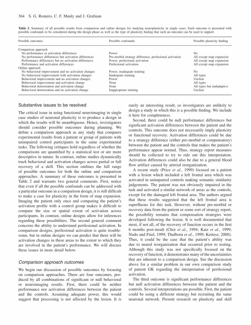
Table 2. Summary of all possible results from comparison and online designs for studying neuroplasticity in single cases. Each outcome is presented withpossible confounds to be considered during the design phase as well as the type of plasticity finding that such an outcome can be used to support
Possible outcomes Possible confounds Possible plasticity finding
Comparison approachNo performance or activation differences Power NoNo performance differences but activation differences Pre-morbid strategy difference, perilesional activation All except map expansionPerformance differences but no activation differences Power, perilesional activation All except map expansionPerformance and activation differences Perilesional activation All except map expansion
Online approachNo behavioral improvement and no activation changes Power, inadequate training NoNo behavioral improvement with activation changes Inadequate training All typesBehavioral improvement and no activation changes Power UnclearBehavioral improvement and activation change None All typesBehavioral deterioration and activation change None All types but maladaptiveBehavioral deterioration and no activation change Inappropriate training Unclear
Substantive issues to be resolved
The critical issue in using functional neuroimaging in singlecase studies of neuronal plasticity is to produce a design inwhich the results will be unambiguous. Hence, investigatorsshould consider possible outcomes during planning. Wedefine a comparison approach as any study that comparesexperimental results from a patient or group of patients withunimpaired control participants in the same experimentaltasks. The following critiques hold regardless of whether thecomparisons are quantified by a statistical test or are moredescriptive in nature. In contrast, online studies dynamicallytrack behavioral and activation changes across partial or fullrecovery of a skill. This section outlines the full rangeof possible outcomes for both the online and comparisonapproaches. A summary of these outcomes is presented inTable 2 and warrants two general comments. First, noticethat even if all the possible confounds can be addressed witha particular outcome in a comparison design, it is still difficultto make a case for plasticity in the form of map expansion.Imaging the patient only once and comparing the patient’sactivation profile with a control group makes it difficult tocompare the size or spatial extent of activation acrossparticipants. In contrast, online designs allow for inferencesregarding these possibilities. The second general commentconcerns the ability to understand perilesional activation. Incomparison designs, perilesional activation is quite trouble-some, but in online designs we can predict that there will beactivation changes in these areas to the extent to which theyare involved in the patient’s performance. We will discussthese issues in more detail below.
Comparison approach outcomes
We begin our discussion of possible outcomes by focusingon comparison approaches. There are four outcomes, pro-duced by all combinations of significant or null behavioralor neuroimaging results. First, there could be neitherperformance nor activation differences between the patientand the controls. Assuming adequate power, this wouldsuggest that processing is not affected by the lesion. It is
rarely an interesting result, so investigators are unlikely todesign a study in which this is a possible finding. We includeit here for completeness.
Second, there could be null performance differences butsignificant activation differences between the patient and thecontrols. This outcome does not necessarily imply plasticityor functional recovery. Activation differences could be dueto a strategy difference (either pre-morbid or compensatory)between the patient and the controls that makes the patient’sperformance appear normal. Thus, strategy report measuresshould be collected to try to rule out this interpretation.Activation differences could also be due to a general bloodflow artifact caused by arterial reorganization.
A recent study (Price et al., 1999) focused on a patientwith a lesion which included a left frontal area which wasactivated in unimpaired controls making semantic similarityjudgements. The patient was not obviously impaired in thetask and activated a similar network of areas as the controls,except for the damaged left frontal area. The authors arguedthat these results suggested that the left frontal area issuperfluous for this task. However, without pre-morbid orrecovery data from the patient or some sort of strategy report,the possibility remains that compensation strategies weredeveloped following the lesion. It is well documented thatmost, if not all, of the recovery of function occurs in the first6 months post-insult (Choi et al., 1994; Katz et al., 1999;Nudo and Friel, 1999; Thulborn et al., 1999; Kertesz, 2000).Thus, it could be the case that the patient’s ability wasdue to neural reorganization that occurred prior to testing.Although this study was not specifically focused on therecovery of function, it demonstrates many of the uncertaintiesthat are inherent to a comparison design. See the discussionabove for a similar problem in our own comparison studyof patient GK regarding the interpretation of perilesionalactivation.
The third outcome is significant performance differencesbut null activation differences between the patient and thecontrols. Several interpretations are possible. First, the patientcould be using a different strategy but recruiting the sameneuronal network. Present research on plasticity and skill

Neuroimaging of single cases 365
learning has yet to address the relationship between strategyselection and activation patterns. Second, the neuroimagingdesign might have insufficient statistical power for a singlecase. A group analysis of neuroimaging data (from thecontrols) is more powerful than a single subject analysis (forthe patient). Third, the patient may be using a typical butdamaged network which is not processing with normalefficiency or the damaged network may have undergonesome reorganization (i.e. map expansion). This may be thecase with JS, but it is difficult to make such a quantitativeargument with a comparison study. Specifically, with JS thepeak of activation seems to be the same for him and thecontrols but the geometric center of JS’s region of activationseems to be shifted superiorly. This finding suggests that theneural topography of this simple numerical processing shiftedover time to use territory (i.e. superior parietal cortical region)that is typically used for more complex numerical processing(cf. Rickard et al., 2000). Unfortunately, it is difficult to testthis difference statistically or to make strong claims aboutthe shift being due to plasticity unless it is possible to observethis change with repeated activation studies. We believe thatonline designs will enable this type of inference because anyshift in processing should result in a detectable shift in thepattern of activity.
The fourth outcome is significant performance and activa-tion differences between the patient and the controls. Thesimplest interpretation is that different processes are used bythe patient, which may or may not involve map expansion,homologous area adaptation or compensatory masquerade. Itwould be difficult to make strong inferences in favor of anytype of plasticity without strategy data from both the patientand the controls. Even with strategy data, arterial reorganiza-tion artifacts could lead the investigator into spuriouslythinking that a perilesional area is crucial to the recoveredprocessing when it is not involved. The most troublesomeproblem with making inferences in this case is the factthat there are no data regarding pre-morbid behavioral andneural activation baselines for the patient to demonstrate andquantify any change that may have occurred in the singlecase. Without any data prior to injury concerning how thepatient performed on these tasks and what cortical areas wereinvolved, comparisons cannot be made concerning howmuch change may have occurred at both the cognitive andneural levels.
Online studies
Now we turn to online studies. There are six possibleoutcomes, produced by all combinations of significant or nullneuroimaging results and improvement, deterioration, orno change in behavioral performance. Some outcomes aretroublesome, but one in particular allows strong conclusionsregarding neuronal plasticity.
The first outcome is no behavioral change and no activationchanges across training. This reflects one of the risks of theonline design, although in some cases one might infer that
the task or skill involved cannot be recovered. Null resultsmight also be due to training that was too short or otherwiseineffective. Careful thought should be given to the trainingregimen and again it may prove useful to consult previousinvestigations from the behavioral skill learning literatureregarding the factors that affect training and the extent towhich transfer of training can be expected. Of course, itshould be noted that completely null results could just bedue to low statistical power.
Second, there might be null performance changes butsignificant activation changes. Given that behavioral measuresare usually more powerful, this outcome might seemimplausible. However, skill learning theories allow learningin the absence of performance improvement at the beginningof skill acquisition and during strategy changes, both timeswhen the processes for more effective performance are beingset up. Interpretation depends on the particular change inactivation patterns, although to make any case for plasticity,it would be necessary to argue that there would have beenbehavioral changes had training continued. That is to saythat practice was too short to allow for improvement, butthis argument would have to be considered in relation to theparticular training program. If the training was carried outfor a significant period this would be a difficult argument tomake. If the changes in activation excluded any new areasor areas different from those found in previous studies (orfrom controls), it would suggest a subtle reallocation ofprocessing resources indicating possible map expansion butperhaps not a strategy shift or compensatory masquerade. Onthe other hand, if a completely different pattern of activationemerged, it might be easier to argue for compensatorymasquerade because it seems more likely that new (i.e. non-homologous) areas would be recruited when an atypicalstrategy is being employed. In either case it is important toassess possible strategy shifts. At present there are few datasuggesting how a cognitive strategy change is manifested inthe brain. In general, early studies regarding this issuesuggest that cognitive strategy differences are accompaniedby activation intensity differences between areas in adistributed cortical network underlying a skill, as well asdifferences in the set of areas that make up these networks(Burbaud et al., 2000; Reichle et al., 2000). These findingsare consistent with earlier results from dichotic listeningexperiments (e.g. Gordon, 1980), from other studies regardinggoal instantiation (Koechlin et al., 2000), and from somework looking at cortical changes in the motor cortex withthe serial reaction time task (Pascual-Leone et al., 1994;Zhuang et al., 1997). Furthermore, as the relationship betweenneuronal changes and skill learning is better delineated, wewill be able to make better predictions regarding the possibleoutcomes of online designs.
The third outcome is significant behavioral improvementbut null activation changes. Low statistical power is mostlikely to lead to this outcome. Although it may be possiblefor behavioral change to occur without concomitant activation

366 S. G. Romero, C. F. Manly and J. Grafman
changes, there is no evidence for such a finding in the earlyneuroimaging studies of skill learning.
The fourth and best outcome is behavioral improvementand significant activation changes. As we have advocated,this would be the clearest demonstration of neuronal plasticityand is the main advantage of the online approach. Further-more, training-related changes in imaging results wouldpermit the identification of areas that are important infunctional recovery. Systematic imaging changes should alsodelineate the extent to which there are consistencies inthe recovery of patients with similar injuries as well asconsistencies in recoveries that are observed in patientswith different types of lesion and in different cortical areas.It is important to note that strategy reports will again benecessary to make strong inferences regarding what type ofplasticity has occurred. For example, strategy reports will becrucial for differentiating between cases of compensatorymasquerade and map expansion or homologous area adapta-tion involving the same cognitive strategy (i.e. same cognitivestrategy carried out by different cortical processing areas).As we have alluded to previously, this outcome also allowsinvestigators to sidestep issues of perilesional activation andarterial reorganization. For example, if a perilesional area isinvolved in recovered processing, it should show training-related changes. More generally, to the extent that areas arenecessary (or at least useful) for processing in a certaindomain, there should be learning-related changes in theseareas in the form of correlations with behavioral data, or,better yet, significant trend components in activation data(across and within scanning sessions) that are similar to thosefound in the behavioral data. Although activation changesassociated with the learning of new skills have been demon-strated, activation changes related to training in a highlylearned skill have not been established, but training-relatedbehavioral changes are known to occur. We speculate thatif activation changes do occur during further training ofestablished skills, it may be possible to assess the differencesbetween the areas necessary for unimpaired processing andthose used in recovered function, as suggested by previousstudies using comparison designs (Price et al., 1999).
The last two outcomes are behavioral deterioration withor without activation changes. The interpretation is similarto the corresponding outcomes with behavioral improvement,but special attention should be paid to these maladaptivechanges, which may indicate possible mechanisms of inter-ference with the recovery of function. For example, mal-adaptive changes might be due to the recovery of anotherbiologically more important function affecting performancein the process of interest (e.g. the crowding hypothesis;Teuber, 1974).
In summary, we have outlined two current lines ofinvestigation using functional neuroimaging to studyneuronal plasticity: skill learning and the recovery offunction in patient populations. Our own case studies, twoof which we described, have shown many of the advantagesof applying neuroimaging techniques to the study of single
cases, as well as some concerns. We have advocated asystematic way to study functional reorganization or recoveryin patients, blending the results from the behavioral skilllearning literature and from early neuroimaging studies ofthe brain mechanisms involved in skill learning. In bothcases presented here, the online framework may have circum-vented or overcome many concerns in these studies thatreflect current issues regarding drawing inferences fromneuroimaging data about the mechanisms of neuronal plasti-city. In the study of patient GK, the online approach mighthave allowed us to differentiate if his recovery was due tohomologous area adaptation or compensatory masquerade.Strategy reports would have allowed us to assess whetherhis recovery was due to the same cognitive strategy used bythe unimpaired controls. The online approach might alsohave delineated the extent to which GK’s perilesional activa-tion was involved in his recovered processing. Dynamicchanges in perilesional activation across GK’s recovery wouldhave provided strong evidence that his abilities were not theproduct of only right hemisphere processing.
In our study of patient JS, the online approach might haveallowed us to quantify statistically the subtle map expansionthat we have speculated about. With JS, the online designwould be much harder to implement because of the develop-mental nature of his deficit. Early detection might have beenentirely impossible. It may, however, be possible to documentJS’s inability to learn these facts with an online design, andthis is a topic for further research with developmental cases.Finally, it is important to note that it would be entirelypossible with an online design to document subtle mapexpansion with patients who have other etiologies.
Although functional neuroimaging has already greatlyaided the neuropsychological study of neuronal plasticityrelated to the recovery of function in patients, we have justbegun to scratch the surface of what is possible with thesetechniques. More clever experimental designs with thesetechniques will continue to further our ability to makeinferences regarding neuronal plasticity in the study ofpatients. We present the online approach as a step towardsthis goal.
References
Aguirre GK, D’Esposito M. Experimental design for brain fMRI. In: MoonenCTW, Bandettini PA, editors. Functional MRI. Berlin: Springer, 1999:369–80.
Anderson J. Knowledge representation. In: The architecture of cognition.Cambridge: Harvard University Press, 1983: 45–84.
Anderson JR. Rules of the mind. Hillsdale: Lawrence Erlbaum, 1992.Binder J. Functional magnetic resonance imaging. Language mapping.
Neurosurgery Clinics of North America 1997; 8: 383–92.Bryan WI, Harter N. Studies on the telegraphic language: The acquisition of
a hierarchy of habits. Psychological Review 1899; 6: 345–75.Buonomano DV, Merzenich MM. Cortical plasticity: From synapses to maps.
Annual Review of Neuroscience 1998; 21: 149–86.Burbaud P, Camus O, Guehl D, Bioulac B, Caille JM, Allard M. Influence of
cognitive strategies on the pattern of cortical activation during mentalsubtraction. A functional imaging study in human subjects. NeuroscienceLetters 2000; 287: 76–80.
Neuroimaging of single cases 367
Chochon F, Cohen L, van de Moortele PF, Dehaene S. Differential contributionsof the left and right inferior parietal lobules to number processing. Journalof Cognitive Neuroscience 1999; 11: 617–30.
Choi SC, Barnes TY, Bullock R, Germanson TA, Marmarou A, Young HF.Temporal profile of outcomes in severe head injury. Journal of Neurosurgery1994; 81: 169–73.
Chollet F, DiPiero V, Wise RJS, Brooks DJ, Dolan RJ, Frackowiak RSJ. Thefunctional anatomy of motor recovery after stroke in humans: A study withpositron emission tomography. Annals of Neurology 1991; 29: 63–71.
Connolly AJ, Nachtman W, Pritchett EM. Key Math Diagnostic ArithmeticTest. Circle Pines, MN: American Guidance Services, 1976.
Crossman RRFW. A theory of the acquisition of speed-skill. Ergonomics1959; 2: 153–66.
Dehaene S, Tzourio N, Frak V, Raynaud L, Cohen L, Mehler J et al. Cerebralactivations during number multiplication and comparison: A PET study.Neuropsychologia 1996; 34: 1097–106.
Elbert T, Pantev C, Wienbruch C, Rockstroh B, Taub E. Increased corticalrepresentation of the fingers of the left hand in string players. Science 1995;170: 305–7.
Ericsson KA, Simon HA. Protocol analysis: Verbal reports as data. Cambridge:MIT Press, 1993.
Fitts PM. Perceptual-motor skill learning. In: Melton AW, editor. Categoriesof human learning. New York: Academic Press, 1964: 243–85.
Gordon H. Degree of ear asymmetries for perception of dichotic chords andfor illusory chord localization in musicians of different levels of competence.Journal of Experimental Psychology: Human Perception and Performance1980; 6: 516–27.
Grafman J, Christen Y, editors. Neuronal plasticity: Building a bridge fromthe laboratory to the clinic. Berlin: Springer, 1999.
Grafman J, Litvan I. Evidence for four forms of neuroplasticity. In: GrafmanJ, Christen Y, editors. Neuronal plasticity: Building a bridge from thelaboratory to the clinic. Berlin: Springer, 1999: 131–9.
Grafman J, Rickard TC. Acalculia. In: Feinberg TE, Farah MJ, editors.Handbook of neuropsychology. Amsterdam: Elsevier, 1996: 415–31.
Grafman J, Passafiume D, Fagliono P, Boller F. Calculation disturbances inadults with focal hemispheric damage. Cortex 1982; 18: 37–50.
Healy AF, Bourne LE Jr. Learning and memory of knowledge and skills.Thousand Oaks: Sage, 1995.
Heiss WD, Kessler J, Karbe H, Fink GR, Pawlik G. Cerebral glucosemetabolism as a predictor of recovery from aphasia in ischemic stroke.Archives of Neurology 1993; 50: 958–64.
Hittmair-Delazer M, Semenza C, Denes G. Concepts and facts in calculation.Brain 1994; 117: 715–28.
Humphreys GW, Price CJ. Cognitive neuropsychology and functional brainimaging: Implications for functional and anatomical models of cognition.Acta Psychologica 2001; 107: 119–53.
Kaplan EF, Goodglass H, Weintraub S. The Boston Naming Test. Boston:Lea & Febiger, 1983.
Karbe H, Thiel A, Weber-Luxenburger G, Herholz K, Kessler J, Heiss WD.Brain plasticity in poststroke aphasia: What is the contribution of the righthemisphere? Brain and Language 1998; 64: 215–30.
Karni A, Meyer G, Jezzard P, Adams MM et al. Functional MRI evidencefor adult motor cortex plasticity during motor skill learning. Nature 1995;377: 155–8.
Katz N, Hartman-Maeir A, Ring H, Soroker N. Functional disability andrehabilitation outcome in right hemisphere damaged patients with andwithout unilateral spatial neglect. Archives of Physical Medicine andRehabilitation 1999; 80: 379–84.
Kay J, Lesser R, Coltheart M. Psycholinguistic Assessment of LanguageProcessing in Aphasia (PALPA). Hove: Lawrence Erlbaum, 1992.
Kertesz A. Behavioral and cognitive disorders. In: Baskin DS, Yatsu FM,editors. Prognosis of neurological disorders. New York: Oxford UniversityPress, 2000: 610–22.
Koechlin E, Corrado G, Pietrini P, Grafman J. Dissociating the role of themedial and lateral anterior prefrontal cortex in human planning. Proceedingsof the National Academy of Sciences 2000; 97: 7651–6.
Krings T, Topper R, Foltys H, Erberich S, Sparing R, Willmes K et al.Cortical activation patterns during complex motor tasks in piano playersand control subjects. A functional magnetic resonance imaging study.Neuroscience Letters 2000; 278: 189–93.
Krupinski J, Kaluza J, Kumar P, Kumar S, Wang JM. Role of angiogenesisin patients with cerebral ischemic stroke. Stroke 1994; 25: 1794–8.
Levy LM, Reis IL, Grafman J. Metabolic abnormalities detected by 1H-MRSin dyscalculia and dysgraphia. Neurology 1999; 53: 639–41.
Liu HM. Neovasculature and blood–brain barrier in ischemic brain infarct.Acta Neuropathologica 1988; 75: 422–6.
Logan GD. Toward an instance theory of automatization. PsychologicalReview 1988; 95: 492–527.
McCloskey M, Lindemann AM. MATHNET: Preliminary results from adistributed model of arithmetic fact retrieval. In: Campbell JID, editor. Thenature and origins of mathematical skills. Amsterdam: Elsevier, 1992:365–409.
McCloskey M, Caramazza A, Basili A. Cognitive mechanisms in numberprocessing and calculation: Evidence from dyscalculia. Brain and Cognition1985; 4: 174–96.
Merzenich MM, Kaas JH, Wall J, Nelson RJ, Sur M, Felleman D. Topographicreorganization of somatosensory areas 3b and 1 in adult monkeys followingrestricted deafferentation. Neuroscience 1983; 8: 33–55.
Newell A, Rosenbloom PS. Mechanisms of skill acquisition and the law ofpractice. In: Anderson J, editor. Cognitive skills and their acquisition.Hillsdale: Erlbaum, 1981: 1–55.
Nudo RJ, Friel KM. Cortical plasticity after stroke: Implications forrehabilitation. Revue Neurologique 1999; 155: 713–7.
Nudo RJ, Milliken GW. Reorganization of movement representations inprimary motor cortex following focal ischemic infarcts in adult squirrelmonkeys. Journal of Neurophysiology 1996; 75: 2144–9.
Pajevic S, Pierpaoli C. Color schemes to represent the orientation of anisotropictissues from diffusion tensor data: Application to white matter fiber tractmapping in the human brain. Magnetic Resonance in Medicine 1999; 42:526–40.
Pascual-Leone A, Grafman J, Hallett M. Modulation of cortical motor outputmaps during development of implicit and explicit knowledge. Science 1994;263: 1287–9.
Pascual-Leone A, Nguyet D, Cohen LG, Brasil-Neto JP, Cammarota A, HallettM. Modulation of muscle responses evoked by transcranial magneticstimulation during the acquisition of new fine motor skills. Journal ofNeurophysiology 1995; 74: 1037–45.
Price CJ, Friston KJ. Scanning patients with tasks they can perform. HumanBrain Mapping 1999; 8: 102–8.
Price C, Warburton W, Swinburn K, Wise R, Frackowiak R. Monitoring therecovery of aphasia using positron emission tomography. Journal of CerebralBlood Flow and Metabolism 1995; 15(Suppl. 1): 696.
Price CJ, Mummery CJ, Moore CJ, Frackowiak RSJ, Friston KJ. Delineatingnecessary and sufficient neural systems with functional imaging studies ofneuropsychological patients. Journal of Cognitive Neuroscience 1999; 11:371–82.
Proctor RW, Dutta A. Skill acquisition and human performance. ThousandOaks: Sage, 1995.
Rae C, Lee M, Dixon R et al. Metabolic abnormalities in developmentaldyslexia detected by 1H magnetic resonance spectroscopy. Lancet 1998;351: 1849–52.
Rapcsak S, Beeson PM, Rubens AB. Writing with the right hemisphere. Brainand Language 1991; 41: 510–30.
Raven JC. Standard progressive matrices. London: H.K. Lewis & Co., 1958.Reichle ED, Carpenter PA, Just MA. The neural bases of strategy and skill
in sentence–picture verification. Cognitive Psychology 2000; 40: 261–95.Rickard TC. Bending the power law: A CMPL theory of strategy shifts and
the automatization of cognitive skills. Journal of Experimental Psychology:General 1997; 126: 288–311.
Rickard TC, Romero SG, Basso G, Wharton C, Flitman S, Grafman J. Thecalculating brain: An fMRI study. Neuropsychologia 2000; 38: 325–35.
Sadato N, Pascual-Leone A, Grafman J, Ibanez V, Deiber M, Dold G et al.Activation of the primary visual cortex by Braille reading in blind subjects.Nature 1996; 380: 526–8.
Schlaug G, Jancke L, Huang Y, Steinmetz H. In vivo evidence for structuralbrain asymmetry in musicians. Science 1995; 167: 699–701.
Schmidt RA, Bjork RA. New conceptualizations of practice: Commonprinciples in three paradigms suggest new concepts for training.Psychological Science 1992; 3: 207–17.
Talairach P, Tournoux J. A stereotactic coplanar atlas of the human brain.Stuttgart: Thieme, 1988.
Teuber HL. Why two brains? In: Schmitt FO, Worden FG, editors. Theneurosciences: Third study program. Cambridge: MIT Press, 1974: 71–4.
Thulborn KR, Carpenter PA, Just MA. Plasticity of language-related brainfunction during recovery from stroke. Stroke 1999; 30: 749–54.
Viscuso SR, Anderson JA, Spoehr KT. Representing simple arithmetic inneural networks. In: Tiberghien G, editor. Advances in cognitive science,Vol. 2: Theory and applications. New York: Wiley, 1989: 141–64.

368 S. G. Romero, C. F. Manly and J. Grafman
Weiller C. Imaging recovery from stroke. Experimental Brain Research 1998;123: 13–7.
Weiller C, Ramsay SC, Wise RJS, Friston KJ, Frackowiak RSJ. Individualpatterns of functional reorganization in the human cerebral cortex aftercapsular infarction. Annals of Neurology 1993; 33: 181–9.
Weiller C, Chollet F, Frackowiak R. Physiological aspects of functionalrecovery from stroke. In: Bogousslavsky J, Ginsberg M, Hennerici M,editors. Cerebrovascular disease. Oxford: Blackwell, 1997: 2057–67.
Zhuang P, Toro C, Grafman J, Mangonotti P, Leocani L, Hallett M. Event-related desynchronization (ERD) in the alpha frequency during developmentof implicit and explicit learning. Electroencephalography and ClinicalNeurophysiology 1997; 102: 374–81.
Investigating cognitive neuroplasticity insingle cases: lessons learned fromapplying functional neuroimagingtechniques to the traditionalneuropsychological case studyframework
S. G. Romero, C. F. Manly and J. GrafmanAbstractWe summarize two case studies as a context for discussing the use ofneuroimaging as a convergent methodology in the study of neuroplasticity insingle subjects. Throughout this paper we argue for a different approach forincluding neuroimaging in these types of study. Previous case studies ofneuroplasticity in patients (ours as well as others reported elsewhere) haveadded neuroimaging to the traditional neuropsychological framework ofcomparing patient results with matched control groups, and synthesized resultsthrough descriptions of anatomical and behavioral dissociations. This type ofapproach is referred to as the comparison approach. We advocate a differentapproach that builds on findings from previous behavioral skill learningresearch. Specifically, we propose adding neuroimaging throughout learningor recovery of the ability of interest and making inferences from systematicchanges in activation topography and intensity that occur within the contextof predicted behavioral changes. We dub this approach the online approach.This approach should allow future investigators to circumvent many of theinterpretation pitfalls that are common in comparison studies.
JournalNeurocase 2002; 8: 355–68
Neurocase Reference Number:O269
Primary diagnosis of interestTumour, stroke, developmental disorder
Author’s designation of caseGK, JS
Key theoretical issued Functional neuroimaging in single case studies
Key words: neuroplasticity; skill learning; functional magnetic resonanceimaging; neuroimaging
Scan, EEG and related measuresfMRI; PET; TMS; MR spectroscopy; MR diffusion
Standardized assessmentGK: Psycholinguistic Assessment of Language Processing in Aphasia(PALPA), Boston NamingJS: Key Math Diagnostic Arithmetic Test, Raven Standard Progressive Matrices
Lesion locationd GK: majority of left hemisphere
JS: no lesion (i.e. developmental case)
Lesion typeGK: strokeJS: developmental disorder
LanguageEnglish