invertebrate lysozymes: diversity and distribution, molecular mechanism and in vivo function
TRANSCRIPT

Invertebrate lysozymes: Diversity and distribution, molecularmechanism and in vivo function
JORIS M VAN HERREWEGHE and CHRIS W MICHIELS*Laboratory of Food Microbiology and Leuven Food Science and Nutrition Research Centre (LFoRCe),
Katholieke Universiteit Leuven, Kasteelpark Arenberg 22, 3001 Leuven, Belgium
*Corresponding author (Fax, +32-16-321960; Email, [email protected])
Lysozymes are antibacterial enzymes widely distributed among organisms. Within the animal kingdom, mainly threemajor lysozyme types occur. Chicken (c)-type lysozyme and goose (g)-type lysozyme are predominantly, but notexclusively, found in vertebrate animals, while the invertebrate (i)-type lysozyme is typical for invertebrate organisms,and hence its name. Since their discovery in 1975, numerous research articles report on the identification of i-typelysozymes in a variety of invertebrate phyla. This review describes the current knowledge on i-type lysozymes,outlining their distribution, molecular mechanism and in vivo function taking the representative from Venerupisphilippinarum (formerly Tapes japonica) (Vp-ilys) as a model. In addition, invertebrate g-type and ch-type (chalar-opsis) lysozymes, which have been described in molluscs and nematodes, respectively, are also briefly discussed.
[Van Herreweghe JM and Michiels CW 2012 Invertebrate lysozymes: Diversity and distribution, molecular mechanism and in vivofunction. J. Biosci. 37 327–348] DOI 10.1007/s12038-012-9201-y
1. Introduction
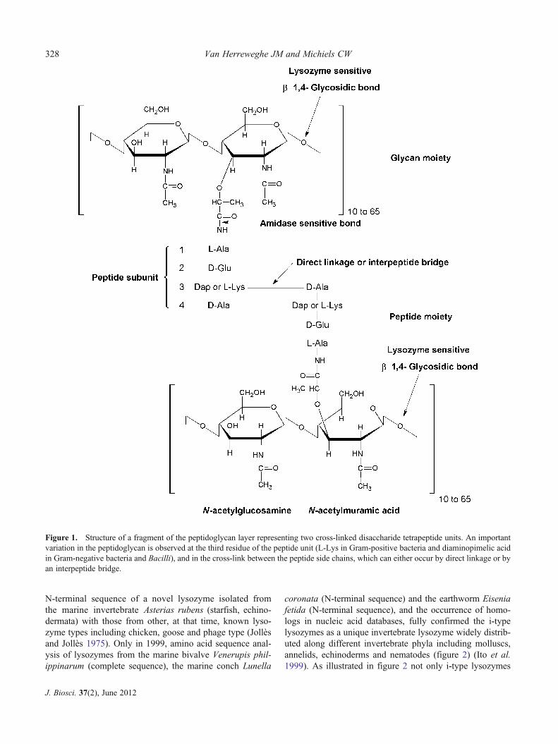
Lysozymes (EC 3.2.1.17), discovered in 1921 by AlexanderFleming, are a heterogeneous family of enzymes, all sharingthe capacity of specifically hydrolysing the β-1,4-glycosidicbond between the N-acetylmuramic acid (NAM) and N-acetylglucosamine (NAG) residues in the glycan moiety ofpeptidoglycan (figure 1). Peptidoglycan is an essential andunique cross-linked bacterial cell wall heteropolymer. Byforming a mesh-like structure surrounding the entire bacte-rial cell, it provides structural strength and protects theosmotically sensitive protoplast. Since cleavage of peptido-glycan by lysozymes and other hydrolases results in bacterialcell lysis, the peptidoglycan layer is considered as theAchilles’ heel of bacteria (Coyette and van der Ende 2008).Based on differences in amino acid sequence as well asbiochemical and enzymatic properties, lysozymes can beclassified into several types, and each type shows a particularphylogenetic distribution. The major lysozymes that havebeen found in animals up to now belong to the c-type(chicken or conventional type), g-type (goose type), i-type(invertebrate type) and ch-type (chalaropsis). Vertebrate ani-mals have only the former two types, while all four types
have been found in invertebrates. Additional lysozyme typesexist in plants, bacteriophages (viruses) and microorganisms,but since this review focuses on invertebrates, these will notbe further discussed. In the classification system of thecarbohydrate active enzymes database (CAZy, http://www.cazy.org/) (Cantarel et al. 2009), g-type lysozyme isin the glycoside hydrolase (GH) family 23 and ch-typelysozyme is part of the GH family 25, while c- and i-typelysozyme are part of the GH family 22.
Both in vertebrates and invertebrates, lysozymes are be-lieved to contribute to antibacterial defence and, in somecases, to digestion. Further, additional functions may beassociated with the isopeptidase activity which some i-typelysozymes exhibit besides lysozyme activity. For furtherreading on the function of lysozymes in vertebrate animals,we refer you to Callewaert and Michiels (2010).
2. Invertebrate-type lysozyme: Discoveryand phylogenetic distribution
The existence of a typical invertebrate lysozyme type (i-type)was first proposed in 1975 based on the comparison of the
http://www.ias.ac.in/jbiosci J. Biosci. 37(2), June 2012, 327–348, * Indian Academy of Sciences 327
Published online: 1 May 2012
Keywords. Catalytic mechanism; crystal structure; in vivo function; invertebrates; lysozymes; phylogeny
Review
N-terminal sequence of a novel lysozyme isolated fromthe marine invertebrate Asterias rubens (starfish, echino-dermata) with those from other, at that time, known lyso-zyme types including chicken, goose and phage type (Jollèsand Jollès 1975). Only in 1999, amino acid sequence anal-ysis of lysozymes from the marine bivalve Venerupis phil-ippinarum (complete sequence), the marine conch Lunella
coronata (N-terminal sequence) and the earthworm Eiseniafetida (N-terminal sequence), and the occurrence of homo-logs in nucleic acid databases, fully confirmed the i-typelysozymes as a unique invertebrate lysozyme widely distrib-uted along different invertebrate phyla including molluscs,annelids, echinoderms and nematodes (figure 2) (Ito et al.1999). As illustrated in figure 2 not only i-type lysozymes
Figure 1. Structure of a fragment of the peptidoglycan layer representing two cross-linked disaccharide tetrapeptide units. An importantvariation in the peptidoglycan is observed at the third residue of the peptide unit (L-Lys in Gram-positive bacteria and diaminopimelic acidin Gram-negative bacteria and Bacilli), and in the cross-link between the peptide side chains, which can either occur by direct linkage or byan interpeptide bridge.
328 Van Herreweghe JM and Michiels CW
J. Biosci. 37(2), June 2012

occur in invertebrates, but within some phyla c-type (mol-luscs and arthropods), ch-type (nematodes) (section 6.2) org-type lysozymes (molluscs and arthropods) are additionallyfound. During a search for homologs with the completeprotein sequence of the Venerupis philippinarum lysozyme(Vp-ilys), 46% primary sequence identity was observed witha salivary gland protein (section 5.2) of the medicinal leechHirudo medicinalis known as destabilase, which was laterproven to be an i-type lysozyme (Hm-iLys) (Zavalova et al.2000). Homology was also observed with several hypothet-ical proteins of the nematode Caenorhabditis elegans. Amultiple alignment of Vp-ilys, Hm-iLys and a homolog
from C. elegans revealed a high content and conservationof cysteines, and suggested the formation of intramoleculardisulphide bridges, which would explain the observed struc-tural stability of the proteins (Ito et al. 1999). Meanwhile,experimental studies reported on i-type lysozymes in var-ious invertebrates, based on different types of evidence(see table 1 for an overview).
Combined with a BLASTp analysis with the Vp-ilyssequence in the NCBI database for i-type lysozymes notexperimentally studied (see table 2 for an overview), thisreveals the occurrence of i-type lysozyme homologs in thefollowing phyla: porifers, molluscs, annelids, nematodes,
Figure 2. Phylogenetic distribution of the different lysozyme types along the invertebrate phyla indicated on a cladogram adapted fromDunn et al. (2008). Indication of the presence of a certain lysozyme type in a certain phylum is based on one or more of the following: (i)publications describing the isolation and characterization; (ii) literature describing the cloning and/or expression; (iii) existence of annotatedlysozyme genes in publicly available databases and (iv) presence of putative lysozyme homologs as based on BLAST analysis of the NCBInon-redundant protein database (conducted may 2011). The numbers (#/#) indicate the number of species in which a certain lysozyme typehas been experimentally studied (table 1)/ the number of species in which the same lysozyme type is found based on a BLAST search, butnot studied (table 2 for a complete list of the latter).
Current knowledge on invertebrate lysozymes 329
J. Biosci. 37(2), June 2012

Tab
le1.
Listof
experimentally
studiedi-type
lysozymes
Phylum
and
species
Organ/cells
Lysozym
ename
Typ
eof
evidence*
Accession
number
Reference
AB
CD
E
Echinod
ermata
Asteriasrubens
who
lebo
dyX
XAAR29
291
JollèsandJollès19
75Apo
sticho
pusjapo
nicus
extractedfrom
body
wall,
intestineanddigestivetract
SjLys
XX
ABK34
500
Con
get
al.2
009
Molluscs
Venerup
isph
ilipp
inarum
(Tap
esjapo
nica)
hemocytes,g
illsand
digestivegland
Vp-ily
sX
XX
XBAB33389
Itoet
al.1
999
lesser
degree
infoot,
mantle
andmuscle
Takeshita
etal.2
004
Zhaoet
al.2
010
Lunella
corona
tavisceraandmuscles
XX
Itoet
al.1
999
Chlam
ysisland
ica
viscera,crystalline
style
chlamysin
XX
CAB63451
Nilsen
etal.1
999
Mytilu
sedulis
softbo
dy,d
igestiv
egland
Me-iLys-Bm
XX
XAF3
3466
2Olsen
etal.2
003
Bachaliet
al.2
002
crystalline
style
Me-iLys-sA,
XMcH
eneryandBirkb
eck
1982;Olsen
etal.2
003
Me-iLys-sB,
XMe-iLys-sC,
Xunknow
nMe-ily
s2ABB76765
Caponeraand
Raw
son*
*Mytilu
sga
lloprovincialis
digestivegland
Mg-iLys1
XX
AAN16
210
Bachaliet
al.2
002
unkn
own
Mg-iLys2
BAF6
3423
Itoh
andTakahashi
(2007)**
Bathymod
iulusthermop
hilus
digestivegland
XX
AF3
3466
4Bachaliet
al.2
002
Bathymod
iulusazoricus
digestivegland
XX
AF3
3466
3Bachaliet
al.2
002
Crassostrea
virginica
digestivegland,
crystalline
style
Cv-iLys
2X
XX
XX
BAE47
520
Xue
etal.2
007
basoph
ilcells
ofdigestivetubules
stylesacmidgu
t,digestive
gland,
gills
labial
palps,
mantle,g
onad
Cv-iLys
1X
XX
XBAE93
114
Xue
etal.2
007
digestivegland,
mantle,
labial
palps,gills,style
sac-midgu
t,hemoctyes
Cv-iLys3
XX
XX
BAG41
979
Xue
etal.2
010
Saxido
mus
purpurata
digestivegland
XX
Miyauchiet
al.2
006
Ostreaedulis
digestiveorgans
XBAD19
060
Matsumotoet
al.2
006
Crassostrea
giga
sdigestivegland,
gills,m
antle,
gonads,h
emocytes
Cg-iLys1
XX
XBAD19
059
basoph
ilcells
ofdigestivetubules
digestivegland,
gills
Cg-iLys2
XX
BAF4
8044
Itoh
andTakahashi
2007
mantle
Cg-iLys3
XX
BAF9
4156
Itoh
etal.2
010
unkn
own
Cg-iLys4
***
330 Van Herreweghe JM and Michiels CW
J. Biosci. 37(2), June 2012
Tab
le1.
(con
tinued)
Phylum
and
species
Organ/cells
Lysozym
ename
Typ
eof
evidence*
Accession
number
Reference
AB
CD
E
Calyp
togena
sp.1
gills
XX
AF3
3466
6Bachaliet
al.2
002
Calyp
togena
sp.2
XX
AF3
3466
7Nem
atod
esCaeno
rhab
ditis
elegan
sun
know
nCe-ily
s1Genom
eanalysis
CE1754
8Schu
lenb
urgand
Boehn
isch
2008
intestine
Ce-ily
s2CE1754
9intestine
Ce-ily
s3CE2485
0un
kown
Ce-ily
s4CE3458
intestine
Ce-ily
s5CE0444
2Ann
elids
Hirud
omedicinalis
saliv
arygland
Hm-iLys
XX
AAA96
144
Zavalov
aet
al.1
996
Eisenia
andrei
coelom
ocytes
Ea-iLys
XX
ABC68610
Joskováet
al.2
009
Eisenia
fetid
aho
mog
enatewho
lebo
dyX
XItoet
al.1
999
Arthrop
ods
Ano
pheles
gambiae
fatbo
dyandmalph
igin
tubules
Ag-iLys
1X
AAT51799
Paskew
itzet
al.2
008
fatbo
dyAg-iLys2
XABP3
5929
Penaeus
mon
odon
digestivegland
Pm-iLys
1X
XACZ6347
1.1
Supu
ngul
etal.2
010
antenn
algland,
eyestalk,
hemocytes
andheart
(highestexpression)
Pm-iLys
2X
XACZ6347
2.1
Procamba
rusclarkii
hemocytes,h
eartand
stom
ach
Pc-iLys1
XX
Cop
iedfrom
article
Zhang
etal.2
010
Pc-iLys2
XX
Porifers
Suberitesdo
mun
cula
gray
cells
mesoh
yl**
**Sd
-iLys
XX
CAG27
844
Thaku
ret
al.2
005
*A:Proteinextractio
nandpu
rificatio
n,B:RNA
isolationandcD
NA
synthesis,C:N-terminal
proteinsequ
ence
determ
ination,
D:RT-PCRandE:(insitu)hy
bridization.
**Directsubm
ission
inNCBIproteindatabank
.
***D
irectsubm
ission
inNCBIEST
databank
.
****
Mesoh
yl:G
elatinou
smatrixwith
inaspon
gefilling
thespacebetweentheexternalpinacoderm
(epiderm
is)andtheinternalchoano
derm
(celllayer
thatlin
estheinnercentral
cavity
ofspon
ges).
Current knowledge on invertebrate lysozymes 331
J. Biosci. 37(2), June 2012
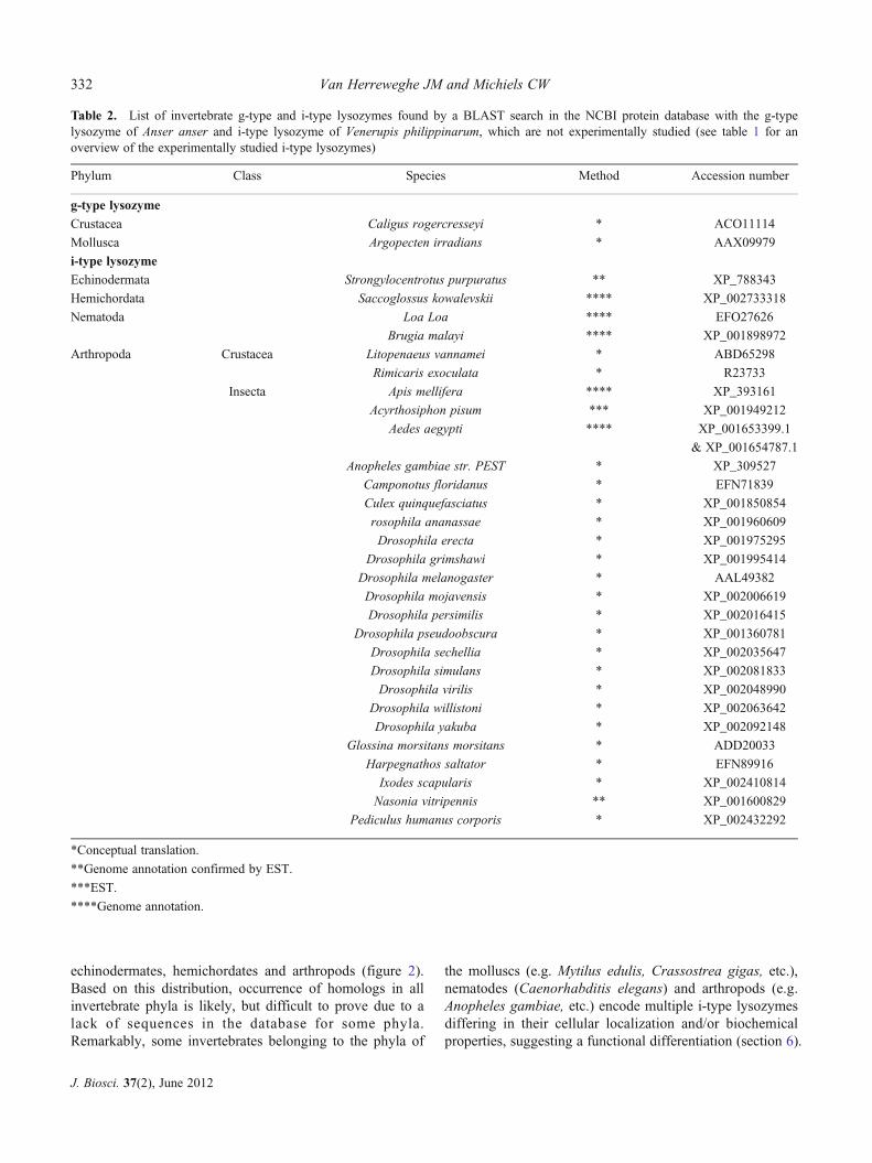
echinodermates, hemichordates and arthropods (figure 2).Based on this distribution, occurrence of homologs in allinvertebrate phyla is likely, but difficult to prove due to alack of sequences in the database for some phyla.Remarkably, some invertebrates belonging to the phyla of
the molluscs (e.g. Mytilus edulis, Crassostrea gigas, etc.),nematodes (Caenorhabditis elegans) and arthropods (e.g.Anopheles gambiae, etc.) encode multiple i-type lysozymesdiffering in their cellular localization and/or biochemicalproperties, suggesting a functional differentiation (section 6).
Table 2. List of invertebrate g-type and i-type lysozymes found by a BLAST search in the NCBI protein database with the g-typelysozyme of Anser anser and i-type lysozyme of Venerupis philippinarum, which are not experimentally studied (see table 1 for anoverview of the experimentally studied i-type lysozymes)
Phylum Class Species Method Accession number
g-type lysozymeCrustacea Caligus rogercresseyi * ACO11114
Mollusca Argopecten irradians * AAX09979
i-type lysozymeEchinodermata Strongylocentrotus purpuratus ** XP_788343
Hemichordata Saccoglossus kowalevskii **** XP_002733318
Nematoda Loa Loa **** EFO27626
Brugia malayi **** XP_001898972
Arthropoda Crustacea Litopenaeus vannamei * ABD65298
Rimicaris exoculata * R23733
Insecta Apis mellifera **** XP_393161
Acyrthosiphon pisum *** XP_001949212
Aedes aegypti **** XP_001653399.1
& XP_001654787.1
Anopheles gambiae str. PEST * XP_309527
Camponotus floridanus * EFN71839
Culex quinquefasciatus * XP_001850854
rosophila ananassae * XP_001960609
Drosophila erecta * XP_001975295
Drosophila grimshawi * XP_001995414
Drosophila melanogaster * AAL49382
Drosophila mojavensis * XP_002006619
Drosophila persimilis * XP_002016415
Drosophila pseudoobscura * XP_001360781
Drosophila sechellia * XP_002035647
Drosophila simulans * XP_002081833
Drosophila virilis * XP_002048990
Drosophila willistoni * XP_002063642
Drosophila yakuba * XP_002092148
Glossina morsitans morsitans * ADD20033
Harpegnathos saltator * EFN89916
Ixodes scapularis * XP_002410814
Nasonia vitripennis ** XP_001600829
Pediculus humanus corporis * XP_002432292
*Conceptual translation.
**Genome annotation confirmed by EST.
***EST.
****Genome annotation.
332 Van Herreweghe JM and Michiels CW
J. Biosci. 37(2), June 2012
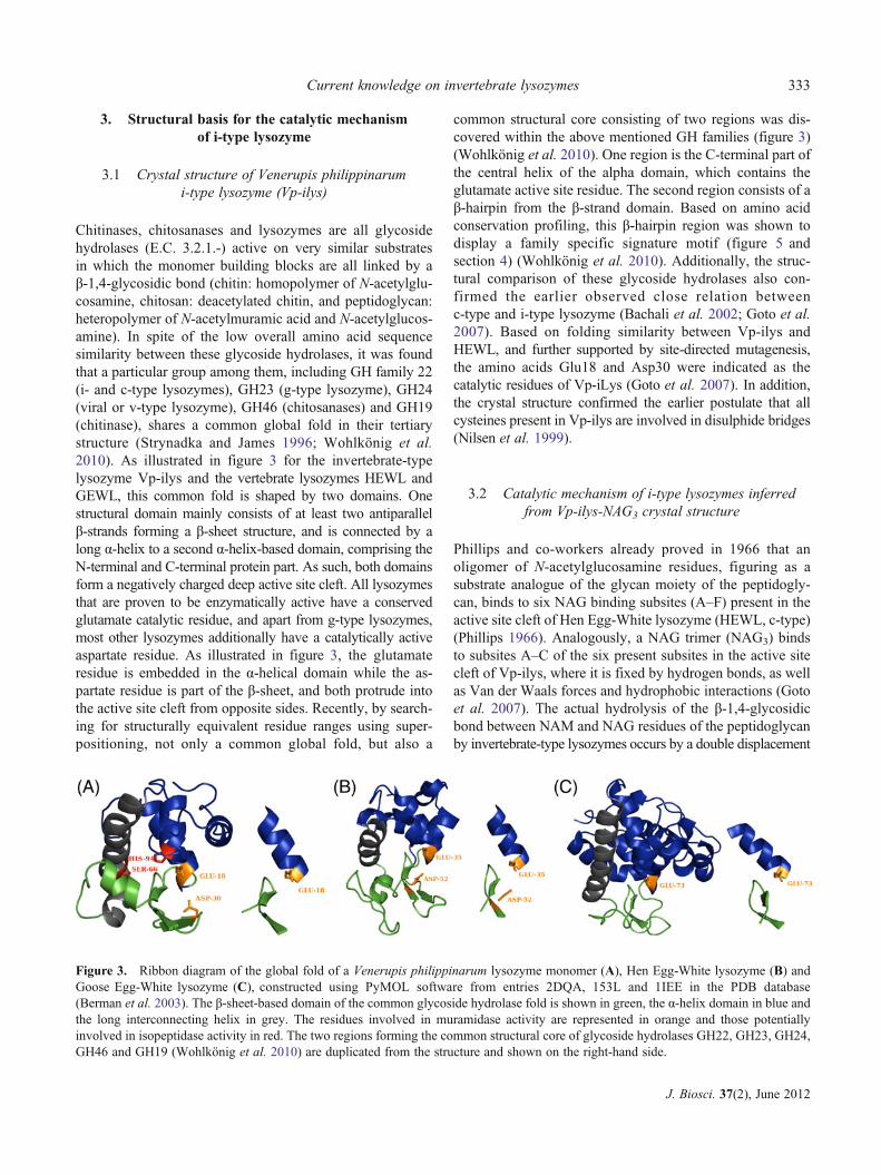
3. Structural basis for the catalytic mechanismof i-type lysozyme
3.1 Crystal structure of Venerupis philippinarumi-type lysozyme (Vp-ilys)
Chitinases, chitosanases and lysozymes are all glycosidehydrolases (E.C. 3.2.1.-) active on very similar substratesin which the monomer building blocks are all linked by aβ-1,4-glycosidic bond (chitin: homopolymer of N-acetylglu-cosamine, chitosan: deacetylated chitin, and peptidoglycan:heteropolymer of N-acetylmuramic acid and N-acetylglucos-amine). In spite of the low overall amino acid sequencesimilarity between these glycoside hydrolases, it was foundthat a particular group among them, including GH family 22(i- and c-type lysozymes), GH23 (g-type lysozyme), GH24(viral or v-type lysozyme), GH46 (chitosanases) and GH19(chitinase), shares a common global fold in their tertiarystructure (Strynadka and James 1996; Wohlkönig et al.2010). As illustrated in figure 3 for the invertebrate-typelysozyme Vp-ilys and the vertebrate lysozymes HEWL andGEWL, this common fold is shaped by two domains. Onestructural domain mainly consists of at least two antiparallelβ-strands forming a β-sheet structure, and is connected by along α-helix to a second α-helix-based domain, comprising theN-terminal and C-terminal protein part. As such, both domainsform a negatively charged deep active site cleft. All lysozymesthat are proven to be enzymatically active have a conservedglutamate catalytic residue, and apart from g-type lysozymes,most other lysozymes additionally have a catalytically activeaspartate residue. As illustrated in figure 3, the glutamateresidue is embedded in the α-helical domain while the as-partate residue is part of the β-sheet, and both protrude intothe active site cleft from opposite sides. Recently, by search-ing for structurally equivalent residue ranges using super-positioning, not only a common global fold, but also a
common structural core consisting of two regions was dis-covered within the above mentioned GH families (figure 3)(Wohlkönig et al. 2010). One region is the C-terminal part ofthe central helix of the alpha domain, which contains theglutamate active site residue. The second region consists of aβ-hairpin from the β-strand domain. Based on amino acidconservation profiling, this β-hairpin region was shown todisplay a family specific signature motif (figure 5 andsection 4) (Wohlkönig et al. 2010). Additionally, the struc-tural comparison of these glycoside hydrolases also con-firmed the earlier observed close relation betweenc-type and i-type lysozyme (Bachali et al. 2002; Goto et al.2007). Based on folding similarity between Vp-ilys andHEWL, and further supported by site-directed mutagenesis,the amino acids Glu18 and Asp30 were indicated as thecatalytic residues of Vp-iLys (Goto et al. 2007). In addition,the crystal structure confirmed the earlier postulate that allcysteines present in Vp-ilys are involved in disulphide bridges(Nilsen et al. 1999).
3.2 Catalytic mechanism of i-type lysozymes inferredfrom Vp-ilys-NAG3 crystal structure
Phillips and co-workers already proved in 1966 that anoligomer of N-acetylglucosamine residues, figuring as asubstrate analogue of the glycan moiety of the peptidogly-can, binds to six NAG binding subsites (A–F) present in theactive site cleft of Hen Egg-White lysozyme (HEWL, c-type)(Phillips 1966). Analogously, a NAG trimer (NAG3) bindsto subsites A–C of the six present subsites in the active sitecleft of Vp-ilys, where it is fixed by hydrogen bonds, as wellas Van der Waals forces and hydrophobic interactions (Gotoet al. 2007). The actual hydrolysis of the β-1,4-glycosidicbond between NAM and NAG residues of the peptidoglycanby invertebrate-type lysozymes occurs by a double displacement
Figure 3. Ribbon diagram of the global fold of a Venerupis philippinarum lysozyme monomer (A), Hen Egg-White lysozyme (B) andGoose Egg-White lysozyme (C), constructed using PyMOL software from entries 2DQA, 153L and 1IEE in the PDB database(Berman et al. 2003). The β-sheet-based domain of the common glycoside hydrolase fold is shown in green, the α-helix domain in blue andthe long interconnecting helix in grey. The residues involved in muramidase activity are represented in orange and those potentiallyinvolved in isopeptidase activity in red. The two regions forming the common structural core of glycoside hydrolases GH22, GH23, GH24,GH46 and GH19 (Wohlkönig et al. 2010) are duplicated from the structure and shown on the right-hand side.
Current knowledge on invertebrate lysozymes 333
J. Biosci. 37(2), June 2012
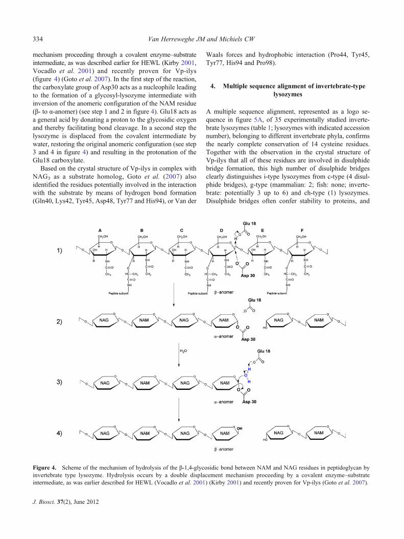
mechanism proceeding through a covalent enzyme–substrateintermediate, as was described earlier for HEWL (Kirby 2001,Vocadlo et al. 2001) and recently proven for Vp-ilys(figure 4) (Goto et al. 2007). In the first step of the reaction,the carboxylate group of Asp30 acts as a nucleophile leadingto the formation of a glycosyl-lysozyme intermediate withinversion of the anomeric configuration of the NAM residue(β- to α-anomer) (see step 1 and 2 in figure 4). Glu18 acts asa general acid by donating a proton to the glycosidic oxygenand thereby facilitating bond cleavage. In a second step thelysozyme is displaced from the covalent intermediate bywater, restoring the original anomeric configuration (see step3 and 4 in figure 4) and resulting in the protonation of theGlu18 carboxylate.
Based on the crystal structure of Vp-ilys in complex withNAG3 as a substrate homolog, Goto et al. (2007) alsoidentified the residues potentially involved in the interactionwith the substrate by means of hydrogen bond formation(Gln40, Lys42, Tyr45, Asp48, Tyr77 and His94), or Van der
Waals forces and hydrophobic interaction (Pro44, Tyr45,Tyr77, His94 and Pro98).
4. Multiple sequence alignment of invertebrate-typelysozymes
A multiple sequence alignment, represented as a logo se-quence in figure 5A, of 35 experimentally studied inverte-brate lysozymes (table 1; lysozymes with indicated accessionnumber), belonging to different invertebrate phyla, confirmsthe nearly complete conservation of 14 cysteine residues.Together with the observation in the crystal structure ofVp-ilys that all of these residues are involved in disulphidebridge formation, this high number of disulphide bridgesclearly distinguishes i-type lysozymes from c-type (4 disul-phide bridges), g-type (mammalian: 2; fish: none; inverte-brate: potentially 3 up to 6) and ch-type (1) lysozymes.Disulphide bridges often confer stability to proteins, and
Figure 4. Scheme of the mechanism of hydrolysis of the β-1,4-glycosidic bond between NAM and NAG residues in peptidoglycan byinvertebrate type lysozyme. Hydrolysis occurs by a double displacement mechanism proceeding by a covalent enzyme–substrateintermediate, as was earlier described for HEWL (Vocadlo et al. 2001) (Kirby 2001) and recently proven for Vp-ilys (Goto et al. 2007).
334 Van Herreweghe JM and Michiels CW
J. Biosci. 37(2), June 2012

Figure 5. (A) Sequence logo built with Weblogo 3. (Crooks et al. 2004) , showing the conservation of amino acids in i-type lysozymesbased on a multiple sequence alignment (built with Clustal X (Larkin et al. 2007)) of 35 invertebrate- type lysozymes with superposition of thesecondary structure of Vp-ilys. Lysozymes included in the analysis are represented in boldface in (B); see table 2 for their correspondingaccession numbers. Signal peptides were predicted by Signal P 3.0 (Bendtsen et al. 2004) and removed prior to alignment. Poorly aligningN-terminal and C-terminal parts were removed. Amino acids are coloured by hydrophobicity: hydrophobic, blue; neutral, green; hydrophilic,black. Disulphide bridge forming cysteines are indicated by c-cx, active site residues formuramidase activity by red boxes and for isopeptidase activityby yellow boxes, all based on those identified in Vp-ilys. Additional residues involved in the interaction with the substrate are indicated by a blackarrow and those involved in lysozyme dimer formation by an orange line. The grey box illustrates the conservation of the GH22i (i-type lysozymes)specific signature (LSCGYFQIK for Vp-iLys) (B) Multiple sequence alignment (built with Clustal X (Larkin et al. 2007)) of the muramidaseand isopeptidase active site region of experimentally studied i-type lysozymes (bold) and i-type lysozymes found by a BLASTp search in theNCBI database restricted to the phylum of the arthropods which are not experimentally studied (non-bold), showing the conservation of the activesite residues for muramidase and isopeptidase activity in invertebrate lysozymes of several phyla based on those identified in Vp-ilys. The grey boxillustrates the conservation of the GH22 i-type lysozymes specific signature (LSCGYFQIK for Vp-iLys).
Current knowledge on invertebrate lysozymes 335
J. Biosci. 37(2), June 2012
Vp-ilys was indeed proven to resist better to denaturationthan c-type lysozymes (HEWL and human) in a guanidine-HCl denaturation assay (Ito et al. 1999). Additionally, thealignment (figure 5) also confirms the complete conservationof the earlier identified active site residues responsible forthe muramidase activity of Vp-ilys (Glu18 and Asp30 inVp-ilys), in all characterized invertebrate-type lysozymesof the mollusc phylum, including the multiple lysozymegene paralogs identified in the same species (M. edulis,M. galloprovincialis, C. gigas and C. virginica). The sameobservation is true for the lysozymes from echinoderm,porifer and annelid phyla, although with reservation sinceonly a few lysozyme sequences are available in the databasesfor these phyla. The nematode Caenorhabditis elegans pos-sesses a large repertoire of fifteen putative lysozyme genesincluding five i-type lysozymes. However, based on the mul-tiple alignment (figure 5B), two among these (Ce-ilys1 andCe-ilys4) are predicted to be inactive due to the absence ofone (Ce-iLys4) or both (Ce-iLys1) active site residues.
Remarkably, predicted amino acid sequences from cDNAof the experimentally studied i-type lysozymes from thearthropod species Anopheles gambiae (Ag-iLys1 and 2)(mosquito), Penaeus monodon (Pm-ilys1 and 2) (giant tigerprawn) and Procambarus clarkii (Pc-iLys1 and 2) (redswamp crawfish) revealed that these enzymes are all missingthe glutamate and all except Ag-iLys1 also the aspartateresidue, both critical for muramidase activity of i-type lyso-zymes (Paskewitz et al. 2008). Inclusion of 13 additionalsequences of arthropod i-type lysozyme homologs, identi-fied by BLASTp search with Vp-ilys in the NCBI proteindatabase, in the multiple sequence alignment (figure 5), con-firmed that muramidase deficiency due to the absence of atleast one of the catalytic residues might be a common featureof arthropod i-type lysozymes.
It is also worthwhile to note that the non-catalytic residuesin Vp-ilys, earlier postulated by Goto et al. (2007) to beinvolved in the interaction with the substrate analogueNAG3, are also fairly well conserved in i-type lysozymes.Finally, the alignment in figure 5 confirms that the specificsignature motif discovered within the β-hairpin region of thecommon structural core of GH19, GH22, GH23, GH24 andGH46 families (section 3) is conserved in all i-type lyso-zymes (LSCGYFQIK for Vp-iLys) except for the arthropodrepresentatives.
5. Invertebrate lysozyme as a multifunctional enzyme
5.1 Muramidase/chitinase activity and its modulationby dimer formation
Most lysozymes exhibit, besides muramidase activity, alsochitinase activity, probably as a result of the similarity be-tween peptidoglycan (heteropolymer of β-1,4 linked N-
acetylmuramic acid and N-acetylglucosamine), the naturalsubstrate of lysozymes, and chitin (homopolymer of β-1,4linked N-acetylglucosamine), the natural substrate of chiti-nases. Moreover, some lysozymes and chitinases/chitosa-nases were found to be structurally related (section 3.1)(Wohlkönig et al. 2010). Besides warding off fungal infec-tions there is no other clear function of the chitinase activitypossessed by lysozymes. Among the i-type lysozymes, somehave chitinase activity, while others, like chlamysin fromChlamys islandica and the i-type lysozyme from the seacucumber Asostichopus japonicus, were shown to be unableof hydrolyzing chitin (Nilsen et al. 1999; Cong et al. 2009).The molecular basis for this difference is unknown. One ofthe i-type lysozymes which possess chitinase activity is theVenerupis philippinarum lysozyme (Vp-ilys). The quaterna-ry structure in the Vp-iLys crystal revealed dimer formationby two Vp-ilys molecules, which was confirmed to be main-tained in an aqueous solution with a low salt concentration(83 mM NaCl) by means of gel filtration chromatography(Goto et al. 2007). This dimer formation was proposed to besustained by electrostatic interactions between the catalyticresidues (Glu18 and Asp30) in one molecule and positiveresidues (Lys108 and 115) at the C-terminal helix 6 of theother molecule (marked by an orange line in figure 5A;figure 6). As a result, the active sites of both moleculesinvolved are blocked by dimer formation, thereby suppressingchitinase and muramidase activity. The observation that highsalt concentrations (≥ 133 mM) increased chitinase activity atlow, but not at high protein concentration (> 10 μM), wasassumed to result from the dissociation of the Vp-ilys dimer athigh ionic strength. From these observations emerged the ideathat the chitinase (and hence also muramidase activity) ofVp-ilys is modulated by environmental salt concentrationsand by its own concentration. Whether this feature is common
Figure 6. Ribbon diagram of a dimer of the Venerupis philippi-narum lysozyme. The residues involved in muramidase activity arerepresented in green and those potentially involved in isopeptidaseactivity in red. Helix 6 and residues Lys 108 and 115 are shown inorange. Figure adapted from entry 2DQA ‘Crystal structure ofTapes japonica’ in the PDB database using PyMOL software.
336 Van Herreweghe JM and Michiels CW
J. Biosci. 37(2), June 2012
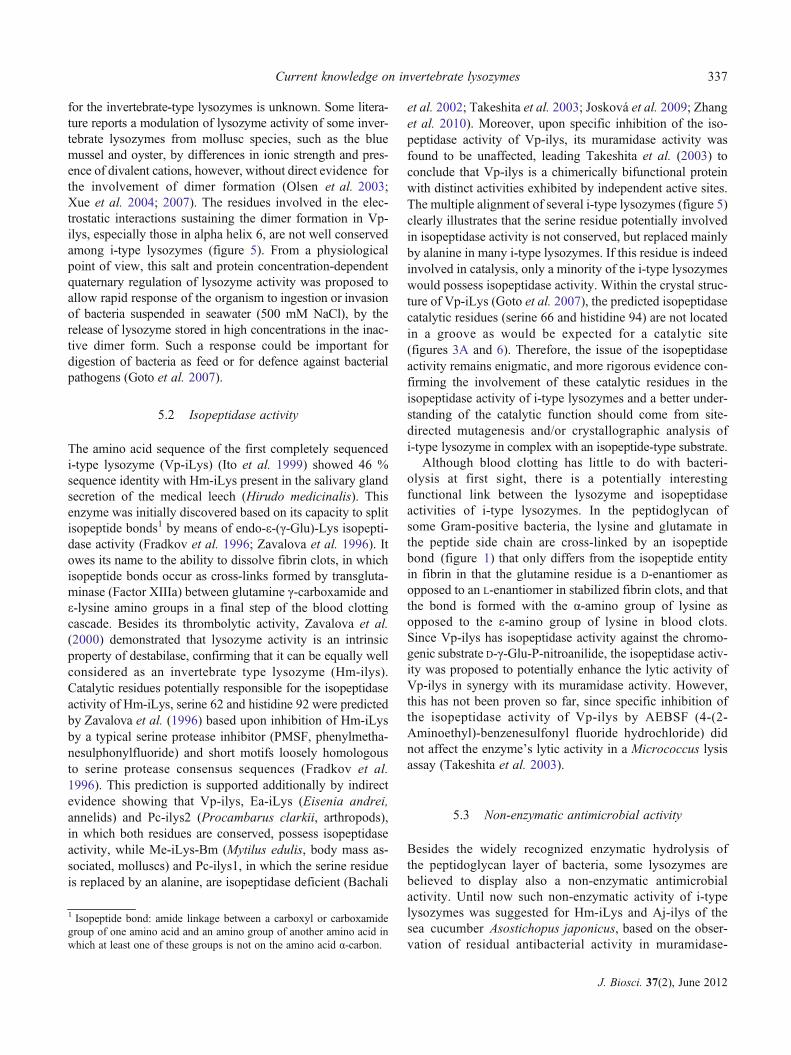
for the invertebrate-type lysozymes is unknown. Some litera-ture reports a modulation of lysozyme activity of some inver-tebrate lysozymes from mollusc species, such as the bluemussel and oyster, by differences in ionic strength and pres-ence of divalent cations, however, without direct evidence forthe involvement of dimer formation (Olsen et al. 2003;Xue et al. 2004; 2007). The residues involved in the elec-trostatic interactions sustaining the dimer formation in Vp-ilys, especially those in alpha helix 6, are not well conservedamong i-type lysozymes (figure 5). From a physiologicalpoint of view, this salt and protein concentration-dependentquaternary regulation of lysozyme activity was proposed toallow rapid response of the organism to ingestion or invasionof bacteria suspended in seawater (500 mM NaCl), by therelease of lysozyme stored in high concentrations in the inac-tive dimer form. Such a response could be important fordigestion of bacteria as feed or for defence against bacterialpathogens (Goto et al. 2007).
5.2 Isopeptidase activity
The amino acid sequence of the first completely sequencedi-type lysozyme (Vp-iLys) (Ito et al. 1999) showed 46 %sequence identity with Hm-iLys present in the salivary glandsecretion of the medical leech (Hirudo medicinalis). Thisenzyme was initially discovered based on its capacity to splitisopeptide bonds1 by means of endo-ε-(γ-Glu)-Lys isopepti-dase activity (Fradkov et al. 1996; Zavalova et al. 1996). Itowes its name to the ability to dissolve fibrin clots, in whichisopeptide bonds occur as cross-links formed by transgluta-minase (Factor XIIIa) between glutamine γ-carboxamide andε-lysine amino groups in a final step of the blood clottingcascade. Besides its thrombolytic activity, Zavalova et al.(2000) demonstrated that lysozyme activity is an intrinsicproperty of destabilase, confirming that it can be equally wellconsidered as an invertebrate type lysozyme (Hm-ilys).Catalytic residues potentially responsible for the isopeptidaseactivity of Hm-iLys, serine 62 and histidine 92 were predictedby Zavalova et al. (1996) based upon inhibition of Hm-iLysby a typical serine protease inhibitor (PMSF, phenylmetha-nesulphonylfluoride) and short motifs loosely homologousto serine protease consensus sequences (Fradkov et al.1996). This prediction is supported additionally by indirectevidence showing that Vp-ilys, Ea-iLys (Eisenia andrei,annelids) and Pc-ilys2 (Procambarus clarkii, arthropods),in which both residues are conserved, possess isopeptidaseactivity, while Me-iLys-Bm (Mytilus edulis, body mass as-sociated, molluscs) and Pc-ilys1, in which the serine residueis replaced by an alanine, are isopeptidase deficient (Bachali
et al. 2002; Takeshita et al. 2003; Josková et al. 2009; Zhanget al. 2010). Moreover, upon specific inhibition of the iso-peptidase activity of Vp-ilys, its muramidase activity wasfound to be unaffected, leading Takeshita et al. (2003) toconclude that Vp-ilys is a chimerically bifunctional proteinwith distinct activities exhibited by independent active sites.The multiple alignment of several i-type lysozymes (figure 5)clearly illustrates that the serine residue potentially involvedin isopeptidase activity is not conserved, but replaced mainlyby alanine in many i-type lysozymes. If this residue is indeedinvolved in catalysis, only a minority of the i-type lysozymeswould possess isopeptidase activity. Within the crystal struc-ture of Vp-iLys (Goto et al. 2007), the predicted isopeptidasecatalytic residues (serine 66 and histidine 94) are not locatedin a groove as would be expected for a catalytic site(figures 3A and 6). Therefore, the issue of the isopeptidaseactivity remains enigmatic, and more rigorous evidence con-firming the involvement of these catalytic residues in theisopeptidase activity of i-type lysozymes and a better under-standing of the catalytic function should come from site-directed mutagenesis and/or crystallographic analysis ofi-type lysozyme in complex with an isopeptide-type substrate.
Although blood clotting has little to do with bacteri-olysis at first sight, there is a potentially interestingfunctional link between the lysozyme and isopeptidaseactivities of i-type lysozymes. In the peptidoglycan ofsome Gram-positive bacteria, the lysine and glutamate inthe peptide side chain are cross-linked by an isopeptidebond (figure 1) that only differs from the isopeptide entityin fibrin in that the glutamine residue is a D-enantiomer asopposed to an L-enantiomer in stabilized fibrin clots, and thatthe bond is formed with the α-amino group of lysine asopposed to the ε-amino group of lysine in blood clots.Since Vp-ilys has isopeptidase activity against the chromo-genic substrate D-γ-Glu-P-nitroanilide, the isopeptidase activ-ity was proposed to potentially enhance the lytic activity ofVp-ilys in synergy with its muramidase activity. However,this has not been proven so far, since specific inhibition ofthe isopeptidase activity of Vp-ilys by AEBSF (4-(2-Aminoethyl)-benzenesulfonyl fluoride hydrochloride) didnot affect the enzyme’s lytic activity in a Micrococcus lysisassay (Takeshita et al. 2003).
5.3 Non-enzymatic antimicrobial activity
Besides the widely recognized enzymatic hydrolysis ofthe peptidoglycan layer of bacteria, some lysozymes arebelieved to display also a non-enzymatic antimicrobialactivity. Until now such non-enzymatic activity of i-typelysozymes was suggested for Hm-iLys and Aj-ilys of thesea cucumber Asostichopus japonicus, based on the obser-vation of residual antibacterial activity in muramidase-
1 Isopeptide bond: amide linkage between a carboxyl or carboxamidegroup of one amino acid and an amino group of another amino acid inwhich at least one of these groups is not on the amino acid α-carbon.
Current knowledge on invertebrate lysozymes 337
J. Biosci. 37(2), June 2012

deficient heat-inactivated enzymes (Cong et al. 2009), and inPc-ilys1 of the red swamp crayfish Procambarus clarkia,which is naturally muramidase-deficient. As the non-enzymatic activity was only proven for a limited number ofi-type lysozymes and the molecular basis for this activity islacking, additional research is needed to confirm the consis-tency of this feature.
6. Contribution of lysozymes to the immuneand digestive system of invertebrate organisms
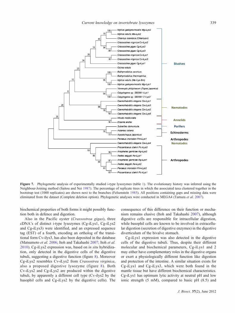
Most vertebrate and invertebrate organisms share the envi-ronment in which they live with bacteria, with which theymay enter into mutualistic, commensalic or parasitic inter-actions. Unlike vertebrate organisms, invertebrates lack anadaptive immune system and as a result solely depend ontheir innate immune system to cope with the threat of path-ogenic bacteria. Lysozymes, together with other effectormolecules, are considered to be key components of this hostdefence. Several invertebrates are known to feed on bacteria.For example, marine bivalves (including scallops, clams,oysters and mussels) are known as filter feeders. By drawingwater over their gills, suspended food particles includingbacteria are trapped on the mucus of the gills from where itis passed on to the gastrointestinal tract. Worms and fliesoften feed on decomposing organic matter including thelarge biomass of microorganisms causing the decomposition.This implies that some invertebrates not only require a well-developed immune system but also a digestive system capa-ble of digesting bacteria. For this reason, i-type lysozymes ofsome invertebrates are also believed to be involved indigestion, similar to some c-type lysozymes in vertebrateorganisms (for example ruminants). The following para-graphs give an overview on the current knowledge ofthe function of i-type lysozymes in several invertebratephyla. Figure 7 shows a phylogenetic analysis of all i-typelysozymes discussed.
6.1 Bivalves
6.1.1 Bivalve i-type lysozymes: Based on the occurrence oflysozyme(−like) activity in hemolymph and crude extracts ofspecific cells (hemocytes) and organs (digestive gland, crys-talline style, gills) of bivalves, lysozymes were hypothesizedto take part in both digestion (McHenery et al. 1979) anddefence (Carballala et al. 1997; Allam and Paillard 1998;Allam et al. 2000) in marine bivalves. Recent studies ofbivalve i-type lysozymes including biochemical character-ization of naturally extracted or by recombinant DNA-technology produced lysozymes, RT-PCR analysis andin situ hybridization revealed the existence in several organ-isms of multiple paralogous i-type lysozymes varying inbiochemical characteristics and spatiotemporal expression
patterns (Olsen et al. 2003). These findings suggest thatbivalve invertebrates may have evolved a battery of lyso-zymes with specialized functions. Based on tissue-specificprotein extraction, subsequent purification and biochemicalcharacterization (pH, ionic strength, divalent cations),M. edulis was found to produce four i-type lysozymes, threecrystalline-style-associated lysozymes (Me-ilys-sA, Me-ilys-sB, Me-ilys-sC) and one soft-body lysozyme (Me-ilys-Bm)considered as the major mussel lysozyme. The soft bodylysozyme (Me-ilys-Bm) is believed to be a hemocyte-produced enzyme involved in antibacterial defence, whilelysozymes from the digestive gland-associated crystallinestyle (figure 8) are believed to be involved in digestion.However, since Me-ilys-Bm was also purified from thedigestive gland, a digestive role cannot be ruled out (Olsenet al. 2003). Independent from this study, a complete proteinsequence of an i-type lysozyme from M. edulis (Me-iLys2),differing from Me-iLys-Bm, was submitted directly to theNCBI protein database (Bachali et al. 2002; Olsen et al.2003). Since the primary sequences of the earlier mentionedcrystalline-style-associated lysozymes, believed to be in-volved in digestion, are unknown, this might be one of theselysozymes or a fifth i-type lysozyme with a proposed diges-tive function in Mytilus edulis
Also from the Eastern oyster (Crassostrea virginica),initially two i-type lysozymes (Cv-iLys1 and Cv-iLys2),differing in molecular and biochemical parameters werepurified and subsequently identified and sequenced bymass spectrometry. The tissue distribution of both lyso-zymes (figure 8) determined by RT-PCR, protein extractionand in situ hybridization revealed a different expressionpattern for both lysozymes, suggesting a different function.Cv-iLys2 was mainly found in the digestive gland, in loweramounts in the crystalline style and was expressed in thebasophil cells of digestive tubules, which is in line with adigestive function. In contrast, Cv-iLys1was mainly found inthe labial palps, the mantle and in lower amounts in thegills, style sac, midgut, digestive gland and gonads. Theepithelia of the latter organs are exposed to the externalenvironment and can be used as a portal of entry byoyster pathogens, therefore suggesting a defence functionfor Cv-iLys1. Very recently, Xue and co-workers iden-tified a third i-type lysozyme in C. virginica (Cv-iLys3)that, based on its biochemical characteristics, was proposedto represent a transitional form between the earlier identifieddefensive i-type lysozyme (Cv-iLys1) and digestive i-typelysozyme (Cv-ilys2) of this species (Xue et al. 2010). Themolecular weight of Cv-iLys3 and N-terminal sequence re-semble that of Cv-iLys1, while its other biochemical param-eters including optimal ionic strength and spatial expressionpattern resemble those of Cv-ilys2 (figure 8). The optimalpH of Cv-iLys 3 (7,5-8,5), however, clearly differs from thatof Cv-iLys1 and 2 (5,5-6,5). Since Cv-iLys3 shares
338 Van Herreweghe JM and Michiels CW
J. Biosci. 37(2), June 2012
biochemical properties of both forms it might possibly func-tion both in defence and digestion.
Also in the Pacific oyster (Crassostrea gigas), threecDNA’s of distinct i-type lysozymes (Cg-iLys1, Cg-iLys2and Cg-iLys3) were identified, and an expressed sequencetag (EST) of a fourth, encoding an ortholog of the transi-tional form Cv-ilys3, has also been deposited in the database(Matsumoto et al. 2006; Itoh and Takahashi 2007; Itoh et al.2010). Cg-iLys2 expression was, based on in situ hybridiza-tion, only detected in the digestive cells of the digestivetubuli, suggesting a digestive function (figure 8). MoreoverCg-iLys2 resembles Cv-iLys2 from Crassostrea virginica,also a proposed digestive lysozyme (figure 8). BothCv-iLys2 and Cg-iLys2 are produced within the digestivetubuli, by apparently a different cell type (Cv-ilys2 by thebasophil cells and Cg-iLys2 by the digestive cells). The
consequence of this difference on their function or mecha-nism remains elusive (Itoh and Takahashi 2007), althoughdigestive cells are responsible for intracellular digestion,while basophil cells are known to be involved in extracellu-lar digestion (secretion of digestive enzymes) in the digestivediverticulum of the bivalve stomach.
Cg-iLys1 expression was also detected in the digestivecells of the digestive tubuli. Thus, despite their differentmolecular and biochemical parameters, Cg-iLys1 and 2may either have complementary roles in the digestive organsor exert a physiologically different function like digestionand protection of the intestine. A similar situation exists forCg-iLys1 and Cg-iLys3, which were both found in themantle tissue but have different biochemical characteristics.Cg-iLys1 has optimum lytic activity at neutral pH and lowionic strength (5 mM), compared to basic pH (8.5) and
Figure 7. Phylogenetic analysis of experimentally studied i-type lysozymes (table 1). The evolutionary history was inferred using theNeighbour-Joining method (Saitou and Nei 1987). The percentage of replicate trees in which the associated taxa clustered together in thebootstrap test (1000 replicates) are shown next to the branches (Felsenstein 1985). All positions containing gaps and missing data wereeliminated from the dataset (Complete deletion option). Phylogenetic analyses were conducted in MEGA4 (Tamura et al. 2007).
Current knowledge on invertebrate lysozymes 339
J. Biosci. 37(2), June 2012
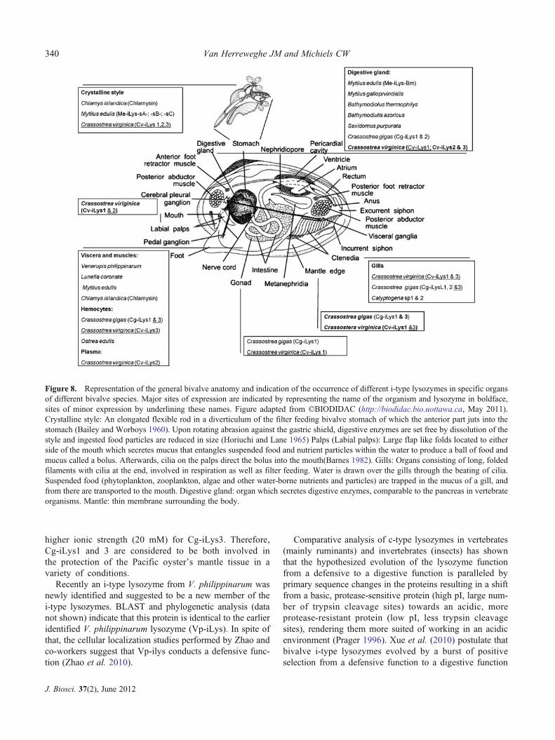
higher ionic strength (20 mM) for Cg-iLys3. Therefore,Cg-iLys1 and 3 are considered to be both involved inthe protection of the Pacific oyster’s mantle tissue in avariety of conditions.
Recently an i-type lysozyme from V. philippinarum wasnewly identified and suggested to be a new member of thei-type lysozymes. BLAST and phylogenetic analysis (datanot shown) indicate that this protein is identical to the earlieridentified V. philippinarum lysozyme (Vp-iLys). In spite ofthat, the cellular localization studies performed by Zhao andco-workers suggest that Vp-ilys conducts a defensive func-tion (Zhao et al. 2010).
Comparative analysis of c-type lysozymes in vertebrates(mainly ruminants) and invertebrates (insects) has shownthat the hypothesized evolution of the lysozyme functionfrom a defensive to a digestive function is paralleled byprimary sequence changes in the proteins resulting in a shiftfrom a basic, protease-sensitive protein (high pI, large num-ber of trypsin cleavage sites) towards an acidic, moreprotease-resistant protein (low pI, less trypsin cleavagesites), rendering them more suited of working in an acidicenvironment (Prager 1996). Xue et al. (2010) postulate thatbivalve i-type lysozymes evolved by a burst of positiveselection from a defensive function to a digestive function
Figure 8. Representation of the general bivalve anatomy and indication of the occurrence of different i-type lysozymes in specific organsof different bivalve species. Major sites of expression are indicated by representing the name of the organism and lysozyme in boldface,sites of minor expression by underlining these names. Figure adapted from ©BIODIDAC (http://biodidac.bio.uottawa.ca, May 2011).Crystalline style: An elongated flexible rod in a diverticulum of the filter feeding bivalve stomach of which the anterior part juts into thestomach (Bailey and Worboys 1960). Upon rotating abrasion against the gastric shield, digestive enzymes are set free by dissolution of thestyle and ingested food particles are reduced in size (Horiuchi and Lane 1965) Palps (Labial palps): Large flap like folds located to eitherside of the mouth which secretes mucus that entangles suspended food and nutrient particles within the water to produce a ball of food andmucus called a bolus. Afterwards, cilia on the palps direct the bolus into the mouth(Barnes 1982). Gills: Organs consisting of long, foldedfilaments with cilia at the end, involved in respiration as well as filter feeding. Water is drawn over the gills through the beating of cilia.Suspended food (phytoplankton, zooplankton, algae and other water-borne nutrients and particles) are trapped in the mucus of a gill, andfrom there are transported to the mouth. Digestive gland: organ which secretes digestive enzymes, comparable to the pancreas in vertebrateorganisms. Mantle: thin membrane surrounding the body.
340 Van Herreweghe JM and Michiels CW
J. Biosci. 37(2), June 2012
in a similar way c-type lysozymes did in vertebrates.However, some remarks can be made concerning this theory.First, Prager (1996) postulated that for digestive c-type lyso-zymes, their ‘low isoelectric point could account for the lowpH optimum in terms of the electrostatic model of lysozymecatalysis’. However, it is now known that c-type and i-typelysozyme catalysis is not based on an electrostatic model(stabilization of oxicarbonium ion by the Asp residue) but bya covalent lysozyme–substrate intermediate. More in gener-al, Talley and Alexov recently disapproved any correlationbetween pH optimum and the isoelectric point of an enzyme(Talley and Alexov 2010). Furthermore, while the pH of theruminant true stomach is highly acidic (pH 3.0–3.5), the pHof a bivalve stomach and crystalline style is only slightlyacidic to neutral (pH 6.0–6.9) (Morton 1983). Finally,experimental evidence for a correlation between the numberof trypsin cleavage sites and trypsin resistance of a proteinis nonexistent, per our knowledge. Therefore, parametersas pI and number of trypsin cleavage sites were notconsidered as proper indications for either a digestive ordefensive function.
6.1.2 Bivalve g-type lysozymes: Besides i-type lysozymes, alimited number of g-type lysozymes were identified inthe bivalve class of the invertebrates. The first inverte-brate g-type lysozyme (Cf-LysG) was discovered inChlamys farreri and shown to be enzymatically active. Atranscriptional study pointed to the digestive gland, gills andgonads as the major sites of expression. While the presencein the digestive gland points towards a digestive function,the expression in the gills points to a defensive function,leading to the suggestion that Cf-LysG is a multifunctionallysozyme (Zhao et al. 2007). The second identified g-typelysozyme (Cg-PGRP-L) is actually a PeptidoglycanRecognition Protein (PGRP) found in Crassostrea gigas.PGRPs are innate immune molecules currently known tooccur in invertebrates (insects, molluscs and echinoderms,but not nematodes) as well as vertebrates and at least containone C-terminal PGRP domain including a substrate bindingsite, Zn2+ binding residues and an amidase catalytic site.Based on their functionality, PGRPs can be classified intoamidase active PGRPs (known in invertebrates and verte-brates), bactericidal PGRPs (only known in mammals) andamidase inactive activators of immune pathways (onlyknown in insects) (Dziarski and Gupta 2006). An exampleof the latter can be found in the fruit fly Drosophila mela-nogaster where the Tracheal Cyto Toxin (TCT), a disaccha-ride tetrapeptide peptidoglycan fragment (NAG-NAM-1,6anhydro L-Ala-D-γ-Glu-mesoDap-D-Ala), is known to acti-vate the Imd (immuno deficiency) immune pathway2 byinteracting with an amidase inactive PGRP. The amidase
activity in some PGRP’s is anticipated to prevent overacti-vation of the immune response by removing the peptide sidechains from peptidoglycan fragments (figure 1), thus render-ing them immunologically inactive.
The newly identified PGRP in Crassostrea gigas (Cg-PGRP-L) is a unique member of the PGRP family since it isthe only one known to combine a PGRP and a goose-typelysozyme domain, suggesting a dual functionality for thisprotein (Itoh and Takahashi 2009). Alignment with otherg-type lysozymes confirmed the presence of the necessaryactive site residue for lysozyme activity while an alignmentwith other PGRPs showed that Cg-PGRP-L may have ami-dase and specific binding ability towards DAP-type peptido-glycan, which is typical for Gram-negative bacteria andGram-positive bacilli. Expression of Cg-PGRP-L was main-ly detected in the circulatory hemocytes by RT-PCR, and thepresence of a signal peptide suggests that it is secreted. Itsexpression was shown to be up-regulated upon infectionwith a Gram-positive (Marinococcus halophilus) as well asa Gram-negative bacterium (Vibrio tubiashii). These find-ings suggest that Cg-PGRPL-L functions in bacterial recog-nition and bacteriolysis. However, further research thatinvestigates the contribution of the amidase and lyso-zyme activity to its general function is needed to get abetter understanding of the anticipated bifunctionality ofthis protein.
6.1.3 Bivalve c-type lysozyme: Only very recently, a cDNAof c-type lysozyme was identified for the first time in amollusc species (Haliotis discus hannai Ino (Hd-LysC))(Ding et al. 2011). Based on RT-PCR analysis, its expres-sion was observed in hemocytes (lowest expression level),gills, mantle (highest expression level), digestive tract andmuscle. Hd-LysC was produced in E. coli by recombinantDNA-technology, whereupon the purified and refolded en-zyme was shown to possess lytic activity against both Gram-positive and Gram-negative bacteria. This finding suggeststhat molluscs are the first invertebrate phylum discovered inwhich i-, g- and c-type lysozymes coexist. However, basedon BLASTp analysis, a g-type lysozyme of arthropod originwas found in a recent release of the NCBI sequence data-base, suggesting that some arthropods may also have thethree lysozyme types (section 6.4.1).
6.2 Nematodes
6.2.1 Large repertoire of lysozymes in the Caenorhabditisgenus: The bacteriovorous free living soil nematodeCaenorhabditis elegans was the first multicellular organismwith a fully sequenced genome (Consortium 1998). Genomicanalysis of the congeneric nematode species C. elegans,C. briggsae and C. remanei uncovered a surprisingly largerepertoire of up to 15 putative lysozyme genes, which
2 Imd pathway: Immune pathway primarly involved in defence againstGram-negative bacteria in Drosophila melanogaster.
Current knowledge on invertebrate lysozymes 341
J. Biosci. 37(2), June 2012
evolutionary analysis demonstrated to be the result of geneduplications and subsequent differentiation from one ormore ancestor genes (Schulenburg and Boehnisch 2008). Aminority of these genes encode i-type lysozymes (5 inC. elegans; 2 in C. briggsae and 3 in C. remanei), whilethe rest encodes a lysozyme type (ch-type; section 6.2.2),initially discovered in a fungus and meanwhile found tooccur in amoebae and mainly (Gram-positive) bacteria andbacteriophages (10 in C. elegans and 7 in C. briggsae andC. ramanei). To our knowledge, Caenorhabditis is the onlymulticellular eukaryotic species known to encode this lyso-zyme type. Phylogenetic analysis has shown that the lyso-zymes from C. elegans fall into three main clades, onecomprising the i-type lysozymes (Ce-ilys1-5) and two othercomprising the ch-type lysozymes (Clade1: Ce-lys 1, 2, 3, 7,8 and 9; Clade 2: Ce-lys 4, 5, 6 and 10) (Schulenburg andBoehnisch 2008).
6.2.2 ch-type lysozyme: an unexpected lysozyme type inthe Caenorhabditis genus: All 10 non-i-type lysozymes ofC. elegans contain a conserved GH 25 domain. Since thecharacterization of the first lysozyme with this signature inthe fungus Chalaropsis, these lysozymes are better knownas the ch-type lysozymes. Unlike all other lysozyme types,ch-type lysozymes exhibit β-1,4-N-,6-O-diacetyl murami-dase activity in addition to β-1,4-N-acetyl muramidase ac-tivity, and hence are the only lysozymes capable ofhydrolyzing 6-O-acetylated peptidoglycan. This modifiedpeptidoglycan is produced in some Gram-positive bacterialike Staphylococcus aureus (Bera et al. 2005) and Gram-negative bacteria like Neisseria gonorrhoeae (Moynihan andClarke 2010), where it contributes to virulence by renderingthe bacteria insensitive to the lysozyme of the host. The firstelucidated crystal structure of a ch-type lysozyme was that ofCellosyl from Streptomyces coelicolor, and it revealed anunusual lysozyme fold. While at least c-, g- i- and v-typelysozymes, together with chitinases and chitosanases, consistof two domains linked by a long α-helix (section 3), cellosylconsists of a single domain showing an unusual β/α-barrelfold, representing a new class of polysaccharide-hydrolyzingβ/α-barrels (Rau et al. 2001, Wohlkönig et al. 2010). Similarto other lysozymes, the active site geometry reveals a grooveculminating in a negatively charged deep hole lined by theacidic residues Asp and Glu, which were earlier proposed tobe involved in catalysis of ch-type lysozymes (Fouche andHash 1978).
6.2.3 Functional analysis of the nematode lysozymes: Allnematodal lysozyme genes for which expression localizationstudies are available, are expressed in the intestine, whileCel-Lys1, 7 and 8 are additionally expressed in the nervoussystem, the muscle cells of L1 larvea and the terminalpharyngeal bulb, respectively (Schulenburg and Boehnisch
2008). The predicted physico-chemical properties of thededuced nematodal lysozymes, their pathogen-induced ex-pression and regulation by the immune system differ dis-tinctly for all three clades and even for the members withinthe clades (Schulenburg and Boehnisch 2008). The clade 1ch-type lysozymes are considered as important immuneeffectors, since dependent on the infecting pathogen, specificmembers of this lysozyme clade are induced. In support ofthis hypothesis, RNAi knock-down or knock-out of clade1 ch-type lysozymes resulted in an increased susceptibil-ity against the following pathogens: Microbacterium nem-atophilum (Ce-lys7) (O'Rourke et al. 2006), Bacillusthuringiensis (Ce-lys1, 2, 7) (Boehnisch et al. 2011),Pseudomonas aeruginosa (Ce-lys2, 7) (Nandakumar andTan 2008) , Escherichia coli LF82 (Ce-lys7) (Simonsenet al. 2011) and the fungus Cryptococcus neoformans(Ce-lys7) (Marsh et al. 2011), while increased resistancewas observed upon clade 1 ch-type lysozyme overexpressionfor: Bacillus thuringiensis (Ce-lys 7) (Boehnisch et al. 2011)and Serratia marcescens (Ce-lys1) (Mallo et al. 2002,O'Rourke et al. 2006).
The clade 2 ch-type lysozymes and the i-type lysozymes,in contrast, are generally down-regulated upon pathogeninfection or by immune regulatory pathways, as if theirnatural functionality interferes with the elicited immune re-sponse. A similar finding was observed for the digestive(c-type) lysozymes of the fruit fly Drosophila melanogasterduring immune challenge (Hultmark 1996). Experimentalevidence, however, indicates that a knock-down ofCe-ilys3 results in a higher susceptibility for M. nematophi-lum, while in the same study no effect was observed for aknock-down of Ce-ilys2 (O'Rourke et al. 2006).
The fact that induction of the different members of theclade 1 ch-type lysozymes depends on the infecting patho-gen and different immune pathways leads to the assumptionthat C. elegansmodulates its immune response in function ofthe bacteria that it encounters. The genetic diversification ofpathogen recognition receptors like lectins and immuneeffectors, besides lysozymes, is believed to be responsiblefor this capacity (Du Pasquier 2006, Schulenburg andBoehnisch 2008, Schulenburg et al. 2007, Schulenburget al. 2008). Further, special attention needs to be given tothe potential synergistic interactions between several im-mune components, which contributes to a quick and effec-tive immune response, as is well documented for lysozymesand membrane-active antimicrobial peptides ( Hultmark1996; Leippe 1999; Brown and Hancock 2006).
6.3 Annelids
The lysozyme Hm-iLys (section 5.2), present in the salivarygland secretion of the medical leech, is a unique polyfunc-tional representative of the i-type lysozymes, reported to
342 Van Herreweghe JM and Michiels CW
J. Biosci. 37(2), June 2012
exhibit muramidase, isopeptidase, chitinase and a non-enzymatic antibacterial activity. In view of its isopeptidase-dependent ability to dissolve fibrin clots, it is believed tocontribute to blood preservation in the crop of the leech,where the blood is stored between two meals for severalmonths as a viscous intraluminal fluid of which small por-tions are gradually released into the intestinal tract. Kikuchiand Graf demonstrated that blood feeding is accompanied bya spatial and temporal change in the natural two-species(Aeromonas veronii bv. sobria and Rikenella-like bacterium)microbial community in the digestive tract of the medicalleech (Kikuchi and Graf 2007). Briefly, the A. veronii pop-ulation first expands and subsequently declines from day 3post-feeding. The Rikenella population also increases andreaches a plateau on day 7 post-feeding. The latter formmicrocolonies encased in a polysaccharide N-acetylglucos-amine matrix and may form an erythrocyte attached orgranular (self-immobilized) biofilm in the intraluminal fluid.On day seven this biofilm breaks apart whereupon the micro-colony size increases. In this dynamic process, the lysozymeHm-iLys might conduct additional functions, for example, incontrolling the expansion of the symbiotic population by itsantibacterial activity and/or in dismantling the N-acetylglu-cosamine matrix around the encased Rikenellamicrocoloniesby its chitinase activity.
The expression of i-type lysozyme from the earth-worm Eisenia andrei (Ea-iLys) by the coelomocytes wasstudied by RT-PCR, and found to be up-regulated uponbacterial challenge with both the Gram-positive bacteriumBacillus subtilis and the Gram-negative bacteriumEscherichia coli. Since coelomocytes are considered as im-portant immune cells in coelomate animals (molluscs, annel-ids and arthropods), these findings suggest Ea-iLys tofunction as an inducible immune effector providing protec-tion upon infection with Gram-positive and negative bacteria(Josková et al. 2009).
6.4 Arthropods
6.4.1 Arthropod c-type lysozymes: Arthropods have c-typeas well as i-type lysozyme genes (figure 2). This was con-sidered to be a unique combination, but recently a c-typelysozyme was also documented in a mollusc (Ding et al.2011). g-type lysozymes were long considered to be absentin the arthropods based on the absence of g-type lyso-zyme encoding genes in the available genomes of themost abundant class of arthropods: the insects. ABLASTp search with the g-type lysozyme from Anseranser in the NCBI protein database revealed a putativeg-type lysozyme in the crustacean Caligus rogercresseyi.The dipterian insects Drosophila melanogaster (Hultmark1996) and Anopheles gambiae (Kang et al. 1996, Li et al.2005) are known to carry multiple c-type lysozyme genes,
11 and 9 , respectively. c-type lysozyme encoding geneswere further reported in the arthropod orders lepidoptera(e.g. the silkworm (moth) Bombyx mori; Jolles et al. 1979;Lee and Brey 1995), hemiptera (e.g. the bug Tritoma infes-tans; Araujo et al. 2006; Waniek et al. 2009), isoptera (e.g.the Japanese termite Reticulitermes speratus; Fujita et al.2002) as well as in the class of the arachnid (e.g. the softthick Ornithodoros moubata; Grunclova et al. 2003) and thesubphylum of the crustaceae (e.g. the giant tiger prawnPenaeus mondon; Supungul et al. 2010). Since the focus ofthis literature review is limited to the i-type lysozyme and somespecial observations on g-type and ch-type in invertebrates, thenumerous studies on c-type lysozymes in invertebrates (Dunnand Dai 1990; Russell and Dunn 1991; Sun et al. 1991;Mulnix and Dunn 1994; Moreira-Ferro et al. 1998; Gaoand Fallon 2000; Fujimoto et al. 2001; Hikima et al. 2003;Kollien et al. 2003; Bedoya et al. 2005; Cancado et al. 2008;Ren et al. 2009; Zhang et al. 2009) will not be discussed inthis literature review.
6.4.2 Arthropod i-type lysozymes: Recently, two i-type lyso-zyme genes (Ag-iLys1 and Ag-iLys2) from the mosquitoAnopheles gambiae, two cDNAs (Pm-iLys1 and Pm-iLys2)of the giant tiger prawn Penaeus monodon and two cDNAs(Pc-iLys1 and Pc-iLys2) of the red swamp crayfishProcambarus clarkii were identified by BLAST analysiswithin the A. gambiae genome database, the P. monodonEST database and a hemocyte cDNA library of P. clarkii,respectively. Analysis of the amino acid sequence of thesesix i-type lysozymes revealed that these enzymes are allmissing the glutamate and all except Ag-iLys1 also theaspartate residue, both critical for muramidase activity ofi-type lysozymes (cfr. section 4). Both i-type lysozymesfrom Penaeus monodon and Procambarus clarkii wereexpressed by recombinant DNA-technology in E. coli, puri-fied and experimentally found to lack muramidase activity ina Micrococcus lysodeikticus turbidity assay (Supungul et al.2010; Zhang et al. 2010). However, prokaryote expressionsystems are known to cause problems for proteins carryingmultiple disulphide bonds, and thus, misfolding or malfor-mation of disulphide bonds cannot be ruled out as a cause ofenzymatic inactivity in these studies (Demain and Vaishnav2009). A multiple sequence alignment of arthropod i-typelysozymes (section 4 and figure 5) confirms that muramidasedeficiency is probably a common feature amongst arthropodi-type lysozymes. In this respect, it is noteworthy that Pm-iLys1 and 2 exhibit antimicrobial activity despite their mur-amidase deficiency. Antimicrobial activity was also reportedfor Pc-iLys1 (muramidase and isopeptidase deficient) but notfor Pc-iLys2 (muramidase-deficient and isopeptidase active).
Paskewitz et al. (2008) postulated that the inactivity ofi-type lysozymes in arthropods might be related to the factthat the functional redundancy with c-type lysozymes in
Current knowledge on invertebrate lysozymes 343
J. Biosci. 37(2), June 2012
arthropods may have allowed diversification of i-type lyso-zymes to other (yet to be discovered) functional roles. On thecontrary, such a functional diversification seems not to haveoccurred in other animals with redundant lysozymes, such asthe molluscs, which have both active i- and g-type lyso-zymes, or vertebrates, which have active c- and g-type lyso-zymes. Interestingly, some of the g-type lysozymesdiscovered in molluscs are actually PGRPs which have a g-type lysozyme domain (section 6.1.2) and are thereforebelieved to be involved in bacterial recognition, signallingand bacteriolysis. In line with the postulate of Paskewitz, thepresence of two lysozyme types in the same species mightpromote functional diversification but is not necessarily ac-companied by a loss of enzymatic activity.
RT-PCR analysis revealed that Ag-iLys1 is constitutivelyexpressed at all life stages, while Ag-iLys2 is only abun-dantly expressed in adults and late instar larvae, which mightreflect a difference in dietary or environmental exposure oflarvae and adults to bacteria. Ag-iLys1 is expressed in the fatbody3 and Malphighian tubules4 while Ag-iLys 2 is onlyexpressed in the fat body. Inoculation of bacteria into thehaemocoel of adult female mosquitoes, however, did notresult in an increased expression of any of these lysozymes,thus making a function in immunity unlikely. On the con-trary, an increased transcription of Ag-iLys1 and 2 in themidgut of the mosquito Anopheles gambiae after blood-feeding was observed, suggesting a function in the digestionof blood (Paskewitz et al. 2008). However, because theylack the isopeptidase catalytic residues (figure 5), both i-type lysozymes from A. gambiae are predicted to be isopep-tidase deficient, making this proposed function unlikely. Analternative function that has been proposed for Ag-iLys1 and2 in relation to blood feeding, which is to control excessiveoutgrowth of bacteria after a blood meal, and this could bebased on the non-enzymatic antibacterial effect of the pro-teins (DeMaio et al. 1996).
In Penaeus monodon, Pm-iLys1 is expressed in the hepa-topancreas (digestive gland) and Pm-iLys2 in different tis-sues but not in the gills, lymphoid organs and intestine.Therefore, Pm-iLys1 possibly conducts a digestive functionwhile that of Pm-iLys2 remains unknown.
Both Procambarus clarkii i-type lysozymes were foundto have a similar tissue expression pattern (hemocytes, heartand stomach). They were proposed to have an immunefunction, because of hemocyte expression and increasedexpression upon challenge with Vibrio anguillarum andStaphylococcus aureus. However, since both lysozymeswere also found in the stomach, a digestive function cannotbe ruled out. In view of the bactericidal deficiency of Pc-iLys2, the authors speculated on a potential involvement of
its isopeptidase activity in bacterial cell lysis. This seemsrather unlikely, however, since Pc-iLys2 was not bactericidaltowards Gram-positive bacteria known to have an isopeptidebond in their peptidoglycan (Staphylococcus aureus,Bacillus subtilis, Micrococcus lysodeikticus), and the obser-vation that inhibition of the isopeptidase activity in mollusci-type lysozyme Vp-ilys did not affect its bactericidal activ-ity (section 5.2).
6.5 Porifera
The most obvious evidence of the involvement of an i-typelysozyme in the elimination of bacteria was obtained bystudying the responsiveness of the marine demospongeSuberitus domuncula towards peptidoglycan fragments ofGram-positive bacteria. The elimination of Gram-positivebacteria from the mesohyl5 compartment in this organismproceeds in two phases, the activation of the gray cells andthe effector phase. In the activation phase, peptidoglycanfragments, released during bacterial lysis within the graycells present in the mesohyl, act as an elicitor leading tothe endocytosis of bacteria and further release of peptidogly-can. This causes an increased expression of an adaptorprotein (AdaPTin-1) involved in endocytosis and subse-quently, with a delay of one day, in an increased expressionof i-type lysozyme by the gray cells present in the mesohyl.During the effector phase, bacteria are lysed extracellularlyin the mesohyl by lysozyme (Thakur et al. 2005). AdaPTinsare well-conserved proteins in both vertebrate and inverte-brate metazoan, suggesting that similar mechanisms mightbe present in other organisms (Boehm and Bonifacino 2001).
7. Periplasmic inhibitor of i-type lysozyme
In response to lysozyme as an important effector molecule inthe innate immune system of vertebrate as well as inverte-brate organisms, bacteria have developed various strategiesto prevent the lethal impact of lysozymes. In Gram-negativebacteria, one of these strategies comprises the production ofspecific periplasmic or membrane-linked lysozyme inhibi-tors. Such inhibitors were first discovered for c-type lyso-zyme in E. coli (Inhibitor of Vertebrate Lysozyme, Ivy;Membrane bound lysozyme inhibitor of c-type lysozyme,MliC) and Salmonella Enteritidis (Periplasmic lysozymeinhibitor of c-type lysozyme, PliC)(Callewaert et al. 2008,Deckers et al. 2008, Deckers et al. 2004, Monchois et al.2001). Recently also a specific, high-affinity Periplasmiclysozyme inhibitor of i-type lysozyme (PliI) was discoveredin Aeromonas hydrophila and a Periplasmic lysozyme
3 Fat body: organ in insects similar in function to the vertebrate liver4 Malphighian tubules: excretory and osmoregulatory organ of insectsthat opens near the junction of the midgut and hindgut.
5 Mesohyl: Gelatinous matrix within a sponge filling the space betweenthe external pinacoderm (epidermis) and the internal choanoderm (celllayer that lines the inner central cavity of sponges).
344 Van Herreweghe JM and Michiels CW
J. Biosci. 37(2), June 2012
inhibitor of g-type lysozyme (PliG) in Escherichia coli (VanHerreweghe et al. 2010, Vanderkelen et al. 2010).
For all these inhibitors, the molecular mechanism of in-teraction with their corresponding lysozyme has been inves-tigated: Ivy (Abergel et al. 2007), MliC/PliC (Voet et al.2011; Yum et al. 2009), PliI (Leysen et al. 2011) and PliG(unpublished results). Based on X-ray crystallography andsite-directed mutagenesis of PliI, Leysen et al. (2011) sug-gested that the molecular interaction between PliI and Vp-iLys proceeds by insertion of a loop of PliI into the activesite of Vp-iLys. This loop contains an SG(x)xY domain thatis conserved across the PliI, PliG and MliC/PliC families ofinhibitors, and in which the serine 104 and tyrosine 107residues (numbering for PliI from Aeromonas hydrophila)are critical for successful interaction. The same authors alsoconstructed a molecular interaction model for PliI and Vp-Lys using in silico docking, but this should be corroboratedby experimental elucidation of the structure of the complex.
Analogous to the contribution of c-type lysozyme inhib-itors to bacterial c-type lysozyme tolerance also, PliI andPliG were shown to contribute to i-type lysozyme (Vp-ilys)tolerance of A. hydrophila and g-type lysozyme (Salmon g-type lysozyme, SalG) (Kyomuhendo et al. 2007) tolerance ofE. coli respectively in case of outer membrane permeabilisa-tion (Vp-ilys and SalG cannot autonomously cross the outermembrane of Gram-negative bacteria). These and previousfindings on c-type lysozyme inhibitors suggest that bacteriallysozyme inhibitors may have an important function, forexample, in bacteria–host interactions. Validating this as-sumption, however, remains a challenging task.
References
Abergel C, Monchois V, Byrne D, Chenivesse S, Lembo F,Lazzaroni JC and Claverie JM 2007 Structure and evolution ofthe Ivy protein family, unexpected lysozyme inhibitors in Gram-negative bacteria. Proc. Natl. Acad. Sci. USA 104 6394–6399
Allam B and Paillard C 1998 Defense factors in clam extrapallialfluids. Dis. Aquat. Organ. 33 123–128
Allam B, Paillard C and Auffret M 2000 Alterations in hemolymphand extrapallial fluid parameters in the Manila clam, Ruditapesphilippinarum, challenged with the pathogen Vibrio tapetis.J. Invertebr. Pathol. 76 63–69
Araujo CAC, Waniek PJ, Stock P, Mayer C, Jansen AM andSchaub GA 2006 Sequence characterization and expressionpatterns of defensin and lysozyme encoding genes from thegut of the reduviid bug Triatoma brasiliensis. Insect Biochem.Mol. Biol. 36 547–560
Bachali S, Jager M, Hassanin A, Schoentgen F, Jolles P, Fiala-Medioni A and Deutsch JS 2002 Phylogenetic analysis of inver-tebrate lysozymes and the evolution of lysozyme function.J. Mol. Evol. 54 652–664
Bailey K and Worboys B 1960 The lamellibranch crystalline style.Biochem. J. 76 487–491
Barnes RD 1982 Invertebrate zoology (Philadelphia: SaundersCollege)
Bedoya RJU, Mitzey AM, Obraztsova M and Lowenberger C 2005Molecular cloning and transcriptional activation of lysozyme-encoding cDNAs in the mosquito Aedes aegypti. Insect Mol.Biol. 14 89–94
Bendtsen JD, Nielsen H, von Heijne G and Brunak S 2004Improved prediction of signal peptides: SignalP 3.0. J. Mol.Biol. 340 783–795
Bera A, Herbert S, Jakob A, Vollmer W and Götz F 2005 Why arepathogenic staphylococci so lysozyme resistant? The peptido-glycan O-acetyltransferase OatA is the major determinant forlysozyme resistance of Staphylococcus aureus. Mol. Microbiol.55 778–787
Berman H, Henrick K and Nakamura H 2003 Announcing theworldwide Protein Data Bank. Nat. Struct. Biol. 10 980
Boehm M and Bonifacino JS 2001 Adaptins - The final recount.Mol. Biol. Cell 12 2907–2920
Boehnisch C, Wong D, Habig M, Isermann K, Michiels NK, RoederT, May RC, Schulenburg H 2011 Protist-Type Lysozymes of theNematode Caenorhabditis elegans Contribute to Resistanceagainst Pathogenic Bacillus thuringiensis. PLoS One 6
Brown KL and Hancock REW 2006 Cationic host defense (antimi-crobial) peptides. Curr. Opin. Immunol. 18 24–30
Callewaert L and Michiels CW 2010 Lysozymes in the animalkingdom. J. Biosci. 35 127–160
Callewaert L, Aertsen A, Deckers D, Vanoirbeek KG, VanderkelenL, Van Herreweghe JM, Masschalck B, Nakimbugwe D,Robben J and Michiels CW 2008 A new family of lysozymeinhibitors contributing to lysozyme tolerance in gram-negativebacteria. PLoS Pathog. 4
Cancado FC, Effio PC, Terra WR and Marana SR 2008 Cloning,purification and comparative characterization of two digestivelysozymes from Musca domestica larvae. Braz. J. Med. Biol.Res. 41 969–977
Cantarel BL, Coutinho PM, Rancurel C, Bernard T, Lombard V andHenrissat B 2009 The Carbohydrate-Active EnZymes database(CAZy): an expert resource for Glycogenomics. Nucleic AcidsRes. 37 D233–238
Carballala MJ, Lópeza C, Azevedob C and Villalbaa A 1997Enzymes involved in defense functions of hemocytes of musselMytilus galloprovincialis. J. Invertebr. Pathol. 70 96–105
Cong L, Yang X, Wang X, Tada M, Lu M, Liu H and Zhu B 2009Characterization of an i-type lysozyme gene from the sea cu-cumber Stichopus japonicus, and enzymatic and nonenzymaticantimicrobial activities of its recombinant protein. J. Biosci.Bioeng. 107 583–588
Coyette J and van der Ende A 2008 Peptidoglycan: the bacterialAchilles heel. FEMS Microbiol. Rev. 32 147–148
Crooks G, Hon G, Chandonia J and Brenner S 2004 WebLogo: asequence logo generator.. Genome Res. 14 1188–1190
Deckers D, Masschalck B, Aertsen A, Callewaert L, Van TiggelenCGM, Atanassova M and Michiels CW 2004 Periplasmic lyso-zyme inhibitor contributes to lysozyme resistance in Escherichiacoli. Cell. Mol. Life Sci. 61 1229–1237
Deckers D, Vanlint D, Callewaert L, Aertsen A and Michiels CW2008 Role of the lysozyme inhibitor Ivy in growth or survival ofEscherichia coli and Pseudomonas aeruginosa bacteria in hen
Current knowledge on invertebrate lysozymes 345
J. Biosci. 37(2), June 2012

egg white and in human saliva and breast milk. Appl. Environ.Microbiol. 74 4434–4439
Demain AL and Vaishnav P 2009 Production of recombinantproteins by microbes and higher organisms. Biotechnol. Adv.27 297–306
DeMaio J, Pumpuni CB, Kent M and Beier JC 1996 The midgutbacterial flora of wild Aedes triseriatus, Culex pipiens, andPsorophora columbiae mosquitoes. Am. J. Trop. Med. Hyg. 54219–223
Ding J, Li J, Bao Y, Li L, Wu F and Zhang G 2011 Molecularcharacterization of a mollusk chicken-type lysozyme gene fromHaliotis discus hannai Ino, and the antimicrobial activity of itsrecombinant protein. Fish Shellfish Immunol. 30 163–172
Du Pasquier L 2006 Germline and somatic diversification of im-mune recognition elements in Metazoa. Immunol. Lett. 104 2–17
Dunn PE and Dai W 1990 Bacterial peptidoglycan: A signal forinitiation of the bacteria regulated synthesis and secretion oflysozyme in Manduca sexta. Defense Molecules 121 33–46
Dunn C, Dunn CW, Hejnol A, Matus DQ, Pang K, BrowneWE, Smith SA, Seaver E, et al. 2008 Broad phylogenomicsampling improves resolution of the animal tree of life. Nature452 745–749
Dziarski R and Gupta D 2006 The peptidoglycan recognition pro-teins (PGRPs). Genome Biol. 7 232–245
Felsenstein J 1985 Confidence-limits on phylogenies- and approachusing the bootstrap. Evolution 39 783–791
Fouche PB and Hash JH 1978 The N,O-diacetylmuramidase ofChalaropsis species. Identificaiton of aspartyl and glutamylresidues in the active site. J. Biol. Chem. 253 6787–6793
Fradkov A, Berezhnoy S, Barsova E, Zavalova L, Lukyanov S,Baskova I and Sverdlov E 1996 Enzyme from the medicinalleech (Hirudo medicinalis) that specifically splits endo-epsilon(−gamma-Glu)-Lys isopeptide bonds: cDNA cloning and pro-tein primary structure. FEBS Lett. 390 145–148
Fujimoto S, Toshimori-Tsuda I, Kishimoto K, Yamano Y andMorishima I 2001 Protein purification, cDNA cloning andgene expression of lysozyme from eri-silkworm, Samia cyn-thia ricini. Comp. Biochem. Physiol. B, Biochem. Mol. Biol. 128709–718
Fujita A, Minamoto T, Shimizu I and Abe T 2002 Molecularcloning of lysozyme-encoding cDNAs expressed in the salivarygland of a wood-feeding termite, Reticulitermes speratus. InsectBiochem. Mol. Biol. 32 1615–1624
Gao Y and Fallon AM 2000 Immune activation upregulates lyso-zyme gene expression in Aedes aegypti mosquito cell culture.Insect Mol. Biol. 9 553–558
Goto T, Abe Y, Kakuta Y, Takeshita K, Imoto T and Ueda T 2007Crystal structure of Tapes japonica lysozyme with substrateanalogue - Structural basis of the catalytic mechanism andmanifestation of its chitinase activity accompanied by quaterna-ry structural change. J. Biol. Chem. 282 27459–27467
Grunclova L, Fouquier H, Hypsa V and Kopacek P 2003 Lysozymefrom the gut of the soft tick Ornithodoros moubata: the se-quence, phylogeny and post-feeding regulation. Dev. Comp.Immunol. 27 651–660
Hikima S, Hikima J, Rojtinnakorn J, Hirono I and Aoki T 2003Characterization and function of kuruma shrimp lysozyme pos-sessing lytic activity against Vibrio species. Gene 316 187–195
Horiuchi S and Lane CE 1965 Digestive enzymes of crystallinestyle of Strombus gigas linné. I. Cellulase and some othercarbohydrases. Biol. Bull. 129 273–281
Hultmark D 1996 Insect lysozymes; in Lysozymes: model enzymesin biochemistry and biology (ed) P Jollès (Basel: BirkhäuserVerlag) pp 87–102
Ito Y, Yoshikawa A, Hotani T, Fukuda S, Sugimura K and Imoto T1999 Amino acid sequences of lysozymes newly purified frominvertebrates imply wide distribution of a novel class in thelysozyme family. Eur. J. Biochem. 259 456–461
Itoh N and Takahashi K 2007 cDNA cloning and in situ hybridiza-tion of a novel lysozyme in the Pacific oyster, Crassostreagigas.. Comp. Biochem. Physiol. B, Biochem. Mol. Biol. 148160–166
Itoh N and Takahashi KG 2009 A novel peptidoglycan recognitionprotein containing a goose-type lysozyme domain from thePacific oyster, Crassostrea gigas. Mol. Immunol. 46 1768–1774
Itoh N, Okada Y, Takahashi K and Osada M 2010 Presence andcharacterization of multiple mantle lysozymes in the Pacificoyster, Crassostrea gigas. Fish Shellfish Immunol. 29 126–135
Jollès J and Jollès P 1975 The lysozyme from Asterias rubens. Eur.J. Biochem. 54 19–23
Jolles J, Schoentgen F, Croizier G, Croizier L and Jolles P 1979Insect lysozymes from 3 species of lepidoptera: Their structuralrelatedness to the c-(chicken)-type lysozyme. J. Mol. Evol. 14267–271
Josková R, Silerová M, Procházková P and Bilej M 2009Identification and cloning of an invertebrate-type lysozyme fromEisenia andrei. Dev. Comp. Immunol. 33 932–938
Kang DW, Romans P and Lee JY 1996 Analysis of a lysozymegene from the malaria vector mosquito, Anopheles gambiae.Gene 174 239–244
Kikuchi Y and Graf J 2007 Spatial and temporal population dy-namics of a naturally occurring two-species microbial commu-nity inside the digestive tract of the medicinal leech. Appl.Environ. Microbiol. 73 1984–1991
Kirby AJ 2001 The lysozyme mechanism sorted - after 50years.Nat. Struct. Biol. 8 737–739
Kollien AH, Fechner S, Waniek PJ and Schaub GA 2003 Isolationand characterization of a cDNA encoding for a lysozyme fromthe gut of the reduviid bug Triatoma infestans. Arch. InsectBiochem. Physiol. 53 134–145
Kyomuhendo P, Myrnes B and Nilsen I 2007 A cold-active salmongoose-type lysozyme with high heat tolerance. Cell. Mol. LifeSci. 64 2841–2847
Larkin MA, Blackshields G, Brown NP, Chenna R, McGettiganPA, McWilliam H, Valentin F, Wallace IM, et al. 2007Clustal W and Clustal X version 2.0. Bioinformatics 232947–2948
Lee WJ and Brey PT 1995 Isolation and characterization of thelysozyme encoding gene from the silkworm Bombyx mori. Gene161 199–203
Leippe M 1999 Antimicrobial and cytolytic polypeptides of amoe-boid protozoa-effector molecules of primitive phagocytes. Dev.Comp. Immunol. 23 267–279
Leysen S, Van Herreweghe JM, Callewaert L, Heirbaut M, BuntinxP, Michiels CW and Strelkov SV 2011 Molecular Basis ofbacterial defense against host lysozymes: x-ray structures of
346 Van Herreweghe JM and Michiels CW
J. Biosci. 37(2), June 2012
periplasmic lysozyme inhibitors PliI and PliC. J. Mol. Biol. 4051233–1245
Li B, Calvo E, Marinotti O, James AA and Paskewitz SM 2005Characterization of the c-type lysozyme gene family inAnopheles gambiae. Gene 360 131–139
Mallo GV, Kurz CL, Couillault C, Pujol N, Granjeaud S, Kohara Yand Ewbank JJ 2002 Inducible antibacterial defense system in C.elegans. Curr. Biol. 12 1209–1214
Marsh EK, van den Berg MCW and May RC 2011 A Two-GeneBalance Regulates Salmonella Typhimurium Tolerance in theNematode Caenorhabditis elegans. PLoS One 6 7
Matsumoto T, Nakamura AM and Takahashi KG 2006 Cloning ofcDNAs and hybridization analysis of lysozymes from two oysterspecies, Crassostrea gigas and Ostrea edulis. Comp. Biochem.Physiol. B, Biochem. Mol. Biol. 145 325–330
McHenery J and Birkbeck T 1982 Characterization of the lysozymeof Mytilus edulis (L). Comp. Biochem. Physiol. B, Biochem.Mol. Biol. 71 583–589
McHenery JG, Birkbeck TH and Allen JA 1979 Occurrence oflysozyme in marine bivalves. Comp. Biochem. Physiol. B,Biochem. Mol. Biol.63 25–28
Miyauchi K, Matsumiya M and Mochizuki A 2006 Purification andcharacterization of lysozyme from purple washington clamSaxidomus purpurata. Fish. Sci. 72 1300–1305
Monchois V, Abergel C, Sturgis J, Jeudy S and Claverie J 2001Escherichia coli ykfE ORFan gene encodes a potent inhibitor ofC-type lysozyme.. J. Biol. Chem. 276 18437–18441
Moreira-Ferro CK, Daffre S, James AA and Marinotti O 1998 Alysozyme in the salivary glands of the malaria vector Anophelesdarlingi. Insect Mol. Biol. 7 257–264
Morton B. 1983. Feeding and digestion in Bivalvia; in The mol-lusca vol 5 part 2 (eds) ASM Saleuddin and KM Wilbur (NewYork: Academic Press)
Moynihan P and Clarke A 2010 O-acetylation of peptidoglycan ingram-negative bacteria: identification and characterization ofpeptidoglycan O-acetyltransferase in Neisseria gonorrhoeae. J.Biol. Chem. 285 13264–13273
Mulnix AB and Dunn PE 1994 Structure and induction of a lyso-zyme gene from the tobacco hornworm, Manduca sexta. InsectBiochem. Mol. Biol. 24 271–281
Nandakumar M and Tan M-W 2008 Gamma-linolenic and stear-idonic acids are required for basal immunity in caenorhabditiselegans through their effects on p38 MAP kinase activity. PLoSGenet. 4 e1000273
Nilsen I, Overbø K, Sandsdalen E, Sandaker E, Sletten K andMyrnes B 1999 Protein purification and gene isolation of chla-mysin, a cold-active lysozyme-like enzyme with antibacterialactivity. FEBS Lett. 464 153–158
Olsen OM, Nilsen IW, Sletten K and Myrnes B 2003 Multipleinvertebrate lysozymes in blue mussel (Mytilus edulis). Comp.Biochem. Physiol. B, Biochem. Mol. Biol. 136 107–115
O'Rourke D, Baban D, Demidova M, Mott R and Hodgkin J 2006Genomic clusters, putative pathogen recognition molecules, andantimicrobial genes are induced by infection of C. elegans withM. nematophilum. Genome Res. 16 1005–1016
Paskewitz SM, Li B and Kajla MK 2008 Cloning and molecularcharacterization of two invertebrate-type lysozymes fromAnopheles gambiae. Insect Mol. Biol. 17 217–225
Phillips DC 1966 Crystallographic studies of enzyme properties oflysozyme. Acta Crystallogr. S 21 A163
Prager EM 1996 Adaptive evolution of lysozyme: changes inamino acid sequence, regulation of expression and genenumber; in Lysozymes: model enzymes in biochemistry andbiology (ed) P Jollès (Basel: Birkhäuser Verlag) pp 323–334
Rau A, Hogg T, Marquardt R and Hilgenfeld R 2001 A newlysozyme fold. Crystal structure of the muramidase fromStreptomyces coelicolor at 1.65 A resolution. J. Biol. Chem.276 31994–31999
Ren Q, Zhao XF and Wang JX 2009 Molecular characteriza-tion and expression analysis of a chicken-type lysozymegene from housefly (Musca domestica). J. Genet. Genomics.36 7–16
Russell VW and Dunn PE 1991 Lysozyme in the midgut ofManduca sexta during metamorphosis. Arch. Insect Biochem.Physiol. 17 67–80
Saitou N and Nei M 1987 The neighbor joining method: a newmethod for reconstructing phylogenetic trees. Mol. Biol. Evol. 4406–425
Schulenburg H and Boehnisch C 2008 Diversification and adaptivesequence evolution of Caenorhabditis lysozymes (Nematoda:Rhabditidae). BMC Evol. Biol. 8 114 doi: 10.1186/1471–2148–1188–1114
Schulenburg H, Boehnisch C and Michiels NK 2007 How doinvertebrates generate a highly specific innate immune re-sponse? Mol. Immunol. 44 3338–3344
Schulenburg H, Hoeppner MP, Weiner J and Bornberg-Bauer E2008 Specificity of the innate immune system and diversity ofC-type lectin domain (CTLD) proteins in the nematodeCaenorhabditis elegans. Immunobiology 213 237–250
Simonsen KT, Moller-Jensen J, Kristensen AR, Andersen JS,Riddle DL and Kallipolitis BH 2011 Quantitative proteomicsidentifies ferritin in the innate immune response of C. elegans.Virulence 2 120–130
Strynadka NC and James MN 1996 Lysozyme: a model enzyme inprotein crystallography; in Lysozymes: model enzymes in bio-chemistry and biology (ed) P Jollès (Basel: Birkhäuser Verlag)pp 185–222
Sun SC, Asling B and Faye I 1991 Organization and ex-pression of the immunoresponsive lysozyme gene in thegiant silk moth, Hyalophora cecropia. J. Biol. Chem. 2666644–6649
Supungul P, Rimphanitchayakit V, Aoki T, Hirono I andTassanakajon A 2010 Molecular characterization and expressionanalysis of a c-type and two novel muramidase-deficient i-typelysozymes from Penaeus monodon. Fish Shellfish Immunol. 28490–498
Takeshita K, Hashimoto Y, Ueda T and Imoto T 2003 A smallchimerically bifunctional monomeric protein: Tapes japonicalysozyme. Cell. Mol. Life Sci. 60 1944–1951
Takeshita K, Hashimoto Y, Thujihata Y, So T, Ueda T and IomotoT 2004 Determination of the complete cDNA sequence, con-struction of expression systems, and elucidation of fibrinolyticactivity for Tapes japonica lysozyme. Protein Expr. Purif. 36254–262
Talley K and Alexov E 2010 On the pH-optimum of activity andstability of proteins. Proteins. 78 2699–2706
Current knowledge on invertebrate lysozymes 347
J. Biosci. 37(2), June 2012

Tamura K, Dudley J, Nei M and Kumar S 2007 MEGA4:Molecular evolutionary genetics analysis (MEGA) software ver-sion 4.0. Mol. Biol. Evol. 24 1596–1599
Thakur NL, Perovic-Ottstadt S, Batel R, Korzhev M, Diehl-Seifert B, Muller IM and Muller WEG 2005 Innate immunedefense of the sponge Suberites domuncula against gram-positive bacteria: induction of lysozyme and AdaPTin. Mar.Biol. 146 271–282
Van Herreweghe JM, Vanderkelen L, Callewaert L, Aertsen A,Compernolle G, Declerck PJ and Michiels CW 2010Lysozyme inhibitor conferring bacterial tolerance to invertebratetype lysozyme. Cell. Mol. Life Sci. 67 1177–1188
Vanderkelen L, Van Herreweghe JM, Vanoirbeek KG, BaggermanG, Myrnes B, Declerck PJ, Nilsen IW, Michiels CW andCallewaert L 2010 Identification of a bacterial inhibitor againstg-type lysozyme. Cell. Mol. Life Sci. 68 1053–1064
Vocadlo DJ, Davies GJ, Laine R and Withers SG 2001 Catalysis byhen egg-white lysozyme proceeds via a covalent intermediate.Nature 412 835–838
Voet A, Callewaert L, Ulens T, Vanderkelen L, VanherrewegheJM, Michiels CW and De Maeyer M 2011 Structure baseddiscovery of small molecule suppressors targeting bacterial ly-sozyme inhibitors. Biochem. Biophys. Res. Commun. 405 527–532
Waniek PJ, Mendonca-Lima L, Menezes GB, Jansen AM andAraujo CAC 2009 Recombinant expression and characterizationof a lysozyme from the midgut of Triatoma brasiliensis(Hemiptera, Reduviidae) in comparison with intestinal murami-dase activity. Physiol. Entomol. 34 309–317
Wohlkönig A, Huet J, Looze Y and Wintjens R 2010Structural relationships in the lysozyme superfamily: signif-icant evidence for glycoside hydrolase signature motifs..PLoS ONE 5 e15388
Worm Sequencing Consortium 1998 Genome sequence of thenematode C. elegans: A platform for investigating biology.Science 282 2012–2018
Xue QG, Schey KL, Volety AK, Chu FL E and La Peyre JF 2004Purification and characterization of lysozyme from plasma of theeastern oyster (Crassostrea virginica). Comp. Biochem. Physiol.B, Biochem. Mol. Biol. 139 11–25
Xue Q, Itoh N, Schey K, Li Y, Cooper R and La Peyre J 2007 Anew lysozyme from the eastern oyster (Crassostrea virginica)indicates adaptive evolution of i-type lysozymes.. Cell. Mol. LifeSci. 64 82–95
Xue Q, Hellberg M, Schey K, Itoh N, Eytan R, Cooper R and LaPeyre J 2010 A new lysozyme from the eastern oyster,Crassostrea virginica, and a possible evolutionary pathway fori-type lysozymes in bivalves from host defense to digestion.BMC Evol. Biol. 10 213
Yum S, Kim MJ, Xu Y, Jin XL, Yoo HY, Park JW, Gong JH, ChoeKM, Lee BL and Ha NC 2009 Structural basis for the recogni-tion of lysozyme by MliC, a periplasmic lysozyme inhibitor inGram-negative bacteria. Biochem. Biophys. Res. Commun. 378244–248
Zavalova L, Lukyanov S, Baskova I, Snezhkov E, Akopov S,Berezhnoy S, Bogdanova E, Barsova E, Sverdlov ED 1996Genes from the medicinal leech (Hirudo medicinalis) codingfor unusual enzymes that specifically cleave endo-epsilon(gam-ma-Glu)-Lys isopeptide bonds and help to dissolve blood clots.Mol. Gen. Genet. 253 20–25
Zavalova LL, Baskova IP, Lukyanov SA, Sass AV, Snezhkov EV,Akopov SB, Artamonova II, Archipova VS, et al. 2000Destabilase from the medicinal leech is a representative of anovel family of lysozymes. Biochim. Biophys. Acta 1478 69–77
Zhang Y, Huang JH, Zhou B, Zhang CL, Liu WB, Miao XX andHuang YP 2009 Upregulation of lysozyme gene expression duringmetamorphosis and immune challenge of the cotton bollworm,Helicover armigera. Arch. Insect Biochem. Physiol. 70 18–29
Zhang H, Sun C, Sun S, Zhao X and Wang J 2010 Functionalanalysis of two invertebrate-type lysozymes from red swampcrayfish, Procambarus clarkii. Fish Shellfish Immunol. 291066–1072
Zhao J, Song L, Li C, Zou H, Ni D, Wang W and Xu W 2007Molecular cloning of an invertebrate goose-type lysozyme genefrom Chlamys farreri, and lytic activity of the recombinantprotein. Mol. Immunol. 44 1198–1208
Zhao JM, Qiu LH, Ning XX, Chen AQ, Wu HF and Li CH 2010Cloning and characterization of an invertebrate type lysozymefrom Venerupis philippinarum. Comp. Biochem. Physiol. B,Biochem. Mol. Biol. 156 56–60
MS received 24 November 2011; accepted 07 March 2012
Corresponding editor: STUART A NEWMAN
348 Van Herreweghe JM and Michiels CW
J. Biosci. 37(2), June 2012