inverse relationship between hyaluronan and collagens in development and angiogenesis
TRANSCRIPT
Differentiation (1993) 54: 1-9
Ontogeny, Neoplasis and Differentiation Therapy
0 Springer-Verlag t 993
Short review
Inverse relationship between hyaluronan and collagens in development and angiogenesis Paul Rooney ', Shant Kumar
Department of Pathological Sciences, University of Manchester, Manchester M13 9PT, UK Christie Hospital NHS Trust, Manchester, M20 9BX, UK
Accepted in revised form February 4, 1993
Abstract. The extracellular matrix plays a vital role in regulating normal tissue development and function ~
largely via the specific arrangement of macromolecules such as collagens, proteoglycans, glycosaminoglycans and glycoproteins. Previous reports have concentrated on associations between combinations of collagens/pro- teoglycans, collagens/glycoproteins and proteoglycans/ glycosaminoglycans whilst little information is available on associations between collagens and free glycosamin- oglycans.
In this review, we discuss possible associations be- tween collagens and the glycosaminoglycan hyaluronan ; macromolecules which are known to exhibit changes in amount and composition during development and under pathological conditions. We demonstrate two types of collagen/hyaluronan association in vivo : the first, during the formation of extracellular matrix structures where neither collagens nor hyaluronan are degraded, resulting in the regulation of collagen fibrillogenesis, and the sec- ond, involving an inverse correlation between collagen synthesis and hyaluronan degradation and vice versa. We suggest that associations between collagens and hya- luronan play an important role in the initiation and maintenance of angiogenesis and put forward a model of cartilage vascularisation which relies on these associa- tions.
Introduction
Connective tissue is a term commonly used to describe an association of cells with an extracellular matrix (ECM) [2, 911. The ECM was once thought to be a relatively inert structural scaffolding but it is now real- ised that it plays a vital role in influencing cellular devel- opment, migration, proliferation and morphology. The role played by the ECM is largely due to the specific spatial arrangement of a number of macromolecules
Correspondence to: P. Rooney
such as Collagens, proteoglycans, glycosaminoglycans, glycoproteins and often elastin [23, 30, 70, 711. The main components of the ECM, by quantity and by function, are collagens and proteoglycans ; collagens provide ten- sile strength whilst proteoglycans form a hydrated gel which resists compressive forces and allows diffusion of nutrients and other soluble molecules [23, 701. The amount of ECM associated with tissues can vary greatly e.g. skin, bone or cartilage are almost entirely comprised of ECM whilst brain and spinal cord have very little.
Two distinct types of ECM have been described - one associated with interstitial connective tissues and one associated with basement membrane. The main dif- ferences between these two forms is that basement mem- brane ECM takes the shape of a condensed sheet-like structure (composed primarily of type IV collagen, the glycoprotein laminin and proteoglycans such as heparan sulphate) 1861, whilst the ECM of interstitial connective tissue is generally an expanded mass [composed of one or more of several types of collagen (excluding type IV), glycoproteins such as fibronectin, several types of pro- teoglycans and free glycosaminoglycans such as hyalur- onan (HA)] [2, 311.
Many studies have been performed on the structure, biosynthesis and biological activities of the ECM com- ponents and several have looked at associations between combinations of collagen/proteoglycan, collagen/glyco- protein (particularly fibronectin) and proteoglycan/hya- luronan [l, 44, 58, 621. Surprisingly, very few studies have looked at the possibility of associations between collagen and free glycosaminoglycans, particularly HA [lo, 561. These macromolecules are known to exhibit changes in amount and composition both spatially and temporally during development and under pathological conditions, however, the possibility that they may work in conjunction has not been examined. Here we review occasions, both in vivo and in vitro where alterations in the amount and/or composition of collagens and HA occur simultaneously. We demonstrate that two types of distinct, but possibly related, associations are ob- served both during development and in post-natal pro-
2
cesses such as tumour formation, wound healing and angiogenesis. The first association, in the formation of ECM structures, occurs when neither HA nor collagens are being degraded, and results in the regulation of colla- gen fibrillogenesis. The second type of association is ob- served in more dynamic situations and involves changes in the relative amounts and synthesis of existing ECM macromolecules, in particular, either the destruction of existing collagens with subsequent stimulation of HA synthesis or the degradation of existing HA and the up regulation of collagen synthesis.
Collagen
Collagens are the most abundant proteins in mammals and are defined as proteins within the ECM which play a role in matrix assembly or organisation and have as a central feature one or more triple-helical rod-like struc- tures composed of three LY chains (either in a homotri- meric or a heterotrimeric form) (Fig. 1) [55, 671. Within the triple helix, every third amino acid is glycine but there is also high concentrations of hydroxyproline and hydroxylysine. The family of collagen proteins includes 14 numbered members with other possible members be- ing investigated. Many collagen molecules have special- ised functions and are found in specific locations (Ta- ble 1). The collagen family can be sub-divided into five groups depending on whether the molecules form fibrils (collagens types I, 11, 111, V and XI) [6, 8, 39, 571; are associated with fibrils and have interrupted triple helices (collagens types IX, XI1 and XIV ~ FACIT collagens) [20, 21, 28, 681; form sheets (collagens types IV, VIII and possibly X) [86, 971 ; form beaded filaments (colla- gen type VI) [9, 411 or form anchoring fibrils (collagen type VII) [65]. The presence of the protein form of colla- gen type XI11 has not yet been discovered and this colla- gen is identified by cDNA and antibody studies [64].
Hyaluronan
HA is a non-sulphated glycosaminoglycan composed of a repeating disaccharide unit of D-glucuronic acid and N-acetyl-D-glucosamine (Fig. 2) [ 121. It is a major com- ponent of the ECM during morphogenesis and sur- rounds proliferating and migrating cells during regenera- tion, remodelling and healing of tissues [46, 87, 891. HA may exert a role on cells in two ways, either by its hydro- dynamic properties or by cell-surface interactions [88]. HA binds to fibronectin, chondroitin sulphate and the
...
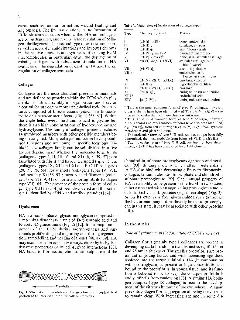
... *- Triple helix .-+
Fig. 1. Schematic rcpresentation of the structure of the triple-helical portion of an interstitial, fibrillar collagen molecule
Table 1. Major sites of localisation of collagen types
Type Chemical formula Tissues
I I1 I11 IV V VI
VTI VIII c
IX X XI XI1 XI11 XIV
bone, tendon, skin cartilage, vitreous skin, blood vessels basement, membrane bone, skin, articular cartilage articular cartilage, skin,
blood vessels anchoring plaques endothelial cells,
cartilage, vitreous hypertrophic cartilage cartilage embryonic skin and tendon endothelial cells embryonic skin and tendon
Descemet's membrane
a This is the most common form of type IV collagen, however, other a chains have been identified - x3(IV), a4(IV), x5(IV) - the precise molecular form of these chains is unknown
This is the most common form of type V collagen, however, other x chains and other molecular forms have also been identified, e.g., [al(V)], from cell cultures; xl(V), a2(V), a3(V) from synovial membranes and placental tissue c The molecular form of type VIII collagen has not yet been fully determined, the most probable form being [EI(VIII)]~, x2(VIII)
The molecular form of type XI11 collagen has not been deter- mined, al(X1II) has been discovered by cDNA cloning
chondroitin sulphate proteoglycans aggrecan and versi- can [93]. Binding proteins which attach preferentially to HA also bind with decreasing affinity to fibronectin, collagen, laminin, chondroitin sulphate and chondroitin sulphate proteoglycans [92]. One unusual property of HA is its ability to be present in the ECM in two states, either associated with an aggregating proteoglycan mole- cule, linked via link proteins (e.g. in cartilage) (Fig. 2), or on its own as a free glycosaminoglycan (although the hyaluronan may not be directly linked to proteogly- can in this state, it may be associated with other proteins [lo]).
In vivo studies
Role of hyaluronan in the,formation of ECM structures
Collagen fibrils (mainly type I collagen) are present in developing rat tail tendon in two distinct sizes, 10-1 5 nm and 25 nm in thickness. The smaller protofibrils are pro- minant in young tissues and with increasing age these coalesce into the larger subfibrils. HA (in combination with proteoglycan) is present at high concentration, is bound to the protofibrils, in young tissue, and its func- tion is believed to be to keep the collagen protofibrils and subfibrils from coalescing [78]. A similar HA/colla- gen complex (type IX collagen) is seen in the develop- ment of the vitreous humour of the eye, where HA again prevents collagen fibril aggregation allowing the vitreous to remain clear. With increasing age and in some dis-

3
Inverse correlation between collagen synthesis and hyaluronan degradation
I I
I I
I ,
Hyaluronan core
I I I I L
I I c=o I I CH3 2
D-Glucuronic acid N-Acetyl-D-Glucosamine
Fig. 2. Diagrammatic representation depicting the association be- tween proteoglycans and the hyaluronan core in a proteoglycan aggregate. Hyaluronan is composed of a repeating disaccharide unit of glucuronic acid and N-acetylglucosamine which is con- nected to individual proteoglycan monomers via link protein. Each proteoglycan monomer consists of a core protein linked to glycosa- minoglycan side chains
eases, this complex is broken down, the collagen fibrils aggregate, and macroscopic fibres can be observed [79, 801. As yet, no direct physical interaction between HA and collagen has been observed in the vitreous [66].
HA also regulates collagen fibrillogenesis and bundle formation in skin, e.g. the skin of the mouse steel mutant has normal levels of both collagens (mainly type I and type 111) and HA. The mutation alters the organisation of the collagen bundles and HA within the ECM result- ing in easily extractible HA [59]. In contrast, low concen- trations of HA are present in the skin of scoliotic chick- ens allowing easily extractible collagen [53]. Thus HA and collagens have to be present at not only the correct concentration but also in the correct conformation for normal bundle formation. In addition, recent studies have demonstrated a direct interaction between HA and type VI collagen in skin [42, 561.
Development and tumour formation. High molecular weight HA and collagens are involved in normal mor- phogenesis and differentiation. The normal sequence of events during morphogenesis often involves the synthesis of (high molecular weight) HA which promotes the mi- gration of mesenchymal cells to appropriate sites. Dur- ing this migration, collagen synthesis is minimal, how- ever, when the appropriate site is reached, HA synthesis is down regulated, the HA is degraded, collagen synthe- sis is up regulated and the cells begin to differentiate. This programme of events has been studied extensively in limb development (involving types I, I1 and I11 colla- gen) [46, 87, 891 but it is also a feature of neural crest cell migration in the gut (type I and type IV collagen) [27], myoblast formation (type I and I11 collagen) [47], somite chondrogenesis (type I1 collagen) [94], testes de- velopment (type I collagen) [26] and the migration of cells into the cardiac jelly of the developing heart (type I collagen) [ 51. Although the precise biological actions are not fully understood, it is believed that the hydrody- namic properties of HA allow the existing ECM spaces to open up further and, perhaps with the aid of fibronec- tin [27], cells are directed through the open spaces until the appropriate site is reached.
A similar process whereby the existing ECM is modi- fied to allow cell migration prior to cell attachment, pro- liferation and differentiation can also be observed in tumour formation and spread. Several invasive tumours are enriched in HA which is synthesised by host fibro- blastic cells [45, 631, and evidence indicates that the site of tumour growth and invasion has enhanced levels of HA synthesis [7, 34, 461. Recently, tumour cells have also been found to synthesise a low molecular weight collagen stimulating factor which results in increased collagen synthesis by host fibroblasts [61]. Taken togeth- er, these observations imply that tumour cells ‘migrate’ (metastasise) by a mechanism recapitulating that ob- served during development; once at the appropriate site the tumour cells then induce HA breakdown and colla- gen synthesis allowing differentiation to proceed. An in- crease in serum levels of low molecular weight HA (i.e. degraded HA) have also been noted in human and ani- mal tumours. Sera from children with a rare type of renal tumour, which has a predilection for bone metasta- sis, have increased levels of HA oligosaccharides whilst sera from a non-bone metastasizing renal tumour pos- sessed no such low molecular mass HA [48].
Wound healing. The normal process of wound healing broadly involves three overlapping phases; inflamma- tion, granulation tissue production and ECM formation and remodelling [13]. The earliest events in wound heal- ing involve the destruction/perturbation of the existing ECM (collagens, proteoglycans, glycoproteins etc.), and the production of a temporary ECM composed of fibro- nectin, HA and fibrin (HA is by far the most abundant glycosaminoglycan synthesised during the first four days). Macrophages, fibroblasts and angiogenic tissue

4
migrate into the wound site, high molecular weight HA and fibrin are degraded, the fibroblasts continue to syn- thesise fibronectin, synthesis of type I11 and then type I collagen is initiated and the macromolecules associate to form a loose ECM. Proteoglycans with associated sulphated glycosaminoglycans accumulate from the fifth day. With increasing time the fibroblasts accumulate ac- tin filaments within their cytoplasm, the cells align up along the collagen fibrils and begin to contract the wound. During repair of skin wounds, re-epithelialisa- tion is also dependant on the synthesis of type V collagen
The amount of collagens present and the amount of contraction determines the degree of scarring observed in a wound. In contrast, wounds to foetal skin demon- strate no scarring [14]. This has been linked to the scar- city of collagens and the abundance of HA in foetal skin [15, 541. The ability of HA to down regulate colla- gen synthesis during wound healing has been demon- strated experimentally by the addition of exogenous HA, both topically and by injection, to wounds [32, 82, 901. In addition to a decrease in collagen synthesis, the degree of subsequent scarring was also reduced. In other reports where the synthesis of HA has been experimentally stim- ulated, e.g. by the addition of interferon gamma, a con- comitant decrease in collagen synthesis is observed and once again the degree of scarring is reduced [29]. Colla- gen synthesis and subsequent scarring are also greatly reduced when the action of transforming growth factor- 8, a known fibrotic agent, is eliminated by the addition of a neutralising antibody [81]. It is not yet known whether HA levels are enhanced in this system.
In each of the cases outlined above, development, tumour formation and wound healing, the migration of cells is triggered by the synthesis of high molecular weight HA, and this migration ceases as the HA is de- graded and collagen synthesis is initiated. A different pattern of events is observed in angiogenesis.
~ 4 1 .
Angiogenesis. Changes in the relative levels and types of collagens and HA in angiogenesis are well known, but the roles played by these macromolecules are un-
Fig. 3. Electron micrograph of a portion of the chorioallantoic membrane of the chick embryo which has been treated with an- giogenic oligosaccharides of hya- luronan. Large amounts of colla- gen fibrils are clearly evident. Bur, 0.1 pm
clear. Type I collagen synthesis is known to be initiated de novo when bovine aortic endothelial cells undergo capillary sprout formation [37], and metabolic reduction of collagen synthesis inhibits capillary formation on the chorioallantoic membrane (CAM) [33], suggesting that type I collagen may play a role as a substrate for endo- thelial cell migration during capillary sprout formation. Recently, type VIII collagen has also been proposed to play a role in angiogenesis [36, 721. Type VIII collagen is produced by vascular endothelial cells but, only if undergoing active cell proliferation [73, 741 and it has been immunolocalised to vascular tissues [38, 431. Type VIII collagen is thought to interact with extracellular matrix components such as HA or some other polyan- ionic glycosaminoglycan and it has been postulated that type VIII collagen may facilitate the assembly of endo- thelial cords and tubes during angiogenesis [35, 361.
Hyaluronun plays u dual role in angiogenesis. An ECM rich in high molecular weight HA inhibits blood vessel formation in chick embryo limb buds and within granu- lation tissue [4, 22, 241, whereas HA oligosaccharides (which should be considered as equivalent to degrada- tion products of high molecular weight HA) stimulate angiogenesis on the CAM [96]. We have shown that this stimulation of angiogenesis on the CAM is associat- ed with the appearance of a very marked increase in the number of collagen fibrils when viewed under the electron microscope (Fig. 3) [49]. The tips of the induced capillary sprouts contain HA but how the breakdown of HA regulates the initiation of angiogenesis is un- known [3]. It is likely that breakdown of HA stimulates angiogenesis associated with synthesis of existing and novel collagens.
The sequence of events observed in angiogenesis differs from those in development and wound healing in that the presence of high molecular weight HA inhib- its endothelial cell migration. Rather, i t is the breakdown products of HA which stimulate migration. Once HA degradation is initiated, collagen synthesis is up regulat- ed as described above. One possible explanation for this could be that endothelial cells require type I collagen

5
as a substrate for cell migration and since they are en- cased within a basement membrane consisting of type IV collagen, one of the initial events must be the de novo synthesis of type I collagen, thus HA is broken down into oligosaccharides capable of stimulating colla- gen synthesis.
The angiogenic potential of HA oligosaccharides may play a role in the repair of internal tissue damage due to disease. Once again, in the repair phase of disease, there appears to be a direct relationship between colla- gens and HA. At early stages of alveolar lung disease, collagen is broken down and HA synthesis is enhanced prior to the migration of fibroblasts with subsequent new collagen synthesis [60]. As this collagen synthesis proceeds, HA synthesis decreases but the HA remaining is closely associated with the invading proliferating cells [60]. It is important to note that degradation of HA will result in the production of angiogenic oligosacchar- ides enabling ready access of nutrients to the repairing tissue.
Hyaluronan - collagen associations in vitro
Many of the observations noted above are emulated by cells in vitro, where most of the experimental data have been obtained from the study of the action of cells in collagen gels or sponges in the presence or absence of external factors such as HA. The general interpretations from these experiments are that in the absence of HA, various types of cells attach to collagen, align along the collagen fibres and do not migrate into the substrate. When HA is added, the collagen expands [93] and cells migrate into the centre of the gel where they begin to synthesise their own ECM [16, 18, 191. The size of the HA and the concentration added influences the cellular behaviour, e.g. low concentrations alter the structure of the collagen and make it more chemoattractant [ 171 whereas a concentration in excess of 5% prevents attach- ment of fibroblasts [83]. This may be in accordance with hyaluronan aiding cell migration, by not allowing strong cell-ECM attachments, in vivo [27]. Since HA and its oligosaccharides have a notable effect on endothelial cells during angiogenesis, we have studied the effect of HA on endothelial cells in vitro.
The addition of moderate amounts of high molecular weight HA to endothelial cells, grown on plastic, has little effect on the morphology and proliferation of the cells. However, if oligosaccharides of two-ten disacchar- ide units are added to the cells, they exhibit increased proliferation and migration in an apparent emulation of angiogenesis [95]. These cells have now been shown to greatly increase the synthesis of both type I and type VIII collagens (Table 2) [69]. These results imply that the production of large bundles of collagen fibres when HA oligosaccharides are added to the CAM (Fig. 3) [49] may be due to the production of type I collagen by endothelial cells in the newly forming capillary sprouts. The production of type VIII collagen has also been ob- served by other workers when endothelial cells are al- lowed to undergo spontaneous angiogenesis in vitro [36].
Table 2. Enhancement of collagen production by bovine aortic en- dothelial cells (BAEC) in vitro by hyaluronan
Type of collagen Relative proportions of collagens produced by BAEC
Control Hyaluronan treated
Type I 1 4.5 Type VIII 1 5.8
Relative proportions of collagens were evaluated from sodium do- decyl sulfate gels following pepsin treatment and laser densitomet- ric analysis
Since type VIII collagen is only produced by actively dividing endothelial cells and as it appears to be stimu- lated by angiogenic factors, or by spontaneous angiogen- esis in vitro, it has been proposed to play an important role in angiogenesis [36]. It seems possible that, in vivo, breakdown of native, high molecular weight HA could result in the production of angiogenic oligosaccharides capable of inducing cell proliferation, collagen synthesis and initiating cell migration and differentiation. If type I collagen is the substrate used for the cells to migrate [33, 371 then type VIII collagen production may be the initial trigger for the proliferation or migration observed [36, 691.
Hyaluronan - collagen associations in vascularisation of cartilage
The observations that the addition of HA oligosacchar- ides to the CAM and to endothelial cells in vitro both result in alterations in collagen synthesis concomitant with the onset of angiogenesis, have led us to propose that HA may interact with collagens and that this inter- action may be the initiating factor in angiogenesis. The models currently in use to study angiogenesis in vivo generally involve the growth of new vessels from existing vessels; we are interested in the growth of blood vessels where none previously existed. Consequently we are ex- amining a model of vascularisation in cartilaginous long bone rudiments where the avascular, cartilage rapidly becomes angiogenic, becomes vascularised and is eroded by bone marrow. Figure4 is a hypothetical model in- volving HA, type X and type VIII collagens, which could account for this vascularisation.
The biological functions of type VIII and type X col- lagens are unknown. Type VIII collagen is very similar in size and chemical characteristics to type X collagen [85, 981, which is a gene product unique to hypertrophic chondrocytes [40, 501. Several reports have linked the production of type X collagen with the process of miner- alisation and have suggested that type X collagen may in some way initiate calcification [St]. However, in the developing cartilage long bone rudiment, particularly of the chick embryo, type X collagen is synthesised in the tibia at 8-9 days of incubation [76] whilst mineralisation does not begin until at least 1 week later [25]. It is inter-

6
Diagrammatic representation of vascularisation of a chick embryo cartilage rudiment
L
7-8 days incubation
R F H F R R Rounded chondrocyte F Flattened chondrocyte H Hypertrophic chondrocyte
-Direction of spread of cell hypertrophy and maior axis of growth
9 days
7 Point of entry of first blood vessels
10-12 days
Erosion of hypertrophic cartilage - Migration and invasion of Mood vessels but always within the hypertrophic cell zone
15- 16 days
Areas of cartilage erosion 0 and replacement by bone marrow
Leading front of blood vessel invasion
There IS 00 mineralisation of the carti\age at this time
Fig. 4. Hypothetical model outlining possible interactions between hyaluronan and collagen during vascularisation of long bone rudi- ments. A cartilaginous long bone rudiment is produced containing the three zones of chondrocytes and an extracellular matrix (ECM) consisting of collagens, proteoglycans, hyaluronan and glycopro- teins (7-8 duys). The hypertrophic chondrocytes synthesise and secrete type X collagen which may induce ECM conformational changes resulting in hyaluronan breakdown, perhaps by activating hyaluronidase. Hyaluronan breakdown may attract vascular tissue to the cartilage/perichondrium junction where endothelial cells di- vide, synthesise type VIII collagen and migrate into the cartilage (9 d u p ) . Erosion of the cartilage ECM occurs producing more hyaluronan oligosaccharides, self-perpetuating the erosion (1 0-12 days). The direction of the erosion is controlled by the continued synthesis of type X collagen by hypertrophic chondrocytes. The leading endothelial cells continue to produce type VIII collagen facilitating further cellular infiltration (15-16 duys). Erosion and vascular invasion ceases when hypertrophic chondrocytes are re- moved and no more type X collagen can be synthesised
esting to note that vascular invasion and bone marrow cavity formation also begin in the centre of the hyper- trophic zone at 8-9 days [ l l , 251. In addition, type X collagen is known to produce a hexagonal structure which is remarkably similar to the hexagonal structure formed by type VIII collagen in the Descemet’s mem- brane of the eye 152, 75, 771. The formation of a regular network in the ECM of hypertrophic cartilage may rep- resent part of a modification process which changes an existing anti-angiogenic ECM into an angiogenic matrix.
We would suggest that the production of type X collagen may influence angiogenesis rather than mineralisation and by altering the existing cartilage ECM, may influ- ence the proliferation and migration of endothelial cells within the well vascularised perichondrium/periosteum. It is important to note that type X collagen selectively binds to HA but not to collagens type TI, VI, IX and XI (A.P.L. Kwan, personal communication). If this is the case then it is possible that type VIII collagen and type X collagen may play a similar role in non-skeletal and skeletal tissues respectively.
In conclusion, cell-cell and cell-matrix interactions are prerequisites for normal tissue development and func- tion. Alterations in these interactions can lead to changes in the nature of the tissue. Most studies on these interac- tions have focused on collagens, in combination with either proteogiycans or glycoproteins such as fibronec- tin. Little information is available on the interactions between collagens and free giycosaminoglycans such as HA. This is surprising since reports often show an in- verse relationship between collagens and HA synthesis both during development and in pathological states. This review draws together these diverse reports and demonstrates that the regulation of synthesis of HA and collagens are intimately related, with enhanced HA syn- thesis as collagen is broken down and vice versa. In addition, it demonstrates that following tissue damage, HA may act as a temporary ECM, allowing the incorpo- ration into the provisional matrix of instructive moie- cules (e.g. fibronectin, tenascin, thrombospondin etc.), until the arrival of appropriate collagen forming cells. Once collagen synthesis begins, HA is broken down and the angiogenic oligosaccharides produced may facilitate the supply of blood and nutrients to the repairing tissue. In this way, HA is involved in the migration of cells, the nutrition of these cells and in the deposition of the final remodelled ECM.
References
1. Akiyama SK, Nagata K, Yamada KM (1990) Cell surface re- ceptors for extracellular matrix components. Biochim Biophys Acta 1031:91-110
2. Alberts B, Bray D, Lewis J, Raff M, Roberts K, Watson JD (1983) The extracellular matrix. In: Molecular biology of the cell. Garland Publishing, London, pp 692-71 3
3. Ausprunk DH, Boudreau CL, Nelson DA (1981) Proteoglycans in the microvasculature. 11. Histochemical localisation in prolif- erating capillaries of the rabbit cornea. Am J Pathol 103:367- 375
4. Balazs EA, Darzynkiewicz Z (1973) The effect of hyaluronic acid on fibroblasts, mononuclear phagocytes and lymphocytes. In: Kulonen E, Pikkarainen J (eds) Biology of the fibroblast. Academic Press, New York, pp 237-252
5. Bernanke DH, Markwald RR (1984) Effects of two glycosamin- oglycans on seeding of cardiac cushion tissue cells into a colla- gen-lattice culture system. Anat Rec 210:25-31
6. Bernard MP, Yoshioka H, Rodriguez E, Rest M van der, Ki- mura T, Ninomiya Y, Olsen BR, Ramirez F (1988) Cloning and sequencing of pro-al(X1) collagen cDNA demonstrates that type XI belongs to the fibrillar class of collagens and re- veals that the expression of the gene is not restricted to cartilagi- nous tissue. J Biol Chem 263:13910-13916
7. Bertrannd P, Girard N, Delpech B, Duval C, D’Anjou J , Dauce JP (1 992) Hyaluronan (hyaluronic acid) and hyaluronectin in the extracellular matrix of human breast carcinomas: Compari- son between invasive and non-invasive areas. Int J Cancer
8. Birk DE, Fitch JM, Babiarz JP, Linsenmayer TF (1988) Colla- gen type I and V are present in the same fibril in the avian corneal stroma. J Cell Biol 106:999-1008
9. Bonanldo P, Russo V, Bucciotti F, Doliana R, Colombatti A (1990) Structural and functional features of the a3 chain indi- cate a bridging role for chicken collagen type VI in connective tissue. Biochemistry 29: 1245-1254
10. Burd DAR, Siebert JW, Ehrlich HP, Garg HG (1989) Human skin and post-burn scar hyaluronan: Demonstration of the as- sociation with collagen and other proteins. Matrix 9: 322-327
11. Caplan AI, Pechak DG (1987) The cellular and molecular em- bryology of bone formation. Bone Min Res 5 : 11 7-1 81
12. Chakrabarti B, Park JW (1980) Glycosaminoglycans: Structure and interaction. CRC Crit Rev Biochem 8:225-313
13. Clark RAF (1989) Overview and general considerations of wound repair. In: Clarke RAF (ed) The molecular and cell biology of wound healing. Plenum Press, New York, pp 3-33
14. Cohen IK (1987) Fetal response to injury in the rabbit. J Pediatr Surg 22 : 640-644
15. DePalma RL, Krummel TM, Durham LA, Michna BA, Thom- as BL, Nelson JM, Diegelman R F (1989) Characterisation and quantitation of wound matrix in the fetal rabbit. Matrix 9: 224- 23 1
16. Docherty R, Forrester JV, Lackie JM, Gregory DW (1989) Glycosaminoglycans facilitate the movement of fibroblasts through three-dimensional collagen matrices. J Cell Sci 92: 263- 270
17. Doillon CJ, Silver FH (1986) Collagen-based wound dressing: Effects of hyaluronic acid and fibronectin on wound healing. Biomaterials 7 : 3-8
18. Doillon CJ, Silver FH, Berg RA (1987) Fibroblast growth on a porous collagen sponge containing hyaluronic acid and fibro- nectin. Biomaterials 8: 195-200
19. Doillon CJ, Silver FH, Olsen RM, Kamath CY, Berg RA (1988) Fibroblast and epidermal cell-type I collagen sponge interactions : Cell culture and human studies. Scanning Microsc
20. Dublet B, Rest M van der (1991) Type XIV collagen: A homo- trimeric molecule extracted from bovine skin and tendon, with a triple helical disulphide-bonded domain homologous to type IX and type XI1 collagens. J Biol Chem 266:6853-6858
21. Dublet B, Oh S, Sugrue SP, Gordon MK, Gerecke DR, Olsen BR, Rest M van der (1989) The structure of avian type XI1 collagen a1 (XII) chains contain 190-kDa non-triple helical ami- no-terminal domains and form homotrimeric molecules. J Biol Chem 264:13150-13156
22. Dvorak HF, Harvey VS, Estrella P, Brown LF, McDonagh J, Dvorak AM (1987) Fibrin containing gels induce angiogene- sis. Implications for tumor stroma generation and wound heal- ing. Lab Invest 57:673-686
23. Eyre DR (1980) Collagen: Molecular diversity in the body’s protein scaffold. Science 207: 131 5-1322
24. Feinberg RN, Beebe DL (1983) Hyaluronate in vasculogenesis. Science 220 : 1 177-1 179
25. Fell HB (1925) The histogenesis of cartilage formation in the long bones of the embryonic fowl. J Morphol Physiol 40:417- 459
26. Fentener van Vlissingen JM, Koch CA, Delpech B, Wensing CJ (1989) Growth and differentiation of the gubernaculum tes- tis during testicular descent in the pig: Changes in the extracel- Mar matrix, DNA content, and hyaluronidase, beta-glucuroni- dase, and beta-N-acetylglucosaminidase activities. J Urol
27. Fujimoto T, Hata J, Yokoyama S, Mitomi T (1989) A study of the extracellular matrix protein as the migration pathway of neural crest cells in the gut: Analyses in human embryos
52:1-6
2: 985-992
142: 837-845
with special reference to the pathogenesis of Hirschsprungs dis- ease. J Pediatr Surg 24: 550-556
28. Gordon MK, Gerecke DR, Olsen BR (1987) Type XI1 collagen: Distinct extracellular matrix component discovered by cDNA cloning. Proc Natl Acad Sci USA 84: 6040-6044
29. Granstein RD, Flotte TJ, Amento EP (1990) Interferons and collagen production. J Invest Derniatol95 : 75s-85s
30. Hardingham TE (1 986) Structure and biosynthesis of proteo- glycans. Rheumatology 10: 143-183
31. Hay ED (1983) Cell and extracellular matrix: their organisation and mutual dependence. Mol Cell Biol2: 509-549
32. Hellstrom S, Laurent C (1987) Hyaluronan and healing of tym- panic membrane perforations. An experimental study. Acta Otolaryngol Suppl442 : 54-61
33. Ingber DE, Folkman J (1988) Inhibition of angiogenesis through modulation of collagen metabolism. Lab Invest 59: 44- 51
34. Iozzo RV, Muller-Glauser W (1985) Neoplastic modulation of extracellular matrix : proteoglycan changes in the rabbit mesen- tery induced by V2 carcinoma cells. Cancer Res 45 : 5677-5687
35. Truela-Arispe ML, Sage EH (1991) Expression of type VIII collagen during morphogenesis of the chicken and mouse heart. Dev Biol 144:107-118
36. Iruela-Arispe ML, Diglio CA, Sage EH (1991) Modulation of extracellular matrix proteins by endothelial cells undergoing angiogenesis in vitro. Arterioscler Thromb 11 :805-815
37. Iruela-Arispe ML, Hasselaar P, Sage EH (1991) Differential expression of extracellular proteins is correlated with angiogen- esis in vitro. Lab Invest 64:174-176
38. Kapoor R, Sakai LY, Funk S, Roux E, Bornstein P, Sage EH (1988) Type VIII collagen has a restricted distribution in spe- cialised extracellular matrices. J Cell Biol 107 : 721-730
39. Keene DR, Sakai LY, Bachinger HP, Burgeson RE (1987) Type 111 collagen can be present on banded collagen fibrils regardless of fibril diameter. J Cell Biol 105: 2393-2402
40. Kielty CM, Kwan APL, Holmes DF, Schor SL, Grant ME (1985) Type X collagen, a product of hypertrophic chondro- cytes. Biochem J 27 : 545-554
41. Kielty CM, Boot-Handford RP, Ayad S, Shuttleworth CA, Grant ME (1990) Molecular composition of type VI collagen: Evidence for chain heterogeneity in mammalian tissues and cultured cells. Biochem J 272:787-795
42. Kielty CM, Cummings C, Whittaker SP, Shuttleworth CA, Grant ME (1991) Isolation and ultrastructural analysis of mi- crofibrillar structures from foetal bovine elastic tissues. J Cell Sci 99 : 797-807
43. Kittelberger R, Davis PF, Flynn DW, Greenhill NS (1990) Dis- tribution of type VIII collagen in tissues: An immunohisto- chemical study. Connect Tissue Res 24: 303-318
44. Kleinman HK, Klebe RJ, Martin GR (1981) Role of collage- nous matrices in the adhesion and growth of cells. J Cell Biol 88:473-485
45. Kundson W, Biswas C, Toole BP (1984) Interactions between human tumor cells and fibroblasts stimulate hyaluronate syn- thesis. Proc Natl Acad Sci USA 81 : 6767-6771
46. Knudson W, Biswas C, Li XQ, Nemec RE, Toole BP (1989) The role and regulation of tumour associated hyaluronan. In: The biology of hyaluronan. Ciba Found Symp 143, pp 150-169
47. Kujawa MJ, Pechak DG, Fiszman MY, Caplan A1 (1986) Hya- luronic acid bonded to cell culture surfaces inhibits the program of myogenesis. Dev Biol 11 3 : 1C16
48. Kumar S, West DC, Ponting J, Gattamameni HR (1989) Sera of children with renal tumours contain low molecular mass hyaluronic acid. Int J Cancer 44:445-448
49. Kumar S, Kumar P, Ponting JM, Sattar A, Rooney P, Pye D, Hunter RD (1992) Hyaluronic acid promotes and inhibits angiogenesis. In: Maragoudakis ME, Lelkes P, Gullino PM (eds) Angiogenesis in health and disease. Plenum Press, New York, pp 253-263

8
SO. Kwan APL, Freemont AJ, Grant ME (1986) Immunoperoxi- dase localisation of type X collagen in chick tibiae. Biosci Rep
51. Kwan APL, Dickson IR, Freemont AJ, Grant ME (1989) Com- parative studies of type X collagen expression in normal and rachitic chicken apiphyseal cartilage. J Cell Biol 109: 1849-1 856
52. Kwan APL, Cummings CE, Chapman JA, Grant ME (1991) Macromolecular organization of chicken type X collagen in vitro. J Cell Biol 114:597-604
53. Lien YH, Fu J, Rucker RB, Scheck M, Abbott U, Stern R (1990) Collagen, proteoglycan and hyaluronidase activity in cultures from normal and scoliotic chicken fibroblasts. Biochim Biophys Acta 1034: 318-325
54. Mast BA, Flood LC, Haynes JH, DePdlma RL, Cohen IK, Diegelmann RF, Krummel TM (1991) Hyaluronic acid is a major component ofthe matrix of fetal skin and wounds: Impli- cation for healing by regeneration. Matrix 11 : 63-68
55. Mayne R, Burgeson RE (1987) (eds) Structure and function of collagen types. Academic Press, New York
56. McDevitt CA, Marcelino J, Tucker L (1991) Interaction of in- tact type VI collagen with hyaluronan. FEBS Lett 294: 167-170
57. Mendler M, Eich-Bender SG, Vaughan L, Winterhalter KH, Bruckner P (1989) Cartilage contains mixed fibrils of collagen types 11, IX and XI. J Cell Biol 108: 191-197
58. Morgelin M, Paulsson M, Hardingham TE, Heinegard D, Engel J (1988) Cartilage proteoglycans: Assembly with hyaluronate and link protein as studied by electron microscopy. Biochem
59. Morrison-Graham K, Bork T, Weston JA (1990) Association between collagen and glycosaminoglycans is altered in dermal extracellular matrix of fetal Steel (Sld/Sld) mice. Dev Biol
60. Nettlebladt 0, Bergh J, Schenholm M, Tengblad A, Haellgren R (1989) Accumulation of hyaluronic acid in the alveolar inter- stitial tissue in bleomycin-induced alveolitis. Am Rev Respir Dis 139 : 759-762
61. Noel A, Munaut C, Nusgens B, Foidart JM, Lapiere ChM (1 992) The stimulation of fibroblast collagen synthesis by neo- plastic cells is modulated by the extracellular matrix. Matrix
62. Orkin RW, Pratt RM, Martin GR (1976) Undersulphated chondroitin sulphate in the cartilage matrix of brachymorphic mice. Dev Biol 50: 82-94
63. Pauli BU, Knudson W (1988) Tumor invasion: A consequence of destructive and compositional matrix alterations. Hum Pa-
64. Pihlajaniemi T, Tamminen M (1990) The ctl chain of type XI11 collagen consists of three collagenous and four non-collagenous domains, and its primary transcript undergoes complex alterna- tive splicing. J Biol Chem 265: 16922-16928
65. Regauer S, Seiler GR, Barrandon Y, Easley KW, Compton CC (1990) Epithelial origin of cutaneous anchoring fibrils. J Cell Biol 11 1 : 2109-21 15
66. Ren ZX, Brewton RG, Mayne R (1991) An analysis by rotary shadowing of the structure of the mammalian vitreous humour and zonular apparatus. J Struct Biol 106: 57-63
67. Rest M van der, Garrone R (1991) Collagen family of proteins. FASEB J 5:2814-2823
68. Rest M van der, Mayne R (1988) Type IX collagen from carti- lage is covalently cross-linked to type I1 collagen. J Biol Chem
69. Rooney P, Wang JM, Kumar P, Kumar S (1993) Angiogenic oligosacchdrides of hyaluronan enhance the production of col- lagen by endothelial cells. J Cell Sci: In Press
70. Ruoslahti E (1989) Proteoglycans in cell regulation. J Biol Chem 264: 13369-13372
71. Ruoslahti E (1990) Extracellular matrix in the regulation of cellular functions. In: Burger MM, Sordat B, Zinkernagel RM (eds) Cell to cell interaction. Karger, Basel, pp 88-98
6: 155-162
J 253:175-185
139:308-313
12:213-220
tho1 19~628-639
263: 1615-161 8
72. Sage H, Iruela-Arispe ML (1990) Type VIII collagen in murine development: Association with capillary formation in vitro. In : Fleischmajer R, Olsen BR, Kuhn K (eds) Structure, molecular biology and pathology of collagen. New York Acad Sci, New York, pp 17-31
73. Sage H, Trueb B, Bornstein P (1983) Biosynthetic and structural properties of endothelial cell type VIII collagen. J Biol Chem
74. Sage H, Pritzl P, Bornstein P (1980) A unique pepsin-sensitive collagen synthesised by endothelial cells in culture. Biochemis- try 19: 5747-5755
75. Sawada H (1982) The fine structure of the bovine Descemet’s membrane with special reference to biochemical nature. Cell Tissue Res 226:241-255
76. Schmid TM, Linsenmayer TF (198s) Developmental acquisi- tion of type X collagen in the embryonic chick tibiotarsus. Dev Biol 107 : 373-381
77. Schmid TM, Linsenmayer T F (1990) Immunoelectron micros- copy of type X collagen: Supramolecular forms within embry- onic chick cartilage. Dev Biol 138 : 53-62
78. Scott JE (1990) Proteoglycan: collagen interactions and subfi- brillar structure in collagen fibrils : Implications in the develop- ment and ageing of connective tissues. J Anat 169: 23-35
79. Sebag J (1987) Age-related changes in human vitreous struc- ture. Graefes Arch Clin Exp Ophthalmol225: 89-93
80. Sebag J, Balazs EA (1989) Morphology and ultrastructure of human vitreous fibres. Invest Ophthalmol Vis Sci 30: 1867-1 871
81. Shah M, Foreman DM, Ferguson MWJ (1992) Control of scar- ring in adult wounds by neutralising antibody to transforming growth factor-/I. Lancet 339:213-214
82. Songer MN, Ghosh L, Spencer DL (1990) Effects of sodium hyaluronate on peridural fibrosis after lumbar laminotomy and discectomy. Spine 1 5 : 550-554
83. Srivastava S, Gorham SD, Courtney JM (1990) The attachment and growth of an established cell line on collagen, chemically modified collagen and collagen composite surfaces. Biomater- ials 11 : 162-168
84. Stenn KS, Madri JA, Roll FJ (1979) Migrating epidermis pro- duces AB, collagen and requires continued collagen synthesis for movement. Nature 277:229-232
85. Thomas JT, Kwan APL, Grant ME, Boot-Handford RP (1991) Isolation of cDNAs encoding the complete sequence of bovine type X collagen. Biochem J 273: 141-148
86. Timpl R (1989) Structure and biological activity of basement membrane proteins. Eur J Biochem 180:487-502
87. Toole BP (1981) Glycosaminoglycans in morphogenesis. In : Hay ED (ed) Cell biology of the extracellular matrix. Plenum Press, New York, pp 259-294
88. Toole BP, Goldberg RL, Chi-Rosso G, Underhill CB, Orkin RW (1984) Hyaluronate-cell interactions. In: Trelstad RL (ed) The role of extracellular matrix in development. Alan R Liss, New York, pp 43-66
89. Toole BP, Munaim SI, Welles S, Knudson CB (1989) Hyaluron- ate-cell interactions and growth factor regulation of hyaluron- ate synthesis during limb development. In: The biology of hya- luronan. Ciba Found Symp 143, pp 138-149
YO. Trabucchi E, Preis Baruffaldi F, Baratti F, Montorsi W (1986) Topical treatment of experimental skin lesions in rats: Macros- copic, microscopic and scanning electron-microscopic evalua- tion of the healing process. Int J Tissue React 8 : 533-544
91. Trelstad RL, ed (1984) The role of extracellular matrix in devel- opment. Alan R Liss, New York
92. Turley E, Moore D (1984) Hyaluronate binding proteins also hind to fibronectin, laminin and collagen. Biochem Biophys Res Commun 121 : 808-81 4
93. Turley E, Erickson CA, Tucker RP (1985) The retention and ultrastructural appearances of various extracellular matrix mol- ecules incorporated into three-dimensional hydrated collagen lattices. Dev Biol 109: 347-367
258: 13391-13401

9
94. Vassan NS, Miller E (1985) Somite chondrogenesis in vitro: Differential induction by modified matrix - a biochemical and morphological study. Dev Growth Differ 27: 405-41 7
95. West DC, Kumar S (1989) The effect of hyaluronate and its oligosaccharides on endothelial cell proliferation and monolay- er integrity. Exp Cell Res 183 ; 179-1 96
96. West DC, Hampson IN, Arnold F, Kumar S (1985) Angiogene- sis induced by degradation products of hyaluronic acid. Science 228.1324-1326
97. Yamaguchi N, Benya PD, Rest M van der, Ninomiya Y (1989) The cloning and sequencing of al(VII1) collagen cDNAs dem- onstrate that type VIII collagen is a short chain collagen and contains triple-helical and carboxyl-terminal non-triple-helical domains similar to those of type X collagen. J Biol Chem
98. Yamaguchi N, Mayne R, Ninomiya A (1991) The a1 (VIII) collagen gene is homologous to the a1 (X) collagen gene and contains a large exon encoding the entire triple helical and carboxyl-terminal non-triple helical domains of the a1 (VIII) polypeptide. J Biol Chem 266:4508-4513
264 : 16022-1 6029