intuitive 3d catheter guidance - philips
TRANSCRIPT

Intuitive 3D catheter guidancePhilips EP navigator and 3D rotational scan
452296278537.indd 1 26-7-2012 10:38:02

Intuitive 3D catheter guidance2
For patients with symptomatic paroxysmal atrial fibrillation (AF) who do not respond adequately to drug therapy, catheter ablation has emerged a highly effective therapeutic option and is increasingly being regarded as the standard of care. The electrical isolation of the pulmonary veins (PVs) requires the best possible understanding of the anatomy of a patient’s left atrium (LA) and PVs to establish the exact position and orientation of the PV ostia. Isolation of the PVs is always a technically challenging procedure, even in the hands of an experienced electrophysiologist. Each patient presents their own innate difficulties due to the complexity and variability of the LA-PV anatomy.
“We used EP navigator to successfully treat a patient who had persistent AF since 1999. This patient had two prior ablations in 2001 and 2009, and used several antiarrhythmic medications for over 10 years. During the third procedure, we had to target the fibrillation which was intermittent. Altogether this was a really difficult case. Ablation at the mitral isthmus line and at the roof recovered after a complete block and required re-ablation. The EP navigator images were very helpful during this procedure. The loop shows you the most external aspect of the left atrium. Registration is also very fast. It hardly takes any time.”
Prof. P. Jaïs, Hopital Cardiologique Haut-Lévéque, Bordeaux-Pessac, France
3D - efficiently and accurately guide your catheter
See Professor Jaïs discussing the treatment of this patient and demonstrating the use of EP navigator on-line at http://www.philips.com/ep
Real-time imaging of cardiac structures is a cornerstone of the electrophysiologist’s ability to efficiently and accurately guide a catheter through the anatomy of a beating heart. Conventional fluoroscopy plays an essential role in this, but 2D
imaging has obvious limitations when navigating through complex and highly variable 3D anatomical structures. The simultaneous provision of 3D data with real-time 2D imaging has the potential to increase the speed and precision of procedures.
452296278537.indd 2 26-7-2012 10:38:09

Philips EP navigator and 3D rotational scan 3
3D - efficiently and accurately guide your catheter
452296278537.indd 3 26-7-2012 10:38:16

Intuitive 3D catheter guidance4
EP navigator and 3D rotational scan EP navigator is an intuitive real-time imaging system, developed by Philips, which allows the electrophysiologist to follow the precise position of catheters during navigation and AF ablation. EP navigator has been developed with the full support of a team of expert electrophysiologists, identifying and addressing their precise needs and making use of their practical knowledge of the complexities of cardiac anatomy. The 3D imaging registered with live 2D fl uoroscopy provided by EP navigator has become an essential tool in many of the foremost EP labs around the world. Its utility has been demonstrated in clinical studies.1, 2
3D rotational scan While previously acquired CT or MR images can be imported into EP navigator, this may not always be the optimal solution for the patient or for the electrophysiologist. Acquiring CT images involves exposing the patient to a certain radiation dose and requires an extra hospital visit. Also, the cardiac anatomy of the patient may have changed between the point of acquiring the CT or MR images and the time of the ablation procedure. A recent development in the functionality of EP navigator is reduced angular 3D rotational scanning, discussed in detail in subsequent pages. 3D rotational scanning (previously known as 3D ATG) immediately prior to the procedure itself, quickly provides excellent, accurate images of cardiac structures and helps to reduce radiation dose.
“We’ve had several cases where we were dealing with very diffi cult anatomy and EP navigator really helps.”
Dr. M. V. Orlov, St Elizabeth’s Medical Center, Boston, USA
452296278537.indd 4 26-7-2012 10:38:24

Philips EP navigator and 3D rotational scan 5
EP navigator facilitates intuitive 3D catheter image guidance during AF ablation procedures. It provides detailed 3D anatomy, which can be overlaid onto live fl uoroscopy, removing the need for a mapping system, or exported to a compatible mapping system to help minimize dose and reduce mapping time.
EP navigator - a clear insight into cardiac anatomy
Benefi ts of EP navigator • Confi dence
Navigate and ablate in a familiar context using precisely registered 3D and 2D live fl uoroscopy images.
• Effi ciency Automatic image segmentation, rapidly identifying cardiac structures.
• Flexibility Supports the import of preprocedural CT or MRI data, or the acquisition of an intraprocedural 3D rotational scan.
Live fl uoroscopy or a compatible electroanatomical mapping system can be used for catheter image guidance.
Import imaging datasets Prior to undergoing ablation, some patients will have already undergone cardiac CT or MR imaging. Preprocedural CT data or MRI data can be imported into EP navigator to provide a precise 3D anatomical image of the patient’s left atrium and pulmonary veins. This fl exibility of EP navigator may provide the best option for some patients, reducing the need for further radiation exposure. A valuable alternative to preprocedural imaging is the import of intraprocedural 3D rotational scan data.
Fast and easy segmentation Imported images undergo zero-click segmentation using a fully automatic algorithm. All non-relevant structures are fi ltered out, leaving only the cardiac structures of interest. Segmentation of the left atrium takes only about 2 minutes. Following segmentation, the electrophysiologist can extract the right and left atria, right and left ventricles, myocardium, aorta, coronary sinus, vena cava, and bronchi.
“The advantage is that we have a more up-to-date left atrium volume. It is the volume and the fi lling status of the patient during that date. The advantage with the 3D rotational angio is that it happens in only one room, which is the cath lab and there is no interaction needed with the radiology department etcetera. And fi nally the advantage is that using the 3D rotational angio allows a more actual registration compared to integration with CT or MRI.”
Prof. Dr. M. Duytschaever, St. Jan Hospital, Bruges, Belgium
452296278537.indd 5 26-7-2012 10:38:30

Intuitive 3D catheter guidance6
Live Overlay in EP navigator
Point Tagging in EP navigator
Registration and live overlay EP navigator precisely registers the 3D volume with the live 2D fluoroscopy images obtained in the EP lab and displays these on a single monitor. Once the 3D image is transparently overlaid onto the fluoroscopy image, the exact position of each catheter in relation to the 3D cardiac structures is clearly revealed. Navigation is supported by the low-radiation-dose fluoroscopy. The EP navigator image moves in-sync with the C-arm geometry of the Allura Xper system, allowing the electrophysiologist to choose the viewing angle of the composite image, providing optimal support for catheter and device navigation.
Point TaggingIn combination with the 3D overlay functionality, EP navigator offers Point Tagging to accurately mark ablation points. The point tagging functionality can be used in combination with all catheters in the field.
452296278537.indd 6 26-7-2012 10:38:31

Philips EP navigator and 3D rotational scan 7
Viewing the heart from inside out Endoview is a useful feature of EP navigator, helping to provide the most complete possible picture of the cardiac anatomy of the patient. It allows the electrophysiologist to look inside the 3D structures to view the posterior side of the atrial wall, as well as the ostia of the pulmonary veins, the ridge, and other cardiac features.
Endoview in EP navigator
“I think when we did the first procedure with EP navigator here it was like a new dimension, because there was so much new information… 3D information adds a certain level of confidence when you do mapping and ablation procedures.”
Dr. M. V. Orlov, St Elizabeth’s Medical Center, Boston, USA
452296278537.indd 7 26-7-2012 10:38:32

Intuitive 3D catheter guidance8
In certain cases the importation of pre-existing data for overlay with live fluoroscopy may be the best approach. The flexibility to utilize radiation-free MRI data, for example, may be a valuable option for some patients. However, preprocedural data sets are usually obtained at least one day before ablation therapy and sometimes much earlier. It is possible that the volume status of the patient at the time of the ablation procedure is different from the day that the 3D volume was obtained.3 Additionally, during the acquisition of CT and MRI scans, the patient’s arms are held in the up position while during the ablation procedure they are generally kept at the patient’s sides. This can result in a different chest position during preoperative imaging. Clinical studies have shown that intraprocedural 3D rotational scanning offers an efficient solution to these problems.3, 4, 6, 7
Tang et al. (2009) performed a clinical study including 46 patients undergoing ablation therapy.6 All patients had preprocedural CT scans and also underwent intraprocedural 3D rotational scanning. Successful 3D reconstruction was achieved in 44 patients (95.7%). A direct comparison of
images reconstructed from CT and 3D rotational scans found good correlations in LA volume and PV ostial diameter.
A similar clinical study by Li et al. (2009) compared preprocedural CT scanning and intraprocedural 3D rotational scanning in 30 patients undergoing ablation therapy.4 The study showed close concordance between PV ostial diameters measured by the two imaging modalities. Li et al. concluded that, “Our findings demonstrate that 3DATG provides real-time images that are easily segmented and registered with excellent accuracy and concordance with live fluoroscopy and CT.”
Kriatselis et al. (2009) performed a clinical study of 70 patients undergoing ablation therapy.3 A 3D rotational scan was performed during adenosine-induced asystole. In 90% of patients, the intersection or ‘ridge’ between the left superior PV and left atrial appendage was successfully delineated and could be clearly visualized. The study found that, “…contrast-enhanced rotational X-ray angiography of the LA and PVs can be safely performed during adenosine-induced ventricular asystole, and it provides detailed and
exact anatomical information about the size of the PV ostia, peripheral branches, and the presence of additional PVs.”
A 3D rotational scan results in an excellent 3D image of cardiac structures at the precise time of the procedure.5 The 3D image is directly available for use and is of comparable quality to CT imaging. The electrophysiologist can work independently of radiology departments.
The previously discussed clinical study by Tang et al. (2009) comparing preprocedural CT and intraprocedural 3D rotational scanning followed by successful ablation in 44 patients found that the radiation dose used during the 3D rotational scan was significantly lower than had been used during the preprocedural CT scan (2.7 ± 0.9 mSv vs 24.9 ± 3.1 mSv; p < 0.001).6 Similarly, the clinical comparison of preprocedural CT and intraprocedural 3D rotational angiography performed by Li et al. (2009) found significantly lower radiation doses associated with 3D rotational scanning compared with CT (2.1 ± 0.3 mSv vs 13.8 ± 2.4 mSv; p < 0.001).4 3D rotational scan using a reduced angular range
The 3D rotational scan - accurate visualization of 3D anatomy
“EP Navigator with 3DATG has been an incredibly useful tool…with a preprocedural CT, volume changes are surprisingly high. CT is not ideal, 3DATG has made a big difference in anatomical accuracy”.
Dr. L. Chinitz, NYU, New York, USA
Why use a 3D rotational scan?
452296278537.indd 8 26-7-2012 10:38:32
Philips EP navigator and 3D rotational scan 9
The 3D rotational scan - accurate visualization of 3D anatomy
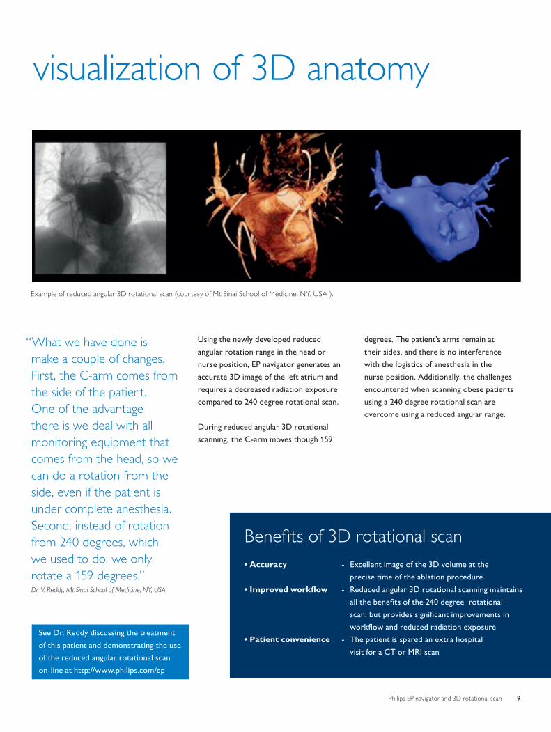
Using the newly developed reduced angular rotation range in the head or nurse position, EP navigator generates an accurate 3D image of the left atrium and requires a decreased radiation exposure compared to 240 degree rotational scan.
During reduced angular 3D rotational scanning, the C-arm moves though 159
degrees. The patient’s arms remain at their sides, and there is no interference with the logistics of anesthesia in the nurse position. Additionally, the challenges encountered when scanning obese patients using a 240 degree rotational scan are overcome using a reduced angular range.
“What we have done is make a couple of changes. First, the C-arm comes from the side of the patient. One of the advantage there is we deal with all monitoring equipment that comes from the head, so we can do a rotation from the side, even if the patient is under complete anesthesia. Second, instead of rotation from 240 degrees, which we used to do, we only rotate a 159 degrees.”
Dr. V. Reddy, Mt Sinai School of Medicine, NY, USA
See Dr. Reddy discussing the treatment of this patient and demonstrating the use of the reduced angular rotational scan on-line at http://www.philips.com/ep
Example of reduced angular 3D rotational scan (courtesy of Mt Sinai School of Medicine, NY, USA ).
Benefits of 3D rotational scan • Accuracy - Excellent image of the 3D volume at the
precise time of the ablation procedure• Improved workflow - Reduced angular 3D rotational scanning maintains
all the benefits of the 240 degree rotational scan, but provides significant improvements in workflow and reduced radiation exposure
• Patient convenience - The patient is spared an extra hospital visit for a CT or MRI scan
452296278537.indd 9 26-7-2012 10:38:33

Intuitive 3D catheter guidance10
It is essential that whether the electrophysiologist is performing fast, fluoro-based procedures or more elaborate, low-dose mapping procedures, EP navigator can be tailored to accommodate their needs. Partnering with other companies that produce EP solutions, so that EP navigator can be seamlessly interfaced with their systems, has been a vitally important part of the development process. Easy integration with multi-vendor equipment provides enhanced flexibility, reducing costs and enabling secondary hospitals to expand into performing electrophysiology procedures.
Collaboration is key to success
Export of 3D volume to compatible mapping systems EP navigator creates and automatically segments the 3D anatomy which can be transferred to a compatible mapping system. Biosense Webster Carto™ and St Jude Medical Ensite™ are fully supported. The procedure can then be carried out mainly on the mapping system to help manage dose and reduce mapping time. The EP navigator fluoroscopy overlay is always available, to help confirm the catheter positions of the mapping system, if needed. In addition, robotic catheter control from Hansen is integrated into the workflow, and fully supported and guided by Philips imaging technology.Transfer of data to a compatible mapping system is achieved seamlessly via the network. To achieve this, the EP lab requires networking applications for their mapping system. The Carto 3™ system requires the CartoMerge module and the Ensite Velocity™ system requires the EnSite Courier™ module.
Biosense Webster Carto with exported 3D anatomy from EP navigator
“Actually I was quite surprised when we got the integration of the rotational angiography into the Carto system and how easily it works. There is a direct connection between the Carto system and the rotational angiography which makes it very easy and fast to integrate the 3D rotational angiography into the Carto systems.”
Dr. R. Tilz, Asklepios Klinik St. George, Department of Cardiology, Hamburg, Germany
See the full interview with Dr Tilz on-line at http://www.philips.com/ep
452296278537.indd 10 26-7-2012 10:38:40

Philips EP navigator and 3D rotational scan 11
References
1. Knecht S, et al. Computed tomography-fluoroscopy overlay evaluation during catheter ablation of left atrial arrhythmia. Europace. 2008;10:931-8.
2. Knecht S, et al. Prospective randomized comparison between the conventional electroanatomical system and three-
dimensional rotational angiography during catheter ablation for atrial fibrillation. Heart Rhythm. 2010; 7:459-465.
3. Kriatselis C, et al. A new approach for contrast-enhanced X-ray imaging of the left atrium and pulmonary veins for atrial
fibrillation ablation: rotational angiography during adenosine-induced asystole. Europace. 2009;11:35-41.
4. Li JH, et al. Segmentation and registration of three-dimensional rotational angiogram on live fluoroscopy to
guide atrial fibrillation ablation: a new online imaging tool. Heart Rhythm. 2009;6:231-7.
5. Orlov MV. How to perform and interpret rotational angiography in the electrophysiology laboratory. Heart Rhythm. 2009;6:1830-6.
6. Tang M, et al. Reconstructing and registering three-dimensional rotational angiogram of left atrium
during ablation of atrial fibrillation. Pacing Clin Electrophysiol. 2009;32:1407-16.
7. Tang M, et al. Optimal fluoroscopic projections for angiographic imaging of the pulmonary vein ostia: lessons learned from
the intraprocedural reconstruction of the left atrium and pulmonary veins. Europace. 2010;12:37-44.
“The big advantage is that by merging the Carto with the real 3D anatomy of the patient, is that you can more anatomically correct delineate the function between the left atrium and pulmonary vein entrance and this allows us to ablate in an efficient and effective way.”
Prof. Dr. M. Duytschaever, St. Jan Hospital, Bruges, Belgium
452296278537.indd 11 26-7-2012 10:38:46

© 2012 Koninklijke Philips Electronics N.V.All rights are reserved.
Philips Healthcare reserves the right to make changes in specifications and/or to discontinue any product at any time without notice or obligation and will not be liable for any consequences resulting from the use of this publication.
Printed in The Netherlands.4522 962 78537 * JuL 2012
Please visit www.philips.com/EP
Philips Healthcare is part of Royal Philips Electronics
How to reach uswww.philips.com/[email protected]
Asia+49 7031 463 2254
Europe, Middle East, Africa+49 7031 463 2254
Latin America+55 11 2125 0744
North America+1 425 487 7000800 285 5585 (toll free, uS only)
452296278537.indd 12 26-7-2012 10:38:46