introduction to urinalysis laboratory procedures
TRANSCRIPT

Introduction to Urinalysis
Laboratory Procedures

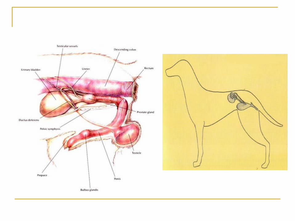
Urinary System
Designed to maintain a balance between fluid, electrolytes, and acid-base homeostasis by selectively eliminating waste products from the body.
Urine is formed through glomerular filtration, tubular reabsorption, and tubular secretion (remember everything you were taught in A&P). These are influenced by hormones

Medical Terminology
Pollakiuria Frequent urination
Polyuria Increased urine output or production
Oliguria Decrease in the formation or elimination of urine
Anuria Complete absence of urine formation or elimination
Dysuria Difficult urination

Urinalysis
Urine collection can be accomplished through mid-stream free catch, manual expression, catheterization, and cystocentesis.

Advantages to Urinalysis
Fast Simple Inexpensive Provides useful information (urinary tract
and/or other body systems)

Voided Urine Sample
Easiest to obtain May be contaminated
from distal genital tract Not satisfactory if
examining for bacteria.

Voided Urine Sample Collection Use a clean container Wash prepuce or vulva (when possible) Try to collect mid-stream urine

Disadvantages to Voided Sample Contamination Difficult in cats May be difficult in easily scared dogs and
short breeds
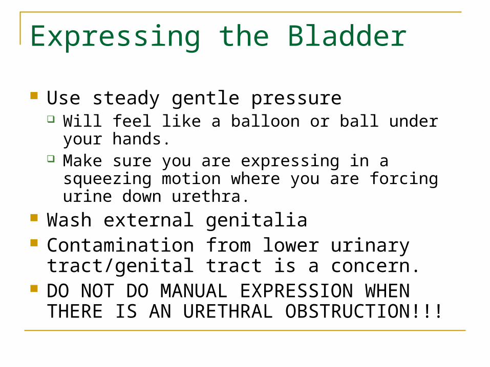
Expressing the Bladder
Use steady gentle pressure Will feel like a balloon or ball under your hands. Make sure you are expressing in a squeezing
motion where you are forcing urine down urethra. Wash external genitalia Contamination from lower urinary tract/genital
tract is a concern. DO NOT DO MANUAL EXPRESSION
WHEN THERE IS AN URETHRAL OBSTRUCTION!!!
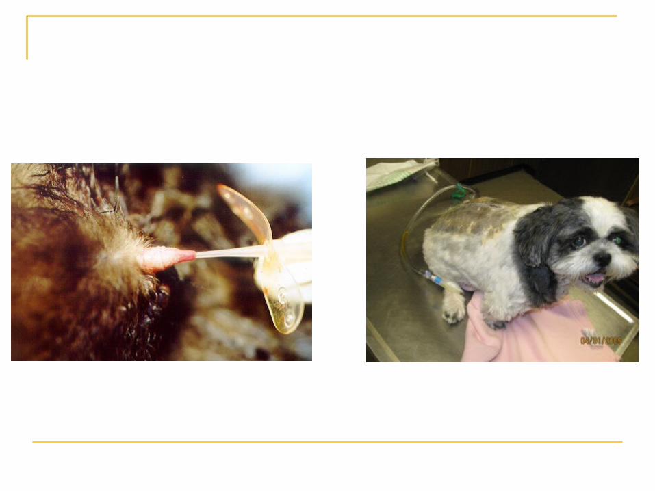
Urinary Catheterization
Act of placing a catheter through urethra into bladder
Advantages: Less possibility of contamination from lower
genital tract. Helpful in obese animals when bladder is difficult
to palpate Disadvantages
Trauma to sensitive urethral mucosa Possible contamination
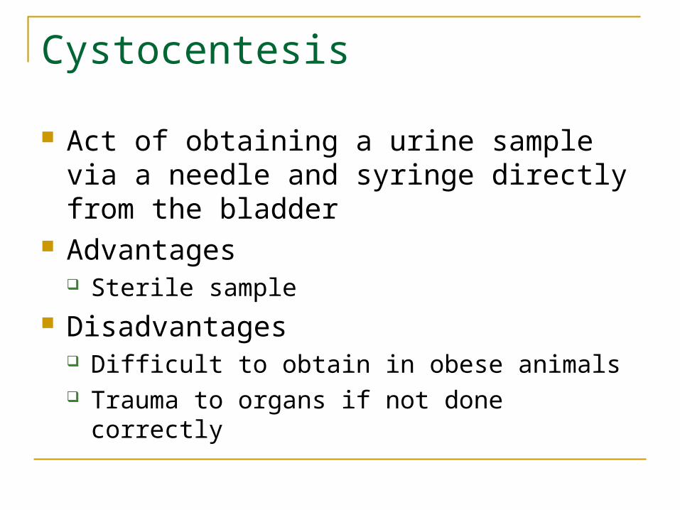
Cystocentesis
Act of obtaining a urine sample via a needle and syringe directly from the bladder
Advantages Sterile sample
Disadvantages Difficult to obtain in obese animals Trauma to organs if not done correctly


Urine Sample Preservation
Analyze all urine within 30 minutes if possible
May refrigerate for 6-12 hours if needed Bring to room temperature
before anlaysis Morning samples are
more concentrated If allowed to stand at
room temperature, may get false results.

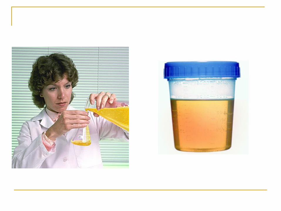
Physical Characteristics of Urine Color Transparency Odor Specific gravity Volume

Urine Color
Normal color is due to pigments called urochromes
Normal: light yellow to amber color Abnormals:
Red: blood (hematuria) Reddish-brown: Hemoglobin or Myoglobin Dark yellow-brown: Bilirubin (bilirubinuria) Orange-Reddish brown: Normal in rabbits

Urine Transparency
Clear vs. Cloudy Cloudy could indicate increase cells, mucus,
casts, crystals and bacteria. Horses and some rabbits have cloudy urine
due to high content of mucus and calcium carbonate crystals.
Urine Odor
Not Very diagnostic Strong odor may suggest bacterial production Male cats, goats, and pigs have a very strong
urine odor
Urine Specific Gravity
Measure urine concentration which is dependent on the number, molecular size, and weight of urine solutes
Measures the density of urine as it compares to water
Specific gravity of water is always 1.000

“Normal” Urine Specific Gravities Man: 1.003-1.037 Dog: 1.013-1.030 Cat: 1.013-1.050 Horse: 1.015-1.045
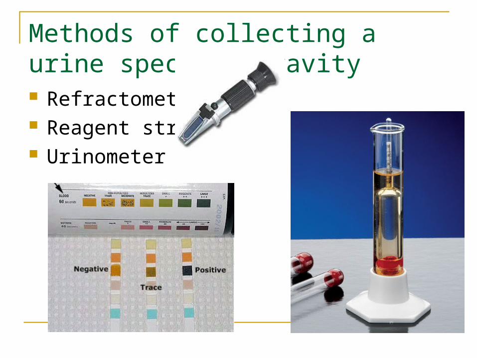
Methods of collecting a urine specific gravity Refractometer Reagent strip Urinometer
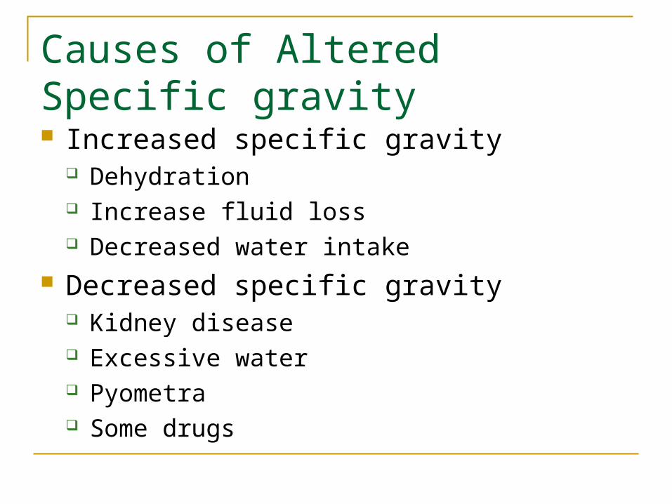
Causes of Altered Specific gravity Increased specific gravity
Dehydration Increase fluid loss Decreased water intake
Decreased specific gravity Kidney disease Excessive water Pyometra Some drugs

Urine Volume
Influenced by several factors Water intake Size of animal Species of animal

Crystals
The presence of crystals in the urine is called crystalluria.
There are many types of crystals that can form in urine, we are going to cover just a few. (More to come in Clinical Pathology!)
Crystalluria may or may not be of clinical significance. Certain crystals form as a result of elements being
secreted into the urine by normal renal activity.

Crystal formation
Crystal formation depends on the pH of the patients urine.
Some crystals form in acidic urine, while others form in basic or neutral urine.
If a urine sample is allowed to stand and cool to room temperature, the number of crystals in the urine will increase because the material that forms the crystals is less likely to dissolve at cooler temperatures.
Types of crystals
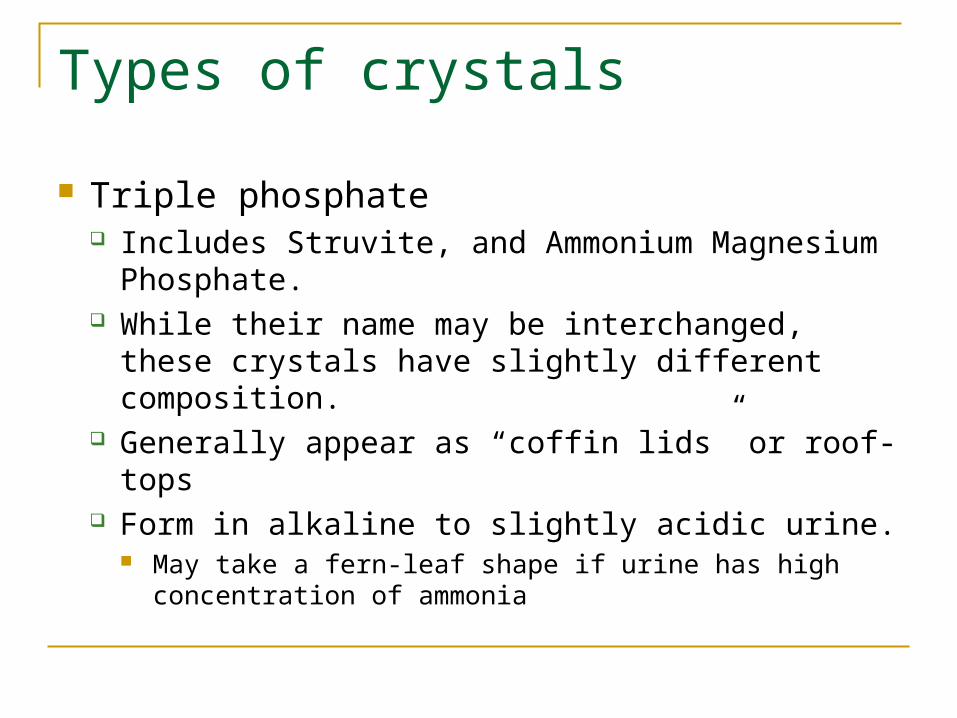
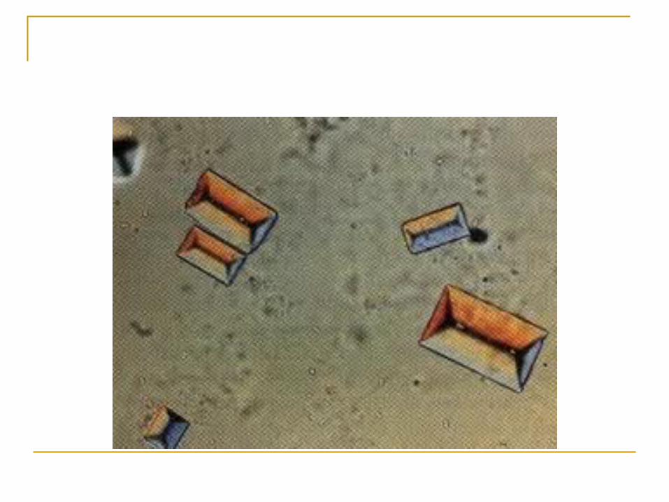
Triple phosphate Includes Struvite, and Ammonium Magnesium
Phosphate. While their name may be interchanged, these
crystals have slightly different composition. Generally appear as “coffin lids” or roof-tops Form in alkaline to slightly acidic urine.
May take a fern-leaf shape if urine has high concentration of ammonia

Calcium Oxalate Dihydrate
Generally appear as small squares with a visible “X” across the top of the crystal.
Most often form in acidic and neutral urine. Are commonly seen in small numbers in dogs
and horses. If seen in large numbers, can indicate calculi
formation.

Calcium Oxalate Monohydrate May be small and “dumbbell” shaped, can
also appear as a slat from a picket fence. Generally form in any pH urine, but are a key
indicator if an animal is experiencing ethylene glycol toxicity!

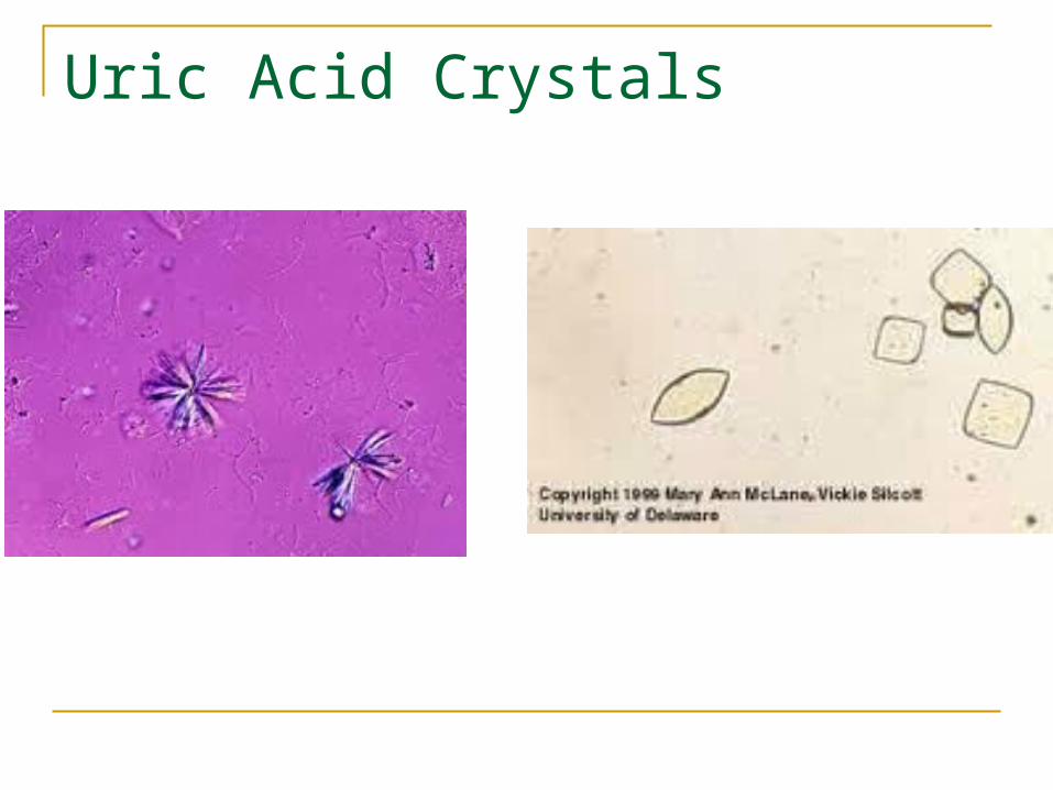
Uric Acid/Ammonium Urate Crystals Uric Acid crystals often appear as yellow-
brown “rosettes” or as diamond-shaped “plates”. Can form in any pH urine!
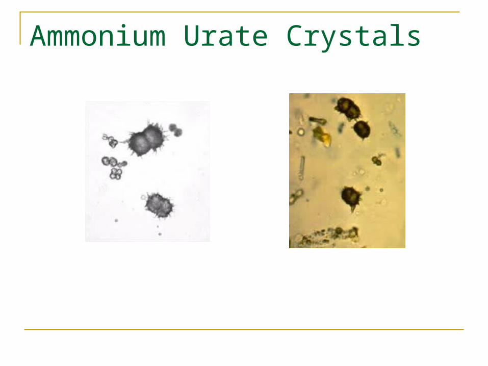
Ammonium Urates often appear as “thorn-apples” Generally form in neutral to alkaline urine.
Are very rarely seen in dogs and cats. (Except Dalmatians!!)
Uric Acid Crystals
Ammonium Urate Crystals