introduction to pathology - ksumsc.comksumsc.com/download_center/1st/1. foundation block... ·...
TRANSCRIPT
INTRODUCTIONTOPATHOLOGY
SUFIAHUSAIN(AssociateProfessor&Consultant)
PATHOLOGY
COLLEGEOFMEDICINE
KSU
RIYADH
SEPTEMBER2019
OBJECTIVES:
Thestudentshould:
A. Understands the role of pathology and its various subspecialities in the diagnostic process with special emphasis on histopathology and cytology.
B. Understands the meaning of the terminology used during the study of a disease like aetiology, pathogenesis, prognosis, sequelae, symptoms, signs, incidence etc.
C. Role of diagnostic pathology in disease management.
D. Be aware of some of the principle techniques used in pathology like light microscopy, cytology, immunohistochemistry and molecular pathology.
E. Have a basic knowledge of the definition of autopsy and its indications.

DEFINITIONOFPATHOLOGY
• Pathology is the study of disease by scientific methods. It is the study of changes which occur in cells and tissues as a result of any injury to the cell or tissue.
• Disease is defined as an abnormality in structure or function of any part of the body.
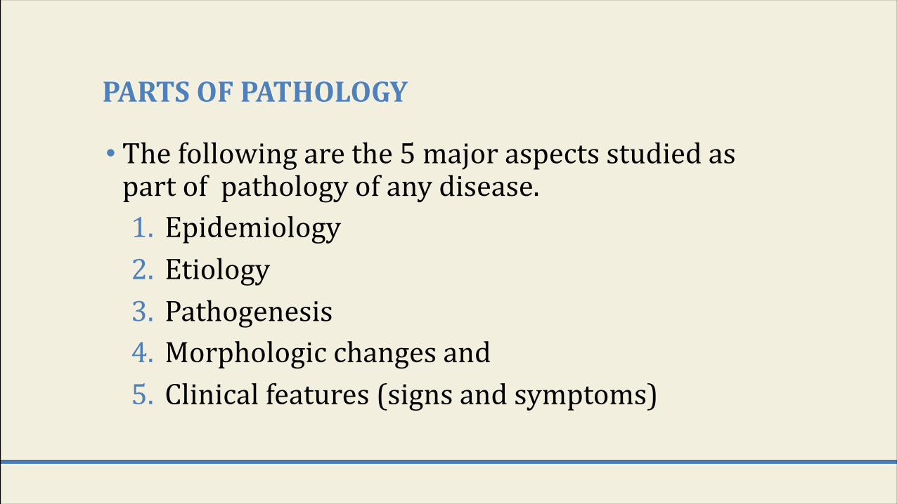
PARTSOFPATHOLOGY
• The following are the 5 major aspects studied as part of pathology of any disease.1. Epidemiology2. Etiology3. Pathogenesis4. Morphologic changes and5. Clinical features (signs and symptoms)
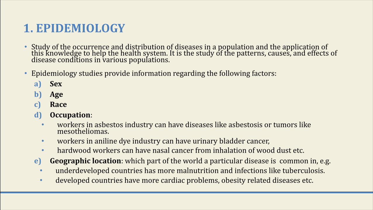
1.EPIDEMIOLOGY• Study of the occurrence and distribution of diseases in a population and the application of
this knowledge to help the health system. It is the study of the patterns, causes, and effects of disease conditions in various populations.
• Epidemiology studies provide information regarding the following factors:a) Sexb) Agec) Raced) Occupation: • workers in asbestos industry can have diseases like asbestosis or tumors like
mesotheliomas. • workers in aniline dye industry can have urinary bladder cancer, • hardwood workers can have nasal cancer from inhalation of wood dust etc.
e) Geographiclocation: which part of the world a particular disease is common in, e.g. • underdeveloped countries has more malnutrition and infections like tuberculosis. • developed countries have more cardiac problems, obesity related diseases etc.
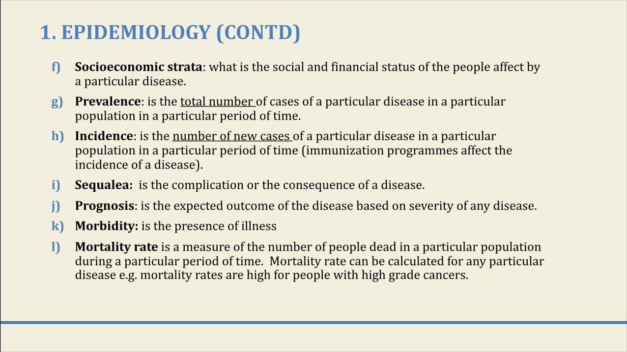
1.EPIDEMIOLOGY(CONTD)f) Socioeconomicstrata: what is the social and financial status of the people affect by
a particular disease.g) Prevalence: is the total number of cases of a particular disease in a particular
population in a particular period of time.h) Incidence: is the number of new cases of a particular disease in a particular
population in a particular period of time (immunization programmes affect the incidence of a disease).
i) Sequalea: is the complication or the consequence of a disease.j) Prognosis: is the expected outcome of the disease based on severity of any disease. k) Morbidity:is the presence of illnessl) Mortalityrate is a measure of the number of people dead in a particular population
during a particular period of time. Mortality rate can be calculated for any particular disease e.g. mortality rates are high for people with high grade cancers.

WHATISTHEPURPOSESORIMPORTANCEOFEPIDEMIOLOGY?
1. To investigate the extent of a disease in a community.2. To study natural pattern/history and prognosis of disease.
3. To identify causes and risk factors.4. To provide good health care based on the findings.
5. To recommend and assist in various health programmes to prevent or treat disease (preventive and therapeutic measures), e.g. immunizations and screening programs for different disease etc.
6. To evaluate all health care facilities and programs.7. Provide information on public health in order to help the health
care system and develop health policies.
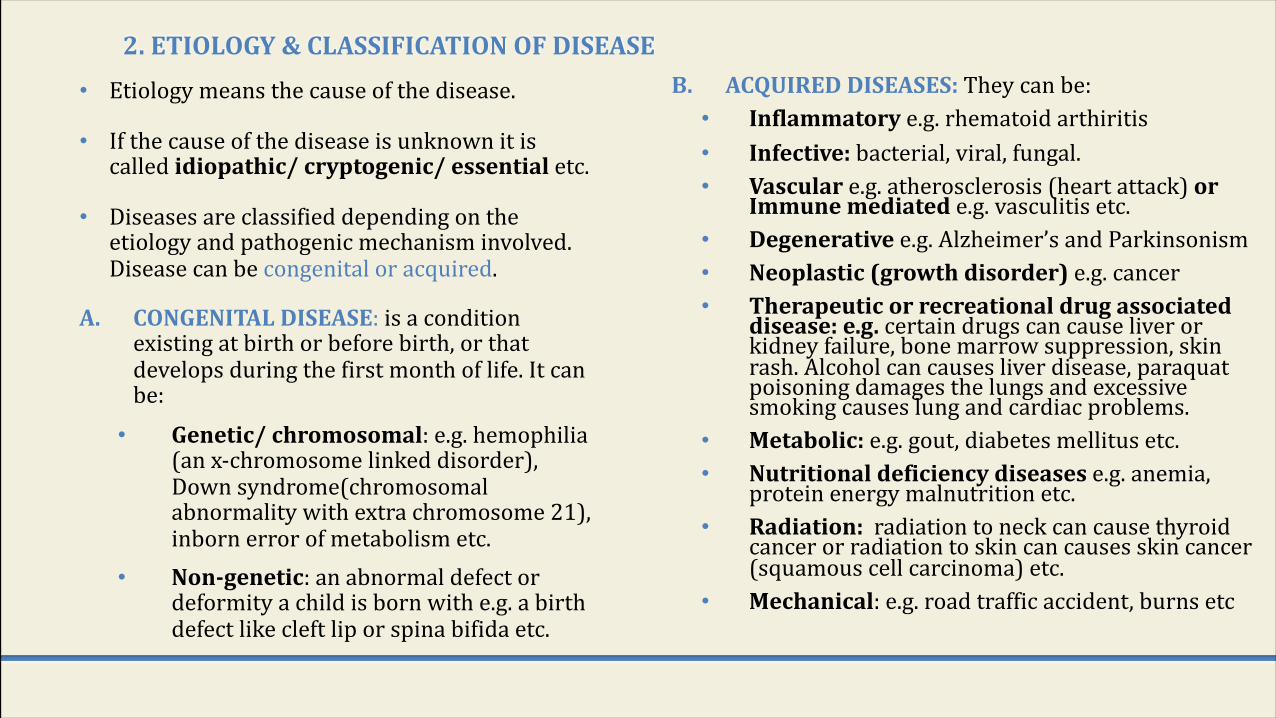
2.ETIOLOGY&CLASSIFICATIONOFDISEASE• Etiology means the cause of the disease.
• If the cause of the disease is unknown it is called idiopathic/cryptogenic/essentialetc.
• Diseases are classified depending on the etiology and pathogenic mechanism involved. Disease can be congenital or acquired.
A. CONGENITALDISEASE: is a condition existing at birth or before birth, or that develops during the first month of life. It can be:• Genetic/chromosomal: e.g. hemophilia
(an x-chromosome linked disorder), Down syndrome(chromosomal abnormality with extra chromosome 21), inborn error of metabolism etc.
• Non-genetic: an abnormal defect or deformity a child is born with e.g. a birth defect like cleft lip or spina bifida etc.
B. ACQUIREDDISEASES:They can be:• Inflammatory e.g. rhematoid arthiritis• Infective:bacterial, viral, fungal.• Vasculare.g. atherosclerosis (heart attack) or
Immunemediated e.g. vasculitis etc.• Degenerativee.g. Alzheimer’s and Parkinsonism• Neoplastic(growthdisorder)e.g. cancer• Therapeuticorrecreationaldrugassociated
disease:e.g.certain drugs can cause liver or kidney failure, bone marrow suppression, skin rash. Alcohol can causes liver disease, paraquatpoisoning damages the lungs and excessive smoking causes lung and cardiac problems.
• Metabolic:e.g. gout, diabetes mellitus etc.• Nutritionaldeficiencydiseasese.g. anemia,
protein energy malnutrition etc.• Radiation: radiation to neck can cause thyroid
cancer or radiation to skin can causes skin cancer (squamous cell carcinoma) etc.
• Mechanical: e.g. road traffic accident, burns etc

3.PATHOGENESIS
• Pathogenesis: it is the steps that take place in the body once the problem begins (whatever it may be) that finally lead to tissue injury (pathological manifestations).
• The four basic pathogenetic mechanisms (or steps that usually take place in diseases) are as follows:
• Inflammatory process• Degenerative process• Carcinogenesis: transformation of normal cells to malignant.• Immunological process
• All these will be dealt with in later chapters
• Pathogenesis leads to morphologic changes (changes in the gross or microscopic appearance of human tissue).

4.MORPHOLOGICCHANGES
• The morphologic changes are the structural changes that take place in cells or tissues due to any disease.
• These morphological changes can be seen Øgrossly (called macroscopicfindings) with the naked eye Øor sometimes they can only be seen under the light microscope
(called microscopic/histologic findings).
• Commonly diseases have certain specific gross or microscopic changes and this helps in the diagnosis of that disease.
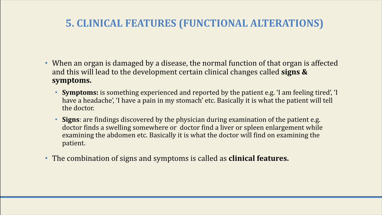
5.CLINICALFEATURES(FUNCTIONALALTERATIONS)
• When an organ is damaged by a disease, the normal function of that organ is affected and this will lead to the development certain clinical changes called signs&symptoms.• Symptoms: is something experienced and reported by the patient e.g. ‘I am feeling tired’, ‘I
have a headache’, ‘I have a pain in my stomach’ etc. Basically it is what the patient will tell the doctor.
• Signs: are findings discovered by the physician during examination of the patient e.g. doctor finds a swelling somewhere or doctor find a liver or spleen enlargement while examining the abdomen etc. Basically it is what the doctor will find on examining the patient.
• The combination of signs and symptoms is called as clinicalfeatures.
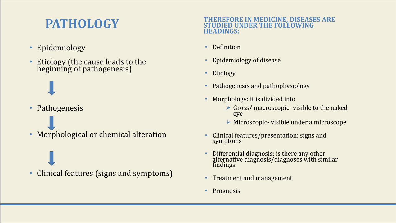
PATHOLOGY
• Epidemiology
• Etiology (the cause leads to the beginning of pathogenesis)
• Pathogenesis
• Morphological or chemical alteration
• Clinical features (signs and symptoms)
THEREFOREINMEDICINE,DISEASESARESTUDIEDUNDERTHEFOLLOWINGHEADINGS:
• Definition
• Epidemiology of disease
• Etiology
• Pathogenesis and pathophysiology
• Morphology: it is divided intoØ Gross/ macroscopic- visible to the naked
eyeØ Microscopic- visible under a microscope
• Clinical features/presentation: signs and symptoms
• Differential diagnosis: is there any other alternative diagnosis/diagnoses with similar findings
• Treatment and management
• Prognosis

COURSEOFDISEASE• The course of a disease is the different stages in the natural history or
progression of a disease in the absence of any intervention.
• The different stages in the natural history or course of a disease especially infectious are as follows:a) Exposure to causative agents or risk factors b) There is a latentperiodbetween exposure and onset of disease. The time period from
the exposure to the development of signs or symptoms in called as incubation(induction)period.
c) Onsetofdisease: the beginning of signs or symptoms.d) Outcomeandconsequencesofdisease:Following clinical onset, disease may follow any
of the following trends:Ø Recovery/resolution of disease without complication or sequalae. Person is
back to normal health.Ø The disease recovery but with sequelae.Ø Complications: development of complications in any disease can make
things worse.Ø Death.
THEDIAGNOSTICPROCESSANDTHEROLEOFPATHOLOGIST
• Any patient going to a clinic meets clinician who will take history and do clinical examination. He may ask for radiological and pathological examination in order to come to a diagnosis.
• The common pathological examinations are blood, urine and stool tests. Sometimes the patient is also asked to undergo a cytopathology or a histopathology test or other special pathological tests in order to obtain an accurate diagnosis.
• This way pathology plays an essential role in the diagnosis of a disease and management and treatment of patient.
THEBRANCHES/SUBDIVISIONSOFPATHOLOGY:
1. Histopathology:study of tissue biopsied/excised from body
2. Cytopathology:study of cell morphology, exfoliated or aspirated from body.
3. Hematology:a study of blood, blood cells and bone marrow, used in the diagnosis of anemias & leukemias.
4. Immunohistochemistry: a special staining procedure is used to detect antigens in the tissue.
5. Chemicalpathology/clinicalbiochemistry:is the analysis of bodily fluids (blood, urine, etc) for diagnosis.
6. Microbiology: is the study of micro-organisms
7.Immunology: is the analysis of the immune system of the body.
8.Toxicology: study of various poisonous and toxic substances.
9.Cytogenetics (clinicalgenetics): is a study of chromosomal abnormalities.
10.Molecularpathology:e.g. fluorescent in situ hybridization, Southern blot tests etc.
11.Autopsy:see later
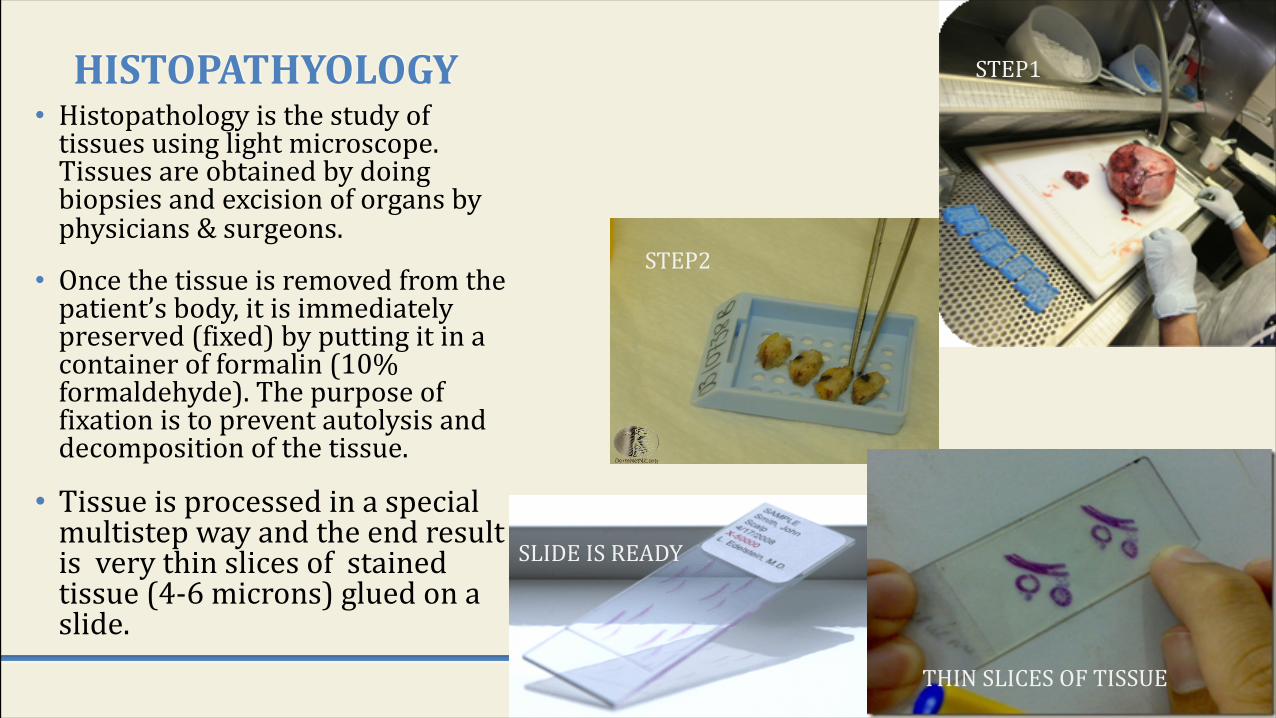
HISTOPATHYOLOGY• Histopathology is the study of
tissues using light microscope. Tissues are obtained by doing biopsies and excision of organs by physicians & surgeons.
• Once the tissue is removed from the patient’s body, it is immediately preserved (fixed) by putting it in a container of formalin (10% formaldehyde). The purpose of fixation is to prevent autolysis and decomposition of the tissue.
• Tissue is processed in a special multistep way and the end result is very thin slices of stained tissue (4-6 microns) glued on a slide.
STEP1
STEP2
THIN SLICES OF TISSUE
SLIDE IS READY
HISTOPATHYOLOGY CONTD…
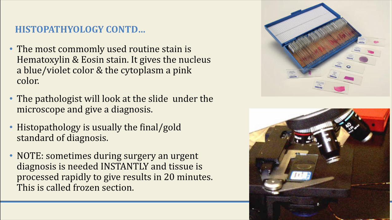
• The most commomly used routine stain is Hematoxylin & Eosin stain. It gives the nucleus a blue/violet color & the cytoplasm a pink color.
• The pathologist will look at the slide under the microscope and give a diagnosis.
• Histopathology is usually the final/gold standard of diagnosis.
• NOTE: sometimes during surgery an urgent diagnosis is needed INSTANTLY and tissue is processed rapidly to give results in 20 minutes. This is called frozen section.

HISTOPATHOLOGYSLIDESREADYTOBEEXAMINEDUNDERALIGHTMICROSCOPE
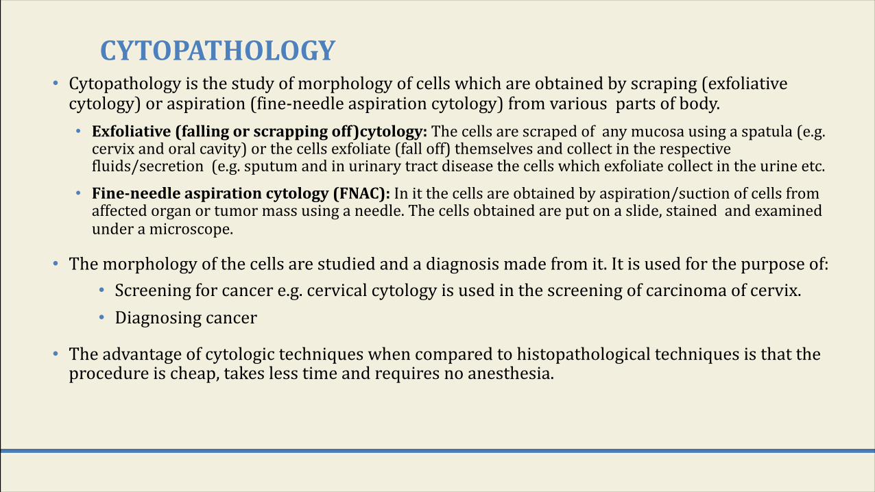
CYTOPATHOLOGY• Cytopathology is the study of morphology of cells which are obtained by scraping (exfoliative
cytology) or aspiration (fine-needle aspiration cytology) from various parts of body. • Exfoliative(fallingorscrappingoff)cytology:The cells are scraped of any mucosa using a spatula (e.g.
cervix and oral cavity) or the cells exfoliate (fall off) themselves and collect in the respective fluids/secretion (e.g. sputum and in urinary tract disease the cells which exfoliate collect in the urine etc.
• Fine-needleaspirationcytology(FNAC):In it the cells are obtained by aspiration/suction of cells from affected organ or tumor mass using a needle. The cells obtained are put on a slide, stained and examined under a microscope.
• The morphology of the cells are studied and a diagnosis made from it. It is used for the purpose of:• Screening for cancer e.g. cervical cytology is used in the screening of carcinoma of cervix.• Diagnosing cancer
• The advantage of cytologic techniques when compared to histopathological techniques is that the procedure is cheap, takes less time and requires no anesthesia.
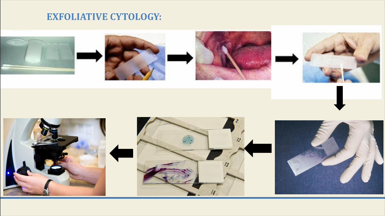
EXFOLIATIVE CYTOLOGY:
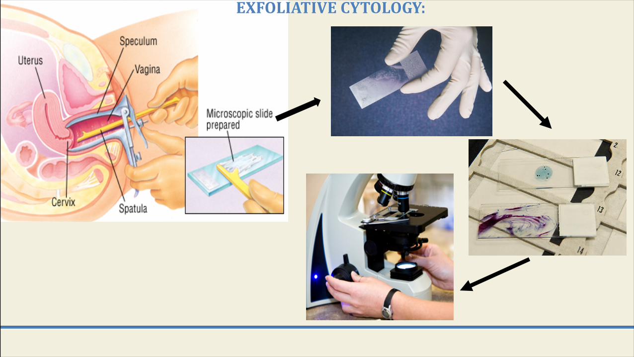
EXFOLIATIVECYTOLOGY:
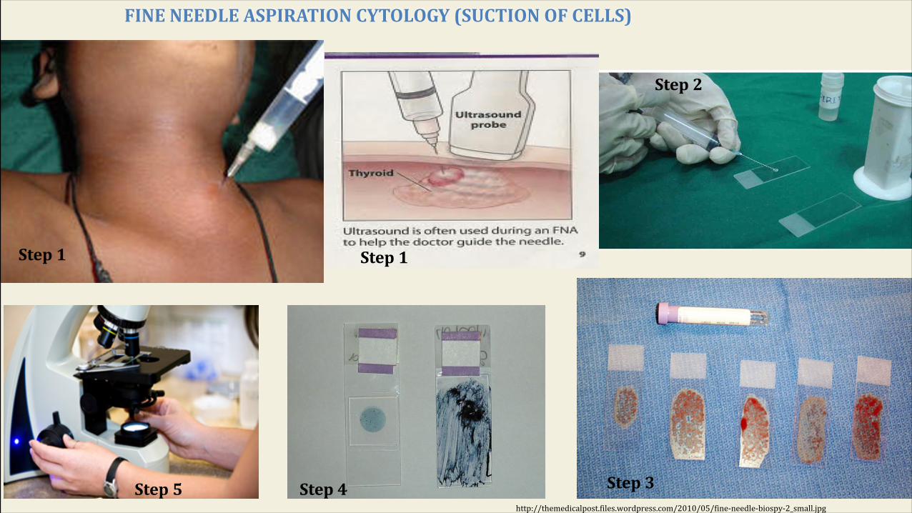
FINENEEDLEASPIRATIONCYTOLOGY(SUCTIONOFCELLS)
http://themedicalpost.files.wordpress.com/2010/05/fine-needle-biospy-2_small.jpg
Step1
Step2
Step3Step4Step5
Step1

b)Cytologyunderthemicroscopea)Histologyunderthemicroscope
a) Histology b) Cytology

11.AUTOPSY• It is a sub-specialty of pathology which involves examining a dead body
• An autopsy is done toØ To determine the cause of death (this is the main reason why autopsy is done). It can be
performed in any of the following situations:v Homicidalv Suicidalv Accidentalv To identify the disease
Ø To provide useful information about various disease.Ø To do research.Ø Also it can be used as a tool to educate students, surgeons etc
• Who does the autopsy? The pathologist.
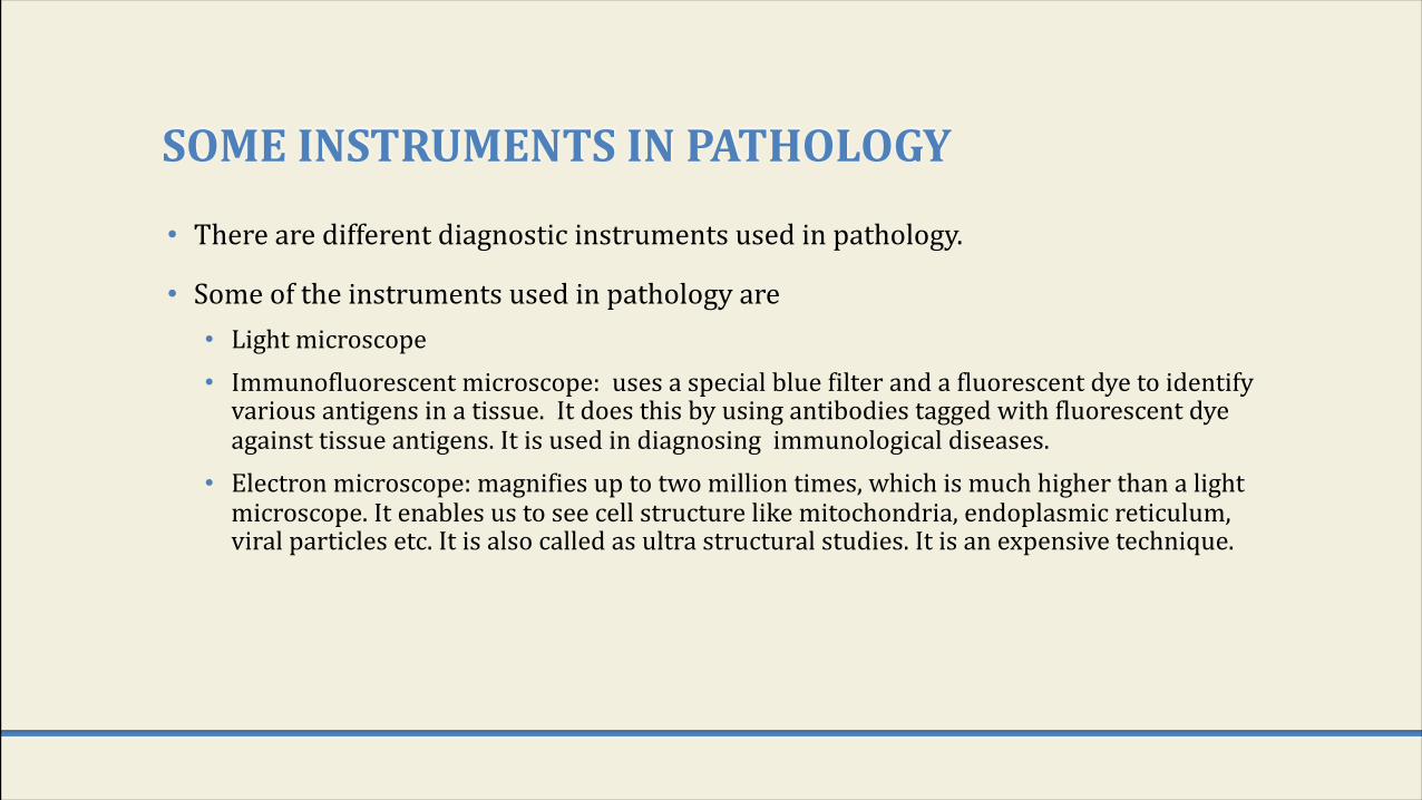
SOMEINSTRUMENTSINPATHOLOGY
• There are different diagnostic instruments used in pathology.
• Some of the instruments used in pathology are • Light microscope• Immunofluorescent microscope: uses a special blue filter and a fluorescent dye to identify
various antigens in a tissue. It does this by using antibodies tagged with fluorescent dye against tissue antigens. It is used in diagnosing immunological diseases.
• Electron microscope: magnifies up to two million times, which is much higher than a light microscope. It enables us to see cell structure like mitochondria, endoplasmic reticulum, viral particles etc. It is also called as ultra structural studies. It is an expensive technique.
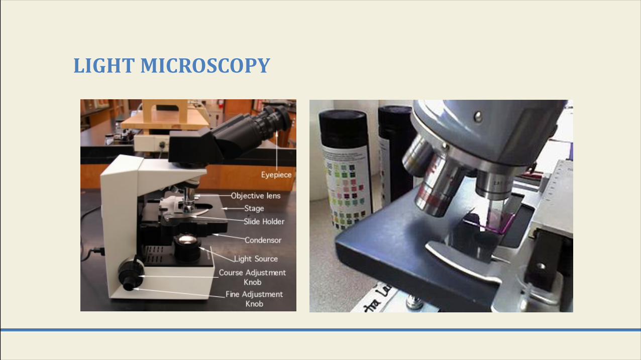
LIGHTMICROSCOPY
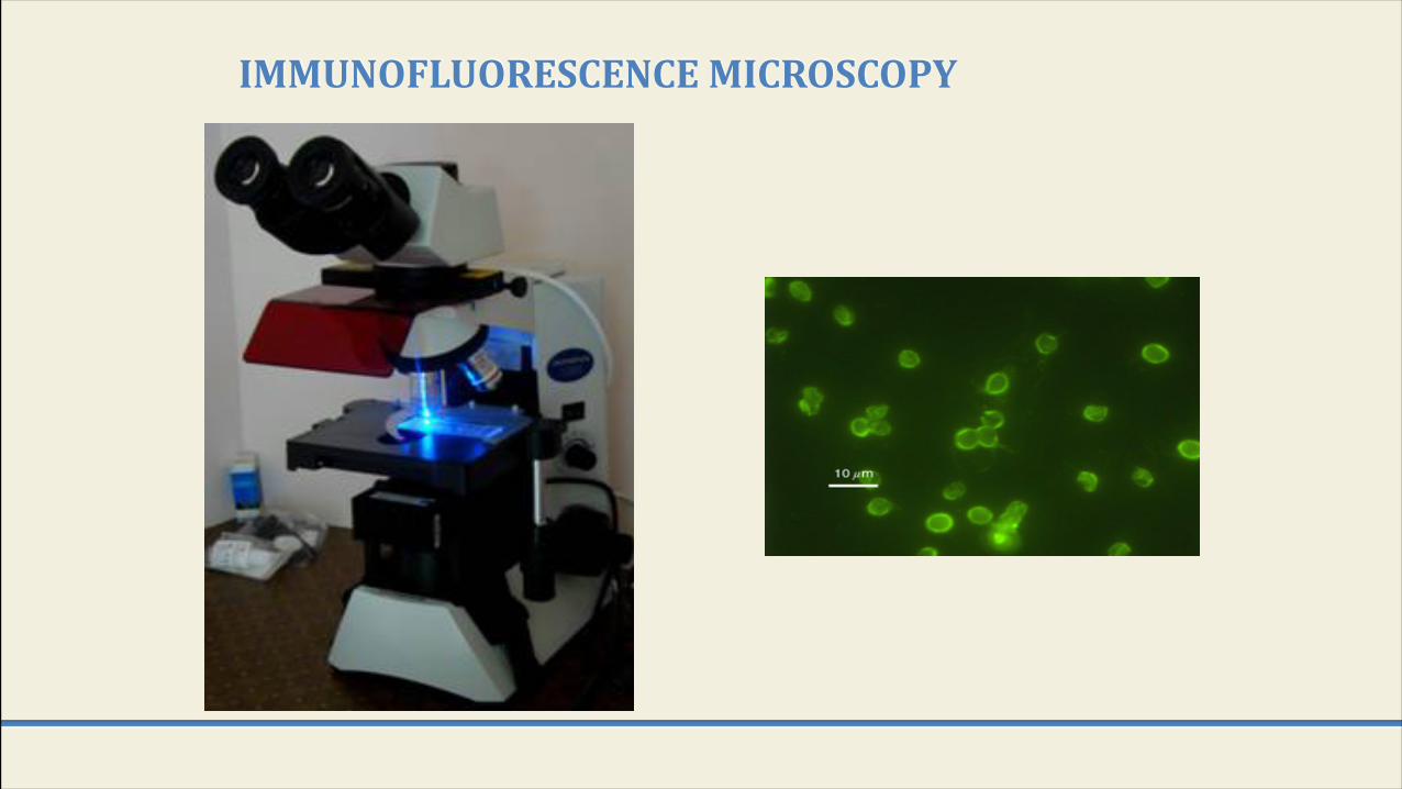
IMMUNOFLUORESCENCEMICROSCOPY
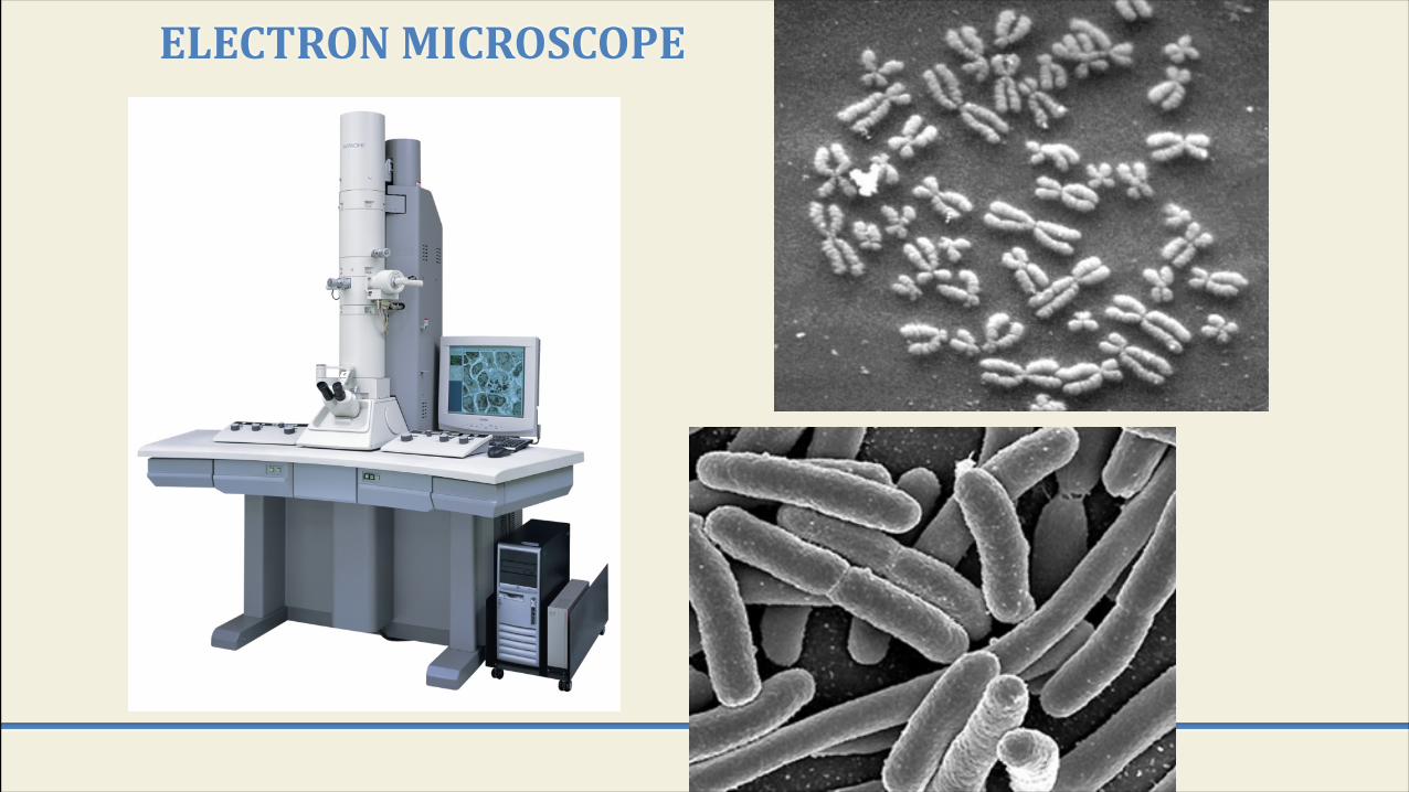
ELECTRONMICROSCOPE