introduction to carotid duplex scanning...• describe how carotid plaque appears on an ultrasound...
TRANSCRIPT
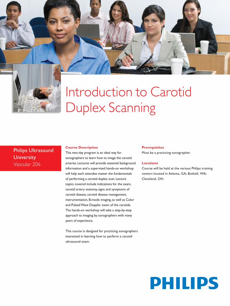
Course DescriptionThis two-day program is an ideal way for sonographers to learn how to image the carotid arteries. Lectures will provide essential background information and a supervised hands-on workshop will help each attendee master the fundamentals of performing a carotid duplex scan. Lecture topics covered include indications for the exam, carotid artery anatomy, signs and symptoms of carotid disease, carotid disease management, instrumentation, B-mode imaging, as well as Color and Pulsed Wave Doppler exam of the carotids. The hands-on workshop will take a step-by-step approach to imaging by sonographers with many years of experience.
This course is designed for practicing sonographers interested in learning how to perform a carotid ultrasound exam.
Introduction to Carotid Duplex Scanning
Philips Ultrasound UniversityVascular 206
PrerequisitesMust be a practicing sonographer.
LocationsCourse will be held at the various Philips training centers located in Atlanta, GA; Bothell, WA; Cleveland, OH.
© 2013 Koninklijke Philips Electronics N.V.All rights are reserved.Apr 2013
Philips Healthcare reserves the right to make changes in specifications and/or to discontinue any product at any time without notice or obligation and will not be liable for any consequences resulting from the use of this publication.
Philips Healthcare is part of Royal Philips Electronics
www.philips.com/[email protected]
Philips Healthcare22100 Bothell Everett HighwayBothell, Washington 98021
Introduction to Carotid Duplex Scanning (Vasc 206)
Course objectivesUpon successful completion of this program, you should be able to:• Discuss the indications for exam, risk factors, as well as signs and
symptoms• Discuss intra and extracranial carotid, subclavian, aortic arch, and
vertebral anatomy• Discuss how carotid disease forms and how it is managed• Discuss what Intimal Medial Thickening is and how it is measured• Explain key clinical trials that have taken place• Explain other examinations used to confirm carotid duplex results• Explain the equipment needed to perform a quality carotid duplex
scan• Explain a basic carotid duplex scan protocol• Explain how system controls are optimized for B-Mode, color, and
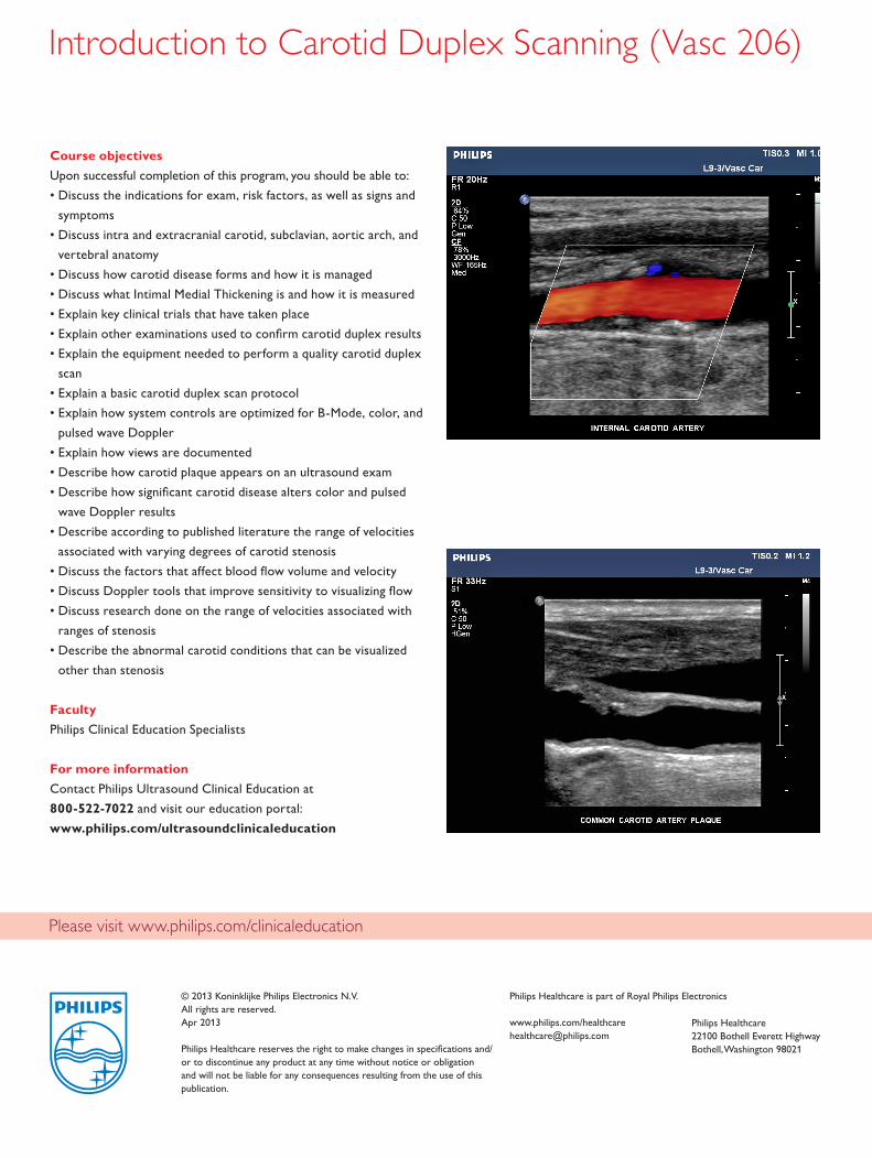
pulsed wave Doppler• Explain how views are documented• Describe how carotid plaque appears on an ultrasound exam• Describe how significant carotid disease alters color and pulsed
wave Doppler results• Describe according to published literature the range of velocities
associated with varying degrees of carotid stenosis• Discuss the factors that affect blood flow volume and velocity• Discuss Doppler tools that improve sensitivity to visualizing flow• Discuss research done on the range of velocities associated with
ranges of stenosis• Describe the abnormal carotid conditions that can be visualized
other than stenosis
FacultyPhilips Clinical Education Specialists
For more informationContact Philips Ultrasound Clinical Education at 800-522-7022 and visit our education portal:www.philips.com/ultrasoundclinicaleducation
Please visit www.philips.com/clinicaleducation