introduction to binocular vision testing: lecture 1 · – patient has an npc that his...
TRANSCRIPT
This presentation has been created for Orbis International trainees by New England College of Optometry Volunteer Faculty.
This presentation is property of the New England College of Optometry and any
attempt to reproduce material will be in violation of US copyright law.
INTRODUCTION TO BINOCULAR VISION TESTING: LECTURE 1
Dr Hilary Gaiser OD, MSc Assistant Professor of Optometry
New England College of Optometry
ACKNOWLEDGEMENTS
Thank you to the Orbis team for the opportunity to present these lectures to the international eye care provider community.
Additional thanks to Dr. Sarah Wassnig for her guidance and support and to Dr. Catherine Johnson for her permission to use her
binocular vision training videos

LECTURE OBJECTIVES
1. Learn an introductory battery of binocular vision testing procedures
2. Gain an introductory understanding of the theory of each
binocular vision test 3. Confidently conduct a basic binocular vision exam

POLL QUESTION # 1
• How familiar are you as a provider with completing a basic binocular vision assessment?
1) I perform basic binocular vision tests regularly as part of a routine vision exam
2) I have learned basic binocular vision tests but I am not confident in applying them in clinic
3) I have seen basic binocular vision tests being performed but I have not learned and/or used them
4) I have no knowledge of basic binocular vision testing
FUNCTIONAL BINOCULAR VISION
• Goal is to have clear, comfortable, single vision when using both eyes
• Determined by a balance between the patient’s refraction, phoria,
vergence and accommodation
• Imbalances can cause symptoms that negatively impact a patient’s quality of life

INDICATIONS FOR BINOCULAR VISION TESTING
• Abnormalities noted on binocular vision “screening” procedures during a standard comprehensive exam ie. Cover test, stereovision
• Patient is symptomatic for visual fatigue, headache, intermittent diplopia, fluctuating vision, difficulty “focusing”

BINOCULAR VISION ABNORMALITIES
• Common and can affect all ages
• Considered an anomaly not a disease as they are non-pathological, non sight threatening but can significantly impact quality of life
• Quality of life concerns include school, work performance, hobbies and general visual comfort
CORE BINOCULAR VISION EXAM
1) Determine need based on case history or previous diagnosis
2) Accurate and current max full plus refraction with binocular balance
3) Measurement of magnitude and direction of phoria (rule out tropia) at distance and near and AC/A ratio
4) Assessment of (+) and (-) fusional vergence using direct and indirect methods
5) Assess convergence amplitude
6) Evaluate sensory status and suppression

INTRODUCTORY BINOCULAR VISION TESTING
• Phoria/tropia: Cover test
• Vergences: Step vergences with prism bars, near point of convergence
• AC/A ratio: Accommodative convergence to accommodation ratio • Accommodation: Amplitude of accommodation (AMPS) • Sensory status and suppression: stereovision
• Must always start out with an accurate and up to date refraction

CAUTION
• Be careful to take a full patient history to rule out possible pathology from functional binocular conditions ie. headaches that do not correlate with binocular vision abnormalities
• Complete dilated ocular health assessment
• Updated and accurate glasses prescription

COVER TEST: COVER/UNCOVER AND ALTERNATING (PHORIA VS. TROPIA)
1

PHORIA AND TROPIAS • Phoria:
- The position of the eyes when binocularity is disassociated (sometimes referred to as the eyes resting position) - Disassociation eliminates fusional vergence - You disassociate the eyes by presenting images that are unfusible (occlusion or prism)
• Tropia:
-A misalignment of the eyes when a fusible image is presented -Since binocularity is impaired often cannot perform binocular vision tests
-Important to remember tropias can be variable – Time of day – Length of testing and/or near work – Case history is invaluable

COVER TEST PROCEDURE
• Purpose: to determine if a patient has a phoria vs. a tropia and the characteristics of a tropia when present
• Objective and can be performed quickly and easily on most patients
• Can be used to measure AC/A ratio (accommodative convergence to accommodation)
ACCOMMODATION CONTROL: TARGET SIZE
• Accommodation affects vergence which affects eye alignment ( synkinetic triad of miosis, convergence and accommodation)
• Accommodation is controlled by choosing a properly sized target and having the patient attend to the target and keep it clear
• Underaccommodation will overestimate an exophoria and overaccommodation will underestimate it

COVER TEST TARGETS
• For near and distance cover testing use an “accommodative target” – 20/30 or 1-2 lines above BCVA in the poorer
seeing eye – Keep fixation target isolated – Make sure patient keeps the target clear
2
3
COVER TEST SET-UP
• Habitual correction for distance or near (40 cm) depending on testing distance or correction through which you would like more information about alignment
• Eye level with patient and behind target for near testing and as in front of patient as possible without obscuring target for distance
• Good lighting to maximize contrast on target and to pick up small
ocular movements

COVER/UNCOVER TEST: PHORIA VS. TROPIA
• Determine which eye is fixating Cover the right eye with the
paddle pausing for three full seconds and then uncover, watching for the eye to move to fixate. Repeat this procedure twice before switching to the left eye. If neither eye moves than proceed to the alternating cover test.
4
Childhood Eye Examination
Figure 4.
Screening tests for abnormal alignment. (A through D) The cover test. (A) On simple observation, one eye (the
http://www.aafp.org/afp/2013/0815/p241.html
1 of 2 12/5/17, 8:31 am
Childhood Eye Examination
Figure 4.
Screening tests for abnormal alignment. (A through D) The cover test. (A) On simple observation, one eye (the
http://www.aafp.org/afp/2013/0815/p241.html
1 of 2 12/5/17, 8:31 am
ALTERNATING COVER TEST
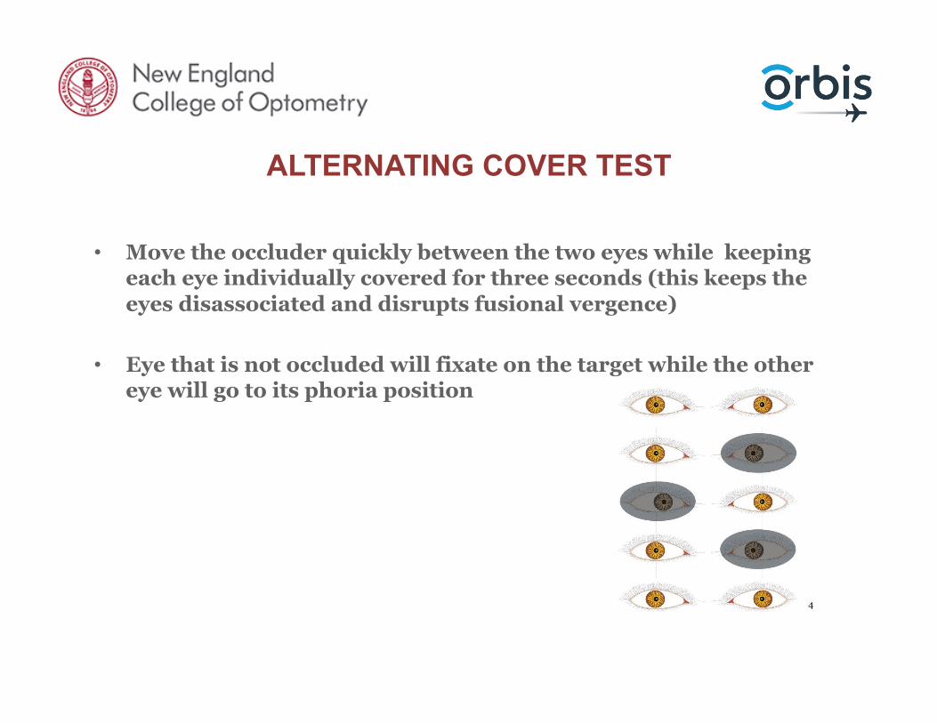
• Move the occluder quickly between the two eyes while keeping each eye individually covered for three seconds (this keeps the eyes disassociated and disrupts fusional vergence)
• Eye that is not occluded will fixate on the target while the other
eye will go to its phoria position
4
COVER TEST ANALYSIS
• If an eye moves to fixate during cover/uncover then the patient has a tropia – Laterality: left, right or alternating – Frequency: constant or intermittent – Magnitude: neutralize with prism bars or loose prism lenses – Direction: hyper, hypo, exo or eso
• If the eyes do not move during cover/uncover but do with alternating CT – Magnitude – Direction

COVER TEST
COVER TEST ANALYSIS
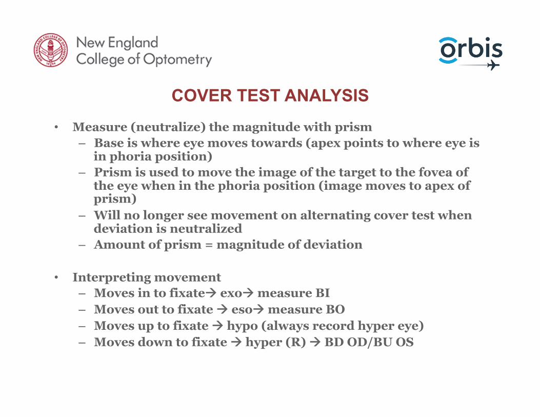
• Measure (neutralize) the magnitude with prism – Base is where eye moves towards (apex points to where eye is
in phoria position) – Prism is used to move the image of the target to the fovea of
the eye when in the phoria position (image moves to apex of prism)
– Will no longer see movement on alternating cover test when deviation is neutralized
– Amount of prism = magnitude of deviation
• Interpreting movement – Moves in to fixateà exoà measure BI – Moves out to fixate à esoà measure BO – Moves up to fixate à hypo (always record hyper eye) – Moves down to fixate à hyper (R) à BD OD/BU OS
POLL QUESTION # 2
On cover test you note that on the cover uncover portion of the test that the patient’s left eye always moves in to fixate when the right eye is covered. When the cover uncover test is performed on the left eye the right eye does not move in to fixate. How would you describe the patient’s findings? 1) Constant left exophoria
2) Intermittent left exotropia
3) Constant left exotropia
4) Constant left esotropia

COVER TEST NORMATIVE DATA AND RECORDING
• Distance: 1 pd esophoria to 3 pd exophoria • Near: orthophoria to 6 pd exophoria • Tropia: never normal
• Recording – SC or CC and through which Rx – Test distance – Direction and magnitude – Tropias add laterality and frequency – Ie. CT cc @ 40 cm: ~ 4 esophoria (EP) – Ie. CT sc @ dist: 5 Δ exotropia (XT)

COVER TEST TROUBLESHOOTING
• Repeat testing multiple times or even at the end of the exam if large phoria is noted (may break down into a tropia)
• Have patient read the letters to ensure they are keeping the target
clear
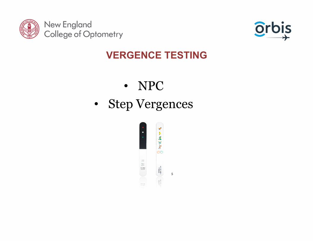
VERGENCE TESTING
• NPC • Step Vergences
5
VERGENCE
• Eye alignment (motor fusion) depends on the balance between fusional vergence and the patient’s phoria (supply and demand)
• Vergence: movement of the visual axis towards or away from each other in order to keep images single (maintain motor fusion) – Convergence towards each other – Divergence away from each other – Hypervergence one eye up relative to each other
*hypervergence of one eye = hypovergence of the other
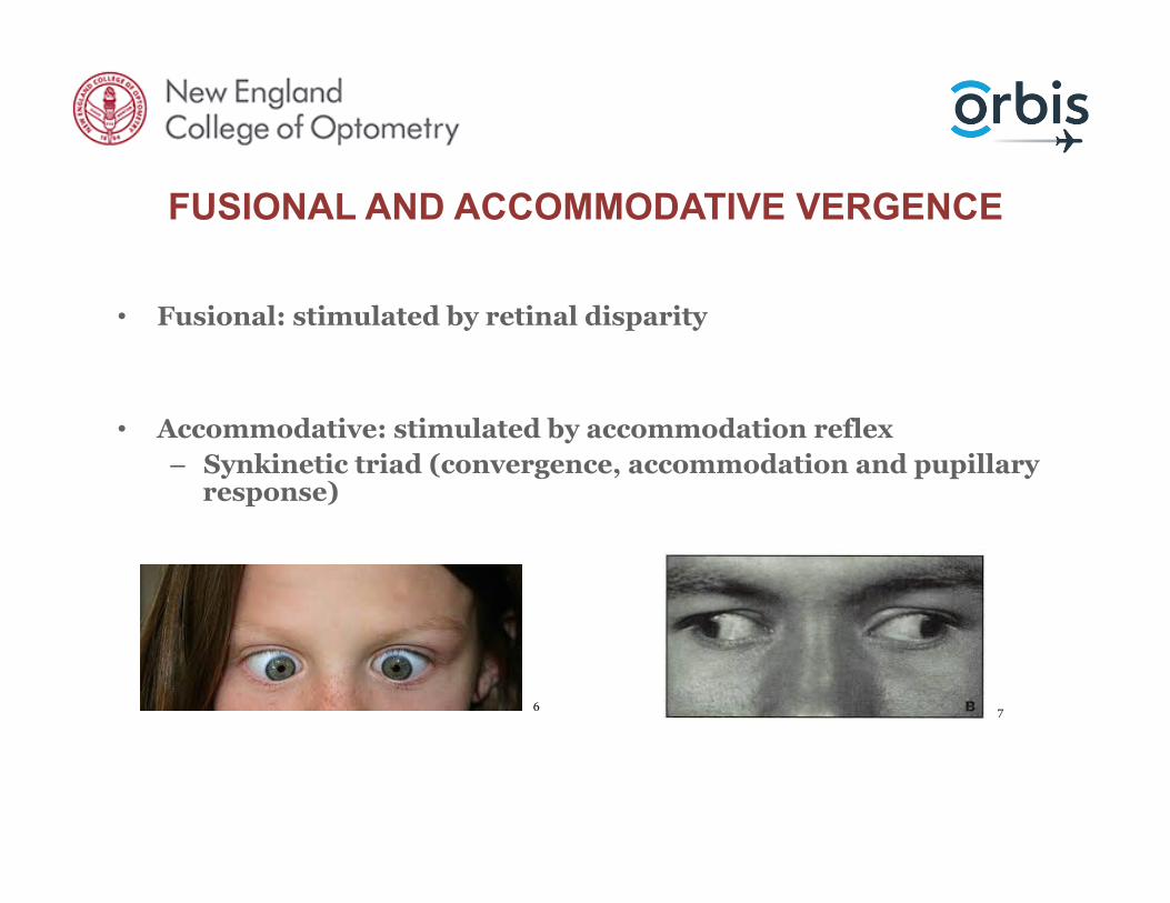
FUSIONAL AND ACCOMMODATIVE VERGENCE
• Fusional: stimulated by retinal disparity
• Accommodative: stimulated by accommodation reflex – Synkinetic triad (convergence, accommodation and pupillary
response)
6 7
VERGENCE SUPPLY AND DEMAND
• Demand exoà compensating convergence (positive)
• Demand eso à compensating divergence (negative)
• Demand right hyper à right hypovergence
• Demand right hypoà right hypervergence

FUSIONAL VERGENCE IN CONTEXT
1. Patient has no motor fusionà constant strabismus
2. Patient has intermittent motor fusion à intermittent strabismus/decompensating phoria
3. Patient has motor fusion but difficulty (discomfort) maintaining ità symptomatic
4. Patient has comfortable motor fusionà typically normal phoria

NEAR POINT OF CONVERGENCE (NPC)
8

NEAR POINT OF CONVERGENCE (NPC)
• Purpose: To assess the absolute convergence limit
• Important in diagnosing convergence insufficiency (CI)
• Objective and subjective test

NPC SET-UP
• Habitual correction or the refractive correction through which you would like information about the absolute convergence limit
• Different targets:
-Accommodative -Light -Red lens
NPC PROCEDURE • Move target towards the bridge of the patient’s nose and
ask the patient to tell you when the target appears double – Observe the patient for loss of binocular fixation (break
point) – Patient reports diplopia (break point) – Should observe and record the eye that loses fixation in
either case
• Once the target becomes double or an eye loses fixation then move the target away from the patient and ask them to tell you when the target becomes single again (recovery point)
• If patient does not report diplopia but an eye loses fixation then they are likely suppressing
• Measure the break and recovery point with a ruler (can
estimate as you gain experience)

NPC PROCEDURE

NPC RECORDING
• Test used and type of stimulus used (acc, light, red lens) • sc or cc (and with which Rx) • Break/recovery (in cm or inches) • Diplopia (per patient report) or suppression (if patient lost
binocular fixation but did not report diplopia) • Which eye loses fixation
Ex 1. NPCcc (acc): 6cm/10cm (diplopia) OD out Ex 2. NPCcc (light): 8cm/12cm (suppression) OS out

NPC IN CONTEXT • Pt. maintains binocular fixation and never reports diplopia
– Patient has an NPC that his “to-the-nose” (TTN); no break point
• Pt. reports diplopia and you observe that the eyes lose binocular fixation – Your observation should match patient’s report – Break point = when pt. reported double/lost fixation
• You observe that the eyes lose binocular fixation but the pt. does not report diplopia – The patient is suppressing the deviated eye or does not
understand the test – Break point = when patient lost binocular fixation
• Objective (recording eye out) assessment may be all we have for children

NPC NORMS
• For accommodative target: – Break < 5cm – Recovery < 7cm – Least receded NPC because engages both accommodative and
fusional vergence
• For penlight target: – Break < 7cm – Recovery < 10cm – More recession with red lens suggests more significant
convergence problem
• Failure to report diplopia as eye turns out indicates suppression
NPC TROUBLESHOOTING
• NPC that is more receded (further away) than norms is suggestive of CI
• If a patient has a normal initial NPC but is symptomatic or has other abnormal BV complaints/findings
- Repeat multiple times (5x is recommended) to assess for fatigue -Repeat with red lens to add another barrier to fusion

POLL QUESTION # 3
You are performing NPC on a 6 year-old child and you note the right eye loses fixation around 4 cm and recovers at 7 cm, but that the child does not report that the target has doubled. What is the most likely assessment of the patient’s findings. 1) 4 cm break/7 cm recovery, OD out, suppression
2) 4 cm break/7 cm recovery, OD out, diplopia
3) Cannot complete testing without a response from the patient
4) 4 cm break/7cm recovery OD out (unable to definitively determine suppression or diplopia rely on objective findings)
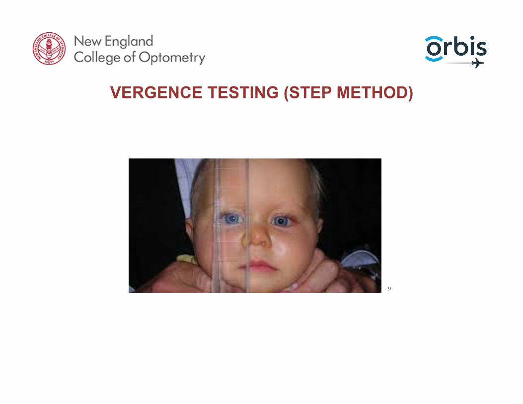
VERGENCE TESTING (STEP METHOD)
9

FUSIONAL VERGENCE
• Introducing prism under binocular conditions allows us to test fusional vergence ability
• The prism creates retinal image disparity (moves image away from the fovea of each eye) – this is a direct stimulus to fusional vergence

FUSIONAL VERGENCE
• The patient will continue to move his eyes until his supply of vergence innervation is depleted – Since accommodation and convergence are linked the patient
will first use up his supply of accommodation convergence (reports blur) and then will use up the rest of their fusional vergence (reports double)
10
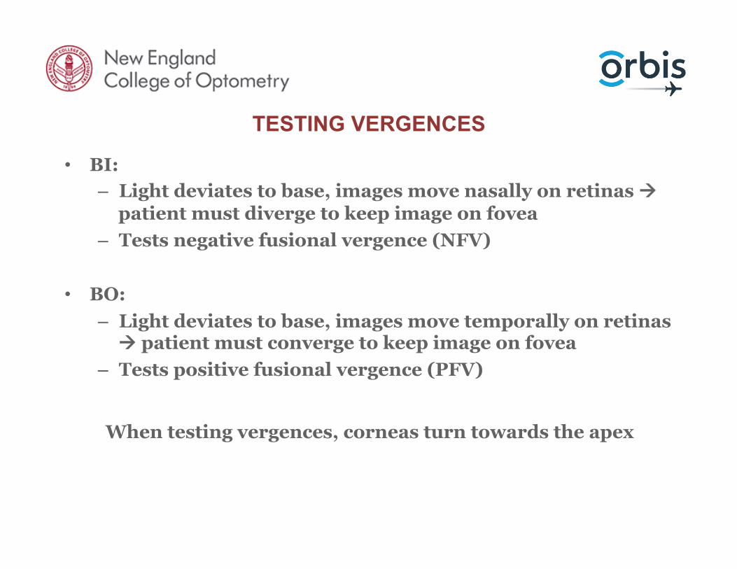
TESTING VERGENCES
• BI: – Light deviates to base, images move nasally on retinas à
patient must diverge to keep image on fovea – Tests negative fusional vergence (NFV)
• BO: – Light deviates to base, images move temporally on retinas
à patient must converge to keep image on fovea – Tests positive fusional vergence (PFV)
When testing vergences, corneas turn towards the apex

STEP VERGENCES
• Purpose: To measure a patient’s ability to maintain clear and single binocular vision while changing vergence and holding accommodation constant
• Must use an accommodative target at a constant viewing distance
• Free space technique (smooth vergences phoropter technique )
• Subjective and objective test
• Patient must be able to fuse in order to measure fusional vergences - you can’t do vergence testing on a patient when they are exhibiting a tropia!

STEP VERGENCE TECHNIQUE
• Habitual correction or correction through which you want vergence information
• Can test at both distance and near
• Use an appropriate near and distance target (isolated line or letter 1-2 lines above BCVA worse seeing eye)
• Place prism bar base-in before one eye and move it step-by-step as the patient attends to the target
• Ask the patient to report when the target becomes blurry, breaks into two and comes back into one
STEP VERGENCE TECHNIQUE
• If the patient fails to report blur, break or recovery watch for the patient’s eyes to flick off the target (break point) when the eyes return to their initial position (recovery)
• Repeat with base-out prism (always perform base-in before base-out) to avoid prism adaptation
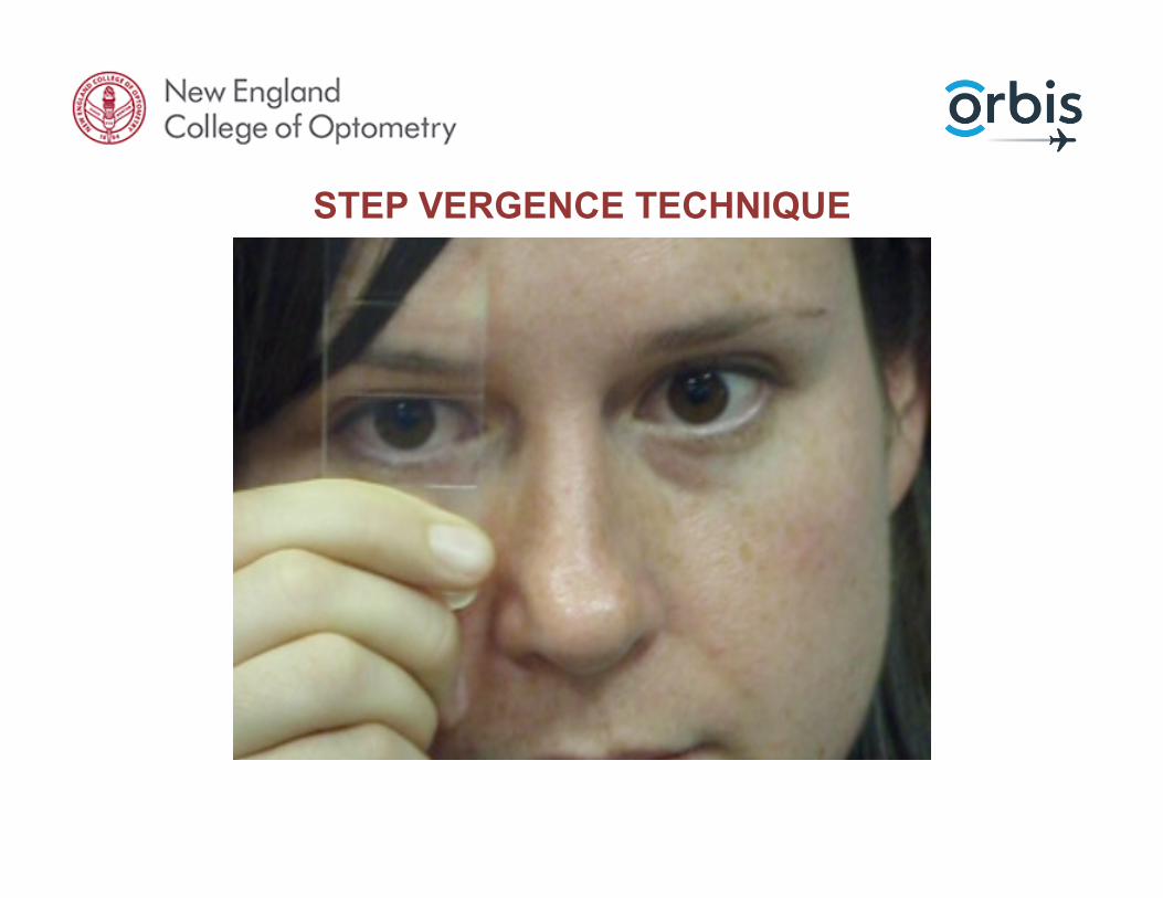
STEP VERGENCE TECHNIQUE

STEP VERGENCE NORMATIVE DATA AND ANALYSIS
• Normative values: – Distance: BO x/11/7 – Distance: BI x/7/4 – Near: BO x/23/16 – Near: BI x/12/7 According to Sheard’s criteria ideally the patient would expect to have comfortable binocular vision if the blur findings (can use break if no blur) is at least twice the patient’s phoria
* Adapted from Scheiman, Wick. Clinical Management of Binocular Vision, 2nd ed. Philadelphia: Lippincott, Williams & Wilkins, 2002: 3-50, 74
POLL QUESTION # 4 On positive vergence testing (BO) you record the following findings. BO x/15/7 and on cover test you find the patient has a 5 pd exophoria. Do you expect the patient to be symptomatic? 1) Cannot determine due to incomplete data. Missing blur findings for
vergences.
2) No, according to Sheard’s criteria and the break findings the patient has at least twice the phoria’s worth of compensating positive fusional vergence
3) Yes, the patient would be symptomatic as the phoria findings are outside norms
4) Should complete BI testing as well in order to determine the patient’s full fusional vergence ability

CLINICAL TESTS OF ACCOMMODATION
• Amplitude of Accommodation (AMPS)

INDICATIONS FOR TESTING ACCOMMODATION
• Reduced near VA, especially in non-presbyope
• Symptoms that suggest accommodative anomaly – Near blur, fluctuating near vision – Increased distance blur after reading – Headache, eye strain after near work – Fluctuating vision not associated with health risk factor
• DDx: dry eye – Pseudomyopia (VA suggests more minus than ret/ref or
ret/ref more minus than entering VA)

ACCOMMODATION TESTING
1. Amplitude of accommodation: (AMPS)
2. Relative accommodation: AC/A, NRA/PRA
3. Accuracy of accommodation: MEM, FCC 4. Accommodative facility: speed of accommodation

AMPLITUDE OF ACCOMMODATION TESTING (AMPS)
11

AMPS PUSH-UP METHOD
• Purpose: to measure (in diopters) a patient’s ability to change focus in response to a near stimulus
• Subjective, monocular technique

AMPS TESTING PROCEDURE
• With the patients near habitual Rx or through the Rx you want more information from (must record through which Rx you are testing)
• Target: well illuminated row of letters one or two lines larger than near visual acuity
• Test each eye separately
• Instruct the patient to keep the letters clear and slowly move the chart closer to the patient and ask them to report with the letters first become and stay blurry (first sustained blur)
• Measure the distance from the card to the spectacle plane in cm and convert into diopters ( ie. 100/10 cm = 10 D)
AMPS RECORDING
• Test used • CC or SC (and Rx through which tested) • AMPS in diopters • OD and OS separately
• Ie. Amp (push-up) cc: OD 7 D and OS 7 D
AMPS NORMS AND ANALYSIS
• ½ Amp in Reserve: sustained clear and comfortable single vision occurs when only using half of the pts. total accommodation during near work
• Make sure to evaluate and correct for - Under-corrected hyperopia - Over-corrected myopia - Presbyopia - Then consider evaluating for an accommodative anomaly
• Hoffstetter’s Formula
– Amp expected based on patient’s age – Minimum = 15- (0.25 x age in years) – Average = 18.5 – (0.30 x age in years)

AMPS PROCEDURE

AC/A RATIO: LINKING ACCOMMODATION AND VERGENCE
12
ACCOMMODATIVE CONVERGENCE/ACCOMMODATION
(AC/A) RATIO
• Concepts of blur and convergence accommodation – Blur (reflex): small changes in accommodation in response to
the detection of blur – Convergence: stimulated directly by changes in fusional
vergence • Increase in accommodationà increase in PFV • Decrease in accommodation à decrease in PFV
AC/A RATIO
• Purpose: To determine the change in accommodative convergence that occurs when the patient increases accommodation by a given amount (fusional vergence stimulus stays constant)
• Plus lenses relax accommodation and reflexively convergence decreases (makes exos larger and esos smaller)
• Minus lenses stimulate accommodation and we expect convergence to increase ( makes exos smaller and esos larger)

AC/A RATIO PROCEDURE
• Re-measure the patient’s near lateral phoria using (+1.00 and/or -1.00 lenses) over the patient’s habitual Rx and using cover test
• This forces accommodation to change by 1 D (A portion of AC/A)
• The difference between the two measurements gives the AC part of the AC/A ratio
• Can also use +/- 2 D just need to convert ratio to x/1 ie. 8/2à 4/1

AC/A RATIO EXAMPLE
• Refraction: -2.00 OD and OS • Measured phoria through habitual: 4 exophoria • Measured phoria through +1.00 D: 8 exophoria • Measured phoria through -1.00 D: orthophoria • AC/A ratio: 4/1
• Convergence decreased by 4 prism diopters when the stimulus to accommodation was decreased by 1 (increase in exophoria)
• Convergence increased by 4 prism diopters when the stimulus to accommodation was increased by 1 (decrease in exophoria)
AC/A RATIO NORMS AND ANALYSIS
• Normal: 4/1 to 6/1 • Low: < 4/1 • High: > 6/1
• Also consider using both +1.00 and -1.00 to understand if anomalies are noted in either direction (ratio should be the same regardless) as the patient may have difficulty relaxing or stimulating accommodation
POLL QUESTION # 5
• When measuring a patient’s near phoria using cover test you find 6 pd esophoria and 6 pd exophoria when repeating the test using +2.00 D flippers. What is the patient’s AC/A ratio and is it within norms?
1) 12/2, not within norms
2) 6/1, not within norms
3) 0/2, not within norms
4) 0/1, within norms
SENSORY STATUS
13

SENSORY FUSION
• Testing the ability to combine sensory information (form, color, size, illuminance, location in space) from the right and left eyes into a single perception (different information presented to each eye)
• Stereopsis is the binocular perception of depth (true depth perception) can also use monocular cues
MONOCULAR DEPTH CUES
• Learned as a result of dynamic interaction with the visual world
• Used by everyone for distances > 200 meters
• Geometric perspective, aerial, overlay, height in relation to the horizon, light and shadow, parallax, accommodation and convergence (internal cues)

NORMAL STEREOPSIS
1. Two eyes that function normally and equally 2. Similar retinal image in OD and OS
3. Ability of the eyes to maintain bifoveal fixation (retinal images of fixated objects can be placed and maintained on the fovea of each eye) aka motor fusion
• Motor fusion and sensory fusion are linked. Motor fusion is
needed for sensory fusion and sensory fusion promotes motor fusion
• If there is an imbalance than the patient will have reduced
stereopsis and binocularity
DEVELOPMENT OF STEREOPSIS
• Patient with disruption to any of the requirements for normal stereopsis – Child during the critical period for development ~ < 7 years
(likely to not develop stereopsis normally unless caught and corrected early)
– Adult with previous normal stereopsis (will not have stereopsis when there is disruption to any of the requirements but if corrected it can be restored)
• Neuroplasticity (new research has suggested that the
critical period may not fully be set in stone and that adults may lose stereopsis if strabismus is constant for 2-3 months)

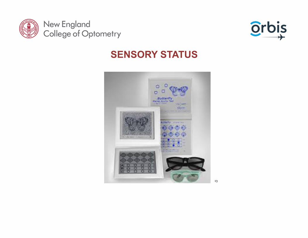
STEREOVISION TESTING
• Variety of methods for testing (*polaroid filters, color filters, synoptophore, stereoscope)
• * More common (Random Dot 3, Random Dot 2, Randot Preschool Test, Titmus Fly Stereo Test, Preschool Assessment of Stereopsis with a Smile (PASS) 2, Random Dot E)
• Level of stereopsis is measured in the angular separation of the target in sec of arc
• Smaller separation= smaller/finer retinal disparity= better
stereovision

STEREOPSIS TESTING
• Test with habitual correction for near
• May need to use testing glasses (polaroid or red-green) over patient’s glasses
• Good overhead illumination with no glare
• Hold book at eye level without moving (best if practitioner holds so extra cues are eliminated)
• Correct testing distance that test is calibrated for (longer distance can equal finer levels of stereo)
• Make sure to read the instruction manual for the test you are using

STEREOPSIS RECORDING
• Test used • CC or SC • Type of stereo if applicable (local or global) • Test distance • Record the measurement in sec of arc • Stereopsis norms are variable per patient’s age and type of test • Generally around 25’’ sec of arc is considered good or normal

LEARNING OBJECTIVE SUMMARY
• Able to complete testing for a basic binocular vision exam – Cover test distance and near (phoria vs. tropia) – NPC and step prism bar (vergences) – AMPS (accommodation) – AC/A Ratio (accommodative convergence to accommodation) – Sensory Status ( stereovision)
• Next lecture will cover common binocular vision anomalies and patient cases

IMAGE REFERENCES • 1. Covertest materials: https://www.google.com/search?
q=cover+test&source=lnms&tbm=isch&sa=X&ved=0ahUKEwi8nJDO5MTbAhWIrVQKHaaWCPEQ_AUICygC&biw=1200&bih=527#imgrc=iZXfn24xwOP52M:
• 2. Snellen letter E: https://www.google.com/search?q=snellen+E&source=lnms&tbm=isch&sa=X&ved=0ahUKEwjZ2_DY5cTbAhUBI3wKHTsSD3MQ_AUICigB&biw=1200&bih=527#imgrc=zuEW8HAXLUiETM:
• 3. Near stick: https://www.google.com/search?q=cover+test+target&source=lnms&tbm=isch&sa=X&ved=0ahUKEwiC64uN5sTbAhUKiVQKHUMrCTYQ_AUICygC&biw=1200&bih=527#imgrc=qHjqjhhXb5Y0NM:
• 4. Cover test diagrams: aafp.org
• 5. Burnell near sticks: https://www.bernell.com/product/728/Fixation-Devices
• 6. Convergence patient: https://www.google.com/search?biw=1200&bih=527&tbm=isch&sa=1&ei=dd0aW4XlEceF0wKh04KQBQ&q=convergence+eyes&oq=convergence+eyes&gs_l=img.3...2043.2660.0.2803.5.5.0.0.0.0.0.0..0.0....0...1c.1.64.img..5.0.0....0.7DAryxcEvi8#imgrc=2VavyC7w1UOMLM:
• 7. Divergence patient: https://www.google.com/search?q=divergence+eyes&source=lnms&tbm=isch&sa=X&ved=0ahUKEwj99ueW68TbAhVliVQKHQNXAecQ_AUICigB&biw=1200&bih=527#imgrc=OJD5QLmBPnlHKM:
• 8. Near point of convergence: https://www.google.com/search?q=near+point+of+convergence&source=lnms&tbm=isch&sa=X&ved=0ahUKEwiQhbOexMbbAhXtHDQIHarlB7wQ_AUICigB&biw=1200&bih=527#imgrc=m7St5N_FX2G39M:
• 9. Step vergence testing infant: https://www.google.com/search?q=step+vergence+testing&source=lnms&tbm=isch&sa=X&ved=0ahUKEwjtytzDxMbbAhV6CDQIHRTwCxIQ_AUICygC&biw=1200&bih=527#imgrc=NZNwZKUdretMAM:
• 10. fusional vergence testing: https://www.google.com/search?biw=1200&bih=527&tbm=isch&sa=1&ei=hL8bW5q0OYfm0gL3p4TABw&q=vergence+testing&oq=vergence+testing&gs_l=img.
3..0i30k1j0i8i30k1.28319.31366.0.31562.17.16.0.0.0.0.289.1965.0j9j3.12.0....0...1c.1.64.img..5.11.1840.0..0j0i67k1j0i10k1.0.lr4dOZjE6yw#imgrc=8cGCbf_X96F62M:
• 11. Amplitude of accommodation testing: https://www.google.com/search?q=amplitude+of+accommodation+testing&source=lnms&tbm=isch&sa=X&ved=0ahUKEwiauuD1xMbbAhUH7p8KHZtHDxAQ_AUICigB&biw=1200&bih=527#imgrc=eAsgPMKTCQWSoM:
• 12. AC/A ratio: https://www.google.com/search?q=ac/a+ratio&source=lnms&tbm=isch&sa=X&ved=0ahUKEwjsnOiK9sTbAhURWqwKHZ8_APsQ_AUICigB&biw=1200&bih=527#imgrc=X2-Hp7ndL_9P6M:
• 13. Stereo test: https://www.google.com/search?q=stere/o+testing&source=lnms&tbm=isch&sa=X&ved=0ahUKEwiL1d7K_MTbAhUFbKwKHQlnANYQ_AUIDCgD&biw=1200&bih=527#imgrc=eJKkMIyjYwIyAM:

QUESTIONS?