introduction to ain and hrs introduction to ain and hrs anna jin, m.d. lbva/uci 8/14/2017 university...
TRANSCRIPT

1
Introduction to AIN and HRS
Anna Jin, M.D.
LBVA/UCI8/14/2017
University of California Irvine Division of Nephrology & Hypertension
Active Faculty/Professors 2017/18
Jason Chouy, MD Yoshi Ob, MD Kam Kalantar-Zadeh, MD PhD
Uttam Reddy, MD Wei Ling Lau, MDYongen Chang, MD,PhDConnie Rhee, MD
Ekamol Tantisattamo, MD
DNH: Division of Nephrology & Hypertension @ UCIMC. ©K. Kalantar-Zadeh 2017

2
VA Long Beach HealthcareNephrology Faculty/Professors
Hamid Moradi, MDJoline Chen, MD
K. Kalantar-ZadehHamid Said, PhD
Elani Streja, MPH, PhD
Anna Jin, MD
Veedamali S Subramanian, PhD
DNH: Division of Nephrology & Hypertension @ UCIMC. ©K. Kalantar-Zadeh 2017
TBA, MD

3
UC Irvine Kidney & Pancreas TransplantDonald Dafoe, MD Hirohito Ichii, MD, PhD Uttam Reddy, MD Ekamol Tantisattamo, MD
• Professor of Surgery
• Chief, Division of Transplantation, SurgerySchool of Medicine
• Medical School: University of Wisconsin Medical School, 1975
• Fellowship: University of Pennsylvania School of Medicine, 1982
• Hirohito Ichii, MD, PhD
• Assistant Professor of Medicine,
• Assistant Clinical Professor, SurgerySchool of Medicine
• M.D., Kobe University school of MedicinePh.D., Kobe university graduate school of Medicine
• Uttam Reddy, MD
• Assistant Professor of Medicine,
• School of Medicine
• M.D., NY College
• school of Medicine
• Nephrology: Harbor-UCLA
• Transplant Nephrology: UCLA
Ekamol Tantisattamo, MD Assistant Professor of Medicine,
Nephrology: Transplant
DNH: Division of Nephrology & Hypertension @ UCIMC. ©K. Kalantar-Zadeh 2017
Yongen Chang, MD, PhDUpcoming Director of Nephrology Fellowship
ProgramUC Irvine Nephrology Center including VA Long Beach Healthcare System
Connie Rhee, MD, MScAssociate Program Director, UCIMC
University of California Irvine Nephrology Training Program The only nephrology fellowship program in OC and Long Beach!
Hamid Moradi, MDAssociate Program Director, VALBHCS
DNH: Division of Nephrology & Hypertension @ UCIMC. ©K. Kalantar‐Zadeh 2017

4
Unprecedented Improvements in UCI Nephrology fellowship
1. UCIMC Call schedule: Every 4th weekend (used to be every 2nd until Aug 2015)
2. UCIMC consult service: 2 fellows (used to be 1 until June 2015)
3. Fellows office in 7th floor next to ICU.4. VA weekday call coverage on Tuesdays and Thursdays
by research fellow5. Meaningful productivity of research rotation6. Coming soon: Phasing out sending fellows to UCLA
Transplant one month a year7. The most important features: BEST TEACHERS! World
Class Nephrology Centers in UCIMC and Long Beach!
DNH: Division of Nephrology & Hypertension @ UCIMC. ©K. Kalantar‐Zadeh 2017
Learning Objectives
Review acute interstitial nephritis (AIN) and hepatorenal syndrome (HRS)
Recognize the clinical featuresBecome familiar with diagnosis Review treatment options and
outcomes
© Anna Jin, MD 2017

5
Hou SH, Bushinsky DA, Wish JB. Am J Med 1983; 74: 243-8.Nash K, Hafeez A, Hou S. Am J Kidney Dis. 2002; 39: 930-6.
Kaufman J, Dhakal M, Patel B, Et al. Am J Kidney Dis 1991; 17: 191-8.
Acute Kidney Injury
© Anna Jin, MD 2017
Pre RENAL vs Intra RenalPre-Renal
(incl Hepato-renal)Intrinsic Renal
(incl ATN)
Urine Volume Decreased Variable
Urine Sp. Gravity > 1.020 ~1.010
Urine Osmolality, mosm/kg > 500 ** < 350
FENa, % < 0.5% > 2%
FEurea, % < 35% > 35%
FEuric acid, % < 7% > 15%
Urine Sediment None to Hyaline casts(bland)
Others, e.g. granular casts
U-creatinine <100 mg/dL <50 mg/dL
Renal US Normal Variable (e.g. ATN)
BUN/Crea ratio >20 * <10
UrineCr./PlasmaCr. >40 <20
U-Na <20 mEq/L >40 mEq/L
Response Responds intra-vascular volume expander
Usually no response to volume
© K. Kalantar, MD 2017

6
Clinical Case One
60 yo Caucasian Male with Type II DM, HTN, GERD, and Hyperlipidemia who was found to have a Cr of 5.4 (Baseline Cr 1.2 six months prior).
ROS is negative. Home medications include
Lisinopril 40mg QDGlipizide 5mg TidOmeprazole 20mg QDSimvastatin 40mg QHS
© Anna Jin, MD 2017
Admission Labs
BMP 2/28 9/17
Na 137 140K 4.4 4.9Cl 109CO2 19BUN 15 51Cr 1.2 5.4Glu 147 127Ca 9.2
CBC 2/28 9/17
WBC 6.7 10.8Hgb 16.2 11.3Hct 46.3 32.5Plt 295 468MCV 92 89
UA 9/17
Color Clear
SG 1.011
pH 6
Protein 30
Ketones None
Billi None
Blood Small
Nitrite None
Glucose None
WBC 3
RBC <1Cystatin C ?

7
Additional studies
Urine Cr 67.4 mg/dL
Urine Na 105 mEq/L
Urine Protein 77 mg/dLFEna 6%Urine Eos 0%
Urine Sediment
Several WBCs, One non-dysmorphic RBC, 3 renal tubular epithelial cells/HPF
Creatinine 41.6 mg/dL
Protein 46 mg/dLTotal Volume 3700 mL
24hr Urine Protein
1.7 g
24hr Urine Cr 1.5 g
© Anna Jin, MD 2017
Additional studies
HIV NegHCV Ab Neg
HBV Surface Ag Neg
HBV Core Ab NegRPR NR
ESR 10HS-CRP 34.4ANA <1:40
ANCA <1:20
c-ANCA Negp-ANCA NegC3 103C4 27.5Anti-GBM <1.3
SPEP NormalUPEP No Bence Jones
Protein
© Anna Jin, MD 2017

8
Renal biopsy
© Anna Jin, MD 2017
AIN: Diagnosis
Best confirmed by renal biopsy
Major histologic changes Inflammatory interstitial infiltrate
of T-lymphocytes and monocytes (fewer eosinophils, plasma cells, and neutrophils)
Interstitial edemaGlomerular and vascular sparing
© Anna Jin, MD 2017

9
Renal biopsy
Inflammatory infiltrates within the interstitium © Anna Jin, MD 2017
Back to our patient
Pt was started on prednisone 60mg po qd and omeprazole was discontinued.
Cr was improved from 5.2 to 2.9 after one week.
Prednisone was decreased to 40mg qd after 1 month, and tapered off slowly over the next month. His crcame down to 1.5.
© Anna Jin, MD 2017

10
AIN: Clinical Presentation
Nonspecific in clinical features Diverse in etiology Symptoms and signs of an allergic-type reaction:
Fever (27%) Eosinophilia (23%) Rash (15%) Triad of rash, fever, and eosinophilia (10%)
Time from drug exposure to development varies the onset ranges from 3 - 5 days with a second
exposure, to as long as several wks with a first exposure.
the latent period may be as short as 1 day with rifampin or as long as 18 months with a NSAIDs.
© Anna Jin, MD 2017
The changing profile of acute interstitial nephritis
A review of three series that totaled 128 pts Drugs (71%) Infection-related (15%) Idiopathic (8%) Tubulointerstitial nephritis and uveitis (TINU)
syndrome (5%) Sarcoidosis (1%)
Backer RJ; Pusey CD, Nephrol Dial Transplant 2004 Jan;19(1):8-11
© Anna Jin, MD 2017

11
Etiology: medications
Antiboitics: 1/3 of these cases Penicillins and Cephalosporins, Bactrim,
Ciprofloxacin/quinolones, Rifampin NSAIDs Proton pump inhibitors: omeprazole and
lansoprazole Cimetidine (rare cases reported) Diuretics: furosemide, bumetanide, thiazide Allopurinol Phenytoin Indinavir 5-aminosalicylates (mesalamine), Interferon alfa
© Anna Jin, MD 2017
Etiology
Infections-Pneumonia with AIN: Legionella
-Leptospirosis
-CMV
-Streptococcus
Autoimmune disorders:-SLE
-Sjogren’s
-Sarcodiosis
-Tubulointerstitial Nephritis and Uveitis Syndrome (TINU)
© Anna Jin, MD 2017

12
AIN: Laboratory Manifestations
An acute rise in Creatinine (or Cystatin C)
A urine sediment: white cell casts, sterile pyuria, microscopic hematuria.
Eosinophilia and eosinophiluriawhen eosinophils >1% of urinary white
cells by Hansel's stain
© Anna Jin, MD 2017
Noninvasive diagnostic test: eosinophiluria
• Corwin, HL, Korbet, SM, Schwartz, MM: Clinical correlates of eosinophiluria. Arch Intern Med 1985 145:1097–1099• Nolan, CR, Anger, MS, Kelleher, SP: Eosinophiluria: A new detection and definition of the clinical spectrum. N Engl J Med 1986 315:1516–1519• Corwin, HL, Bray, RA, Haber, MH: The detection and interpretation of urinary eosinophils. Arch Pathol Lab Med 1989 113:1256–1258• Ruffing, KA, Hoppes, P, Blend, D, et al: Eosinophils in urine revisited. Clin Nephrol 1994 41:163–166
© Anna Jin, MD 2017

13
AIN: Diagnosis
Best confirmed by renal biopsy
Major histologic changes Inflammatory interstitial infiltrate
of T-lymphocytes and monocytes (fewer eosinophils, plasma cells, and neutrophils)
Interstitial edemaGlomerular and vascular sparing
© Anna Jin, MD 2017
© Anna Jin, MD 2017

14
AIN: Treatment
The optimal therapy is unknown d/t a paucity of data in the literature.
Identify the cause and careful review of medication list. Withdraw the offending drug and treat underlying infection/disease.
With suspected-drug induced AIN, no further therapy is required if renal function begins to improve within a week after drug withdrawal.
© Anna Jin, MD 2017
Treatment• A trial of corticosteroids (1 mg/kg of prednisone
/day to a maximum of 40-60 mg for 6-12 weeks) can be considered.
• Several small retrospective studies have suggested that corticosteroid therapy improves clinical outcome; however, no prospective randomized controlled trials exist.
• For steroid-unresponsive or dependent cases, some anecdotal success with MMF and cyclosporine.
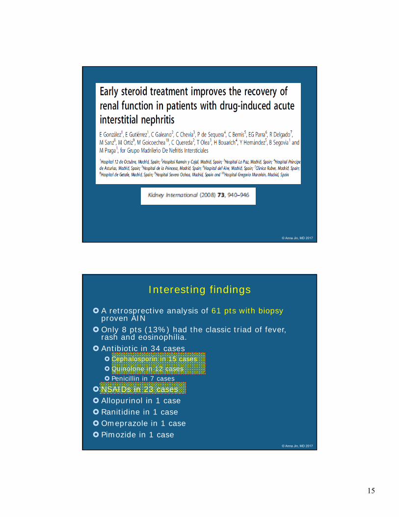
Gonzalez E, Gutierrez E, Galeano C, et al. Early steroid treatment improves the recovery of renal function in patients with drug-induced acute interstitial nephritis. Kidney international 2008;73:940-6.
Pusey CD, Saltissi D, Bloodworth L, Rainford DJ, Christie JL. Drug associated acute interstitial nephritis: clinical and pathological features and the response to high dose steroid therapy. Q J Med 1983; 52: 194–211
Buysen JG, Houthoff HJ, Krediet RT, Arisz L. Acute interstitial nephritis: a clinical and morphological study in 27 patients. Nephrol Dial Transplant 1990; 5: 94–99
Enriquez R, Gonzalez C, Cabezuelo JB et al. Relapsing steroid-responsive idiopathic acute interstitial nephritis. Nephron 1993; 63: 462–465 © Anna Jin, MD 2017
15
© Anna Jin, MD 2017
Interesting findings
A retrosprective analysis of 61 pts with biopsy proven AIN
Only 8 pts (13%) had the classic triad of fever, rash and eosinophilia.
Antibiotic in 34 cases Cephalosporin in 15 cases Quinolone in 12 cases Penicillin in 7 cases
NSAIDs in 23 cases Allopurinol in 1 case Ranitidine in 1 case Omeprazole in 1 case Pimozide in 1 case
© Anna Jin, MD 2017

16
Key Finding
© Anna Jin, MD 2017
AIN: Prognostic factor
Clinical and histologic indicators of a decreased likelihood of recovery
Prolonged renal failure (>3 wks), ? SeverityAIN associated with NSAIDs useHistologydegree of interstitial fibrosis and tubular atrophy diffuse vs patchy infiltrateinterstitial granulomas
© Anna Jin, MD 2017

17
Take Home Messages: AIN
A syndrome that is becoming both increasingly non-specific in clinical
features and diverse in etiologyUnexplained acute renal injury with
normal sized kidneys (+/- increased echogenicity)
ALWAYS get a thorough history on pt’smedications
Consider steroid therapy in certain pts. (do it early)
© Anna Jin, MD 2017
Clinical Case Two:
58 y/o white male with Hep C cirrhosis, advanced recurrent HCC (with evidence of tumor thrombus in portal vein) s/p TACE x 2 (2014 and 2016), DM2, HTN, HL, who presents with severe abdominal distension due to ascites, anasarca with lower extremity edema, decreased urine output and AKI. His baseline Cr 2 wks. prior was 0.9, and current Cr is 4.8, BUN 70, Na 123, K 6.2, and Bicarb 17. His ascites is refractory to diuretics (lasix and aldactone). He underwent large volume paracentesis ~8L removed 2 weeks ago.
Exam: BP 97/58 HR=82, JVP=12
Icteric, muscle wasting, tense ascites, 3+ pitting edema on LE
T Bili=9.7, INR=1.2; WBC=5.5; Hgb 9.4, albumin 2.0
Urine sediment nl; Urine Na=<10
© Anna Jin, MD 2017

18
Hepatorenal Syndrome (HRS)
What is the most common cause of AKI in pts with cirrhosis?
A. Prerenal azotemiaB. Acute tubular necrosisC. HRS
© Anna Jin, MD 2017
Definition of HRS
Hepatorenal syndrome represents the end-stage of a sequence of reductions of renal perfusion induced by severe hepatic injury • Functional renal failure, with absence of histological changes• Development of diuretic resistant or refractory ascites • The Hallmark is renal vasoconstriction. • Worst prognosis of all complications of cirrhosis
© Anna Jin, MD 2017

19
Types of HRS Type 1 HRS (AKI)
Rapid deterioration in renal function within 2 wks. (double initial Cr >2.5mg/dl)
Three-fourth with “second” hit: SBP (20%), a large volume paracentesis (15%),
GI bleed, and rapid diuresis. One-fourth without precipitating factor Survival in weeks (median 2 wks)
Type 2 HRS (CKD) Slowly progressive renal impairment Spontaneous Associated with refractory ascites in 75%
(resistant to diuretics) Survival in months (median 6 months)
© Anna Jin, MD 2017
Probability of survival: Type 1 vs Type 2
Alessandria et al, Hepatol 2005© Anna Jin, MD 2017

20
Assessing kidney function in pts with cirrhosis
Q: Why is Cr falsely low in cirrhosis?
a). Decreased synthesis d/t lower hepatic production of creatine b). Malnutrition and less muscle massc). Dilutional d/t the edematous state in end-stage liver disease leading to large distribution of Cr in the body d). Cr assays are subject to interference by chromogens, hyperbilirubenemia being the major one e). Complications such as variceal bleeding, spontaneous bacterial peritonitis or sepsis lead to increased Cr tubular excretionf). All of the above.
© Anna Jin, MD 2017
Pathogenesis
© Anna Jin, MD 2017

21
Endogenous vasoactive factors The hallmark of HRS is intense renal vasoconstriction with
predominant peripheral arterial vasodilation.
Arch intern med 1993 © Anna Jin, MD 2017
Ascites and HRS as a Continuum
© Anna Jin, MD 2017
Cystatin C ?

22
Diagnosis and management of acute kidney injury in patients with cirrhosis: Revised consensus
recommendations of the International Club of Ascites
Paolo Angeli, Pere Ginès, Florence Wong, Mauro Bernardi, Thomas D. Boyer, Alexander Gerbes, Richard Moreau, Rajiv Jalan, ShivK. Sarin, Salvatore Piano, Kevin Moore, Samuel S. Lee, Francois Durand, Francesco Salerno, Paolo Caraceni, W. Ray Kim, Vicente
Arroyo, Guadalupe Garcia-Tsao
Journal of HepatologyVolume 62, Issue 4, Pages 968-974 (April 2015)
© Anna Jin, MD 2017
Diagnostic Criteria defined by the International Club of Ascites (ICA)
Advanced hepatic failure and portal hypertensionDiagnosis of AKI according to ICA-AKI criteria
an increase in Cr by >0.3 mg/dL within 48 hrs, or an increase from baseline by >50% within 7 days
No response after 2 consecutive days with diuretic withdrawal and volume expansion with albumin 1g/kg of body weight (max 100 g/day)
Absence of shock No current or recent use with nephrotoxic drugs. No macroscopic signs of structural kidney injury
Absence of proteinuria >500 mg/day, microhematuria (>50 RBC/hpf), and abnormal renal ultrasonography.
Journal of Hepatology 2015 62, 968-974 © Anna Jin, MD 2017

23
HRS is diagnosis of exclusion
Hypotension (DDx pre-renal) Serum sodium < 130 mEq/L
(hypervolemic hyponatremia ) Urine sodium <10 mEq/L Urine volume <500 ml/day Urine Osm > Plasma Osm Bland urine sediment
© Anna Jin, MD 2017
Diagnostic Approach to AKI in Cirrhotics
AKI in a pt with cirrhosis
ECF fluid losses;rapid/excessive diureticsVomiting,diarrhea,hemorrhage,recentLVP /hemodynamic changes due to use of NSAIDS (or) ACEI/ARB
hold diuretics/offending medicationsTrial of intravascular volume expander (albumin) if renal func on ↑ with trial,Diagnosis of prerenal is made
Recent use of nephrotoxic medications Hypotension(sepsis,hemorrhage)
Glomerular proteinuria&hematuriai.e.,dysmorphic RBCs and RBC cast
Toxic or ischemic renal injury
Glomerular diseaseYES
Imaging (USG,CT scan)shows Hydronephrosis,urinary retention
Obstructive uropathy
Patient has evidence of Portal Hypertension & fulfills HRS‐AKI diagnostic criteria
Diagnosis of HRS can be made
YES
YES
YES
YES
NO
NO
NO
NO
© Anna Jin, MD 2017

24
Treatment of HRS
Liver Transplantation: the only effective permanent tx.
Transjugular Intrahepatic Portosystemic Shunt (TIPS) ? For HRS type 2 and refractory
ascites
Reduce portal hypertension, increase effective arterial volume and reverse splanchnic vasodilatation
Complications (Encephalopathy, Shunt stenosis, Hemolysis and Hyperbilirubinemia)
© Anna Jin, MD 2017
Treatment of HRS
Optimize Renal Perfusion:
Hold Beta Blockers and diuretics
[non-renal] vasoconstrictors combined with albumin
ICU-Treatment:
• Norepinephrine in combination with albumin with goal to raise MAP by 10mmHg
• Meta-analysis, 154 pts, Type 1 HRS, N+A vs Terlipressin + A, similar results, N+A less side effects
Non-ICU Treatment:
• Terlipressin + Albumin if available
• Midodrine + Ocreotide + AlbuminAntonio Paulo Nassar Junior, et al. Terlipressin versus Norepinephrine in the Treatment of Hepatorenal Syndrome: A Systematic Review and Meta-Analysis. PLOS ONE 2014 © Anna Jin, MD 2017

25
HRS: Management
Vasoconstrictors to increase MAP by 10-15 mmHg.Midodrine: alpha 1-adrenergic agonist
systemic vasoconstrictor5mg tid up to a maximum of 15mg tid
Octreotide: analogue of somatostatin inhibitor of vasodilation 100mcg sc tid, with maximum 200mcg tid
Terlipressin: vasopressin analogues, V-1 receptor agonist splanchnic vasoconstriction
© Anna Jin, MD 2017
A 60-yr-old man with alcoholic cirrhosis is admitted with SBP. Despite antibiotic therapy, he develops AKI (Cr increased from 0.6 to 1.8 mg/dl). Which ONE Of the following therapies has been shown in a randomized, controlled trial to improve renal function in patients with hepatorenal syndrome?
A. Albumin and octreotide
B. Fenoldopam and terlipressin
C. Dopamine and octreotide
D. Terlipressin and albumin
E. Midodrine and octreotide
© Anna Jin, MD 2017

26
© Anna Jin, MD 2017
© Anna Jin, MD 2017

27
THANK YOU!
© Anna Jin, MD 2017
Acute Kidney Injury (AKI)
[a.k.a. Acute Renal Failure, ARF]
Kamyar Kalantar-Zadeh, MD, PhD, MPHProfessor of Medicine, Pediatrics, Public Health & Epidemiology
Division of Nephrology and HypertensionUniv. California Irvine
Key elements:
Cr or Scr = serum creatinine
BUN = blood urea nitrogen (SUN)
HCO3- = bicarbonate
Na = sodium
Normal blood range:
Cr or Scr =
men ≤ 1.5 mg/dL
women ≤ 1.3 mg/dL
BUN = 5-25 mg/dL
HCO3- = 23-24 mEq/L

28
Nutritional Needs and Guideline Support for AKI

29
Nutrient Needs of AKI Patients as Defined by ESPEN Guidelines*
aAdapted to catabolism levels and to individual needs in case of underweight or obesity.*Adjust as necessary for obese patients.Cano NJ, et al. Clin Nutr. 2009;28(4):401–414.
Macronutrients
Energy (non-protein calories) 20–30 kcal/kg/da
Carbohydrates 3–5 (max. 7) g/kg/d
Fat 0.8–1.2 (max. 1.5) g/kg/d
Protein (Essential and Non-essential Amino Acids)
Conservative therapy, mild catabolism (mild Aki on CKD)
0.6–0.8 (max. 1.0) g/kg/d
Extracorporeal therapy, moderate catabolism
1.0–1.5 g/kg/d
CCRT, severe hypercatabolismUp to maximum 1.7 g/kg/d (or 2.0 or higher!)
Variable Estimate P-value Odds ratio
SAPS II score 0.00798 0.166 1.08Cardiopulmonary resuscitation 0.06160 0.004 1.86Multiple vasoactive medication 0.02930 <0.001 1.34Mechanical ventilation 0.02930 <0.001 1.34Single vasoactive medication 0.01160 0.012 1.13Treatment of complicated metabolic acidosis/alkalosis
0.00768 0.034 1.08
Care of drains -0.00883 0.002 0.91
Enteral nutrition -0.01480 <0.001 0.86
SAPS, Simplified Acute Physiology Score.Variables express the proportion of days on which this activity was performed. The odds ratios reflect the change in the risk of dying during the intensive care unit stay if the proportion of days with intervention increases by 10%.Metnitz PGH, et al. Crit Care Med. 2002;30(9):2051-2058.
Effect of AKI Requiring RRT on Outcome in Critically Ill Patients (N=19,067)
Enteral Nutrition is Associated with Favorable Outcomes in AKI

30
Acute Renal Failure: Mechanisms
Volume ↓ Prerenal
Renal Vascular: Stenosis, vasculitis
Acute Glomerulo-
nephritisAcute tubular damage
Interstitial damage
Post-renal Obstruction
Case 1
• A 50-year-old male was brought to the ER complaining of vomiting for two weeks. There was no history of drug ingestion and no prior history of renal disease.
• Physical examination on admission revealed: BP 100/60 supine and 85/50 upright; pulse 120; wt 135 lbs. The rest of the physical examination was unremarkable.
• Lab data: BUN 100 mg/dl; Cr 3.7 mg/dl; Na 125 mEq/L; K 4.2 mEq/L; HCO3 30 mEq/L.
• Urinalysis was negative for protein and showed noRBC or casts.

31
Case 1
1.i. What is the most likely cause of renal failure?
A:A
RF due
to v
olum
e...
B:A
RF due
to re
nal v
...
C:A
RF due
to a
cute
...
D:A
RF due
to a
cute
...
E:A
RF due
to a
cute
in...
F:A
RF due
to u
rinar
y ...
G:C
KD with
chr
onic
...
0% 0% 0% 0%0%0%0%
A: ARF due to volume depletion(pre-renal azotemia)
B: ARF due to renal vascular dis.(renal artery stenosis,
vasculitis, atheroembolic dis)
C: ARF due to acute glomerulonephritis (GN)
D: ARF due to acute tubular necrosis (ATN)
E: ARF due to acute interstitial nephritis (AIN)
F: ARF due to urinary tract obstruction, e.g., prostate
hypertrophy (post-renal azotemia)
G: CKD with chronic renal insufficiency
:10
Acute Renal Failure: Mechanisms
Volume ↓ Prerenal
Renal Vascular: Stenosis, vasculitis
Acute Glomerulo-
nephritisAcute tubular damage
Interstitial damage
Post-renal Obstruction

32
Case 11.ii. What information obtained from the physical examination and laboratory data is helpful in establishing the diagnosis of
ARF?
A:H
isto
ry o
f vom
iting
B:O
rthost
atic
chan
ge...
C. T
achyc
ardia
consi
...
D:T
he hig
h BUN/S
cr r.
..
E:E
leva
ted s
erum
HCO
3
F:L
ack
of his
tory
of .
..
G: A
ll of
the
above
0% 0% 0% 0%0%0%0%
A: History of vomiting
B: Orthostatic changes in BP
C. Tachycardia consistent with volume depletion
D: The high BUN/Scr ratio
E: Elevated serum HCO3
F: Lack of history of drug ingestion and previous renal disease and bland urine sediment.
G: All of the above
:10
Case 11.iii. What other laboratory data would be
helpful?
A:U
rine
lyte
s an
d os.
..
B:E
leva
ted H
ct a
nd u
...
C:B
oth.
D:N
eith
er
0% 0%0%0%
A: Urine lytes and osmolality showing a pre-renal profile
B: Elevated Hct and uric acid levels
C: Both.
D: Neither
:10

33
Case 11.iv. What treatment is indicated and what is
the expected effect on renal function?
A:K
eep
patie
nt NPO ..
.
B:G
ive
high d
ose lo
o...
C:E
mer
gent d
ialy
sis
...
D:V
olum
e re
plet
ion w
...
0% 0%0%0%
A: Keep patient NPO and no IV fluid
B: Give high dose loop diuretics such as furosemide (Lasix)
C: Emergent dialysis treatment, because patient’s renal function will not improve
D: Volume repletion with normal saline will likely improve renal function
:10
It's Important To Keep Hydrated While On the Job

34
Pre‐Renal AKI: Some Causes
• Hypovolemia– Volume Loss – GI, renal, skin– Blood Loss – GI, MVA
• Cardiac Causes (hemodynamic CRS)– Acute cardiogenic shock– Chronic congestive heart failure
• Liver Disease
• Nephrotic syndrome
• Renovascular– Renal venous thrombosis
Pre RENAL vs Intra RenalPre-Renal
(incl Hepato-renal)Intrinsic Renal
(incl ATN)
Urine Volume Decreased Variable
Urine Sp. Gravity > 1.020 ~1.010
Urine Osmolality, mosm/kg > 500 ** < 300
FENa, % < 0.5% > 2%
FEurea, % < 35% > 35%
FEuric acid, % < 7% > 15%
Urine Sediment None to Hyaline casts(bland)
Others, e.g. granular casts
U-creatinine >100 mg/dL <50 mg/dL
Renal US Normal Variable, echo-gen. (ATN)
BUN/Crea ratio >20 * <10
UrineCr./PlasmaCr. >40 <20
U-Na <10 mEq/L >40 mEq/L
Response Responds intra-vascular volume expander
Usually no response to volume

35
Case 5
• A 30-year-old female noted dysuria and frequency and was given ampicillin for a presumed urinary tract infection. Two to 3 days after beginning the medication, she noted the onset of a diffuse skin rashand a fever to 101 F.
• Physical examination revealed: BP 130/80 supine and upright. There was an erythematous rash on the abdomen and extremities, but the physical examination was otherwise negative.
• Lab data: BUN 75 mg/dl; Cr 5.0 mg/dl; Na 140 mEq/L; K 5.0 mEq/L; HCO3 20 mEq/L. Urinalysis showed 2+ protein, many RBCs and WBCs, no RBC casts.
Case 55.i. What is the most likely cause of renal
failure?
A:A
RF due
to v
olum
e...
B:A
RF due
to re
nal v
...
C:A
RF due
to a
cute
...
D:A
RF due
to a
cute
...
E:A
RF due
to a
cute
in...
F. A
RF due
to u
rinar
y ...
G:C
KD with
chr
onic
...
0% 0% 0% 0%0%0%0%
A: ARF due to volume depletion(pre-renal azotemia)
B: ARF due to renal vascular dis.(renal artery stenosis,
vasculitis, atheroembolic dis)C: ARF due to acute
glomerulonephritis (GN)D: ARF due to acute tubular
necrosis (ATN)E: ARF due to acute interstitial
nephritis (AIN)F. ARF due to urinary tract
obstruction, e.g., prostate hypertrophy (post-renal
azotemia)G: CKD with chronic renal
insufficiency :10

36
Acute Renal Failure: Mechanisms
Volume ↓ Prerenal
Renal Vascular: Stenosis, vasculitis
Acute Glomerulo-
nephritisAcute tubular damage
Interstitial damage
Post-renal Obstruction
Case 55.ii. What information in the history, physical examination and lab data is helpful in making a diagnosis of the cause of the
renal disease?
A:S
kin ra
sh a
fter s
tar..
.
B:L
ow-gra
de fe
ver a
ft..
C:P
rese
nce o
f ski
n r..
.
D:P
rese
nce o
f WBCs.
..
E:M
odera
te p
rote
inur..
F:O
nly A
, B a
nd C
G:A
ll of t
he ab
ove
0% 0% 0% 0%0%0%0%
A: Skin rash after starting ampicillin
B: Low-grade fever after starting ampicillin
C: Presence of skin rash on physical exam
D: Presence of WBCs and RBCs, but no casts
E: Moderate proteinuria (2+ protein)
F: Only A, B and C
G: All of the above
:10

37
Acute Interstitial Nephritis
Case 55.iii. What single laboratory test would be the most helpful in determining the cause of renal failure?
A:E
osinoph
iluria
(e...
B:R
ecta
l exa
min
atio
n
C:C
hest X
-ray
D:O
nly A
and B
E:A
ll of t
he ab
ove
0% 0% 0%0%0%
A: Eosinophiluria (eosinophils in the urine) esp. if > 5% [Hansel stain of urine]
B: Rectal examination
C: Chest X-ray
D: Only A and B
E: All of the above
38
Case 55.iv. What treatment is indicated?
A:W
ithdra
wal o
f am
pici
...
B:A
dmin
iste
r IV c
ontra
st
C:R
epla
ce a
mpic
illin
...
D:G
ive
high d
ose N
SAID
E:A
ll of t
he ab
ove
0% 0% 0%0%0%
A: Withdrawal of ampicillin (+/-steroid)
B: Administer IV contrast
C: Replace ampicillin with aminoglycoside
D: Give high dose NSAID
E: All of the above
:10
Acknowledgement
Investigators and Staff• Elani Streja, MPH, PhD• Connie M. Rhee, MD, MSc• Hamid Moradi, MD• Wei Ling Lau, MD• Joline Chen, MD, MPH• Vanessa Ravel, MPH• Foad Ahamdi, MD• Paungpaga Lertdumrongluk,
MD• Mega Doshi, MD• Sepideh Rezakhani, MD• Melissa Soohoo, MPH• Bryan Shapiro, MPH• Rochelle Roger, MPH• Amanda Brown, RD• Tracy Nakata • Nany Lopez• Catherine Guillen• Jennie Jing, MS• Kamyar Kalantar-Zadeh, MD,
MPH, PhD
Collaborators:• Csaba P. Kovesdy, MD• Rajnish Mehrotra, MD• Joel D Kopple, MD• Matthew Budoff, MD• Steven S. Jacobsen, MD,
PhD• Rajiv Saran, MD, MSc.• Miklos Z. Molnar, MD, PhD• Jongha Park, MD• Daniel Gillen, PhD• Danh Nguyen, PhD• Allen Nissenson, MD, • Steven Brunelli, MD, MS
The Harold Simmons Center for Kidney Disease Research & Epidemiology

39
Professional Social Media
Dr. Kamyar Kalantar‐Zadeh’s social media links for professional networking: Twitter: @KamKalantar https://twitter.com/kamkalantarFacebook: @KamKalantar https://www.facebook.com/KamKalantarMendeley: https://www.mendeley.com/profiles/kamyar-kalantar-zadeh3Google+: https://plus.google.com/109239031645120656897Wikipedia: https://en.wikipedia.org/wiki/Kamyar_Kalantar-ZadehResearchGate: https://www.researchgate.net/profile/Kamyar_Kalantar-ZadehGoogle Scholar: https://scholar.google.com/citations?user=kYonxxoAAAAJ&hl=en