introduction: the immune system–the basics, … inflammation in health and disease. 20.400 spring...
TRANSCRIPT
20.380 S10Introduction:
the Immune System– the basics, inflammation in health
and disease
20.400 spring 2008 1
Overview of the immune system
2

Two arms of immunity: the innate and adaptive immune systems
20.400 spring 2008 2
KEY EFFECTORS OF ADAPTIVE IMMUNITY
Diagram of how B lymphocytes, Helper T lymphocytes, and Cytolytic T lymphocytes recognize particular antigens and effect immunity has been removed due to copyright restrictions.
20.400 spring 2008 (Abbas) 2
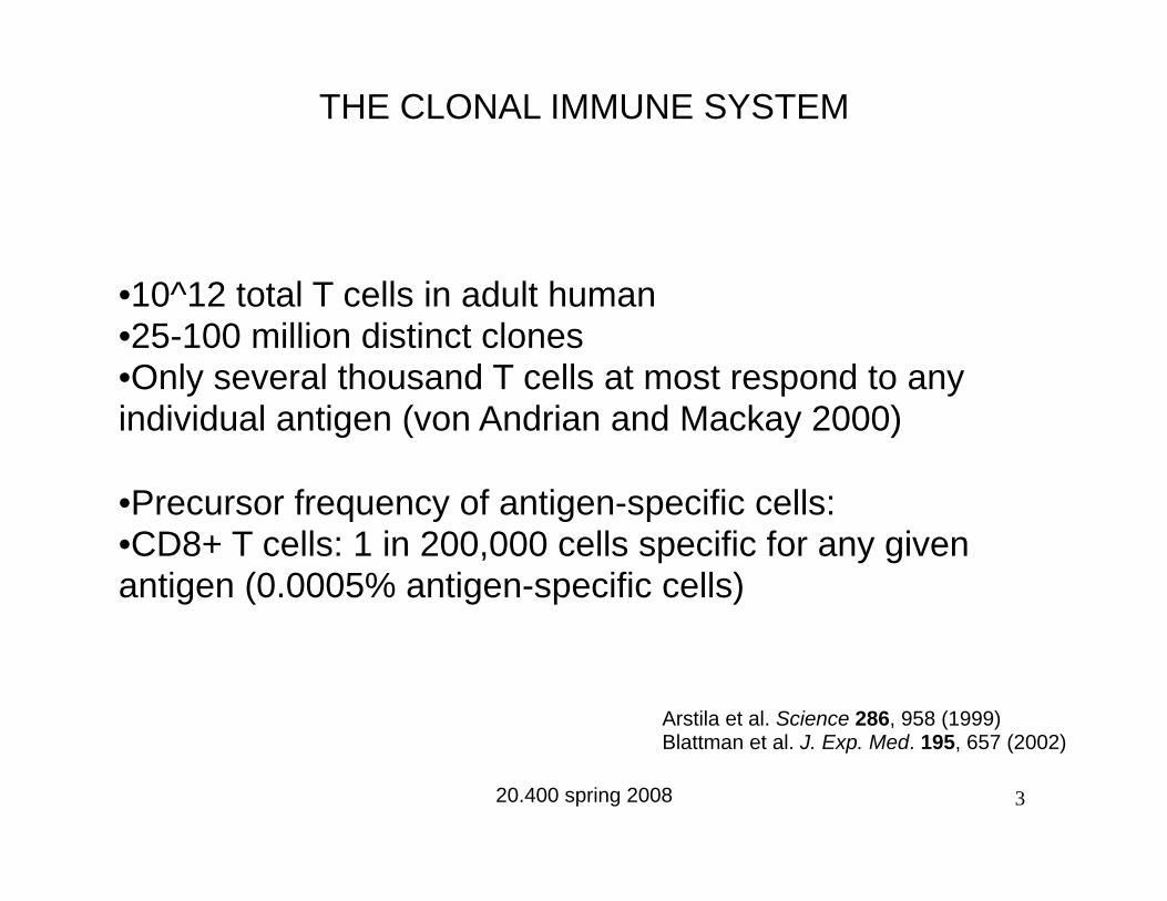
THE CLONAL IMMUNE SYSTEM
•10^12 total T cells in adult human •25-100 million distinct clones •Only several thousand T cells at most respond to any individual antigen (von Andrian and Mackay 2000)
•Precursor frequency of antigen-specific cells: •CD8+ T cells: 1 in 200,000 cells specific for any given antigen (0.0005% antigen-specific cells)
Arstila et al. Science 286, 958 (1999) Blattman et al. J. Exp. Med. 195, 657 (2002)
20.400 spring 2008 3
B cell
T cell
Dendritic cell
T B
LYMPH NODE PERIPHERAL TISSUE
1 2
3
4
5 LYMPHATICS
BLOOD
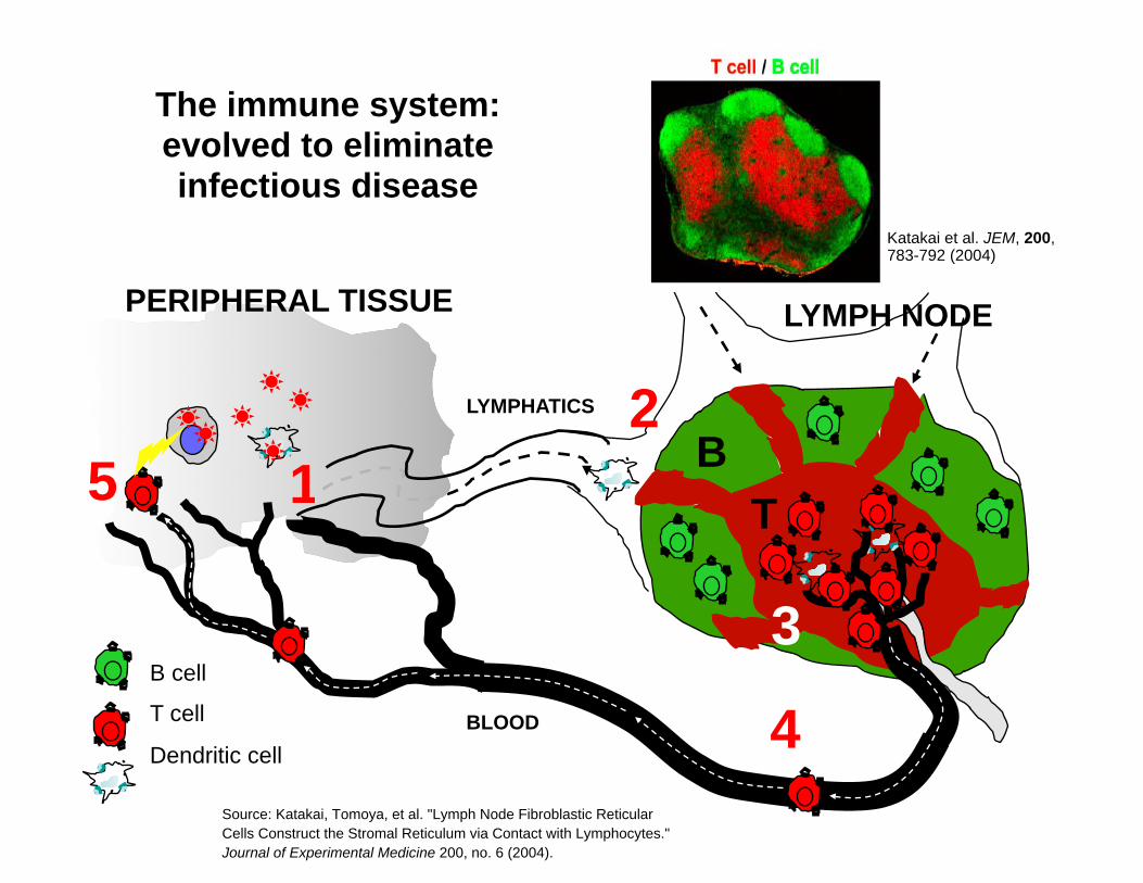
Katakai et al. JEM, 200, 783-792 (2004)
The immune system: evolved to eliminate infectious disease
Source: Katakai, Tomoya, et al. "Lymph Node Fibroblastic Reticular Cells Construct the Stromal Reticulum via Contact with Lymphocytes." Journal of Experimental Medicine 200, no. 6 (2004).
B cell activation
Diagram of antigen recognition, B cell proliferation, and Ig secretion and isotype switching has been removed due to copyright restrictions.
Lecture 20 Spring 2006 (Abbas) 13
Lecture 20 Spring 2006
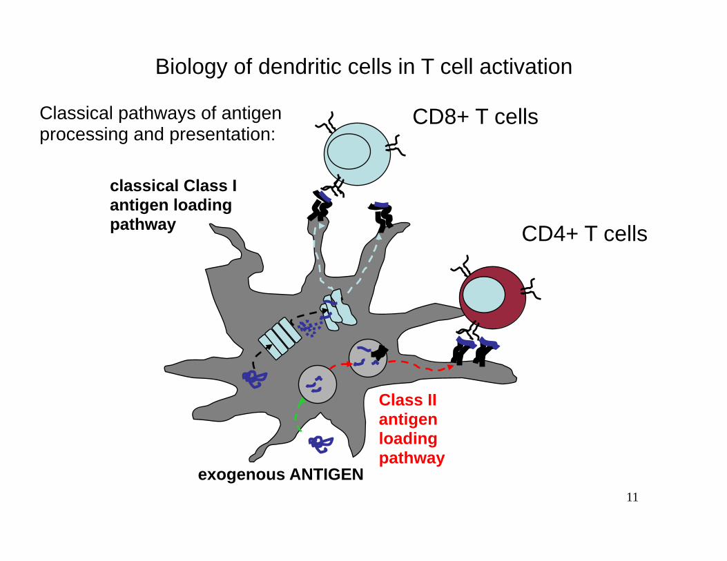
Biology of dendritic cells in T cell activation
Classical pathways of antigen CD8+ T cells processing and presentation:
Class II antigen loading pathway
classical Class I antigen loading pathway CD4+ T cells
exogenous ANTIGEN 11

interactions in the lymph node
Three electron micrographs of T cells and dendritic cells interacting with reticular fibers have been removed due to copyright restrictions.

5 !
T-cell activation
infected cell or (1) antigen recognitiontumor cell target cell
T-cell
T T cell receptor (TCR)
Two electron micrographs removed due to copyright restrictions.peptide-MHC
(2) immunological synapse (IS) formation
T T
Grakoui et al. Science 285, 221 (1999) Monks et al. Nature 395, 82-86 (1998)

The immune system: a distributed network
11
lymphocyte trafficking is “addressed” by combinations ofadhesion molecules and chemokine signals
Diagram of lymphocyte trafficking removed due to copyright restrictions.There are four steps: rolling adhesion, tight binding, diapedesis, and migration.See Figure 2-44 part 3 of 3, Janeway, Charles, et al. Immunobiology. 6th ed.New York: Garland Science, 2005. ISBN: 9780815341017.
12
or by promoting the survival or proliferation of specific lympho-
cyte subpopulations. For example, in an in vitro system IL-2,
IL-4, IL-10, and CD40L direct homing receptor expression,
as sessile GC B cells differentiate into recirculating memory
B cells (145). During CD4! T-cell activation and differentiation
in vitro, IL-12 and IL-4 can directly regulate the generation
ligands for P- and E-selectin by controlling the expression of
a1,3-fucosyltransferase VII, a key enzyme required for selectin
ligand biosynthesis (146). However, selectin ligands are not
restricted to Th1 cells in vivo, and thus the role of IL-12 in
directing their expression during activation in cutaneous and
intestinal lymphoid tissues is unclear (101, 102). Similarly,
transforming growth factor-b (TGF-b) response elements have
been found in the b7 integrin promoter, but treating cells
in vitro with this cytokine inhibits a4 expression. Therefore,
this cytokine alone cannot account for the upregulation of
a4b7 in intestinal lymphoid tissues (147). Taken together,
these data suggest that lymphocytes integrate a complex array
of TCR, costimulatory, chemokine, and cytokine signals that
together specify unique microenvironments within cutaneous
and intestinal lymphoid tissues and direct expression of appro-
priate combinations of adhesion and chemoattractant receptors.
Understanding these complex pathways of lymphocyte
differentiation clearly requires further research, will undoubt-
edly lead to novel insights into lymphocyte biology, and has
important implications for the development of effective sys-
temic and mucosal vaccines.
Concluding remarks
The regulated expression of chemokines and chemokine
receptors underlies the structural and systemic organization
of the immune system, serving essential functions in leukocyte
development, lymphocyte recirculation, immune surveillance,
and effector lymphocyte differentiation and targeting (Fig. 2).
While much has been learned about chemokine and chemo-
kine receptor expression and function in immunity, a com-
plete understanding of these critical molecules is still a long
way off. In addition to mediating integrin activation and
chemotaxis, chemokines and chemokine receptors have been
implicated in lymphocyte differentiation and as leukocyte
growth/survival factors, but these aspects of their biology
remain largely unexplored (36, 148). Moreover, the complete
sequencing of the human and mouse genomes has revealed
many ‘orphan’ chemokine receptors whose ligands, expres-
sion patterns, and function remain poorly understood (149).
In addition, very little is known about the signaling pathways
and transcription factors that regulate the very complex tissue-
Campbell et al " Immune system organization
66 Immunological Reviews 195/2003
Chemokines in the systemicorganization of immunity
Daniel J. Campbell
Chang H. Kim
Eugene C. Butcher
Authors’ addresses
Daniel J. Campbell1,2, Chang H. Kim3,Eugene C. Butcher1,2,1Laboratory of Immunology and Vascular
Biology, Department of Pathology, Stanford
University School of Medicine, Stanford, CA, USA.2The Center for Molecular Biology and Medicine,
Veterans Affairs Palo Alto Health Care System, Palo
Alto, CA, USA.3Laboratory of Immunology and Hematopoiesis,
Department of Veterinary Pathobiology and
Purdue Cancer Center, Purdue University, West
Lafayette, IN, USA.
Correspondence to:
Daniel J. CampbellBenaroya Research Institute at Virginia Mason1201 Ninth AvenueSeattle, WA 98101–2795USATel.: !12065836525Fax: !12062237543E-mail: [email protected]
Acknowledgements
We thank Tracy Staton and Gudrun Debes forcomments on the manuscript. E.C.B. is sup-ported by grants from the NIH and theVeterans administration. D.J.C. is the recipientof a postdoctoral fellowship from the ArthritisFoundation. C.H.K. is supported by grantsfrom The Eli and Edythe L. Broad Foundationand Leukemia and Lymphoma Society.
Summary: Directed cellular migrations underlie immune system organ-ization. Chemokines and their receptors (along with surface-adhesionmolecules) are central to these migrations, targeting developing andmature leukocytes to tissues and microenvironments suitable for theirdifferentiation and function. The chemokine CXCL12 and its receptorCXCR4 play a central role in the migration of hematopoietic stem cells,and several chemokine receptors are transiently expressed during distinctstages of B- and T-cell development. In the periphery, mature naı̈ve B andT cells utilize the receptors CCR7, CXCR4, and CXCR5 to recirculatethrough specialized microenvironments within the secondary lymphoidtissues, while effector and memory lymphocytes express bewilderingpatterns of adhesion molecules and chemokine receptors that allowthem to function within microenvironments and non-lymphoid tissuesinaccessible to naı̈ve cells. Here, we summarize the role of chemokinesand their receptors in the spatial organization of the immune system andconsider the implications for immune function.
The mechanisms of leukocyte homing
The mammalian immune system is designed to combat infec-
tion while maintaining self-tolerance and limiting immune-
mediated pathology. In order to accomplish these tasks, the
cells and tissues of the immune system are precisely organized
to ensure the proper development, activation, and function of
diverse leukocyte populations. Tissue- and microenvironment-
selective leukocyte homing is the basis for this organization,
and leukocyte responses to the chemoattractant cytokines
(chemokines) function prominently in these homing path-
ways (1).
Leukocyte homing from blood to tissues generally occurs in
specialized post-capillary venules through a series of sequential
interactions with the vascular endothelium (reviewed exten-
sively in 2, 3). Initial interactions mediated by surface-adhe-
sion molecules, either selectins or low-affinity integrins, cause
the leukocytes to roll along the endothelium. Chemokines
presented on the endothelial surface trigger integrin affinity
and avidity activation and mediate leukocyte arrest on
the endothelial wall (4–6). Subsequently, cells undergo
Copyright ! Blackwell Munksgaard 2003
Immunological Reviews0105-2896
58
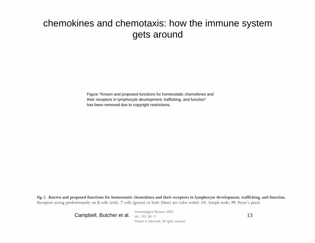
chemokines and chemotaxis: how the immune system gets around
Figure "Known and proposed functions for homeostatic chemokines and their receptors in lymphocyte development, trafficking, and function" has been removed due to copyright restrictions.
Fig. 2. Known and proposed functions for homeostatic chemokines and their receptors in lymphocyte development, trafficking, and function. Receptors acting predominantly on B cells (red), T cells (green) or both (blue) are color coded. LN, lymph node; PP, Peyer’s patch.
Campbell, Butcher et al.Immunological Reviews 2003 Vol. 195: 58–71 13 Printed in Denmark. All rights reserved
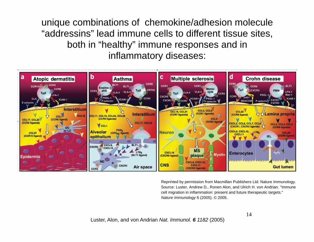
unique combinations of chemokine/adhesion molecule “addressins” lead immune cells to different tissue sites,
both in “healthy” immune responses and in inflammatory diseases:
Reprinted by permission from Macmillan Publishers Ltd: Nature Immunology. Source: Luster, Andrew D., Ronen Alon, and Ulrich H. von Andrian. "Immune cell migration in inflammation: present and future therapeutic targets." Nature Immunology 6 (2005). © 2005.
14 Luster, Alon, and von Andrian Nat. Immunol. 6 1182 (2005)
Diagram of chemotaxis directing cell migration has been removed due to copyright restrictions.
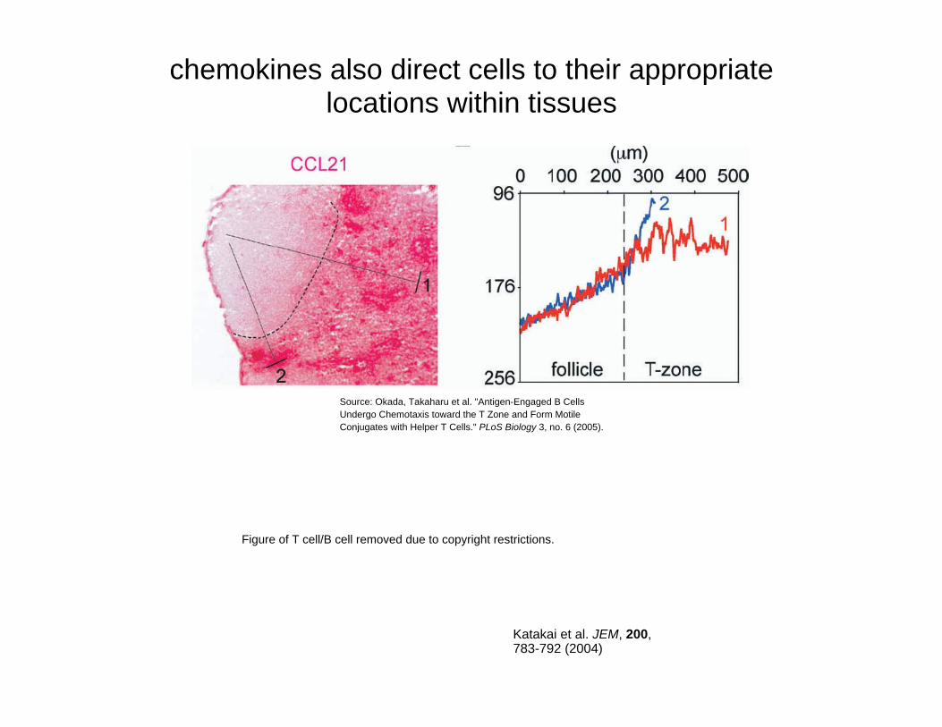
chemokines also direct cells to their appropriate locations within tissues
Figure of T cell/B cell removed due to copyright restrictions.
Katakai et al. JEM, 200, 783-792 (2004)
Source: Okada, Takaharu et al. "Antigen-Engaged B Cells Undergo Chemotaxis toward the T Zone and Form Motile Conjugates with Helper T Cells." PLoS Biology 3, no. 6 (2005).

Steps in the immune response to infection
17

innate immune sentinels
HIV illustration removed due to copyright restrictions.
http://www.northwestern.edu/newscenter/images/2008/12/hiv-illustration.jpg18

infection site in peripheral tissue
(3) DCs travel to lymph nodes
5
resident DCs in skin
(1) chemoattraction of dendritic cells/DC precursors
to sites of infection/inflammation
Randolph, Angeli, and Swartz Nat. Rev. Immunol. 5 617 (2005)
5
(4) DCs activatelymphocytes bycell-cell contact
Dendritic cells and initiation of adaptive immune responses
APC
peptide-MHC
pathogen
(2) dendritic cellscollect antigen
and become activated
Reprinted by permission from Macmillan Publishers Ltd: Nature Reviews Immunology. Source: Randolph, Gwendalyn J. et al. "Dendritic-cell trafficking to lymph nodes through lymphatic vessels." Nature Reviews Immunology 5 (2005). © 2005.
Electron micrograph of resident dendritic cells in skin has been removed due to copyright restrictions.
infection site in peripheral
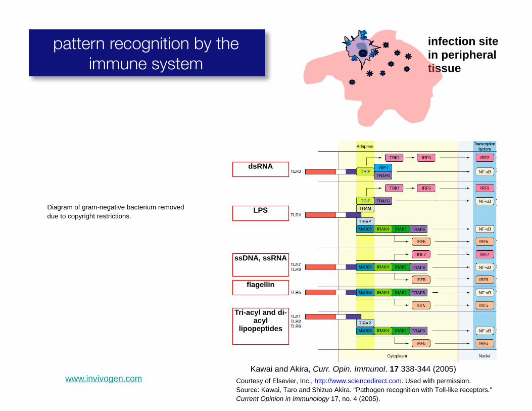
pattern recognition by the immune system tissue
Diagram of gram-negative bacterium removed due to copyright restrictions.
Kawai and Akira, Curr. Opin. Immunol. 17 338-344 (2005)
Tri-acyl and di-acyl
lipopeptides
ssDNA, ssRNA
LPS
dsRNA
flagellin
www.invivogen.com Courtesy of Elsevier, Inc., http://www.sciencedirect.com. Used with permission. Source: Kawai, Taro and Shizuo Akira. "Pathogen recognition with Toll-like receptors." Current Opinion in Immunology 17, no. 4 (2005).

PAMP recognition Immune cells integrate many signals toof microbes by ‘fingerprint’ pathogens: dendritic cells
Kawai and Akira, Curr. Opin. Immunol. 17 338-344 (2005)
LPS
dsRNA
ssDNA, ssRNA
Diagram from Science magazine removed due to copyright restrictions.
Huang et al., Science 294 3870 (2001)
flagellin
Tri-acyl and di-acyl
lipopeptides
Courtesy of Elsevier, Inc., http://www.sciencedirect.com. Used with permission. Lecture 20 Spring 2006Source: Kawai, Taro and Shizuo Akira. "Pathogen recognition with Toll-like receptors."Current Opinion in Immunology 17, no. 4 (2005).
7

TLR signaling is likely one of the earliest steps in the host response to infection
22

440 ! Februar y 12, 1998
The New England Journal of Medicine
Figure 2. Chemokine Regulation of Leukocyte Movement.Chemokines are secreted at sites of inflammation and infection by resident tissue cells, resident and recruited leukocytes, and cy-tokine-activated endothelial cells. Chemokines are locally retained on matrix and cell-surface heparan sulfate proteoglycans, estab-lishing a chemokine concentration gradient surrounding the inflammatory stimulus, as well as on the surface of the overlyingendothelium. Leukocytes rolling on the endothelium in a selectin-mediated process are brought into contact with chemokines re-tained on cell-surface heparan sulfate proteoglycans. Chemokine signaling activates leukocyte integrins, leading to firm adherenceand extravasation. The recruited leukocytes are activated by local proinflammatory cytokines and may become desensitized to fur-ther chemokine signaling because of high local concentrations of chemokines. The Duffy antigen receptor for chemokines (DARC),a nonsignaling erythrocyte chemokine receptor, functions as a sink, removing chemokines from the circulation and thus helpingto maintain a tissue–bloodstream chemokine gradient.
Copyright © 1998 Massachusetts Medical Society. All rights reserved. Downloaded from www.nejm.org at MASS INST TECH LIBRARY on December 2, 2004 .
what we typically think of as inflammation: recruitment of innate and adaptive immune cells to peripheral tissue sites:
inflammatory agent applied to epithelium:
Figure removed due to copyright restrictions.See Figure 2 from Luster, Andrew D. "Chemokines — ChemotacticCytokines That Mediate Inflammation." New England Journal of Medicine 338 (2006).
Le Borgne, Dubois et al. Immunity 24 191-201 (2006)
Courtesy of Elsevier, Inc., http://www.sciencedirect.com. Used with permission. Source: Le Borgne, Marie, et al. " Dendritic Cells Rapidly Recruited into Epithelial Tissues via CCR6/CCL20 Are Responsible for CD8+ T Cell Crosspriming In Vivo." Immunity 24 (2006).
Diagram explaining process of inflammation removed due to copyright restrictions.
Robbins and Cotran Pathol. Basis of Disease 7th ed. 24
recruitment of DCs: chemotaxis into inflammation sites
chemoattractants bring monocytes and
DCs to sites of infection
Two electron micrograph images removed due to copyright restrictions.

resident DCs in skin
(1) chemoattraction of dendritic cells/DC precursors
to sites of infection/inflammation
Reprinted by permission from Macmillan Publishers Ltd: Nature Reviews Immunology. Source: Randolph, Gwendalyn J. et al. "Dendritic-cell trafficking to lymph nodes through lymphatic vessels." Nature Reviews Immunology 5 (2005). © 2005.
5
(4) DCs activatelymphocytes bycell-cell contact
Randolph, Angeli, and Swartz Nat. Rev. Immunol. 5 617 (2005)
infection site in peripheral tissue
(3) DCs travel tolymph nodes APC
peptide-MHC
pathogen
(2) dendritic cellscollect antigen
and become activated
Lecture 20 Spring 2006
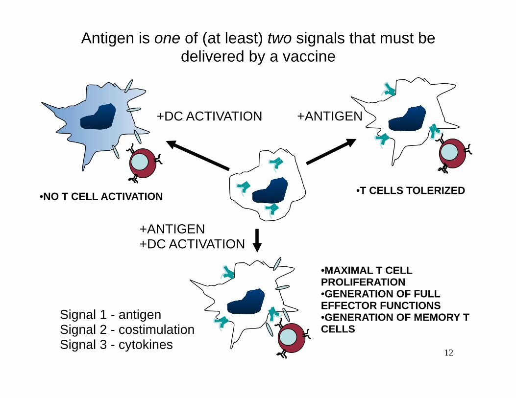
Antigen is one of (at least) two signals that must be delivered by a vaccine
•T CELLS TOLERIZED •NO T CELL ACTIVATION
+ANTIGEN+DC ACTIVATION
•MAXIMAL T CELL PROLIFERATION •GENERATION OF FULL EFFECTOR FUNCTIONS
Signal 1 - antigen •GENERATION OF MEMORY T Signal 2 - costimulation CELLS Signal 3 - cytokines
+DC ACTIVATION +ANTIGEN
12

(1) antigen carried tolymph nodes: (3) Antigen-specific T and B cells
meet at follicular border:ORCHESTRATION OF THEPRIMARY IMMUNE
RESPONSE
(2a) B cells encounter antigen,likely in follicles:
(Pape et al. Immunity 26 491 (2007))
3.5 min antigen B cells T cells
(2b) T cells encounter antigen in T zones: pMHC B cells
Figure from New England Journal of Medicine removed due to copyright restrictions. See Figure 3 from Ada, Gordon. "Advances in Immunology: Vaccines and Vaccination." New England Journal of Medicine 345 (2001).
Courtesy of Elsevier, Inc., http://www.sciencedirect.com . Used with permission. Source: Itano, Andrea A., et al. "Distinct Dendritic Cell Populations Sequentially Present Antigen to CD4 T Cells and Stimulate Different Aspects of Cell-Mediated Immunity." Immunity 19, no. 1 (2003).
Source: Okada, Takaharu et al. "Antigen-Engaged B Cells Undergo Chemotaxis toward the T Zone and Form Motile Conjugates with Helper T Cells." PLoS Biology 3, no. 6 (2005).

B cell
T cell
Dendritic cell
T B
LYMPH NODE PERIPHERAL TISSUE
1 2
3
4
5 LYMPHATICS
BLOOD
Katakai et al. JEM, 200, 783-792 (2004)
Source: Katakai, Tomoya, et al. "Lymph Node Fibroblastic Reticular Cells Construct the Stromal Reticulum via Contact with Lymphocytes." Journal of Experimental Medicine 200, no. 6 (2004).

440 ! Februar y 12, 1998
The New England Journal of Medicine
Figure 2. Chemokine Regulation of Leukocyte Movement.Chemokines are secreted at sites of inflammation and infection by resident tissue cells, resident and recruited leukocytes, and cy-tokine-activated endothelial cells. Chemokines are locally retained on matrix and cell-surface heparan sulfate proteoglycans, estab-lishing a chemokine concentration gradient surrounding the inflammatory stimulus, as well as on the surface of the overlyingendothelium. Leukocytes rolling on the endothelium in a selectin-mediated process are brought into contact with chemokines re-tained on cell-surface heparan sulfate proteoglycans. Chemokine signaling activates leukocyte integrins, leading to firm adherenceand extravasation. The recruited leukocytes are activated by local proinflammatory cytokines and may become desensitized to fur-ther chemokine signaling because of high local concentrations of chemokines. The Duffy antigen receptor for chemokines (DARC),a nonsignaling erythrocyte chemokine receptor, functions as a sink, removing chemokines from the circulation and thus helpingto maintain a tissue–bloodstream chemokine gradient.
Copyright © 1998 Massachusetts Medical Society. All rights reserved. Downloaded from www.nejm.org at MASS INST TECH LIBRARY on December 2, 2004 .
or by promoting the survival or proliferation of specific lympho-
cyte subpopulations. For example, in an in vitro system IL-2,
IL-4, IL-10, and CD40L direct homing receptor expression,
as sessile GC B cells differentiate into recirculating memory
B cells (145). During CD4! T-cell activation and differentiation
in vitro, IL-12 and IL-4 can directly regulate the generation
ligands for P- and E-selectin by controlling the expression of
a1,3-fucosyltransferase VII, a key enzyme required for selectin
ligand biosynthesis (146). However, selectin ligands are not
restricted to Th1 cells in vivo, and thus the role of IL-12 in
directing their expression during activation in cutaneous and
intestinal lymphoid tissues is unclear (101, 102). Similarly,
transforming growth factor-b (TGF-b) response elements have
been found in the b7 integrin promoter, but treating cells
in vitro with this cytokine inhibits a4 expression. Therefore,
this cytokine alone cannot account for the upregulation of
a4b7 in intestinal lymphoid tissues (147). Taken together,
these data suggest that lymphocytes integrate a complex array
of TCR, costimulatory, chemokine, and cytokine signals that
together specify unique microenvironments within cutaneous
and intestinal lymphoid tissues and direct expression of appro-
priate combinations of adhesion and chemoattractant receptors.
Understanding these complex pathways of lymphocyte
differentiation clearly requires further research, will undoubt-
edly lead to novel insights into lymphocyte biology, and has
important implications for the development of effective sys-
temic and mucosal vaccines.
Concluding remarks
The regulated expression of chemokines and chemokine
receptors underlies the structural and systemic organization
of the immune system, serving essential functions in leukocyte
development, lymphocyte recirculation, immune surveillance,
and effector lymphocyte differentiation and targeting (Fig. 2).
While much has been learned about chemokine and chemo-
kine receptor expression and function in immunity, a com-
plete understanding of these critical molecules is still a long
way off. In addition to mediating integrin activation and
chemotaxis, chemokines and chemokine receptors have been
implicated in lymphocyte differentiation and as leukocyte
growth/survival factors, but these aspects of their biology
remain largely unexplored (36, 148). Moreover, the complete
sequencing of the human and mouse genomes has revealed
many ‘orphan’ chemokine receptors whose ligands, expres-
sion patterns, and function remain poorly understood (149).
In addition, very little is known about the signaling pathways
and transcription factors that regulate the very complex tissue-
Development in primarylymphoid tissues
Lymphocyte recirculation andimmune surveillance
Microenvironmental positioningand non-lymphoid tissue homing
ThymusCXCR4/CXCL12
CCR9/CCL25CCR4/CCL17/CCL22CCR7/CCL19/CC21
Bone marrowCXCR4/CXCL12
CCR9/CCL25
LN/spleenCCR7/CCL19/CCL21
CXCR4/CXCL12
CCR7/CCL19/CCL21PP
CXCR4/CXCL12CXCR5/CXCL13
Fig. 2. Known and proposed functions for homeostatic chemokines and their receptors in lymphocyte development, trafficking, and function.Receptors acting predominantly on B cells (red), T cells (green) or both (blue) are color coded. LN, lymph node; PP, Peyer’s patch.
Campbell et al " Immune system organization
66 Immunological Reviews 195/2003
Chemokines in the systemicorganization of immunity
Daniel J. Campbell
Chang H. Kim
Eugene C. Butcher
Authors’ addresses
Daniel J. Campbell1,2, Chang H. Kim3,Eugene C. Butcher1,2,1Laboratory of Immunology and Vascular
Biology, Department of Pathology, Stanford
University School of Medicine, Stanford, CA, USA.2The Center for Molecular Biology and Medicine,
Veterans Affairs Palo Alto Health Care System, Palo
Alto, CA, USA.3Laboratory of Immunology and Hematopoiesis,
Department of Veterinary Pathobiology and
Purdue Cancer Center, Purdue University, West
Lafayette, IN, USA.
Correspondence to:
Daniel J. CampbellBenaroya Research Institute at Virginia Mason1201 Ninth AvenueSeattle, WA 98101–2795USATel.: !12065836525Fax: !12062237543E-mail: [email protected]
Acknowledgements
We thank Tracy Staton and Gudrun Debes forcomments on the manuscript. E.C.B. is sup-ported by grants from the NIH and theVeterans administration. D.J.C. is the recipientof a postdoctoral fellowship from the ArthritisFoundation. C.H.K. is supported by grantsfrom The Eli and Edythe L. Broad Foundationand Leukemia and Lymphoma Society.
Summary: Directed cellular migrations underlie immune system organ-ization. Chemokines and their receptors (along with surface-adhesionmolecules) are central to these migrations, targeting developing andmature leukocytes to tissues and microenvironments suitable for theirdifferentiation and function. The chemokine CXCL12 and its receptorCXCR4 play a central role in the migration of hematopoietic stem cells,and several chemokine receptors are transiently expressed during distinctstages of B- and T-cell development. In the periphery, mature naı̈ve B andT cells utilize the receptors CCR7, CXCR4, and CXCR5 to recirculatethrough specialized microenvironments within the secondary lymphoidtissues, while effector and memory lymphocytes express bewilderingpatterns of adhesion molecules and chemokine receptors that allowthem to function within microenvironments and non-lymphoid tissuesinaccessible to naı̈ve cells. Here, we summarize the role of chemokinesand their receptors in the spatial organization of the immune system andconsider the implications for immune function.
The mechanisms of leukocyte homing
The mammalian immune system is designed to combat infec-
tion while maintaining self-tolerance and limiting immune-
mediated pathology. In order to accomplish these tasks, the
cells and tissues of the immune system are precisely organized
to ensure the proper development, activation, and function of
diverse leukocyte populations. Tissue- and microenvironment-
selective leukocyte homing is the basis for this organization,
and leukocyte responses to the chemoattractant cytokines
(chemokines) function prominently in these homing path-
ways (1).
Leukocyte homing from blood to tissues generally occurs in
specialized post-capillary venules through a series of sequential
interactions with the vascular endothelium (reviewed exten-
sively in 2, 3). Initial interactions mediated by surface-adhe-
sion molecules, either selectins or low-affinity integrins, cause
the leukocytes to roll along the endothelium. Chemokines
presented on the endothelial surface trigger integrin affinity
and avidity activation and mediate leukocyte arrest on
the endothelial wall (4–6). Subsequently, cells undergo
Copyright ! Blackwell Munksgaard 2003
Immunological Reviews0105-2896
58
Adaptive immune cell effectors home back to infection site:
Figure removed due to copyright restrictions.See Figure 2 from Luster, Andrew D. "Chemokines — Chemotactic Figure removed due to copyright restrictions.
Cytokines That Mediate Inflammation." New England Journal of See Campbell, Daniel J., Chang H. Kim, and
Medicine 338 (2006). Eugene C. Butcher. "Chemokines in the systemic organization of immunity." Immunological Reviews 195, no. 1 (2003).

molecular warfare involved in clearing infections:
Figure of chemical processes within a blood vessel removed due to copyright restrictions.
31
regulatory T
Nature Reviews | Immunology
In this Review, I discuss how regulatory T cells can control infection as part of the microbial life cycle or as a by-product of the diseases they produce. I discuss recent findings involving regulatory T cells in the control of primary and secondary responses against pathogens, and how this control can be beneficial or detrimental to the host.
Natural TReg cellsRole of natural TReg cells during infection in mouse
models. Natural TReg cells were initially described as a unique population of CD4+ T cells that prevent the expansion of self-reactive lymphocytes and sub-sequent autoimmune disease (reviewed in REFS 4,5). Natural TReg cells are classically defined by their con-stitutive expression of CD25 (also known as the IL-2 receptor A-chain). These cells also express cytotoxic T-lymphocyte antigen 4 (CTLA4) and the tumour-necrosis factor (TNF)-receptor family members GITR (glucocorticoid-induced TNF-receptor-related protein), OX40 (REF. 5), CD39 and CD73 (REF. 6) and high levels of folate receptor 4 (FR4)7. However, none of these markers are specific for natural TReg cells, as they can also be expressed by activated T cells. Expression of the transcription factor FOXP3 is the most definitive signature of natural TReg cells in mice5, but its expression can also be transiently upregulated by activated human T cells.
Despite extensive studies in various models, the mechanism by which natural TReg cells limit effector responses in vivo remains poorly understood. Recently performed in vivo imaging indicates that natural TReg cells form long-lasting interactions with dendritic cells (DCs) soon after they enter the lymph nodes, and this impairs the ability of DCs to subsequently activate effector T cells, indicating that, in vivo, natural TReg cells may inhibit T-cell responses indirectly by modulating the function of APCs8. In addition, the production of anti-inflammatory cytokines, such as TGFB and IL-10, has been shown to also contribute to natural TReg-cell suppressive activity in vivo9. CTLA4-expressing natural TReg cells induce the expression by APCs of the enzyme indoleamine 2,3-dioxygenase (IDO), which degrades tryptophan, and lack of this essential amino acid has been shown to inhibit T-cell activation and promote T-cell apoptosis10. More recently, adenosine and cyclic AMP have also been shown to contribute to TReg-cell suppressive activity6,11. However, in most cases, the mechanisms of suppression by TReg cells are still largely unclear. During infection, such mechanisms are likely to be redundant and vary according to the site of infection or the degree of inflammation (FIG. 2).
Some of the earliest studies of natural TReg cells emphasized that such cells control the extent of immune-mediated pathology. Activated natural TReg cells efficiently control self-reactive T cells and innate
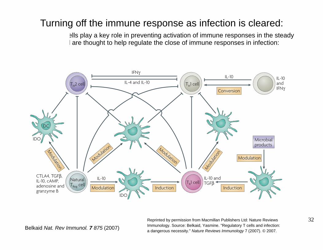
Figure 1 | Regulatory T cells during infection. Various populations of regulatory T cellshave been shown to have a role during infection. T helper 1 (TH1)-cell and TH2-cell populations can regulate each other via their production of cytokines. Naturally occurring forkhead box P3 (FOXP3)+CD4+CD25+ regulatory T cells (natural TReg cells) can limit TH1- and TH2-cell responses either indirectly by modulating antigen-presenting cell (APC) function or directly by cell–cell contact. Indirect regulatory mechanisms involve the release of transforming growth factor-B (TGFB, interleukin-10 (IL-10) or adenosine, or the induction of the tryptophan-degrading enzyme indoleamine 2,3-dioxygenase (IDO) by APCs recognizing cytotoxic T-lymphocyte antigen 4 (CTLA4) on natural TReg cells. Direct regulation of effector T cells includes the release of cyclic AMP (cAMP) by TReg cells. IL-10-producing T regulatory 1 (T R1) cells can be induced by dendritic cells (DCs) under certain conditions (such as following manipulation of DCs by microbial products). Natural TReg cells can also favour the development of TR1 cells via their modulation of APC functions. TR1 cells can limit immune responses during infections through their ability to release IL-10 and/or TGFB. During sustained TH1-cell responses, TH1 cells can secrete IL-10 that in a regulatory-feedback loop will limit TH1-cell responses. These cells can revert to a TH1-cell phenotype. IFNG, interferon-G.
REVIEWS
876 | NOVEMBER 2007 | VOLUME 7 www.nature.com/reviews/immunol
Turning off the immune response as infection is cleared: -cells play a key role in preventing activation of immune responses in the steady
state and are thought to help regulate the close of immune responses in infection:
IL-10 and IFN�
TR1 cell
IDO
IDO
IL-10
DC
Natural TReg cell
Modulation
Modulation
Modulation Modulation
Modulation
IL-10 and TGF�
CTLA4, TGF�, IL-10, cAMP, adenosine and granzyme B
Microbial products
Induction Induction
Conversion
IL-10 IFN�
IL-4 and IL-10
Mod
ulatio
n
TH2 cell TH1 cell
Reprinted by permission from Macmillan Publishers Ltd: Nature Reviews Immunology. Source: Belkaid, Yasmine. "Regulatory T cells and infection:Belkaid Nat. Rev Immunol. 7 875 (2007) a dangerous necessity." Nature Reviews Immunology 7 (2007). © 2007.
32

Turning off the immune response as infection is cleared:role of hypoxia/adenosine receptor signaling
TCR
A2AR
CyclicAMP
Inhibited productionof interferon-!
Inhibited T-cell response
T cell
[O2]low
Hypoxia[O2]low
AMP
[Adenosine]hi
Adenosine
AK NT5
HIF1"
A2BR
Figure 3 | The hypothesis: role of hypoxia in local tissues in the regulation of T cells in inflamed and hypoxic areas. We think that excessive collateral immune damage to the local-tissue microcirculation, and therefore to the oxygen (O2) supply, creates deepening tissue hypoxia, which functions as a signal to stop immune responses. Hypoxia, in turn, inhibits adenosine kinase (AK) and upregulates 5$-nucleotidase (NT5) activity, which results in the accumulation of extracellular adenosine. Adenosine signals through the immunosuppressive adenosine receptors A2AR and/or A2BR at the surface of surrounding activated T cells, and it downregulates T-cell-receptor (TCR)-mediated responses in a delayed negative-feedback manner. The regulatory effects of hypoxia-inducible factor 1" (HIF1") on T cells remain to be directly established, but it is expected that the increased expression of HIF1 in response to hypoxia will also be inhibitory.
Immune cell
Inspired airPo2 = 21 kPa (159 mm Hg)
LungsPo2 = 20 kPa (150 mm Hg)
TissuesPo2 = 0.5–2.5 kPa (4–20 mm Hg)(hypoxia)
CirculationPo2 = 5–13 kPa (40 –100 mm Hg)
Sitkovsky Nat. Rev Immunol. 5 712 (2005)
Reprinted by permission from Macmillan Publishers Ltd: NatureReviews Immunology. Source: Sitkovsky, Michail and DmitriyLukashev. "Regulation of immune cells by local-tissue oxygentension: HIF1and adenosine receptors." Nature ReviewsImmunology 5 (2005). © 2005.
©!!""#!Nature Publishing Group!
!
(A2AR and A2BR), which are involved in immunosup-pression1,68,69. In T cells, A2AR, A2BR and HIF1" might function together1 (FIG. 3).
Regulation of HIF1 expression and activity. HIF1" is a fundamental mediator of adaptation of cells to hypoxia66 !BOX 2". Oxygen sensing is a universal cel-lular event, and it is now accepted that HIF1" is the main mediator of the regulation of oxygen homeo-stasis and many other cellular processes in normal cells and tumour cells2,12,67,70,71.
HIF1 is a heterodimeric transcription factor: it consists of the constitutively expressed HIF1# subunit and the oxygen-tension-regulated HIF1" subunit. In the presence of oxygen, iron and 2-oxoglutarate, hydroxylation of the proline residues at positions 402 and 564 of HIF1", in the oxygen-dependent degradation domain, is catalysed by the prolyl-hydroxylase-domain-containing proteins (PHD1, PHD2 and PHD3)72–74, with a contribution from OS9 !REF. 75". Prolyl-hydroxylated HIF1" is subsequently targeted for ubiquitin-dependent degradation that is mediated by von Hippel–Lindau tumour-suppressor protein (VHL)76. The resulting deg-radation of HIF1" explains why HIF1" is expressed at low levels under normal oxygen tensions (FIG. 4a). Under hypoxic conditions, the prolyl hydroxylases PHD1 and PHD3 are, in turn, targeted by SIAH1A (seven in absen-tia homologue 1A) and SIAH2 for ubiquitin-dependent degradation77 (FIG. 4b). This complex, multilevel control mechanism ensures that HIF1" protein is stabilized under hypoxic conditions and is degraded under nor-moxic conditions. Increased HIF1" protein stability and activity of the HIF1 complex, in turn, regulate the transcription of many hypoxia-responsive genes, includ-ing those encoding many glycolytic enzymes, erythro-poietin, adrenomedullin, and growth factors such as vascular endothelial growth factor (VEGF)78,79.
In addition, the transcriptional activity of HIF1 can be negatively regulated by hydroxylation of the aspara-gine residue at position 803 of HIF1" by the oxygen-dependent hydroxylase FIH1 (factor inhibiting HIF1), which inhibits the interaction of HIF1 with either of its co-activators, p300 and CBP (cyclic AMP (cAMP)-responsive-element-binding protein (CREB)-binding protein)80.
Regulation of extracellular adenosine and the adenos-ine receptors A2AR and A2BR. Hypoxia markedly changes cellular metabolism and causes the accumulation of extracellular adenosine16,81. This accumulation is at least partially explained by hypoxia-mediated regulation of enzymes that are involved in adenosine metabolism — adenosine kinase82 and 5$-nucleotidase83–85 (FIG. 3). Adenosine kinase, which rephosphorylates adenosine to convert it to AMP, was shown to be inhibited by hypoxia in isolated-heart models82, but this observation remains to be extended to other tissues. It will be important to establish the mechanism of adenosine-kinase inhibi-tion to provide clues for novel immunomodulatory strategies. The accumulation of adenosine in hypoxic tissues can also be explained by the hypoxia-mediated upregulation of 5$-nucleotidase activity, an enzyme that converts AMP to adenosine83–85. Moreover, HIF1 can upregulate transcription from an ecto-5$-nucleotidase promoter85, implying that there might be a direct link between HIF1-mediated mechanisms and adenosine-receptor-mediated mechanisms of immunoregulation.
Pharmacological studies have firmly established that exogenously added extracellular adenosine has immunosuppressive effects1; however, for many years, extracellular adenosine was only one of several can-didate physiological regulators of immune responses.
Box 2 | HIF1 and cancer
Hypoxia-inducible factor 1 (HIF1) was first identified as an activator of expression of the gene encoding erythropoietin in response to hypoxia117. It was shown that HIF1 binds specific sites, which are known as hypoxia-response elements, in the erythropoietin gene promoter. These elements were later found in the promoters of various other genes that are induced by hypoxia, which indicates that induction of HIF1 activity is a key cellular response to hypoxia. It was later shown that HIF1 activates the transcription of genes encoding enzymes that are responsible for glycolysis (such as phosphoglycerate kinase 1, pyruvate kinase M and aldolase A)118 and angiogenesis (such as vascular endothelial growth factor)119.
Tumour cells often outgrow their blood supply, and this results in hypoxia of the tumour. Overexpression of HIF1" has been observed in tumour cells, causing them to switch to glycolysis and promoting angiogenesis. Indeed, HIF1" expression in tumours is associated with neovascularization and tumour growth79. So, the inhibition of HIF1 activity in tumour cells can suppress tumour growth70, which has encouraged the development of new antitumour strategies that are based on targeting HIF1".
716 | SEPTEMBER 2005 | VOLUME 5 www.nature.com/reviews/immunol
R E V I EWS
©!!""#!Nature Publishing Group!
!
The understanding of molecular mechanisms that allow immune cells to adapt to local-tissue hypoxic microenvironments is therefore important for devel-oping rational strategies to treat diseases that have an inflammatory pathogenesis and for improving the immunotherapy of hypoxic cancerous tissues. We think that adaptations to tissue hypoxia have evolved as an important immunoregulatory mechanism that should be taken into account in immunological studies and when considering pharmacological effects on the behaviour and responses of immune cells in different compartments of normal or inflamed tissues.
In this article, we discuss the immunological implications of studies of two important mediators of the effects of hypoxia: hypoxia-inducible factor 1! (HIF1!)12, and extracellular adenosine and adenosine receptors (which are purinergic receptors)13.
Hypoxic microenvironments in tissuesLow oxygen tension has been shown in many compart-ments of normal, inflamed and cancerous tissues14–19. Tumour growth often outpaces the expansion of the supporting vascular bed, thereby creating a hypoxic microenvironment18. By contrast, in inflamed tissues, the blood supply is often interrupted because the vessels are clogged with phagocytes20,21. Direct in vivo genetic evidence of the importance of hypoxic envi-ronments in the regulation of immune responses was recently provided by a study of neutrophil-mediated lung inflammation22, in which there was a marked exacerbation of inflammation after oxygenation. This was shown to result from weakening of the anti-inflammatory mechanism that is driven by local-tissue hypoxia22.
In many cases, local-tissue hypoxia might not result from tissue damage but might reflect the normal tissue microenvironment, vasculature and geometry. Tissue compartments differ in their extent of vascularization and therefore their level of oxygenation. In normal situations, the oxygen tension in tissues is proportional to the distance from the end of the nearest capillary. Detailed studies using different methods of oxygen measurement have shown that oxygen tensions in dif-ferent tissues are often low and could be considered to be hypoxic23. Even the ‘non-hypoxic’, normal levels of oxygen in many compartments of normal spleen and lymph-node tissues are much lower than would be expected from the Po2 of inspired air, which is 21 kPa (that is, 159 mm Hg or a concentration of 21%, at sea level)4,24. For example, it was shown that the Po2 is "1.3 kPa ("10 mm Hg) in the thymus and "2.1 kPa ("16 mm Hg) in the spleen25. In vivo measurements of Po2 in the spleen of live, anaesthetized mice have shown that it is low, 0.5–4.5 kPa (4–34 mm Hg)4: the Po2 was found to be highest near the splenic artery and to gradually decrease with distance from the artery.
Further detailed investigations are required to establish the pattern of changing oxygen tensions that immune cells encounter as they reside in dif-ferent compartments of lymphoid organs or as they migrate from the circulation to the tissues in search
of pathogens (FIG. 1). This might allow investigators to more accurately mimic in vivo conditions in in vitro experiments. In vitro experiments are usually carried out in incubators that have an atmosphere of 5% car-bon dioxide and 21% oxygen, which is not likely to be physiological26. Instead, physiological levels of oxygen in tissues are likely to be ‘hypoxic’4,23. Therefore, the choice of oxygen tension during in vitro culture of immune cells should be made carefully and should be varied depending on the specific experiment, to better simulate physiological levels of oxygen tension in the relevant tissue in vivo.
Generation of cellular energyThe oxygen supply in inflamed tissues, and even in normal tissues, is known to be low23. Although there is no direct evidence for metabolic adaptation to hypoxia by immune cells, that such adaptation occurs can be deduced from the ability of immune cells to func-tion in all tissues, including those that are hypoxic27. Otherwise, pathogens would take advantage of hypoxic ‘shelters’, where ‘non-adapted’ immune cells would not be able to function.
Under aerobic conditions, the source of cellular energy is glycolysis coupled to oxidative phosphory-lation (collectively known as respiration) (FIG. 2). During glycolysis, glucose is converted to pyruvate, with ATP being concomitantly produced as a result
Figure 1 | Low oxygen concentration in tissue environments. Oxygen is inspired from the air; the oxygen tension (Po2) in air is 21 kPa (159 mm Hg), which is a concentration of 21%. When warmed air arrives in the lungs, it loses some pressure: Po2 = 20 kPa (150 mm Hg). The oxygen pressure continues to decrease in the circulation: Po2 = 13 kPa (100 mm Hg) in alveoli and 5 kPa (40 mm Hg) in veins that reach the heart. The oxygen pressure in the tissues then decreases with increasing distance from the blood vessels: Po2 = 0.5 –2.5 kPa (4–20 mm Hg).
NATURE REVIEWS | IMMUNOLOGY VOLUME 5 | SEPTEMBER 2005 | 713
R E V I EWS
Induction of immunologicalmemory (the basis of
vaccination)
Figure removed due to copyright restrictions.See Kaech, Susan M. and Rafi Ahmed. "CD8 T CellsRemember with a Little Help." Science 300, no. 5617 (2003).

MIT OpenCourseWarehttp://ocw.mit.edu
20.380J / 5.22J Biological Engineering Design Spring 2010
For information about citing these materials or our Terms of Use, visit: http://ocw.mit.edu/terms.