introduction - · pdf fileintroduction 1) isomorphous ... (uv-vis) diffuse reflectance...
TRANSCRIPT
Introduction1)
Isomorphous substitution of silicon and/or aluminum by
various heteroatoms into the framework of different zeo-
lites has received considerable attention in the utilization
of zeolites as catalysts. These elements can impart cata-
lytic properties to the zeolite for many reactions other
than those catalyzed by acid sites while offering the ad-
vantages of atomic dispersion of the elements, which is
difficult to achieve with traditional catalyst preparation
methods. In this respect, isomorphous substitution of tita-
nium, zirconium, or vanadium into the framework of
high-silica zeolites has expanded the scope of application
of these materials. These isomorphously substituted zeo-
lites are reported to be very promising in the liquid-phase
catalytic oxidation of hydrocarbons. Taramasso and cow-
†To whom all correspondence should be addressed.
(e-mail: [email protected])
orkers [1] first reported the hydrothermal synthesis of a
titanium-containing zeolite, denoted as titanium silica-
lite-1 (TS-1). Titanium silicalite-1 was found to be active
in the oxidation of a variety of organic substrates in the
presence of aqueous hydrogen peroxide as oxidizing
agent. Much the same as titanium-substituted molecular
sieves, zirconium or vanadium silicalite-1 (ZS-1 or VS-
1) can be synthesized by isomorphous substitution of
Zr4+or V
5+for Si
4+in the MFI structure framework, and
has very interesting properties towards catalytic oxida-
tion [2-7]. The oxidative property of vanadium could be
combined with the shape-selective characteristics im-
parted by the micropore structure in which the vanadium
is located [8].
Niobium - which belongs to the same group in the
Periodic Table as vanadium (Group V) - has been re-
ported to have the capability of the photocatalytic and se-
lective oxidations [9-11]. Although electronegativity and
ionic radius between niobium and its neighbors (V, Mo,
Yong Sig Ko†, Hyun Tae Jang , andWha Seung Ahn
Department of Advanced Material Chemistry, Shinsung College, Dangjin-gun 343-860, Korea
*Department of Chemical Engineering, Hanseo University, Seosan 356-706, Korea
**Department of Chemical Engineering, College of Engineering, Inha University, Inchon 402-751, Korea
Received January 29, 2007; Accepted May 3, 2007
Abstract:Niobium silicalite-1 (NbS-1) molecular sieves with MFI structure were prepared using the hydro-
thermal synthesis method. X-ray diffraction, scanning and transmission electron microscopies, Fourier transform
infrared spectroscopy, ultraviolet-visible (UV-vis) diffuse reflectance spectroscopy,29Si magic-angle spinning
nuclear magnetic resonance spectroscopy, ammonia temperature-programmed desorption (NH3-TPD), physical
adsorption of nitrogen and elemental analysis were then performed to evaluate their physico-chemical properties
and to provide evidences for the incorporation of Nb5+into the zeolite framework. The unit cell volume of niobi-
um silicalite-1 increased linearly with increasing niobium content, suggesting isomorphous substitution of Si4+
by Nb5+in the lattice framework. The framework infrared spectrum showed a characteristic absorption band at
963 cm-1, probably due to Si-O-Nb linkages. In the UV-vis spectrum, the absorption assigned to the presence of
Nb5+
in the zeolite framework was observed at around 200 nm. In addition, NH3-TPD studies revealed weak
acidity in niobium silicalite-1 due possibly to [Nb (OH)-Si] sites in the framework.․․․
Keywords: niobium, niobium silicalite-1, hydrothermal synthesis, NH3-TPD, isomorphous substitution, charac-
terization, FT-IR, UV-vis DRS
Hydrothermal Synthesis and Characterization of Niobium-containing Silicalite-1 Molecular Sieves with MFI Structure 765
Ta, and Zr) in the Periodic Table are different, it is re-
ported that the promoter effect, support effect and acidic
nature of niobium compounds are quite different from
those of compounds of the surrounding elements [12].
Niobium compounds and niobium materials have re-
cently been shown to enhance catalytic properties when
used as a component of catalysts or when small amounts
are added to known catalysts. Depending on the catalyst
composition and structure, they are used in the processes
involving acidic active centers as well as redox character.
These niobium-containing catalysts have been used for
dehydration and dehydrogenation of alcohols [13-15],
oxidative dehydrogenation of alkanes [16], alkylation of
benzene [17], NO reduction by NH3 [18], dimerization
and oligomerization of olefins [19], oxidation of meth-
anol [20,21] and polycondensation reactions [22]. The
presence of niobium in molecular sieves has the potential
of generating catalysts with shape-selective properties
and acidic or redox characteristics. Only a few reports
are available on porous niobium silicates; they include
work on the incorporation of niobium in micro/meso-
porous materials, ETS-10 [23] denoted as ETNbS-10, to
MFI [24] designated as NbS-1, and also to MCM-41
[25].
Though synthesis methods of niobium silicate molec-
ular sieves have been reported [24,26], further inves-
tigation concerning the hydrothermal synthesis of niobi-
um-containing silicalite molecular sieves with more de-
tailed characterization would contribute towards a better
understanding of the system.
In this work, we report the hydrothermal synthesis and
characterization of crystalline niobium silicalite molec-
ular sieves with the MFI structure prepared using silicon
and niobium alkoxides. Results from X-ray diffraction
(XRD), scanning electron microscopy (SEM), transmi-
ssion electron microscopy (TEM), framework infrared
(IR) spectroscopy, ultraviolet-visible (UV-vis) diffuse re-
flectance spectroscopy (DRS),29Si magic-angle spinning
(MAS) nuclear magnetic resonance spectroscopy
(NMR), temperature-programmed desorption of ammo-
nia (NH3-TPD), nitrogen adsorption and elemental analy-
sis are discussed to verify the incorporation of niobium
in the zeolite lattice framework.
Experimental
Synthesis of niobium silicalite-1 catalyst
NbS-1 samples were prepared from substrates having
the following composition ratio: SiO2/Nb2O5 = 32.7∼
99, TPA+/SiO2 = 0.46, H2O/SiO2 = 35. In a typical prepa-
ration, a Pyrex beaker was placed in a glove box and ni-
trogen was flushed over to minimize the adverse effect of
moisture in air. After 27.01 g of tetraethylorthosilicate
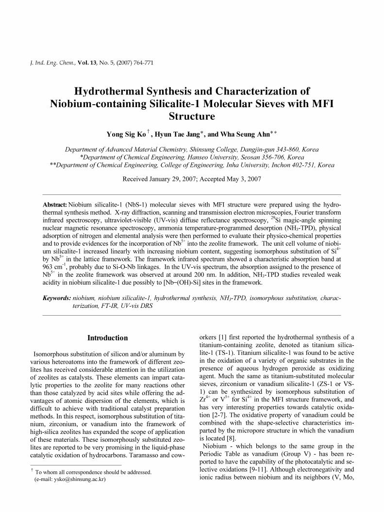
Figure 1. Schematic diagram for the hydrothermal synthesis of
niobium silicalite-1.
(Aldrich, 99.999 %) was transferred to the Pyrex beaker
and vigorously stirred, 0.43 g of niobium(V) ethoxide
(Aldrich, 99.95 %) was carefully introduced into this sol-
ution, and the mixture was cooled to about 273 K. After
a few minutes, 60.65 g of tetrapropylammonium hydrox-
ide (TPAOH) aqueous solution (Aldrich, 20 %), also
cooled to 273 K, was slowly added dropwise into the
mixture. Stirring and cooling were maintained during this
process. After addition of all TPAOH the synthesis mix-
ture was kept for 5 6 h at 343 353 K in order to accel∼ ∼ -
erate hydrolysis and to evaporate the ethyl alcohol
produced. Deionized water was then added to increase
the volume of the mixture to its original value. This clear
homogeneous solution was then transferred to a Teflon-
lined stainless steel autoclave and kept in a convection
oven at 448 K under autogenous pressure. In order to in-
vestigate the crystallization process, autoclaves were tak-
en out from the oven at different time intervals and were
quenched immediately in cold water for sample identifi-
cation. The solid products were separated by means of
suction-filtration or centrifugation, washed several times
with hot deionized water and dried in an air oven at 383
K overnight. The products were finally calcined at 823 K
for 6 h. The synthesis procedure of niobium silicalite-1
catalyst can be represented schematically as in Figure 1.
A reference sample of silicalite-1 (MFI) was prepared
following the same procedure without adding niobium
ethoxide to the substrate mixture. In addition, a sample
Yong Sig Ko, Hyun Tae Jang, and Wha Seung Ahn766
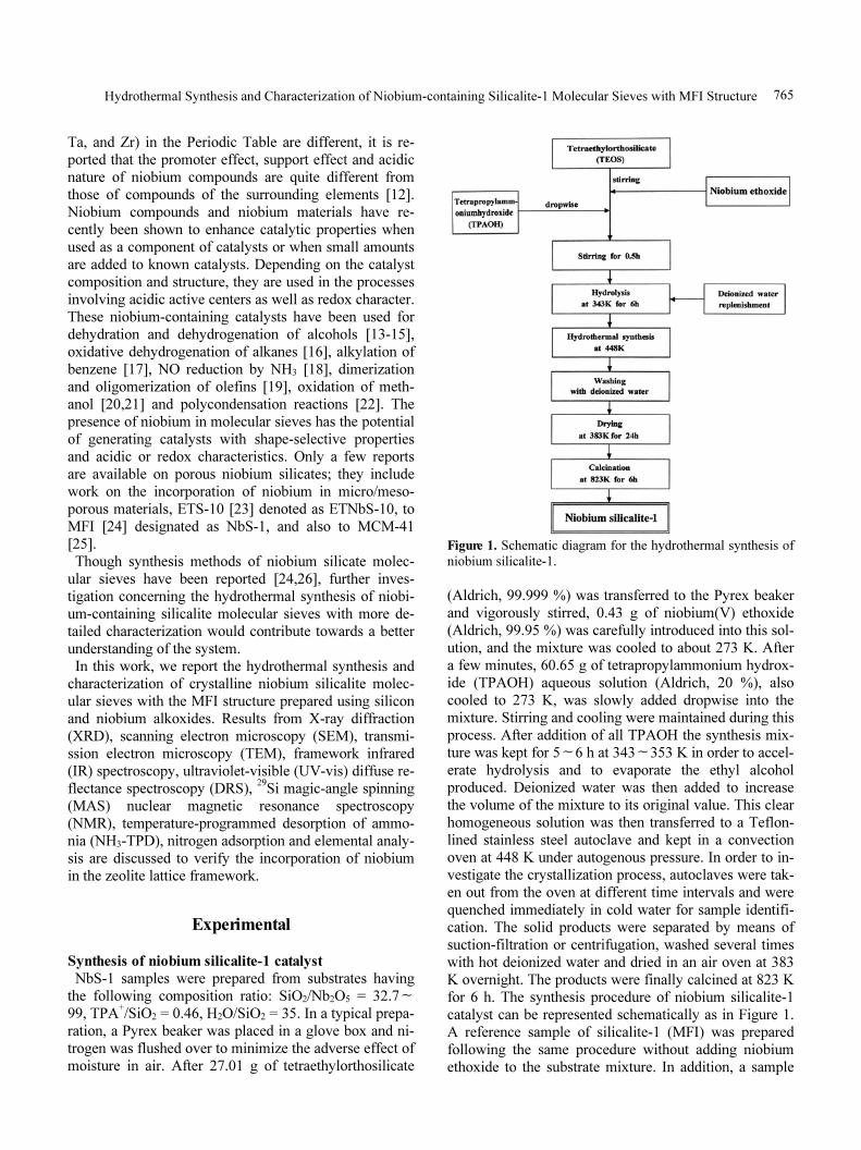
Figure 2. XRD patterns of niobium silicalite-1 and silicalite-
1 (a) silicalite-1, (b) ZSM-5, (c) NbS-1 (3 mol% niobium),:
and (d) Nb2O5 powder.
of about 3 mol% niobium-impregnated silicalite-1 [Nb/
silicalite-1 (IMP)] was also prepared for comparison pur-
poses by the incipient wetness method.
Characterization
The samples synthesized were analyzed by X-ray dif-
fraction (XRD) for both qualitative and quantitative
phase identification. The unit used was a powder X-ray
diffractometer (Philips, PW-1700) with a scintillation
counter and a graphite monochromator attachment, uti-
lizing nickel-filtered CuK radiation (40 kV, 25 mA).α
Unit cell parameters were obtained by a least-squares fit
to the interplanar spacings measured in the 5 45° (2 )∼ θ
angular region, using silicon as an internal standard. The
crystal size and morphology of the crystalline samples
were examined using a scanning electron microscope
(Hitachi, X-650) after coating with a Au-Pd evaporated
film. For TEM analysis, samples were dispersed ultra-
sonically in ethanol, and a drop of the suspension was
deposited on a holey carbon copper grid. Micrographs
were recorded with a 1024 × 1024 Gatan CCD camera on
a Philips CM20 microscope operated at 200 kV. Frame-
work infrared (IR) spectra of samples were recorded in
air at room temperature on a Perkin Elmer 221 spec-
trometer (in the range of 400 4000 cm∼-1) with wafers of
zeolites mixed with dry KBr. The elemental analyses of
crystalline samples were performed with an inductively
coupled plasma (ICP) spectrometer (Jobin Yuon, JY-38
VHR) and X-ray fluorescence (XRF; Rigaku, 3070).
UV-vis diffuse reflectance spectroscopy was performed
on a Varian CARY 3E double-beam spectrometer and
dehydrated MgO was used as a reference in the range
190 500 nm. The solid-state NMR spectra were ob∼ -
tained with a Bruker AM 300 spectrometer at a fre-
quency of 59.6 MHz and spinning rate of 3.5 kHz with a
pulse width of 3 µs, a relaxation delay of 5 s and 100∼
200 acquisitions. The chemical shift was referenced with
respect to the29Si signal of tetramethylsilane (TMS). The
nitrogen-adsorption isotherms and specific surface areas
were determined by nitrogen physisorption with the
Brunauer-Emmett-Teller (BET) method at liquid-nitro-
gen temperature using a Micromeritics ASAP 2010 auto-
matic analyzer. Ammonia temperature-programmed de-
sorption (NH3-TPD) tests were carried out in a quartz mi-
croreactor with continuous analysis of the released NH3
concentration via a TCD detector using a Micromeritics
TPD/TPR 2900 analyzer. Before the tests, the sample
was pretreated in situ with a flow of argon at 773 K in or-
der to remove adsorbed species, and the adsorption was
conducted after the sample was cooled to 373 K. The
physisorbed NH3 was flushed by a dry argon flow at the
same temperature for 3 h. The temperature was then in-
creased at the rate of 10 K min-1.
Results and Discussion
The X-ray diffraction patterns of calcined NbS-1, ZSM-
5, silicalite-1, and N2O5 powder are shown in Figure 2.
The X-ray diffraction pattern of the calcined NbS-1 mo-
lecular sieve was similar to that of ZSM-5 zeolite with
MFI structure. Almost identical diffractograms of NbS-1
were obtained irrespective of the niobium contents of the
sample up to 3 mol% and no detectable niobium oxide
peaks were observed. The symmetry of the calcined
NbS-1 was orthorhombic, comprising single reflections
around 2 at 24.5θoand 29.2
o. As expected, calcined sili-
calite-1 had monoclinic symmetry which presents double
reflections at the same 2θ. The persistence of the ortho-
rhombic symmetry in the calcined state of the niobium
silicalites can be considered as a result of niobium ions
present in the framework positions in NbS-1.
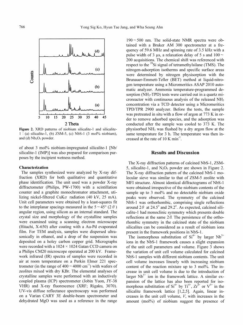
The isomorphous substitution of Si4+
by larger Nb5+
ions in the NbS-1 framework causes a slight expansion
of the unit cell parameters and volume. Figure 3 shows
the variation of unit cell volume calculated for calcined
NbS-1 samples with different niobium contents. The unit
cell volume increases linearly with increasing niobium
content of the reaction mixture up to 3 mol%. The in-
crease in unit cell volume is due to the introduction of
larger Nb5+
ion in the framework lattice. A similar ex-
pansion of the lattice has also been reported for iso-
morphous substitution of Si4+
by Ti4+, Zr
4+or V
5+in the
silicalite framework lattice [1,2,5]. Again, linear in-
creases in the unit cell volume, V, with increases in the
amount (mol%) of niobium suggest the presence of
Hydrothermal Synthesis and Characterization of Niobium-containing Silicalite-1 Molecular Sieves with MFI Structure 767
Figure 3. Unit cell volume of NbS-1 as a function of niobium
content.
Figure 4. Comparison between the crystallization rates of TS-1,
ZrS-1 and NbS-1.
niobium in the framework. Earlier studies [27,28] have
shown that the expansion of the unit cell volume is re-
lated to the concentration of the framework vanadium in
the V5+containing zeolite samples.
The overall crystallization rates at constant gel compo-
sition and temperature for TS-1, Zr-containing zeolite
ZrS-1 [29] and NbS-1 are compared in Figure 4. While
the crystallization time was kept to 3 or 4 days to obtain
the maximum crystallinity for TS-1 or ZrS-1, it was nec-
essary for NbS-1 sample to be kept to 10 days in order to
obtain the same degree crystallinity. The fact that a rela-
tively longer crystallization time was necessary to main-
tain the same degree of crystallinity for NbS-1 may be a
consequence of the radius of niobium atoms being larger
than that of titanium or zirconium atoms, and conse-
quently it is believed to be more difficult for niobium to
Figure 5. Relationship between the niobium content of the
NbS-1 samples and the gel mixtures.
enter the silicalite framework than titanium or zirconium.
In addition, while ZSM-5 (which has the same MFI
structure) can be prepared in 1 day, it takes substantially
longer when titanium, zirconium or niobium is sub-
stituted into the framework. Such a trend is generally ob-
served in the synthesis of metal-substituted silicate mo-
lecular sieves [29-31], but it can be also considered as a
consequence of the mineralizing agent NaOH/KOH be-
ing absent in the reaction mixture in such cases.
We have synthesized a series of NbS-1 samples by
varying the amount of niobium in the synthesis gel.
Figure 5 shows the bulk niobium contents of NbS-1 crys-
tals plotted against the niobium content of their reaction
mixtures. It is seen that the niobium content of the NbS-1
crystals obtained is linearly correlated with the niobium
content of the reaction mixtures. This indicates that vary-
ing amounts of niobium can be incorporated in the crys-
tal by changing the niobium content at the stage of gel
preparation, and most of the niobium present in the sub-
strate can be incorporated into the zeolite framework
within the range of less than about 3 mol%.
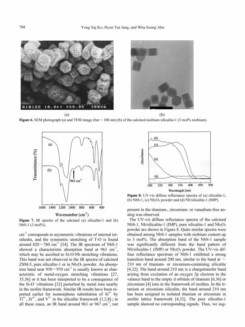
A scanning electron micrograph and transmission elec-
tron micrograph of NbS-1 (3 mol% niobium) sample are
presented in Figure 6. The niobium content in the sub-
strate mixture did not lead to a significant modification
in the morphology of NbS-1. All NbS-1 samples were
made up of uniform crystals of size about 0.13 µm and
hexagonal shape. The transmission electron micrograph
of the NbS-1 sample shows the absence of amorphous
matters outside the crystals of NbS-1.
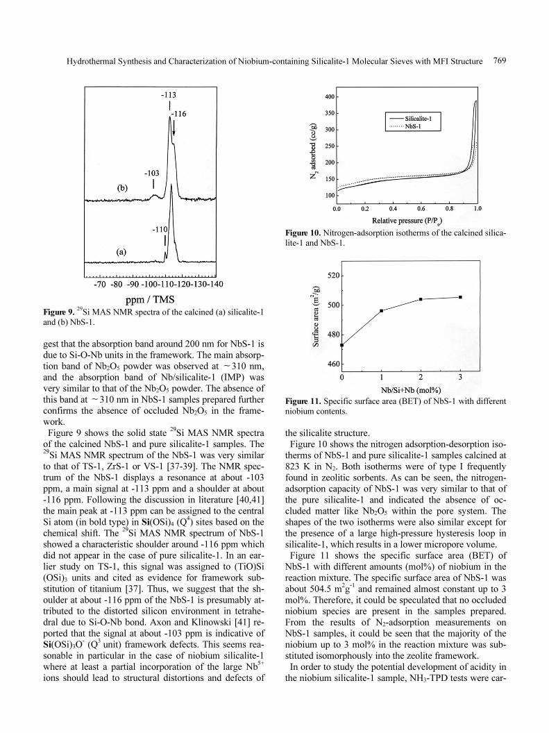
IR spectra of the calcined silicalite-1 and NbS-1 are
compared in Figure 7. As is known, IR spectra in the
mid-infrared region can provide evidence of the iso-
morphous substitution of large heteroatoms into the
framework of zeolites [32,33]. The band around 1100
Yong Sig Ko, Hyun Tae Jang, and Wha Seung Ahn768
(a) (b)
Figure 6. SEM photograph (a) and TEM image (bar = 100 nm) (b) of the calcined niobium silicalite-1 (3 mol% niobium).
Figure 7. IR spectra of the calcined (a) silicalite-1 and (b)
NbS-1 (3 mol%).
cm-1corresponds to asymmetric vibrations of internal tet-
rahedra, and the symmetric stretching of T-O is found
around 820 780 cm∼-1[34]. The IR spectrum of NbS-1
showed a characteristic absorption band at 963 cm-1,
which may be ascribed to Si-O-Nb stretching vibrations.
This band was not observed in the IR spectra of calcined
ZSM-5, pure silicalite-1 or in Nb2O5 powder. An absorp-
tion band near 950 970 cm∼-1is usually known as char-
acteristic of metal-oxygen stretching vibrations [27,
35,36] or it has been interpreted to be a consequence of
the Si-O-vibrations [32] perturbed by metal ions nearby
in the zeolite framework. Similar IR results have been re-
ported earlier for isomorphous substitution of Si4+
by
Ti4+, Zr
4+, and V
5+in the silicalite framework [1,2,8] ; in
all these cases, an IR band around 963 or 967 cm-1, not
Figure 8. UV-vis diffuse reflectance spectra of (a) silicalite-1,
(b) NbS-1, (c) Nb2O5 powder and (d) Nb/silicalite-1 (IMP).
present in the titanium-, zirconium- or vanadium-free an-
alog was observed.
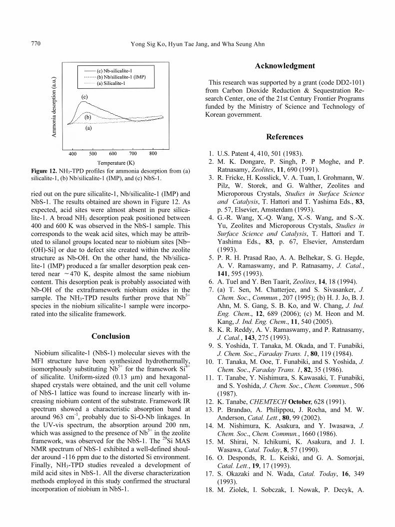
The UV-vis diffuse reflectance spectra of the calcined
NbS-1, Nb/silicalite-1 (IMP), pure silicalite-1 and Nb2O5
powder are shown in Figure 8. Quite similar spectra were
obtained among NbS-1 samples with niobium content up
to 3 mol%. The absorption band of the NbS-1 sample
was significantly different from the band pattern of
Nb/silicalite-1 (IMP) or Nb2O5 powder. The UV-vis dif-
fuse reflectance spectrum of NbS-1 exhibited a strong
transition band around 200 nm, similar to the band at ∼
210 nm of titanium- or zirconium-containing silicalite
[4,32]. The band around 210 nm is a chargetransfer band
arising from excitation of an oxygen 2p electron in the
valance band to the empty d orbitals of titanium [6,36] or
zirconium [4] ions in the framework of zeolites. In the ti-
tanium or zirconium silicalite, the band around 210 nm
has been assigned to isolated titanium or zirconium in
zeolite lattice framework [4,32]. The pure silicalite-1
sample showed no corresponding signals. Thus, we sug-
Hydrothermal Synthesis and Characterization of Niobium-containing Silicalite-1 Molecular Sieves with MFI Structure 769
Figure 9.29Si MAS NMR spectra of the calcined (a) silicalite-1
and (b) NbS-1.
gest that the absorption band around 200 nm for NbS-1 is
due to Si-O-Nb units in the framework. The main absorp-
tion band of Nb2O5 powder was observed at 310 nm,∼
and the absorption band of Nb/silicalite-1 (IMP) was
very similar to that of the Nb2O5 powder. The absence of
this band at 310 nm in NbS-1 samples prepared further∼
confirms the absence of occluded Nb2O5 in the frame-
work.
Figure 9 shows the solid state29Si MAS NMR spectra
of the calcined NbS-1 and pure silicalite-1 samples. The29Si MAS NMR spectrum of the NbS-1 was very similar
to that of TS-1, ZrS-1 or VS-1 [37-39]. The NMR spec-
trum of the NbS-1 displays a resonance at about -103
ppm, a main signal at -113 ppm and a shoulder at about
-116 ppm. Following the discussion in literature [40,41]
the main peak at -113 ppm can be assigned to the central
Si atom (in bold type) in Si(OSi)4 (Q4) sites based on the
chemical shift. The29Si MAS NMR spectrum of NbS-1
showed a characteristic shoulder around -116 ppm which
did not appear in the case of pure silicalite-1. In an ear-
lier study on TS-1, this signal was assigned to (TiO)Si
(OSi)3 units and cited as evidence for framework sub-
stitution of titanium [37]. Thus, we suggest that the sh-
oulder at about -116 ppm of the NbS-1 is presumably at-
tributed to the distorted silicon environment in tetrahe-
dral due to Si-O-Nb bond. Axon and Klinowski [41] re-
ported that the signal at about -103 ppm is indicative of
Si(OSi)3O-(Q
3unit) framework defects. This seems rea-
sonable in particular in the case of niobium silicalite-1
where at least a partial incorporation of the large Nb5+
ions should lead to structural distortions and defects of
Figure 10. Nitrogen-adsorption isotherms of the calcined silica-
lite-1 and NbS-1.
Figure 11. Specific surface area (BET) of NbS-1 with different
niobium contents.
the silicalite structure.
Figure 10 shows the nitrogen adsorption-desorption iso-
therms of NbS-1 and pure silicalite-1 samples calcined at
823 K in N2. Both isotherms were of type I frequently
found in zeolitic sorbents. As can be seen, the nitrogen-
adsorption capacity of NbS-1 was very similar to that of
the pure silicalite-1 and indicated the absence of oc-
cluded matter like Nb2O5 within the pore system. The
shapes of the two isotherms were also similar except for
the presence of a large high-pressure hysteresis loop in
silicalite-1, which results in a lower micropore volume.
Figure 11 shows the specific surface area (BET) of
NbS-1 with different amounts (mol%) of niobium in the
reaction mixture. The specific surface area of NbS-1 was
about 504.5 m2g-1and remained almost constant up to 3
mol%. Therefore, it could be speculated that no occluded
niobium species are present in the samples prepared.
From the results of N2-adsorption measurements on
NbS-1 samples, it could be seen that the majority of the
niobium up to 3 mol% in the reaction mixture was sub-
stituted isomorphously into the zeolite framework.
In order to study the potential development of acidity in
the niobium silicalite-1 sample, NH3-TPD tests were car-
Yong Sig Ko, Hyun Tae Jang, and Wha Seung Ahn770
Figure 12. NH3-TPD profiles for ammonia desorption from (a)
silicalite-1, (b) Nb/silicalite-1 (IMP), and (c) NbS-1.
ried out on the pure silicalite-1, Nb/silicalite-1 (IMP) and
NbS-1. The results obtained are shown in Figure 12. As
expected, acid sites were almost absent in pure silica-
lite-1. A broad NH3 desorption peak positioned between
400 and 600 K was observed in the NbS-1 sample. This
corresponds to the weak acid sites, which may be attrib-
uted to silanol groups located near to niobium sites [Nb․․․
(OH)-Si] or due to defect site created within the zeolite
structure as Nb-OH. On the other hand, the Nb/silica-
lite-1 (IMP) produced a far smaller desorption peak cen-
tered near 470 K, despite almost the same niobium∼
content. This desorption peak is probably associated with
Nb-OH of the extraframework niobium oxides in the
sample. The NH3-TPD results further prove that Nb5+
species in the niobium silicalite-1 sample were incorpo-
rated into the silicalite framework.
Conclusion
Niobium silicalite-1 (NbS-1) molecular sieves with the
MFI structure have been synthesized hydrothermally,
isomorphously substituting Nb5+
for the framework Si4+
of silicalite. Uniform-sized (0.13 µm) and hexagonal-
shaped crystals were obtained, and the unit cell volume
of NbS-1 lattice was found to increase linearly with in-
creasing niobium content of the substrate. Framework IR
spectrum showed a characteristic absorption band at
around 963 cm-1, probably due to Si-O-Nb linkages. In
the UV-vis spectrum, the absorption around 200 nm,
which was assigned to the presence of Nb5+in the zeolite
framework, was observed for the NbS-1. The29Si MAS
NMR spectrum of NbS-1 exhibited a well-defined shoul-
der around -116 ppm due to the distorted Si environment.
Finally, NH3-TPD studies revealed a development of
mild acid sites in NbS-1. All the diverse characterization
methods employed in this study confirmed the structural
incorporation of niobium in NbS-1.
Acknowledgment
This research was supported by a grant (code DD2-101)
from Carbon Dioxide Reduction & Sequestration Re-
search Center, one of the 21st Century Frontier Programs
funded by the Ministry of Science and Technology of
Korean government.
References
1. U.S. Patent 4, 410, 501 (1983).
2. M. K. Dongare, P. Singh, P. P Moghe, and P.
Ratnasamy, Zeolites, 11, 690 (1991).
3. R. Fricke, H. Kosslick, V. A. Tuan, I. Grohmann, W.
Pilz, W. Storek, and G. Walther, Zeolites and
Microporous Crystals, Studies in Surface Science
and Catalysis, T. Hattori and T. Yashima Eds., 83,
p. 57, Elsevier, Amsterdam (1993).
4. G.-R. Wang, X.-Q. Wang, X.-S. Wang, and S.-X.
Yu, Zeolites and Microporous Crystals, Studies in
Surface Science and Catalysis, T. Hattori and T.
Yashima Eds., 83, p. 67, Elsevier, Amsterdam
(1993).
5. P. R. H. Prasad Rao, A. A. Belhekar, S. G. Hegde,
A. V. Ramaswamy, and P. Ratnasamy, J. Catal.,
141, 595 (1993).
6. A. Tuel and Y. Ben Taarit, Zeolites, 14, 18 (1994).
7. (a) T. Sen, M. Chatterjee, and S. Sivasanker, J.
Chem. Soc., Commun., 207 (1995); (b) H. J. Jo, B. J.
Ahn, M. S. Gang, S. B. Ko, and W. Chang, J. Ind.
Eng. Chem., 12, 689 (2006); (c) M. Heon and M.
Kang, J. Ind. Eng. Chem., 11, 540 (2005).
8. K. R. Reddy, A. V. Ramaswamy, and P. Ratnasamy,
J. Catal., 143, 275 (1993).
9. S. Yoshida, T. Tanaka, M. Okada, and T. Funabiki,
J. Chem. Soc., Faraday Trans. 1, 80, 119 (1984).
10. T. Tanaka, M. Ooe, T. Funabiki, and S. Yoshida, J.
Chem. Soc., Faraday Trans. 1, 82, 35 (1986).
11. T. Tanabe, Y. Nishimura, S. Kawasaki, T. Funabiki,
and S. Yoshida, J. Chem. Soc., Chem. Commun., 506
(1987).
12. K. Tanabe, CHEMTECH October, 628 (1991).
13. P. Brandao, A. Philippou, J. Rocha, and M. W.
Anderson, Catal. Lett., 80, 99 (2002).
14. M. Nishimura, K. Asakura, and Y. Iwasawa, J.
Chem. Soc., Chem. Commun., 1660 (1986).
15. M. Shirai, N. Ichikumi, K. Asakura, and J. I.
Wasawa, Catal. Today, 8, 57 (1990).
16. O. Desponds, R. L. Keiski, and G. A. Somorjai,
Catal. Lett., 19, 17 (1993).
17. S. Okazaki and N. Wada, Catal. Today, 16, 349
(1993).
18. M. Ziolek, I. Sobczak, I. Nowak, P. Decyk, A.
Hydrothermal Synthesis and Characterization of Niobium-containing Silicalite-1 Molecular Sieves with MFI Structure 771
Lewandowska, and J. Kujawa, Microporous and
Mesoporous Materials, 35, 195 (2000).
19. A. Morikawa and A. Togashi, Catal. Today, 16, 333
(1993).
20. A. Philippou, P. Brandao, A. Ghanbari-Siahkali, J.
Dwyer, J. Rocha, and M. W. Anderson, Appl. Catal.
A, 207, 229 (2001).
21. J. M. Jehng and I. E. Wachs, Catal. Today, 16, 417
(1993).
22. T. Kushimoto, Y. Ozawa, A. Baba, and H. Matsuda,
Catal. Today, 16, 571 (1993).
23. J. Rocha, P. Brandao, J. D. Pedrosa de Jesus, A.
Philippou, and M. W. Anderson, Chem. Commun.,
471 (1999).
24. A. M. Prakash and L. Kevan, J. Am. Chem. Soc.,
120, 13148 (1998).
25. M. Ziolek and I. Nowak, Zeolites, 18, 356 (1997).
26. I. Sobczak, P. Decyk, M. Ziolek, M. Daturi, J. C.
Lavalley, L. Kevan, and A. M. Prakash, J. Catal.,
207, 101 (2002).
27. P. R. Hari Prasad Rao, A. V. Ramaswamy, and P.
Ratnasamy, J. Catal., 137, 225 (1992).
28. K. R. Reddy, A. V. Ramaswamy, and P. Ratnasamy,
J. Chem. Soc., Chem. Commun., 1613 (1992).
29. Y. S. Ko and W. S. Ahn, Korean J. Chem. Eng., 15,
423 (1998).
30. M. Shibata and Z. Gabelica, Zeolites, 19, 246
(1997).
31. N. K. Mal, V. Ramaswamy, P. R. Rajamohanan, and
A. V. Ramaswamy, Microporous Materials, 12, 331
(1997).
32. M. R. Boccuti, K. M. Rao, A. Zecchina, G. Leofanti,
and G. Petrini, Structure and Reactivity of Surfaces,
Studies in Surface Science and Catalysis, C. Mor-
terra, A. Zecchina and G. Costa Eds., 48, p. 133,
Elsevier, Amsterdam (1989).
33. D. R. C. Huybrechts, I. Vaesen, H. X. Li, and P. A.
Jacobs, Catal. Lett., 8, 237 (1991).
34. D. W. Breck, Zeolite Molecular Sieves: Structure,
Chemistry and Use, p. 415, Wiley, New York
(1974).
35. J. S. Reddy, R. Kumar, and P. Ratnasamy, Appl.
Catal., 57, L1 (1990).
36. A. Thangaraj, R. Kumar, S. P. Mirajkar, and P.
Ratnasamy, J. Catal., 130, 1 (1991).
37. A. J. H. P. van der Pol, A. J. Verduyn, and J. H. C.
Hooff, Appl. Catal. A, 92, 113 (1992).
38. B. Rakshe, V. Ramaswamy, S. G. Hegde, R.
Vetrivel, and A. V. Ramaswamy, Catal. Lett., 45, 41
(1997).
39. Y. Ben Taarit, A. Tuel, N. Velmasco, and C. Na-
ccache, Fr. Patent 9 203 364 (1992).
40. A. Tuel and Y. Ben Taarit, J. Chem. Soc., Chem.
Commun., 1578 (1992).
41. S. A. Axon and Klinowski, Appl. Catal. A, 81, 27
(1992).