introducing n-, p-, and s-donor leaving groups: an
TRANSCRIPT

DaltonTransactions
PAPER
Cite this: Dalton Trans., 2020, 49,15693
Received 9th September 2020,Accepted 16th October 2020
DOI: 10.1039/d0dt03165h
rsc.li/dalton
Introducing N-, P-, and S-donor leaving groups: aninvestigation of the chemical and biologicalproperties of ruthenium, rhodium and iridiumthiopyridone piano stool complexes†
Sophia Harringer, a Debora Wernitznig, a Natalie Gajic,a Andreas Diridl,a
Dominik Wenisch, a Michaela Hejl,a Michael A. Jakupec, a,b Sarah Theiner, c
Gunda Koellensperger, c Wolfgang Kandioller *a,b and Bernhard K. Keppler a,b
A series of 15 piano-stool complexes featuring either a RuII, RhIII or IrIII metal center, a bidentate thiopyri-
done ligand, and different leaving groups was synthesized. The leaving groups were selected in order to
cover a broad range of different donor atoms. Thus, 1-methylimidazole served as a N-donor, 1,3,5-triaza-
7-phosphaadamantane (pta) as a P-donor, and thiourea as a S-donor. Additionally, three complexes fea-
turing different halido leaving groups (Cl, Br, I) were added. Leaving group alterations were carried out
with respect to a possible influence on pharmacokinetic and pharmacodynamic parameters, as well as
the cytotoxicity of the respective compounds. The complexes were characterized via NMR spectroscopy,
X-ray diffraction (where possible), mass spectrometry, and elemental analysis. Cytotoxicity was assessed
in 2D cultures of human cancer cell lines by microculture and clonogenic assays as well as in multicellular
tumor spheroids. Furthermore, cellular accumulation studies, flow-cytometric apoptosis and ROS assays,
DNA plasmid assays, and laser ablation ICP-MS studies for analyzing the distribution in sections of multi-
cellular tumor spheroids were conducted. This work demonstrates the importance of investigating each
piano-stool complexes’ properties, as the most promising candidates showed advantages over each other
in certain tests/assays. Thus, it was not possible to single out one lead compound, but rather a group of
complexes with enhanced cytotoxicity and activity.
Introduction
It is widely acknowledged that antitumoral metal complexesoffer a vast window of possible modes of action which are inac-cessible for purely organic compounds.1 A huge part of thisdiversity is due to the various oxidation states of the centralatom, accompanied by different coordination numbers andgeometries, and the possibility of fine-tuning pharmacokinetic
and pharmacodynamic properties via ligand variation.2
However, the miraculous rise of Pt(II) drugs that followed thediscovery of their antineoplastic properties was compromisedby their adverse side effects and resistances.3 While manytreatment regimens are still based on these drugs (e.g. cispla-tin), research is focused on alternatives in order to overcomethese problems.4,5 Various approaches have been tried andbrought to light the class of organometallic anticancer com-plexes, where titanocene dichloride,6,7 budotitane,8 and thetamoxifen-ferrocenyl derivative ferrocifen9 are just a fewexamples of pioneer drug candidates (Chart 1A–C). However,none of these drugs were pursued after initial clinical trials, asthey revealed stability, solubility or formulation issues, or anundesirable toxicity profile.1 Another great hope lies withruthenium anticancer compounds, as the central atom isaccessible in various oxidation states under physiologicallyrelevant conditions, and its complexes feature slow ligandexchange rates.2 Indeed, the first RuIII drug candidates dis-played low overall toxicity, and pronounced antitumor or atleast antimetastatic potency in preclinical models (e.g. NAMI-Aand BOLD-100; Chart 1D and E).10 Both RuII and RuIII com-
†Electronic supplementary information (ESI) available: NMR characterization,crystallographic data, UV-Vis spectra, as well as further data concerning MTTassay, apoptosis assays, clonogenic assay, confocal microscopy, plasmid assay,laser ablation ICP-MS. CCDC 2018340–2018345 and 2018351–2018356. For ESIand crystallographic data in CIF or other electronic format see DOI: 10.1039/d0dt03165h
aInstitute of Inorganic Chemistry, Faculty of Chemistry, University of Vienna,
Waehringer Strasse 42, 1090 Vienna, Austria.
E-mail: [email protected]; Tel: +43 1 4277 52609bResearch Cluster “Translational Cancer Therapy Research”, Waehringer Strasse 42,
1090 Vienna, AustriacInstitute of Analytical Chemistry, Faculty of Chemistry, University of Vienna,
Waehringer Strasse 38, 1090 Vienna, Austria
This journal is © The Royal Society of Chemistry 2020 Dalton Trans., 2020, 49, 15693–15711 | 15693
Ope
n A
cces
s A
rtic
le. P
ublis
hed
on 0
2 N
ovem
ber
2020
. Dow
nloa
ded
on 4
/2/2
022
8:10
:02
PM.
Thi
s ar
ticle
is li
cens
ed u
nder
a C
reat
ive
Com
mon
s A
ttrib
utio
n-N
onC
omm
erci
al 3
.0 U
npor
ted
Lic
ence
.
View Article OnlineView Journal | View Issue

pounds may act as prodrugs, but they differ fundamentally inone respect: while the poorly reactive RuIII parent compoundsare assumed to be activated by reduction to their more activeRuII congeners in vivo, piano-stool complexes already featurethe more active RuII species, which is stabilized by its coordi-nation sphere.11,12 Amongst this group of transition metalcomplexes, RM175 and RAPTA-C (Chart 2, A and B) are twowell-studied examples, which highlight the importance for thecoordination sphere and slight modifications thereof on theactivity profile. RM175 features a biphenyl, a bidentate ethyl-ene diamine (en) ligand, and a chlorido leaving group, and itsmode of action is supposedly based on DNA intercalation, aswell as DNA and GSH adduct formation.13,14 On the otherhand, RAPTA-C’s coordination sphere is built of a p-cymene,two monodentate chlorido ligands and a 1,3,5-triaza-7-phos-phaadamantane (pta) leaving group. These changes lead to adistinctly different mode of action, where protein adduct for-mation is the alleged key factor.15
In this context, organometallic Rh and Ir complexes andtheir possible clinical application are also noteworthy. WhilePt and Ru agents are well studied at this point, Rh andespecially Ir drugs are still in their infancy. Nevertheless,
promising results were obtained with RhI and IrI compounds[RhacacNBD] and [IracacCOD], which cured a 100% of micebearing Ehrlich ascites carcinoma at sublethal dosages(Chart 2, C and D).16 On the other hand, several RhIII and IrIII
congeners of literature-known RuIII drugs failed inbiological tests, which highlights the tremendous impact ofligand scaffold and metal center on anticancer properties.For instance, [ImH][trans-{IrCl4(DMSO)(Im)}] and[(DMSO)2H][trans-{IrCl4(DMSO)2}] which are structurallyrelated to NAMI-A, were found to be too inert with regard totheir kinetics and poorly active in biological studies.17 Anotherexample is the RhIII complex ([ImH][trans-{RhCl4(Im)2}]),which showed significantly slower ligand exchange reactionsand was found to be biologically inactive.18
While many RuII piano-stool compounds and their RhIII
and IrIII counterparts feature almost the same ligand sphere,one major difference is the stabilizing arene moiety. Whilebenzene derivatives are employed in the case of RuII and OsII
complexes, electron-rich cyclopentadienyl ligands (Cp) arebetter suited for RhIII and IrIII compounds.19 Apart from that,it is possible to generate libraries of organometallics withslight variations of their coordination sphere, which is anexcellent opportunity for in-depth studies of each buildingblock on anticancer activity (e.g. RAPTA-C, OSPTA-C, [Ir(η5-C5Me5)(PTA)Cl2]).
20 The findings of the RAPTA studies showedthat the Ru compound was most active, followed by OSPTA-Cand finally its Ir congener. While anticancer activity was gener-ally very low in the ovarian cancer cell line A2780, RAPTA-Cshowed the ability to reduce the number and weight of lungmetastases in vivo.20,21
Another compound class are thiopyridones based on a thio-maltol scaffold. While thiomaltol organometallics showed con-siderably low IC50 values in cytotoxicity tests but lacked stabi-lity, the exchange of the heterocyclic oxygen for an amine func-tionality resulted in even lower IC50 values, combined withexcellent water solubility and increased stability under biologi-cally relevant conditions.22,23
In this paper, we investigate a set of closely related thiopyri-done complexes featuring different metal centers (RuII, RhIII,IrIII), thiopyridone ligands (methyl, benzyl), and leaving
Chart 1 Overview of five exemplary transition-metal complexes: titanocene dichloride (A), budotitane (B), ferrocifen (C; where n = 2, 3, 4, 5, 8),NAMI-A (D), and BOLD-100 (E).
Chart 2 Structures of RuII piano-stool complexes which are inadvanced preclinical stages of drug development (A and B) and RhI
[RhacacNBD] and IrI [IracacCOD] organometallics (C and D) which wereevaluated in vivo.
Paper Dalton Transactions
15694 | Dalton Trans., 2020, 49, 15693–15711 This journal is © The Royal Society of Chemistry 2020
Ope
n A
cces
s A
rtic
le. P
ublis
hed
on 0
2 N
ovem
ber
2020
. Dow
nloa
ded
on 4
/2/2
022
8:10
:02
PM.
Thi
s ar
ticle
is li
cens
ed u
nder
a C
reat
ive
Com
mon
s A
ttrib
utio
n-N
onC
omm
erci
al 3
.0 U
npor
ted
Lic
ence
.View Article Online

groups (1-methylimidazole, pta, and thiourea). The two thio-pyridone ligands were chosen based on our previously pub-lished results, where benzyl-thiopyridone complexes were themost active representatives in a series of 16 complexes.23 Onthe other hand, thiodeferiprone (sulfur analogue of deferi-prone) was chosen as a negative control as the respective com-plexes were the least active of the established substance class.This work investigates the impact of the leaving group onstability and biological behavior (e.g. interaction, IC50 values indifferent cell culture models, cellular accumulation).
Results and discussionSynthesis and characterization
The complexes of this work were designed in order to gatherfurther insights into the chemical and biological behavior ofthiopyridone organometallics via leaving group variation.These organometallics were synthesized with special focus onthe influence of this building block on their properties. Theintroduced monodentate leaving groups feature differentdonor atoms (e.g. N, P, and S), as well as steric volume, whichinfluences characteristics such as stability under biologicallyrelevant conditions, as well as solubility (depending on theemployed counter ion where applicable). For a detailed over-view of the chemical structures see Chart 3.
In a first approach the chloride leaving group wasexchanged for bromide and iodide (H1, H2), as well as thep-cymene arene for toluene (H3) in order to estimate theimpact on dimerization in aqueous solution, which was pre-
viously published.23 To gain more detailed insights, well-studied leaving groups such as 1-methylimidazole (N1–N6)and 1,3,5-triaza-7-phosphaadamantane (pta; P1–P4), as well asbioactive building blocks (e.g. thiourea (tu); S1, S2) wereemployed, resulting in 15 new complexes (Chart 3). The syn-thetic route started from commercially available maltol, whichwas reacted with methyl or benzyl amine to give the respectivepyridones. In a second step, the pyridone of choice was thio-nated under inert conditions by use of Lawesson’s reagentaccording to literature.22–24 In the case of organometallics H1–H3 complexation was performed according to standard pro-cedures.25 Thus, the respective ligand was deprotonated withNaOMe by means of Schlenk technique and brought to reac-tion with the desired dimeric metal precursor. This procedurehad to be extended by another step for pta compounds P1–P4and thiourea derivatives S1, S2. As previously reported, thiopyr-idone complexes featuring a chloride leaving group undergodimerization under polar protic conditions.23 This species fea-tures a double positive charge. In this state, leaving groupexchange seemed unfeasible and therefore, the solvent systemwas changed from polar protic (MeOH) to polar aprotic (DCM)where the intermediate chloride complex is present in itsmonomeric form. Subsequently, the desired leaving group(either pta or tu) was added and the mixture was stirred foranother 1–24 hours. Noticeably shorter reaction times(1.5–2.5 hours) have been observed for pta complexes, whilethe exchange for tu took 24 hours. In the case of pta com-pounds (P1–P4) silver hexafluorophosphate (AgPF6) was addedto exchange the counter ion and simplify work up. In the caseof 1-methylimidazole piano-stools (N1–N6) it was reported that
Chart 3 Overview of the synthesized thiopyridone organometallics featuring different halides (H1, H2), 1-methylimidazole (N1–N6), pta (P1–P4), orthiourea (S1–S2) as leaving groups, and RuII complex H3 with a toluene arene moiety.
Dalton Transactions Paper
This journal is © The Royal Society of Chemistry 2020 Dalton Trans., 2020, 49, 15693–15711 | 15695
Ope
n A
cces
s A
rtic
le. P
ublis
hed
on 0
2 N
ovem
ber
2020
. Dow
nloa
ded
on 4
/2/2
022
8:10
:02
PM.
Thi
s ar
ticle
is li
cens
ed u
nder
a C
reat
ive
Com
mon
s A
ttrib
utio
n-N
onC
omm
erci
al 3
.0 U
npor
ted
Lic
ence
.View Article Online

the above described standard procedure led to product mix-tures.22 Therefore, a monomeric 1-methylimidazole metal pre-cursor was synthesized under microwave conditions and usedinstead of the dimeric precursor.26 Again, the thiopyridoneligand was deprotonated by use of NaOMe, the 1-methyl-imidazole precursor was added, and after a prolonged reactiontime of 24 hours, AgPF6 was added to exchange the counterion. All products were obtained after filtration and precipi-tation from DCM/Et2O or DCM/n-hex in poor to good yields(30–83%; Scheme 1). The isolated compounds were character-ized by 1H- and 13C-NMR, elemental analysis, ESI-MS measure-ments, and X-ray diffraction analysis (where possible).Successful complexation was confirmed via 1H-NMR spec-troscopy due to the vanishing of the ligand’s OH peak, as wellas shifts of the coordinated leaving group signals. Forexample, in comparison to free 1-methylimidazole the signalsshifted an average of 0.45 ppm downfield upon coordinationto IrIII in the spectrum of complex N3 in d4-MeOD. Anadditional indicator for successful coordination was observedfor pta metalorganics (P1–P4), as 1H-NMR signals of free ptaand coordinated pta look significantly different. While ptagives a doublet at 4.06 ppm and a singlet at 4.62 ppm, theychange to a doublet of doublets at 4.04 ppm and a multipletfrom 4.37–4.56 ppm when coordinated to the metal center inRuII methyl pta complex P1 (Fig. 1). This observation can beexplained by loss of rotational freedom upon coordination tothe sterically more demanding organometallic scaffold. NMRspectra of complexes bearing bromide, and iodide halides aswell as a toluene arene (H1–H3) were recorded in deuterateddimethyl sulphoxide (d6-DMSO; Fig. S1–6†) as well as deute-rated water (D2O; Fig. S31–36†) in order to investigate if thesemodifications had any effect on dimerization. According to
expectations, dimerization was immediately observed for allthree compounds in D2O (dimeric compounds will be anno-tated with a * henceforth). However, for organometallics fea-turing either 1-methylimidazole (N1–N6), pta (P1–P4), orthiourea (S1, S2) no dimerization in polar protic solvents canbe reported. These findings prove that the affinity of theleaving group’s donor atom as well as its bulkiness have con-siderable impact on the behavior of these compounds in solu-tion. As aqueous solubility for these complexes is lower due totheir single positive charge compared to the double positivelycharged dimers, spectra were recorded in deuterated organicsolvents (e.g. d6-DMSO, d4-MeOD or CDCl3; Fig. S7–30†).Counterintuitively, RuII chlorido compounds H1 and H2showed only two doublets attributable to the p-cymene’s aro-matic protons, which has been observed for maltol-based com-plexes before.27 However, the aromatic shifts change drasticallywhen employing sterically more demanding leaving groups(N1–N6, P1–P4, S1, S2) where four distinct signals could beobserved. According to literature, this observation can berationalized by the hindered inversion at the metal center.22
ESI-MS investigations produced [M-L]+ fragments for all com-plexes, and [M]+ fragments for selected organometallics (P1,P3, P4).
X-ray diffraction analysis
Crystals suitable for X-ray diffraction analysis were obtainedfor two Ru benzyl dimers (H1*, H2*), 1-methylimidazole (N1–N6), pta (P1–P4), and thiourea complex (S2). Single crystalssuitable for diffraction analysis were obtained via vapordiffusion from DCM/Et2O. Their structures are shown in Fig. 2and Fig. S37–48,† and selected bond lengths and angles arelisted in Table 1 and Tables S2–25.† The CCDC numbers canbe found in Table S1.† These findings confirm the piano-stoolconfiguration, where the metal center is surrounded by a stabi-lizing arene moiety constituting the seat, as well as a bidentatethiopyridone ligand and a monodentate leaving group(bromide, iodide, 1-methylimidazole, pta, or thiourea) formingits legs. All 1-methylimidazole complexes (N1, N2, N4–N6) crys-tallized in the triclinic space group P1̄. The same observationscould be made for pta complexes P1, P3 and P4, while Rhmethyl compound P2 crystallized in the monoclinic spacegroup P21/c. Thiourea complex S2 crystallized in the mono-
Scheme 1 Overview of synthesized complexes. (i) [(p-cym)/(tol)/(Cp*)MCl2]2, NaOMe, MeOH. (ii) [(p-cym)/(Cp*)MCl2]2, NaOMe, MeOH, pta, AgPF6.(iii) [(p-cym)/(Cp*)MCl2]2, NaOMe, MeOH, tu. (iv) [(p-cym)/(Cp*)M(MeIm)2Cl]Cl, NaOMe, MeOH, AgPF6. Charges omitted for clarity.
Fig. 1 Stacked 1H-NMR spectra of complex P1 (A) and free pta (B)recorded in CDCl3 at 500.10 MHz.
Paper Dalton Transactions
15696 | Dalton Trans., 2020, 49, 15693–15711 This journal is © The Royal Society of Chemistry 2020
Ope
n A
cces
s A
rtic
le. P
ublis
hed
on 0
2 N
ovem
ber
2020
. Dow
nloa
ded
on 4
/2/2
022
8:10
:02
PM.
Thi
s ar
ticle
is li
cens
ed u
nder
a C
reat
ive
Com
mon
s A
ttrib
utio
n-N
onC
omm
erci
al 3
.0 U
npor
ted
Lic
ence
.View Article Online

meric space group P21/n. Selected lattice values for RuII methylcompounds featuring 1-methylimidazole (N1) and pta (P1)leaving groups are listed in Table 1. The Ru–S and Ru–O dis-tances are in the same range for both compounds. On theother hand, the bond length between the metal center and theleaving group increases in the order N1 (2.110 Å) < P1(2.294 Å). These results are according to expectations, as thenitrogen donor in N1 features a smaller atom radius combinedwith a higher electronegativity than the phosphorus contain-ing compound P1. The findings for pta compound P1 are inthe same range as literature known values for [Ru(η6-p-cymene)Cl2(pta-Me)]Cl [Ru–P: 2.2753(11)].28 Furthermore, theCvS, C–O, and π-plane distances are approximately the same,indicating that the leaving group variation has no majoreffects on the remaining complex bonds.
Stability investigations
One step in elucidating the mode of action or possible drugcandidates is to determine their fate under physiologically rele-vant conditions. These experiments help in establishing stabi-lities, delineating differences in aqueous behavior, and identi-fication of the biologically active species. Consequently, thehydrolysis of all compounds was investigated by UV-vis spec-trophotometry and incubation in PBS at a pH of 7.2 over24 hours at 25 °C. RuII complexes featuring bromide (H1) andiodide (H2) leaving groups, as well as a toluene arene (H3)gave almost identical UV spectra with an absorption maximumat 413 nm (H1, H2), or 407 nm (H3) and confirmed stabilityover 24 hours (Fig. S49A–C†). Comparing the spectra of the1-methylimidazole bearing group (N1–N6; Fig. S50†) it
becomes apparent that RuII and RhIII complexes featuringmethyl ligands (N1, N2) and benzyl ligands (N4, N5) givesimilar results, with absorption bands around 390–400 nm.Additionally, stability over 24 hours could be confirmed for allsubstances with the slight deviation of RhIII methyl complexN2, which showed decreasing absorption bands due to slowprecipitation from solution over time (Fig. S50, A and B†). Onthe contrary, the respective IrIII 1-methylimidazole congenersrevealed distinctly different absorption curves. IrIII complexesN3 featuring a methyl ligand, and N6 featuring a benzyl ligandexhibited an absorption maximum at 373 nm, followed byanother maximum at 310 nm (Fig. S50, E and F†). However,RhIII compound N3 showed a slight shift to lower absorptionwavelengths over time and isosbestic points at 325 and370 nm, while the spectrum for complex N6 remainedunchanged (Fig. S50, C and D†). The third group consisted ofRuII and RhIII organometallics bearing a pta leaving group(P1–P4; Fig. S51†). In this group all spectra look approximatelythe same with a strong absorption band at 389 nm and stabi-lity over 24 hours. Finally, the UV-vis results for thiourea com-pounds (S1, S2; Fig. S52, A and B†) revealed hydrolysis overtime. While RuII compound bearing a methyl ligand (S1)showed only slight shifts to lower wave lengths over a largewindow of time, its counterpart featuring a benzyl ligand (S2)was stable over 18 hours followed by a drastic drop of theabsorption intensity at 400 nm. These findings indicate thatS2 is stable during a certain time window, followed by quickdeterioration.
Cytotoxicity
Several representatives of (thio)maltol-based organometallicshave already been tested for their cytotoxic potency in vitro andrevealed a great range of IC50 values. While RuII and OsII
piano-stool complexes bearing the bidentate O,O-maltolcoordination motif were deemed completely inactive,27 theintroduction of S,O-ligands brought to light a markedly highercytotoxicity of the respective compounds. Accordingly, thio-maltol and thiopyridone piano-stool complexes showed mod-erate to good activity against human lung cancer A549, coloncarcinoma SW480, and ovarian teratocarcinoma CH1/PA-1 celllines.22,23 In order to gather further insights into the cytotoxicbehavior of the present monomeric thiopyridone library, the 3-(3,5-dimethylthiazol-2-yl)-2,5-diphenyltetrazolium bromide(MTT) test was carried out with all 15 complexes in the threecell lines mentioned above. In the MTT assay the capacity ofreducing a tetrazolium salt to a formazan product serves as anindicator for cell viability.29 The results are listed in Table 2and illustrated in Fig. S53–S56.† The first group of three, fea-turing the dimeric RuII compounds with a benzyl ligand andeither bromide (H1*) or iodide (H2*) as leaving group ortoluene (H3*) as an arene, showed IC50 values in the samerange as cisplatin and their related thiopyridone compoundsfeaturing a chloride leaving group and p-cymene as an arene.23
Therefore, it can be concluded that neither the halogen northe arene have a significant impact on the cytotoxic potency ofthese organometallics. The second sub-group featuring
Fig. 2 ORTEP views of complex N1 (A) bearing a 1-methylimidazoleN-donor leaving group and P1 (B) featuring a pta P-donor. Hydrogenatoms and counter ions were omitted for clarity.
Table 1 Selected crystal parameters for RuII methyl complexes N1, andP1
Compound N1 P1
RuS [Å] 2.3724(5) 2.3737(11)Ru–O [Å] 2.0655(5) 2.0895(9)Ru–L [Å] 2.1098(6) 2.2945(9)CvS [Å] 1.7346(4) 1.7381(7)C–O [Å] 1.3177(3) 1.3155(5)π-Plane centroid distance [Å] 1.7108(4) 1.7259(9)
Dalton Transactions Paper
This journal is © The Royal Society of Chemistry 2020 Dalton Trans., 2020, 49, 15693–15711 | 15697
Ope
n A
cces
s A
rtic
le. P
ublis
hed
on 0
2 N
ovem
ber
2020
. Dow
nloa
ded
on 4
/2/2
022
8:10
:02
PM.
Thi
s ar
ticle
is li
cens
ed u
nder
a C
reat
ive
Com
mon
s A
ttrib
utio
n-N
onC
omm
erci
al 3
.0 U
npor
ted
Lic
ence
.View Article Online

1-methylimidazole as a leaving group (N1–N6) showed thesame ligand-dependent trends that were previously reportedfor dimeric thiopyridone complexes.23 Thus, the RuII com-pound featuring a methyl ligand (N1) was least active overall,followed by the RhIII (N2) and IrIII (N3) congeners.
When employing a benzyl ligand, IC50 values were signifi-cantly lower for all compounds, but again the RuII complex(N4) revealed higher IC50 values than its RhIII (N5) and IrIII
(N6) counterparts, which were in the nanomolar range.Generally, it can be seen in 2D settings that all N-donorbearing compounds were least active in the multidrug-resistantA549 cell line, with IC50 values ranging from 1.2 µM to 45 µM.However, they exhibited high activities in partly chemoresis-tant SW480 and chemosensitive CH1/PA-1 cells where thelowest 50% inhibitory concentrations were observed for IrIII
benzyl compound N6 with 0.33 µM and 0.46 µM, respectively.Similar results could be observed for pta complexes (P1–P4).Again, organometallics bearing methyl ligands (P1, P2) wereless active than their benzyl containing counterparts (P3, P4)and RhIII complexes were more active than the structurallyrelated RuII complexes. Also the cell line trend was the samefor P- and N-donor compounds, with moderate to low activitiesin A549 cells and activities in the nanomolar range in SW480and CH1/PA-1 cell lines. The thiourea sub-group only containstwo RuII analogues with either a methyl (S1) or a benzyl (S2)ligand. Again, the more active complex is S2 with a benzyl thio-pyridone scaffold. In summary, the obtained IC50 values fromthese 2D MTT assays are similar to those previously reportedfor their halogen leaving group congeners.23 Furthermore, the
same cytotoxicity trends could be observed. Thus, 50% inhibi-tory concentrations increase in the order Ir < Rh < Ru, andgenerally benzyl complexes are more active than methyl com-plexes. Leaving group variation had only a marginal effect inthis setting. However, the most active organometallics were Rhcompound N5 featuring a benzyl ligand and a 1-methyl-imidazole leaving group and its Ir congener N6. Additionally,Rh complex P4 featuring a benzyl ligand and a pta leavinggroup showed activities in the same range. IC50 values are inthe nanomolar range for these three complexes in SW480 andCH1/PA-1 cells.
Lipophilicity and cellular accumulation
In order to gather further insights into the influence of the N-,P-, and S-donor leaving groups on the pharmacokinetic pro-perties, cellular accumulation was determined for seven repre-sentatives and compared to the values of their chlorido par-ental compounds (Table 3). These findings are in accordancewith expectations, as higher cellular accumulation levels corre-late with lower IC50 values in most cases (Fig. 3). This is nicelyexemplified by compounds with a 1-methylimidazole group:while Ru methyl complex N1 had the lowest uptake level andthe highest IC50 value, accumulation of its structurally relatedbenzyl congener N4 was 8 times as high and the 50% inhibi-tory concentration 5.5 times lower. Accumulation of Rh benzylcomplex N5 was 8.5 times as high as that of N4, associatedwith an IC50 value in the nanomolar range. The only exceptionfrom this trend appears in the pta series. On the one hand,higher intracellular concentrations led to a higher cytotoxicactivity for Ru pta P1 featuring a methyl ligand, as well as Rhpta featuring a benzyl ligand (P4). On the other hand, higheruptake of Ru pta complex P3 featuring a benzyl ligand did notincrease cytotoxicity. In a series of related structures, miLogPvalues allow the comparison of relative lipophilicity.
Table 2 50% Inhibitory concentrations of thiopyridone organometallicsfeaturing different halides (H1, H2), 1-methylimidazole (N1–N6), pta(P1–P4), or thiourea (S1, S2) as leaving groups, and RuII complex H3 fea-turing a toluene arene
IC50a/µM
A549 SW480 CH1/PA-1
Me Im >200 >200 >200pta — ~200 147 ± 2Thiourea >200 >200 >200H1 1.8 ± 0.4 3.2 ± 0.5 1.2 ± 0.2H2 1.4 ± 0.1 2.9 ± 0.8 1.2 ± 0.1H3 1.4 ± 0.3 — 1.0 ± 0.1N1 45 ± 23 23 ± 10 26 ± 1N2 18 ± 1 9.8 ± 3.5 15.1 ± 0.4N3 14 ± 6 6.0 ± 0.7 14 ± 2N4 3.0 ± 1.2 3.8 ± 0.3 1.2 ± 0.2N5 1.2 ± 0.1 0.57 ± 0.01 0.53 ± 0.04N6 1.2 ± 0.1 0.33 ± 0.04 0.46 ± 0.02P1 0.60 ± 0.03 1.7 ± 0.4 0.96 ± 0.08P2 18 ± 3 4.9 ± 0.3 3.3 ± 0.7P3 — 20 ± 9 1.0 ± 0.1P4 1.3 ± 0.1 0.47 ± 0.06 0.51 ± 0.04S1 23 ± 1 22 ± 4 26 ± 2S2 2.0 ± 0.6 2.6 ± 0.3 1.3 ± 0.1Cisplatin30 1.3 ± 0.4 3.5 ± 0.3 0.16 ± 0.03BOLD-10031 156 ± 11 88 ± 19 62 ± 9
a IC50 concentrations were determined in three different human cancercell lines by use of MTT assay with an exposure time of 96 h.
Table 3 Comparison of cellular accumulation and IC50 values inSW480 cells, as well as miLogP values of the free leaving groups ofcomplexes N1, N4, N5, P1, P3, P4, and S2, for the parent chloridocomplexes
Centralatom
Cellular accumulationa
[fg Ru or Rh per cell]
miLogP(leavinggroup)
IC50[µM]
N1 RuII 18 ± 8 −0.01 23 ± 10N4 RuII 147 ± 36 −0.01 3.8 ± 0.3N5 RhIII 1275 ± 257 −0.01 0.57 ± 0.01P1 RuII 2.4 ± 0.8 −0.67 1.7 ± 0.4P3 RuII 20 ± 2 −0.67 20 ± 9P4 RhIII 54 ± 2 −0.67 0.47 ± 0.06S2 RuII 444 ± 118 −0.46 2.6 ± 0.3Ru–Me23 RuII 21 ± 5 — 55 ± 18Ru–Bz23 RuII 147 ± 54 — 3.1 ± 0.2Rh–Bz23 RhIII 855 ± 66 — 0.28 ± 0.02
a Cellular accumulation in SW480 cells, determined by inductivelycoupled plasma mass spectrometry (ICP-MS); exposure time 2 h; c =50 µM. Abbreviations refer to the respective chlorido parent complexes(e.g. Ru–Me features the same scaffold as N1, and P1, but is equippedwith a chlorido leaving group).
Paper Dalton Transactions
15698 | Dalton Trans., 2020, 49, 15693–15711 This journal is © The Royal Society of Chemistry 2020
Ope
n A
cces
s A
rtic
le. P
ublis
hed
on 0
2 N
ovem
ber
2020
. Dow
nloa
ded
on 4
/2/2
022
8:10
:02
PM.
Thi
s ar
ticle
is li
cens
ed u
nder
a C
reat
ive
Com
mon
s A
ttrib
utio
n-N
onC
omm
erci
al 3
.0 U
npor
ted
Lic
ence
.View Article Online

Recently, lipophilicity indices of the parental chlorido com-pounds have been determined. These experiments showedthat compounds bearing a methyl ligand are less lipophilicthan their respective benzyl congeners, which was in goodaccordance with the reported IC50 values and uptake studies.23
In this series of organometallics the molecule scaffold remainsthe same and is thus assumed to contribute a constant lipo-philicity value.
Therefore, changes in lipophilicity can be attributed to therespective leaving group. The predicted miLogP values are inaccordance with expectation, as the most lipophilic leavinggroup is 1-methylimidazole, followed by thiourea and, finally,pta. These values further support cellular accumulation levels,which in the case of the Ru methyl complex bearing a1-methylimidazole leaving group (N1) was 7 times higher thanthat of the pta counterpart (P1). A comparable increase inaccumulation was observed for Ru benzyl MeIm complex (N4)relative to the pta analogue (P3). In the case of the Rh ana-logues, the difference was even more pronounced, as the cellu-lar concentration of benzyl 1-methylimidazole organometallic(N5) was 24 times as high as that of the pta compound (P4).Comparing these values to those previously reported for thechlorido parent compounds reveals that leaving group vari-ation has a noticeable impact on this parameter. In this seriesof seven, both RhIII compounds showed the highest accumu-lation and cytotoxicity (in the nanomolar range) in SW480cells. In summary, leaving group variation has a noticeableimpact on properties such as lipophilicity, cellular accumu-lation, reflecting a good correlation between these parameters.While the least lipophilic leaving group was associated withlower cellular accumulation, higher intracellular metal levelscould be attributed to a more lipophilic character of therespective organometallic compound.
Cytotoxicity in spheroid tumor models
The cytotoxic behavior of the aforementioned seven com-pounds was also tested in multicellular spheroids of fourdifferent cancer cell lines (A549, CH1/PA-1, HCT-116, as well asHT29). It is possible to get further insights into the cytotoxic
behavior of drug candidates, as spheroids better recapitulateproperties of solid tumors than 2D monolayer cultures do.32
Therefore, multicellular spheroids were grown and exposed tothe complexes for 96 h (Fig. 4 and Fig. S57†). In nearly allcases, cytotoxic activity was noticeably reduced in these 3D cul-tures (Table 4), which has been reported for structurallyrelated compounds, too (IC50 values up to 183 timeshigher).33,34 Overall, the most active compound in the sensitivemodel CH1/PA-1 was Ru methyl complex P1, followed by itsbenzyl counterpart P3, and finally P4 featuring a RhIII
center. It is noteworthy that the 50% inhibitory concentrationfor P1 was unaltered in A549 cells, and only slightly elevatedin CH1/PA-1 cells compared to the 2D results (Table 2).Generally, the majority of complexes were most active in thechemosensitive CH1/PA-1 cell line (Fig. S57†). However, due tothe variable patterns, it is not possible to identify one complexwith the highest activity in spheroids of all four cell linesbased on the obtained data. Further 3D in vitro studies focus-ing on specific tumor types are required to address the ques-tion whether the cytotoxic profile would point to a specificindication.
Apoptosis induction in a flow cytometry assay
The investigation of cellular apoptosis has gathered interestsince the introduction of potent flow cytometry assays.35 Inthese assays, differences in plasma membrane integrity andpermeability are exploited – by combining propidium iodide(PI) and fluorochrome-conjugated annexin-V staining.36–38 Thepenetration of the PI dye into the nuclei is dependent on thepermeabilization of the cell membrane. Viable and early apop-totic cells have intact cell membranes, leading to no PIsignal.39 On the other hand, during late apoptotic or necroticstages, the integrity of their membranes decreases, thus allow-ing PI to enter the cells and intercalate into DNA, resulting inincreased fluorescence signal.36,37,40–42 In this work, the induc-tion of apoptotic cell death by complexes N1, N4, N5, P1, P3,P4 and S2 was investigated via annexin V-FITC and PI doublestaining of HTC-116 cells and flow cytometric analysis (Fig. 5
Fig. 3 Scatter plot of cellular accumulation versus log(IC50) of com-pounds N1, N4, N5, P1, P3, P4, and S2. Fig. 4 Comparison of untreated HCT-116 spheroids with cells treated
at the IC50 concentration of the respective complexes for 96 h.
Dalton Transactions Paper
This journal is © The Royal Society of Chemistry 2020 Dalton Trans., 2020, 49, 15693–15711 | 15699
Ope
n A
cces
s A
rtic
le. P
ublis
hed
on 0
2 N
ovem
ber
2020
. Dow
nloa
ded
on 4
/2/2
022
8:10
:02
PM.
Thi
s ar
ticle
is li
cens
ed u
nder
a C
reat
ive
Com
mon
s A
ttrib
utio
n-N
onC
omm
erci
al 3
.0 U
npor
ted
Lic
ence
.View Article Online

and Fig. S58, S59†). All assays were carried out with the IC50
concentrations obtained in Alamar Blue 3D assays and treat-ment for 96 h. Compound N1 showed the strongest induction
of apoptosis with 70% apoptosis after treatment for 48 h. Celldeath in HCT-116 spheroids was significantly induced by com-pounds N1, P4, S2, P3, with the strongest effect observed in N1treated cells. However, compared to the untreated control nosignificant induction of apoptosis could be observed for com-pounds N4, N5, and P1. After treatment with compound N1most cells were found to be in a late apoptotic state, whereascomparably small percentages were early apoptotic or necrotic(Fig. S58 and S59†). The same pattern was established for com-plexes P3, P4, and S2 to a lower extent. Necrosis was notinduced to a high percentage by the complexes of this work,with the highest necrotic potential of 10% for P4.
Apoptosis induction in a confocal microscopy assay
Furthermore, in order to support and better visualize the find-ings obtained by the flow cytometry assay, treated HCT-116spheroids were stained with annexin-V and PI and live cellimages were obtained by confocal microscopy. The imagesobtained from a stack of optical sections from the spheroidsdepict green dots that correspond to annexin V-FITC positivecells (early apoptotic), red dots to PI positive cells (necrotic),and yellow dots to annexin V-FITC/PI double positive (lateapoptotic) cells. These findings support the flow cytometrydata, where compounds N1 and S2 induced the strongest fluo-rescence signals (corresponding to a late apoptotic or necroticstage; Fig. 6 and Fig. S60†). The combination of the AlamarBlue and apoptosis assays (flow cytometry and live cellimaging) highlighted an important aspect in the activity ofmetal-based drugs. Here, we focused on the ability of the com-pounds to induce apoptotic cell death according to theirrespective IC50 values. Even though compound N1 featured ahigh IC50, it induced the highest apoptosis levels, while othercompounds such as N4 and N5 despite having lower IC50
values, were not able to induce significant levels of apoptoticcell death compared to the untreated controls. These resultssuggest that these compounds might be more cytostatic ratherthan cytocidal.
Table 4 IC50 values of compounds N1, N4, N5, P1, P3, P4, and S2 inmulticellular tumor spheroids of four different cell lines, determined bythe Alamar Blue assay after 96 h exposure time
IC50/µM
A549 CH1/PA-1 HCT-116 HT29
N1 >400 253 ± 16 283 ± 7 235 ± 59N4 130 ± 6 146 ± 5 108 ± 10 236 ± 14N5 128 ± 5 65 ± 3 56 ± 10 74 ± 2P1 0.44 ± 0.10 1.4 ± 0.6 318 ± 8 >400P3 153 ± 23 39 ± 1 263 ± 43 >400P4 245 ± 29 71 ± 1 17 ± 10 175 ± 7S2 139 ± 2 117 ± 4 242 ± 29 293 ± 28
Fig. 5 Induction of apoptosis in HCT-116 spheroids after 96 h of treat-ment with the indicated complexes at the respective 50% inhibitory con-centration presented as means ± SDs. Compounds N1, P3, P4, and S2induced elevated apoptosis compared to the untreated control, indi-cated by asterisks (*p < 0.05, **p < 0.01, ***p < 0.001, ns = notsignificant).
Fig. 6 Representative 3D reconstructions of HCT-116 spheroids, untreated or treated with complexes N1 and S2 at their respective 50% inhibitoryconcentrations after 48 h. Confocal microscope images were obtained from a stack of optical sections from the spheroids.
Paper Dalton Transactions
15700 | Dalton Trans., 2020, 49, 15693–15711 This journal is © The Royal Society of Chemistry 2020
Ope
n A
cces
s A
rtic
le. P
ublis
hed
on 0
2 N
ovem
ber
2020
. Dow
nloa
ded
on 4
/2/2
022
8:10
:02
PM.
Thi
s ar
ticle
is li
cens
ed u
nder
a C
reat
ive
Com
mon
s A
ttrib
utio
n-N
onC
omm
erci
al 3
.0 U
npor
ted
Lic
ence
.View Article Online

ROS generation
Oxidative stress can be linked to a set of commonly known dis-eases such as cancer, Alzheimer’s and Parkinson’s.43 Undernormal conditions, the ratio between reducing and oxidizingagents in cells is tightly balanced in order to maintain thewell-functioning cellular machinery. Imbalances betweenthese two groups of oxidants (e.g. reactive oxygen and nitrogenspecies; ROS/RNS) and anti-oxidants may cause modificationsin gene expression, signaling pathways, as well as bio-molecules (e.g. DNA, lipids or proteins).44 ROS levels arenoticeably elevated in many cancer cells compared to normalcells, which is explained by their augmented energy supplyleading to an increased metabolic burden on the electrontransport chain.45 Despite the well-known fact that ROS canpromote cancer cell survival and even increase cancer cell pro-liferation, but also DNA damage, it is believed that thisimbalance can be exploited in chemotherapy.46 In this way,the cancer cell’s buffer capacity may be pushed over a criticallimit and ultimately the cell could be destroyed.45 In previousstudies, piano-stool complexes featuring a thiomaltol ligandand 1-methylimidazole leaving group only induced slightlyincreased ROS levels in cancer cells.22 Therefore, seven repre-sentatives of this series have been evaluated regarding theircapacity of ROS generation (Fig. 7). A flow cytometryCellROX®-based assay was carried out at the IC50 concen-tration of the respective drug, where HCT-116 cells weretreated for 24 h and stained with CellROX® and PI, followedby flow cytometry analysis. Compared to the control, only com-pounds P1, P3 and S2 induced a significant increase in ROSlevels with a maximum of 20% for P1. Contrary, complexes N1,N4, N5, and P4 did not induce significant changes in ROS con-centrations compared to the untreated cells.
Clonogenic assay
We employed the colony formation assay in order to assesscell survival and proliferation based on the ability of one cell
to grow into a colony.47 Amongst others, Sadler et al.48 as wellas Samuelson et al.4 have designed organometallic RuII com-plexes with the ability to significantly inhibit colony formation.In this experimental setting three different human cancer celllines (A549, SW480, CH1/PA-1; Fig. 8 and Fig. S61–63†) havebeen treated with N1, N4, N5, P1, P3, P4, and S2 at the respect-ive IC50 concentration for seven days. Overall, colony formationin A549 cells was markedly inhibited by all complexes exceptfor S2. In CH1/PA-1 cells colony formation was inhibited byP3, and N1. Finally, in SW480 cells P3 showed the highestinhibitory potency. Treatment of these cells with all othercompounds lead to a decrease in colonies (except for RhIII
complex N5). Compound P3 inhibited colony formation in allthree cell lines, which suggests that this compound is able toreduce survival and proliferation under prolonged treatmentregimens.
Fig. 7 Levels of ROS in HTC-116 cells. The graph shows that com-pounds P1, P3, and S2 elevate intracellular ROS levels after 24 h of treat-ment. Columns show mean ± SD (*p < 0.05, **p < 0.01, ***p < 0.001,ns = not significant).
Fig. 8 Selected cell colony images of SW480, CH1, and A549 cells. Theuntreated control group, and cells treated with N1, P3, and S2 for 7 days(from left to right).
Fig. 9 Graph showing the ratio between OC and SC DNA plasmid after15 min, 30 min, 1 h, 2 h, 4 h, and 6 h of treatment with compound P3 atthe respective IC50 inhibitory concentration.
Dalton Transactions Paper
This journal is © The Royal Society of Chemistry 2020 Dalton Trans., 2020, 49, 15693–15711 | 15701
Ope
n A
cces
s A
rtic
le. P
ublis
hed
on 0
2 N
ovem
ber
2020
. Dow
nloa
ded
on 4
/2/2
022
8:10
:02
PM.
Thi
s ar
ticle
is li
cens
ed u
nder
a C
reat
ive
Com
mon
s A
ttrib
utio
n-N
onC
omm
erci
al 3
.0 U
npor
ted
Lic
ence
.View Article Online

DNA plasmid assay
It is known that DNA can adopt different conformations insolution, with their mobility depending on the topology of theprobe.49,50 Interaction of test compounds with a supercoileddsDNA plasmid (e.g. pUC19) may alter its migratory propertiesin gel electrophoresis in a variety of ways: while in untreatedcontrols most DNA plasmids are present in their supercoiled(SC) form, induction of single- or double-strand breaks leadsto the instant adoption of an open-circular (OC) or linearform, respectively; whereas untwisting or bending of theplasmid by adduct formation or intercalation results ingradual convergence of the SC and OC forms.51–53 However,the present piano-stool complexes showed little to no DNAinteraction (Fig. S64–S79;† agarose gel pictures Fig. S70†),apart from pta complexes P1 and P3 generating OC levels>10% after 6 h of incubation (Fig. 9 and Fig. S67†). On theother hand, no noticeable interaction could be observed for1-methylimidazole derivatives N4 and N5. Overall, it can beassumed that DNA is not the primary target of these com-plexes, which is in accordance with previously publishedresults for various organometallic complexes.15,20,54–58
Laser ablation-ICP MS studies of 3D tumor models
Another important and useful tool in the determination ofcompound distribution within tumors and small organisms islaser ablation-inductively coupled plasma-mass spectrometry(LA-ICP-MS).59–61 In this work, LA-ICP-MS was used to deter-mine the ruthenium and rhodium distribution in colon cancerHCT-116 multicellular tumor spheroids after treatment withthe respective compounds (Fig. 10 and Fig. S71†). Tumorspheroids were treated with compounds N1, N4, N5, P1, P3, orS2 at the respective IC50 concentration for 96 h. For com-pounds N1, N4, N5 and S2 (N4 in Fig. 9 and N1, N5 inFig. S71†) highest ruthenium or rhodium accumulation wasobserved in the proliferating cells at the outer rim of the spher-oid sections. Significantly lower metal levels were detected inthe middle part of the spheroids, which correspond to quies-cent cells. This is in accordance with previous LA-ICP-MSstudies on platinum compounds in tumor spheroids wherehighest platinum levels corresponded to proliferating cells,
whereas quiescent cells displayed low levels of platinum.59,62
For compounds N1, P1 and P3 pronounced accumulation wasobserved in the spheroid’s inner area corresponding to thenecrotic core. Additionally, it is possible to compare thesefindings to our previously reported LA-ICP-MS results of theaccumulation of thiopyridone compounds in tumor spher-oids.23 The chlorido congener of complexes N1 and P1 showedhigher accumulation in the necrotic core.23 However, the chlor-ido counterpart to compounds N4, N5, P3 and S2 only pene-trated the outer layers of the spheroids.23 In the case of RuII
methyl compound N1 introduction of the 1-methylimidazoleleaving group decreased the complexes’ ability to migrate intothe cell core compared to the chlorido parent molecule(Fig. S71A†).23 However, introduction of a pta leaving groupincreased ruthenium levels in the spheroid’s center for boththe methyl P1 and benzyl P3 congener. This highlights aninteresting trend where organometallics featuring a pta leavinggroup (P1, P3) are able to penetrate the tumor core, whereas1-methylimidazole organometallics remain in the spheroid’souter rims. These findings again highlight the role of theleaving group’s influence on the intracellular fate of the hereinpresented complexes.
Conclusions
Metal complexes remain at the center of cancer therapy andresearch into possible future drugs with improved selectivityand efficacy as compared to established PtII drugs is continu-ing. In the scope of this paper, 15 piano-stool complexes havebeen synthesized with a special focus on leaving group vari-ation (e.g. 1-methylimidazole, pta, thiourea). Additionally, thesmall library of this work has been characterized by use ofstandard techniques and thoroughly investigated via diversebiological assays. The results brought to light that it is notpossible to single out one optimal leaving group, but ratherrepresentatives of each group had special advantages over theothers. These trends were dependent upon various factors,such as the chosen molecule scaffold (e.g. metal center,ligand, leaving group), as well as the treated cell line.Generally, the complexes were mildly to highly cytotoxic,
Fig. 10 Signal intensity maps of 102Ru+ obtained by LA-ICP-MS analysis of HCT-116 tumor spheroids after treatment with N4 (A), P1 (B), or P3 (C).High resolution LA-ICPMS images were obtained with a pixel size of 5 µm; scale bar 100 µm.
Paper Dalton Transactions
15702 | Dalton Trans., 2020, 49, 15693–15711 This journal is © The Royal Society of Chemistry 2020
Ope
n A
cces
s A
rtic
le. P
ublis
hed
on 0
2 N
ovem
ber
2020
. Dow
nloa
ded
on 4
/2/2
022
8:10
:02
PM.
Thi
s ar
ticle
is li
cens
ed u
nder
a C
reat
ive
Com
mon
s A
ttrib
utio
n-N
onC
omm
erci
al 3
.0 U
npor
ted
Lic
ence
.View Article Online

induced apoptosis, partly generated ROS, and inhibited colonyformation of cancer cells. However, plasmid assays revealedlittle to no DNA interactions, which is according to expec-tations. In detail, complexes P4, N5, and N6 had 50% inhibi-tory concentrations in the nanomolar range in SW480 andCH1/PA-1 cells. Cellular accumulation studies suggest a con-nection between complex lipophilicity and permeation intocells, as more lipophilic compounds generally showed elevatedcellular accumulation. Cytotoxicity assays in multicellulartumor spheroids confirmed trends from experiments in mono-layer cell cultures; however, IC50 values were considerablyhigher. A notable exception was RuII methyl pta complex P1,which revealed an outstandingly high activity in the nano-molar range in A549 and very low micromolar range in CH1/PA-1 spheroids. Flow-cytometric apoptosis assays showed thatcompounds N1, P4, S2 and P3 induced apoptosis, which wasfurther supported by confocal microscopy. Furthermore, com-pounds P1, P3 and S2 generated elevated ROS levels inHCT-116 cells. Colony formation of A549 cells was inhibited byall investigated complexes, while only compound P3 hadinhibitory activity in all three tested cell lines (A549, SW480,CH1/PA-1) in the clonogenic assay. Finally, plasmid assaysshowed little to no overall interaction with cellular DNA.However, highest interaction was observed for RuII benzyl ptacomplex P3.
These results make it impossible to identify one lead drugcandidate, as all compounds showed a different activityprofile. Thus, this library highlights once more the importanceof each part of a piano-stool complex including the leavinggroup, as well as the thorough investigation of each drug can-didate. In order to determine if one candidate’s unique set ofproperties is properly suited for the complex biologicalenvironment of a mammalian organism, in vivo tests shouldbe carried out.
Experimental partMaterials and methods
All dimeric metal precursors [Ru(p-cym)Cl2]2,63 [Ru(p-cym)
Br2]2,64 [Ru(p-cym)I2]2,
64 [Ru(toluene)Cl2]2,65 [Rh(Cp*)Cl2]2,
66
and [Ir(Cp*)Cl2]2,66 monomeric MeIm precursors [Ru(p-cym)
(MeIm)2Cl]Cl,26 [Rh(Cp*)(MeIm)2Cl]Cl,
26 [Ir(Cp*)(MeIm)2Cl]Cl,26 and ligands 2a,67 and 2b 23 were prepared according to lit-erature. The solvents used were purchased from commercialsources and dried before use if needed. MeOH (HPLC grade,Fisher), DCM (HPLC grade, Fisher), and silica gel (mesh40–63 µM) were used for column chromatography. 3-Hydroxy-2-methyl-4H-pyran-4-one (≥99.0%, Sigma-Aldrich), Lawesson’sreagent (99%, Acros-Fisher), hydrochloric acid (37%, Acros-Fisher), MeOH (HPLC grade, Fisher), n-hexane (Reag. Ph. Eur.ACS), DCM (stab. with 0.2% EtOH; VWR), toluene (HPLCgrade, VWR), α-terpinene (90%, Alfa Aesar), 1,2,3,4,5-penta-methylcyclopentadiene (>93%, TCI Europe), iridium(III) chlor-ide (Johnson Matthey), ruthenium(III) chloride·H2O (JohnsonMatthey), rhodium(III) chloride·H2O (Johnson Matthey),
sodium methoxide (ca. 95%, Fluka), benzylamine (99%,Sigma-Aldrich), methyl amine (40% in water; Riedel-de-Haen),AgPF6 (>98%, TCI Europe), KBr (99%, Fluka), KI (99%, AlfaAesar), 1,3,5-triaza-7-phosphaadamantane (97%, Alfa Aesar),thiourea (99%, TCI Europe), 1-methylimidazole (99%, Sigma-Aldrich) were purchased and used as received. HCl and HNO3
were purchased from Sigma–Aldrich in puriss quality.Microwave reactions were carried out on a Biotage® Initiator +system. 1H-, 13C- and 2D-NMR spectra were recorded on aBruker Avance III™ 500 MHz FT-NMR or Bruker Avance III600 MHz spectrometers at 500.21/600.25 MHz (1H) or 125.75/150.95 MHz (13C) at 298 K from solutions in deuterateddimethyl sulfoxide (d6-DMSO), methanol (d4-MeOD), chloro-form (CDCl3) or water (D2O). CHNS elemental analyses werecarried out on a Eurovector EA3000 elemental analyzer in theMicroanalytical Laboratory of the University of Vienna. Highresolution electronspray ionization mass spectra were recordedon a Bruker Maxis UHR qTOF Mass Spectrometer at the CoreFacility for Mass Spectrometry of the University of Vienna(Faculty of Chemistry).
UV/Vis spectra were recorded using a Hewlett Packard8452A diode-array spectrophotometer between 200–800 nmwith PTP (Peltier Temperature Programmer) and Julabo AWC100 recirculating cooler. The path length (l) was either 1 or2 cm.
Single-crystal X-ray diffraction analysis
The X-ray intensity data were measured on a Bruker D8Venture diffractometer equipped with multilayer monochro-mator, Mo and Cu K/α INCOATEC micro focus sealed tube andOxford cooling system. The structures were solved by DirectMethods, Patterson Methods and Intrinsic Phasing. Non-hydrogen atoms were refined with anisotropic displacementparameters. Hydrogen atoms were inserted at calculated posi-tions and refined with riding model. The following softwarewas used: Bruker SAINT software package68 using a narrow-frame algorithm for frame integration, SADABS69 for absorp-tion correction, OLEX270 for structure solution, refinement,molecular diagrams and graphical user-interface, Shelxle71 forrefinement and graphical user-interface SHELXS-201572 forstructure solution, SHELXL-201573 for refinement, Platon74 forsymmetry check. Experimental data and CCDC-codes experi-mental data (available online: http://www.ccdc.cam.ac.uk/conts/retrieving.html) can be found in Table S1.† Crystal data,data collection parameters, and structure refinement detailsare given in Tables S1–25.† Asymmetric unit visualized inFig. 2 and Fig. S37–48.†
Cell culture
CH1/PA-1 (ovarian teratocarcinoma) cells were a gift fromL. R. Kelland (CRC Centre for Cancer Therapeutics, Institute ofCancer Research, Sutton, UK). A549 (non-small cell lungcancer), SW480, HCT-116 and HT29 (all colorectal carcinoma)cells were kindly provided by the Institute of Cancer Research,Dept. of Medicine I, Medical University of Vienna, Austria.Monolayer cultures were grown in Eagle’s minimal essential
Dalton Transactions Paper
This journal is © The Royal Society of Chemistry 2020 Dalton Trans., 2020, 49, 15693–15711 | 15703
Ope
n A
cces
s A
rtic
le. P
ublis
hed
on 0
2 N
ovem
ber
2020
. Dow
nloa
ded
on 4
/2/2
022
8:10
:02
PM.
Thi
s ar
ticle
is li
cens
ed u
nder
a C
reat
ive
Com
mon
s A
ttrib
utio
n-N
onC
omm
erci
al 3
.0 U
npor
ted
Lic
ence
.View Article Online

medium (MEM) supplemented with L-glutamine (4 mM),sodium pyruvate (1 mM) and 1% v/v non-essential amino acidsolution (all from Sigma-Aldrich) and 10% v/v heat-inactivatedfetal calf serum (FCS, from BioWest) in 75 cm2 flasks (Starlab)at 37 °C under a humidified atmosphere containing 5% CO2
in air.
MTT assay
Antiproliferative activity of the compounds was determinedwith the colorimetric MTT (3-(4,5-dimethyl-2-thiazolyl)-2,5-diphenyl-2H-tetrazolium bromide) assay. 1 × 10³ CH1/PA-1, 2 ×10³ SW480 and 3 × 10³ A549 cells were seeded in 100 μL perwell into 96-well microculture plates (Starlab). After 24 h, testcompounds were dissolved in DMSO (Fisher Scientific), seriallydiluted in complete MEM not to exceed a final DMSO contentof 0.5% v/v and added in 100 μL per well. After 96 h, the drug-containing medium was replaced with 100 µL of RPMI 1640/MTT mixture [6 parts of RPMI 1640 medium (Sigma-Aldrich;supplemented with 10% heat-inactivated fetal bovine serumand 4 mM L-glutamine), 1 part of MTT in phosphate-bufferedsaline (5 mg mL−1; both from Sigma-Aldrich)]. After incu-bation for 4 h, the MTT-containing medium was replaced with150 μL DMSO per well to dissolve the formazan productformed by viable cells. Optical densities at 550 nm (and areference wavelength of 690 nm) were measured with a micro-plate reader (ELx808, Bio-Tek). The 50% inhibitory concen-trations (IC50) relative to untreated controls were interpolatedfrom concentration-effect curves. At least three independentexperiments were performed, with triplicates per concentrationlevel each.
Cellular accumulation/ICP-MS
1.8 × 10⁵ SW480 cells per well were seeded into 12-well platesin aliquots of 1 mL supplemented MEM (see above) and incu-bated at 37 °C for 24 h. Then, cells were exposed for 2 h at37 °C to 50 μM solutions of the test compounds (containing≤0.5% DMSO) in fresh 0.5 mL of complete MEM per well uponexchange of the medium. Afterwards, cells were washed threetimes with 1 mL PBS and lysed with 0.4 mL subboiled HNO3
per well for 1 h at room temperature. Aliquots of 300 μL lysatewere diluted with Milli-Q water to a total volume of 8 mL each.Ruthenium and rhodium contents were quantified by induc-tively coupled plasma mass spectrometry (ICP-MS) using anAgilent 7800 ICP-MS instrument, equipped with an SPS 4 auto-sampler and a MicroMist nebulizer, at a sample uptake rate ofapprox. 0.2 mL min−1. Ruthenium, rhodium and indium stan-dards were obtained from CPI International. The instrumentwas equipped with nickel cones and operated at an RF powerof 1550 W, with argon as the plasma gas (15 L min−1), nebuli-zer gas (1.06 L min−1) and auxiliary gas (0.9 L min−1). TheAgilent MassHunter software package (Workstation Software,Version C.01.04, 2018) was used for data processing.Robustness was ensured by tuning the ICP-MS on a dailybasis, by using an internal standard (115In) and measuringcalibration standards with each measurement. Data fromadsorption/desorption blanks were subtracted from the corres-
ponding sample data, and metal content is given relative tothe cell number. Results are based on at least three indepen-dent experiments, each performed in triplicates.
Spheroid formation
For spheroid generation, HCT-116, HT-29, CH1/PA-1 and A549cells were harvested from culture flasks by trypsinization,resuspended in their respective supplemented medium andseeded in ultra-low attachment round-bottom 96-well plates(Corning) at a density of 500 viable cells per well. Plates wereincubated at 37 °C with 5% CO2 for 96 h to allow spheroid for-mation and then used for the experiments.
Cytotoxicity test in spheroids
Test compounds were first dissolved in DMSO, and stock solu-tions were prepared in the appropriate medium according tothe cell line and diluted stepwise to obtain a serial dilution.100 µL of the respective dilutions were added to each well, andthe plates were incubated for 96 h at 37 °C with 5% CO2. 16 hbefore the end of the incubation time, a 440 µM resazurinsodium salt (Sigma-Aldrich, Austria) in PBS solution was pre-pared and 20 µL were added to each well. Fluorescence wasmeasured with a Synergy HT reader (BioTek).
Clonogenic assay
SW480, CH1/PA-1 and A549 cells were harvested from cultureflasks by trypsinization, resuspended in their respective sup-plemented medium and seeded in 6-well plates at a density of3 000 viable cells per well. Plates were incubated at 37 °C with5% CO2 for 24 h to allow proper cell attachment to the wells.Cells were treated and incubated with test compounds for 7days. Plates were washed with PBS and fixed with methanol for30 min at 4 °C. Cells were washed twice with PBS and stainedwith a crystal violet solution (Sigma-Aldrich) for 5 min. Plateswere washed with tap water to remove excess dye and air dried,and the extent of colony formation was compared withuntreated controls.
Spheroid growth
HCT-116, HCT-15 and HT-29 spheroids were treated with testcompounds (at their respective IC50s according to the cyto-toxicity test) at 37 °C with 5% CO2 for 96 h. Pictures were takenbefore treatment and after treatment to compare growthamong the different treatment groups, by using an OlympusCKX41 microscope and Cell^F.
Flow cytometry analysis – apoptosis
After 48 h of treatment with the test compounds, spheroidswere collected and pooled. Samples were washed with PBS fol-lowed by dissociation with Tryple Express (Gibco, Austria) for15 min at 37 °C. 500 µL medium containing 10% FCS wereadded to each tube to stop trypsinization. Samples were centri-fuged at 2200g for 3 min and were incubated with anti-annexin-V FITC conjugated antibody (eBioscience) for 15 minfollowed by nuclei staining using propidium iodide (Sigma-Aldrich) in annexin-V binding buffer solution. Samples were
Paper Dalton Transactions
15704 | Dalton Trans., 2020, 49, 15693–15711 This journal is © The Royal Society of Chemistry 2020
Ope
n A
cces
s A
rtic
le. P
ublis
hed
on 0
2 N
ovem
ber
2020
. Dow
nloa
ded
on 4
/2/2
022
8:10
:02
PM.
Thi
s ar
ticle
is li
cens
ed u
nder
a C
reat
ive
Com
mon
s A
ttrib
utio
n-N
onC
omm
erci
al 3
.0 U
npor
ted
Lic
ence
.View Article Online

analyzed immediately by using a Guava EasyCyte flow cyt-ometer (Merck/Millipore). The results were analyzed using theFlowJo software. For the confocal imaging approach, spheroidswere treated for 48 h with the test compounds and labelledwith Annexin-V-FITC Apoptosis Staining/Detection Kit(ab14085 Abcam) according to manufacturer’s instructions.Spheroids were analyzed with a confocal microscope (ZeissLSM 780). Images were processed with the software Zen(Zeiss).
Detection of reactive oxygen species (ROS)
After treatment, spheroids were collected and pooled. Sampleswere washed with PBS followed by dissociation with TrypleExpress (Gibco, Austria) for 15 min at 37 °C. 500 µL mediumcontaining 10% FCS were added to each tube to stop trypsini-zation. Samples were centrifuged at 2200g for 3 min and wereincubated with Cellrox® (C10492 Molecular Probes) dyeaccording to manufacturer’s instructions. Samples were ana-lyzed immediately by a Guava EasyCyte flow cytometer (Merck/Millipore). The results were analyzed using the FlowJosoftware.
Electrophoretic dsDNA plasmid assay
For the cell-free plasmid DNA interaction assay, 400 ng of thebacterial-derived pUC19 plasmid were incubated for differentperiods of time (from 15 min to 6 h) with 50 μM of the testcompound at 37 °C under continuous shaking. After 6 h, inter-action was stopped by cooling samples on ice, 20 μL of theprobes were mixed with 4 μL 6× DNA loading dye (ThermoFisher Scientific), and the obtained solutions were loaded on a1% agarose gel (in 1× TBE). Electrophoresis was accomplishedin 1× TBE-buffer: infiltration of the gel was achieved at 60 V for5 min and separation of the distinct DNA conformations at120 V for about 90 min. Then, the gel was stained for 20 minwith ethidium bromide in 1× TBE (0.75 μg mL−1) under con-tinuous shaking. Images of the stained gel were taken with aGelDoc-It Imaging System Fusion Fx7 (Vilber Lourmat), andquantitative evaluation was performed with ImageJ software.
LA-ICP-TOFMS analysis
Spheroids were collected, pooled and embedded in TissueTek(Sakura). Samples were cut into sections of 5 µm thicknessusing a Cryostat (Leica) and placed onto Superfrost slides. AnAnalyte Excite Excimer 193 nm laser ablation system (TeledynePhoton Machines, Bozeman, MT, USA) was coupled to anicpTOF 2R (TOFWERK AG, Thun, Switzerland) TOF-basedICP-MS instrument. The laser ablation system is equippedwith a prototype COBALT ablation cell and the aerosol rapidintroduction system (ARIS). Through the low-dispersionmixing bulb of the ARIS an Ar make-up gas flow (∼1 L min−1)is introduced into the optimized He carrier gas flow (0.50 Lmin−1) before entering the plasma. Laser ablation samplingwas performed in fixed dosage mode 2, at a repetition rate of100 Hz and using a square spot size with 10 µm in diameter.The line scans overlapped one another by 5 µm and the usedlaser ablation parameters resulted in a pixel size of 5 µm.
Tumor spheroids were removed quantitatively using a fluenceof 1.0 J cm−2. The standard operation mode was used forICP-TOFMS measurements, which allows the analysis of ionsfrom m/z = 14–256. The integration and read-out rate matchthe laser ablation repetition rate. The instrument wasequipped with a torch injector of 2.5 mm inner diameter andnickel sample and skimmer cones with a skimmer cone insertof 2.8 mm in diameter. A radio frequency power of 1440 W, anauxiliary Ar gas flow rate of ∼0.80 L min−1 and a plasma Ar gasflow rate of 15 L min−1 was used. Data was recorded usingTofPilot 1.3.4.0 (TOFWERK AG, Thun, Switzerland). Post-acqui-sition data processing was performed with Tofware v3.2.0. Thedata was further processed with HDIP version 1.3.1.1038(Teledyne Photon Machines, Bozeman, MT, USA).
miLogP calculation
Molinspiration (v2018.10) was used to predict the octanol–water partition coefficient log P.
General protocol for RuII p-cymene and IrIII/RhIII Cp* halidecomplex syntheses (H1–H3). Syntheses of all complexes wereperformed by dissolving the respective ligand (1 eq.) andsodium methoxide (1.2 eq.) in absolute methanol (10 mL). Inorder to deprotonate the ligand, the solution was stirred underAr atmosphere at RT for 20 min. The respective dimeric metalprecursor (0.45 eq.) was added and the resulting dark colouredmixture was stirred at RT or 40 °C for 5–60 min depending onthe complex. Afterwards, the solvent was evaporated underreduced pressure and the crude product was dissolved in di-chloromethane. In order to remove by-products, the solutionwas filtrated and the filtrate was concentrated in vacuo.Precipitation or crystallization from DCM/EtOAc afforded thedesired products in moderate to good yields (50–83%).
General protocol for RuII p-cymene and IrIII/RhIII Cp* MeImcomplex syntheses (N1–N6). Syntheses of all complexes wereperformed by dissolving the respective ligand (1 eq.) andsodium methoxide (1.1 eq.) in absolute methanol (15 mL). Inorder to deprotonate the ligand, the solution was stirred underAr atmosphere at RT for 20 min. The respective metal precur-sor (0.9 eq.) was added and the resulting dark colouredmixture was stirred at RT or 40 °C for several hours (6–24 hdepending on the complex). Afterwards, AgPF6 (1.1 eq.) wasadded and the mixture was stirred for 1–2.5 h. In order toremove by-products, the solution was concentrated, the crudeproduct was taken up in DCM; filtrated and the filtrate wasconcentrated in vacuo. Precipitation or crystallization fromDCM/Et2O or DCM/n-hex afforded the desired products inmoderate to good yields (30–62%).
General protocol for RuII p-cymene and IrIII/RhIII Cp* ptaand thiourea complex syntheses (P1–P4; S1–2). Syntheses of allcomplexes were performed by dissolving the respective ligand(1 eq.) and sodium methoxide (1.1 eq.) in absolute methanol(15 mL). In order to deprotonate the ligand, the solution wasstirred under Ar atmosphere at RT for 20 min. The respectivedimeric metal precursor (0.45 eq.) was added and the resultingdark coloured mixture was stirred at RT or 40 °C for severalhours (3–24 h depending on the complex). Afterwards, the
Dalton Transactions Paper
This journal is © The Royal Society of Chemistry 2020 Dalton Trans., 2020, 49, 15693–15711 | 15705
Ope
n A
cces
s A
rtic
le. P
ublis
hed
on 0
2 N
ovem
ber
2020
. Dow
nloa
ded
on 4
/2/2
022
8:10
:02
PM.
Thi
s ar
ticle
is li
cens
ed u
nder
a C
reat
ive
Com
mon
s A
ttrib
utio
n-N
onC
omm
erci
al 3
.0 U
npor
ted
Lic
ence
.View Article Online

solvent was evaporated under reduced pressure and the crudeproduct was dissolved in dichloromethane. The desiredleaving group pta (1 eq.) and AgPF6 (1.1 eq.), or thiourea (1eq.) were then added and the mixture was stirred for 1 h. Inorder to remove by-products, the solution was filtrated and thefiltrate was concentrated in vacuo. Precipitation or crystalliza-tion from DCM/Et2O or DCM/n-hex afforded the desired pro-ducts in moderate to good yields (39–77%).
[Bromido(1-benzyl-2-methyl-3-(oxo-κO)-pyridine-4(1H)-thio-nato-κS)(η6-p-cymene)ruthenium(II)] (H1). The synthesis wasperformed according to the general complexation protocol forhalide complexes using ligand 2b (50 mg, 0.216 mmol, 1.0eq.), sodium methoxide (17.5 mg, 0.324 mmol, 3.0 eq.) and[Ru(p-cym)Br2]2 (85.4 mg, 0.108 mmol, 0.5 eq.) and a reactiontime of 1 h. The product was isolated as a red powder. Yield:97.5 mg (83%). ESI-HR-MS+ m/z found (calculated): [M]+
466.0776 (466.0778). Elemental analysis calcd (%) forC23H26BrNORuS·1.5H2O: C 48.25, H 5.11, N 2.45, S 5.60;found: C 48.11, H 4.88, N 2.50, S 5.58.
Monomer H1. 1H-NMR (500.10 MHz, d6-DMSO, 25 °C): δ =7.73 (d, 3J (H,H) = 6 Hz, 1H, H6), 7.41–7.36 (m, 3H, H10, H11,H12), 7.34 (s, 1H, H5), 7.10 (d, 3J (H,H) = 7 Hz, 2H, H9, H13),6.02 (d, 3J (H,H) = 6 Hz, 2H, Hd), 5.87 (d, 3J (H,H) = 6 Hz, 2H,Hc), 5.51 (s, 2H, H7), 2.69 (hept, 3J (H,H) = 7 Hz, 1H, Hf), 2.34(s, 3H, H1), 2.12 (s, 3H, Ha), 1.19 (d, 3J (H,H) = 7 Hz, 6H, Hg)ppm; 13C-NMR (125.75 MHz, d6-DMSO, 25 °C): δ = 167.4 (C3),160.6 (C4), 136.9 (C2), 135.1 (C8), 129.7 (C6), 129.1 (C10, C12),128.3 (C11), 126.6 (C9, C13), 121.2 (C5), 107.0 (Cb), 102.4 (Ce),89.1 (Cd), 87.5 (Cc), 58.2 (C7), 30.4 (Cf), 22.1 (Cg), 17.5 (Ca),12.0 (C1) ppm.
Dimer H1*. 1H-NMR (500.10 MHz, D2O, 25 °C): δ = 7.69 (d,3J (H,H) = 6 Hz, 2H, H6), 7.49–7.43 (m, 8H, H5, H10, H11,H12), 7.20–7.15 (m, 4H, H9, H13), 5.75 (d, 3J (H,H) = 7 Hz, 2H,Hc), 5.59 (d, 3J (H,H) = 7 Hz, 2H, Hd), 5.53 (d, 3J (H,H) = 7 Hz,2H, Hc), 5.48 (d, 3J (H,H) = 6 Hz, 2H, Hd), 5.23 (d, 2J (H,H) = 15Hz, 2H, H7), 5.14 (d, 2J (H,H) = 15 Hz, 2H, H7), 2.60 (hept, 3J(H,H) = 7 Hz, 2H, Hf), 2.25 (s, 6H, Ha), 1.66 (s, 6H, H1), 1.07(d, 3J (H,H) = 7 Hz, 6H, Hg), 0.97 (d, 3J (H,H) = 7 Hz, 6H, Hg)ppm; 13C-NMR (125.75 MHz, D2O, 25 °C): δ = 172.0 (C3), 147.4(C4), 140.8 (C2), 132.7 (C8), 129.5 (C11), 129.4 (C10, C12),129.1 (C6), 127.7 (C9, C13), 125.2 (C5), 106.7 (Ce), 101.6 (Cb),86.0 (Cd), 85.4 (Cd), 84.4 (Cc), 81.2 (Cc), 60.3 (C7), 30.0 (Cf),21.6 (Cg), 20.8 (Cg), 17.5 (Ca), 12.1 (C1) ppm.
[Iodido(1-benzyl-2-methyl-3-(oxo-κO)-pyridine-4(1H)-thionato-κS)(η6-p-cymene)ruthenium(II)] (H2). The synthesis was per-formed according to the general complexation protocol forhalide complexes using ligand 2b (50 mg, 0.216 mmol, 1.0eq.), sodium methoxide (17.5 mg, 0.324 mmol, 3.0 eq.) and[Ru(p-cym)I2]2 (105.7 mg, 0.108 mmol, 0.5 eq.) and a reactiontime of 1 h. The product was isolated as a red powder. Yield:93.7 mg (73%). ESI-HR-MS+ m/z found (calculated): [M]+
466.0765 (466.0778). Elemental analysis calcd (%) forC23H26INORuS·H2O: C 45.25, H 4.62, N 2.29, S 5.25; found: C45.63, H 4.33, N 2.38, S 5.28.
Monomer H2. 1H-NMR (500.10 MHz, d6-DMSO, 25 °C): δ =7.73 (d, 3J (H,H) = 6 Hz, 1H, H6), 7.41–7.36 (m, 3H, H10, H11,
H12), 7.34 (s, 1H, H5), 7.10 (d, 3J (H,H) = 7 Hz, 2H, H9, H13),6.02 (d, 3J (H,H) = 6 Hz, 2H, Hd), 5.87 (d, 3J (H,H) = 6 Hz, 2H,Hc), 5.51 (s, 2H, H7), 2.69 (hept, 3J (H,H) = 7 Hz, 1H, Hf), 2.34(s, 3H, H1), 2.12 (s, 3H, Ha), 1.19 (d, 3J (H,H) = 7 Hz, 6H, Hg)ppm; 13C-NMR (125.75 MHz, d6-DMSO, 25 °C): δ = 167.4 (C3),160.6 (C4), 136.9 (C2), 135.0 (C8), 129.7 (C6), 129.1 (C10, C12),128.3 (C11), 126.6 (C9, C13), 121.3 (C5), 107.0 (Cb), 102.4 (Ce),89.1 (Cd), 87.5 (Cc), 58.2 (C7), 30.4 (Cf), 22.1 (Cg), 17.5 (Ca),12.0 (C1) ppm.
Dimer H2*. 1H-NMR (500.10 MHz, D2O, 25 °C): δ = 7.69 (d,3J (H,H) = 6 Hz, 2H, H6), 7.49–7.43 (m, 8H, H5, H10, H11,H12), 7.20–7.15 (m, 4H, H9, H13), 5.75 (d, 3J (H,H) = 7 Hz, 2H,Hc), 5.58 (d, 3J (H,H) = 7 Hz, 2H, Hd), 5.54 (d, 3J (H,H) = 7 Hz,2H, Hc), 5.49 (d, 3J (H,H) = 6 Hz, 2H, Hd), 5.22 (d, 2J (H,H) = 15Hz, 2H, H7), 5.14 (d, 2J (H,H) = 15 Hz, 2H, H7), 2.60 (hept, 3J(H,H) = 7 Hz, 2H, Hf), 2.25 (s, 6H, Ha), 1.66 (s, 6H, H1), 1.07(d, 3J (H,H) = 7 Hz, 6H, Hg), 0.97 (d, 3J (H,H) = 7 Hz, 6H, Hg)ppm; 13C-NMR (125.75 MHz, D2O, 25 °C): δ = 172.0 (C3), 147.4(C4), 140.8 (C2), 132.7 (C8), 129.5 (C11), 129.4 (C10, C12),129.1 (C6), 127.7 (C9, C13), 125.2 (C5), 106.7 (Ce), 101.6 (Cb),86.0 (Cd), 85.4 (Cd), 84.4 (Cc), 81.2 (Cc), 60.3 (C7), 30.0 (Cf),21.5 (Cg), 20.7 (Cg), 17.4 (Ca), 12.1 (C1) ppm.
Chlorido[1-benzyl-2-methyl-3-(oxo-κO)-pyridine-4(1H)-thio-nato-κS](toluene)ruthenium(II) (H3). The synthesis was per-formed according to the general complexation protocol forhalide complexes using ligand 2b (100 mg, 0.432 mmol, 1.0eq.), sodium methoxide (27.0 mg, 0.519 mmol, 1.2 eq.) and[Ru(toluene)Cl2]2(102.7 mg, 0.195 mmol, 0.5 eq.) and a reac-tion time of 3.5 h. The product was isolated as a red powder.Yield: 99.8 mg (50%). ESI-HR-MS+ m/z found (calculated): [M]+
424.0327 (424.0308). Elemental analysis calcd (%) forC20H20ClNORuS·1.2H2O: C 49.98, H 4.70, N 2.91, S 6.67;found: C 49.79, H 4.53, N 3.13, S 6.68.
Monomer H3. 1H-NMR (500.10 MHz, d6-DMSO, 25 °C): δ =7.73 (d, 3J (H,H) = 6 Hz, 1H, H6), 7.43–7.38 (m, 3H, H10, H11,H12), 7.37–7.33 (m, 1H, H5), 7.12 (d, 3J (H,H) = 7 Hz, 2H, H9,H13), 6.14 (d, 3J (H,H) = 6 Hz, 2H, Hd), 5.76 (d, 3J (H,H) = 6 Hz,3H, Hc, He), 5.52 (s, 2H, H7), 2.37 (s, 3H, Ha), 2.06 (s, 3H, H1)ppm; 13C-NMR (500.10 MHz, d6-DMSO, 25 °C): δ = 167.3 (C3),162.0 (C4), 137.4 (C2), 135.5 (C8), 129.7 (C6), 129.1 (C10, C12),128.1 (C5), 126.6 (C9, C13), 121.6 (C11), 109.7 (Cb), 92.3 (Cd),89.5 (Cc), 85.3 (Cd), 84.8 (Cc), 80.0 (Ce), 58.0 (C7), 17.8 (C1),12.0 (Ca) ppm.
Dimer H3*. 1H-NMR (500.10 MHz, D2O, 25 °C): δ = 7.76 (d,3J (H,H) = 6 Hz, 2H, H6), 7.49–7.41 (m, 8H, H5, H10, H11,H12), 7.20 (d, 3J (H,H) = 6 Hz, 4H, H9, H13), 5.89 (dd, 3J (H,H)= 6 Hz, 3J (H,H) = 6 Hz, 2H, Hd), 5.85 (dd, 3J (H,H) = 6 Hz, 3J(H,H) = 6 Hz, 2H, Hd), 5.65 (dd, 3J (H,H) = 6 Hz, 3J (H,H) = 6Hz, 2H, He), 5.48 (d, 3J (H,H) = 6 Hz, 2H, Hc), 5.45 (d, 3J (H,H)= 6 Hz, 2H, Hc), 5.22 (d, 3J (H,H) = 15 Hz, 2H, H7), 5.06 (d, 3J(H,H) = 15 Hz, 2H, H7), 2.05 (s, 6H, H1), 1.81 (s, 6H, Ha) ppm;13C-NMR (500.10 MHz, d6-DMSO, 25 °C): δ = 172.3 (C8), 147.0(C4), 141.1 (C2), 132.7 (C3), 129.5 (C10, C11, C12), 129.2 (C6),127.7 (C9, C13), 125.9 (C5), 104.6 (Cb), 89.7 (Cd), 87.5 (Cd),83.9 (Cc), 83.5 (Cc), 80.7 (Ce), 60.1 (C7), 17.5 (C1), 12.4 (Ca)ppm.
Paper Dalton Transactions
15706 | Dalton Trans., 2020, 49, 15693–15711 This journal is © The Royal Society of Chemistry 2020
Ope
n A
cces
s A
rtic
le. P
ublis
hed
on 0
2 N
ovem
ber
2020
. Dow
nloa
ded
on 4
/2/2
022
8:10
:02
PM.
Thi
s ar
ticle
is li
cens
ed u
nder
a C
reat
ive
Com
mon
s A
ttrib
utio
n-N
onC
omm
erci
al 3
.0 U
npor
ted
Lic
ence
.View Article Online

[(1-Methyl-1H-imidazole-κN3)(1,2-dimethyl-3-oxo-κO-pyridine-4(1H)-thionato-κS)(η6-p-cymene)ruthenium(II)]hexafluorophosphate(N1). The synthesis was performed according to general pro-cedure for 1-methylimidazole complexes using ligand 2a(50.0 mg, 0.322 mmol, 1.0 eq.), sodium methoxide (19.1 mg,0.354 mmol, 1.1 eq.), [Ru(p-cym)(MeIm)2Cl]Cl (136.2 mg,0.290 mmol, 0.9 eq.), AgPF6 (89.6 mg, 0.354 mmol, 1.1eq.) and a reaction time of 24 h at 40 °C. Theproduct was isolated as red crystals. Yield: 62.5 mg (35%).ESI-HR-MS+ m/z found (calculated): [M-MeIm]+ 390.0458(390.0464). Elemental analysis calcd (%) forC21H28F6N3OPRuS: C 40.91, H 4.58, N 6.82, S 5.20; found: C40.71, H 4.49, N 6.77, S 5.28.
1H-NMR (500.10 MHz, d6-DMSO, 25 °C): δ = 7.76 (s, 1H,H4′), 7.32 (s, 1H, H6), 7.13 (s, 1H, H2′), 7.05 (s, 1H, H5), 6.80(s, 1H, H3′), 5.68 (s, 1H, Hc), 5.57 (s, 1H, Hd), 5.47 (s, 1H, Hd),5.36 (s, 1H, Hc), 3.78 (s, 3H, H7), 3.64 (s, 3H, H1′), 2.63 (s, 1H,Hf), 2.43 (s, 3H, H1), 2.03 (s, 3H, Ha), 1.18 (d, J = 6 Hz, 6H,Hg) ppm; 13C-NMR (125.75 MHz, d6-DMSO, 25 °C): δ = 166.7(C3), 160.0 (C4), 139.8 (C4′), 135.2 (C2), 128.9 (C3′), 128.5 (C6),121.5 (C2′), 120.6 (C5), 101.1 (Ce), 98.5 (Cb), 84.2 (Cc), 83.4(Cd), 81.4 (Cd), 80.8 (Cc), 43.1 (C1′), 34.1 (C7), 30.6 (Cf), 22.6(Cg), 22.1 (Cg), 17.6 (C1), 11.9 (Ca) ppm.
[(1-Methyl-1H-imidazole-κN3)(1,2-dimethyl-3-oxo-κO-pyridine-4(1H)-thionato-κS)(η5-1,2,3,4,5-pentamethylcyclopentadienyl)rhodium(III)]hexafluorophosphate (N2). The synthesis was per-formed according to general procedure for 1-methylimidazolecomplexes using ligand 2a (50.0 mg, 0.322 mmol, 1.0 eq.),sodium methoxide (19.1 mg, 0.354 mmol, 1.1 eq.), [Rh(Cp*)(MeIm)2Cl]Cl (136.9 mg, 0.290 mmol, 0.9 eq.), AgPF6 (89.6 mg,0.354 mmol, 1.1 eq.) and a reaction time of 3 h at 40 °C. Theproduct was isolated as red crystals. Yield: 53.9 mg (30%).ESI-HR-MS+ m/z found (calculated): [M-MeIm]+ 392.0551(392.0555). Elemental analysis calcd (%) forC21H29F6N3OPRhS·0.25CH2Cl2: C 39.84, H 4.64, N 6.56, S 5.01;found: C 39.93, H 4.31, N 6.92, S 4.91.
1H-NMR (500.10 MHz, d6-DMSO, 25 °C): δ = 7.82 (s, 1H,H4′), 7.40 (d, J = 5 Hz, 1H, H6), 7.22 (s, 1H, H2′), 7.09 (s, 1H,H5), 6.91 (s, 1H, H3′), 3.81 (s, 3H, H7), 3.68 (s, 3H, H1′), 2.45(s, 3H, H1), 1.61 (s, 15H, Cp*) ppm; 13C-NMR (125.75 MHz, d6-DMSO, 25 °C): δ = 166.7 (C3, C4), 139.0 (C4′), 136.8 (C2), 129.4(C6), 128.2 (C3′), 122.6 (C2′), 121.8 (C5), 79.3 (Cp*), 43.8 (C7),34.7 (C1′), 12.6 (C1), 8.9 (Cp*) ppm.
[(1-Methyl-1H-imidazole-κN3)(1,2-dimethyl-3-oxo-κO-pyridine-4(1H)-thionato-κS)(η5-1,2,3,4,5-pentamethylcyclopentadienyl)iridium(III)]hexafluorphosphate (N3). The synthesis was per-formed according to general procedure for 1-methylimidazolecomplexes using ligand 2a (50.0 mg, 0.322 mmol, 1.0 eq.),sodium methoxide (19.1 mg, 0.354 mmol, 1.1 eq.), [Ir(Cp*)(MeIm)2Cl]Cl (167.2 mg, 0.290 mmol, 0.9 eq.), AgPF6 (89.6 mg,0.354 mmol, 1.1 eq.) and a reaction time of 3 h at 40 °C. Theproduct was isolated as a yellow powder. Yield: 127.4 mg(62%). ESI-HR-MS+ m/z found (calculated): [M-MeIm]+
482.1115 (482.1130). Elemental analysis calcd (%) forC21H29F6IrN3OPS: C 35.59, H 4.12, N 5.93, S 4.52; found: C35.29, H 4.02, N 5.75, S 4.66.
1H-NMR (600.25 MHz, d4-MeOD, 25 °C): δ = 7.73 (s, 1H,H4′), 7.30 (s, 1H, H6), 7.16 (s, 1H, H2′), 7.10 (s, 1H, H5), 6.99(s, 1H, H3′), 3.86 (s, 3H, H7), 3.75 (s, 3H, H1′), 2.57 (s, 3H, H1),1.72 (s, 15H, Cp*) ppm; 13C-NMR (150.95 MHz, d4-MeOD,25 °C): δ = 170.0 (C3), 139.7 (C4′), 130.4 (C2), 129.0 (C6, C3′),122.9 (C2′, C5), 43.9 (C7), 34.5 (C1′), 12.6 (C1), 8.9 (Cp*) ppm.
[(1-Methyl-1H-imidazole-κN3)(1-benzyl-2-methyl-3-oxo-κO-pyridine-4(1H)-thionato-κS)(η6-p-cymene)ruthenium(II)]hexa-fluorophosphate (N4). The synthesis was performed accordingto general procedure for 1-methylimidazole complexes usingligand 2b (50.0 mg, 0.216 mmol, 1.0 eq.), sodium methoxide(12.8 mg, 0.238 mmol, 1.1 eq.), [Ru(p-cym)(MeIm)2Cl]Cl(91.6 mg, 0.195 mmol, 0.9 eq.), AgPF6 (60.2 mg, 0.238 mmol,1.1 eq.) and a reaction time of 24 h at 40 °C. The product wasisolated as red crystals. Yield: 67.1 mg (50%). ESI-HR-MS+ m/zfound (calculated): [M-MeIm]+ 466.0776 (466.0778). Elementalanalysis calcd (%) for C27H32F6N3OPRuS·0.25H2O: C 46.52, H4.70, N 6.03, S 4.60; found: C 46.52, H 4.70, N 6.03, S 4.60.
1H-NMR (500.10 MHz, d6-DMSO, 25 °C): δ = 7.66 (s, 1H,H4′), 7.42–7.30 (m, 4H, H6, H10–12), 7.16 (d, J = 6 Hz, 1H, H5),6.99 (m, 3H, H2′, H9, H13), 6.95 (s, 1H, H3′), 5.61 (s, 1H, Hc),5.54 (s, 1H, Hc), 5.40–5.33 (m, 4H, Hd, H7), 3.67 (s, 3H, H1′),2.73–2.63 (m, 1H, Hf), 2.37 (s, 3H, H1), 2.05 (s, 3H, Ha),1.26–1.21 (m, 6H, Hg) ppm; 13C-NMR (125.75 MHz, d6-DMSO,25 °C): δ = 167.3 (C3), 161.6 (C4), 139.9 (C4′), 135.3 (C2), 134.4(C8), 129.1 (C10, C12), 128.9 (C3′), 128.8 (C6), 128.2 (C11),126.5 (C9, C13), 121.5 (C2′), 121.0 (C5), 101.5 (Ce), 98.4 (Cb),84.3 (Cc), 83.3 (Cc), 81.5 (Cd), 80.9 (Cd), 58.0 (C7), 34.1 (C1′),30.6 (Cf), 22.5 (Cg), 22.0 (Ca), 17.7 (Ca), 12.0 (C1) ppm.
[(1-Methyl-1H-imidazole-κN3)(1-benzyl-2-methyl-3-oxo-κO-pyri-dine-4(1H)-thionato-κS)(η5-1,2,3,4,5-pentamethylcyclopentadienyl)rhodium(III)]hexafluorophosphate (N5). The synthesis was per-formed according to general procedure for 1-methylimidazolecomplexes using ligand 2b (50.0 mg, 0.216 mmol, 1.0 eq.),sodium methoxide (12.8 mg, 0.238 mmol, 1.1 eq.), [Rh(Cp*)(MeIm)2Cl]Cl (91.9 mg, 0.195 mmol, 0.9 eq.), AgPF6 (49.2 mg,0.195 mmol, 1.1 eq.) and a reaction time of 24 h at 40 °C. Theproduct was isolated as a red powder. Yield: 71.7 mg (53%).ESI-HR-MS+ m/z found (calculated): [M-MeIm]+ 468.0859(468.0863). Elemental analysis calcd (%) forC27H33F6N3OPRhS: C 46.63, H 4.78, N 6.04, S 4.61; found: C46.36, H 4.71, N 6.21, S 4.53.
1H-NMR (500.10 MHz, d6-DMSO, 25 °C): δ = 7.84 (s, 1H,H4′), 7.56 (d, J = 6.4 Hz, 1H, H6), 7.41–7.36 (m, 2H, H10, H12),7.35–7.31 (m, 1H, H11), 7.25–7.22 (m, 1H, H5), 7.18 (d, J = 6Hz, 1H, H2′), 7.11–7.07 (m, 2H, H9, H13), 6.92 (s, 1H, H3′),5.44 (s, 2H, H7), 3.68 (s, 3H, H1′), 2.36 (s, 3H, H1), 1.59 (s,15H, Cp*) ppm; 13C-NMR (125.75 MHz, d6-DMSO, 25 °C): δ =167.0 (C3), 160.8 (C4), 138.6 (C4′), 135.2 (C2, C8), 129.1 (C10,C12), 128.7 (C6), 128.2 (C11), 127.6 (C3′), 126.7 (C9, C13),121.9 (C2′), 121.6 (C5), 58.1 (C7), 33.9 (C1′), 12.1 (C1), 8.5 (Cp*)ppm.
[(1-Methyl-1H-imidazole-κN3)(1-benzyl-2-methyl-3-oxo-κO-pyri-dine-4(1H)-thionato-κS)(η5-1,2,3,4,5-pentamethylcyclopentadienyl)iridium(III)]hexafluorophosphate (N6). The synthesis was per-formed according to general procedure for 1-methylimidazole
Dalton Transactions Paper
This journal is © The Royal Society of Chemistry 2020 Dalton Trans., 2020, 49, 15693–15711 | 15707
Ope
n A
cces
s A
rtic
le. P
ublis
hed
on 0
2 N
ovem
ber
2020
. Dow
nloa
ded
on 4
/2/2
022
8:10
:02
PM.
Thi
s ar
ticle
is li
cens
ed u
nder
a C
reat
ive
Com
mon
s A
ttrib
utio
n-N
onC
omm
erci
al 3
.0 U
npor
ted
Lic
ence
.View Article Online
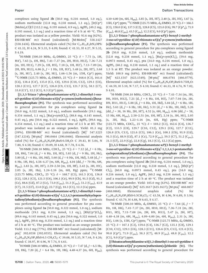
complexes using ligand 2b (50.0 mg, 0.216 mmol, 1.0 eq.),sodium methoxide (12.8 mg, 0.238 mmol, 1.1 eq.), [Ir(Cp*)(MeIm)2Cl]Cl (112.2 mg, 0.195 mmol, 0.9 eq.), AgPF6 (49.2 mg,0.195 mmol, 1.1 eq.) and a reaction time of 4 h at 40 °C. Theproduct was isolated as a yellow powder. Yield: 93.6 mg (62%).ESI-HR-MS+ m/z found (calculated): [M-MeIm]+ 558.1427(558.1436). Elemental analysis calcd (%) for C27H33F6IrN3OPS:C 41.32, H 4.24, N 5.35, S 4.09; found: C 41.53, H 4.27, N 5.32,S 4.19.
1H-NMR (500.10 MHz, d6-DMSO, 25 °C): δ = 7.75 (s, 1H,H4′), 7.65 (s, 1H, H6), 7.41–7.37 (m, 2H, H10, H12), 7.36–7.31(m, 1H, H11), 7.29 (s, 1H, H5), 7.18 (s, 1H, H2′), 7.13–7.09 (m,2H, H9, H13), 6.86 (t, J = 1 Hz, 1H, H3′), 5.48 (s, 2H, H7), 3.68(s, 3H, H1′), 2.40 (s, 3H, H1), 1.66–1.56 (m, 15H, Cp*) ppm;13C-NMR (125.75 MHz, d6-DMSO, 25 °C): δ = 168.6 (C3), 161.6(C4), 138.4 (C4′), 135.0 (C2, C8), 129.6 (C10, C12), 129.1 (C6),128.3 (C11), 127.7 (C3′), 126.8 (C9, C13), 121.7 (C5), 58.1 (C7),33.8 (C1′), 12.2 (C1), 8.3 (Cp*) ppm.
[(1,3,5-Triaza-7-phosphaadamantane-κP)(1,2-dimethyl-3-oxo-κO-pyridine-4(1H)-thionato-κS)(η6-p-cymene)ruthenium(II)]hexa-fluorophosphate (P1). The synthesis was performed accordingto general procedure for pta complexes using ligand 2a(50.0 mg, 0.322 mmol, 1.0 eq.), sodium methoxide (19.1 mg,0.354 mmol, 1.1 eq.), [Ru(p-cym)Cl2]2 (88.8 mg, 0.145 mmol,0.45 eq.), pta (50.6 mg, 0.322 mmol, 1 eq.), AgPF6 (89.6 mg,0.354 mmol, 1.1 eq.) and a reaction time of 2.5 h at RT. Theproduct was isolated as an orange powder. Yield: 81.3 mg(39%). ESI-HR-MS+ m/z found (calculated): [M]+ 547.1225(547.1234); [M-pta]+ 390.0464 (390.0466). Elemental analysiscalcd (%) for C23H34F6N4OP2RuS·0.5H2O: C 39.43, H 5.04, N7.80, S 4.58; found: C 39.09, H 4.68, N 7.78, S 4.56.
1H-NMR (500.10 MHz, CDCl3, 25 °C): δ = 7.12 (d, J = 6 Hz,1H, H6), 7.04 (d, J = 6 Hz, 1H, H5), 5.83 (d, J = 9 Hz, 1H, Hc),5.80 (d, J = 6 Hz, 1H, Hd), 5.60 (d, J = 6 Hz, 1H, Hd), 5.48 (d, J =6 Hz, 1H, Hc), 4.56–4.37 (m, 6H, Hpta), 4.04 (dd, J = 70 Hz, 6H,Hpta), 3.83 (s, 3H, H7), 2.59–2.50 (m, 1H, Hf), 2.42 (s, 3H, H1),2.05 (s, 3H, Ha), 1.24–1.16 (m, 6H, Hg) ppm; 13C-NMR(125.75 MHz, CDCl3, 25 °C): δ = 168.7 (C3), 161.5 (C4), 136.8(C2), 128.1 (C5), 121.5 (C6), 106.1 (Ce), 99.9 (Cb), 91.5 (Cd), 91.3(Cc), 88.6 (Cd), 87.2 (Cc), 73.0 (Cpta), 51.3 (Cpta), 51.2 (Cpta), 43.9(C7), 31.5 (Cf), 23.0 (Cg), 22.7 (Cg), 18.3 (C1), 12.3 (Ca) ppm.
[(1,3,5-Triaza-7-phosphaadamantane-κP)(1,2-dimethyl-3-oxo-κO-pyridine-4(1H)-thionato-κS)(η5-1,2,3,4,5-pentamethylcyclopen-tadienyl)rhodium(III)]hexafluorophosphate (P2). The synthesiswas performed according to general procedure for pta com-plexes using ligand 2a (50.0 mg, 0.322 mmol, 1.0 eq.), sodiummethoxide (19.1 mg, 0.354 mmol, 1.1 eq.), [Rh(Cp*)Cl2]2(89.6 mg, 0.145 mmol, 0.45 eq.), pta (50.6 mg, 0.322 mmol, 1.0eq.), AgPF6 (89.6 mg, 0.354 mmol, 1.1 eq.) and a reaction timeof 2 h at 40 °C. The product was isolated as an orange powder.Yield: 113.2 mg (77%). ESI-HR-MS+ m/z found (calculated): [M-pta]+ 392.0558 (392.0555). Elemental analysis calcd (%) forC24H39F6N4OP2RhS·0.5 CH2Cl2: C 39.08, H 5.35, N 7.44, S 4.26;found: C 38.97, H 4.96, N 7.74, S 4.61.
1H-NMR (500.10 MHz, d6-DMSO, 25 °C): δ = 7.47 (d, J = 6.5 Hz,1H, H6), 7.20 (d, J = 6.4 Hz, 1H, H6), 4.48–4.37 (m, 6H, Hpta),
4.10–4.00 (m, 6H, Hpta), 3.85 (s, 3H, H7), 2.40 (s, 3H, H1), 1.67 (s,15H, Cp*) ppm; 13C-NMR (125.75 MHz, d6-DMSO, 25 °C): δ = 166.5(C3), 158.0 (C4), 137.5 (C2), 128.8 (C5), 121.0 (C6), 99.7 (Cp*q), 71.9(Cpta), 48.8 (Cpta), 43.5 (Cpta), 12.2 (C1), 9.0 (Cp*) ppm.
[(1,3,5-Triaza-7-phosphaadamantane-κP)(1-benzyl-2-methyl-3-oxo-κO-pyridine-4(1H)-thionato-κS)(η6-p-cymene)ruthenium(II)]hexafluorophosphate (P3). The synthesis was performedaccording to general procedure for pta complexes using ligand2b (50.0 mg, 0.216 mmol, 1.0 eq.), sodium methoxide(12.8 mg, 0.238 mmol, 1.1 eq.), [Ru(p-cym)Cl2]2 (59.6 mg,0.0973 mmol, 0.45 eq.), pta (34.0 mg, 0.216 mmol, 1.0 eq.),AgPF6 (60.2 mg, 0.238 mmol, 1.1 eq.) and a reaction time of1.5 h at RT. The product was isolated as an orange powder.Yield: 100.9 mg (68%). ESI-HR-MS+ m/z found (calculated):[M]+ 623.1547 (623.1549); [M-pta]+ 466.0781 (466.0779).Elemental anaylsis calcd (%) for C29H38F6N4OP2RuS·0.75H2O:C 44.59, H 5.10, N 7.17, S 4.10; found: C 44.33, H 4.74, N 7.02,S 4.01.
1H-NMR (500.10 MHz, CDCl3, 25 °C): δ = 7.41–7.34 (m, 3H,H6, H10, H12), 7.21 (d, J = 6 Hz, 1H, H5), 7.07–7.04 (m, 3H,H9, H11, H13), 5.88 (d, J = 6 Hz, 1H, Hd), 5.84 (d, J = 6 Hz, 1H,Hc), 5.61 (d, J = 6 Hz, 1H, Hc), 5.51 (d, J = 6 Hz, 1H, Hd), 5.26(dd, J = 38, 16 Hz, 2H, H7), 4.52 (s, 6H, Hpta), 4.06 (dd, J = 58,15 Hz, 6H, Hpta), 2.58–2.51 (m, 1H, Hf), 2.34 (s, 3H, H1), 2.05(s, 3H, Ha), 1.23–1.16 (m, 6H, Hg) ppm; 13C-NMR(125.75 MHz, CDCl3, 25 °C): δ = 169.2 (C3), 162.6 (C4), 136.7(C2), 133.5 (C8), 129.7 (C10, C12), 129.1 (C6), 127.7 (C11),126.8 (C9, C13), 121.6 (C5), 106.1 (Ce), 100.1 (Cb), 91.9 (Cd),91.5 (Cc), 88.9 (Cd), 87.1 (Cc), 72.9 (Cpta), 59.5 (C7), 51.2 (Cpta),31.5 (Cf), 22.9 (Cg), 22.8 (Cg), 18.3 (C1), 12.3 (Ca) ppm.
[(1,3,5-Triaza-7-phosphaadamantane-κP)(1-benzyl-2-methyl-3-oxo-κO-pyridine-4(1H)-thionato-κS)(η5-1,2,3,4,5-pentamethyl-cyclopentadienyl)rhodium(III)]hexafluorophosphate (P4). Thesynthesis was performed according to general procedure forpta complexes using ligand 2b (50.0 mg, 0.216 mmol, 1.0 eq.),sodium methoxide (12.8 mg, 0.238 mmol, 1.1 eq.), [Rh(Cp*)Cl2]2 (60.0 mg, 0.0973 mmol, 0.45 eq.), pta (34.0 mg,0.216 mmol, 1.0 eq.), AgPF6 (60.2 mg, 0.238 mmol, 1.1 eq.)and a reaction time of 2 h at 40 °C. The product was isolatedas an orange powder. Yield: 105.0 mg (63%). ESI-HR-MS+ m/zfound (calculated): [M]+ 625.1617 (625.1637); [M-pta]+ 468.0857(468.0868). Elemental anaylsis calcd (%) forC29H39F6N4OP2RhS·0.5CH2Cl2: C 43.58, H 4.96, N 6.89, S 3.94;found: C 43.79, H 4.88, N 6.65, S 4.17.
1H-NMR (500.10 MHz, d6-DMSO, 25 °C): δ = 7.64 (d, J = 7Hz, 1H, H6), 7.41–7.37 (m, 2H, H10, H12), 7.36–7.31 (m, 2H,H11, H5), 7.11–7.08 (m, 2H, H9, H13), 5.47 (s, 2H, H7),4.48–4.38 (m, 6H, Hpta), 4.09–4.00 (m, 6H, Hpta), 2.31 (s, 3H,H1), 1.66 (s, 15H, Cp*) ppm; 13C-NMR (125.75 MHz, d6-DMSO,25 °C): δ = 167.1 (C3), 159.6 (C4), 136.7 (C2), 135.1 (C8), 129.1(C10, C12), 129.1 (C6), 128.3 (C11), 126.6 (C9, C13), 121.4 (C5),99.8 (Cp*), 71.9 (Cpta), 58.3 (C7), 48.9 (Cpta), 48.8 (Cpta), 12.2(C1), 9.0 (Cp*) ppm.
[(Thioatocarbonyldiamine-κS)(1,2-dimethyl-3-oxo-κO-pyridine-4(1H)-thionato-κS)(η6-p-cymene)ruthenium(II)]chloride (S1). Thesynthesis was performed according to general procedure for
Paper Dalton Transactions
15708 | Dalton Trans., 2020, 49, 15693–15711 This journal is © The Royal Society of Chemistry 2020
Ope
n A
cces
s A
rtic
le. P
ublis
hed
on 0
2 N
ovem
ber
2020
. Dow
nloa
ded
on 4
/2/2
022
8:10
:02
PM.
Thi
s ar
ticle
is li
cens
ed u
nder
a C
reat
ive
Com
mon
s A
ttrib
utio
n-N
onC
omm
erci
al 3
.0 U
npor
ted
Lic
ence
.View Article Online

pta complexes using ligand 2a (50.0 mg, 0.322 mmol, 1.0 eq.),sodium methoxide (19.1 mg, 0.354 mmol, 1.1 eq.), [Ru(p-cym)Cl2]2 (88.8 mg, 0.145 mmol, 0.45 eq.), thiourea (24.5 mg,0.322 mmol, 1.0 eq.) and a reaction time of 24 h at RT. Theproduct was isolated as a red powder. Yield: 108.4 mg (74%).ESI-HR-MS+ m/z found (calculated): [M-tu]+ 390.0468(390.0466). Elemental analysis calcd (%) forC18H26ClN3ORuS2·0.25H2O: C 42.76, H 5.28, N 8.31, S 12.68;found: C 42.48, H 4.96, N 8.50, S 12.99.
1H-NMR (500.10 MHz, CDCl3, 25 °C): δ = 8.94 (s, 2H, NH2),7.15 (d, J = 6 Hz, 1H, H6), 7.07 (s, 2H, NH2), 6.96 (d, J = 6 Hz,1H, H5), 3.76 (s, 3H, H7), 2.70–2.62 (m, 1H, Hf), 2.38 (s, 3H,H1), 2.12 (s, 3H, Ha), 1.18 (d, J = 7 Hz, 6H, Hg) ppm; 13C-NMR(125.75 MHz, CDCl3, 25 °C): δ = 183.0 (tu), 167.0 (C3), 164.2(C4), 135.6 (C2), 128.4 (C6), 122.1 (C5), 102.1 (Ce), 100.5 (Cb),85.9 (Cd), 84.8 (Cc), 83.9 (Cd), 83.0 (Cc), 43.8 (C7), 31.0 (Cg),18.2 (C1), 12.7 (Ca) ppm.
[(Thioatocarbonyldiamine-κS)(1-benzyl-2-methyl-3-oxo-κO-pyri-dine-4(1H)-thionato-κS)(η6-p-cymene)ruthenium(II)]chloride (S2).The synthesis was performed according to general procedurefor thiourea complexes using ligand 2b (50.0 mg, 0.216 mmol,1.0 eq.), sodium methoxide (12.8 mg, 0.238 mmol, 1.1 eq.),[Ru(p-cym)Cl2]2 (59.6 mg, 0.0973 mmol, 0.45 eq.), tu (16.5 mg,0.216 mmol, 1.0 eq.), and a reaction time of 24 h at RT. Theproduct was isolated as a red powder. Yield: 84.5 mg (75%).ESI-HR-MS+ m/z found (calculated): [M-tu]+ 466.0779(466.0778). Elemental anaylsis calcd (%) forC24H30ClN3ORuS2·0.5H2O: C 49.18, H 5.33, N 7.17, S 10.94;found: C 48.95, H 5.34, N 7.19, S 11.07.
1H-NMR (500.10 MHz, CDCl3, 25 °C): δ = 8.83 (s, 2H, NH2),7.40–7.35 (m, 3H, H6, H10, H12), 7.22 (d, J = 7 Hz, 1H, H5),7.06 (s, 2H, NH2), 7.04–6.98 (m, 3H, H9, H11, H13), 5.57–5.33(m, 3H, Hc, Hd), 5.28–5.12 (m, 3H, Hd, H7), 2.71–2.63 (m, 1H,Hf), 2.32 (s, 3H, H1), 2.12 (s, 3H, Ha), 1.19 (d, J = 7 Hz, 6H,Hg) ppm; 13C-NMR (125.75 MHz, CDCl3, 25 °C): δ = 183.0 (tu),167.3 (C3), 165.0 (C4), 135.6 (C2), 133.4 (C8), 129.7 (C10, C12),129.2 (C6), 128.2 (C11), 126.7 (C9, C13), 122.3 (C5), 101.9 (Ce),100.7 (Cb), 86.3 (Cd), 84.7 (Cc), 84.0 (Cd), 83.4 (Cc), 59.3 (C7),31.0 (Cf), 23.0 (Cg), 22.4 (Cg), 18.3 (Ca), 12.6 (C1).
Conflicts of interest
There are no conflicts to declare.
Acknowledgements
We thank the University of Vienna for financial support.Furthermore, we would like to thank Claudia Cseh for herkind support with the evaluation of DNA plasmid assays.
Notes and references
1 Y. K. Yan, M. Melchart, A. Habtemariam and P. J. Sadler,Chem. Commun., 2005, 4764–4776.
2 C. S. Allardyce and P. J. Dyson, Platinum Met. Rev., 2001, 45,62–69.
3 I. Kostova, Curr. Med. Chem., 2006, 13, 1085–1107.4 R. Mitra, S. Das, S. V. Shinde, S. Sinha, K. Somasundaram
and A. G. Samuelson, Chem. – Eur. J., 2012, 18, 12278–12291.
5 L. Ma, R. Ma, Z. Wang, S. M. Yiu and G. Zhu, Chem.Commun., 2016, 52, 10735–10738.
6 H. Köpf and P. Köpf-Maier, Angew. Chem., Int. Ed. Engl.,1979, 91, 509–509.
7 M. Guo, Z. Guo and P. Sadler, J. Biol. Inorg. Chem., 2001, 6,698–707.
8 B. K. Keppler, M. R. Berger and M. E. Heim, Cancer Treat.Rev., 1990, 17, 261–277.
9 G. Jaouen, S. Top, A. Vessieres, G. Leclercq andM. McGlinchey, Curr. Med. Chem., 2004, 11, 2505–2517.
10 M. Hanif, H. Henke, S. M. Meier, S. Martic, M. Labib,W. Kandioller, M. A. Jakupec, V. B. Arion, H. B. Kraatz,B. K. Keppler and C. G. Hartinger, Inorg. Chem., 2010, 49,7953–7963.
11 S. M. Meier, M. Hanif, W. Kandioller, B. K. Keppler andC. G. Hartinger, J. Inorg. Biochem., 2012, 108, 91–95.
12 M. Caterino, M. Herrmann, A. Merlino, C. Riccardi,D. Montesarchio, M. A. Mroginski, D. Musumeci, F. Ruffo,L. Paduano, P. Hildebrandt, J. Kozuch and A. Vergara,Inorg. Chem., 2019, 58, 1216–1223.
13 R. E. Morris, R. E. Aird, P. del Socorro Murdoch, H. Chen,J. Cummings, N. D. Hughes, S. Parsons, A. Parkin, G. Boyd,D. I. Jodrell and P. J. Sadler, J. Med. Chem., 2001, 44, 3616–3621.
14 F. Wang, J. Xu, K. Wu, S. K. Weidt, C. L. MacKay,P. R. R. Langridge-Smith and P. J. Sadler, Dalton Trans.,2013, 42, 3188–3195.
15 B. Wu, M. S. Ong, M. Groessl, Z. Adhireksan,C. G. Hartinger, P. J. Dyson and C. A. Davey, Chem. – Eur. J.,2011, 17, 3562–3566.
16 T. Giraldi, G. Sava, G. Mestroni, G. Zassinovich andD. Stolfa, Chem.–Biol. Interact., 1978, 22, 231–238.
17 L. Messori, J. Inorg. Biochem., 2003, 95, 37–46.18 G. Mestroni, E. Alessio, A. Sessanta o Santi, S. Geremia,
A. Bergamo, G. Sava, A. Boccarelli, A. Schettino andM. Coluccia, Inorg. Chim. Acta, 1998, 273, 62–71.
19 Z. Liu and P. J. Sadler, Acc. Chem. Res., 2014, 47, 1174–1185.
20 A. Casini, F. Edafe, M. Erlandsson, L. Gonsalvi,A. Ciancetta, N. Re, A. Ienco, L. Messori, M. Peruzzini andP. J. Dyson, Dalton Trans., 2010, 39, 5556–5563.
21 C. Scolaro, A. Bergamo, L. Brescacin, R. Delfino,M. Cocchietto, G. Laurenczy, T. J. Geldbach, G. Sava andP. J. Dyson, J. Med. Chem., 2005, 48, 4161–4171.
22 C. M. Hackl, M. S. Legina, V. Pichler, M. Schmidlehner,A. Roller, O. Dömötör, E. A. Enyedy, M. A. Jakupec,W. Kandioller and B. K. Keppler, Chem. – Eur. J., 2016, 22,17269–17281.
23 S. Harringer, B. Happl, M. Ozenil, C. Kast, M. Hejl,D. Wernitznig, A. A. Legin, A. Schweikert, N. Gajic,
Dalton Transactions Paper
This journal is © The Royal Society of Chemistry 2020 Dalton Trans., 2020, 49, 15693–15711 | 15709
Ope
n A
cces
s A
rtic
le. P
ublis
hed
on 0
2 N
ovem
ber
2020
. Dow
nloa
ded
on 4
/2/2
022
8:10
:02
PM.
Thi
s ar
ticle
is li
cens
ed u
nder
a C
reat
ive
Com
mon
s A
ttrib
utio
n-N
onC
omm
erci
al 3
.0 U
npor
ted
Lic
ence
.View Article Online

A. Roller, G. Koellensperger, M. A. Jakupec, W. Kandiollerand B. K. Keppler, Chem. – Eur. J., 2020, 26, 5419–5433.
24 M. P. Cava and M. I. Levinson, Tetrahedron, 1985, 41, 5061–5087.
25 C. A. Riedl, M. Hejl, M. H. M. Klose, A. Roller,M. A. Jakupec, W. Kandioller and B. K. Keppler, DaltonTrans., 2018, 47, 4625–4638.
26 M. Schmidlehner, P. S. Kuhn, C. M. Hackl, A. Roller,W. Kandioller and B. K. Keppler, J. Organomet. Chem.,2014, 772, 93–99.
27 A. F. A. Peacock, M. Melchart, R. J. Deeth, A Habtemariam,S. Parsons and P. J. Sadler, Chem. – Eur. J., 2007, 13, 2601–2613.
28 A. Dorcier, P. J. Dyson, C. Gossens, U. Rothlisberger,R. Scopelliti and I. Tavernelli, Organometallics, 2005, 24,2114–2123.
29 T. Mosmann, J. Immunol. Methods, 1983, 65, 55–63.30 V. Pichler, S. M. Valiahdi, M. A. Jakupec, V. B. Arion,
M. Galanski and B. K. Keppler, Dalton Trans., 2011, 40,8187–8192.
31 P. S. Kuhn, V. Pichler, A. Roller, M. Hejl, M. A. Jakupec,W. Kandioller and B. K. Keppler, Dalton Trans., 2015, 44,659–668.
32 A. S. Nunes, A. S. Barros, E. C. Costa, A. F. Moreira andI. J. Correia, Biotechnol. Bioeng., 2019, 116, 206–226.
33 E. Schreiber-Brynzak, E. Klapproth, C. Unger,I. Lichtscheidl-Schultz, S. Göschl, S. Schweighofer,R. Trondl, H. Dolznig, M. A. Jakupec and B. K. Keppler,Invest. New Drugs, 2015, 33, 835–847.
34 H. Geisler, D. Wernitznig, M. Hejl, N. Gajic, M. A. Jakupec,W. Kandioller and B. K. Keppler, Dalton Trans., 2020, 49,1393–1397.
35 A. M. Rieger, K. L. Nelson, J. D. Konowalchuk andD. R. Barreda, J. Visualized Exp., 2011, 3–6.
36 I. Vermes, C. Haanen, H. Steffens-Nakken andC. Reutelingsperger, J. Immunol. Methods, 1995, 184, 39–51.
37 I. Vermes, C. Haanen and C. Reutelingsperger, J. Immunol.Methods, 2000, 243, 167–190.
38 M. Cornelissen, J. Philippé, S. De Sitter and L. De Ridder,Apoptosis, 2002, 7, 41–47.
39 Z. Darzynkiewicz, S. Bruno, G. Del Bino, W. Gorczyca,M. A. Hotz, P. Lassota and F. Traganos, Cytometry, 1992, 13,795–808.
40 G. Kroemer, B. Dallaporta and M. Resche-Rigon, Annu. Rev.Physiol., 1998, 60, 619–642.
41 G. Denecker, D. Vercammen, W. Declercq andP. Vandenabeele, Cell. Mol. Life Sci., 2001, 58, 356–370.
42 L. Faleiro and Y. Lazebnik, J. Cell Biol., 2000, 151, 951–960.43 J. A. Khan, F. Forouhar, X. Tao and L. Tong, Expert Opin.
Ther. Targets, 2007, 11, 695–705.44 J. J. Soldevila-Barreda and P. J. Sadler, Curr. Opin. Chem.
Biol., 2015, 25, 172–183.45 K. W. Wellington, RSC Adv., 2015, 5, 20309–20338.46 G.-Y. Liou and P. Storz, Free Radic. Res., 2010, 44, 479–496.47 N. A. P. Franken, H. M. Rodermond, J. Stap, J. Haveman
and C. van Bree, Nat. Protoc., 2006, 1, 2315–2319.
48 P. Zhang and P. J. Sadler, J. Organomet. Chem., 2017, 839,5–14.
49 S. Zorbas-Seifried, C. G. Hartinger, K. Meelich, M. Galanskiand B. K. Keppler, DNA Repair, 2006, 14817–14825.
50 H. Li, H. Bo, J. Wang, H. Shao and S. Huang,Cytotechnology, 2011, 63, 7–12.
51 H.-K. Liu and P. J. Sadler, Acc. Chem. Res., 2011, 44, 349–359.
52 S. Göschl, H. P. Varbanov, S. Theiner, M. A. Jakupec,M. Galanski and B. K. Keppler, J. Inorg. Biochem., 2016,160, 264–274.
53 S. Göschl, E. Schreiber-Brynzak, V. Pichler, K. Cseh,P. Heffeter, U. Jungwirth, M. A. Jakupec, W. Berger andB. K. Keppler, Metallomics, 2017, 9, 309–322.
54 S. Moon, M. Hanif, M. Kubanik, H. Holtkamp, T. Söhnel,S. M. F. Jamieson and C. G. Hartinger, ChemPlusChem,2015, 80, 231–236.
55 M. Hanif, A. A. Nazarov, A. Legin, M. Groessl, V. B. Arion,M. A. Jakupec, Y. O. Tsybin, P. J. Dyson, B. K. Keppler andC. G. Hartinger, Chem. Commun., 2012, 48, 1475–1477.
56 M. Zaki, S. Hairat and E. S. Aazam, Scope of organometalliccompounds based on transition metal-arene systems as anti-cancer agents: Starting from the classical paradigm to target-ing multiple strategies, Royal Society of Chemistry, 2019, vol.9.
57 U. Ndagi, N. Mhlongo and M. E. Soliman, Drug Des., Dev.Ther., 2017, 11, 599–616.
58 S. J. Dougan, A. Habtemariam, S. E. McHale, S. Parsonsand P. J. Sadler, Proc. Natl. Acad. Sci. U. S. A., 2008, 105,11628–11633.
59 S. Theiner, S. J. M. Van Malderen, T. Van Acker, A. Legin,B. K. Keppler, F. Vanhaecke and G. Koellensperger, Anal.Chem., 2017, 89, 12641–12645.
60 M. Zoriy, A. Matusch, T. Spruss and J. S. Becker, Int. J. MassSpectrom., 2007, 260, 102–106.
61 R. Lobinski, C. Moulin and R. Ortega, Biochimie, 2006, 88,1591–1604.
62 S. Theiner, E. Schreiber-Brynzak, M. A. Jakupec,M. Galanski, G. Koellensperger and B. K. Keppler,Metallomics, 2016, 8, 398–402.
63 S. B. Jensen, S. J. Rodger and M. D. Spicer, J. Organomet.Chem., 1998, 556, 151–158.
64 M. G. Mendoza-Ferri, C. G. Hartinger, A. A. Nazarov,R. E. Eichinger, M. A. Jakupec, K. Severin andB. K. Keppler, Organometallics, 2009, 28, 6260–6265.
65 L. E. Heim, S. Vallazza, D. van der Waals andM. H. G. Prechtl, Green Chem., 2016, 18, 1469–1474.
66 J. W. Kang, K. Moseley and P. M. Maitiis, J. Am. Chem. Soc.,1969, 91, 5970–5977.
67 W. Tejchman, E. Zesławska, K. Zborowski, W. Nitek andM. Zylewski, ARKIVOC, 2015, 2015, 216–230.
68 Bruker SAINT v8.38B Copyright © 2005-2019, Bruker AXS.69 G. M. Sheldrick, SADABS, University of Göttingen,
Germany, 1996.70 O. V. Dolomanov, L. J. Bourhis, R. J. Gildea, J. A. K. Howard
and H. Puschmann, J. Appl. Crystallogr., 2009, 42, 339–341.
Paper Dalton Transactions
15710 | Dalton Trans., 2020, 49, 15693–15711 This journal is © The Royal Society of Chemistry 2020
Ope
n A
cces
s A
rtic
le. P
ublis
hed
on 0
2 N
ovem
ber
2020
. Dow
nloa
ded
on 4
/2/2
022
8:10
:02
PM.
Thi
s ar
ticle
is li
cens
ed u
nder
a C
reat
ive
Com
mon
s A
ttrib
utio
n-N
onC
omm
erci
al 3
.0 U
npor
ted
Lic
ence
.View Article Online

71 C. B. Hübschle, G. M. Sheldrick and B. Dittrich, J. Appl.Crystallogr., 2011, 44, 1281–1284.
72 G. M. Sheldrick, SHELXS v 2016/4, University of Göttingen,Germany, 2015.
73 G. M. Sheldrick, SHELXL v 2016/4, University of Göttingen,Germany, 2015.
74 A. L. Spek, Acta Crystallogr., Sect. D: Biol. Crystallogr., 2009,65, 148–155.
Dalton Transactions Paper
This journal is © The Royal Society of Chemistry 2020 Dalton Trans., 2020, 49, 15693–15711 | 15711
Ope
n A
cces
s A
rtic
le. P
ublis
hed
on 0
2 N
ovem
ber
2020
. Dow
nloa
ded
on 4
/2/2
022
8:10
:02
PM.
Thi
s ar
ticle
is li
cens
ed u
nder
a C
reat
ive
Com
mon
s A
ttrib
utio
n-N
onC
omm
erci
al 3
.0 U
npor
ted
Lic
ence
.View Article Online