intraoperative neurophysiological monitoring for
TRANSCRIPT

Thomas Jefferson University Thomas Jefferson University
Jefferson Digital Commons Jefferson Digital Commons
Department of Neurosurgery Faculty Papers Department of Neurosurgery
5-16-2016
Intraoperative Neurophysiological Monitoring for Endoscopic Intraoperative Neurophysiological Monitoring for Endoscopic
Endonasal Approaches to the Skull Base: A Technical Guide. Endonasal Approaches to the Skull Base: A Technical Guide.
Harminder Singh Stanford Hospitals and Clinics
Richard W. Vogel Safe Passage Neuromonitoring
Robert M. Lober Stanford Hospitals and Clinics
Adam T. Doan Safe Passage Neuromonitoring
Craig I. Matsumoto Sentient Medical Systems
See next page for additional authors
Follow this and additional works at: https://jdc.jefferson.edu/neurosurgeryfp
Part of the Neurology Commons, and the Surgery Commons
Let us know how access to this document benefits you
Recommended Citation Recommended Citation
Singh, Harminder; Vogel, Richard W.; Lober, Robert M.; Doan, Adam T.; Matsumoto, Craig I.;
Kenning, Tyler J.; and Evans, James J., "Intraoperative Neurophysiological Monitoring for
Endoscopic Endonasal Approaches to the Skull Base: A Technical Guide." (2016). Department of
Neurosurgery Faculty Papers. Paper 84.
https://jdc.jefferson.edu/neurosurgeryfp/84
This Article is brought to you for free and open access by the Jefferson Digital Commons. The Jefferson Digital Commons is a service of Thomas Jefferson University's Center for Teaching and Learning (CTL). The Commons is a showcase for Jefferson books and journals, peer-reviewed scholarly publications, unique historical collections from the University archives, and teaching tools. The Jefferson Digital Commons allows researchers and interested readers anywhere in the world to learn about and keep up to date with Jefferson scholarship. This article has been accepted for inclusion in Department of Neurosurgery Faculty Papers by an authorized administrator of the Jefferson Digital Commons. For more information, please contact: [email protected].

Authors Authors Harminder Singh, Richard W. Vogel, Robert M. Lober, Adam T. Doan, Craig I. Matsumoto, Tyler J. Kenning, and James J. Evans
This article is available at Jefferson Digital Commons: https://jdc.jefferson.edu/neurosurgeryfp/84
Review ArticleIntraoperative Neurophysiological Monitoring forEndoscopic Endonasal Approaches to the Skull Base:A Technical Guide
Harminder Singh,1 Richard W. Vogel,2 Robert M. Lober,1 Adam T. Doan,2
Craig I. Matsumoto,3 Tyler J. Kenning,4 and James J. Evans5
1Stanford Hospitals and Clinics, Department of Neurosurgery, 300 Pasteur Drive, Stanford, CA 94305, USA2Safe Passage Neuromonitoring, 915 Broadway, Suite 1200, New York, NY 10010, USA3Sentient Medical Systems, 11011 McCormick Road, Suite 200, Hunt Valley, MD 21031, USA4Albany Medical Center Neurosurgery Group, 47 New Scotland Avenue, MC 10, Physicians Pavilion, First Floor, Albany,NY 12208, USA5Thomas Jefferson University Hospital, Department of Neurosurgery, 909 Walnut Street, Third Floor, Philadelphia, PA 19107, USA
Correspondence should be addressed to Harminder Singh; [email protected]
Received 4 December 2015; Revised 4 April 2016; Accepted 11 April 2016
Academic Editor: Kosmas Paraskevas
Copyright © 2016 Harminder Singh et al. This is an open access article distributed under the Creative Commons AttributionLicense, which permits unrestricted use, distribution, and reproduction in any medium, provided the original work is properlycited.
Intraoperative neurophysiological monitoring during endoscopic, endonasal approaches to the skull base is both feasible and safe.Numerous reports have recently emerged from the literature evaluating the efficacy of different neuromonitoring tests duringendonasal procedures, making them relatively well-studied. The authors report on a comprehensive, multimodality approach tomonitoring the functional integrity of at risk nervous system structures, including the cerebral cortex, brainstem, cranial nerves,corticospinal tract, corticobulbar tract, and the thalamocortical somatosensory system during endonasal surgery of the skull base.Themodalities employed include electroencephalography, somatosensory evoked potentials, free-running and electrically triggeredelectromyography, transcranial electric motor evoked potentials, and auditory evoked potentials. Methodological considerationsas well as benefits and limitations are discussed. The authors argue that, while individual modalities have their limitations,multimodality neuromonitoring provides a real-time, comprehensive assessment of nervous system function and allows for safer,more aggressive management of skull base tumors via the endonasal route.
1. Introduction
For over a century in neurosurgery, the endonasal approachto the anterior skull base was recognized as a means toaccess sellar lesions [1–3]. First introduced by Schloffer in1907, the reach of the endonasal approach greatly expandedwith the introduction of numerous imaging technologies. Forexample, when Hardy introduced the intraoperative micro-scope in 1967, it revolutionized transsphenoidal surgery andimproved its safety by combining improved magnificationand illumination [4]. The most recent and, arguably, equallysignificant advancement in endonasal neurosurgery was thedescription of the use of the endoscope by Jankowski et al.
in 1992 [5]. The subsequent rapid expansion of endoscopicintracranial surgery has permitted access to large areas of thecranial base and its associated pathology. Today, the reachof the endoscopic, endonasal approach (EEA) extends farbeyond the sphenoid and sella to the entire ventral skullbase via transcribiform, transplanum, transdorsum sellae,transclival, and transpterygopalatine fossa corridors [6–9].Thus,multiple reports in the literature have demonstrated theutility of these approaches in reaching the anterior, middle,infratemporal, and posterior fossae.
Endoscopic endonasal surgery often requires working inclose proximity to, and occasionally directly on, the criticalneurovascular structures and cranial nerves that traverse the
Hindawi Publishing CorporationScientificaVolume 2016, Article ID 1751245, 20 pageshttp://dx.doi.org/10.1155/2016/1751245

2 Scientifica
cranial base. Although the reported rates of vascular injuryand cranial nerve deficits following endonasal cranial basesurgery are low, these complications can have devastatingeffects. There is a relatively high potential for postoperativemorbidity when addressing cranial base pathology, and allpossible efforts must be made to limit that potential. Use ofan endoscope clearly serves that goal by providing superiorillumination and visualization when compared to the oper-ating microscope. Any additional measures that improve thesafety of endonasal surgery should be considered.
Intraoperative neurophysiological monitoring (IONM;neuromonitoring) can be used to further mitigate the risksassociated with the EEA. IONM involves the use of phys-iological tests that can identify nervous system structures,as well as evaluating their functions, in real-time duringsurgery. IONM is based on the premise that the patient’sneurophysiology changes in a measureable way, prior tothe onset of permanent neurological deficit. Thus, critical,nontechnical changes in the neurophysiology data alert theneurosurgeon to perturbations in the nervous system which,if left unattended, could result in transient or permanent post-operative motor and/or sensory deficits. IONM endeavorsnot only to detect and identify iatrogenic nervous systemdysfunction, but also to guide the use of surgical inter-ventions and monitor their efficacy. Thoughtful data trendanalysis, a keen understanding of neural elements at risk forinjury, and knowledge of surgical technique (time-lockingdata changes to surgical/anesthetic conditions) are all criticalin the neurophysiologist’s assessment of these measures. Inaddition to monitoring the function of neural tissue, IONMis frequently used to identify and differentiate specific neuralelements, such asmotor cranial nerves, with the ultimate goalof preserving their baseline function through the duration ofthe surgical procedure.
Multimodality IONM has emerged as a standardapproach to monitoring the nervous system under generalanesthesia to improve safety and optimize surgical outcomesacross a wide range of surgical procedures involving thecentral and peripheral nervous system. In the context ofthe EEA to the skull base, global cortical monitoring withelectroencephalography, somatosensory evoked potentials,and transcranial, electrical motor evoked potentials is likelyto be helpful in situations where the internal carotid arteriesor their branches are at risk [10]. In the case of skull baseapproaches, this includes exposure of the parasellar regionand cavernous sinus [11, 12]. Similarly, brainstem auditoryevoked potentials (BAEP) are useful for detecting brainstemischemia during surgery at or around the vertebrobasilarjunction, as is the case for transclival approaches [12–14].
Intraoperative monitoring of the oculomotor, trochlear,and abducens nerves with needle electrodes placed in theinferior rectus, superior oblique, and lateral rectus mus-cles, respectively, has long been used during transcranialapproaches to the cavernous sinus [15–17] and is equallyapplicable in many EEA procedures [18, 19], including casesin which the cavernous sinus is not accessed.The oculomotornerve, for example, is vulnerable in the interpeduncularcistern via the transsphenoidal [20] and transplanum routes[21], with vascular compromise possible from injury to the
inferolateral trunk of the cavernous carotid or its branches[20]. The trochlear nerve may be exposed at the ambiensdivision of the cisternal segment through the transellartranstubercular route, and ischemic injury may occur withinjury to the superior cerebellar artery [22]. The abducensnerve, being the longest and most ventrally located cranialnerve at the level of the clivus and cavernous sinus, isparticularly at risk during approaches to petroclival lesionsvia the midline transclival, paramedian suprapetrous, andmedial petrous apex approaches [23]. In these cases, therisk may be increased by abnormal anatomy (e.g., medialdisplacement of the nerve by a petroclival tumor or upwarddisplacement by a cisternal mass). Similar to the oculomotornerve, it may also suffer vascular compromise by injury tothe inferolateral trunk from the cavernous segment of theinternal carotid artery. The trigeminal nerve, also at risk insome EEA procedures, may be violated in Meckel’s cave viathe transpterygoid corridor [24]. As EEAs are extended tothe inferior clivus, as well as through the transcondylar, andtransjugular corridors [25], attentionmust be placed on lowercranial nerve monitoring, including the glossopharyngeal,vagus, accessory, and hypoglossal nerves [26].
The techniques of multimodality monitoring, as theypertain to the EEA, are now relatively well-studied [11, 12,14, 18, 19, 23, 26]. Each IONM modality has its benefits andlimitations, and the power of neuromonitoring to detect orprevent iatrogenic injury emerges from the ability of the sur-geon and neurophysiologist to combine these tests tomonitormultiple neural structures and systems simultaneously. Therisks of the surgical procedure guide the selection of IONMtests that form the multimodality monitoring plan for eachindividual patient. The surgical team is further empoweredby the neurophysiologist’s ability to quickly and accuratelyinterpret and communicate the IONM data [27]. Here wereport the IONM techniques that are available for the EEA.Each test modality is presented with a review of its utilityin skull base surgery, recommendations for implementation,as well as benefits and limitations. We conclude with adiscussion of how different IONM tests may be combined tooptimize monitoring for different EEAs to the skull base.
2. Materials and Methods
IONM has been used during various surgical proceduresfor decades, gaining popularity in its modern form with theintroduction of somatosensory evoked potentials for surgeryin the 1970s [30, 31].The ultimate goals of IONMare to reducethe risk of iatrogenic neural injury and to provide functionalguidance to the surgical/anesthesia team, as necessary. In itsevolution, IONM has expanded its scope and understanding,allowing it to be used effectively in myriad surgical proce-dures that includemuch of the central and peripheral nervoussystem. IONM is presently used in a wide range of intracra-nial procedures and has been shown to optimize surgical out-comes by significantly reducing the risk of iatrogenic injuryto the nervous system. Indeed, iatrogenic injury is alwaysone of the most feared complications of neurosurgery, andusing the EEA to reach skull base pathology is no exception.

Scientifica 3
Table 1: Surgical approaches using the endoscopic, endonasal route and recommended IONM modalities based on pathologies commonlyencountered via that approach.
Surgical approach IONMMontage Common pathologyTranssphenoidal to sella None Adenoma, Rathke’s cleft cystTranssphenoidal, transplanum,transtuberculum to suprasellar region EEG, SSEPs, MEPs Meningioma, craniopharyngioma,
giant pituitary adenomasTo orbital apex EEG, SSEPs, MEPs, EMG (CN III, IV, VI) Hemangioma, meningioma, neoplasmTransethmoidal, transcribiform to anteriorcranial fossa EEG, SSEPs, MEPs Meningioma, esthesioneuroblastoma,
meningoceleTransclival/transpetrous to posterior fossa EEG, SSEPs, MEPs, EMG (CN VI, VII) Chordoma, chondrosarcoma
Transpterygoid EEG, SSEPs, MEPs, EMG (CN V) Meningocele, meningoencephalocele,schwannoma
To cavernous sinus EEG, SSEPs, MEPs, EMG (CN III, IV, VI) Adenoma, meningiomaTranscondylar/transjugular EEG, SSEPs, MEPs, EMG (CN IX, X, XI, XII) Chordoma, chondrosarcoma
Given the complex anatomy and physiology encountered inthis approach, a multimodality monitoring plan is requiredto adequately assess the areas at risk and to increase the sen-sitivity and specificity of the totality of the monitoring plan.Depending on the location of the pathology, we commonlyuse electroencephalography (EEG), somatosensory evokedpotentials (SSEPs), motor evoked potentials (MEPs), free-running and stimulus-triggered electromyography (EMG) ofmuscles innervated by cranial nerves, and brainstem auditoryevoked potentials (BAEPs) to ensure full coverage of at riskneural structures (Table 1). Understanding the techniques forimplementing these tests and knowing the advantages anddrawbacks of each test, are necessary prerequisites for correctinterpretation of response data.
2.1. Electroencephalography. Electroencephalography (EEG)began to be routinely used in the operating room in the 1970sto monitor cerebral perfusion in neurovascular procedures[32]. Today, EEG is often the standard of care at manyinstitutions for both extracranial and intracranial vascularmonitoring. While the intraoperative use of EEG recordinghas gained widespread popularity for vascular monitoringduring carotid endarterectomy (CEA) [33–36] and cerebralaneurysm surgery [37, 38], it is not known whether or notEEG is routinely used in EEA to the cavernous sinus orparasellar regionswhere the internal carotid arteries and theirbranches are at risk. Preoperative clinical imaging helps toshow the relationship between the lesion and the vascularanatomy, which is often enveloped or compressed by thetumor. The demonstrated utility of EEG as an intraoperativemeasure of cerebral blood flow [39] and the fact that itis a relatively noninvasive measure both make this IONMmodality appropriate for EEA to the skull base.
EEG is a free-running, real-time graphical representationof electrical potentials produced by neuronal activity in thecerebral cortex. EEG can be recorded using either subdermalneedle electrodes or cup electrodes positioned on the surfaceof the scalp using the International 10–20 system for electrodeplacement (Figure 1) [28]. An 8–16 channel longitudinalbipolar montage can be assembled to adequately monitorgross cerebral cortical perfusion and with enough specificity
Fpz
Cz
Pz
Fz
Oz
C4
F4
C3
P4
O2
Fp2
F8
T4
T6
F7
T3
T5
F3
Fp1
P3
O1
A1 A2
Figure 1: Common EEG recording locations using the International10–20 System for Electrode Placement [28]. F: frontal; C: central; T:temporal; P: parietal; O: occipital; A: auricular; z: midline.
to evaluate different aspects of the anterior and posteriorcirculation [37, 40]. Increasing the number of recordingchannels will help to increase sensitivity and specificity andcan be considered in rare cases where amultimodal approachto neuromonitoring is not feasible. Extended 12 and 16channel recording montages permit global cortical coverageof all 4 cerebral lobes across both hemispheres but maynot be pragmatic in most cases. Depending on the vascularstructures involved in the surgery, and whether anterior orposterior circulation is more at risk, the neurophysiologistwill adopt a recording montage that evaluates relevant areasof cortex, maximizes sensitivity/specificity, and minimizescognitive noise (i.e., excessive recording that provides noadditional benefit to the analysis). As a matter of preference,we use 8–10 channel EEG recordings in the EEA, and this hasbeen shown to provide 100% sensitivity and 100% specificityfor detecting ischemia, at least during carotid endarterectomysurgery [41].
4 Scientifica
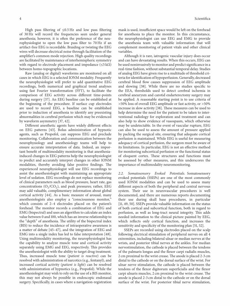
High pass filtering of ≤0.5Hz and low pass filteringof 30Hz will record the frequencies seen under generalanesthesia; however, it is often the preference of the neu-rophysiologist to open the low pass filter to 70Hz if anartifact-free EEG is recordable. Braiding or twisting the EEGwires will decrease electrical noise through facilitation of theamplifier’s common mode rejection. High quality recordingsare facilitated by maintenance of interhemispheric symmetrywith regard to electrode placement and impedance (≤5 kΩ)between homo-topographic locations.
Raw (analog or digital) waveforms are monitored on allcases in which EEG is a selected IONMmodality. Frequentlythe neurophysiologist will prefer to add quantitative EEGrecordings, both numerical and graphical trend analysesusing fast Fourier transformation (FFT), to facilitate thecomparison of EEG activity across different time-pointsduring surgery [37]. An EEG baseline can be established atthe beginning of the procedure. If surface cup electrodesare used to record EEG, a baseline can be establishedprior to induction of anesthesia to identify any preexistingabnormalities in cerebral perfusion which may be evidencedby waveform asymmetry [37, 42].
Different anesthetic agents have widely different effectson EEG patterns [43]. Bolus administration of hypnoticagents, such as Propofol, can suppress EEG and precludemonitoring. Collaboration and communication between theneurophysiology and anesthesiology teams will help toensure accurate interpretation of data. Indeed, an impor-tant benefit of multimodality monitoring is that anesthesia-induced changes in EEG patterns help the neurophysiologistto predict and accurately interpret changes in other IONMmodalities, thereby avoiding false positive findings. Theexperienced neurophysiologist will use EEG recordings toassist the anesthesiologist with maintaining an appropriatelevel of sedation. EEG recordings do not replace monitoringof clinical parameters such as blood pressure, heart rate, gasconcentrations (O
2/CO2), and peak pressures; rather, EEG
may add valuable, complimentary information about globalcortical activity [44]. As a basic measure of arousal, manyanesthesiologists also employ a “consciousness monitor,”which consists of 2–4 electrodes placed on the patient’sforehead. The monitor records a combination of EEG andEMG (bispectral) and uses an algorithm to calculate an indexvalue between 0 and 100, which has an inverse relationship tothe “depth” of anesthesia. The utility of the bispectral index(BIS) to reduce the incidence of intraoperative awareness isa matter of debate [45–47], and the integration of EEG andEMG into a single index has led to false interpretation [48].Using multimodality monitoring, the neurophysiologist hasthe capability to analyze muscle tone and cortical activityseparately using EMG and EEG, respectively. This providesthe anesthesiologist with the ability to target drug treatment.Thus, increased muscle tone (patient is reactive) can beresolved with administration of narcotics (e.g., fentanyl), andincreased cortical activity (patient is light) can be resolvedwith administration of hypnotics (e.g., Propofol). While theanesthesiologist may wish to rely on the use of a BIS monitor,this may not always be possible in endoscopic endonasalsurgery. Specifically, in cases where a navigation-registration
mask is used, insufficient space would be left on the foreheadfor anesthesia to place the monitor. In this circumstance,the neurophysiologist can use EEG and EMG to providethe anesthesia team with valuable information that willcomplement monitoring of patient vitals and other clinicalvariables.
Although it is rare, iatrogenic vascular injury does occurand can have devastating results. When this occurs, EEG canbe used noninvasively tomonitor and predict significance in areal-time fashion, without substantial temporal delay. Studiesof analog EEG have given rise to a multitude of threshold cri-teria for identification of hypoperfusion.Generally, decreasedcerebral blood flow causes suppression of EEG amplitudeand slowing [38]. While there are no studies specific tothe EEA, thresholds used to detect cerebral ischemia incerebral aneurysm and carotid endarterectomy surgery maybe applied. A reasonable starting point is to use criteria of>50% loss of overall EEG amplitude or fast activity, or >50%increase in slow activity [38]. These measures can be used tohelp determine the need for the patient to be taken to inter-ventional radiology for exploration and treatment and canalso help to show evidence of vasospasm, which otherwisemay be undetectable. In the event of vascular rupture, EEGcan also be used to assess the amount of pressure appliedby packing the surgical site, ensuring that adequate corticalperfusion is maintained. While EEG is useful for measuringadequacy of cortical perfusion, the surgeon must be aware ofits limitations. In particular, EEG is not an effective methodfor monitoring subcortical perfusion or the functional statusof eloquent cortex. These structures and functions mustbe assessed by other measures, and this underscores theimportance of multimodality neuromonitoring.
2.2. Somatosensory Evoked Potentials. Somatosensoryevoked potentials (SSEPs) are one of the most commonlyused IONM modalities and can be used to assess manydifferent aspects of both the peripheral and central nervoussystem. Their use in neurovascular procedures is welldocumented, and there are numerous reports documentingtheir use during skull base procedures, in particular[11, 49, 50]. SSEPs provide valuable information on the statusof both cortical and subcortical function, both in regard toperfusion, as well as long-tract neural integrity. This addsneeded information to the clinical picture painted by EEG,which reflects only cortical function, and increases thesensitivity and specificity of the monitoring plan [51, 52].
SSEPs are recorded using electrodes placed on the scalpfollowing electrical stimulation of peripheral nerves on all 4extremities, including bilateral ulnar or median nerves at thewrists, and posterior tibial nerves at the ankles. For mediannervestimulation, the cathode is placed between the tendonsof the palmaris longus and the flexor carpi radialis muscles,2 cm proximal to the wrist crease.The anode is placed 2-3 cmdistal to the cathode or on the dorsal surface of the wrist. Forulnar nerve stimulation, the cathode is placed between thetendons of the flexor digitorum superficialis and the flexorcarpi ulnaris muscles, 2 cm proximal to the wrist crease. Theanode is placed 2-3 cm distal to the cathode or on the dorsalsurface of the wrist. For posterior tibial nerve stimulation,

Scientifica 5
Fpz
Cz
Pz
Fz
Oz
C4
F4
C3
P4
O2
Fp2
F8
T4
T6
F7
T3
T5
F3
Fp1
P3
O1
A1 A2
CP3 CP4CPz
C1 C2
Figure 2: Electrode positions used for stimulating tceMEP, andrecording SSEP, BAEP, and VEP. All recording locations are basedon the International 10–20 System for Electrode Placement [28].F: frontal; C: central; CP: midway between central and parietal; O:occipital; A: auricular; z: midline; Cs2: cervical spine (not shown).
the cathode is placed over the posterior portion of the medialsurface of the ankle, 1-2 cm distal and posterior to the medialmalleolus. The anode is placed 2-3 cm distal to the cathode.Alternate stimulation sites or alternate peripheral nerves canbe used when comorbidities preclude recording from thesepreferred sites.
Peripheral nerve stimulation is commonly achieved withthe use of stick-on surface electrodes or subdermal needleelectrodes.The authors prefer the latter, particularly for long-duration procedures in which the adhesive from surfaceelectrodes may degrade and cause stimulation failure and/orstimulus shunting due to the development of a salt bridge.
Interleaving square wave pulses of 200–400𝜇sec durationare used at a frequency of 2–5Hz and using a supramax-imal stimulation intensity. This intensity is 20% above thethreshold for muscle twitch in the distal muscles innervatedby the stimulated nerve. While each patient requires slightlydifferent stimulation parameters to optimize SSEP data, theauthors recommend a pulse duration of 300 𝜇sec, a frequencyof 4.7Hz, and an intensity of 25–45mA for the ulnar/mediannerves or 35–50mA for the posterior tibial nerves.
SSEPs can be recorded using either subdermal needleelectrodes, or cup electrodes positioned on the surface ofthe scalp using locations modified from the International10–20 system for electrode placement (Figure 2). Followingulnar or median nerve stimulation, subcortical SSEPs arerecorded with a latency of 13msec (N13) over the 2nd cervicalvertebra (Cs2), and cortical SSEPs are recorded with a latencyof 20msec (N20) from the contralateral cerebral hemisphere(CP3 or CP4). All recording sites are referenced to Fpz. Fol-lowing posterior tibial nerve stimulation, subcortical SSEPsare recorded with a latency of 29msec (N29) over the 2ndcervical vertebra (Cs2), and cortical SSEPs are recorded with
a latency of 37msec (P37) from the cerebral vertex (CPz).The cortical SSEP may also be recorded from the ipsilateralcerebral hemisphere (CP3 or CP4) due to paradoxical later-alization. A transcortical montage (CP3-CP4, CP4-CP3) canoften be used to facilitate signal acquisition if initial corticalamplitudes are low.
Bandpass filters of 30–500Hz are used for subcorticalrecordings, while 30–300Hz is typically optimal for corticalrecordings. Peripheral recording sites, such as Erb’s point orpopliteal fossa, are used by some labs to assist with techni-cal troubleshooting, but the authors have not found thesemethods to be of sufficient benefit as to warrant inclusion inour monitoring plan. If the reader opts to employ peripheralrecordings, we recommend bandpass filters of 30–1500Hz.A recording epoch of at least 50msec is recommended forupper extremity SSEPs, and 100msec for lower extremitySSEPs. The SSEP is an averaged response, which can takedozens to hundreds of trials to fully resolve, ranging in timefrom 30 seconds to 2 minutes, depending on the amountof unresolved electrical noise in the environment. Contem-porary IONM systems usually permit full resolution of anSSEP waveform in under 30 seconds, which is a significantimprovement over decades past. Braiding or twisting theSSEP recording wires will decrease electrical noise throughfacilitation of the amplifier’s common mode rejection. Highquality recordings are facilitated by maintenance of inter-hemispheric symmetry with regard to electrode placementand impedance (≤5 kΩ) for all recording locations.
SSEP baselines should be recorded after induction, butprior to any significant patient positioning. This will helpto detect and correct pressure or traction on the brachialplexus or peripheral nervous system. An alarm criterion ofa 50% amplitude decrease and/or a 10% latency increaseare traditionally used, both for positioning issues and fortrue iatrogenic changes. While SSEPs provide informationregarding the functional status of eloquent cortex and patientpositioning, they still have several limitations. For example,these long-tract sensory pathways are not fully sensitive tosubcortical ischemia [53] and do not provide any informationspecific to the motor pathways. In cases where the ischemicpenumbra is small or slow to develop, or in cases whereonly the motor pathways are affected, SSEPs may remainunchanged from baseline parameters, despite evolving hemi-paresis [54]. These limitations can lead to false negativeneurophysiologic findings and may need to be supplementedby additional modalities, such as EEG and motor evokedpotentials.
2.3. Motor Evoked Potentials. Transcranial electrical motorevoked potentials (tceMEPs) have played a role in the oper-ating room since it was first demonstrated that the pulse-train stimulation technique could successfully evoke MEPsin the anesthetized patient [31, 55]. While routine use oftceMEP monitoring began in spinal surgery in the 1980s, itis now also commonly used in many supratentorial [56–61]and infratentorial [62–66] procedures, as well as proceduresin which peripheral nerves or spinal nerve roots are at risk[67–69]. Given the limitations of EEG and SSEPs mentionedabove, the addition of tceMEPs to the multimodality IONM

6 Scientifica
plan can paint a more comprehensive picture of nervoussystem functionwhenmonitoring cases using the EEA. Inclu-sion of tceMEPs monitoring is the only means of detectinginsult to the long-tract motor pathways. Although the utilityof tceMEPs in detecting functional motor change has beendemonstrated in a wide range of intracranial procedures [56–66], their efficacy using the EEA is scarce.
Motor evoked potentials are recorded from skeletal mus-culature following electrical stimulation of primary motorcortex via electrodes placed over C1-C2 or C3-C4 cranially(Figure 2). Corkscrew or subdermal needle electrodes arecommonly used. Anodal stimulation works best to activatethe corticospinal tract and elicit MEPs from upper and lowerextremity muscles contralateral to the stimulated cerebralhemisphere. A multipulsed train of square wave constantvoltage stimuli of 50 to 75 𝜇sec duration is used. The pulse-train stimulation method is required to overcome the effectsof anesthesia. The amount of voltage required to elicit MEPscan vary significantly between patients.The number of pulsescan range from 3 to 9 and the interstimulus interval (ISI)can range from 1 to 4msec (equal to a frequency of 1000–250Hz, resp.). All stimulation parameters, including voltage,are tailored to the individual patient to optimize data andminimize the possibility of false positive or false negativefindings.
Motor evoked potentials are recorded using bipolar sub-dermal needle electrodes placed in upper and lower extremitymuscles bilaterally. For example, the neurophysiologist mayelect to record from the extensor carpi radialis, first dorsalinterosseous, tibialis anterior, and abductor halluces muscles.Many alternate recording sites are available. Use of multi-ple recording sites ensures adequate coverage of long-tractcorticospinal function and has the added benefit of helpingto detect and correct positional changes. A recording epochof at least 100msec is required and bandpass filters of 10–3000Hz are recommended. The tceMEP is not an averagedresponse; thus, each test gives immediate feedback regardingcorticospinal tract function. Stimulation may cause slightpatient movement, often requiring a brief surgical pause fortesting.This can be done in the seconds duringwhich surgicalinstruments are exchanged. The surgeon must remain awarethat tceMEP testing is not performed continuously duringsurgery.
One concern regarding tceMEP monitoring duringintracranial procedures is the risk of false negative findingsdue to excessive stimulation. To mitigate this risk, the voltageand all stimulating parameters are kept purposefully lowto help limit electrical spread to the cortical layers of thestimulated cerebral hemisphere. Stimulation parameters thatare set too high will bypass the cerebral cortex and depolarizethe pyramidal tracts at subcortical levels, potentially leadingto false negative recordings in the event of eloquent motorcortex ischemia. While this is not of concern in spinalprocedures, it should always be considered in cranial cases.Including contralateral myotomes in the ipsilateral recordingtrace window helps to identify this limitation. For example,anodal stimulation of the right cerebral hemisphere willproduce muscle recordings on the left side of the body. Inthe recording trace window for the left side of the body,
one can include right side myotomes as well. Responsesgenerated from the left myotomes, with absence of responsesfrom the right myotomes, can help to demonstrate focalstimulation that is confined to the right cortical hemisphere.These techniques can help to predict and prevent those rareoccasions when other monitoring modalities, such as SSEPor EEG, do not display signal change in the face of evolvinghemiparesis.
When the EEA to the skull base poses risk to the brain-stem via the transclival approach, for example, the medullarypyramids are at risk and tceMEPs provide the added benefitofmonitoring long-tractmotor function.While corticospinaltractmonitoring has been increasingly utilized across variousneurosurgical procedures, stimulation of the corticobulbartract for monitoring cranial nerve motor evoked potentials(CrN MEPs) has not gained the same popularity. As previ-ously mentioned, lesions in the cavernous sinus or aroundthe clivus will frequently compress or surround the cranialnerves. Although the use of electrically triggered EMG (seeSection 2.4) is the gold-standard for cranial nerve IONM, itis often the case that large tumors must be partially debulkedprior to localization of neural elements.This initial debulkingof the tumor can result in iatrogenic injury, prior to thebaseline stimulation. CrN MEPs allow one to establish abaseline response prior to surgical manipulation, much inthe same way where upper and lower extremity baselinesare established. CrN MEP testing also permits assessmentto occur on a more frequent basis, as it is much faster thanpausing the surgery to stimulate with a handheld probe.
CrN MEPs are recorded from cranial-nerve-innervatedmuscles following transcranial electrical stimulation of pri-mary motor cortex. Dong and colleagues [70] first describedthe technique for eliciting CrN MEPs from muscles inner-vated by the facial nerve, and the utility of this technique hassince been investigated for various other cranial nerves [71–73]. In order to limit electrical stimulation to the most lateralaspects of the motor homunculus, a hemispheric stimulationmontage is used. The anode is placed contralateral to theoperative side (C3 or C4; Figure 2), and the cathode is pacedat the vertex (Cz).
Stimulus parameters are similar to those listed above forcorticospinal tract monitoring. A multipulsed train stimulusis required to overcome the effects of anesthesia. It isimperative to limit electrical stimulation to the poly-neuronalcorticobulbar tract and not allow electrical spread throughdeeper structures or around the periphery of the scalp andface. Any response that is recorded following a single-pulsestimulus is likely generated by activation of the peripheralpathways. Failure to limit spread can bypass the corticobulbarpathway and cause direct activation of cranial nerves, whichcan result in false negative findings. The idea that CrNMEP latency is a good predictor of whether or not theresponse is generated centrally or peripherally has recentlybeen challenged [74, 75].
CrN MEPs are recorded using the same electrodes thatare used for EMG recordings (see Section 2.4). Frequently,there is also a great deal of stimulus artifact present inthe responses, which can obscure CrN MEPs due to theirinherent short latency.The hemispheric stimulationmontage

Scientifica 7
produces reliable responses and helps to eliminate the stim-ulus artifact that is common to CrN MEPs. Additionally, wehave found that raising the high pass filter from 10 to 100Hzcan reduce stimulation artifact.
Interpretation of tceMEPs is complicated by their inher-ent trial-to-trial amplitude variability and the lack of con-sensus in the literature regarding criteria for alert. In supra-tentorial and brainstem surgery, major alert criteria for tce-MEPs include disappearance or consistent >50% amplitudereduction [57, 58, 65, 76–78]. While the same alert criteriaare commonly used for CrNMEP interpretation [70, 79–82],the situation is further clouded by the observation that intra-operative test results do not always correlate well with thepostoperative neurological outcome [10, 80, 83]. Due to thepotential for a high number of false positives and negatives,CrN MEPs have not become routine. Further investigationis required before predictive values can be established thatallow CrN MEPs to be used during skull base endoscopicprocedures. Thus, while tceMEPs are frequently utilized tomonitor eloquent cortical and long-tract motor functions,reliable monitoring of cranial nerve motor function requiresother methods, such as free-running and stimulus-triggeredelectromyography.
2.4. Electromyography. Electromyography (EMG) isrecorded in surgery to monitor somatic efferent nerveactivity and assess the functional integrity of individualnerves. First introduced in the 1960s as a means to assessfacial nerve function during exploratory parotid surgery[84, 85], EMG recording techniques were later adapted forintracranial [86], spinal [87], and peripheral nerve surgeries[88]. A large volume of literature devoted to EMG use duringintracranial surgery is devoted to facial nerve monitoring inthe cerebellopontine angle [89, 90], and there is a growingnumber of reports on the use of this technique with theEEA to the skull base [18, 19, 26]. With this approach, EMGrecordings are particularly important to identify cranialnerves and guide tumor resection when the pathologyinvolves the cavernous sinus or retroclival regions. Whileother techniques used in multimodality IONM providevaluable information about nervous system function, EMGis the only method that can (1) provide real-time feedbackabout neural activation throughout the course of surgery, (2)accurately detect and localize motor or mixed motor-sensorynerves embedded within tumor, and (3) reliably assess theintegrity of cranial nerve motor functions before, during andafter tumor resection.
The basic premise of EMG is that depolarization of amotor nerve produces a recordable electrical potential withinone or more muscle(s) innervated by that particular nerve.This activity is recorded using subdermal or intramuscularneedle electrodes, unless otherwise noted below. A bipolarmontage is frequently employed, consisting of two activerecording leads placed within the samemuscle. Alternatively,a referential montagemay be used in which a single electrodeis placed in the muscle of interest, and a reference electrodeis placed in a neutral location.
For each motor or mixed sensory/motor cranial nerve,the following muscles are selected: inferior rectus or superior
rectus (CN III); superior oblique (CN IV); masseter ortemporalis (CN V); lateral rectus (CN VI); orbicularis oculi,orbicularis oris andmentalis (CNVII); stylopharyngeus (CNIX); vocalis (CN X); upper trapezius (CN XI); and tongue(CN XII). Recording from extraocular muscles for monitor-ing CNs III, IV, andVI requires knowledge of ocular anatomyand associated vasculature. Careful electrode placement willensure the integrity of the sclera. Several recording methodshave been reviewed elsewhere [91]. We use a commerciallyavailable subdermal needle electrode which is prebent toapproximately 90∘. With the eye in the closed position, theneurophysiologist inserts the needle into the extraocularmuscle of interest and carefully advances the electrode alongthe bony ridge of the orbit until it is fully inserted. A smallpiece of Transpore� tape is used to secure each electrodeas the others are placed. One electrode is placed for eachof the extraocular muscle monitored, and each recordingis referenced to a needle inserted into the frontalis muscleof the forehead. Tegaderm� film is applied to protect theeyes during surgery. When the glossopharyngeal nerve ismonitored, prebent subdermal needle electrodes are placedin the soft palate with the aid of a curved hemostat (althoughthe utility of glossopharyngeal nerve monitoring has recentlybeen challenged) [92]. A commercially available endotrachealtube with surface-mounted electrode contact can be usedto monitor the vagus nerve. For all other cranial-nerve-innervated muscles, pairs of straight, subdermal needle elec-trodes are used for recording EMG. If a mask registrationdevice is used for neuronavigation, then all EMG electrodesmust be placed after registration is complete.
Bandpass filters of 10–3000Hz will capture all frequencycomponents of EMG. The low pass filter can be reduced toparry excessive high pass noise. A vertical screen resolutionof 50–200𝜇V/division is appropriate to visualize spontaneousand evoked EMG activity. Accurate interpretation of EMG isfacilitated by simultaneous visual and auditory monitoring,so a speaker is used in parallel to provide concurrent auditoryfeedback. Whereas incidental motor cranial nerve activity ismonitored with a free-running, spontaneous EMG recording(S-EMG), stimulus-triggered EMG (T-EMG) recordings aretime-locked to delivery of an electrical pulse and used tomapthe location of cranial motor nerves, as well as assessing theirfunctional status, as described below.
Free-running S-EMG is recorded throughout the courseof the surgery and it provides real-time feedback when-ever a nerve is activated. A recording time base of 200–1000msec/division is used. Several distinct firing patterns ofS-EMG activity may be recorded [29, 93], and the surgeonshould be able to identify them by sound [94]. While thesound of EMG activity may serve to heighten the surgeon’sawareness, not all patterns of EMG activity are cause forconcern and attempting to differentiate EMG patterns bysound alone is inadvisable. Also, in the electrically hostileenvironment of the operating room, one must be able todetect true neuronal activity and distinguish it from 60-cyclenoise and other forms of electrical interference, which shouldbe reduced or eliminated when possible. This underscoresthe importance of having an experienced neurophysiologistpresent to interpret the waveforms.
8 Scientifica
A
100msec
250𝜇V
(a)
C
100msec
250𝜇V
BB
BS
(b)
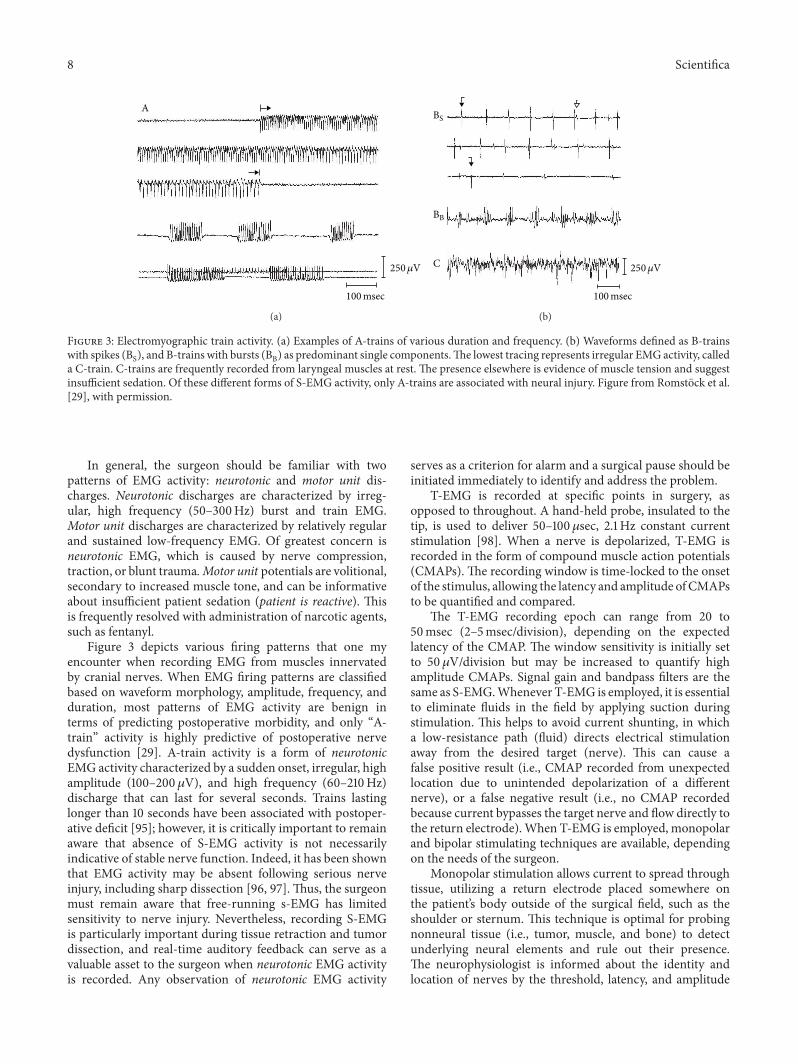
Figure 3: Electromyographic train activity. (a) Examples of A-trains of various duration and frequency. (b) Waveforms defined as B-trainswith spikes (BS), and B-trains with bursts (BB) as predominant single components.The lowest tracing represents irregular EMG activity, calleda C-train. C-trains are frequently recorded from laryngeal muscles at rest. The presence elsewhere is evidence of muscle tension and suggestinsufficient sedation. Of these different forms of S-EMG activity, only A-trains are associated with neural injury. Figure from Romstock et al.[29], with permission.
In general, the surgeon should be familiar with twopatterns of EMG activity: neurotonic and motor unit dis-charges. Neurotonic discharges are characterized by irreg-ular, high frequency (50–300Hz) burst and train EMG.Motor unit discharges are characterized by relatively regularand sustained low-frequency EMG. Of greatest concern isneurotonic EMG, which is caused by nerve compression,traction, or blunt trauma. Motor unit potentials are volitional,secondary to increased muscle tone, and can be informativeabout insufficient patient sedation (patient is reactive). Thisis frequently resolved with administration of narcotic agents,such as fentanyl.
Figure 3 depicts various firing patterns that one myencounter when recording EMG from muscles innervatedby cranial nerves. When EMG firing patterns are classifiedbased on waveform morphology, amplitude, frequency, andduration, most patterns of EMG activity are benign interms of predicting postoperative morbidity, and only “A-train” activity is highly predictive of postoperative nervedysfunction [29]. A-train activity is a form of neurotonicEMG activity characterized by a sudden onset, irregular, highamplitude (100–200𝜇V), and high frequency (60–210Hz)discharge that can last for several seconds. Trains lastinglonger than 10 seconds have been associated with postoper-ative deficit [95]; however, it is critically important to remainaware that absence of S-EMG activity is not necessarilyindicative of stable nerve function. Indeed, it has been shownthat EMG activity may be absent following serious nerveinjury, including sharp dissection [96, 97]. Thus, the surgeonmust remain aware that free-running s-EMG has limitedsensitivity to nerve injury. Nevertheless, recording S-EMGis particularly important during tissue retraction and tumordissection, and real-time auditory feedback can serve as avaluable asset to the surgeon when neurotonic EMG activityis recorded. Any observation of neurotonic EMG activity
serves as a criterion for alarm and a surgical pause should beinitiated immediately to identify and address the problem.
T-EMG is recorded at specific points in surgery, asopposed to throughout. A hand-held probe, insulated to thetip, is used to deliver 50–100𝜇sec, 2.1 Hz constant currentstimulation [98]. When a nerve is depolarized, T-EMG isrecorded in the form of compound muscle action potentials(CMAPs). The recording window is time-locked to the onsetof the stimulus, allowing the latency and amplitude ofCMAPsto be quantified and compared.
The T-EMG recording epoch can range from 20 to50msec (2–5msec/division), depending on the expectedlatency of the CMAP. The window sensitivity is initially setto 50 𝜇V/division but may be increased to quantify highamplitude CMAPs. Signal gain and bandpass filters are thesame as S-EMG.Whenever T-EMG is employed, it is essentialto eliminate fluids in the field by applying suction duringstimulation. This helps to avoid current shunting, in whicha low-resistance path (fluid) directs electrical stimulationaway from the desired target (nerve). This can cause afalse positive result (i.e., CMAP recorded from unexpectedlocation due to unintended depolarization of a differentnerve), or a false negative result (i.e., no CMAP recordedbecause current bypasses the target nerve and flow directly tothe return electrode).When T-EMG is employed, monopolarand bipolar stimulating techniques are available, dependingon the needs of the surgeon.
Monopolar stimulation allows current to spread throughtissue, utilizing a return electrode placed somewhere onthe patient’s body outside of the surgical field, such as theshoulder or sternum. This technique is optimal for probingnonneural tissue (i.e., tumor, muscle, and bone) to detectunderlying neural elements and rule out their presence.The neurophysiologist is informed about the identity andlocation of nerves by the threshold, latency, and amplitude

Scientifica 9
of the CMAP, as well as the muscle(s) from which it isrecorded. Ergo: the closer the neural element is to the locusof stimulation, the lower the threshold required to elicit aCMAP. Additionally, as the locus of stimulation approachesthe nerve, the CMAP is likely to exhibit a shorter latencyand higher amplitude, and with less spread of excitation tonearby nerves. As a general rule, when a CMAP is evokedwith 1.0mA or less, this is evidence of neural proximity andthe surgeon should dissect with caution. More distal cranialnerves may require higher levels of stimulation for depolar-ization, and the CMAP may exhibit a smaller amplitude andlonger latency. Whenever the expected result of stimulationis absence of a response, it is advisable to use a positivecontrol to demonstrate efficacy of stimulation. This can beaccomplished either by increasing the current until a CMAPis recorded, or by stimulating an exposedmotor/mixed nerveand recording the CMAP. While direct nerve stimulation isalways preferred prior to tumor resection, it is often the casethat tumors are of sufficient size and they must partially bedebulked prior to exposure of neural elements. Monopolarstimulation is advantageous in this situation because rulingout the presence of underlying motor/mixed cranial nervespermits rapid, safe extraction of nonneural tissue.
Bipolar stimulation limits the spread of current throughtissue, because the active and return electrodes are very closetogether, usually <3mm apart. This technique is preferredwhen the surgeon endeavors to identify a nerve or determinewhether or not a nerve is functional. Identification of a nerveis accomplished with direct electrical stimulation, and themuscle(s) from which the CMAP is recorded help to revealthe identity of the nerve. For example, if an unidentifiednerve is stimulated and CMAPs are recorded from the lateralrectus muscle, then the nerve in question is the abducensnerve (CN VI). Basic motor/mixed nerve functionality isassessed by establishing a CMAP threshold, which is definedas the minimum current (mA) required to evoke a CMAP.Beginning at 0.00mA, the current is carefully increased by0.01mA increments until a CMAP is recorded with minimalspread to other nerves/muscles. Nerves should be stimulatedfrequently during tumor debulking to assess changes inthreshold. At the end of the procedure, the nerve should bestimulated on each side of the tumor, proximal and distal tothe brainstem, with little expected variation in the CMAPthreshold. In the interest of prognostication, a number ofmethods have been reported for evaluating facial nervefunction [99–105], but it is unclear if these methods can begeneralized to address all motor or mixed sensory/motorcranial nerves.
2.5. Brainstem Auditory Evoked Potentials. The auditorybrainstem response (ABR), also known as the brainstemauditory evoked potential (BAEP), is recorded in surgery tomonitor vascular perfusion, as well as functional integrity, ofthe ascending auditory system, beginning with the vestibulo-cochlear nerve (CNVIII) and including associated brainstemtracts and nuclei up to the inferior colliculus [13, 53, 106–108]. Inclusion of BAEPs into the multimodality IONM planis recommended whenever there is potential for brainstemischemia in surgery. Inclusion of BAEPs in themultimodality
IONM plan is standard for monitoring brainstem perfusionduring open posterior fossa surgery [13]. As the EEA hasexpanded to reach pathology beyond the clivus and to theforamen magnum, inclusion of BAEPs is recommend tocompliment other monitoring modalities [12, 14].
BAEPs are recorded in response to auditory (click)stimulation delivered to the ears. The stimulus is deliveredthrough expanding foam earbuds placed in the externalauditory canal. The click consists of a 99 dB (nHL), 100 𝜇secpulse presented to each ear in interleaving fashion at afrequency range of 9.1–17.1 Hz. The polarity of the click maybe rarefaction, condensation, or both (alternating). BAEPscan be recorded using either subdermal needle electrodesor cup electrodes positioned on the surface of the scalpusing locationsmodified from the International 10–20 systemfor electrode placement (Figure 2). A referential recordingmontage is used in which active recording electrodes areplaced at A1 or A2, and recordings are referenced to Cz.
Bandpass filters of 100–1500Hz are common, and arecording epoch of at 15–20msec is recommended. Becausethe BAEP is a far-field response, it is small in amplitude(usually less than 1 𝜇V), but robust and repeatable whenhearing is intact. The BAEP is an averaged response, whichcan take hundreds to thousands of trials to fully resolve,ranging in time from 1 to 3 minutes, depending on theamount of unresolved electrical noise in the environment.Contemporary IONM systems usually permit full resolutionof BAEP waveform in under 1 minute, which is a significantimprovement over decades past. Braiding or twisting theBAEP recording wires will decrease electrical noise throughfacilitation of the amplifier’s common mode rejection. Highquality recordings are facilitated by maintenance of inter-hemispheric symmetry with regard to electrode impedance(≤5 kΩ) for all recording locations.
The BAEP consists of a waveform with approximately5 distinct peaks, labeled (I)–(V), which reflect neuronalactivity through the ascending auditory pathway. The neuralgenerators for the peaks are (I) distal auditory nerve, (II)proximal auditory nerve, (III) cochlear nucleus, (IV) superiorolivary complex, and (V) lateral lemniscus or inferior col-liculus [109].There are several longer latency (nonbrainstem-generated) peaks that represent higher thalamic and cor-tical auditory processing, but these peaks are suppressedby anesthetic agents [110]. The technical and pathologicalmechanisms that may underlie changes in the BAEP arenumerous [109].
BAEP baselines should be established before traversingthe clivus as the petroclival approach places the brainstemat risk for ischemia secondary to vascular compromise(arterial compression, rupture, or vasospasm). Alarm criteriaare defined as persistent decreases in amplitude of greaterthan 50% of wave (V) and/or persistent absolute latencyincrease of the peak of wave (V) which equals or exceeds0.5 milliseconds [12]. Knowledge of the neural generatorsfor each wave can reveal the location and extent of theinjury. For example, a major ischemic accident secondary tobasilar artery rupture may result in disappearance of waves(I)–(V) because perfusion of the cochlea via the internalauditory artery may be compromised. Ischemia of higher

10 Scientifica
brainstem structuresmay preserve waves (I)–(III) but abolishor delay wave (V). When these changes do not resolveduring the course of surgery, postoperative deficits are to beexpected [109]. Amore comprehensive reviewofmechanismsunderlying changes in the AEP is beyond the scope of thispaper.
2.6. Visual Evoked Potentials (VEP). Visual Evoked Potentials(VEPs) are recorded in surgery tomonitor the visual pathway,beginning with the prechiasmatic optic nerve and endingwith the striate cortex. Surgical approaches to the parasellarregion of the anterior skull base pose risk to the optic nerveand iatrogenic visual field deficits are a serious concern.Since the introduction of VEPs as an intraoperative moni-toring modality [111], their questionable prognostic value hasbeen widely published [112–115]. Nevertheless, the numerousreports of VEP monitoring during the EEA to the anteriorskull base warrant their inclusion in this paper [111–114, 116–126].
Goggles or contact lenses can deliver a flash stimu-lus using light-emitting diodes (LEDs) to the anesthetizedpatient. The flash stimulus is presented for 200–400msec,and a typical stimulation frequency can range from <1 Hz to3Hz. VEPs can be recorded using either subdermal needleelectrodes or cup electrodes positioned on the surface of thescalp using locations modified from the International 10–20 system for electrode placement (Figure 2). A referentialrecording montage is used in which active recording elec-trodes are placed at O1, O2, and Oz, with references eitherto A1/A2.
Bandpass filters of 1 to 300Hz are common and can benarrowed if stimulation artifact is encountered. A recordingepoch of at least 200msec is required.TheVEP is an averagedresponse, which can take dozens to hundreds of trials tofully resolve, ranging in time from seconds to minutes,depending on the amount of unresolved electrical noise inthe environment. Similar to other averaged evoked poten-tials, braiding or twisting the recording wires will decreaseelectrical noise, and high quality recordings are facilitated bymaintenance of interhemispheric symmetry with regard toelectrode impedance (≤5 kΩ) for all recording locations.
The VEP is usually recorded at its first negative deflectionwith a latency of approximately 70msec (N70), followed byits first positive deflection with a latency of approximately100msec (P100). Alert criteria include (1) total loss of wave-form, (2) loss of peak in the waveform, (3) latency increase >2standard deviations from baseline, or (4) amplitude decrease>50% from baseline [112].
Many lesions arising in the parasellar region result incompression/encasement of the optic nerve or chiasm, oftenresulting in preoperative clinical visual disturbances. It hasbeen demonstrated that decompression of these visual path-ways can actually improve postoperative visual neurologicaltesting [127]. Additionally, with the ability to detect evolvinginjury to the healthy, unimpaired optic tract is appealing.Unfortunately, there is little in the way of evidence thatintraoperative VEPs can accurately detect and help to preventiatrogenic injury to the visual pathways. In one study ofVEP monitoring during transnasal surgery of 22 patients,
intraoperative latency or amplitude change did not correlatewith immediate postoperative improvement or deterioration[117]. A larger, more recent study of VEP monitoring duringtransnasal surgery in 53 patients demonstrated no associationbetween VEP waveforms and postoperative visual outcomes[128]. Lack of prognostic value is common with VEPs insurgery. Indeed, the biggest limitations for the use of VEPs toprevent postoperative visual field deficits include variabilityof the response secondary to anesthetic regimen and stimulusdelivery [115]. Owing to the inconsistency of these recordings,which leads to both false positive and false negative findings,VEPs are not recommended during the EEA to the skullbase.
2.7. Anesthesia. Every anesthetic agent administered duringsurgery will affect neurophysiologic recordings to varyingdegrees; VEPs are particularly affected, whereas BAEPsshow little fluctuation despite the anesthetic regimen. Thesuccess of the monitoring plan relies on cooperation andcontinuous communication with the anesthesia team. Werecommend using total intravenous anesthesia (TIVA; noinhalational anesthetic agents) to facilitate IONM duringthis surgical approach. TIVA helps to reduce the dose-dependent attenuation of signal amplitudes that are usuallyseenwhen using inhalational agents, and helps to improve thesignal-to-noise ratio, thereby optimizing themonitoring plan[34, 129, 130].
As it relates to tceMEPs, the common agents used in TIVAproduce less inhibition at the pyramidal tract synapse on 𝛼-motor neurons of the spinal cord [131–137]. This allows themultipulse descending summation to overcome the effectsof anesthesia more readily, ultimately producing recordableCMAPs. The patient must also be sufficiently free of phar-macological blockade of the neuromuscular junction to allowboth tceMEPs and EMG to be sensitively recorded. Absenceof neuromuscular blockade or sufficient clearance/reversalfor reliable monitoring can be documented by using “train-of-four” (TOF) monitoring, which records muscle twitchesin response to stimulation of a peripheral nerve [138]. Usingsupramaximal stimulation at 2.0Hz, TOF can be recordedfrom the distal extremities to stimulation of the ulnar andposterior tibial nerves, and responses can be recorded fromthe first dorsal interosseous and abductor halluces muscles,respectively. Short-acting neuromuscular blocking agents canbe given to facilitate endotracheal intubation but are thendiscontinued for the remainder of the procedure. Currently,there is literature to support partial neuromuscular blockadeto help to limit patient movement during surgery whereIONM is occurring; however, partial blockade affects differ-ent muscle groups to varying degrees [139, 140] and can beespecially variable in a patient with preexisting neurologicaldysfunction [130, 138].
This regimen of anesthesia (TIVA and a full TOF)requires diligent teamwork between the neurophysiologistand the anesthesia team to ensure that there is no patientmovement and that analgesia is adequately controlled. Theneurophysiologist, having means to assess the patient’s levelof sedation and analgesia through the use of EEG and EMG,respectively, can add valuable information about the state of

Scientifica 11
anesthesia, helping to ensure that the patient does not moveor have recall during the surgery.
The most common complication resulting from MEPmonitoring is oral trauma secondary to oromandibular con-traction during motor tract activation [141, 142]. Oral traumacan take the form of hematoma or laceration of soft tissuewithin the oral cavity (e.g., tongue, lips, or gingiva), andfracture or avulsion of the teeth.The risk of oral trauma is alsoheightened during triggered EMG of the facial nerve [143]and presumably the trigeminal, glossopharyngeal, vagus, andhypoglossal nerves.Themost commonmethod formitigatingoral trauma is bilateral placement of soft bite blocks betweenthe mandibular and maxillary denta, or between the gingivain edentulous patients [143–145]. In doing so, the bite blocksoften need to be modified to be small enough so as to notobstruct the endoscopic entrance to the nares. Prolongedsurgical procedures may heighten the risk of tongue necrosis[146], but it is unclear whether or not proper placement of softbite blocks may contribute to this risk. It has been suggestedthat placement of dental guards, in addition to the soft biteblocks, may mitigate the risk of oral trauma [147]. Wheneverpossible, it is best practice to periodically verify the integrityof the oral cavity as well as confirming that the bite blockshave not become displaced.
3. Discussion
The traditional boundaries of the EEA continue to beexpanded with advances in instrumentation, optics, andmicrosurgical techniques. Such expansion requires an inti-mate knowledge of the anatomy encountered along theventral skull base and clivus. Evenwith a profound familiarityof the normal structures encountered, anatomical boundariesand relationships may be distorted by the pathology present,creating difficulty with the identification of landmarks. Thisis particularly true when faced with anomalous vasculature,which confounds the expected anatomy. A multimodalityIONM strategy is required to adequately assess the at riskstructures.
When the anterior skull base is accessed, there is con-siderable concern for the internal carotid arteries and theirbranches which supply blood to cortical and subcorticalstructures collectively representing three-fifths of the cere-brum. The most commonly used IONM modalities formonitoring cerebral blood flow are EEG and SSEPs. Whenone considers the vast array of monitored surgical proce-dures in which cerebral blood flow is a concern, includingcardiovascular, cardiopulmonary, cerebrovascular (e.g., CEA,aneurysm clipping, and vascular malformation), and proce-dures performed in the interventional radiology suite, onewould suspect that EEG is the most widely used vascularmonitoring modality. Despite major advantages over othermeasures such as transcranial Doppler and cerebral oximetry,and numerous studies inwhich EEGhas significantly loweredneurological deficits, shortened postoperative recovery, andreduced hospital costs, there has been little enthusiasm forEEG monitoring in cardiothoracic surgery [37, 148]. The useof EEG in CEA surgery is more widely reported [36, 37], andselective shunting with EEG is safer than routine shunting
[33, 149]. In intracranial aneurysm surgery, limited montageEEG monitoring can be used for induced hypotension andneuroprotective burst suppression [37, 150], but more exten-sive EEG monitoring is impractical, primarily due to thefact that the craniotomy precludes placement of electrodesover the regions at risk for ischemia [151]. This is not thecase during surgical procedures using the EEA. Given therisks to the internal carotid arteries and downstream vascularstructures in these procedures, the absence of reported EEGmonitoring in the literature is unusual, particularly in thecontext of its demonstrated utility for evaluating cerebralperfusion in cardiothoracic and CEA surgery.
The most likely explanation of the lack of reports specif-ically addressing EEG monitoring during EEA is that EEG israther comparable to SSEPs in terms of sensitivity and speci-ficity. Florence and colleagues performed a meta-analysis tocompare EEG with SSEPs in terms of their efficacy for moni-toring cerebral blood flow [152].They analyzed outcomes datafrom different vascular procedures, in which monitoring wasperformed with either EEG or SSEPs, and found that SSEPswere more sensitive than analog EEG (0.60 versus 0.20);however, their sensitivities were comparable if quantitativeEEG was used (0.58). SSEPs and EEG exhibited comparablespecificity (0.97 versus 0.95). Othermeta-analyses examiningEEG and SSEP monitoring during CEA surgery have yieldedsimilar findings [52, 153, 154]. One advantage of extended,multichannel EEG over SSEPs is the ability to localize corticalischemia beyond SSEP-relatedwatershed regions, giving EEGhigher spatial resolution. Also, given the potential for delayin detecting ischemia with SSEPs due to response averaging,EEGpresumably has better temporal specificity aswell.Whilethe use EEG in EEA has not been specifically reported, it hasbeen advocated for as part of a comprehensive multimodalityIONM plan [11, 19, 26].
A major limitation of EEG is that it is purely a measure ofcortical activity and, thus, cannot detect subcortical ischemia.For example, if ischemia were isolated to the internal capsuledue to reduced flow in the anterior choroidal or lenticulostri-ate arteries, then the patient may develop hemiparesis in theabsence of EEG changes. To compensate for this limitation,SSEP monitoring is introduced to the monitoring plan as adirect measure of somatosensory function. SSEP monitoringduring brain surgery has been rather extensively reported.Beyond their sensitivity to cerebral ischemia during a widerange of vascular procedures [51, 155–160], SSEP monitorthe integrity of the entire dorsal column-medial lemniscussystem, including thalamocortical projections [161]. Further-more, SSEPs are sensitive to detecting malpositioning of theneck [162, 163] and compression of the limbs which can resultin postoperative nerve damage [164–166].These observationsmake SSEP monitoring a useful adjunct to EEG monitoring.
The utility of SSEP monitoring during EEA surgery hasbeen investigated in multiple reports by Thirumala andcolleagues [11, 12]. In one retrospective review of 999 patientsundergoing skull base surgery, the incidence of changes inSSEPswas 20, and therewere 5 incidents of newpostoperativedeficit [11]. In this study there were 2 false negative outcomesof patients who had postoperative deficits (i.e., hemiparesis± aphasia) in the absence of intraoperative SSEP changes. In
12 Scientifica
a second study of 138 patients undergoing surgery via EEA,SSEP changes were detected in 5 patients, three of whichwere true positive findings [12]. In both of these studies,intraoperative changes in SSEPs were usually resolved afterraising the mean arterial blood pressure (MAP). The authorsconclude that SSEP monitoring is a useful adjunct to acomprehensive multimodality IONM plan.
The incidence of false negative findings with SSEP mon-itoring is not new. Over the years, multiple studies havereported postoperative symptoms ranging from paresis toplegia in the absence of intraoperative SSEP changes [167–171]. For this reason, it is becoming increasing common fortceMEPmonitoring to be used during intracranial surgery fordirect and indirect (vascular) monitoring of the corticospinaltract [56–66, 172]. Given that SSEPs only directly monitorsomatosensory tract function, it makes sense to includetceMEPs into the multimodality IONM plan in an effort todetect evolving motor deficits. When stimulation parametersare kept low to limit the spread of current, tceMEPs are usefulin supratentorial tumor resection [57, 58] and aneurysmclipping [56, 60, 61], as well as a multitude of infratento-rial/brainstem procedures [62–66]. There are no reports onthe use of tceMEPs for the EEA to the skull base. Whendeveloping a comprehensive and patient-specific IONM planin the context of cerebrovascular monitoring, we agree withThirumala and colleagues [11, 12] in that EEG and SSEPsshould be concurrently monitored in all EEA surgeries inwhich the lesion is in close proximity to critical vascularstructures, including the carotid and vertebrobasilar systems.The utility of tceMEPs in these procedures is yet to be con-clusively demonstrated; however, with numerous EEG/SSEPfalse negative reports in the literature, our approach is tomonitor tceMEPs on all suprasellar and petroclival lesions.
BAEPs should be added to themultimodality IONMplanwhen there is risk to the vertebrobasilar system [12, 14].With the EEA, BAEPs are primarily used to monitor vascularperfusion of the brainstem when the pathology includesthe retroclival structures. Intraoperative BAEPs have reliablyassessed the integrity and perfusion of both CN VIII and thebrainstem during posterior fossa skull base procedures sincethe late 1970s. Pathology that approximates the basilar arteryor the brainstem necessitates the inclusion of this modalityinto the monitoring plan, as it helps to complete the totalclinical picture in regard to potential subcortical ischemia.
The utility of VEPs for intraoperative monitoring ofthe optic tract is questionable [128]. As the technology forstimulation devices advances, VEPs may play a larger role inmonitoring. The TIVA anesthetic protocol helps stabilize thetrial-by-trial variability that is present in the VEP responses.Further research is needed to assess if VEPs can be reliablyused as an adjunct to monitor procedures where the visualtracts are affected. Presently, there is little evidence fromany intracranial procedure that VEPs are reliable predictorsof postoperative function. For that reason, they are notrecommended as part of a comprehensive IONMplan duringEEA to the skull base.
Given the close association withmany skull base patholo-gies to cranial nerves, the utility of cranial nerve monitoringduring EEAs is of significant interest. The methodology
for eliciting CrN MEPs has been established for recordingreliable MEPs from muscles innervated by the facial nerve(CN VII) [70]. Facial CrN MEPs have been successfullyemployed in a multitude of different surgical procedures,including skull base, brainstem, and posterior fossa [10, 70,79–81, 173]. Additionally, there is strong evidence in favor oftheir prognostic value in terms of predicting postoperativefacial nerve motor function [79, 81, 173, 174]. Unfortunately,attempts to record CrN MEPs from muscles innervated byother cranial nerves have been met with limited success. Themost common motor cranial nerves at risk during the EEAare the oculomotor, trochlear, and abducens nerves [20–22].
To our knowledge, CrN MEPs from the extraocularmuscles have not been successfully recorded, probably owingto the fact that the muscles are small and poorly innervated.Additionally, the short onset latency of the response wouldoften be obscured by stimulus artifact. Even if this techniquewas extrapolated to include these nerves, alarm criteria wouldneed to be established before routine use.
Regarding the trigeminal nerve, there are no publishedreports of CrNMEPs being recorded from either themasseteror temporalis muscle. We have successfully recorded MEPsfrom the masseter muscle, but their value remains unclear.First, the origin of the response could be accounted for byvolume conduction from the facial muscles. Second, we donot have enough experience with trigeminalMEPs to analyzetheir prognostic efficacy.
Greater successes have recently been reported for lowercranial nerve monitoring, methods for recording reliableMEPs from the vocal cords were introduced by Deletis etal. [71], and 1 patient with intraoperative unilateral laryn-geal nerve injury exhibited immediate reduction in MEPsrecorded from the ipsilateral vocalis muscle. Motoyama etal. recently reported their success in recording MEPs fromthe stylopharyngeus and vocalis muscles in two patientsundergoing microvascular decompression for glossopharyn-geal neuralgia [175]. Ito et al. successfully recorded MEPsfrom the vocalismuscles in 15 patients undergoing surgery forskull base or brainstem tumors, and intraoperative changescorrelated with postoperative dysphagia [176]. The ability torecord reliable MEPs from the upper trapezius muscles forspinal accessory monitoring and the tongue for hypoglos-sal nerve monitoring have both been reported in isolatedpatients, but their prognostic value remains unknown [73].
Free-running S-EMG provides real-time feedback ofcranial nerve irritation when operating near these sites.Recognition of nerve irritation during a procedure allows fora change in surgical strategy. Given the multitude of differentEMG patterns that one may encounter [29], the importanceof accurate interpretation cannot be overstated as falsepositives cause unnecessary concern and have the potentialto significantly delay the surgical procedure. Additionally,one should not assume that absence of EMG activity equateswith neural integrity [96, 97]. Despite the limited specificity,the utility of S-EMG for monitoring cranial nerve motorfunction been demonstrated in EEA surgery across the fullrange cranial nerves [14, 19, 26]. In these studies, S-EMGwas useful for identifying the location of a nerve, but was oflimited prognostic value.

Scientifica 13
Inferior rectus Lateral rectus
200𝜇V/div200𝜇V/div 2msec/div2msec/div
(a) (b)
Figure 4: Monopolar stimulation of the left oculomotor nerve at 0.50mA with subsequent compound muscle action potentials recordedfrom the left inferior rectus muscle using and intramuscular needle electrode (a). This response is referenced to the contralateral orbicularisoculi muscle. (b) The ipsilateral lateral rectus recording, which was not activated with this stimulation.
The best way to confirm the functional integrity of amotor or mixed sensory/motor cranial nerve is with T-EMG.Whenever possible, the nerve should be stimulated bothproximal and distal to the brainstem, on each side of thelesion. In some cases, nerve stimulation is useful to localizecranial nerves and avoid injury. Electrical stimulation ofthe nerves to the extraocular muscles is feasible and safe(Figure 4) [14, 17, 177–179]. For management of retroclivallesions, the glossopharyngeal nerve (CN IX), vagus (CNX), spinal accessory (CN XI) and hypoglossal (CN XII)nerves can be safely stimulated [10, 180]. Adverse conse-quences include hypotension and bradycardia (CNs IX andX), and muscle and tendon injuries (CN XI). There areno reported adverse consequences of stimulating CN XII.Maintaining low stimulation intensity with a short (25–100 𝜇sec) duration will help to limit stimulation-inducedinjury. Electrical stimulation of any motor cranial nerve hasthe added benefit of evaluating the functional integrity ofthe nerve, especially in the rare event of nerve transection,which does not necessarily result in spontaneous EMGactivity.
Which cranial nerves are monitored will depend on thenature of their involvement of the lesion and the risks associ-ated with the approach (Table 1) [18, 19, 26]. The reliabilityof T-EMG is largely dependent upon the integrity of theneural elements being tested. Thus, a patient presenting withpreoperative neurological deficits confounds the monitoring.Patients with cavernous sinus or suprasellar pathologieswill frequently experience preoperative extraocular palsy orvisual disturbance. Often, a nerve that is compromised willhave an altered threshold, decreased amplitude, increasedlatency, poor morphology, or no response at all. Markedpreoperative clinical neural compromise could create invalidresponses, increasing the rates of false positives and negative.In this context, CrN MEPs would be of significant benefit to
help establish a neural conduction baseline. Methodologicaladvancements are required to test this hypothesis.
The efficacy of IONM and the modalities chosen by theneurophysiologist to monitor during this approach need tobe examined closely, and both case series and case reports areneeded to expand the literature base.Themonitoring regimenthat we present here encompasses a strategy that assesses bothcortical and subcortical structures, as well perfusion, andcan help to increase the sensitivity and specificity of at riskneural structures during this technically demanding surgicalapproach.
4. Conclusion
IONM has gained widespread acceptance in cranial surgeryand has even become standard of care in some settings(e.g., facial nerve monitoring in surgery of the cerebello-pontine angle). In this paper, we have presented the modal-ities employed in varying combinations during endoscopicendonasal skull base surgery. Certainly, not all of thesetechniques are required for every endoscopic endonasalapproach. For example, IONM is unlikely to be beneficialduring the resection of standard pituitary adenomas withoutcavernous sinus invasion. For those lesions that do involve thecavernous sinus and its associated neurovascular structures,or for pathology requiring an EEA, an IONM regimentailored to the specific approach and the at risk anatomyis utilized (Table 1). In doing so, however, it is necessaryto recognize the rationale for each modality as well as itspotential limitations.
Disclosure
Richard W. Vogel and Adam T. Doan are employees ofSafe Passage Neuromonitoring, New York, NY. Craig I.

14 Scientifica
Matsumoto is an employee of SentientMedical Systems,HuntValley,M.D. Current address of RobertM. Lober,M.D., Ph.D.is Dayton Children’s Hospital, Department of Neurosurgery,Boonshoft School of Medicine, Dayton, OH, USA.
Competing Interests
The authors declare that they have no competing interests.
Authors’ Contributions
Harminder Singh and Richard W. Vogel made equal contri-butions to the paper.
Acknowledgments
The authors thank Cindy H. Samos for assistance withpreparation of the paper.
References
[1] O. Hirsch, “Endonasal method of removal of hypophysealtumors with a report of two successful cases,”The Journal of theAmerican Medical Association, vol. 55, no. 9, pp. 772–774, 1910.
[2] M. R. Rosen, K. Saigal, J. Evans, and W. M. Keane, “A review ofthe endoscopic approach to the pituitary through the sphenoidsinus,” Current Opinion in Otolaryngology and Head and NeckSurgery, vol. 14, no. 1, pp. 6–13, 2006.
[3] H. Schloffer, “Erfolgreiche operation eines hypophysentumorsauf nasalem wege,”Wiener Klinische Wochenschrift, vol. 20, pp.621–624, 1907.
[4] J. Hardy, “Surgery of the pituitary gland, using the trans-sphenoidal approach. Comparative study of 2 technical meth-ods,” L’union medicale du Canada, vol. 96, no. 6, pp. 702–712,1967.
[5] R. Jankowski, J. Auque, C. Simon, J. C. Marchal, H. Hepner,and M. Wayoff, “Endoscopic pituitary tumor surgery,” Laryn-goscope, vol. 102, no. 2, pp. 198–202, 1992.
[6] L. M. Cavallo, A. Messina, P. Gardner et al., “Extendedendoscopic endonasal approach to the pterygopalatine fossa:anatomical study and clinical considerations,” NeurosurgicalFocus, vol. 19, no. 1, article E5, 2005.
[7] A. B. Kassam, P. Gardner, C. Snyderman, A. Mintz, andR. Carrau, “Expanded endonasal approach: fully endoscopic,completely transnasal approach to themiddle third of the clivus,petrous bone, middle cranial fossa, and infratemporal fossa,”Neurosurgical Focus, vol. 19, no. 1, article E6, 2005.
[8] A. Kassam, C. H. Snyderman, A. Mintz, P. Gardner, and R.L. Carrau, “Expanded endonasal approach: the rostrocaudalaxis. Part II. Posterior clinoids to the foramen magnum,”Neurosurgical Focus, vol. 19, no. 1, article E4, 2005.
[9] A. Kassam, C. H. Snyderman, A. Mintz, P. Gardner, and R. L.Carrau, “Expanded endonasal approach: the rostrocaudal axis.Part I. Crista galli to the sella turcica,” Neurosurgical Focus, vol.19, no. 1, p. E3, 2005.
[10] F. Sala, P. Manganotti, V. Tramontano, A. Bricolo, and M.Gerosa, “Monitoring of motor pathways during brain stemsurgery: what we have achieved and what we still miss?”Neurophysiologie Clinique, vol. 37, no. 6, pp. 399–406, 2007.
[11] P. D.Thirumala, A. B. Kassasm, M. Habeych et al., “Somatosen-sory evoked potential monitoring during endoscopic endonasalapproach to skull base surgery: analysis of observed changes,”Neurosurgery, vol. 69, no. 1, supplement, pp. ons64–ons76, 2011.
[12] P. D. Thirumala, H. S. Kodavatiganti, M. Habeych et al., “Valueof multimodality monitoring using brainstem auditory evokedpotentials and somatosensory evoked potentials in endoscopicendonasal surgery,” Neurological Research, vol. 35, no. 6, pp.622–630, 2013.
[13] J. R. Little, R. P. Lesser, H. Lueders, and A. J. Furlan, “Brain stemauditory evoked potentials in posterior circulation surgery,”Neurosurgery, vol. 12, no. 5, pp. 496–502, 1983.
[14] C. Elangovan, S. P. Singh, P. Gardner et al., “Intraoperativeneurophysiological monitoring during endoscopic endonasalsurgery for pediatric skull base tumors,” Journal of Neuro-surgery: Pediatrics, vol. 17, no. 2, pp. 147–155, 2016.
[15] L. N. Sekhar and A. R. Moller, “Operative management oftumors involving the cavernous sinus,” Journal of Neurosurgery,vol. 64, no. 6, pp. 879–889, 1986.
[16] H.-P. Schlake, R. Goldbrunner, C. Milewski et al., “Technicaldevelopments in intra-operative monitoring for the preserva-tion of cranial motor nerves and hearing in skull base surgery,”Neurological Research, vol. 21, no. 1, pp. 11–24, 1999.
[17] H.-P. Schlake, R. Goldbrunner, M. Siebert, R. Behr, andK. Roosen, “Intra-operative electromyographic monitoring ofextra-ocular motor nerves (Nn. III, VI) in skull base surgery,”Acta Neurochirurgica, vol. 143, no. 3, pp. 251–261, 2001.
[18] D. San-Juan, J. Barges-Coll, J. L. Gomez Amador et al.,“Intraoperative monitoring of the abducens nerve in extendedendonasal endoscopic approach: a pilot study technical report,”Journal of Electromyography and Kinesiology, vol. 24, no. 4, pp.558–564, 2014.
[19] P. D. Thirumala, S. K. Mohanraj, M. Habeych et al., “Value offree-run electromyographic monitoring of extraocular cranialnerves during Expanded Endonasal Surgery (EES) of the skullbase,” Journal of Neurological Surgery Reports, vol. 74, no. 1, pp.43–50, 2013.
[20] G. Iaconetta, M. de Notaris, L. M. Cavallo et al., “Theoculomotor nerve: microanatomical and endoscopic study,”Neurosurgery, vol. 66, no. 3, pp. 593–601, 2010.
[21] B. Abuzayed, N. Tanriover, Z. Akar, B. S. Eraslan, and N.Gazioglu, “Extended endoscopic endonasal approach to thesuprasellar parachiasmatic cisterns: anatomic study,” Child’sNervous System, vol. 26, no. 9, pp. 1161–1170, 2010.
[22] G. Iaconetta, M. De Notaris, A. Benet et al., “The trochlearnerve: microanatomic and endoscopic study,” NeurosurgicalReview, vol. 36, no. 2, pp. 227–237, 2013.
[23] J. Barges-Coll, J. C. Fernandez-Miranda, D. M. Prevedello etal., “Avoiding injury to the abducens nerve during expandedendonasal endoscopic surgery: anatomic and clinical casestudies,” Neurosurgery, vol. 67, no. 1, pp. 144–154, 2010.
[24] A. B. Kassam, D. M. Prevedello, R. L. Carrau et al., “The frontdoor to Meckel’s cave: an anteromedial corridor via expandedendoscopic endonasal approach-technical considerations andclinical series,” Neurosurgery, vol. 64, no. 3, supplement, pp.ons71–ons82, 2009.
[25] V. A. Morera, J. C. Fernandez-Miranda, D. M. Prevedello etal., “‘Far-medial’ expanded endonasal approach to the inferiorthird of the clivus: the transcondylar and transjugular tubercle

Scientifica 15
approaches,” Neurosurgery, vol. 66, no. 6, supplement, pp. 211–220, 2010.
[26] P. D. Thirumala, S. K. Mohanraj, M. Habeych et al., “Value offree-run electromyographic monitoring of lower cranial nervesin endoscopic endonasal approach to skull base surgeries,”Journal of Neurological Surgery, Part B: Skull Base, vol. 73, no.4, pp. 236–244, 2012.
[27] J. P. Dormans, “Establishing a standard of care for neuromon-itoring during spinal deformity surgery,” Spine, vol. 35, no. 25,pp. 2180–2185, 2010.
[28] G. H. Klem, H. O. Luders, H. H. Jasper, and C. Elger, “The ten-twenty electrode system of the International Federation. TheInternational Federation of Clinical Neurophysiology,” Elec-troencephalography and Clinical Neurophysiology. Supplement,vol. 52, pp. 3–6, 1999.
[29] J. Romstock, C. Strauss, and R. Fahlbusch, “Continuous elec-tromyography monitoring of motor cranial nerves duringcerebellopontine angle surgery,” Journal ofNeurosurgery, vol. 93,no. 4, pp. 586–593, 2000.
[30] C. L. Nash Jr., R. A. Lorig, L. A. Schatzinger, and R. H. Brown,“Spinal cord monitoring during operative treatment of thespine,” Clinical Orthopaedics and Related Research, vol. 126, pp.100–105, 1977.
[31] T. Tamaki and S. Kubota, “History of the development ofintraoperative spinal cordmonitoring,” European Spine Journal,vol. 16, supplement 2, pp. 140–146, 2007.
[32] F. W. Sharbrough, J. M. Messick, and T. M. Sundt, “Correlationof continuous electroencephalograms with cerebral blood flowmeasurements during carotid endarterectomy,” Stroke, vol. 4,no. 4, pp. 674–683, 1973.
[33] E. Ballotta, M. Saladini, M. Gruppo, F. Mazzalai, G. Da Giau,and C. Baracchini, “Predictors of electroencephalographicchanges needing shunting during carotid endarterectomy,”Annals of Vascular Surgery, vol. 24, no. 8, pp. 1045–1052, 2010.
[34] S. Deiner, “Highlights of anesthetic considerations for intra-operative neuromonitoring,” Seminars in Cardiothoracic andVascular Anesthesia, vol. 14, no. 1, pp. 51–53, 2010.
[35] J. R. Schneider and K. E. Novak, “Carotid endarterectomywith routine electroencephalography and selective shunting,”Seminars in Vascular Surgery, vol. 17, no. 3, pp. 230–235, 2004.
[36] M. V. Simon, K. H. Chiappa, R. D. Kilbride et al., “Predic-tors of clamp-induced electroencephalographic changes duringcarotid endarterectomies,” Journal of Clinical Neurophysiology,vol. 29, no. 5, pp. 462–467, 2012.
[37] M. R. Isley, H. L. Edmonds Jr., and M. Stecker, “Guidelines forintraoperative neuromonitoring using raw (analog or digitalwaveforms) and quantitative electroencephalography: a posi-tion statement by the American society of neurophysiologicalmonitoring,” Journal of Clinical Monitoring and Computing, vol.23, no. 6, pp. 369–390, 2009.
[38] M. R. Nuwer, “Intraoperative electroencephalography,” Journalof Clinical Neurophysiology, vol. 10, no. 4, pp. 437–444, 1993.
[39] K. M. McGrail, “Intraoperative use of electroencephalographyas an assessment of cerebral blood flow,”Neurosurgery Clinics ofNorth America, vol. 7, no. 4, pp. 685–692, 1996.
[40] American Electroencephalographic Society, “Guideline seven:a proposal for standard montages to be used in clinical EEG,”Journal of Clinical Neurophysiology, vol. 11, no. 1, pp. 30–36, 1994.
[41] R. M. Craft, T. J. Losasso, W. J. Perkins, M. R. Weglinski, and F.W. Sharbrough, “EEG monitoring for cerebral ischemia duringcarotid endarterectomy (CEA): how much is enough?” Journalof Neurosurgical Anesthesiology, vol. 6, no. 4, p. 301, 1994.
[42] L. A. Kearse Jr., M. Lopez-Bresnahan, K. McPeck, and A.Zaslavsky, “Preoperative cerebrovascular symptoms and elec-troencephalographic abnormalities do not predict cerebralischemia during carotid endarterectomy,” Stroke, vol. 26, no. 7,pp. 1210–1214, 1995.
[43] V. Jantti and T. B. Sloan, “EEG and anesthetic effects,” inIntraoperative Monitoring of Neural Function, M. R. Nuwer,Ed., vol. 8 of Handbook of Clinical Neurophysiology, pp. 77–93,Elsevier, Amsterdam, The Netherlands, 2008.
[44] G. Schneider, D. Jordan, G. Schwarz et al., “Monitoring depth ofanesthesia utilizing a combination of electroencephalographicand standard measures,” Anesthesiology, vol. 120, no. 4, pp. 819–828, 2014.
[45] P. S. Myles, K. Leslie, J. McNeil, A. Forbes, and M. T. V. Chan,“Bispectral index monitoring to prevent awareness duringanaesthesia: the B-Aware randomised controlled trial,” TheLancet, vol. 363, no. 9423, pp. 1757–1763, 2004.
[46] M. S. Avidan, L. Zhang, B. A. Burnside et al., “Anesthesiaawareness and the bispectral index,” The New England Journalof Medicine, vol. 358, no. 11, pp. 1097–1108, 2008.
[47] M. S. Avidan, E. Jacobsohn, D. Glick et al., “Prevention ofintraoperative awareness in a high-risk surgical population,”The New England Journal of Medicine, vol. 365, no. 7, pp. 591–600, 2011.
[48] I. J. Rampil, “Monitoring depth of anesthesia,” Current Opinionin Anaesthesiology, vol. 14, no. 6, pp. 649–653, 2001.
[49] G. K. Bejjani, P. C. Nora, P. L. Vera, L. Broemling, and L. N.Sekhar, “The predictive value of intraoperative somatosensoryevoked potential monitoring: review of 244 procedures,” Neu-rosurgery, vol. 43, no. 3, pp. 491–500, 1998.
[50] F. Gentili, W. M. Lougheed, K. Yamashiro, and C. Corrado,“Monitoring of sensory evoked potentials during surgery ofskull base tumours,” Canadian Journal of Neurological Sciences,vol. 12, no. 4, pp. 336–340, 1985.
[51] A. M. Lam, P. H. Manninen, G. G. Ferguson, and W. Nan-tau, “Monitoring electrophysiologic function during carotidendarterectomy: a comparison of somatosensory evoked poten-tials and conventional electroencephalogram,” Anesthesiology,vol. 75, no. 1, pp. 15–21, 1991.
[52] K. Thiagarajan, H. L. Cheng, J. E. Huang et al., “Is two reallybetter than one? examining the superiority of dual modalityneurophysiological monitoring during carotid endarterectomy:a meta-analysis,” World Neurosurgery, vol. 84, no. 6, pp. 1941–1949.e1, 2015.
[53] N. M. Branston, A. Ladds, L. Symon, and A. D. Wang, “Com-parison of the effects of ischaemia on early components of thesomatosensory evoked potential in brainstem, thalamus, andcerebral cortex,” Journal of Cerebral Blood Flow & Metabolism,vol. 4, no. 1, pp. 68–81, 1984.
[54] P. De Vleeschauwer, S. Horsch, and R.Matamoros, “Monitoringof somatosensory evoked potentials in carotid surgery: results,usefulness and limitations of the method,” Annals of VascularSurgery, vol. 2, no. 1, pp. 63–68, 1988.
[55] P. A. Merton and H. B. Morton, “Stimulation of the cerebralcortex in the intact human subject,” Nature, vol. 285, no. 5762,p. 227, 1980.

16 Scientifica
[56] K. Horiuchi, K. Suzuki, T. Sasaki et al., “Intraoperative mon-itoring of blood flow insufficiency during surgery of middlecerebral artery aneurysms,” Journal of Neurosurgery, vol. 103, no.2, pp. 275–283, 2005.
[57] G. Neuloh, U. Pechstein, and J. Schramm, “Motor tract moni-toring during insular glioma surgery,” Journal of Neurosurgery,vol. 106, no. 4, pp. 582–592, 2007.
[58] G. Neuloh, U. Pechstein, C. Cedzich et al., “Motor evokedpotential monitoring with supratentorial surgery,” Neuro-surgery, vol. 54, no. 5, pp. 1061–1072, 2004.
[59] Y. Maruta, M. Fujii, H. Imoto et al., “Strategies and pitfallsof motor-evoked potential monitoring during supratentorialaneurysm surgery,” Journal of Stroke and Cerebrovascular Dis-eases, vol. 25, no. 2, pp. 484–495, 2016.
[60] R. Holdefer, D. MacDonald, L. Guo, and S. Skinner, “An eval-uation of motor evoked potential surrogate endpoints duringintracranial vascular procedures,” Clinical Neurophysiology, vol.127, no. 2, pp. 1717–1725, 2016.
[61] G. Neuloh and J. Schramm, “Monitoring of motor evokedpotentials comparedwith somatosensory evoked potentials andmicrovascular Doppler ultrasonography in cerebral aneurysmsurgery,” Journal of Neurosurgery, vol. 100, no. 3, pp. 389–399,2004.
[62] J. Sarnthein, O. Bozinov, A. G. Melone, and H. Bertalanffy,“Motor-evoked potentials (MEP) during brainstem surgery topreserve corticospinal function,” Acta Neurochirurgica, vol. 153,no. 9, pp. 1753–1759, 2011.
[63] D.-Z. Kang, Z.-Y. Wu, Q. Lan et al., “Combined monitoringof evoked potentials during microsurgery for lesions adjacentto the brainstem and intracranial aneurysms,” Chinese MedicalJournal, vol. 120, no. 18, pp. 1567–1573, 2007.
[64] K. Kodama, M. Javadi, V. Seifert, and A. Szelenyi, “ConjunctSEP and MEP monitoring in resection of infratentorial lesions:lessons learned in a cohort of 210 patients,” Journal of Neuro-surgery, vol. 121, no. 6, pp. 1453–1461, 2014.
[65] G. Neuloh, J. Bogucki, and J. Schramm, “Intraoperative preser-vation of corticospinal function in the brainstem,” Journal ofNeurology, Neurosurgery and Psychiatry, vol. 80, no. 4, pp. 417–422, 2009.
[66] P. J. Slotty, A. Abdulazim, K. Kodama et al., “Intraoperative neu-rophysiological monitoring during resection of infratentoriallesions: the surgeon’s view,” Journal of Neurosurgery, 2016.
[67] D. Fan, D. M. Schwartz, A. R. Vaccaro, A. S. Hilibrand,and T. J. Albert, “Intraoperative neurophysiologic detection ofiatrogenic C5 nerve root injury during laminectomy for cervicalcompression myelopathy,” Spine, vol. 27, no. 22, pp. 2499–2502,2002.
[68] B. Bose, A. K. Sestokas, and D. M. Schwartz, “Neurophysiolog-ical detection of iatrogenic C-5 nerve deficit during anteriorcervical spinal surgery,” Journal of Neurosurgery: Spine, vol. 6,no. 5, pp. 381–385, 2007.
[69] H. I. Berends, H. L. Journee, I. Racz, J. van Loon, R. Hartl,andM. Spruit, “Multimodality intraoperative neuromonitoringin extreme lateral interbody fusion. Transcranial electricalstimulation as indispensable rearview,” European Spine Journal,vol. 25, no. 5, pp. 1581–1586, 2016.
[70] C. C. J. Dong, D. B. MacDonald, R. Akagami et al., “Intraopera-tive facial motor evoked potential monitoring with transcranial
electrical stimulation during skull base surgery,”Clinical Neuro-physiology, vol. 116, no. 3, pp. 588–596, 2005.
[71] V.Deletis, I. Fernandez-Conejero, S.Ulkatan, andP.Costantino,“Methodology for intraoperatively eliciting motor evokedpotentials in the vocal muscles by electrical stimulation of thecorticobulbar tract,”Clinical Neurophysiology, vol. 120, no. 2, pp.336–341, 2009.
[72] I. Fernandez-Conejero, S. Ulkatan, C. Sen, and V. Deletis,“Intra-operative neurophysiology during microvasculardecompression for hemifacial spasm,”Clinical Neurophysiology,vol. 123, no. 1, pp. 78–83, 2012.
[73] S. A. Skinner, “Neurophysiologic monitoring of the spinalaccessory nerve, hypoglossal nerve, and the spinomedullaryregion,” Journal of Clinical Neurophysiology, vol. 28, no. 6, pp.587–598, 2011.
[74] M. J. Tellez, S. Ulkatan, J. Urriza, B. Arranz-Arranz, and V.Deletis, “Neurophysiological mechanism of possibly confound-ing peripheral activation of the facial nerve during corticobul-bar tract monitoring,” Clinical Neurophysiology, vol. 127, no. 2,pp. 1710–1716, 2016.
[75] S. Skinner and R. Holdefer, “IONM evidence: putting the workand insight of Tellez et al. in context,” Clinical Neurophysiology,vol. 127, no. 2, pp. 1015–1017, 2016.
[76] A. Szelenyi, D. Langer, K. Kothbauer, A. B. De Camargo, E. S.Flamm, and V. Deletis, “Monitoring of muscle motor evokedpotentials during cerebral aneurysm surgery: intraoperativechanges and postoperative outcome,” Journal of Neurosurgery,vol. 105, no. 5, pp. 675–681, 2006.
[77] A. Szelenyi, E. Hattingen, S. Weidauer, V. Seifert, and U.Ziemann, “Intraoperative motor evoked potential alteration inintracranial tumor surgery and its relation to signal alteration inpostoperative magnetic resonance imaging,” Neurosurgery, vol.67, no. 2, pp. 302–313, 2010.
[78] A. Szelenyi, C. Senft, M. Jardan et al., “Intra-operative sub-cortical electrical stimulation: a comparison of two methods,”Clinical Neurophysiology, vol. 122, no. 7, pp. 1470–1475, 2011.
[79] R. Akagami, C. C. J. Dong, and B. D. Westerberg, “Localizedtranscranial electrical motor evoked potentials for monitoringcranial nerves in cranial base surgery,”Neurosurgery, vol. 57, no.1, pp. 78–85, 2005.
[80] M. Fukuda,M.Oishi, T.Hiraishi, A. Saito, andY. Fujii, “Intraop-erative facial nerve motor evoked potential monitoring duringskull base surgery predicts long-term facial nerve functionoutcomes,” Neurological Research, vol. 33, no. 6, pp. 578–582,2011.
[81] M. Fukuda, M. Oishi, T. Takao, A. Saito, and Y. Fujii, “Facialnerve motor-evoked potential monitoring during skull basesurgery predicts facial nerve outcome,” Journal of Neurology,Neurosurgery and Psychiatry, vol. 79, no. 9, pp. 1066–1070, 2008.
[82] C. Matthies, F. Raslan, T. Schweitzer, R. Hagen, K. Roosen, andK. Reiners, “Facial motor evoked potentials in cerebellopontineangle surgery: technique, pitfalls and predictive value,” ClinicalNeurology and Neurosurgery, vol. 113, no. 10, pp. 872–879, 2011.
[83] N. Morota, S. Ihara, and V. Deletis, “Intraoperative neuro-physiology for surgery in and around the brainstem: roleof brainstem mapping and corticobulbar tract motor-evokedpotential monitoring,” Child’s Nervous System, vol. 26, no. 4, pp.513–521, 2010.

Scientifica 17
[84] R. C. Parsons, “Electrical stimulation of the facial nerve,”Laryngoscope, vol. 76, no. 3, pp. 391–406, 1966.
[85] R. C. Parsons, “Electrical nerve stimulation at surgery,” TheLaryngoscope, vol. 78, no. 5, pp. 742–748, 1968.
[86] T. E. Delgado, W. A. Buchheit, H. R. Rosenholtz, and S.Chrissian, “Intraoperative monitoring of facial muscle evokedresponses obtained by intracranial stimulation of the facialnerve: a more accurate technique for facial nerve dissection,”Neurosurgery, vol. 4, no. 5, pp. 418–421, 1979.
[87] J. T. Hormes and J. L. Chappuis, “Monitoring of lumbosacralnerve roots during spinal instrumentation,” Spine, vol. 18, no.14, pp. 2059–2062, 1993.
[88] D. G. Kline, E. R. Hackett, and P. R. May, “Evaluation of nerveinjuries by evoked potentials and electromyography,” Journal ofNeurosurgery, vol. 31, no. 2, pp. 128–136, 1969.
[89] C. D. Yingling and J. N. Gardi, “Intraoperative monitoring offacial and cochlear nerves during acoustic neuroma surgery,”Otolaryngologic Clinics of North America, vol. 25, no. 2, pp. 413–448, 1992.
[90] R. E. Minahan and A. S. Mandir, “Neurophysiologic intraop-erative monitoring of trigeminal and facial nerves,” Journal ofClinical Neurophysiology, vol. 28, no. 6, pp. 551–565, 2011.
[91] J. R. Lopez, “Neurophysiologic intraoperativemonitoring of theoculomotor, trochlear, and abducens nerves,” Journal of ClinicalNeurophysiology, vol. 28, no. 6, pp. 543–550, 2011.
[92] J. M. Kartush, “Commentary: endotracheal tube electrodesto assess vocal cord motor function during surgery in thecerebellopontine angle,” Neurosurgery, vol. 77, no. 5, pp. E838–E841, 2015.
[93] G. S. Nichols and E. Manafov, “Utility of electromyography fornerve rootmonitoring during spinal surgery,” Journal of ClinicalNeurophysiology, vol. 29, no. 2, pp. 140–148, 2012.
[94] R. L. Prass and H. Luders, “Acoustic (loudspeaker) facial elec-tromyographic monitoring: part 1. Evoked electromyographicactivity during acoustic neuroma resection,” Neurosurgery, vol.19, no. 3, pp. 392–400, 1986.
[95] J. Prell, S. Rampp, J. Romstock, R. Fahlbusch, and C. Strauss,“Train time as a quantitative electromyographic parameterfor facial nerve function in patients undergoing surgery forvestibular schwannoma,” Journal of Neurosurgery, vol. 106, no.5, pp. 826–832, 2007.
[96] K. R. Nelson and H. C. Vasconez, “Nerve transection withoutneurotonic discharges during intraoperative electromyographicmonitoring,”Muscle and Nerve, vol. 18, no. 2, pp. 236–238, 1995.
[97] J. M. Kartush, “Electroneurography and intraoperative facialmonitoring in contemporary neurotology,” Otolaryngology—Head and Neck Surgery, vol. 101, no. 4, pp. 496–503, 1989.
[98] R. Prass and H. Luders, “Constant-current versus constant-voltage stimulation,” Journal of Neurosurgery, vol. 62, no. 4, pp.622–623, 1985.
[99] A. K. Lalwani, F. Y.-S. Butt, R. K. Jackler, L. H. Pitts, andC. D. Yingling, “Facial nerve outcome after acoustic neuromasurgery: a study from the era of cranial nerve monitoring,”Otolaryngology—Head and Neck Surgery, vol. 111, no. 5, pp. 561–570, 1994.
[100] A. K. Lalwani, F. Y. Butt, R. K. Jackler, L. H. Pitts, and C.D. Yingling, “Delayed onset facial nerve dysfunction followingacoustic neuroma surgery,”American Journal of Otolaryngology,vol. 16, no. 6, pp. 758–764, 1995.
[101] J. M. Taha, J. M. Tew Jr., and R. W. Keith, “Proximal-to-distalfacial amplitude ratios as predictors of facial nerve functionafter acoustic neuroma excision,” Journal of Neurosurgery, vol.83, no. 6, pp. 994–998, 1995.
[102] G. Magliulo, C. Sepe, S. Varacalli, and J. Crupi, “Acousticneuroma surgery and delayed facial palsy,” European Archivesof Oto-Rhino-Laryngology, vol. 255, no. 3, pp. 124–126, 1998.
[103] B. Isaacson, P. R. Kileny, H. El-Kashlan, and A. K. Gadre,“Intraoperative monitoring and facial nerve outcomes aftervestibular schwannoma resection,” Otology and Neurotology,vol. 24, no. 5, pp. 812–817, 2003.
[104] A. B. Grayeli, M. Kalamarides, B. Fraysse et al., “Comparisonbetween intraoperative observations and electromyographicmonitoring data for facial nerve outcome after vestibularschwannoma surgery,” Acta Oto-Laryngologica, vol. 125, no. 10,pp. 1069–1074, 2005.
[105] A. B. Grayeli, S. Guindi, M. Kalamarides et al., “Four-channelelectromyography of the facial nerve in vestibular schwannomasurgery: sensitivity and prognostic value for short-term facialfunction outcome,” Otology and Neurotology, vol. 26, no. 1, pp.114–120, 2005.
[106] M. V. Simon, “Neurophysiologic intraoperative monitoring ofthe vestibulocochlear nerve,” Journal of Clinical Neurophysiol-ogy, vol. 28, no. 6, pp. 566–581, 2011.
[107] B. J. Stern, A. Krumholz, H. D. Weiss, P. Goldstein, and K.C. Harris, “Evaluation of brainstem stroke using brainstemauditory evoked response,” Stroke, vol. 13, no. 5, pp. 705–711,1982.
[108] H. Luders, “Surgical monitoring with auditory evoked poten-tials,” Journal of Clinical Neurophysiology, vol. 5, no. 3, pp. 261–285, 1988.
[109] A. D. Legatt, “Mechanisms of intraoperative brainstem auditoryevoked potential changes,” Journal of Clinical Neurophysiology,vol. 19, no. 5, pp. 396–408, 2002.
[110] A. D. Legatt, “BAEPs in surgery,” in Intraoperative Monitoringof Neural Function, M. R. Nuwer, Ed., vol. 8 of Handbook ofClinical Neurophysiology, pp. 334–349, Elsevier, Amsterdam,The Netherlands, 2008.
[111] A. L. Albright and R. J. Sclabassi, “Cavitron ultrasonic surgicalaspirator and visual evoked potential monitoring for chiasmalgliomas in children. Report of two cases,” Journal of Neuro-surgery, vol. 63, no. 1, pp. 138–140, 1985.
[112] C. Cedzich, J. Schramm, and R. Fahlbusch, “Are flash-evokedvisual potentials useful for intraoperative monitoring of visualpathway function?” Neurosurgery, vol. 21, no. 5, pp. 709–715,1987.
[113] M. Lorenz and R. R. Renella, “Intraoperative monitoring: visualevoked potentials in surgery of the sellar region,” Zentralblattfur Neurochirurgie, vol. 50, no. 1, pp. 12–15, 1989.
[114] K. Kodama, T. Goto, A. Sato, K. Sakai, Y. Tanaka, and K. Hongo,“Standard and limitation of intraoperative monitoring of thevisual evoked potential,” Acta Neurochirurgica, vol. 152, no. 4,pp. 643–648, 2010.
[115] P. D. Thirumala, M. E. Habeych, D. J. Crammond, and J. R.Balzer, “Neurophysiologic intraoperative monitoring of olfac-tory and optic nerves,” Journal of Clinical Neurophysiology, vol.28, no. 6, pp. 538–542, 2011.
[116] C. Cedzich, J. Schramm, C. F. Mengedoht, and R. Fahlbusch,“Factors that limit the use of flash visual evoked potentials

18 Scientifica
for surgical monitoring,” Electroencephalography and ClinicalNeurophysiology/Evoked Potentials Section, vol. 71, no. 2, pp.142–145, 1988.
[117] A. G. Chacko, K. S. Babu, and M. J. Chandy, “Value of visualevoked potential monitoring during trans-sphenoidal pituitarysurgery,” British Journal of Neurosurgery, vol. 10, no. 3, pp. 275–278, 1996.
[118] E. A. Bonaroti, R. D. Rose, D. Kondziolka, S. Baser, and L.D. Lunsford, “Flash visual evoked potential monitoring ofoptic tract function duringmacroelectrode-based pallidotomy,”Neurosurgical Focus, vol. 2, no. 3, p. e4, 1997.
[119] C. Cedzich and J. Schramm, “Monitoring of flash visual evokedpotentials during neurosurgical operations,” International Anes-thesiology Clinics, vol. 28, no. 3, pp. 165–169, 1990.
[120] P. Christophis, “Visual evoked potentials as an indicator oftranstentorial herniation,” Zentralblatt fur Neurochirurgie, vol.59, no. 4, pp. 256–262, 1998.
[121] G. F. A. Harding, J. D. P. Bland, and V. H. Smith, “Visual evokedpotential monitoring of optic nerve function during surgery,”Journal of Neurology, Neurosurgery and Psychiatry, vol. 53, no.10, pp. 890–895, 1990.
[122] G. F. A. Harding, V. H. Smith, and H. C. Yorke, “A contact lensphotostimulator for surgical monitoring,” Electroencephalogra-phy and Clinical Neurophysiology, vol. 66, no. 3, pp. 322–326,1987.
[123] G. D. Herzon and D. L. Zealear, “Intraoperative monitoring ofthe visual evoked potential during endoscopic sinus surgery,”Otolaryngology—Head andNeck Surgery, vol. 111, no. 5, pp. 575–579, 1994.
[124] S. S. M. Hussain, H. C. Laljee, J. M. Horrocks, H. Tec, andA. R. Grace, “Monitoring of intra-operative visual evokedpotentials during functional endoscopic sinus surgery (FESS)under general anaesthesia,” Journal of Laryngology and Otology,vol. 110, no. 1, pp. 31–36, 1996.
[125] V. E. Oliushin, G. S. Tigliev, L. G. Vaınshteın, V. M. Merkin,and M. M. Sultanov, “Monitoring control of the visual evokedpotentials during the surgical treatment of tumors of thechiasmal-sellar area,” Zhurnal Voprosy Neırokhirurgii Imeni N.N. Burdenko, vol. 3, pp. 22–24, 1990.
[126] T. Sasaki, T. Itakura, K. Suzuki et al., “Intraoperativemonitoringof visual evoked potential: introduction of a clinically usefulmethod: clinical article,” Journal of Neurosurgery, vol. 112, no.2, pp. 273–284, 2010.
[127] H. Z. Gokalp, N. Egemen, A. Culcuoglu, S. Naderi, and A.Zorlutuna, “The use of Nd:YAG laser in pituitary surgery andevaluation of visual function by visual evoked potential (VEP),”Neurosurgical Review, vol. 15, no. 3, pp. 193–197, 1992.
[128] S.-B. Chung, C.-W. Park, D.-W. Seo, D.-S. Kong, and S.-K. Park,“Intraoperative visual evoked potential has no association withpostoperative visual outcomes in transsphenoidal surgery,”ActaNeurochirurgica, vol. 154, no. 8, pp. 1505–1510, 2012.
[129] S. DiCindio and D. M. Schwartz, “Anesthetic management forpediatric spinal fusion: implications of advances in spinal cordmonitoring,” Anesthesiology Clinics of North America, vol. 23,no. 4, pp. 765–787, 2005.
[130] T. B. Sloan and E. J. Heyer, “Anesthesia for intraoperativeneurophysiologic monitoring of the spinal cord,” Journal ofClinical Neurophysiology, vol. 19, no. 5, pp. 430–443, 2002.
[131] T. Kerz, H.-J. Hennes, A. Feve, P. Decq, P. Filipetti, and P.Duvaldestin, “Effects of propofol on H-reflex in humans,”Anesthesiology, vol. 94, no. 1, pp. 32–37, 2001.
[132] B. Rehberg, M. Grunewald, J. Baars, K. Fuegener, B. W.Urban, and W. J. Kox, “Monitoring of immobility to noxiousstimulation during sevoflurane anesthesia using the spinal H-reflex,” Anesthesiology, vol. 100, no. 1, pp. 44–50, 2004.
[133] T. Kammer, B. Rehberg, D. Menne, H.-C. Wartenberg, I.Wenningmann, and B. W. Urban, “Propofol and sevofluranein subanesthetic concentrations act preferentially on the spinalcord: evidence from multimodal electrophysiological assess-ment,” Anesthesiology, vol. 97, no. 6, pp. 1416–1425, 2002.
[134] J. Zentner, T. Albrecht, D. Heuser, J. Oro, and J. R. Daube,“Influence of halothane, enflurane, and isoflurane on motorevoked potentials,” Neurosurgery, vol. 31, no. 2, pp. 298–305,1992.
[135] J. Zentner, C. Thees, U. Pechstein, K.-M. Scheufler, J. Wurker,and J. Nadstawek, “Influence of nitrous oxide on motor-evokedpotentials,” Spine, vol. 22, no. 9, pp. 1002–1006, 1997.
[136] H. H. Zhou and C. Zhu, “Comparison of isoflurane effects onmotor evoked potential and F wave,” Anesthesiology, vol. 93, no.1, pp. 32–38, 2000.
[137] H. H. Zhou, T.-T. Jin, B. Qin, and H. Turndorf, “Suppressionof spinal cord motoneuron excitability correlates with surgicalimmobility during isoflurane anesthesia,” Anesthesiology, vol.88, no. 4, pp. 955–961, 1998.
[138] T. B. Sloan, “Muscle relaxant use during intraoperative neu-rophysiologic monitoring,” Journal of Clinical Monitoring andComputing, vol. 27, no. 1, pp. 35–46, 2013.
[139] Y. Saitoh, H. Tanaka, H. Toyooka, and K. Amaha, “Recoveryof post-tetanic and train-of-four responses at the first dorsalinterosseous and adductor pollicis muscles in patients receivingvecuronium,”Canadian Journal of Anaesthesia, vol. 43, no. 4, pp.362–367, 1996.
[140] B. Gavrancic, A. Lolis, and A. Beric, “Train-of-four test in intra-operative neurophysiologic monitoring: differences betweenhand and foot train-of-four,” Journal of Clinical Neurophysiol-ogy, vol. 31, no. 6, pp. 575–579, 2014.
[141] D. B. MacDonald, “Safety of intraoperative transcranial electri-cal stimulation motor evoked potential monitoring,” Journal ofClinical Neurophysiology, vol. 19, no. 5, pp. 416–429, 2002.
[142] D. M. Schwartz, A. K. Sestokas, J. P. Dormans et al., “Transcra-nial electric motor evoked potential monitoring during spinesurgery: is it safe?” Spine, vol. 36, no. 13, pp. 1046–1049, 2011.
[143] A. Chaturvedi and P. Khanna, “Prevention of airway injuryduring spine surgery: rethinking bite blocks,” Journal of Neu-rosurgical Anesthesiology, vol. 21, no. 4, article 356, 2009.
[144] S. G. Deiner and I. P. Osborn, “Prevention of airway injuryduring spine surgery: rethinking bite blocks,” Journal of Neu-rosurgical Anesthesiology, vol. 21, no. 1, pp. 68–69, 2009.
[145] T.N. Pajewski, V. Arlet, and L.H. Phillips, “Current approach onspinal cordmonitoring: the point of view of the neurologist, theanesthesiologist and the spine surgeon,”European Spine Journal,vol. 16, supplement 2, pp. S115–S129, 2007.
[146] K. Sriram, A. Khorasani, K. E. Mbekeani, and S. Patel, “Tonguenecrosis and cleft after prolonged transesophageal echocardio-graphy probe placement,” Anesthesiology, vol. 105, no. 3, article635, 2006.
Scientifica 19
[147] M. Mahmoud, J. Spaeth, and S. Sadhasivam, “Protection oftongue from injuries during transcranial motor-evoked poten-tial monitoring,” Paediatric Anaesthesia, vol. 18, no. 9, pp. 902–903, 2008.
[148] H. L. Edmonds, “Central nervous system monitoring,” inKaplan’s Cardiac Anesthesia: The Echo Era, J. A. Kaplan, D. L.Reich, and J. S. Savino, Eds., pp. 466–495, Elsevier Saunders,Philadelphia, Pa, USA, 2011.
[149] A. J. Salvian, D. C. Taylor, Y. N. Hsiang et al., “Selective shuntingwith EEG monitoring is safer than routine shunting for carotidendarterectomy,” Cardiovascular Surgery, vol. 5, no. 5, pp. 481–485, 1997.
[150] T. H. Jones, K. H. Chiappa, R. R. Young, and R. G. Ojemann,“EEG monitoring for induced hypotension for surgery ofintracranial aneurysms,” Stroke, vol. 10, no. 3, pp. 292–294, 1979.
[151] J. R. Lopez, “Intraoperative neurophysiological monitoring,”International Anesthesiology Clinics, vol. 34, no. 4, pp. 33–54,1996.
[152] G. Florence, J.-M. Guerit, and B. Gueguen, “Electroencephalog-raphy (EEG) and somatosensory evoked potentials (SEP) toprevent cerebral ischaemia in the operating room,”Neurophysi-ologie Clinique, vol. 34, no. 1, pp. 17–32, 2004.
[153] E. L. Nwachuku, J. R. Balzer, J. G. Yabes, M. E. Habeych,D. J. Crammond, and P. D. Thirumala, “Diagnostic valueof somatosensory evoked potential changes during carotidendarterectomy: a systematic review and meta-analysis,” JAMANeurology, vol. 72, no. 1, pp. 73–80, 2015.
[154] P.D.Thirumala, K.Thiagarajan, S. Gedela, D. J. Crammond, andJ. R. Balzer, “Diagnostic accuracy of EEGchanges during carotidendarterectomy in predicting perioperative strokes,” Journal ofClinical Neuroscience, vol. 25, pp. 1–9, 2016.
[155] J. R. Lopez, S. D. Chang, and G. K. Steinberg, “The use of elec-trophysiological monitoring in the intraoperative managementof intracranial aneurysms,” Journal of Neurology Neurosurgeryand Psychiatry, vol. 66, no. 2, pp. 189–196, 1999.
[156] G. Penchet, P. Arne, E. Cuny, P. Monteil, H. Loiseau, and J.-P. Castel, “Use of intraoperative monitoring of somatosensoryevoked potentials to prevent ischaemic stroke after surgicalexclusion of middle cerebral artery aneurysms,” Acta Neu-rochirurgica, vol. 149, no. 4, pp. 357–364, 2007.
[157] R. W. McPherson, E. F. Niedermeyer, R. J. Otenasek, and D.F. Hanley, “Correlation of transient neurological deficit andsomatosensory evoked potentials after intracranial aneurysmsurgery. Case report,” Journal of Neurosurgery, vol. 59, no. 1, pp.146–149, 1983.
[158] J.-M. Guerit, C. Witdoeckt, M. de Tourtchaninoff et al.,“Somatosensory evoked potential monitoring in carotidsurgery. I. Relationships between qualitative SEP alterationsand intraoperative events,” Electroencephalography and ClinicalNeurophysiology/Evoked Potentials Section, vol. 104, no. 6, pp.459–469, 1997.
[159] C. Witdoeckt, S. Ghariani, and J.-M. Guerit, “Somatosensoryevoked potential monitoring in carotid surgery. II. Compar-ison between qualitative and quantitative scoring systems,”Electroencephalography and Clinical Neurophysiology/EvokedPotentials Section, vol. 104, no. 4, pp. 328–332, 1997.
[160] T. B. Sloan, H. L. Edmonds Jr., and A. Koht, “Intraoperativeelectrophysiologic monitoring in aortic surgery,” Journal ofCardiothoracic and Vascular Anesthesia, vol. 27, no. 6, pp. 1364–1373, 2013.
[161] E.-K. Lee and M. Seyal, “Generators of short latency humansomatosensory-evoked potentials recorded over the spine andscalp,” Journal of Clinical Neurophysiology, vol. 15, no. 3, pp. 227–234, 1998.
[162] R.W.McPherson, J. Szymanski, andM. C. Rogers, “Somatosen-sory evoked potential changes in position-related brain stemischemia,” Anesthesiology, vol. 61, no. 1, pp. 88–90, 1984.
[163] D. M. Schwartz, A. K. Sestokas, A. S. Hilibrand et al., “Neu-rophysiological identification of position-induced neurologicinjury during anterior cervical spine surgery,” Journal of ClinicalMonitoring and Computing, vol. 20, no. 6, pp. 437–444, 2006.
[164] I. Chung, J. A. Glow, V. Dimopoulos et al., “Upper-limbsomatosensory evoked potential monitoring in lumbosacralspine surgery: a prognostic marker for position-related ulnarnerve injury,” Spine Journal, vol. 9, no. 4, pp. 287–295, 2009.
[165] I. R. Kamel, E. T. Drum, S. A. Koch et al., “The use ofsomatosensory evoked potentials to determine the relationshipbetween patient positioning and impending upper extremitynerve injury during spine surgery: a retrospective analysis,”Anesthesia and Analgesia, vol. 102, no. 5, pp. 1538–1542, 2006.
[166] D. M. Schwartz, D. S. Drummond, M. Hahn, M. L. Ecker, and J.P. Dormans, “Prevention of positional brachial plexopathy dur-ing surgical correction of scoliosis,” Journal of Spinal Disorders,vol. 13, no. 2, pp. 178–182, 2000.
[167] H. H. Ginsburg, A. G. Shetter, and P. A. Raudzens, “Postoper-ative paraplegia with preserved intraoperative somatosensoryevoked potentials. Case report,” Journal of Neurosurgery, vol. 63,no. 2, pp. 296–300, 1985.
[168] R. G. Emerson, “Anatomic and physiological bases of posteriornerve somatosensory evoked potentials,”Neurologic Clinics, vol.6, no. 4, pp. 735–749, 1988.
[169] D. Krieger, H. P. Adams, F. Albert,M. vonHaken, andW.Hacke,“Pure motor hemiparesis with stable somatosensory evokedpotential monitoring during aneurysm surgery: case report,”Neurosurgery, vol. 31, no. 1, pp. 145–150, 1992.
[170] A. S. Hilibrand, D. M. Schwartz, V. Sethuraman, A. R. Vaccaro,and T. J. Albert, “Comparison of transcranial electric motor andsomatosensory evoked potential monitoring during cervicalspine surgery,” Journal of Bone and Joint Surgery—Series A, vol.86, no. 6, pp. 1248–1253, 2004.
[171] P. Thirumala, D. Lai, J. Engh, M. Habeych, D. Crammond, andJ. Balzer, “Predictive value of somatosensory evoked potentialmonitoring during resection of intraparenchymal and intraven-tricular tumors using an endoscopic port,” Journal of ClinicalNeurology, vol. 9, no. 4, pp. 244–251, 2013.
[172] A. Quinones-Hinojosa, M. Alam, R. Lyon et al., “Transcra-nial motor evoked potentials during basilar artery aneurysmsurgery: technique application for 30 consecutive patients,”Neurosurgery, vol. 54, no. 4, pp. 916–924, 2004.
[173] M. A. Acioly, M. Liebsch, C. H. Carvalho, A. Gharabaghi,and M. Tatagiba, “Transcranial electrocortical stimulation tomonitor the facial nerve motor function during cerebellopon-tine angle surgery,” Neurosurgery, vol. 66, no. 6, SupplementOperative, pp. 354–362, 2010.
[174] M. A. Acioly, M. Liebsch, P. H. P. de Aguiar, and M. Tatagiba,“Facial nerve monitoring during cerebellopontine angle andskull base tumor surgery: a systematic review from descriptionto current success on function prediction,”World Neurosurgery,vol. 80, no. 6, pp. e271–e300, 2013.

20 Scientifica
[175] Y. Motoyama, I. Nakagawa, T. Takatani et al., “Microvasculardecompression for glossopharyngeal neuralgia using intraop-erative neurophysiological monitoring: Technical case report,”Surgical Neurology International, vol. 7, supplement 2, pp. S28–S35, 2016.
[176] E. Ito, M. Ichikawa, T. Itakura et al., “Motor evoked potentialmonitoring of the vagus nerve with transcranial electrical stim-ulation during skull base surgeries,” Journal of Neurosurgery,vol. 118, no. 1, pp. 195–201, 2013.
[177] O. Alberti, U. Sure, T. Riegel, andH. Bertalanffy, “Image-guidedplacement of eye muscle electrodes for intraoperative cranialnerve monitoring,” Neurosurgery, vol. 49, no. 3, pp. 660–664,2001.
[178] M. Kawaguchi, H. Ohnishi, T. Sakamoto et al., “Intraoperativeelectrophysiologic monitoring of cranial motor nerves in skullbase surgery,” Surgical Neurology, vol. 43, no. 2, pp. 177–181, 1995.
[179] T. Sekiya, T. Hatayama, T. Iwabuchi, S. Maeda, L. N. Sekhar, andJ. R. Daube, “Intraoperative recordings of evoked extraocularmuscle activities to monitor ocular motor nerve function,”Neurosurgery, vol. 32, no. 2, pp. 227–235, 1993.
[180] H.-P. Schlake, R. H. Goldbrunner, C. Milewski et al., “Intra-operative electromyographic monitoring of the lower cranialmotor nerves (LCN IX-XII) in skull base surgery,” ClinicalNeurology and Neurosurgery, vol. 103, no. 2, pp. 72–82, 2001.

Submit your manuscripts athttp://www.hindawi.com
Neurology Research International
Hindawi Publishing Corporationhttp://www.hindawi.com Volume 2014
Alzheimer’s DiseaseHindawi Publishing Corporationhttp://www.hindawi.com Volume 2014
International Journal of
ScientificaHindawi Publishing Corporationhttp://www.hindawi.com Volume 2014
Hindawi Publishing Corporationhttp://www.hindawi.com Volume 2014
BioMed Research International
Hindawi Publishing Corporationhttp://www.hindawi.com Volume 2014
Research and TreatmentSchizophrenia
The Scientific World JournalHindawi Publishing Corporation http://www.hindawi.com Volume 2014
Hindawi Publishing Corporationhttp://www.hindawi.com Volume 2014
Neural Plasticity
Hindawi Publishing Corporationhttp://www.hindawi.com Volume 2014
Parkinson’s Disease
Hindawi Publishing Corporationhttp://www.hindawi.com Volume 2014
Research and TreatmentAutism
Sleep DisordersHindawi Publishing Corporationhttp://www.hindawi.com Volume 2014
Hindawi Publishing Corporationhttp://www.hindawi.com Volume 2014
Neuroscience Journal
Epilepsy Research and TreatmentHindawi Publishing Corporationhttp://www.hindawi.com Volume 2014
Hindawi Publishing Corporationhttp://www.hindawi.com Volume 2014
Psychiatry Journal
Hindawi Publishing Corporationhttp://www.hindawi.com Volume 2014
Computational and Mathematical Methods in Medicine
Depression Research and TreatmentHindawi Publishing Corporationhttp://www.hindawi.com Volume 2014
Hindawi Publishing Corporationhttp://www.hindawi.com Volume 2014
Brain ScienceInternational Journal of
StrokeResearch and TreatmentHindawi Publishing Corporationhttp://www.hindawi.com Volume 2014
Neurodegenerative Diseases
Hindawi Publishing Corporationhttp://www.hindawi.com Volume 2014
Journal of
Cardiovascular Psychiatry and NeurologyHindawi Publishing Corporationhttp://www.hindawi.com Volume 2014