intramedullaryfatnecrosisofmultiplebones ...jnm.snmjournals.org/content/39/8/1401.full.pdf ·...
TRANSCRIPT

Enzinger FM, Lattes R, Torloni H, eds. International classification of tumours, vol. 3.Geneva: World Health Organization; 1969.
10. Armin A, Connelly EM. Rowden G. An immunoperoxidase investigation of S-100protein in granular cell myoblastomas: evidence for Schwann cell derivation. Am JClin Palimi 1983:79:37-44.
11. Avril N, Dose J, JänickeF, et al. Metabolie characterization of breast tumors withpositron emission tomography using F-18 fluorodeoxyglucose. J Clin Oncol 1996; 14:1848-1857.
12. Gordon AB, Fisher C, Palmer B. Greening W. Granular cell tumour of the breast. EarJ Surg Oncol 1985;! 1:269-273.
13. Crawford ES. DeBakey ME. Granular cell myoblastoma. Two unusual cases. Cancer1953:6:786-789.
14. LelléRJ. Park H, Brow CA. Benign granular cell tumour mimicking carcinoma of thebreast. Eur J Gynaec Oncol 1992;13:390-393.
15. Uzoaro I, Firfer B. Ray V. Hubbard-Shepard M. Rhee H. Malignant granular celltumor. Arch Palhol Lab Med 1992:116:206-208.
16. Rickard MT, Sendel A, Burche«I. Case report: granular cell tumour of the breast. ClinRadici 1992:45:347-348.
17. Kittner T, Dziambor U. Bergander S, Theissig F. Der Granularzelltumor der Mamma—die seltene Differentialdiagnose des Mammakarzinoms. Röntgenpraxis 1995;48:185-186.
18. Heywang-Köbrunner SH, Beck R. Contrast enhanced MRI of the hreast, 2nd ed.
Berlin: Springer; 1995.19. Adler LP, Crowe JP, Al-Kaisi NK. Sunshine JL. Evaluation of breast masses and
axillary lymph nodes with (F-18) 2-deoxy-2-fluoro-D-glucose PET. Radiology 1993;187:743-750.
20. Wahl RL, Cody RL. Hutchins GD, Mudgett EE. Primary and metastatic breastcarcinoma. Initial clinical evaluation with PET with the radiolabeled glucose analogue2-(F18)-fluoro-2-deoxy-D-glucose. Radiology 1991:179:765-770.
21. Hahn HJ. Iglesias J. Flenker H. Kreuzer G. Granular cell tumor in differential diagnosisof tumors of the breast. The role of fine needle aspiration cytology. Path Res Pract1992:188:1091-1094.
22. Schneider V. Granular cell tumor in differential diagnosis of tumors of the breast. Therole of fine needle aspiration cytology: letters to the case. Path Res Pract 1992; 188:1095-1097.
Intramedullary Fat Necrosis of Multiple BonesAssociated with PancreatitisByeong C. Ahn, Jaetae Lee, Kyung J. Suh, Kyung A. Chun, Sang K. Sohn, Kyubo Lee and Chun K. KimDepartments of Nuclear Medicine and Diagnostic Radiologi,', Kyungpook National University School of Medicine, Taegu,
Korea; and Division of Nuclear Medicine, Mount Sinai Medical Center, New York, New York
We describe findings of intramedullary fat necrosis on five imagingstudies in a patient with alcoholic pancreatitis. Radiography and CTof extremities showed multiple osteolytic lesions that were initiallyconsidered to be métastases.However, a 99mTc-methylene diphos-phonate whole-body bone scan revealed abnormal areas of increased uptake in only the bones of extremities without involvementof the axial skeleton, a distribution quite unusual for metastaticdisease. Furthermore, 99mTc-sestamibi scintigraphy was essentially
normal. MRI revealed findings compatible with the diagnosis of fatnecrosis/infarct. Findings from bone biopsy demonstrated necroticbone marrow without malignant cells. It may not be necessary toperform all the imaging studies described in this report when clinicalfeatures suggesting metastatic fat necrosis are present. Appearanceand distribution of abnormalities on the whole-body bone scan andMR images show that necrosis/infarct of the marrow may obviatebone biopsy, which is often needed to confirm the diagnosis ofintramedullary fat necrosis and to exclude neoplastic processes.
Key Words: pancreatitis;fat necrosis;radiography;scintigraphyJ NucÃMed 1998; 39:1401-1404
Xancreatic disorders can be complicated by fat necrosis atmultiple distant sites, resulting in subcutaneous nodular lesions,polyarthritis and intramedullary fat necrosis (7 ). Although boneinvolvement of pancreatic disease had been believed to occurrarely, a necropsy study showed a relatively higher prevalenceof bone lesion in postmortem samples with acute pancreatitis(2). The appearance of intramedullary fat necrosis on mostimaging studies can be nonspecific, especially when an individual study is interpreted alone. We describe various radio-
logic and scintigraphic findings that led to the correct diagnosisin a patient with clinical features suggesting intramedullary fatnecrosis.
CASE REPORTA 69-yr-old man with pulmonary emphysema was admitted for
pain in the extremities that had worsened over several weeks. Hehad been experiencing upper abdominal discomfort concurrentwith the appearance of extremity pain that partly subsided afterfasting. His medical history included smoking for 35 yr andalcoholism with multiple previous episodes of alcoholic pancreatitis. Three years earlier, the patient was found to have mesenteric fatnecrosis associated with pancreatitis during emergency exploratorylaparotomy that was performed because of suspicion of acuteintestinal infarction.
On admission, physical examination of the patient showed anemaciated body habitus and a soft, nontender, subcutaneous mass
Received Aug. 8, 1997; revision accepted Nov. 6, 1997.For correspondence or reprints contact: Jaetae Lee, MD, Department of Nuclear
Medicine, Kyungpook National University Hospital, Samduk 2 Ga-50, Taegu 700-412,
Korea. FIGURE 1. A subcutaneous nodule is visible in the left deltoid area.
INTRAMEDULLARYFAT NECROSISASSOCIATEDWITHPANCREATITIS•Ahn et al. 1401
by on August 29, 2018. For personal use only. jnm.snmjournals.org Downloaded from
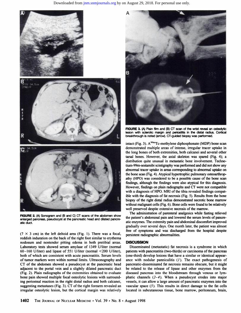
FIGURE 2. (A) Sonogram and (B and C) CT scans of the abdomen showenlarged pancreas, pseudocyst at the pancreatic head and dilated pancreatic duct.
(7X3 cm) in the left deltoid area (Fig. 1). There was a focal,reddish induration on the back of the right foot similar to erythemanodosum and nontender pitting edema in both pretibial areas.Laboratory tests showed serum amylase of 1349 U/liter (normal60-160 U/liter) and lipase of 551 U/liter (normal <200 U/liter),both of which are consistent with acute pancreatitis. Serum levelsof tumor markers were within normal limits. Ultrasonography andCT of the abdomen showed a pseudocyst at the pancreatic headadjacent to the portal vein and a slightly dilated pancreatic duct(Fig. 2). Plain radiographs of the extremities obtained to evaluatebone pain showed multiple focal osteolytic lesions with surrounding periosteal reaction in the right distal radius and both calcanei,suggesting métastases(Fig. 3). CT of the right forearm revealed anirregular osteolytic lesion, but the cortical margin was relatively
FIGURE 3. (A) Plain film and (B) CT scan of the wrist reveal an osteolyticlesion with sclerotic margin and periostitis in the distal radius. Corticalbreakthrough is noted (arrow). CT-guided biopsy was performed.
intact (Fig. 3). A99mTc-methylene diphosphonate (MDP) bone scan
demonstrated multiple areas of intense, irregular tracer uptake inthe long bones of both extremities, both calcanei and several othertarsal bones. However, the axial skeleton was spared (Fig. 4); adistribution quite unusual in metastatic bone involvement. Techne-tium-99m-sestamibi scintigraphy was performed and did not show anyabnormal tracer uptake in areas corresponding to abnormal uptake onthe bone scan (Fig. 4). Atypical hypertrophie pulmonary osteoarthrop-athy (HPO) was considered to be a possible cause of the bone scanfindings, although the findings were also atypical for this diagnosis.However, findings on plain radiographs and CT were not compatiblewith a diagnosis of HPO. MRI of the tibia revealed findings compatible with the diagnosis of fat necrosis (Fig. 5). Results from the bonebiopsy of the right distal radius demonstrated necrotic bone marrowwithout malignant cells (Fig. 6). Bone cells were found to be relativelywell preserved despite extensive necrosis of the marrow.
The administration of parenteral analgesics while fasting relievedthe patient's abdominal pain and lowered the serum levels of pancre
atic enzymes. The extremity pain and abdominal discomfort improvedgradually over several days. One month later, the patient was almostfree of symptoms and was discharged from the hospital despitepersistent radiographie abnormalities.
DISCUSSIONDisseminated (metastatic) fat necrosis is a syndrome in which
patients with pancreatitis (two-thirds) or carcinoma of the pancreas(one-third) develop lesions that have a similar or identical appearance with nodular panniculitis (/). The exact pathogenesis ofpancreatic-disseminated fat necrosis remains obscure, but it mightbe related to the release of lipase and other enzymes from thediseased pancreas into the bloodstream through venous or lymphatic channels (2-4). When a pseudocyst erodes into majorvessels, it can allow a large amount of pancreatic enzymes into thevascular space (5). This results in direct damage to the fat cellslocated in subcutaneous tissue, bone marrow, peritoneum, brain,
1402 THEJOURNALOFNUCLEARMEDICINE•Vol. 39 •No. 8 •August 1998
by on August 29, 2018. For personal use only. jnm.snmjournals.org Downloaded from

••».BONE MIBI•
•
FIGURE 4. Bone scan (left)demonstrates multiple areas of intense, irregulartracer uptake in the long bones of all extremities and feet. A ""Tc-sestamibi
scan (right) shows normal findings.
kidney, mesentery and other distant sites reached by blood flow.Based on the identification of IgG and C3 in the affected areas,immune-mediated injury also has been implicated in the pathogen-esis of fat necrosis (6). In our case, the presence of a pseudocyst inclose proximity to the portal vein may explain the pathogenesis ofthe fat necrosis in the long bones distant from the pancreas.
The diagnosis of fat necrosis in acutely ill patients is not easy.Jackson et al. (7) proposed the following six features as those thatsuggest metastatic fat necrosis: (a) skin lesions resembling erythema nodosum or Bazin's disease; (b) subcutaneous nodules
tending to break down and become sterile abscesses; (c) limb painand a tendency for the necrotic process to involve the joints; (d)tendency for eosinophilia; (e) malaise, high fever and wasting; and(0 duration of illness measured in months. Our patient had all ofthese features except eosinophilia.
Skeletal involvement may occur as an isolated phenomenon orsimultaneously with subcutaneous nodules and polyarthritis. Anecropsy study showed a relatively higher prevalence (approximately 10%) of intramedullary fat necrosis in postmortem samplesobtained from 67 acute pancreatitis patients (2). However, intramedullary fat necrosis is essentially a radiologie entity andlargely has been found incidentally in patients with acute or
FIGURE 5. Coronal MR images. (Top) T1-weighted MR image showsdiminished signal intensity. (Bottom) T2-weighted MR ¡mageshows areas of
decreased signal intensity within a region of overall increased signal intensityin diaphysis and metaphysis of both tibias.
chronic pancreatitis (8). Radiographie findings of intramedullaryfat necrosis include osteolytic lesions with moth-eaten bone destruction, periostitis of the tubular bones of the extremities andcalcification of medullary cavities (9-13). In the carpal and tarsalbones, cystic defects and a coarse trabecular pattern can beapparent, whereas the epiphyses may be unaffected.
Bone scintigraphy of our patient revealed numerous lesions thatwere not apparent on the plain radiographs, as in a previouslyreported case (14). The appearance of individual abnormalitiesassociated with intramedullary fat necrosis on radiographs, CT orbone scanning is nonspecific and is similar to that of malignantmetastatic lesions, osteomyelitis and osteonecrosis. However, metastatic deposits typically affect axial bones containing hematopoi-etic marrow (15), whereas the osseous changes associated withintramedullary fat necrosis are seen predominantly in limb bonesprobably because distal long bones contain primarily fatty marrow.The whole-body bone scan in our patient clearly showed the distribution of the lesions; making metastatic disease unlikely.
The exclusive long-bone involvement in our patient raisedthe possibility of secondary hypertrophie osteoarthropathy thatis commonly associated with lung cancer, chronic pulmonary
INTRAMEDULLARYFAT NECROSISASSOCIATEDWITHPANCREATITIS•Ahn et al. 1403
by on August 29, 2018. For personal use only. jnm.snmjournals.org Downloaded from
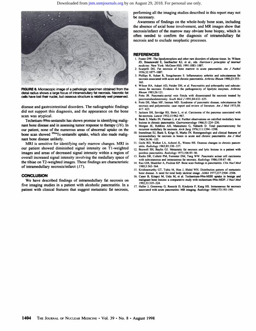
FIGURE 6. Microscopic image of a pathologic specimen obtained from thedistal radius shows a large focus of intramedullary fat necrosis. Necrotic fatcells have lost their nuclei, but osseous structure is relatively well preserved.
disease and gastrointestinal disorders. The radiographie findingsdid not support this diagnosis, and the appearance on the bonescan was atypical.
Techetium-99m-sestamibi has shown promise in identifying malignant bone disease and in assessing tumor response to therapy (16 ). Inour patient, none of the numerous areas of abnormal uptake on thebone scan showed 9%nTc-sestamibiuptake, which also made malig
nant bone disease unlikely.MRI is sensitive for identifying early marrow changes. MRI in
our patient showed diminished signal intensity on T l-weightedimages and areas of decreased signal intensity within a region ofoverall increased signal intensity involving the medullary space ofthe tibiae on T2-weighted images. These findings are characteristicof intramedullary necrosis/infarct (17).
CONCLUSIONWe have described findings of intramedullary fat necrosis on
five imaging studies in a patient with alcoholic pancreatitis. In apatient with clinical features that suggest metastatic fat necrosis.
performing all the imaging studies described in this report may notbe necessary.
Awareness of findings on the whole-body bone scan, includingthe absence of axial bone involvement, and MR images show thatnecrosis/infarct of the marrow may obviate bone biopsy, which isoften needed to confirm the diagnosis of intramedullary fatnecrosis and to exclude neoplastic processes.
REFERENCES1. Foster DW. The lipodystrophies and other rare disorders of adipose tissue. In: Wilson
JD. Braunawald E, Isselbacher KJ, et al., eds. Harrison 's principles of internal
medicine. New York: McGraw-Hill; 1991:1883-1887.
2. Scarpelli DG. Fat necrosis of bone marrow in acute pancreatitis. Am J Puthol1956:32:1077-1087.
3. Phillips R, Sulser R. Songcharoen S. Inflammatory arthritis and subcutaneous fatnecrosis associated with acute and chronic pancreatitis. Arthritis Rheum 1980;23:355-
360.4. Wilson HA, Askari AD, Neider DH. et al. Pancreatitis and arthropathy and subcuta
neous fat necrosis. Evidence for the pathogenecity of lipolytic enzymes. ArthritisRheum 1983:26:121.
5. Potts JR. Pancreatic-portal vein fistula with disseminated fat necrosis treated bypancrealoduodenectomy. South MetÃJ 1991;84:632-635.
6. Potts DE, Mass MF. Iseman MD. Syndrome of pancreatic disease, subcutaneous fatnecrosis and polyserositis: case report and review of literature. Am J Med 1975;58:417-423.
7. Jackson SH, Savidge RS, Stein L, et al. Carcinoma of the pancreas associated withfat-necrosis. Lancet 1952;15:962-967.
8. Bank S. Marks IN, Farman J, et al. Further observations on calcified medullary bonelesions in chronic pancreatitis. Gastroenlerology 1966:51:224-230.
9. Morgan JE, Robbins AH, Matsumoto G, Nabseth D. Total pancreatectomy forrecurrent medullary fat necrosis. Arch Surg 1976;! 11:1394-1398.
10. Immelman EJ, Bank S, Krige H, Marks IN. Roentgenologic and clinical features ofintramedullary fat necrosis in bones in acute and chronic pancreatitis. Am J Med1964:36:96-105.
11. Gerle RD, Walker LA, Achord JL. Weens HS. Osseous changes in chronic pancreatitis. Radiology 1965:85:330-337.
12. Boswell SH, Baylin GJ. Metastatic fat necrosis and lytic lesions in a patient withpainless pancreatitis. Radiology* 1973:106:85-86.
13. Radin DR. Colletti PM, Forrester DM, Tang WW. Pancreatic acinar cell carcinomawith subcutaneous and intraosseous fat necrosis. Radiology 1986:158:67-68.
14. Rao GM, Shadcher A, Poulose KP. Bone scan findings in pancreatitis. Clin NucÃMed1980:5:563-564.
15. Krishnamurthy GT, Tubis M, Hiss J, Blahd WH. Distribution pattern of metastaticbone disease. A need for total body skeletal image. JAMA 1977:237:2504-2506.
16. Caner B, Kitapci M. Unlu M. et al. Technetium-99m-MIBI uptake in benign andmalignant bone lesions: a comparative study with technctium-99m-MDP. J NucÃMed1992:33:319-324.
17. Haller J. Greenway G. Resnick D, Kindynis P, Kang HS. Intraosseous fat necrosisassociated with acute pancreatitis: MR imaging. Radiology 1989:173:193-195.
1404 THEJOURNALOFNUCLEARMEDICINE•Vol. 39 •No. 8 •August 1998
by on August 29, 2018. For personal use only. jnm.snmjournals.org Downloaded from
1998;39:1401-1404.J Nucl Med. Byeong C. Ahn, Jaetae Lee, Kyung J. Suh, Kyung A. Chun, Sang K. Sohn, Kyubo Lee and Chun K. Kim Intramedullary Fat Necrosis of Multiple Bones Associated with Pancreatitis
http://jnm.snmjournals.org/content/39/8/1401This article and updated information are available at:
http://jnm.snmjournals.org/site/subscriptions/online.xhtml
Information about subscriptions to JNM can be found at:
http://jnm.snmjournals.org/site/misc/permission.xhtmlInformation about reproducing figures, tables, or other portions of this article can be found online at:
(Print ISSN: 0161-5505, Online ISSN: 2159-662X)1850 Samuel Morse Drive, Reston, VA 20190.SNMMI | Society of Nuclear Medicine and Molecular Imaging
is published monthly.The Journal of Nuclear Medicine
© Copyright 1998 SNMMI; all rights reserved.
by on August 29, 2018. For personal use only. jnm.snmjournals.org Downloaded from