intracranial tumour complicating pregnancy
TRANSCRIPT
Intracranial Tumour
Complicating Pregnancy
Presentor : Dr. Shilpa
Designation : Fellow, HRP and Perinatology
Hospital : Fernandez Hospital, Hyderabad
Date of Presentation : 09.06.2009

Case Details
Mrs. M, Aged 27 yrs, Para2, Live2
Delivered twice in Fernandez Hospital
1st delivery - July2007
2nd delivery - April 2009
Both pregnancies complicated by intracranial
tumour.

First Pregnancy
Registered at 17 wks
No of visits 09
Normotensive
LMP 9/10/2006
EDD 16/07/2007
First Pregnancy - History
OBS. History: Conceived with treatment
Past History: No significant medical or surgical
history

Investigations
21/02/2007: (at booking visit)
TSH 1.39uIU/ml
Hb10.9 g/dl
GCT 128 mg/dl
CUE- NAD
HIV & HBsAg Negative
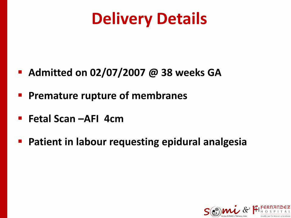
Delivery Details
Admitted on 02/07/2007 @ 38 weeks GA
Premature rupture of membranes
Fetal Scan –AFI 4cm
Patient in labour requesting epidural analgesia
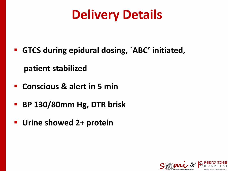
GTCS during epidural dosing, `ABC’ initiated,
patient stabilized
Conscious & alert in 5 min
BP 130/80mm Hg, DTR brisk
Urine showed 2+ protein
Delivery Details

Enigma…
Could this be due to :
Inadvertant intravenous bolus of local anesthetic
causing seizure ?
– ? Intrapartum Eclampsia
Management
? Intrapartum eclampsia –Inj.MgSO4, IV, 4gm
loading dose followed by 1gm/hr maintenance
infusion
Emergency LSCS was decided
Intra Operatively
Patient had a generalized tonic clonic seizure
2 episodes of vomiting
Regained consciousness in 3mins
Epidural block: Dermatomal level of T6 was present

Intraop Dilemmas!
Is it LA toxicity?
Is it intrapartum eclampsia?
Is it CSVT / Seizures?
Is there any other IC pathology / epilepsy?
Any dyselectrolytemia?

Delivery Details
Surgery done under EPIDURAL ANAESTHESIA
uneventfully
Delivered an alive baby girl weighing 2.29KGS
APGAR- 08/09/09

Intra OP Investigations
Preeclampsia profile- LDH, Uric acid, S. Creatinine,
Platelets, LFT-normal
S. electrolytes normal
WBC Count 13,200/cmm

Postop Period Detailed History
H/O gradual onset of neuro deficit since 7th month
of pregnancy
Loss of balance
Loss of smell
Decreased palpebral fissure size

Slurred speech
Altered taste
Decreased hearing in left ear over 2 months
Symptoms not progressing or regressing since onset
Postop Period Detailed History
No H/O :
Seizures in the past
Headache / fever
Bladder / bowel disturbances
Motor or sensory loss
Postop Period Detailed History
Examination On POD-0
Pulse 80/min, BP 90/70mm Hg, RR 18/min
Mental functions normal except Dysarthria
Pupils NSRL, Fundus bilateral papilloedema
Left eye partial ptosis
Horizontal nystagmus Rt gaze>Lt gaze
Left LMN VII Nerve palsy
Examination
DTR brisk bilaterally, Plantars
Other motor/sensory normal
Ataxia, Gait wide based
No meningeal signs
Other systems unremarkable
Impression
VII,VIII CN +/- I CN involvement
Ataxia
CerebelloPontine Angle lesion
– ?Infective
– ?Neoplastic
Physician’s Advise
Stop MgSo4
Consider antiepileptic if further seizures
LMWH after neuroradiology
Neurophysician opinion
MRI Brain plain and contrast

Postpartum Period
Uneventful
MRI Brain not done due to financial problems
Risks explained
Discharged on 06/07/2007 on 4th POD

10-07-07: MRI Brain
Left Vestibular Schwannoma
measuring 4.3 x 3.4 cms
with Obstructive Hydrocephalous!!!

Left Vestibular schawnnoma with
Obstructive hydrocephalus!!!!!!!!

Left Vestibular schawnnoma with
Obstructive hydrocephalus!!!!!!!!

Follow up
Seen by neurosurgeon
Tab Phenytoin100mg 1---2
Tab Decadron 4mg 1---1

July 2007
Tumour resection @CARE Hospital on 24-07-07
Pseudomeningocoele: Occipital craniectomy site
Lt hemiparesis post op - recovery over 3months
Antiepileptic drugs X 1year

Post OP CT Brain
4th ventricle deformed by an isodensity
3rd and both lat. Ventricles- mild dilatation
VP shunt in situ
Lt. occipital pseudo meningocoele

Tumour HPE Report
Cellular lesion- elongated cells with wavy nuclei in
palisades
Verrocay bodies seen with paucicellular areas
Thin walled blood vessels
No mitosis, giant cells, necrosis
Features consistent with Schwannoma, CPangle
Repeat Cect Brain- 1/08/2008
Ventricular shunt tube in situ on left side
Enhancing soft tissue in the left CP angle at the
Internal Auditory Canal 13X9 mm–Possible
recurrence / residual tumour
Left occipital region -Pseudomeningocele
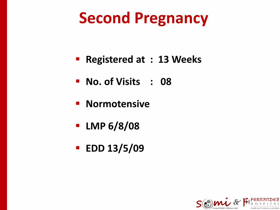
Second Pregnancy
Registered at : 13 Weeks
No. of Visits : 08
Normotensive
LMP 6/8/08
EDD 13/5/09

Second Pregnancy
ANC period – uneventful
Routine investigations normal
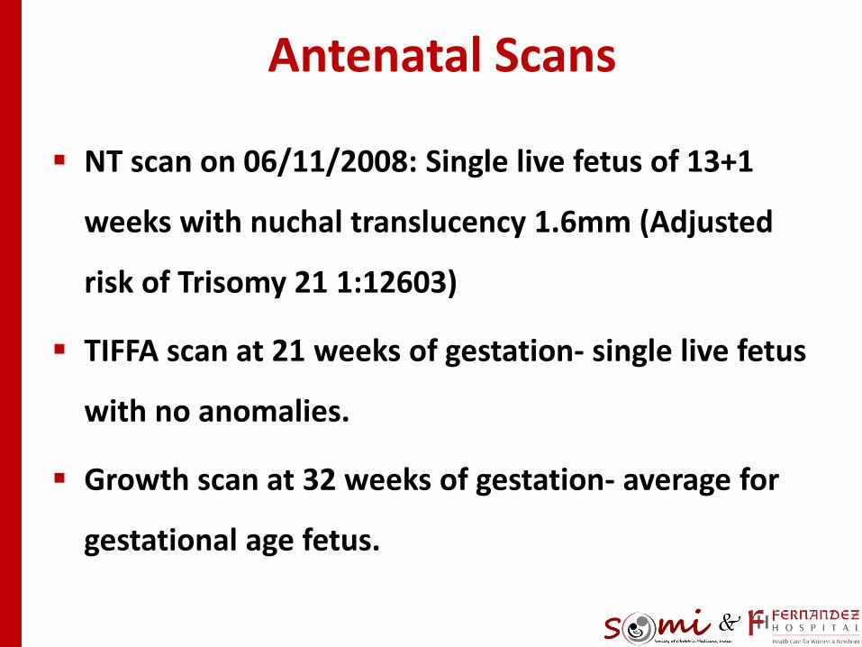
Antenatal Scans
NT scan on 06/11/2008: Single live fetus of 13+1
weeks with nuchal translucency 1.6mm (Adjusted
risk of Trisomy 21 1:12603)
TIFFA scan at 21 weeks of gestation- single live fetus
with no anomalies.
Growth scan at 32 weeks of gestation- average for
gestational age fetus.

Multidisciplinary care involving
Neurosurgeon, Obstetrician, Physician,
Anesthesiologist and Pediatrician was planned

Examination at Term
Left nasal discharge
Left occipital soft & cystic
swelling
(pseudomeningocoele)
Left facial palsy LMN type
Right sided nystagmus
Power normal on Right
side; 4/5 on Left side
Reflexes ++/++
No meningeal /
cerebellar signs
Fundus normal

MRI Brain: 10/04/2009
Recurrent /residual left CP angle mass
consistent with an acoustic schwannoma.
Large pseudo meningocoel

MRI Brain: 10/04/2009

Delivery Plan
Planned for El. LSCS at 37 completed weeks
In view of Previous LSCS with residual Intracranial
Tumour
Under graded epidural anesthesia
BUT….

Delivery Details
Admitted on 16/04/2009 @ 36+1 weeks of
gestation in early labour
An emergency LSCS was decided
Epidural anesthesia- uneventful
Delivered an alive baby boy weighing 2.64KGS
APGAR- 08/09/09

Postpartum Management
Postoperative period uneventful
Antibiotics
Thromboprophylaxis for 3 days
Discharged on 4th POD

Follow up
After 3weeks: Asymptomatic
Pseudomeningocoele size reduced
Advised neurosurgeon follow up