intracranial cavernoma - neurosurgery … cavernoma_new... · seizure (40 -80 %) ... the...
TRANSCRIPT
INTRACRANIAL CAVERNOMA
INTRODUCTION
l CEREBRAL CAVERNOUS MALFORMATION (CCM), CAVERNOUS HEMANGIOMA, CAVERNOUS ANGIOMA, CRYPTIC VASCULAR MALFORMATION,OCCULT VASCULAR MALFORMATION, HEMORRHOID OF BRAIN.
l Developmental abnormalities that affects the blood vessels supplying the brain.
l Dilated sinusoidal channels lined by single layer endothelium without intervening brain parenchyma.
l Can occur anywhere in the CNS
l Angiographically occult vascular malformations.
EPIDEMIOLOGY
l Prevalence: 0.5%
l 5-10% of all Cerebro-vascular malformation
l 80% are supratentorial
l Familial CCM accounts around 20-30%
l Increased prevalence in Mexican American families
l 30- 50% asymptomatic
l 20-30% of cavernomas may be a/w venous anomaly
l Bimodal presentation- Pediatric:3-11 yrs.
Adult : 2nd to 4th decade.
l Age at first diagnosis: – <20 yrs: 25-30% – 20-40 yrs: 60% – >40 yrs: 10-15%
EPIDEMIOLOGY

GENETICS
l Usually sporadic (80%)
l Three autosomal dominant genes (7p/7q/3q) a/w familial form. The precise function not known :
1. ccm1 on 7q chromosome and expresses KRIT1 2. ccm2 on 7p chromosome and expresses MGC4607 3. ccm3 on 3q chromosome and expresses PCD10
l Working model : Alteration in growth control pathway involving the regulation of Krev 1 / rap 1a by KRIT 1 protein following mutation of CCM 1
PATHOLOGY
l Mulberry appearance, soft / hard consistency, usually 1 -5 cm
l Enlarged capillaries with abnormal gap between endothelial cells with thin collagenous wall & lack of smooth muscle / elastic fibers
l Capillaries are immediately adjacent to each other, with no intervening neural tissue.
l No feeding arteries or draining veins. May be a/w venous anomaly. Blood flow is low.
l Blood degradation products (hemosiderin staining) and gliotic reaction in the adjacent brain
CLINICAL PRESENTATION
l Approximately 50% asymptomatic.
l Seizure (40 -80 %) - Males more commonly presents with epilepsy.
- Chronic intractable epilepsy is found in almost half. l Sudden onset neurological deficits caused by hemorrhage,
commonly observed in thalamic and brainstem CCM. l Prior hemorrhage, female sex and pregnancy are the risk
factors for re-bleed.

NATURAL HISTORY
l Depends on the topography
l M.C. presentation is seizure
l Subclinical hemorrhage seen in all and clinical hemorrhage in 12.1%
l Annual hemorrhage rate 0.5 to 1 % & rebleed rate of 4.5 %
l The epileptic risk is correlated to the localization, more frequent for temporal and frontal lesions (1.5 to 2.4 % each year)
l Lesion may arise de novo, may grow / shrink / remain unchanged

NATURAL HISTORY
AUTHOR / YEAR RATE OF SYMPTOMIC HEMORRHAGE
COMMENTS
Del Curling et al 1991 0.1 % per lesion /yr Retrospective study based on pts recall
Robinson et al 1991 0.7 % per lesion /yr Prospective study
Zabramski et al 1995 1.2 % per lesion /yr Prospective MR based study. 60% episodes were symptomatic
Kondziolka et al 1995 1.3 % per lesion /yr 2.6 % per lesion /yr 0.6 % per lesion /yr 4.5 % per lesion /yr
Incidental lesion has lower rate of Hmg
Aiba et al 2005 0 % per lesion /yr 0.4 % per lesion /yr
Prospective study : low hmg rate for incidentaloma / with seizure
Porter et al 2007 4.2 % per lesion /yr Retrospective review from 1st hmg
RADIOLOGY
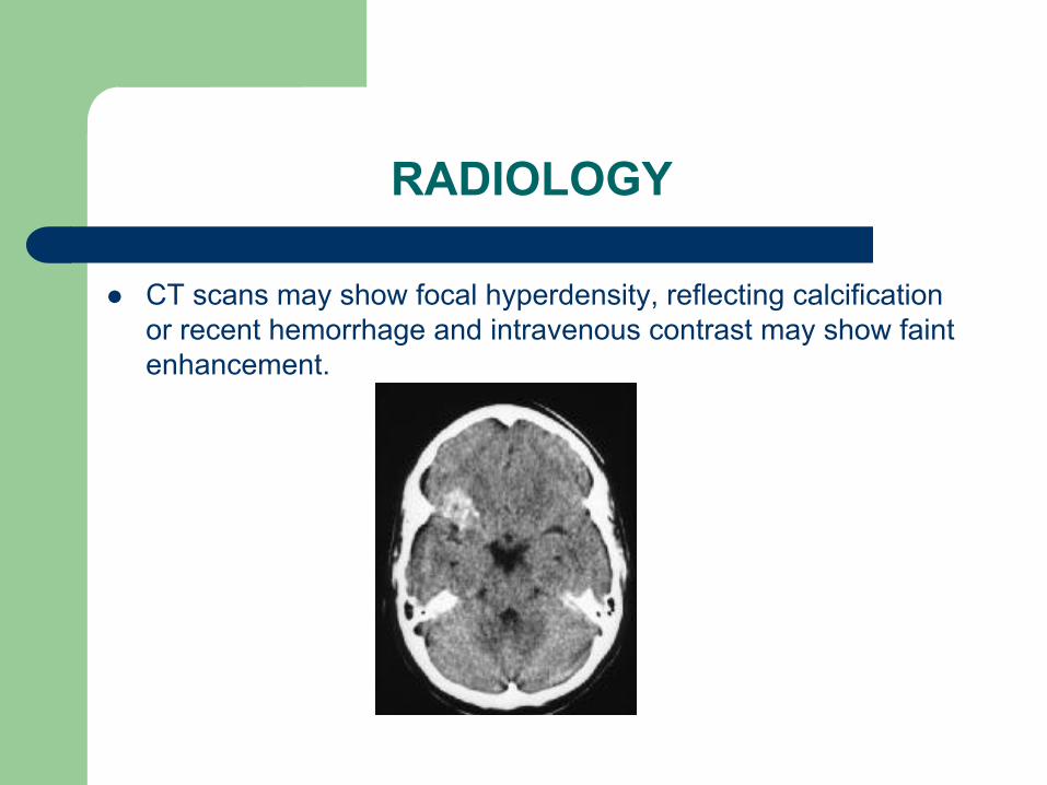
l CT scans may show focal hyperdensity, reflecting calcification or recent hemorrhage and intravenous contrast may show faint enhancement.
RADIOLOGY MRI
l T2WI show reticulated core of lesion “popcorn” appearance with a lucent halo surrounding a lesion.
l This pathognomonic appearance is produced due to repeated hemorrhages and hemosiderin deposition around the lesion.
l No peri-lesional edema.
l Gradient sequences are more sensitive for detection of small lesions
RADIOLOGY
l Cerebral DSA demonstrate no vascular abnormalities. Developmental venous anomalies may be associated cavernoma and may be be seen on angiography.
DIFFEENTIAL DIAGNOSIS
l Hemorrhagic metastasis viz melanoma
l Glioma e.g. oligodendoglioma , pleomorphic xanthoastrocytoma
l AVM – occult / overt
l Hemorrhagic telangiectasia (Osler Weber Rendu disease) / multiple metastasis

Management Options
l Medical treatment. l Stereotactic radiotherapy or gamma knife.
l Surgical resection (gold standard).
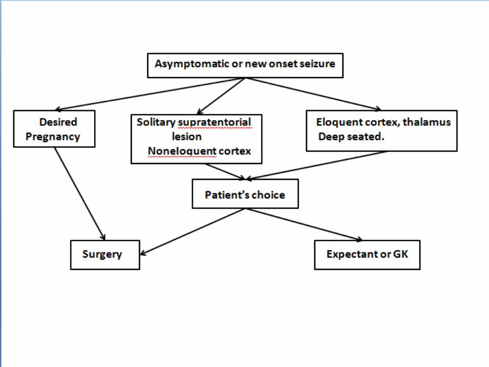
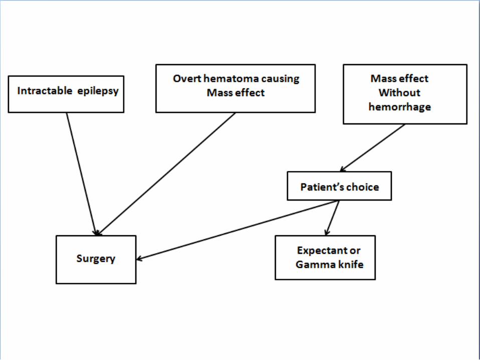
Medical Management
l Offered to asymptomatic, incidentally detected lesions,
surgically inaccessible lesions and unfit for surgery
l Regular follow up MRI to assess increase in size or new hemorrhages.
l Medical treatment is limited to control of seizure activity.
l Patients are refrained from strenuous exercises, anticoagulant use, avoid pregnancy

Surgical Treatment
Merits: l Reduction of mass effect. l No rebleeding from completely excised CCM. l 75-88% of patients become seizure free after complete
resection. l Intractable epilepsy may become well controlled. l Complete cure can be assured.

Surgical Treatment
Demerits: l In brainstem and thalamic CM, surgical treatment may lead to
neurological deficits.
l Standard surgical mortality and morbidity
Planning
l T1WI,T2WI & contrast MRI with GRE / FLAIR sequences
l Venous malformations should be looked for
l If lesion is in the eloquent area, then functional imaging along with cortical mapping should be done.
l Frameless stereotaxy is considered in small and deep seated lesions & dot localizing CT for superficial polar lesion

Surgical Technique
l Surgical technique depends on the location of lesion, presence of associated venous malformations and presence of overt hematoma.
l Position and approach based on location , trans sulcal approach is
preferred .
l Lesion should be localized after opening the dura. l Intraoperative sonography, or stereotaxy can be used. l Surface lesion appear as purple-blue mulberry like structure
surrounded by area of hemosiderosis
l The cortical venous drainage pattern should be inspected.
l Lesion is initially shrunk with bipolar coagulation. l The surrounding gliotic brain (pseudo capsule) offers a plane of
dissection.
l Cotton patties are used for preservation of plane of dissection. l Complete resection is to be attempted .
l Resection bed to be inspected for satellite lesion
Surgical Technique
Associated Venous Anomalies
l Approx. 24% patients of CCM
l These venous anomalies are the normal veins draining brain
l Every effort is made to preserve them
l These may be the only veins supplying the brain
l Coagulation of these may result in venous infarcts and increases the morbidity of surgery
Surgical Excision Options
l Lesionectomy alone.
l Lesionectomy and excision of abnormal tissues.
l Lesionectomy and removal of abnormal area remote from the cavernoma.
Special Considerations
INTRACTABLE EPILEPSY : l Pre op. work up to localize source of seizure.
l If the location non eloquent, surrounding hemosiderin stained brain may be resected because of possible epileptogenicity
l Pre op evaluation detect epileptogenicity is remote from CM or multiple CM = ECOG guided surgery.
l Pre op evaluation detect epileptogenicity is dual pathology (eg- temporal lobe CM with MTS) = resection of both foci.
Special Considerations
CAPSULAR AND THALAMIC CAVERNOMA : l Resection caries greater morbidity l Resection attempted to those lesions that reach pial or ventricular
surface. l Frameless image guidance helpful
l Anterior lesion – transcallosal route l Lateral thalamic lesion – tanscortical route through superior parietal
lobule l Medial posterior lesion – posterior interhemispheric approach

Special Considerations
OPTIC PATHWAY CM :
l May present with acute / subacute visual disturbances
l May mimic SAH on CT
l Urgent decompression for chiasmal apoplexy (Maitland et al)
l Pterional or subfrontal route
l CM may affect any part of optic pathway , lesionectomy is attempted.

Special Considerations
PINEAL CM : l < 1 % of CCM (Slavin et al)
l Present with features of raised ICP , ocular symptoms , neuro-endocrine abnormalities .
l EVD / Shunt to lower ICP
l Interhemispheric transsplenial , sub occipetal transtentorial & supra cerebellar infra tentorial approach depending on location of CM
Special Considerations
MULTIPLE CM : l Multiple in 50 % cases
l Treatment at addressing symptomatic lesion
l Large sized , hemorrhage, FND , seizure focus
l Familial CM are at risk for the formation of de novo CM
Special Considerations
DURAL BASED CM : l Commonly found in middle fossa, CP angle , tentorium and convexity l Present with headache l Rarely bleed l May mimic meningioma l Enhance strongly and homogenously on Gd MRI l Pre op embolizaion / SRS or EBRT is advisable for highly vascular
middle fossa CM l Post op morbidity as high as 38%
Special Considerations
Brain Stem CM:
– Of all CNS cavernoma, 9-35% are found in brain stem
– Acute onset focal neurological deficits are the most common presentation
– The waxing and waning of symptoms mimic multiple sclerosis.
– Hemorrhage rate is 30 times more than that for supratentorial CCMs.
– Rebleeding rate is as high as 35%.
REVIEW OF LITERATURE Brain Stem Cavernoma
Angelo Franzini, Neurosurgery 56:1203-1214, 2005 : l 52 patients undergone micro neurosurgery for brainstem
cavernoma. l Rebleed rate was 34%. l 29 developed temporary neurological deficits which persisted in
10 pts. l Mortality was 1.9%
Surgical Indications Brain Stem CM
l Exophytic lesions reaching pial surface.
l Rapid progressive neurological deterioration.
l Hemorrhage outside the lesion capsule.
l Significant mass effect.
l Multiple debilitating hemorrhages.
Brain Stem CCM
l Lesions are considered for surgical resection only if they reach pial surface.
l Patient can be followed till they suffer one or more hemorrhages.
l Waiting for 3-5 days for hematoma to liquefy is good practice.
l Threshold of intervention should be high for pediatric population.
Goals Of Treatment.. Brain Stem CM
l Minimize amount of normal brainstem traversed.
l Complete excision of the tumor.

Surgical technique.. Brain Stem CM
l Position and approach differs with location of lesion.
l If it doesn’t reach pial surface, then intra operative sonography or stereotaxy can be used along with hemosiderin staining.
l Dark bluish red area or mulberry appearance is classical for cavernoma
Surgical technique.. Brain Stem CM
l Hemosiderin stained area should be considered a normal tissue.
l Internal decompression should be done followed by careful dissection from surrounding tissues.
l Every possible attempt is made to save the normal veins.
Surgical technique.. Brain Stem CM
l Complete excision may not be possible due to technical limitations.
l Better to stage the procedure than to risk the functionality
Post Operative Management.. Brain Stem CM
l Patient usually kept intubated for at least 24 hours.
l Demonstration of gag and cough reflexes guides extubation. l MRI / CT on early post op days for assessment of extent of
excision if patient’s condition permits.

OUTCOME
l When CCM are completely removed , risk of further growth and hemorrhage is essentially permanently eliminated
l More than 80% of patients were same or better after surgery while few worsened in various series.
l Cranial nerve deficits, motor deficits, meningitis, CSF leak, tracheostomies are the usual complications of brainstem CM

FOLLOW UP
l Post op MRI scan
l First degree relative with more than 1 family member having a cavernoma should have CECT or MRI brain to be done along with genetic counseling
REVIEW OF LITERATURE
Shih YH, Pan DH,Clin Neurol Neurosurg. 2005 Feb;107(2):108-112
l 46 patients (16+30) were treated with surgery and GK. l 79 % (11/14) vs. 25 % (4/16) of patients became seizure free after
surgery and GK (p<0.002). l 100 vs. 67 % patients- no rebleed (NS). l Concluded that surgery is better option for supratentorial CM in terms
of seizure control and rebleed rates.
Gamma Knife Therapy…IN CCM
PRINCIPLE :
l Radiation injury to endothelial cell causes release of growth factors and fibroblast proliferation.
l This causes hyperplasia of smooth muscles cells and occlusion of lumen.
l Coagulation necrosis of cavernoma and obliteration of vessels were also a proposed mechanism.

Gamma Knife Therapy
Indications: l Surgically inaccessible lesions like thalamic, brainstem or deep seated
lesion.
l Lesions presented with minor hemorrhages
l Multiple lesions.
l Patients choice.
Gamma Knife Therapy
l Merits:
– Reduction of rebleed rates. – Halting of growth of lesion. – Volume control. – Seizure control.
l Demerits: – Radiation edema and transient or permanent deficits – Lower efficacy of treatment.
Gamma Knife Therapy
MORBIDITY-
l Perilesional edema was found in 27% with transient morbidity
of 20.5% with permanent morbidity of 4.5%.
l Rebleed common in patients who presented with bleed, larger lesion volume and prescription dose less than 13 Gy.
l Edema was common with prescription dose > 13 Gy.
REVIEW OF LITERATURE
Lie AL, Wang CC. Zhongguo Yi Xue Ke Xue Yuan Xue Bao. 2005 Feb;27(1):18-21
l 92 patients with mean follow up of 2-8 yrs.
l 43 patients of them were primarily treated for epilepsy, 83% seizure control achieved .
l Radiological rebleed rate was 9.8%
l Radiation edema developed in 7 patients
l Gk is effective in controlling the seizures and bringing down the rebleed rate.
l Recommend GK therapy for surgically unfit cases.

REVIEW OF LITERATURE
Kim MS and Pyo SY, J Neurosurg. 2005 Jan;102 Suppl:102-6. l 42 patients with deep seated cavernoma located at thalamus, brainstem
and 8 patients with multiple cavernomas were treated by GK. Mean margin dose of 14.55 Gy was given
l Tumor size decreased in 29 patients.
l Seizure control by drugs is achieved in 88% of patients.
l Clinically significant rebleed occurred in 7.1%.
l Recommended GK for deep seated and brainstem lesions as well as multiple ones.
CONCLUSION
l Surgery still remains gold standard for intracranial cavernoma
l Conservative therapy is appropriate for cavernoma that are asymptomatic and for those who had one non devastating episode of hemorrhage
l Complications can occur and extensive informed consent should be obtained
l Meticulous radiography and clinical follow up necessary to monitor residual or recurrent disease
