intracellular translocation of fluorescent sphingolipids
TRANSCRIPT

Intracellular Translocation of Fluorescent Sphingolipids in Cultured Fibroblasts: Endogenously Synthesized Sphingomyelin and Glucocerebroside Analogues Pass through the Golgi Apparatus En Route to the Plasma Membrane
NAOMI G. LIPSKY and RICHARD E. PAGANO Department of Embryology, Carnegie Institution of Washington, Baltimore, Maryland 21210
ABSTRACT When monolayer cultures of Chinese hamster lung fibroblasts are briefly incubated at 2°C with the fluorescent sphingolipid analogue, C6-NBD-ceramide (N-[7-(4-nitrobenzo-2- oxa-l,3-diazole)] aminocaproyl sphingosine), fluorescent labeling of the mitochondria, endo- plasmic reticulum, and nuclear envelope occur. During further incubation at 37°C, the Golgi apparatus, and later the plasma membrane, become intensely fluorescent. Within this period, the C6-NBD-ceramide is converted to equal amounts of fluorescent sphingomyelin and glucocerebroside (Lipsky, N. G., and R. E. Pagano, 1983, Proc. Natl. Acad. Sci. USA., 80:2608- 2612). In the present study, the intracellular translocation of these metabolites and their subsequent appearance at the plasma membrane were investigated by fluorescence micros- copy, the addition of the ionophore monensin, and the technique of "back exchange," in which the amounts and types of fluorescent lipids present at the cell surface are identified after their transfer from the cell surface into recipient vesicles. In control cells, the amount of fluorescent glucocerebroside and sphingomyelin that could be removed from the cell surface by back exchange increased during incubation at 37°C, correlating with the increased fluorescence of the plasma membrane observed by microscopy. In the presence of 10 #M monensin, visible labeling of the plasma membrane was greatly diminished, whereas the Golgi apparatus became highly fluorescent and distended. The ability to remove fluorescent metab- olites from the cell surface by back exchange was significantly but reversibly inhibited by monensin. Monensin also increased the total amount of fluorescent sphingomyelin, but not the glucocerebroside found in cells.
Subcellular fractions were assayed for their ability to convert radiolabeled and fluorescent ceramides to the corresponding sphingomyelins and glucocerebrosides. The activities of parallel fractions coincided, suggesting that the presence of the NBD moiety did not affect the cellular metabolism of ceramide. Furthermore, the major peak of sphingomyelin- and glucocerebroside-synthesizing activity appeared to coincide with an enriched Golgi fraction.
These results strongly suggest that fluorescent sphingomyelin was not synthesized at the plasma membrane as has recently been suggested for endogenous sphingomyelin. Rather, both the sphingomyelin and glucocerebroside analogues were synthesized intracellularly from C6-NBD-ceramide and translocated through the Golgi apparatus to the cell surface.
The sites of lipid biosynthesis in animal cells and the mecha- nism(s) by which newly synthesized molecules are subse- quently translocated to various intracellular membranes pose
many fundamental but, as yet, unresolved questions for biochemists and cell biologists. Sphingomyelin, the predomi- nant sphingolipid in many cell types, illustrates this point (1).
THE JOURNAL OF CELL BIOLOGY. VOLUME 100 JANUARY 1985 27-34 © The Rockefeller University Press • 0021-9525/85/01/0027/08 S 1.00 2 7
on April 3, 2019jcb.rupress.org Downloaded from http://doi.org/10.1083/jcb.100.1.27Published Online: 1 January, 1985 | Supp Info:

Two precursors for the synthesis of this lipid have been proposed. The long-standing evidence that the donor of the phosphorylcholine moiety of this molecule is CDP-choline (2-4) has recently been challenged in favor of phosphatidyl- choline as the more likely donor (5-10). The site of sphingo- myelin biosynthesis is also in dispute. The earliest studies (3) indicating that synthesis occurred in a microsomal fraction were based on the use of the nonnatural threo-ceramide isomer, and employed CDP-choline as the phosphorylcholine donor. In a more recent study that used the naturally occur- ring D-erythroceramide and phosphatidylcholine precursors, Ullman and Radin (5) found activity in a crude liver micro- somal fraction, and later Bernert and Ullman (6) established this for other tissues as well. Unfortunately, this crude micro- somal fraction may have contained endoplasmic reticulum, Golgi apparatus, and plasma membrane. Marggraf et al. (8) found significant activity in a very light sucrose fraction, which they identified as plasma membrane. However, their later study (11) revealed the activity to actually be present in a Golgi fraction, a Golgi apparatus-plasma membrane frac- tion, and a plasma membrane fraction. They note in this report that 5'-nucleotidase, considered a marker for the plasma membrane, was found to an equal degree in the Golgi fraction (this has also been shown cytochemically [12]). Voelker and Kennedy (7), in an elegant study that established phosphatidylcholine as the appropriate precursor for sphin- gomyelin biosynthesis, went on to find significant synthetic activities in a plama membrane fraction. However, this frac- tion was characterized on the basis of 5'-nucleotidase activity and was not assayed for the presence of Golgi membranes. Sphingomyelin synthetic activity has also been reported in mitochondria ( 13, 14).
In the present study, we address the issues of the site(s) of synthesis and route(s) of translocation of sphingolipids in cultured fibroblasts, by using a fluorescent analogue of cer- amide. This approach has the advantage that the fluorescent ceramide and its metabolites may be visualized in living cells by fluorescence microscopy (15), and thus information on the intracellular distribution of these molecules may be obtained without fixation or cell disruption. These data can then be correlated with standard lipid biochemical analyses.
MATERIALS AND METHODS
Materials Chemicals were purchased from the indicated sources: Eagle's minimal
essential medium (Gibco Laboratories, Grand Island, NY); lectins (Vector Laboratories, Inc., Burlingame, CA); ovalbumin grade V, UDP-glueose, UDP- galactose, sphingosine, and sphingosine-l-phosphocholine (Sigma Chemical Co, St. Louis, MO); dioleoyl phosphatidyl choline (DOPC) 1 and N-[7-(4- nitrobenzo-2-oxa- 1,3-diazole)] aminocaproic acid (C~-NBD-aminocaproic acid: (Avanti Biochemicals Birmingham, AL); Aquasol, [9,10-~H(N)] palmitic acid (23.5 Ci/mmol), and [galactose-4,53H(N)] UDP-galactose (48.5 Ci/mmol)
~ Abbreviations used in this paper: DOPC, dioleoyl phosphat idylcho- line; H M E M , HEPES-buffered Eagle's min ima l essential med ium, pH 7.4, without indicator, plus 0.62 m M PO4; C6-NBD-aminocaproic acid, N-[7-(4-nitrobenzo-2-oxa-l,3-diazole)] aminocaproic acid; C6- NBD-ceramide, N-[ 7-(4-nitrobenzo-2-oxa- 1,3-diazole)]-6-aminoca- proyl sphingosine; C6-NBD-glucocerebroside, N-[7-(4-nitrobenzo-2- oxa-l ,3-diazole)]-6-minocaproyl sphingosine glucoside; C6-NBD- sphingomyelin , N-[7-(4-nitrobenzo-2-oxa-l ,3-diazole)]-6-aminoca- proyl sphingosine- l -phosphochol ine; NBD-glucocerebroside, N-[7- (4-nitrobenzo-2-oxa-l,3-diazole)] aminoacyl sphingosine glucoside; NBD-sphingomyel in , N-[7-(4-nitrobenzo-2-oxa-l,3-diazole)] amino- acyl sphingosine- 1 -phosphocholine.
28 THE IOURNAL OI- CELL BIOLOGY • VOLUME 100, 1985
(New England Nuclear, Boston, MA); and monensin (Calbiochem-Behring Corp., San Diego, CA). ~251-wheat germ agglutinin was the generous gift of Ms. J. Lippincott-Schwartz, Carnegie Institution of Washington, and glucosylsphin- gosine was obtained from Dr. Shimon Gatt (Hebrew University).
Preparation of Lipids N-[7-(4-nitrobenzo-2-oxa-l,3-diazole)]-6-aminocaproyl sphingosine (C6-
NBD-ceramide); N-[7-(4-nitrobenzo-2-oxa- 1,3-diazole)]-6-aminocaproyl sphin- gosine glucoside (C6-NBD-glucocerebroside); and N-[7-(4-nitrobenzo-2-oxa- 1,3-diazole)]-6-aminocaproyl sphingosine-l-phosphocholine (C6-NBD-sphin- gomyelin) were synthesized from C6-NBD-aminocaproic acid and sphingosine, glucosylsphingosine, and sphingosine-l-phosphocholine, respectively (16). The concentrations of NBD-labeled lipids were determined fluorometrically (exci- tation 470 nm; emission 530 nm), and for C6-NBD-sphingomyelin, by phos- phate analysis (17) as well. Palmitoyl sphingosine and [9,103H(N)] palmitoyl sphingosine were synthesized (16) and were entirely in the o-erythro form as judged by high-pressure liquid chromatography (studies performed by Dr. Yasuo Kishimoto, Johns Hopkins School of Medicine). [9,10-3H(N)] palmitoyl sphingosine was radiographically pure by thin-layer chromatography.
Vesicle Preparation Small unilamellar fluorescent vesicles were formed by ethanol injection ( 18):
23 mol% C6-NBD-ceramide-77% DOPC or 14 tool% C6-NBD-glucocerebro- side or C6-NBD-sphingomyelin-86% DOPC were mixed, dried under nitrogen and then under reduced pressure, and made to 2.8 mM total lipids in ethanol. This mixture was injected into 13 vol of 10 mM HEPES-buffered calcium- and magnesium-free Puck's saline, dialyzed at 2"C overnight against this buffer, then diluted with HEPES-buffered Eagle's minimal essential medium (HMEM) to a final concentration of 5-10 nmol NBD lipid/ml. DOPC vesicles were prepared in the same manner, except the DOPC was made to 28 mM in ethanol, and the vesicles were dialyzed against HMEM and brought to a final concentration of 1.1 mM in HMEM.
Cell Culture and Incubations with Vesicles Monolayer cultures of Chinese hamster V79 fibroblasts were grown to
confiuency in Eagle's minimal essential medium supplemented with glutamine, gentamicin, fungizone, streptomycin sulfate, penicillin G, and 12% horse serum in a water-saturated atmosphere of 5% CO2 in air. For fluorescent labeling, cultures were washed twice and incubated for 0.5 h in HMEM at 37"C, and then incubated with fluorescent vesicles in HMEM for 0.5 h at 2°C. Cultures were washed three times with HMEM, and then further incubated in HMEM at 37"C for the indicated times. For back-exchange studies, cultures were next incubated with DOPC vesicles for 15 rain at 2"C, the medium was removed, and the incubation was repeated with additional DOPC vesicles. A parallel set of monolayer cultures was incubated with two changes of HMEM only. Cultures were washed three times with HMEM, scraped offthe dish, and cellular lipids were extracted and analyzed (19). Where indicated, all of the above solutions contained 10 uM monensin. A 40 mM solution of monensin in ethanol was made fresh before use, diluted to 100 t~m with HMEM, and then further diluted to a final concentration of 10 uM with the indicated incubation media. An equivalent amount of ethanol was present in all control solutions.
Microscopy Fibroblasts were grown on rat tail collagen-coated glass coverslips, and
incubated as described above. For Golgi apparatus co-localization studies, C6- NBD-ceramide-treated cells were first photographed for NBD fluorescence and then a Golgi apparatus-staining procedure (20) was followed with modifications (15). A Zeiss universal microscope (Carl Zeiss, Inc. New York) equipped with epi-illumination for fluorescence was used.
Thin-layer Chromatography Cellular lipid extracts were applied to silica gel 60 thin-layer plates (Merck
& Co., Rahway, N J) and developed in CHCI~/CH3OH/H20 (65:25:4). Fluores- cent lipids were detected by ultraviolet illumination, and quantified by reference to synthetic standards with a Coming 750 scanning densitometer (Corning Medical and Scientific, Corning Glass Works, Medfield, MA).
Protein Protein was estimated by absorbance at 280 nm and/or the Lowry procedure
(21), There was good agreement between the two methods.

DNA Measurement Aliquots of cell suspensions were precipitated in 0.2 N-perchloric acid and
hydrolyzed, and DNA was estimated spectrophotometrically (22).
Subcellular Fractionation Subcellular fractions were prepared by a modification of the procedure of
Snider and Robbins (23). Cells were washed and scraped offthe dish with PBS, centrifuged, and then resuspended in 10 mM Tris HCI, pH 7.4, I mM MgCI2 at ~ I x 107 cells/ml for 20 rain at 2"C. The suspension was homogenized with 25 strokes in a loose-fitting Dounce homogenizer, 0.5 vol of 0.75 M sucrose- I0 mM Tris HCI, pH 7.4, I mM MgCI2, was added, and this was centrifuged at 600 g to sediment nuclei. The supernatant was centrifuged at I00,000 g for 45 rain in an SW41 rotor (Beckman Instruments, Inc., Fullerton, CA), and the pellet was resuspended in 0.25 M sucrose-10 mM Tris HC1, pH 7.4, 1 mM EDTA. This "membrane pellet" (0.8-1.2 mg of protein) was layered over a gradient containing t.7 ml each of 50, 40, 35, 30, 25, and 15% sucrose in i0 mM Tris HCI, pH 7.4, 1 mM EDTA. Gradients were centrifuged at 100,000 g for 16 h in an SW41 rotor, and 0.5-ml fractions were collected and the percent sucrose was estimated with a refractometer. Fractions were centrifuged at 100,000 g for 45 rain in a 50.2 Ti rotor (Beckman Instruments, Inc.), and pellets were resuspended in 10 mM Tris HC1, pH 7.4. Aliquots of each fraction were frozen and enzymatic activity was assayed within 24 h.
RESULTS
Monensin Affects lntracellular Distribution of Fluorescence
When flbroblast monolayers were incubated briefly with C6-NBD-ceramide-containing vesicles at 2"C, washed, and warmed 30 min at 37"C in HMEM, intense labeling was seen in a perinuclear region and to a lesser degree in mitochondria and endoplasmic reticulum (Fig. 1A). Within 2 h, the plasma membrane became visibly labeled (Fig. 1 C). In the presence of 10 uM monensin, two major differences were noted. First, the bright, perinuclear fluorescence seen after 30 min became even more intense, and appeared to occupy a larger, more diffuse area (Fig. 1B). In addition, mitochondria assumed a spherical shape, rather than their more typical elongated "threadlike" form. The second major difference was the sig- nificant reduction in plasma membrane labeling seen after 2 h at 37°C (Fig. 1 D). Cell viability, as assessed by trypan blue exclusion, was unaffected by Cr-NBD-ceramide or monensin treatments.
Enzyme and Marker Assays PLASMA MEMBRANE: Monolayer cultures were incubated for 30 min at
2"C with t2~I-wheat germ agglutinin (9.2 x 10⁸ cpm/mg) (24), washed, and then subjected to subcellular fractionation. The specific radioactivity in each fraction was measured before and after protein precipitation. No difference was found in the activity profile except in the very-low-density region, indicating that all significant peaks represented protein-bound t25I.
GOLGI APPARATUS: Galactosyltransferase was measured by a modifi- cation of the procedure of Bretz el al. (25). The reaction mixture contained 40 mM sodium cacodylate, pH 7.0, 40 mM MnCI2, 40 mM 2-mercaptoethanol, 17.5 mg/ml ovalbumin, 2 mM ATP, 0.4% Triton X-100, 1/~Ci [galactose-4,5- 3H(N)] UDP-galactose, 0.5 mM UDP-galactose, and 5-10 #g of protein sample, in a fluid volume of 0.1 ml. After incubation at 37"C for 30 min, an additional 50 nmol UDP-galactose was added. Proteins were precipitated and washed with trichloroacetie acid at 2"C, digested with 100 h 1 N NaOH, and neutralized with 20 ~, 6 N HCI. Then 5 ml of Aquasol was added, and radioactivity was measured. The lysosomal enzyme marker ~-hexosaminidase was assayed as described (24).
SPHINGOMYELIN AND GLUCOCEREBROSIDE SYNTHESIS ASSAYS: Sphingomyelin synthesis from Cr-NBD-ceramide was initially assayed by the method of Voelker and Kennedy (7). However, to avoid the use of detergents, an alternative assay system was used that gave identical results. This procedure is a modification of that of Costantino-Ceccarini and Cestelli (26): Cr-NBD- ceramide or [9,10-3H(N)] palmitoyl sphingosine was mixed with DOPC, and the lipids were desiccated. To this was added a solution of 130 mM KCI, 10 mM Tris HCI, pH 7.4, for final lipid concentrations of 670/~M ceramide and 1.3 mM DOPC. After 30 rain at 25"C, the mixture was sonicated in a Bransonic 12 water bath sonicator (Branson Sonic Power Co., Danbury, CT) until completely resuspended (5-10 s). Assay reagents were added to the sample (5- 10 ~g of protein), then the reaction was started with the addition of liposomes. 20 nmol of Cr-NBD-ceramide or [9,10-3H(N)] palmitoyl sphingosine (2 x 10⁶ cpm) and 40 nmol of DOPC were present in each assay. After incubation of the reaction mixture at 37"C for 15 rain, lipids were extracted and applied to silica gel 60 plates. NBD lipids were separated in CHCI3/CH3OH/H20/NH4OH 72:48:9:2 (6). Fluorescent spots were quantified by scanning densitometry. 3H- lipids were separated first in CHCI3/CH3OH/H20 65:25:4, then CHCI~/ CHaOH/2 N NH4OH 40:10:1 in the same dimension. Radioactive plates were sprayed with 7% 2,5-diphenyloxazole in acetone, and autoradiograms were prepared. Radioactive spots were then scraped and counted. This assay results in the production of both sphingomyelin and glucocerebroside. These were identified by their behavior on silica gel plates in several solvent systems (and on borate-treated plates for the glueocerebroside) and by resistance to mild alkaline hydrolysis.
Calculation of results The percent of NBD lipids removed by back exchange was calculated as 100
x ( I - a m o u n t present with back exchange/amount present without back ex- change). All values were on a per DNA basis. Statistical significance was evaluated with a Student's t test.
The Intensely Labeled Region Is the Golgi Apparatus
We have previously demonstrated that the perinuclear re- gion described in Fig. 1 A corresponds to the Golgi apparatus (15). To determine the identity of the highly labeled area seen in the presence of monensin (Fig. I ,B and D), we carded out co-localization studies with rhodamine-labeled wheat germ agglutinin, a lectin that preferentially labels the Golgi appa- ratus (20). Fig. 2 demonstrates the co-localization of NBD fluorescence with the lectin. It was apparent that the NBD- labeled structure corresponded to the Golgi apparatus in monensin-treated cells.
Arrival of NBD Lipids at the Plasma Membrane The identity of the compounds present at the plasma mem-
brane of fluorescently labeled monolayers was investigated using "back exchange." In this procedure Cr-NBD-lipids pres- ent in the plasma membrane of labeled cells are transferred into nonfluorescent recipient DOPC vesicles (27). The vesicles and cells can be separated, and the fluorescent lipids present in each can be extracted and quantified. This technique was applied in the present study in an attempt to remove and analyze those NBD lipids present in the plasma membranes of NBD-ceramide-labeled cells.
Several features of back exchange were investigated before the technique was applied to Cr-NBD-ceramide-treated cells (Table I). This data was obtained from cells that had been labeled with exogenously supplied Cr-NBD-sphingomyelin or C6-NBD-glucocerebroside rather than by allowing these lipids to be synthesized endogenously by metabolism of Cr-NBD- ceramide. At 2"C, these compounds reside exclusively in the plasma membrane as determined by fluorescence microscopy (reference 28 and data not shown) and thus provide a model system for testing back exchange efficiency. The maximum amount of each compound which could be removed from the plasma membrane by this method was 88% for the Cr-NBD- sphingomyelin and 85% for Cr-NBD-glucocerebroside. The presence of monensin in the incubation medium did not affect the efficiency of back exchange. These results indicated that back exchange should efficiently remove almost all of the NBD-sphingomyelin and NBD-glucocerebroside that
LtPslgY AND PAGANO lntraceffular Transfocation o[ Sphingolipids 29

FIGURE 1 Intracellular fluorescence in the presence and absence of monensin: cells were incubated with C6-NBD-ceramide containing vesicles at 2°C, washed, and incubated at 37°C in HMEM for 30 min (A and B) or 2 h (C and D). For B and D, all steps were in the presence of 10 ~M monensin. Bar, 10/~m.
might appear at the plasma membrane following the metab- olism of C6-NBD-ceramide. When cultures labeled for 60 min at 2°C with C6-NBD-sphingomyelin or C6-NBD-glucocerebro- side were washed and incubated for 30 min at 37"C, plasma membrane labeling decreased, while internal or cytoplasmic fluorescence became visible (reference 28 and data not shown). Following this incubation, the amount of fluorescent lipid which could be removed by back exchange decreased from -90% to ~70% for NBD-sphingomyelin and from -90% to ~60% for NBD-glucocerebroside (data not shown). Fluorescence microscopy of these ceils indicated that after back exchange, no plasma membrane fluorescence remained, while strong cytoplasmic fluorescence was still visible. These
results suggest that NBD-lipids inside the cell are much less accessible to back exchange. Similar findings have previously been reported using a fluorescent analogue of phosphatidyl- choline (27).
Fig. 3 shows the results of a back exchange experiment using cells that were first incubated with vesicles containing C6-NBD-ceramide for 60 min at 2"C, washed, and then in- cubated at 37"C. At various intervals they were either sub- jected to back exchange, or, for controls, incubated in HMEM. Cell lipids were extracted and separated by thin- layer chromatography. The amount of each fluorescent lipid present in the control and back exchanged cells was deter- mined and normalized to cell DNA, and the percent removed
30 THE JOURNAL OF CELL BIOLOGY • VOLUME 100, 1985

FIGURE 2 Co-localization of fluorescence in the presence of monensin to the Golgi apparatus: cells were incubated with Cr- NBD-ceramide-containing vesicles at 20C, washed, and incubated in HMEM at 37"C fora half hour. They were then photographed for NBD fluorescence (A). Monolayers were then treated as described in Materials and Methods with Golgi apparatus staining rhodamine-labeled wheat germ agglutinin, and the same field was rephotographed for rhodamine fluorescence (B). All prefixation steps were in the presence of 10/~M monensin. Bar, 10/sm.
TABLE I
Efficiency of Back Exchange with Exogenously Supplied NBD Lipids in the Presence and Absence of Monensin
-DOPC Vesicles pmol
+DOPC Vesicles % Removed by back exchange
A. Cr-NBD-sphingomyel in Control 2,828 ___ 171 349 __+ 31 88 10/~M monensin 3,039 + 152 238 _ 14 92
B. Cr-NBD-glucocerebroside Control 2,823 + 16 428 + 10 85 10 #M monensin 2,958 + 84 530 ___ 23 82
Cells were incubated tor 0.5 h at 2"C with vesicles containing either (A) Cr-NBD-sphingomyelin or (/3) Cr-NBD-glucocerebroside. They were washed and incubated at 2"C with HMEM (-DOPC vesicles), or subjected to back exchange (+DOPC vesicles), as described in Materials and Methods. Where indicated, all steps were in the presence of 10 .aM monensin_ Values are pmol NBD lipid/mg cell DNA, and represent mean + SD of three independent experiments.
by back exchange was calculated. Fig. 3 demonstrates that between 5 and 15 min at 37"C, 30% of the total cellular NBD- sphingomyelin and NBD-glucocerebroside were removed by this procedure. After 45 rain, almost 60% was removed. In contrast, only ~ 15% of the Cr-NBD-ceramide could be re- moved at any time. These results suggest that increasing fractions of each fluorescent metabolite became localized to the plasma membrane over time, in agreement with the increasing fluorescence observed at the plasma membrane by microscopy (Fig. I,A and C).
It is difficult to establish whether back exchange overesti- mates the amount of NBD-lipid present at the plasma mem- brane by removing some fluorescent lipid from the cell inte- rior. This may be the case since at early stages of the 37°C incubation (e.g., 5 min), 15-20% of the NBD-ceramide, and
25-30% each of NBD-sphingomyelin and -glucocerebroside could be removed by back exchange (Fig. 3), even though no plasma membrane labeling could be seen by fluorescence microscopy. This absence of plasma membrane labeling was not because the levels of NBD-metabolites were too low to be seen since exogenously supplied C6-NBD-cerebroside or -sphingomyelin present in the plasma membrane at these concentrations was readily observed. While some removal of NBD-lipids from the cell interior may have occurred during back exchange, this can only cause an overestimate of the amounts of NBD-lipids present at the plasma membrane. Such an overestimate does not alter the conclusion suggested by Fig. 3 and related experiments in Table II (see below), namely that the fluorescent metabolites are synthesized within the cell, and subsequently transported to the cell surface.
LIPSKY AND PAGANO tntraceliular TransJocation of Sphingolipids 31
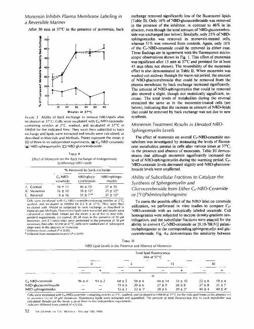
Monensin Inhibits Plasma Membrane Labeling in a Reversible Manner
After 30 min at 37°C in the presence of monensin, back
Tc
[a.I . z 6 0
-o 5C
,~ 4(~ 4)
, o lu
, '7
o
I 0 20 3.0 40 50 6 0
M i n u t e s a t 3 7 ° C
FIGURE 3 Abi l i ty of back exchange to remove NBD-lipids after incubation at 37°C: Cells were incubated with C6-NBD-ceramide- containing vesicles at 2°C, washed, and incubated at 37°C in HMEM for the indicated t ime. They were then subjected to back exchange and lipids were extracted and results were calculated, as described in Materials and Methods. Points represent the mean ± SD of three to six independent experiments. (@) C6-NBD-ceramide; (A) NBD-sphingomyel in; (I-l) NBD-glucocerebroside.
TABLE II
Effect of Monensin on the Back Exchange of Endogenously Synthesized NBD Lipids
% Removed by back exchange
C6-NBD- NBD-gluco- NBD-sphingo- ceramide cerebroside myelin
A. Control 1 6 ± 11 4 6 + 10 51 ± 10 B. Monensin 16 + 10 1 6 ± 12" 25 ± 12" C. Reversal 8 ± 16 31 ± 7*'* 31 ± 12"
Cells were incubated with C6-NBD-ceramide-containing vesicles at 2°C, washed, and incubated in HMEM for 0.5 h at 37°C. They were then incubated with HMEM or subjected to back exchange as described in Materials and Methods. Fluorescent lipids were extracted and results were calculated as described. Values are the mean ± SD of five to nine inde- pendent experiments. (A) control, (B) all steps in the presence of 10 pM monensin, and (C) initial steps were performed in the presence of 10 pM monensin; then after 10 min at 37~C, cells were washed and all subsequent steps were in the absence of monensin.
* Different from control (P < 0.05). * Different from monensin-treated (P < 0.05).
exchange removed significantly less of the fluorescent lipids (Table II). Only 16% of NBD-glucocerebroside was removed in the presence of the inhibitor, in contrast to 46% in its absence, even though the total amount of NBD-glucocerebro- side was unchanged (see below). Similarly, only 25% of NBD- sphingomyelin was removed in monensin-treated cells, whereas 51% was removed from controls. Again, only 16% of the C6-NBD-ceramide could be removed in either case. These findings are in agreement with the fluorescence micro- scopic observations shown in Fig. 1. This effect of monensin was significant after 15 rain at 37"C and persisted for at least 45 min (data not shown). The reversibility of the monensin effect is also demonstrated in Table II. When monensin was washed out midway through the warm-up period, the amount of NBD-glucocerebroside that could be removed from the plasma membrane by back exchange increased significantly. The amount of NBD-sphingomyelin that could be removed also showed a slight, though not statistically significant, in- crease. The total levels of metabolites during the reversal remained the same as in the monensin-treated cells (see below), indicating that the increase in amount of NBD-lipids that could be removed by back exchange was not due to new synthesis.
Monensin Treatment Results in Elevated NBD- Sphingomyelin Levels
The effect of monensin on overall C6-NBD-ceramide me- tabolism was investigated by measuring the levels of fluores- cent metabolites present in cells after various times at 37"C, in the presence and absence of monensin. Table III demon- strates that although monensin significantly increased the level of NBD-sphingomyelin during the warming period, C6- NBD-ceramide levels decreased slightly and NBD-glucocere- broside levels were unaffected.
Ability of Subcellular Fractions to Catalyze the Synthesis of Sphingomyelin and Glucocerebroside from Either C6-NBD-Ceramide or [3H]PalmitoyIsphingosine
To assess the possible effect of the NBD label on ceramide utilization, we performed in vitro studies to compare C6- NBD-ceramide with an isotopically labeled ceramide. Cell homogenates were subjected to sucrose density-gradient cen- trifugation, and the subcellular fractions were assayed for the ability to convert C6-NBD-ceramide or [9,10-3H(N)] palmi- toylsphingosine to the corresponding sphingomyelin and glu- cocerebroside. Fig. 4a demonstrates the similarity between
TABLE III
NBD Lipid Levels in the Presence and Absence of Monensin
Total lipid f luorescence Min at 37°C
0 5 15 30
- - + - - + - - + - - +
%
C6-NBD-ceramide 96 ± 0 93 _ 2 68 -+ 5 58 _ 4 44 ± 14 33 ___ 10 23 + 8 19 + 4 NBD-glucocerebroside - - - - 19 _+ 6 20 + 6 27 _ 9 28 _ 8 37 + 8 31 + 7 NBD-sphingomyel in - - - - 12 + 1 22 ± 1" 28 ± 6 39 ± 3* 40 ± 4 49 +_ 4*
Cells were incubated with C6-NBD-ceramide-containing vesicles at 2°C, washed, and incubated in HMEM at 37°C for the indicated times in the absence (-) or presence (+) of 10 pM monensin. Fluorescent lipids were extracted and quantified. The percent of total fluorescence due to each metabolite was calculated. Results are the mean _+ sE) of three to five independent experiments.
* Indicates different from control (P < 0.05}.

m
~'0.5 o ¢ -
. B r -
A m z
1.0
I
z
t 3 L -
,,~ 1.0 t - O
0 0
t~
0
b
C
I I
i I
/
i
. i | i
i x I
I0 20
, i \ =
w !
,q : :
I
: x - x
30 40 50
!
t . -
E o Ob
I.O .E c -
O .
!
"l"
O
8 t P
I.O -~
c
c
E
1.0 ~ e -
i
% Sucrose FIGURE 4 Sucrose gradient profiles of enzymatic activities and markers of subcellular fractions: Cells were subjected to subcellular fractionation and fractions were assayed for the indicated activities. Representative gradients are shown. The ordinate represents spe- cific activity relative to that of the starting "membrane pellet," calculated in cpm/OD2ao or fluorescence units/OD2ao. ~2sl-WGA is based on precipitated cpm/#g protein. (a) [3H]Sphingomyelin syn- thesis (@---@); NBD-sphingomyelin synthesis (x x); (b) [3H]- glucocerebroside synthesis (@---@); NBD-glucocerebroside synthe- sis (x x); (c) galactosyl transferase (Golgi marker) (@--@); 1251- wheat germ agglutinin (plasma membrane marker) (x---x).
the abilities of each fraction to form the appropriate sphin- gomyelin from either precursor, and Fig. 4b demonstrates that the same was true for formation ofglucocerebroside. The peaks for all activities co-align at ~28% sucrose. This corre- sponds with the presence of the Golgi apparatus marker enzyme, galactosyltransferase (Fig. 4c), and is also in good agreement with other reports of the Golgi apparatus density (29, 30). The same co-distribution of NBD peaks and Golgl fraction was obtained on three independent occasions, always in the region of 27-29% sucrose. Plasma membrane was typically found in a region of~32% sucrose (Fig. 4c), whereas
lysosomes were found at 35-40% and 10% sucrose (data not shown). It is unlikely that mitochondria were present at the density of the Golgi fraction (31), but some smooth or rough endoplasmic reticulum may have been present.
DISCUSSION
We have previously shown that C6-NBD-ceramide is metab- olized to fluorescent glucocerebroside and sphingomyelin in cultured fibroblasts at 37"C (l 5). The cells are initially labeled in the mitochondria, endoplasmic reticulum, and nuclear envelope, but as metabolism progresses, the Golgi apparatus becomes intensely labeled, and later, the plasma membrane becomes strongly fluorescent. The results of the present study are consistent with the hypothesis that both fluorescent sphin- gomyelin and glucocerebroside are synthesized inside the cell and are translocated through the Golgi apparatus before arriv- ing at the plasma membrane. This reasoning is based on data obtained from studies conducted in intact, living cells using the fluorescent ceramide precursor in the presence and ab- sence of the ionophore monensin.
Fluorescence microscopy revealed that if cells that had been labeled with C6-NBD-ceramide at 2"C were washed and warmed to 37"C, the Golgl apparatus became intensely flu- orescent. After longer periods at 37"C, the plasma membrane became strongly labeled with both NBD-sphingomyelin and NBD-glucocerebroside (Table II). The fraction of each metab- olite present in the cell that could be removed by back exchange increased with time at 37"C, suggesting that both were being delivered to the plasma membrane from an intra- cellular site. If either compound were synthesized at the plasma membrane, such a time delay would not be expected.
Because the Golgl apparatus was implicated by microscopy as a participant in this metabolism, further studies were conducted in monensin-treated ceils. It is known that monen- sin has multiple intracellular effects, but is most remarkable for blocking the transport of secretory glycoproteins out of the Golgi apparatus, acting somewhere between the proximal and distal regions (for review, see reference 32). Recently, it has been shown to block the further glycosylation of cerebro- sides (33, 34). Fluorescence microscopy of NBD-ceramide labeled cells in the presence of monensin revealed the striking morphological changes in the Golgi apparatus characteristic of this ionophore. Light and electron microscopy of fixed cells have established that monensin treatment leads to a swollen, vesiculated Golgi apparatus (32), but to our knowledge, this is the first demonstration of monensin's effects in living cells.
Monensin treatment markedly reduced visible plasma membrane labeling (Fig. I D), and inhibited the arrival of both NBD-glucocerebroside and NBD-sphingomyelin at the cell surface (Table II) although cellular levels of the latter actually increased (Table III). The finding that NBD-gluco- cerebroside was inhibited from appearing at the plasma mem- brane was not unexpected as most evidence indicates that glycosylating enzymes are localized to microsomal or Golgi fractions (2, 33-36). The inhibition of NBD-sphingomyelin transport to the cell surface by monensin was more significant, inasmuch as the exact site of sphingomyelin biosynthesis is subject to dispute.
The finding that monensin caused the cellular accumula- tion of NBD-sphingomyelin while retarding its appearance at the plasma membrane most strongly suggested that NBD- sphingomyelin was synthesized intracellularly and trans- ported through the Golgi apparatus to the plasma membrane. Were it to be synthesized at the plasma membrane, several
LmSKY AND PAGANO Intracellolar Translocation of Sphingolipids 33

findings should have resulted in this system. One might expect that back exchange would remove more NBD-sphingomyelin, and more r~pidly than it would NBD-glucocerebroside. How- ever, both compounds were removed in a parallel fashion. In the presence of monensin, which significantly raised NBD- sphingomyelin levels, back exchange should have removed even more NBD-sphingomyelin; instead, the amount was decreased. In addition, fluorescence microscopy indicated a decrease in plasma membrane labeling at times where mo- nensin resulted in significantly increased NBD-sphingomyelin levels. Finally, the observation that when monensin is washed out, back exchange reveals the appearance at the plasma membrane of both fluorescent metabolites, although the total amounts remain unchanged, suggests that these compounds were accumulated inside the cell in the presence of monensin.
To evaluate the possible effects of the fluorescent label itself on ceramide utilization, NBD-ceramide and [3H]ceramide were given to subcellular fractions under identical conditions. No preferential metabolism of the radiolabeled vs. NBD- labeled ceramide was detected (Fig. 4), suggesting that the NBD-label does not significantly alter the ability of subcellular fractions to metabolize ceramide. We stress that the exact intracellular site(s) where ceramide was converted to sphin- gomyelin and glucocerebroside could not be definitively es- tablished from our cell fractionation data alone because of possible contamination of this fraction (see Results). How- ever, the finding of highest activity in the Golgi-enriched fraction corroborates the fluorescence microscopic observa- tions on intact cells.
In conclusion, by using a fluorescent ceramide analogue we have observed sphingolipid metabolism in a living system without fixation or disruption of cells. Our results suggest that both the sphingomyelin and glucocerebroside analogue were synthesized intracetlular/y from Cr-NBD-ceramide, and were then translocated through the Golgi apparatus to the cell surface. Importantly, fluorescent sphingomyelin was appar- ently not synthesized at the plasma membrane as has recently been suggested for endogenous sphingomyelin (7, 8). How- ever, at present we do not know how accurately the metabo- lism and intracellular translocation of C6-NBD-ceramide may reflect the behavior of endogenous ceramide. Our findings that (a) exogenously supplied Cr-NBD-ceramide was metab- olized to the expected fluorescent products in intact ceils; (b) C~-NBD-ceramide and [3H]ceramide were metabolized to equal extents by subcellular fractions; and (c) monensin in- hibits the appearance of both isotopically labeled glycosphin- golipids (33, 34) and fluorescent glucocerebroside at the cell surface suggest that the approach with fluorescent ceramide will be useful in further studies of sphingolipid metabolism and translocation in cells.
We thank Dr. Yasuo Kishimoto for performing the high-pressure liquid chromotography studies.
This work was supported by grant GM-22942 from the U.S. Public Health Service. Dr. Lipsky was supported by a postdoctoral fellowship (GM-08848) from the National Institutes of Health.
Receivedfor publication 16 July 1984, and in revised form 25 Septem- ber 1984.
REFERENCES
1. Kishimoto, Y. 1983. Sphingolipid formation. In The Enzymes. P. D. Boyer, editor. VoL XVI. Academic Press, Inc, New York. 358-409.
2. Van Golde, L M. G., J. Raben, ]. J. Batenburg, B. Fleischer, F. Zambrano, and S. Fleischer. 1974. Biosynthesis of lipids in Golgi complex and other subcellular fractions from rat liver, Biochim. Biophys. Acta. 360:179-192.
3. Sribney, M., and E. P. Kennedy. 1958, The enzymatic synthesis of sphingomyelin. J. Biol. Chem. 233:1315-1322.
4. Stoffel, W., and 1. Melzner. 1980. Stu.dies in vitro on the biosynthesis of ceramide and sphingomyelin--a reevaluation of proposed pathways. Hoppe-Seyler's Z. Physiol. Chem. 361:755-77 I.
5, Ullman, M. D., and N. S. Radio. 1974. The enzymatic formation of sphingomyelin from ceramide and lecithin in mouse liver. J. Biol. Chem 249:1506-1512.
6, Bernert, J. T., Jr,, and M. D. Ullman. 1981. Biosynthesis of sphingomyelin from er)¢hro- ceramides and phosphatidylcholine by a microsomal cholinephosphotransferase, Biochim. Btophys. Acta. 666:99-109.
7. Voclker, D. R., and E. P. Kennedy. 1982, Cellular and enzymatic synthesis of sphingo- myelin. Biochemistry. 21:2753-2759.
8. Marggraf, W.-D., F. A. Aodercr, and J. N. Kaofer. 1981. The formation of sphingomyelin from phosphatidylcholine in plasma membrane preparations from mouse fibrob|asts. Biochim. Biophys, Acta. 664:61-73.
9. Marggraf, W.-D., and J. N. Kanfer. 1984. The phosphorylcholine acceptor in the phosphatidylcholine:ceramide cholinephosphotransferase reaction. Is the enzyme a transferase or a hydrolase? Biochim Biophys. Acta. 793:346-353.
lO. Esko. J. D., and C, R. H. Raetz. 1980. Autoradiographic detection of animal cell membrane mutants altered in phosphatidylcholine synthesis. Proc NatL Aead Sci. USA. 77:5192-5196.
11. Marggraf, W.-D., R. Zertani, F. A. Anderer~ and J. N. Kanfer. 1982. The role of endogenous phosphatidylcholine and ceramide in the biosynthesis of sphingomyelin in mouse fibroblasts. Biochim. Biophys. Acta. 710:314-323.
12. Farquhar, M. G., J. J. M. Bergeron, and G. E. Palade. 1974. Cytochemistry of Golgi fractions prepared from rat liver. Z Cell BioL 60:8-25.
13, Sribney, M, 1971. Stimulation ~f sphingomyelin synthetase by sulthydryl reagents. Can, £ Biochem. 49:306-310,
14. Morell, P,, and P, Braun. 1972. Biosynthesis and metabolic degradation ofsphingolipids not comainingsialic acid, J. Lipid. Res. 13:293-310.
15. Li~ky, N. G., and R. E, Pagano. 1983. Sphingolipid metabolism in cultured fibroblasts: microscopic and biochemical studies employing a fluorescent ceramide analogue. Proc. Natl. Acad. Sci. USA 80:2608-2612.
16. Kishimoto, Y. 1975, A facile synthesis of ceramides. Chem. Phys. Lipids. 15:33-36. 17. Ames, B. N.. and D. T. Dubin. 1960. The role of polyamines in the neutralization of
bacteriophage deoxyribonucleic acid. ,/Biol. Chem. 235:769-775. 18. Kremer, J, M, H., M. W. J. v. d. EskeL C. Pathmamanobaran, and P. H. Wiersema.
1977. Vesicles of variable diameter prepared by a modified injection method. Biochem- istry. 16:3932-3935.
19. Bligh, E. G., and W. J. Dyer. 1959. A rapid method of total lipid extraction and purification, Can. J. Biochem PhysioL 37:911-917.
20. Virtanen, 1., P. Ekblom, and P. Laurila. 1980. Subcellular compartmentalization of saccharide moieties in cultured normal and malignant cells. J, Cell BioL 85:429--434.
21, Lowry, O. H., N. J. Rosebrough, A. L. Farr, and R. J. Randall. 1951. Protein measure- ment wilh the Folin phenol reagent J. Biol. Chem. 193:265-275.
22. Burton, K. 1968. Determination of DNA concentration with diphenylamine. Methods EnzymoL 12t3:163-166.
23. Solder, M. D., and P. W. Robbins. 1982. Transmembrane organization of protein glycosylation--mature oligosaecharide-lipid is located on the luminal side of micro- sprees from chinese hamster ovary cells. J. BioL Chem. 257:6796-6801.
24. Sahaglan, G_ G., and E. F. Neufeld. 1983. Biosynthesis and turnover of the maonose 6- phosphate receptor in cultured chinese hamster ovary cells. J Biol. Chem. 258:7 t21- 7128.
25. Bretz. R., H. Bretz, and G. E. Palade. t980. Distribution of terminal glycosyltransferases in hepatic Golgi fractions. £ Cell Biol. 84:87-101.
26. Costantino-Ceccarini, E., and A. Cestelli. 1981. A novel assay method for the biosyn- thesis of galactosyl- and glucosylceramides, Methods Enzymol. 72:384-391.
27. Struck, D. K., and R. E. Pagano. 1980. Insertion of fluorescent phospholipids into the plasma membrane of a mammalian cell, .L Biol. Chem. 255:5404-5410.
28. Lipsky, N. G., and R. E, Pagano. 1984. Fluorescent sphingomyelin labels the plasma membrane of cultured fibroblasts. Ann N. Y. Aead. ScL In press.
29. Goldberg. D. E., and S. Kornfeld, 1983. Evidence for extensive subcellular organization of aspara$ine-tinked oligosaccharide processing and lysosomal enzyme phosphorylation. .L BioL Chem 258:3159-3165.
30. Dumphy, W. G., and J. E. Rothman. 1983. Compartmentation of asparagineqinked oligosaccharide processing in the Golgi apparatus. £ Cell BioL 97:270-275.
31. Evans, W. H. 1978. General methods for the preparation of plasma membranes. In Preparation and Characterization of Mammalian Plasma Membranes. T. S. Work and E. Work, edilors. North-Honand Publishing Co, Amsterdam. 54-61.
32. Tartakoff, A. M. 1983. Perturbation of vesicular traffic with the carboxylie ionophore monensin. Cell. 32:1026-1028.
33. Saito, M., M. Saito, and A. Rosenberg. 1984. Action of monensin, a monovalent cationophore, on cultured human fibroblasts: evidence that it induces high cellular accumulation ofglucosyl- and lactosylceramide (glu¢o- and iactocerebroside). Biochem- istry. 23:1043-1046.
34. Miller-Prodraza, H., and P. H. Fishman. 1984, Effect of drugs and temperature on biosynthesis and transport ofglycosphingolipids in cultured neurotumor cells. Biochtm. Biophys, Acta. 804:44-51.
35. Suzuki, Y., and H. A. Blough. 1982, Enzymatic deoxyglucosylation of ceramides by microsomes of BHK-21 Cells. Biochim. Biophys. Acta. 710:221-229.
36. Keenan, T. W,, D. J. Morre, and S. Basu. 1974. Ganglioside biosynthesis--concemration of glycosphingolipid glycosyhransferases in Golgi apparatus from rat liver. J. Biol. Chem. 249:310-315,
3 4 THE JOURNAL OF CELL BIOLOGY . VOLUME 100, 1985