intracellular distribution of various boron compounds for use in boron neutron capture therapy
TRANSCRIPT

Biochemical Pharmacology. Vol. 4% No. 1, PP. 147-W 1993. Printed in Great Britain.
Ol-%-2952193 $6.00 + 0.00 @I 1993. Pergamon Press Ltd
INTRACELLULAR DISTRIBUTION OF VARIOUS BORON COMPOUNDS FOR USE IN BORON NEUTRON CAPTURE
THERAPY
TIEN NGuYm,*t GORDON L. BROWNELL,* SYLVIA A. HOLDEN+ and BEVERLY A. TEICHER$$
* Nuclear Engineering Department and Whitaker College, Massachusetts Institute of Technology, Cambridge, MA 02139; and $ Dana-Farber Cancer Institute, Boston, MA 02115, U.S.A.
(Receiued 11 March 1992; accepted 8 September 1992)
Abstract-The neutron capture reaction in boron (‘OB(n,cu)‘Li) generates two short-range particles with high linear energy transfer. The effect of neutron capture therapy depends on the selective localization of 1°B atoms in target cells. The determination of the distribution of boron compounds in cancer cells at the subcellular level is required for the understanding of the effect of this treatment. The monomeric sulfhydryl borane (BSH) compound has been used clinically in Japan and preclinically in the U.S.A. Recently, new compounds have been developed: a dimeric sulfhydryl borane (BSSB), a borono- phenylalanine (BPA), and two porphyrin complexes (BOPP and VCDP). This study demonstrates that the porphyrin complexes (BOPP and VCDP) are more cytotoxic than the other three compounds to the rat 9L gliosarcoma cell line. Using atomic absorption spectrophotometry to determine boron content for cellular uptake studies of these agents, we found that of the five compounds tested BOPP (25 @vi) exposure resulted in the greatest boron uptake averaging 305 ng B/lo” cells. BSSB (500 ,uM) was second averaging 93 ng B/lo6 cells, BSH (500 PM) third averaging 62 ng B/lo” cells, VCDP (25 MM) fourth averaging 58 ng B/lo6 cells, and BPA (500 PM) fifth averaging 7.4 ng B/lo” cells. Data on the distribution of boron in the nuclei, mitochondria, lysosomes, microsomes, and cytosomes of 9L cells are also presented.
Neutron capture therapy, in principle, can achieve cellular selectivity and provide a radiation source within each cancer cell which is activated by neutron delivery. Neutron capture therapy involves the administration of a neutron capture agent, such as ‘OB, 6Li, 235U, and 157Gd, to the tumor site and subsequent irradiation with thermal neutrons. Thus far, l”B has been the neutron capture agent that has received the most attention for biological applications because of its relatively high neutron capture cross- section (a, = 3838 barns, 1 b = 1O-24 cm2), its low toxicity, and because of the a, 7Li, and y ray produced upon irradiation with thermal neutrons [l, 21:
“% f 1n6.31% + ‘Li + 4He + 2.792 MeV
93.69% + 7Li* f 4He + 2.314 MeV
+ ‘Li + 4He + 2.792 MeV
To be used for boron neutron capture therapy (BNCT)II, boron compounds must meet certain criteria. Ideally, they should have a high specificity
t Current address: Nuclear Medicine Department, National Institutes of Health, Bethesda, MD 20892.
i Corresponding author: Dr. Beverly A. Teicher, Dana- Farber Cancer Institute, 44 Binney St., Boston, MA 02115. Tel. (617) 732-3122; FAX (617) 732-3463.
1) Abbreviations: BNCT, boron neutron capture therapy; BSH, monomeric sulfhydryl borane; BSSB, dimeric sulf- hydryl borane; BPA, D,L-p-boronophenylalanine; BOPP, a carborane derivative of protoporphyrin; VCDB, a car- boranyl porphyrin; AAS, atomic absorption spectroscopy; and ICP-AES, inductively coupled plasma-atomic emission spectroscopy.
for malignant cells with concomitantly low con- centrations in adjacent normal tissues and blood. Since it is desirable to confine the radiation solely to the neoplastic cells, an intracellular and optimally intranuclear localization of boron would be preferred. The compounds must be water-soluble, and should be soluble at the physiological pH of 7.4 to avoid toxic effects as a consequence of pH changes in the mammalian system. High chemical and biological stability is also of prime importance. Thus, the boron linkage to carbon, hydrogen, nitrogen, oxygen, sulfur or to another boron atom must have a high order of stability; if not, the injected compound would be degraded rapidly to boric acid under in vivo conditions. For use in human treatment, the compound must have a low toxicity and preferably a high boron content. This is essential since large doses of low boron percentage compounds may result in a serious toxicity problem [3].
We report the subcellular distribution of five of the most promising boron compounds, monomeric sulfhydryl borane (BSH), dimeric sulfhydryl borane (BSSB), D,L-p-boronophenylalanine (BPA), a car- borane derivative of protoporphyrin (BOPP), and a carboranyl porphyrin (VCDP), in the rat 9L gliosarcoma cell line grown in vitro. The first two compounds, BSH and BSSB, are sulfhydryl boranes; the third compound, BPA, is a melanin affinic agent; and the last two are porphyrin complexes. Cytotoxicity studies were performed first to deter- mine the toxicity of each boron compound to 9L gliosarcoma cells. Drug uptake studies were used to elucidate the boron levels achievable intracellularly with each of these boron-containing compounds in
147
148 T. NGUYEN et al.
this cell line at a drug concentration producing a surviving fraction of 0.70. Finally, subcellular fractionation experiments were used to provide the information concerning the intracellular distribution of boron carried by these compounds.
MATERIALS AND METHODS
The 9L cell line. The rat 9L gliosarcoma cell line has a doubling time during exponential growth of about 18 hr [4]. 9L Gliosarcomacells were maintained in culture in MEM-a medium (Grand Island Biological Co., Grand Island, NY) supplemented with 10% fetal bovine serum (Sigma Chemical Co., St. Louis, MO) and antibiotics (penicillin, 10,000 U/ L and streptomycin, lO,OOO~g/L; Grand Island Biological Co.). The plating efficiency for this cell line was 60 * 2%.
Boron compounds. Five boron compounds were investigated. BSH, the monomeric sulfhydryl borane (NazBr2HnSH), was purchased from the Callery Chemical Co., Pittsburgh, PA. It has a molecular weight of 222 and a boron percentage of 59.46% by weight. BSH was obtained in powder form, and is extremely easy to dissolve in water. BSSB, the dimeric sulfhydryl borane (Na4Br4HZZSZ), was supplied by Dr. M. Miura, Medical Department, Brookhaven National Laboratory, Upton, NY. It has a molecular weight of 442, a boron percentage of 59.73% by weight, and is readily water soluble. BPA, the boronophenylalanine (CsH1zBN04), a gift from the Callery Chemical Co., was obtained in powder form. It has a molecular weight of 209 and a boron percentage of 5.26% by weight. BPA was formulated in a small volume of hydrochloric acid (HCI) (1 mol of BPA in 2mol of HCl) and then diluted with phosphate-buffered 0.9% saline (PBS) to the desired concentration and physiological pH. BOPP, a closed cage carborane derivative of protoporphyrin (C4SH77B40N4012K2)r was supplied by Dr. S. B. Kahl, Department of Pharmaceutical Chemistry, University of California at San Francisco, CA. It has a molecular weight of 1398 and a boron percentage of 31.14% by weight. The dipotassium salt is readily water soluble. VCDP, a carboranyl porphyrin (C3sHs2N404B1sK4), was supplied by Dr. M. Miura, Medical Department, Brookhaven National Laboratory. It has a molecular weight of 980 and a boron percentage of 19.90% by weight. VCDP, a natural porphyrin derivative, is water soluble, but it dissolves better at pH 7 than at more acidic levels. As in the case with any porphyrin, VCDP in solution creates free radicals which can cause damage to its surrounding tissue (51.
Cytotoxicity of the boron compounds. 9L Glio- sarcoma cells were grown as monolayers in MEM- a medium. Cytotoxicity assays were performed during the exponential growth period of cells by determining colony formation [6,7]. 9L Cells (lo5 flask) were allowed to grow in complete medium for 2 days. Each experiment included control groups treated with the vehicle only and groups treated with various concentrations of the boron compounds. After exposure of the cells to the vehicle or to each boron compound over a concentration range of 25 to 200 PM for BSH BSSB and BPA, and 5 to 250 ,uM
for BOPP and VCDP in serum-free medium for 60 min at 37” in the dark in an atmosphere of humidified air with 5% COZ, cells were washed three times with sterile PBS, suspended by treatment with 0.01% trypsin, and counted by a hemocytometer. Known numbers of control and drug-treated cells were then plated onto 60 mm culture dishes at three different dilutions, in duplicate, in fresh serum- supplemented medium. After 1 week the dishes were stained with crystal violet to visualize the colonies formed by the surviving cells. Colonies of greater than 50 cells were counted manually, and the results were expressed as the surviving fraction of treated cells compared to untreated (control) cells. Due to their light-sensitive characteristics, when either of the two porphyrins (BOPP and VCDP) was present in the cell medium, the flasks and dish were wrapped or covered with aluminum foil during handling. The experiment was repeated three times with each boron compound.
Cellular uptake. 9L Cells (6.0 x 10⁷) in exponential growth were suspended by treatment with trypsin and then resuspended in serum-free (Y medium at a concentration of 4 x lo6 cells/ml and maintained in a 37”, 5% COr humidified incubator for 1 hr. Silicon oil with a density of 994.6 kg/m3 (General Electric, Waterford, NY) was added to 1.8-mL micro- centrifuge tubes (0.4 ml/tube), and then 1 mL of suspended, well-dispersed cells was added to each tube. Due to different viscosities, the cells and medium stayed above the silicon oil in the tubes. At various time points (60, 50, 40, 30, 20, 10, 5, 3, 2, and 1 min, and 30,15 and 0 set), the boron compound was added to each tube. Since the direct cytotoxicity of the boron compounds varies greatly, the two porphyrins (BOPP and VCDP) being much more toxic to 9L cells than the other three compounds (BSH, BSSB, and BPA), and iso-effective con- centration (the concentration that resulted in a surviving fraction of 0.70 by each of the five boron compounds) was chosen for the uptake and subcellular fractionation studies. This iso-effective concentration for BOPP and VCDP was 25 PM, and for BSH, BSSB, and BPA 5OOpM. At the end of the exposure period, the tubers were centrifuged in a microcentrifuge (Fisher Scientific, model 235B) at 13,000g for 5 min. The cells were pelleted through the medium and silicon oil. The medium and silicon oil were then removed from the tubes using a Pasteur pipet. The excess oil and medium on the inside tube walls were wiped off with cotton sticks. Distilled water (5OpL) was added to each tube, and cells were lysed for lOsec, using a Fisher Sonic demembrator. A known volume of the lysates was injected into the atomic absorption spectro- photometer to measure intracellular boron levels. The zero time-point sample was used to obtain the boron background level, which was subtracted from the boron levels of the other time points. All tubes with porphyrins were wrapped with aluminum foil during the experiment. Each time point was repeated three independent times.
Subcellular fractionation. 9L Cells were allowed to grow in LY medium containing 10% fetal bovine serum for 4 days. The cells were then suspended by treatment with 0.01% trypsin and counted.
Intracellular distribution of boron compounds 149
Suspended cells (9.0 x 10’) were separated into two aliquots of 2.3 x lo6 cells/ml and allowed to rest for 30 min in serum-free medium in a 37’, 5% COr humidi~ed incubator. The drug exposure time was 15 or 60 min. After each time period, the medium containing the boron compound was removed by centrifugation of the cells at 400g for 3 min. Cells were washed by resuspension and centrifugation three times with cold PBS to remove extracellular boron, During the last wash, cells were counted to determine the final cell concentration before fractionation, then centrifuged at 400g for 3 min, resuspended in 7 mL of cold distilled water, and lysed by sonication on ice.
The fractionation was performed at 4” in distilled water, following the procedure of Sharma and Edwards [8]. The cell lysates were centrifuged at 1OOOg for 10 min. The resulting pellet was resuspended in 0.25 M sucrose/l.8 mM CaCl&% Triton X-100 solution. An equal volume of 0.34M sucrose/O.l8mM CaCl, solution was added to the bottom of the tube, pushing the lighter solution up to form a sucrose gradient. After cent~~gation at 600g for 10 min, the nuclear pellet was obtained. The first supernatant was centrifuged at 35OOg for IOmin to obtain the mitochondrial pellet. This supernatant fraction was again centrifuged at 16,000 g for 20 min to obtain the lysosomal pellet. The third su~~at~t fraction was untinged at l~,~ g for 60 min to obtain the microsomal pellet. The final supernatant contained the cytosol. Each subcellular fraction was lysed again in 3OpL of distilled water using a sonic demembrator and then a known volume was injected into the atomic absorption s~~trophotometer for boron measurement. The fractionation experiment was repeated three times for each time point of each boron compound.
To examine the retention of boron compounds in the cells, a second aliquot of cells was exposed to each boron compound for 60min, and then resuspended in boron-free medium for 10 min in a 3P, 5% COr humidi~ed incubator. The cells were then washed three times with cold PBS, lysed, and carried through the subcellular fractionation procedure as described above.
Flameless atomic absorption spectrophotometry (AAS). Boron certified atomic absorption standard (102O~g B/mL, Fisher Scientific Co.) was used to prepare working standards by suitable dilutions. Calcium nitrate (OS%, w/v) was used as a boron matrix modifier [9]. A calibration curve of atomic absorbance versus boron mass was produced using boron atomic absorption standard solutions. A vibration curve of atomic absorbance versus boron mass was also produced using the specific boron compound in each of the uptake and subcellular fractionation studies. This second calibration curve was used to determine the boron level in the experimental samples, while the first curve was combined with the second curve to determine the efficiency of the atomic aborption sp~rophotometer for a boron compound.
Experimental samples (15pL) from uptake or subcellular fractionation studies were atomized from the walls of pyrolytidally coated graphite tubes (Table 1). In the cases of BSSB, BOPP, and VCDP,
S? 413-K
Table 1. Operating parameters for the determination of boron for atomization using graphite tube
Parameter Setting
Wavelength Split width Lamp current Drying Thermal decomposition Atomi~~on Tube reconditioning
249.7 nm 0.7 nm 30 mA 90”, 10 set 13~1400”, 45 set (4 steps) 26.50”, 10 set 20”, 5 set
E 10’ 1
o to-2 *B-l
I 03SSB
I BPA
aBOPP
AVCDP
10 -5-h-I 0 200 600 1000
DRUG t?DNCENT%ITDN. uM
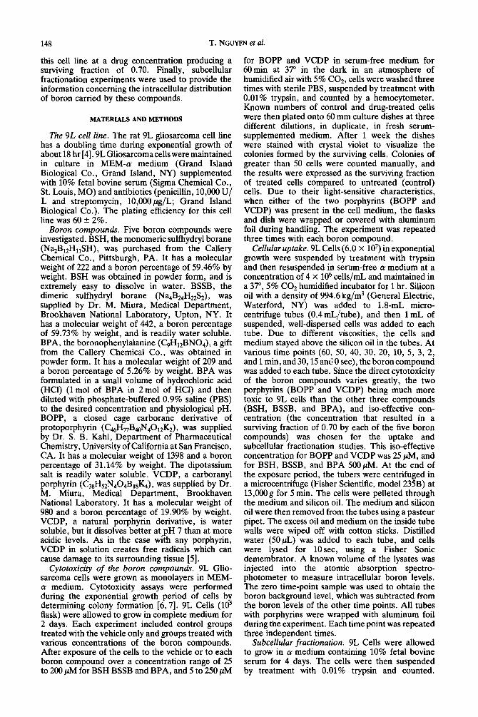
Fig. 1. Survival of 9L gliosarcoma cells exposed to various concentrations of each boron compound for 1 hr. Points are the means of three separate experiments; error bars
represent the SEM.
the samples were further diluted with a known volume of distilled water before being injected into the graphite tube, to bring the absorbance levels to within the calibration range. An atomic absorption spectrophotometer (Perkin Elmer, model 2380), equipped with a graphite furnace (Perkin Elmer, model 400) and a boron hoilow-cathode lamp was used. The operating parameters were set as shown in Table 1. The measurements were made in duplicate in each of the three independent exper- iments.
RESULTS
The number of boron atoms per molecule in the five boron compounds investigated in this study ranged from 40 in BOPP to 1 in BPA. The cytotoxicity of each boron compound toward exponentially growing 9L rat gliosarcoma cells was determined by colony formation. The survival of 9L cells following exposure to various concentrations of each compound for 1 hr is shown in Fig. 1. The two sulfhydryl boranes and the BPA showed very little cytotoxicity toward 9L cells up to a concentration of 1 mM, while the two prophyrins were much more cytotoxic. A 500 &f concentration of either borane
150 T. NGUYEN et al.
0.3-
g 0.2'.
3
$ 0.1'.
% 0.07
/,
0 10 20 30 10
a.
Boron (ng)
[a] -o- B standard
4-
3'.
2-
,8"(
1'
0 20 40 60
Boron (ng)
[c] 0 BSSB
0.
0, 0. s 2 0.
1 on 0. -4
0.
Boron (ng)
[b] o BSH
20
15
10
05
00 i/J 0 5 10 15 20 25
Boron (ng)
[d] A BPA
0 30 60 SO 120 0 20 40 60 80
Boron (ng) Boron (ng)
[e] 0 BOPP’ [fl V VCDP
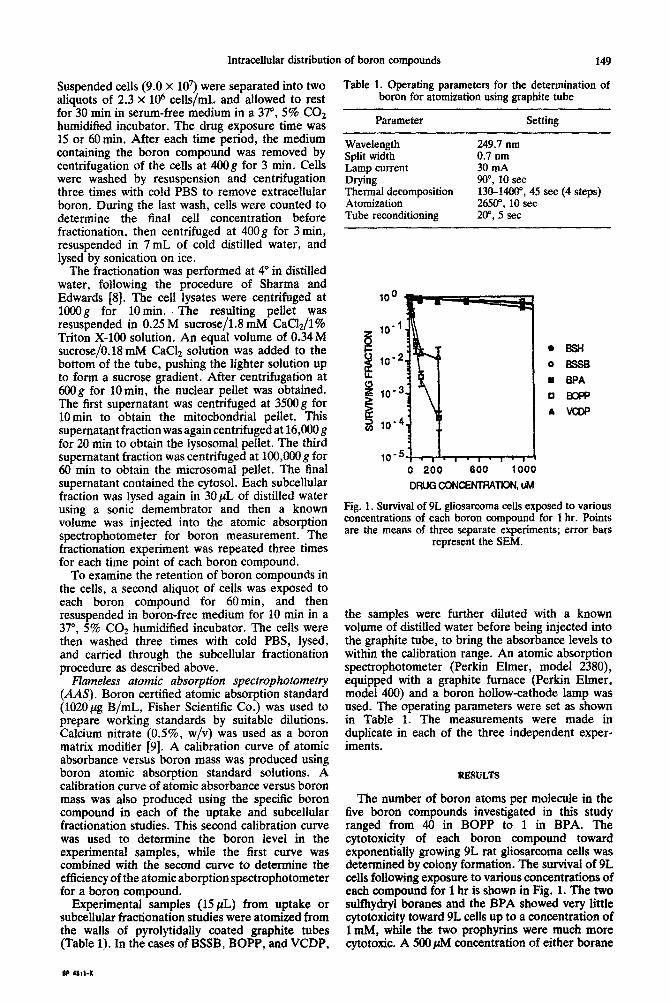
Fig. 2. Calibration curves for boron atomic absorption of standard solution (a) and of the compounds (b-f).
produced a surviving fraction of about 0.70, while that same surviving fraction was obtained with a 25 PM concentration of either porphyrin. Of the two porphyrins, there was 1 log of cell kill with a 30 PM concentration of either BOPP or VCDP, and 2 logs of cell kill with 80 PM BOPP or 250 @rn VCDP. An iso-effective concentration of the five compounds for a 0.70 surviving fraction was chosen for the uptake and subcellular distribution studies. This iso- effective concentration was 500 ,uM for BSH, BSSB, and BPA, and 25 PM for BOPP and VCDP.
Efficiency of AAS for boron measurement in various boron compounds. A boron atomic absorption line at 249.7 nm with a split-width of 0.7 nm was used to detect boron in the lysate cell
samples. The standard calibration curve, obtained by using boron certified atomic absorption standard solution diluted with deionized distilled water and constructed from the absorbance intensity, is shown in Fig. 2a. This calibration curve was linear within the range of 5 to 35 ng B (in 15 PL water). Using the boron standard solution, 25 PM boron (4.12 ng B in 15 pL) could be detected by AAS.
For the five boron compounds, however, the calibration curves deviated somewhat from the standard, probably due to variation in the amount of boron atomized from each compound structure [lo]. Panels b to f of Fig. 2 show the calibration curves of each boron compound. These curves were used to determine the boron content of the

Intracellular distribution of boron compounds 151
0.6 Tl
0.5 I 0.4
0.3
02
0.1
0.0 D 0 102030405060
0 10203040.5060
0.6 I
0.4
‘li!I
I
02
EPA 0.0
0 102030405060
TIME, mln
Fig. 3. Uptake of BSH, BSSB or BPA by 9L cells, measured by boron atomic absorption. 9L cells were exposed to a 500 N concentration of each boron compound. Points are the means of three separate experiments; error bars
represent the SEM.
experimental cell lysates. The efficiencies of AAS in boron measurement for each compounds can be derived from these calibration curves. At a 0.20 absorbance level (equivalent to 24.3 ng B from the standard solution), the efficiencies of AAS for these compounds were 100% for BSH, 89.8% for BSSB, and 100% for BPA, BOPP, and VCDP. At higher absorbance levels the efficiency of boron detection was lower. About lOG200 determinations could be made during the lifespan of the graphite tube, and a sensitivity drift of 11 +- 2% (SD) was observed.
Uptake of boron compounds into whole cells. With a concentration of 5OO@f BSH in the cell culture medium, BSH had an initial uptake rate of approximately 0.03 nmol/min within the first 5 min and the u take reached a saturation of approximately 0.5 nmol P 106 cells after 60min (Fig. 3). When 9L cells were exposed to the same molar concentration of BSSB, this compound had an initial uptake rate of about O.O5nmol/min within the first 5min, and
5 0.00
E 0 102030405060
C
w” 0.30
Y
5 5 0.20
0.10
B
f I
0.00
0 102030405060
TIME, min
Fig. 4. Uptake of BOPP or VCDP by 9L cells exposed to a 25pM concentration of each boron compound. Points are the means of three separate experiments; error bars
represent the SEM.
an uptake saturation of about 0.35 nmol/l06 cells after 60min (Fig. 3). BPA, in contrast, had a very slow uptake rate during the first 5 min when the drug concentration was 5OOpM. The uptake then increased linearly to a rate of approximately 0.01 nmol/min and reached 0.7 nmol/l06 cells at 60 min without showing saturation (Fig. 3). With a 20- fold lower concentration of 25 PM BOPP had a high initial uptake rate, 0.06 nmol/min, and appeared to reach saturation, 0.7 nmol/l06 cells, after 20 min. The level of intracellular BOPP then decreased slightly with time (Fig. 4), perhaps reflecting an efflu process. At the same concentration (25 PM), VCDP had an initial uptake rate of 0.02 nmol/min and a lower saturation level than BOPP. The intracellular level of VCDP continued to slowly increase with time (Fig. 4). Overall, in term of molar uptake by 9L cells, BOPP and BPA had the highest uptake level among the five boron compounds while BSSB and VCDP had the lowest.
In terms of boron concentration localized intracellularly, however, BOPP had the highest uptake into 9L cells, due to the large number of boron atoms per BOPP molecule. The maximal uptake level from BOPP exposure (25 PM, 60 min) was 305 ng B/106 cells, about 3.3 times as high as the next highest uptake level, by BSSB (500 PM, 60 min). Because of the low number of boron atoms per molecule, exposure to BPA (5OO@vf, 60 min) resulted in a boron uptake level 41 times less than that of BOPP. BSH (5OOpM, 60 min) and VCDP (25 PM, 60min) had boron uptake levels 5 times lower than the uptake level of BOPP (Table 2). It should be emphasized that at the iso-effective

152 T. NGUYEN et al.
Table 2. Uptake characteristics of boron compounds in 9L cells
Boron compound BSH BSSB BPA BOPP VCDP
Exposure concentration (PM) 500 500 500 25 25 Initial uptake rate (nmol/min) 0.03 0.05 0.01* 0.06 0.02 Maximum intracellular concentration (nmol/l@ cells) 0.51 0.351 0.7$ 0.76 0.3$ Exposure ~ncentration (+ug B/mL) 60.0 120.0 5.5 10.9 4.9 Initial uptake rate (ng B/min) 12.9 0.11* Maximum intracellular level (ng B/l@ cells) 92.7t 7.4s
* Having an initial delay. t Saturated level at 60 min. $ Still increasing at 60 min. § Peak uptake at 20 min.
SUECEtLUtAR DlSTRlBUTlON
60
40
*30 % 0 "0 20
z gl '0
w" 2.0 Y
s Q
= 1.0
0.0
IA BSH L.i*l.l-l~l*14
10 20 30 40 50 80 70
0 10203040508070
90
SSSB 30 1 *, * I., .I. *. s. r
0 10 203040 506070
12.0
10.0
8.0
6.0
4.0
2.0
0.0 0 10203040506070
TIME, mln
150 :A
BPA
0.0 0 10203040508070
Fig. 5. Boron content of various subcellular fractions of 9L gliosarcoma ceils, measured by boron atomic absorption after exposure to BSH, BSSB or BPA. The cellular fractions were: (1) cytosol (A), (2) nuclei (O), (3) mitochondria (0), (4) lysosomes (W), and (5) microsomes (Cl). The zero time point was the time of addition of the boron compound to the cells. The 60-min time point was immediately after removal of the boron compound. Points are the means of three separate experiments; error bars
represent the SEM.
cbncetittation that resulted in a 0.70 surviving frac- tion to 9L cells (5OOpM for BSH, BSSB and BPA, and 25@4 for BOPP and VCDP), the drug concentrations in terms of boron mass per unit volume of the cell medium were not the same for all five compounds. At this iso-effective concentration, BPA and VCDP had a boron concentration 2 times lower than that of BOPP, while BSH and BSSB provided a boron concentration 6.5 and 13 times higher than the one provided by BOPP, respectively (Table 2).
Subcellular d&tribution. The pattern of uptake and the intracellular distribution of each boron compound in 9L cells are shown in Figs. 5 and 6. When the cells were exposed to 500,uM BSII, the microsomal fraction showed a sharp increase of boron content over the period from 15 to 60min.
Boron content in the cytosol, highest among the subcellular fractions, also increased noticeably, while the other fractions of the cell showed only a slight increase of boron content over the same time course. The mit~hond~al fraction had the lowest percentage of in~acellul~ boron carried by BSH compared to the other fractions.
In the case of BSSB , the microsomes and lysosomes showed the sharpest increases in boron content between 15 and 60 min, with the lysosomal fraction showing a slightly sharper increase and the microsomal fraction showing a higher boron content (Fig. 5). The nuclear fraction had the least amount of boron, with a slower increase with time. The ~tochond~~ fraction initially had a moderate amount of boron, but this amount did not increase with time.

Intracellular distribution of boron compounds
SUBCELLULAR DISTRIBUTION 1800 T T I 4oo I
6ooO 10203040506070
0 0 10203040506070 0 10203040506070
TIME, mln
Fig. 6. Content of various subcellular fractions of 9L gliosarcoma cells, measured by boron atomic absorption after exposure to BOPP or VCDP. The cellular fraction were: (1) cytosol (A), (2) nuclei (O), (3) mitochondria (0), (4) lysosomes (U) and (5) microsomes (0). The zero time point was the time of addition of the boron compound to the cells. The 60-min time point was immediately after removal of the boron compound. Points are the means of three separate experiments; error bars
represent the SEM.
153
When 9L cells were exposed to 500 PM BPA, the cytosol took up the most intracellular boron and the nuclear fraction showed the second highest boron percentage, increasing rapidly with time (Fig. 5). The lysosomal fraction also showed a rapid increase of boron uptake, while the level of boron in the microsomes and the mitochondria increased more slowly with the slowest increase in the mitochondria.
Intracellular boron delivered by BOPP in all the fractions, except the nuclei, had approximately the same rate of uptake between the 15 and 60-min time points, with the cytosolic fraction containing the most intracellular boron, and the microsomal and the lysosomal fractions having the second highest amount of boron (Fig. 6). The mitochondrial fraction initially contained a small amount of boron, but its boron content increased relatively rapidly whereas the level of boron in the nuclear fraction remained small.
Upon exposure to 25pM VCDP, all the cellular fractions seems to increase in boron content very slowly, with the lysosomal and mitochondrial fractions increasing at a faster rate than the other subcellular fractions (Fig. 6). Similar to BOPP, the nuclear fraction took up the least percentage of boron carried by VCDP, and it did not increase significantly with time.
To examine the retention of these five compounds in the 9L cells, the boron compounds were allowed to accumulate in the cells for 60 min and then the cells were exposed to boron-free medium for an additional 10min. In all cases intracellular boron content decreased markedly (Figs. 5 and 6). Micro- somal boron delivered via BSH, microsomal and lysosomal boron delivered by BSSB, nuclear and
lysosomal boron delivered by BPA, and lysosomal, microsomal and cytosolic boron delivered by BOPP showed the largest decrease from their initial boron content. The mitochondrial seemed to retain boron content, even though at the 60-min time point the content was only moderate. With BSH, BSSB and BPA, the boron content in the mitochondrial fractions appeared to increase slightly during the lo- min drug-free period. However, these increases were not statistically significant. In comparison with the intracellular boron at 60 min, nuclear boron carried by BSSB decreased by 92% during the lo-min maintenance in the boron-free medium. Microsomal boron delivered by BOPP and BSH decreased by 6% and 59%, respectively. VCDP was retained to the highest degree by the 9L cells.
DISCUSSION
For use in boron neutron capture therapy, the boron compound usually is formulated for administration to the patients either intravenously or intraarterially in appropriate doses. All five boron compounds examined here were sufficiently water soluble for such formulations.
Other investigators have reported that the two sulfhydryl boranes studied are non-toxic to biological systems at 3Opg “B/mL [ 111 and the borono- phenylalanine at pH7.0 has an LDso value of more than 3000 mg/kg in Sprague-Dawley rats [12]. BOPP has been reported to be non-toxic to V-79 Chinese hamster cells grown in vitro at a concentration of 30 pg ‘OB/mL [13]. In the present study with 9L cells, the two boranes and the phenylalanine were found to be non-cytotoxic at concentrations up to

154 T. NGUYEN et al.
1OClO ,uM for 1 hr, but the two porphyrins were much more cytotoxic. The toxicity of a compound may cause a limit to the dosage given a patient, thus reducing the potential effect of the therapy; therefore, uptake studies were carried out at relatively non-toxic concentrations of the agents.
From the uptake curves for BSH and BSSB, it is likely that the concentration difference between extra- and intracellular boron was the main driving force for the transport of these two boron compounds into the cells, suggesting uptake by passive diffusion. The rapid rate of boron uptake as delivered by BOPP and VCDP over the first 5 min also suggests an important role for concentration gradients in the uptake of these two compounds in the cell/medium system (Fig. 4). The decreased uptake level of boron at 20 min after the cells were treated with BOPP, however, may indicate an efflux of the boron compound from the cells. From the structure of BOPP, it may be suggested that the bond between the -COO-B1zHu group and the -CH2 group may have been cleaved while the BOPP molecule was in the cells. It has been reported, however, that when V-79 Chinese hamster cells were exposed to BOPP at 30 pg ‘OB/mL for 18.5 hr, the concentration of BOPP in these cells steadily increased with time, as measured by prompt-y neutron activation analysis and in terms of boric acid equivalents [13]. In the case of VCDP, after the 9L cells were exposed to this compound for 30 min, the boron level in the cells increased markedly and appeared to be continuing to increase after 60 min of cell exposure to the compound. The uptake of boron delivered by BPA in 9L cells displayed a delay at the beginning and then increased gradually (Fig. 3).
Subcellular fractionation, combined with ASS, was used to measure boron concentrations in cell structures. Several methods have been used to analyze boron content in tissues. The calorimetric assay, with a detection limit of 0.8 pg B, is tedious and also has been reported to present possible problems in the analysis of borane cages [14-161. The use of prompt-y spectroscopy allows the analysis of ‘OB/g at a concentration as low as 1 pg “B/g tissue, and does not require a chemical conversion procedure. However, this technique is not sensitive enough to detect ng “‘B in mg samples [ 171. This technique requires extensive access to a neutron beam and works only with ‘OB-enriched compounds. Track-etch detectors have been used for boron quantitative analysis and have been reported to provide sensitivity below 0.02 pg “B/mL [RX]. This approach is generally delicate and, like prompt-y spectroscopy, requires a neutron source. Recently, boron determination by inductively coupled plasma- atomic emisson spectroscopy (ICP-AES) has been introduced with detection limits as low as 1 pg B/g tissue and has produced reliable results [19].
AAS provides an alternative technique for boron analysis. Even though the AAS detection limit for boron is generally higher than that of ICP-AES (5- 10 ng B/mL vs 0.1-7 ng B/mL, respectively [20]), AAS was able to detect boron with a sensitivity of 4.12 ng B in 15 yL of cell lysate. The AAS procedure is simple, and the results are quite reproducible. The pyrolytic coating on the graphite tube wall has
been found to minimize the memory effects on the tube, thus reducing the standard deviation of the results [9]. Using coated tubes, together with heating at a very high rate during the atomization stage and adding suitable matrix modifier was found to improve the sensitivity of the technique. Non-linear calibration curves as reported here have generally been seen in boron detection using AAS. This is probably due to the doublet 249.71249.8 nm wavelength of boron as well as to carbide formation [20]. Due to the high temperatures applied, the life-span of the graphite tubes was short. The gradual sensitivity drift that was observed during the life-span of the tube was insignificant.
The results of the subcellular fractionation study showed that all five compounds localized mainly in the cytosolic fraction, which accounts for the largest percentage of the cell volume. All five compounds, however, diffused out of the cells quickly when the cells were exposed to boron-free medium, suggesting free diffusion of the boron compounds from the cells and indicating that none of these five compounds forms strong bonds to cellular components.
Boron localized in the cell nuclei would be expected to result in the most efficient cell killing by BNCT [21]. Of the five compounds investigated, however, only BPA produced a high percentage of intranuclear boron. The amount of BPA boron localized in the nuclei, unfortunately, was not high. In fact, even with the highest percentage, due to its low boron carrying capability, BPA still delivered the lowest boron mass to the nuclei in comparison to the other compounds. It may be worth noting that the three compounds that have the highest molecular weights showed the lowest percentages of boron in the nuclear fractions. It is interesting that four (BSH, BSSB, BOPP, and VCDP) of the five compounds investigated localized to a high degree in the microsomal fraction of the 9L cells. This fraction, consisting of cell membrane fragments and of endoplasmic reticular fragments, contains the most lipophilic fraction of the cellular components. The lysosomal fraction, accounting for a small percentage of total cellular volume, appeared in three cases (BSSB, BOPP and VCDP) to contain high percentages of boron. The high concentration of boron in this cell structure may be important, since lysosomes contain many acid hydrolase which, when released as a result of the high linear energy transfer radiations due to the therapy, can result in damaging proteolytic processes within the cell. The mitochondrial fractions generally contained only a small percentage of total intracellular boron.
From a pharmacological standpoint, the selective retention of boron compound in tumor cells is of prime importance. Examination of the retention of each boron compound in the 9L cell system, however, showed that all five of the compounds investigated diffused readily from the cells, indicating no interactive processes between these compounds and cellular components. The ideal boron compound for boron neutron capture therapy would be non-toxic to the host system, would have a high selectivity for radiation-sensitive regions in malignant cells, and would achieve a high boron concentration in the

Intracellular distribution of boron compounds 155
7. Thilly WG, DeLuca JG, Hoppe H and Penman BW, Phenotypiclag and mutation to 6-thioguanine resistance in diploid human lymphoblasts. Mutat Res SO: 137- 144, 1978.
tumor with long retention time. Comparing the five compounds investigated, it appears that each compound partially fulfills these specifications. BOPP provides the greatest intracellular boron levels. Albeit the percentage of nuclear boron delivered by BOPP is not high, its high boron- carrying capacity has resulted in a relatively high boron content in this radiation-sensitive region when compared with other compounds. The cytotoxicity of BOPP, however, limited the drug concentration that could be used in the uptake studies. Perhaps BOPP derivatives can be developed which would have a high retention in neoplastic tissues, providing reliable neutron targets in malignant cells for the therapy. VCDP, the other porphyrin derivative studied, also provides a significant amount of intracellular boron in 9L cells. Its cytotoxicity is tolerable, and this compound appears to have a better retention in the cells than the other compounds. BSSB, on the other hand, has low cytotoxicity and therefore can be administered at high concentrations resulting in a high intracellular boron content. Moreover BSSB produced a relatively high boron content in the nucleus, indicating that this drug may potentially be more efficient in killing cells upon neutron irradiation. The next step for all of these potential BCNT agents is testing in the tumor-bearing whole animals to determine normal toxicity tissue and tumor versus normal tissue localization.
8. Sharma RP and Edwards IR, c&Platinum: Subcellular distribution and binding to cytosolic ligands. Biochem Pharmacol32: 2565-2669, 1983.
9. Szydlowsky FJ, Boron in natural waters by atomic absorption spectrometry with electrothermal atom- ization. Anal Chim Acta 106: 121-125, 1979.
10. Fairchild RG and Bond VP, Enhancement of Tumor Dose Via Neutron Beam Filtration and Dose Rate, and the Effects of These Parameters on Minimum Boron Contents, Brookhaven National Laboratory BNL Report No. 51730, pp. 1-12. Brookhaven National Laboratory, Upton, NY, 1983.
11. Laster BGH, Kahl SB, Kalef-Ezra J, Popenoe EA and Fairchild RG, Biological efficacy of a boronated porphyrin as measured in cell culture. Strahlenther Onkol 165: 203-205, 1989.
12. Taniyama K, Fujiwara H, Kuno T, Saito N, Shuntoh H, Sakaue M and Tanaka C, Acute and subacute toxicity of i”B-paraboronophenylaianine. Pigment Cell Res 4: 291-297, 1989.
13. Farchild RG, Kahl SB, Laster BH, Kalef-Ezra J and Popenoe EA, In vitro determination of uptake, retention, distribution, biological efficacy, and toxicity of boronated compounds for NCT: A comparison of pophyrins with sulfhydryl boron hydrides. Cancer Res 50: 4860-4865, 1990.
14. Kaczmarczyk A, Messer JR and Pierce CE, Rapid determination of boron in biological materials. Anal Chem 43: 271-272, 1971.
Acknowledgements-This work was supported by NIH Grants ROlCA36508 and ROlCA47379.
REFERENCES
1. Kobayashi T and Kanda K, Analytical calculation of boron-10 dosage in cell nucleus for neutron capture therapy. Radiat Res 91: 77-94, 1982.
2. Kitao K, A method for calculating the absorbed dose near interface from i”B(n, (u)‘Li reaction. Radiut Res 61: 304-315, 1975.
15. Strouf 0, Mertenova E, Schneiderova L, Zamecnikova H and Janku I, Boron determination of neutron capture therapy by calorimetry and emission spectrometry. Strahlenther Onkol 165: 174-176, 1989.
16. Slatkin D Micca P, Forman A, Gabel D, Wielopolski L and Fairchild RG. Boron uotake in melanoma. cerebrum, and blood from ’ Na2BuH,,SH and N+B24HZZS2 administered to mice. Biochem Pharmacol 35: 1771-1776, 1986.
17. Fairchild RG, Gabel D, Laster BH, Greenberg D, Kiszenick W and Micca PL, Microanalytical techniques for boron analysis using the ‘OB(n, (u)‘Li reaction. Med Phys 13: 50-56, 1986.
3. Soloway AH, Boron compounds in cancer therapy. In: Progress in Boron Chemistry (Eds. McCloskey AL and Steinberg H), Vol. 1, pp. 203-234. Pergamon Press, New York, 1964.
4. Barker GW, Hoshino T, Gurcay 0, Wilson CB, Nielsen S and Downie R, Development of an animal brain tumor model and its response to therapy with 1,3- bis)2chloroethyl)-1-nitrosourea. Cancer Res 33: 976 986, 1973.
5. Miura M, Gabel D, Fairchild RD, Laster BH and Warkentien LS, Synthesis and in vivo studies of a carboranyl porphyrin. Strahlenther Onkol 165: 131- 134, 1989.
Res 41: 73-81, 1981.
18. Gabel D, Holstein H, Larsson B, Gille L, Ericson G, Sacker D, Som P and Fairchild RG, Quantitative neutron capture radiography for studying the bio- distribution of tumor-seeking boron-containing com- pounds. Cancer Res 47: 5451-5454, 1987.
19. Bauer WF, Johnson DA, Steele SM, Messick K, Miller DL and Propp WA, Gross boron determination in biological samples by inductively coupled plasma- atomic emission spectroscopy. Strahlenther Onkol165: 176179, 1989.
6. Teicher BA, Lazo JS and Sartorelli AC, Classification of antineoplastic agents by their selective toxicities toward oxygenated and hypoxic tumor cells. Cancer
20. Tsalev DL, Atomic Absorption Spectrometry in Occupational and Environmental Health Practice, Vol. 2. CRC Press, Boca Raton, FL, 1984.
21. Gabel D, Foster S and Fairchild RG, The Monte Carlo simulation of the biological effect of the ‘OB(n, o)‘Li reaction in cells and tissue and its implication for boron neutron capture therapy. Radiat Res 111: 14-25, 1987.