intra-articular bupivacaine: a literature review including
TRANSCRIPT

i
Intra-articular bupivacaine: A literature review including a pilot study
investigating the safety of intra-articular bupivacaine to chondrocytes
by Willem Jacobus van der Merwe
Thesis presented in partial fulfilment of the requirements for the degree of Master of
Medicine (Anesthesiology) at the University of Stellenbosch
Supervisors:
Dr. L Firfiray
Senior Consultant
Department of Anesthesiology and Critical Care
University of Stellenbosch
Prof. AR Coetzee
Professor and Executive Head
Department of Anesthesiology and Critical Care
University of Stellenbosch
December 2015

ii
DECLARATION:
By submitting this thesis electronically, I declare that the entirety of the work contained therein is
my own, original work, that I am the authorship owner thereof (unless to the extent explicitly
otherwise stated), that reproduction and publication thereof by Stellenbosch University will not
infringe any third party rights and that I have not previously in its entirety or in part submitted it for
obtaining any qualification.
………………………………
WJ van der Merwe
Date:………………………..
Copyright © 2015 University of Stellenbosch
All rights reserved
Stellenbosch University https://scholar.sun.ac.za

iii
ABSTRACT
Background: Today, intra-articular bupivacaine injections are common practice, with very good
analgesic effects. This method of analgesia is utilized by general practitioners, orthopedic surgeons
and anesthesiologists. Unfortunately, since 2004, more than 200 cases of chondrolysis were noted
following intra-articular bupivacaine infusion. Numerous studies have investigated the safety of
intra-articular bupivacaine. The aim of this thesis was to quantify the risk of chondrolysis with
intra-articular bupivacaine and to guide medical practitioners in safe practice.
Methods: A literature review was done of the most recently published original research, meta-
analysis and review articles. Adverse outcomes were mostly associated with bupivacaine infusions,
rather than with single intra-articular doses of bupivacaine. Limited data on the safety of a single
dose of intra-articular bupivacaine was found. We conducted a pilot study to investigate the safety
of a single dose of intra-articular bupivacaine. A paired, case-controlled, experiment was done
using four merino sheep. The nul hypothesis was that a single dose of intra-articular bupivacaine
did not cause chondrolysis when compared with a dose of intra-articular normal saline. Our results
were added to the thesis to further quantify the risk.
Results: Numerous in vitro and in vivo studies confirmed that bupivacaine is chondrotoxic. The
chondrotoxic effect is time, dose and concentration dependent. Our pilot study revealed that single
dose intra-articular bupivacaine may also be unsafe for chondrocytes.
Conclusion: The administration of intra-articular bupivacaine is toxic to chondrocytes. The
chondrotoxic effect of intra-articular bupivacaine is time, dose and concentration dependent.
Infusions of intra-articular bupivacaine should be contra-indicated. From the limited data available
on single dose intra-articular bupivacaine, it appears that it is also chondrotoxic. Magnesium,
morphine and clonidine may be safer for intra-articular use, but this needs further investigation.
Stellenbosch University https://scholar.sun.ac.za
iv
OPSOMMING
Agtergrond: Vandag word bupivacaine gebruik as intra-artikulêre inspuiting met baie goeie
analgetiese effek. Hierdie metode word ontgin deur, onder andere, algemene praktisyns, ortopediese
chirurge en anestesioloë. Sedert 2004 is meer as 200 gevalle van chondrolise aangeteken na die
toediening van intra-artikulêre bupivacaine infusies. Talle studies het gevolg om die veiligheid van
intra-artikulêre bupivacaine te ondersoek. Die doel van hierdie skripsie is om die risiko verbonde
aan intra-artikulêre bupivacaine te kwantifiseer en aan mediese praktisyns veilige riglyne te stel.
Metode: ‘n Literatuur oorsig is gedoen, insluitende die mees onlangs gepubliseerde oorspronklike
navorsing, meta-analises asook oorsig artikels. Ongunstigde uitkomstes is meestal geassosieer met
intra-artikulêre bupivacaine infusies, eerder as met enkel dosering intra-artikulêre bupivacaine.
Beperkte data met betrekking tot die gebruik van bupivacaine as enkel dosering intra-artikulêr is
beskikbaar. Om die veiligheid van enkel dosering intra-artikulêre bupivacaine te ondersoek is ‘n
loodsstudie gedoen. Dit is ‘n gevalle-kontrole in-vivo eksperiment, met vier merino skape. Die nul
hipotese is dat ‘n enkel dosering intra-artikulêre bupivacaine nie meer chondrolise veroorsaak, as ‘n
enkel dosering intra-artikulêre normale saline nie. Die resultate is ingesluit as deel van die stawende
data om die risiko te kwantifiseer.
Resultate: Talle in-vitro asook in-vivo studies het bevestig dat bupivacaine chondrotoksies is. Die
toksiese effek is tyd, dosis en konsentrasie afhanklik. Ons loodsstudie het ook getoon dat enkel
dosering intra-artikulêre bupivacaine moontlik skadelik vir kraakbeen kan wees.
Gevolgtrekking: Die toediening van intra-artikulêre bupivacaine is toksies vir chondrosiete. Die
chondrotoksiese effek is tyd, dosis en konsentrasie afhanklik. Infusies van intra-artikulêre
bupivacaine is dus gekontraïndikeer. Die beperkte data oor enkel dosering intra-artikulêre
bupivacaine dui ook moontlik chondrotoksisiteit aan. Magnesium, morfien en clonidine kan
moontlik as veiliger alternatief oorweeg word, maar benodig verdere navorsing.
Stellenbosch University https://scholar.sun.ac.za

v
ACKNOWLEDGEMENTS
Contributors:
The author would like to make special mention of the following individuals for their much
appreciated input to make this thesis possible:
Histological analysis:
Dr. EM Geldenhuys
Anatomist and Histologist
Department of Anatomy and Histology
University of Stellenbosch
Prof. BJ Page
Head of Department
Department of Anatomy and Histology
University of Stellenbosch
Mr. R Williams
Manager of Laboratory
Department of Anatomy and Histology
University of Stellenbosch
Management of animals:
Mr. N Markgraaff
Manager
Stellenbosch Animal Unit
University of Stellenbosch
Data interpretation and statistical analysis:
Mr. M Chirehwa
Biostatistics Unit
Centre for Evidence Based Health Care
University of Stellenbosch
Stellenbosch University https://scholar.sun.ac.za

vi
Ethics Approval of in vivo case controlled experiment:
Research Ethics Committee: Animal Care and Use
Financial support:
Harry Crossley Foundation
Supervisors:
Dr. L Firfiray
Senior Consultant
Department of Anesthesiology and Critical Care
University of Stellenbosch
Prof. AR Coetzee
Professor and Executive Head
Department of Anesthesiology and Critical Care
University of Stellenbosch
Stellenbosch University https://scholar.sun.ac.za

vii
DEDICATIONS
I dedicate this thesis to my wife, Annemarie, and my two sons, Guillem and Jakob. Thank you for
all your love, patience and support over the last four years.
Stellenbosch University https://scholar.sun.ac.za

viii
TABLE OF CONTENTS
Declaration ii
Abstract iii
Opsomming iv
Acknowledgements v
Dedications vii
Preface ix
Chapter 1: Literature review: Intra-articular Bupivacaine 1
1.1 Introduction 1
1.2 In vitro studies 2
1.3 In vivo studies 3
Chapter 2: A case controlled experiment to evaluate the in vivo effects of single dose intra-articular 0.5% bupivacaine on cartilage of sheep 6 2.1 Introduction 6
2.2 Methodology 7
2.3 Results 11
2.3.1 Supra-normal single dose in knee joints 11
2.3.2 Therapeutic single dose in stifle joints 15
Chapter 3: Discussion 21
3.1 Literature review 21
3.2 Single dose intra-articular bupivacaine pilot study 21
3.3 Pathophysiology of bupivacaine chondrotoxicity 22
3.4 Alternatives to intra-articular bupivacaine 23
Chapter 4: Conclusion 25
Appendices 26
1. Appendix A: Monitoring sheet for sheep wellbeing 26
2. Appendix B: Modified Mankin Scores of Front Knee joints 27
3. Appendix C: Modified Mankin Scores of Stifle joints 28
Bibliography 32
Stellenbosch University https://scholar.sun.ac.za

ix
PREFACE
The aim of this thesis is to review the literature on the effect of intra-articular bupivacaine on
chondrocytes. As most of the adverse outcomes were associated with intra-articular bupivacaine
infusions, the effect of a single dose of intra-articular bupivacaine on chondrocytes was further
investigated by conducting a pilot study on four adult merino sheep. The literature review will be
discussed in chapter 1 of this thesis and the results of the pilot study will be disclosed in chapter 2,
followed by a discussion of our findings and a conclusion in chapters 3 and 4 respectively. We trust
that it will give more insight and guidance into the safe practice of intra-articular bupivacaine.
WJ van der Merwe
Registrar
Department of Anesthesiology and Critical Care
University of Stellenbosch
Stellenbosch University https://scholar.sun.ac.za

1
CHAPTER 1
LITERATURE REVIEW: INTRA-ARTICULAR BUPIVACAINE
A literature review was done of the available published literature, for the time period of 1985 to
2015.
1.1 INTRODUCTION
Today, intra-articular bupivacaine injections are common practice amongst orthopedic surgeons,
general practitioners and anesthetists with very good analgesic effects.1-2
This method of pain control is utilized in the outpatient, inpatient and perioperative setting.
Bupivacaine has been used either as the sole drug of the injectate, or in combination with additives
such as clonidine, opiates, corticosteroids and epinephrine.3
This practice of analgesia has however not been without local risks to joints. There are even reports
of possible systemic toxicity following intra-articular administration of bupivacaine.4-5
According to Meining et al.6, peak plasma levels after therapeutic intra-articular dosages occur
approximately 20 minutes after administration, but the actual plasma level is normally eight to ten
times less than the human convulsion threshold. Certain surgical techniques compromising the
cartilage and bone structure might increase the risk of systemic uptake.6 However, the reported
incidence of systemic compromise after intra-articular bupivacaine is extremely low.
More prevalent and of greater concern, are reports of a possible chondrotoxic effect of intra-
articular bupivacaine, especially in cases of intra-articular infusion.7-9
Between 2004 and 2012 more than 200 cases of chondrolysis were noted following intra-articular
bupivacaine infusion. The mean age of the patients who developed chondrolysis was only 30 years,
suggesting bupivacaine as the possible cause.11 In a series of 19 patients who received intra-articular
infusions of bupivacaine and epinephrine, 12 developed chondrolysis. 10
Numerous in vitro and in vivo studies have investigated the safety of intra-articular bupivacaine to
chondrocytes.
Stellenbosch University https://scholar.sun.ac.za

2
1.2 IN VITRO STUDIES
Drogoo et al.13 claimed that intra-articular bupivacaine infusions could be utilized with the
provision that the infusion duration did not exceed 48 hours. Interestingly, they found that
bupivacaine in combination with epinephrine caused significantly more chondrotoxicity than
bupivacaine alone. Human chondrocytes, cultured in a bioreactor that mimicked synovial
metabolism, were exposed to 1% lidocaine, and 0.25% and 0.5% bupivacaine. Chondrocytes were
exposed to these solutions alone and with epinephrine. Cell viability was assessed 24, 48 and 72
hours after continuous infusion. Similar chondrocyte necrosis was found in 0.25% and 0.5%
bupivacaine groups at 24 and 48 hours when compared to control groups. When epinephrine was
added, significantly greater necrosis was seen (p < 0.05) at all intervals. At 72 hours, the 0.5%
bupivacaine alone had caused significant necrosis.13
Lo et al.14 confirmed the chondrotoxic effect of 0.25% bupivacaine, 1% lidocaine and 0.5%
ropivacaine on bovine cartilage, which had been sampled from the radial-carpal joint. Significantly
worse chondrotoxicity was shown with longer periods of exposure of the cartilage to the local
anesthetics.14
Hennig et al.15 found intra-articular bupivacaine 0.5% to be toxic to canine cartilage.15
Baker et al.16 compared the effect of different local anesthetics at different concentrations on human
chondrocytes. Cells were exposed to levobupivacaine (0.13%, 0.25%, 0.5%), bupivacaine (0.13%,
0.25%, 0.5%), ropivacaine (0.19%, 0.38%, 0.75%), normal saline and 10% magnesium sulphate, for
15 minutes each. After 24 hours, cell viability was found to be significantly decreased in all local
anesthetic groups and appeared to be concentration dependent (p < 0.05). There was no reduction in
chondrocyte viability after exposure to 10% magnesium sulphate.16
Wang et al.17 evaluated chondrocyte viability and chondrocyte function after exposure to
bupivacaine. They used an intervertebral disc organ model, which they harvested from 10 week old
mice. They waited 3 days before exposing the cells to bupivacaine to compensate for surgically
induced inflammatory responses. The cells were exposed to bupivacaine 0.1%, 0.25% and 0.5%, for
60 minutes. The 0.5% bupivacaine group was also exposed for different time periods of 30, 60 or
120 minutes. Cell viability, collagen synthesis and proteoglycan matrix synthesis was evaluated.
They showed a dose and time dependent reduction in cell viability. Bupivacaine 0.5% killed 20% of
chondrocytes after 60 minutes and up to 70% after 120 minutes (95% CI 53.2 – 70.1%).
Stellenbosch University https://scholar.sun.ac.za

3
Bupivacaine 0.5% reduced the proteoglycan matrix synthesis three-fold and collagen synthesis four-
fold. The authors postulate that the reduced synthetic function that was demonstrated may only be a
reflection of chondrocyte loss, and not necessarily reflect an inhibition of chondrocyte function by
bupivacaine.17
Breu et al.18 attempted to quantify bupivacaine's chondrotoxic effect on human mesenchymal cells,
and compared it to equipotent concentrations of mepivacaine and ropivacaine. After 60 minutes of
exposure to the local anesthetics, bupivacaine was found to be significantly more chondrotoxic,
reducing cell viability by 5% (± 1%) when compared to mepivacaine's 1% (± 0%) reduction.
Ropivacaine proved to be the least toxic.18
Beyzadeoğlu et al.19 showed that both bupivacaine and its enantiomer, levobupivacaine, were
chondrotoxic to rat cartilage. Levobupivacaine proved to be more toxic.19
In contrast, Erden et al.20 could not illustrate any increase in inflammation in articular and peri-
articular surfaces in a rat model after treatment with levobupivacaine. The authors advocated that
levobupivacaine might therefore be a safe alternative.20 A possible cause for their negative results,
might be that the samples were only investigated up to 21 days post intervention, and the changes
become more prevalent at longer intervals after exposure.11
In an experiment done by Gungor et al.21, no statistical difference in chondrocyte apoptosis could be
demonstrated between bupivacaine and levobupivacaine.21
Most of the in vitro studies were not done on full thickness cartilage and therefore might falsely
increase the likelihood of positive results. It is also problematic to make assumptions based on in
vitro cell cultures. The complexity of the synovial environment, extracellular collagen and
proteoglycan matrix interactions can only be mimicked to a certain degree, before compromising
the true milieu. In vivo studies have therefore also been done.
1.3 IN VIVO STUDIES
Gomoll et al.12 divided 30 male New Zealand white rabbits into three groups, to receive either
normal saline, bupivacaine 0.25% or bupivacaine 0.25% with epinephrine (1:200 000), via
continuous infusion into their glenohumoral joints for 48 hours at a rate of 210μL/h. This dosage
was proportional to the normal infusion rate of 4ml/h administered to adult human patients.
Stellenbosch University https://scholar.sun.ac.za
4
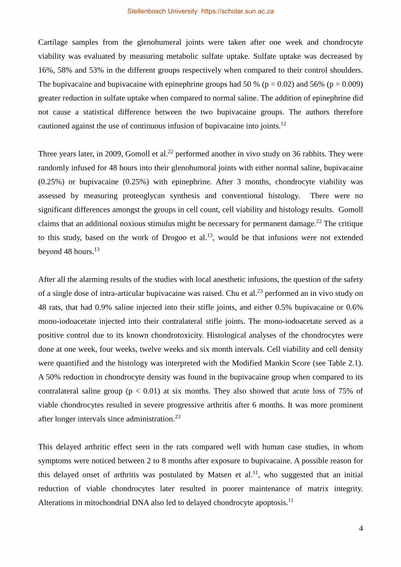
Cartilage samples from the glenohumeral joints were taken after one week and chondrocyte
viability was evaluated by measuring metabolic sulfate uptake. Sulfate uptake was decreased by
16%, 58% and 53% in the different groups respectively when compared to their control shoulders.
The bupivacaine and bupivacaine with epinephrine groups had 50 % (p = 0.02) and 56% (p = 0.009)
greater reduction in sulfate uptake when compared to normal saline. The addition of epinephrine did
not cause a statistical difference between the two bupivacaine groups. The authors therefore
cautioned against the use of continuous infusion of bupivacaine into joints.12
Three years later, in 2009, Gomoll et al.22 performed another in vivo study on 36 rabbits. They were
randomly infused for 48 hours into their glenohumoral joints with either normal saline, bupivacaine
(0.25%) or bupivacaine (0.25%) with epinephrine. After 3 months, chondrocyte viability was
assessed by measuring proteoglycan synthesis and conventional histology. There were no
significant differences amongst the groups in cell count, cell viability and histology results. Gomoll
claims that an additional noxious stimulus might be necessary for permanent damage.22 The critique
to this study, based on the work of Drogoo et al.13, would be that infusions were not extended
beyond 48 hours.13
After all the alarming results of the studies with local anesthetic infusions, the question of the safety
of a single dose of intra-articular bupivacaine was raised. Chu et al.23 performed an in vivo study on
48 rats, that had 0.9% saline injected into their stifle joints, and either 0.5% bupivacaine or 0.6%
mono-iodoacetate injected into their contralateral stifle joints. The mono-iodoacetate served as a
positive control due to its known chondrotoxicity. Histological analyses of the chondrocytes were
done at one week, four weeks, twelve weeks and six month intervals. Cell viability and cell density
were quantified and the histology was interpreted with the Modified Mankin Score (see Table 2.1).
A 50% reduction in chondrocyte density was found in the bupivacaine group when compared to its
contralateral saline group (p < 0.01) at six months. They also showed that acute loss of 75% of
viable chondrocytes resulted in severe progressive arthritis after 6 months. It was more prominent
after longer intervals since administration.23
This delayed arthritic effect seen in the rats compared well with human case studies, in whom
symptoms were noticed between 2 to 8 months after exposure to bupivacaine. A possible reason for
this delayed onset of arthritis was postulated by Matsen et al.11, who suggested that an initial
reduction of viable chondrocytes later resulted in poorer maintenance of matrix integrity.
Alterations in mitochondrial DNA also led to delayed chondrocyte apoptosis.11
Stellenbosch University https://scholar.sun.ac.za

5
Chu et al.23 emphasized that single dose intra-articular bupivacaine needed more investigation,
which led to our pilot study described in chapter 2.
Stellenbosch University https://scholar.sun.ac.za

6
CHAPTER 2
A CASE CONTROLLED EXPERIMENT TO EVALUATE THE IN VIVO EFFECTS OF
SINGLE DOSE INTRA-ARTICULAR 0.5% BUPIVACAINE ON CARTILAGE OF SHEEP
2.1 INTRODUCTION
It is difficult to extrapolate data from rats to the human race, because articular biomechanical
differences exist between species, and simulation of therapeutic dosages of local anesthetics may be
imprecise. The ovine stifle joint has been used increasingly in experimental studies because of its
similarities to the human knee. Besides minor differences, such as a smaller trochlear width and a
narrower femoral intercondylar notch, the articular surfaces and biomechanics are very similar.24
According to Osterhoff et al.24, the ovine stifle joint can be assumed to be approximately a third of
the adult human knee in size.
Stifle joint
Knee joint
Fig 2.1: Illustration of the stifle and knee joints used
With the question concerning the safety of single dose intra-articular bupivacaine not yet clarified,
we conducted a pilot study to simulate a single dose of intra-articular 0.5% bupivacaine on four, 18
month old, merino sheep. The study was a case-controlled experiment and was unpaired, as each
sheep was its own control. The nul hypothesis was that a single dose of intra-articular bupivacaine
did not cause chondrolysis when compared with intra-articular normal saline. Ethics approval was
obtained from the University of Stellenbosch Research Ethics Committee (SU-ACUM13-00004).
The project was funded by the Harry Crossley Foundation.
Stellenbosch University https://scholar.sun.ac.za

7
2.2 METHODOLOGY
The animals were fasted for 8 hours prior to the experiment in order to reduce gastro-oesophageal
reflux and aspiration risks during the sedation that was given. The sheep were sedated with intra-
muscular ketamine 5mg/kg.
In the front legs, the right knee was injected with 7ml 0.5% bupivacaine, while 7ml 0.9% saline was
injected into their left knee. This supra-normal dosage was the maximum amount which would fill
the joint cavity without increasing intra-articular pressure. The knee cavity of the front leg is
considerably smaller than the stifle joint. This dosage addressed the question whether a single intra-
articular high dose of bupivacaine can cause significant chondrolysis.
In the hind legs, 7ml 0.5% bupivacaine was given in the right stifle joint, while 7ml 0.9% saline
was given in the left stifle joint. This was approximately a third of the volume usually administered
to human adult knees. This dosage was comparable to that given to humans in the clinical setting
and investigated whether a single, therapeutic dosage causes significant chondrolysis.
The injections were done according to the same sterility protocols used in humans, to reduce the
risk of intra-articular sepsis. Their pulse and breathing patterns were monitored until fully awake,
and supplemental oxygen was administered throughout the procedure to keep their oxygen
saturation above 90%. The animals were observed for the following 5 days for any acute
complications of intra-articular injections, such as haemarthrosis and septic arthritis. (See Appendix
A for example of monitoring sheet.) After the observation period, the animals were allowed to roam
freely in camps on a farm for 6 months. This was to mimic a normal mobile functional human
being. When the total of 6 months had expired the animals were taken back to the animal unit for
sampling of their joint cartilage. Ketamine 5mg/kg intra-muscularly was given as a premed.
Intravenous access was obtained and the animals were given an intravenous dosage of thiopentone
10mg/kg, and a muscle relaxant, pancuronium 0.2mg/kg, followed by a lethal dose of potassium
chloride 100mg/kg to serve as cardioplegia. After asystole was sustained and the sheep were
declared dead, the knee and stifle joints were collected for histological evaluation of the cartilage.
Cartilage was graded according to the Modified Mankin Score. The parameters used by the
Modified Mankin Grading System are surface integrity, cellularity, cell cloning and safranin-O
staining intensity (safranin-O binds stoichiometrically to chondroitin 6-sulphate and keratin
sulphate in cartilage tissue sections). A higher score implicates more damage to chondrocytes.
Stellenbosch University https://scholar.sun.ac.za

8
These parameters gave each sample a score out of 23 as described below in table 2.1:
Table 2.1: Modified Mankin Score
ARTICULAR SURFACE INTEGRITY SCORE
Normal 0
Slight surface irregularities 1
Moderate surface irregularities 2
Severe surface irregularities 3
Clefts to transitional zone 4
Clefts to radial zone 5
Clefts to calcified zone 6
Fibrillation and/or loss to transitional zone 7
Fibrillation and/or loss to radial zone 8
Fibrillation and/or loss to calcified zone 9
Fibrillation and/or loss to subchondral zone 10
CELLULARITY
Normal 0
Slight focal decrease 1
Moderate decrease 2
Severe decrease (50% of cells) 3
Complete loss of cells 4
CLONE FORMATION
None 0
Several doublets 1
Many doublets 2
Doublets and triplets 3
Multiple cell nests 4
SAFRANIN-O STAINING
Normal 0
Slight reduction 1
Reduction in radial layer 2
Reduction in inter-territorial layer 3
Only present in peri-cellular matrix 4
No staining 5
TOTAL 23
Stellenbosch University https://scholar.sun.ac.za

9
In the front knee joints, 5 cartilage biopsies were taken as illustrated in figure 2.2:
1. os carpi ulnare
2. os carpi intermedium
3. os carpi radiale
4. os carpale quartum
5. os carpale tertium
Each biopsy was given a score out of 23, giving each joint a total score from the 5 samples.
Fig 2.2: Anatomy of the knee of domestic animals33
In each stifle joint, 15 cartilage biopsies were taken from 15 predetermined, anatomically distinct
positions as illustrated in figure 2.3:
1. anterior patella
2. posterior patella
3. anterior femoral groove
4. central femoral groove
5. posterior femoral groove
6. anterior lateral femoral condyle
7. posterior lateral femoral condyle
8. anterior medial femoral condyle
9. posterior medial femoral condyle
10. anterior lateral tibial plateau
11. posterior lateral tibial plateau
12. central lateral tibial plateau
Stellenbosch University https://scholar.sun.ac.za

10
13. anterior medial tibial plateau
14. posterior medial tibial plateau
15. central medial tibial plateau
Fig 2.3: Anatomical predetermined positions for sampling of the stifle joint25
This sampling method and sites were similar to that used by HR Moody et al.25 Each stifle joint was
then given the total score from the 15 samples.
In order to do the histological analysis, thin sections of the biopsied cartilage were made with a
sharp trimming blade. The tissue sections were placed in correctly labeled histology cassettes with
appropriate perforations for tissue processing. Processing in graded alcohols and formaldehyde was
done using the Shandion Elliot Duplex processor overnight for 17 hours. This was followed by
orientating the tissue sections within a mould, which was filled with liquid paraffin wax using the
Leica EG 1160 embedder. Once the liquid wax was set and the cassettes were trimmed to expose
the tissue, tissue sections of approximately 6μm were cut using a Leica TM2125RT microtome. Two
sections were cut from one cassette and placed on two different glass slides. The sections were
placed in an incubator for approximately 2 hours prior to staining with hematoxylin and eosin
(H&E) using a Leica Autostainer XL. The safranin-O special stain was performed at a later stage.
For this stain, the sections were de-paraffinized and hydrated in different graded alcohols (100%,
95%, and 70%) for one minute each. The sections were subsequently stained with Weigert’s iron
hematoxylin solution for 10 minutes, followed by a 10 minute washing step using running tap
water. Fast green (FCF) solution was used to stain the sections for 5 minutes after which 1%
(glacial) acetic acid was used to rinse the sections for a maximum of 15 seconds. The sections were
then placed in 0.1% safranin-O solution for 5 minutes before dehydrating the sections in graded
alcohols. Cover slips were added using a mounting medium, in this case, DPX (Merck, Germany).
Stellenbosch University https://scholar.sun.ac.za

11
The slides were viewed with an Axioskop Zeiss light microscope and microphotographs were
obtained using an attached Zeiss Axiocam camera.
The right front knees formed the experimental group 1, and were compared to the left front knees,
which was our control group 1. The right stifle joints formed experimental group 2, and were
compared to the left stifle joints, which was our control group 2. Figure 2.4 is a diagram illustrating
the layout of the study:
Figure 2.4: Diagram of pilot study
2.3 RESULTS
2.3.1 SUPRA-NORMAL SINGLE DOSE IN KNEE JOINTS
As described above, 5 samples were taken from each knee joint, namely:
1. os carpi ulnare
2. os carpi intermedium
3. os carpi radiale
4. os carpale quartum
5. os carpale tertium.
Each one of these biopsies was graded individually by the Modified Mankin Score. From Sheep 2,
the right os carpale quartum sample was insufficient for histological analysis. The contralateral
(left) os carpale quartum sample from Sheep 2 was therefore also excluded from the data.
Table 2.2 summarizes the Modified Mankin Scores that was found in the knee joints. (See Appendix
4 Adult Merino Sheep
8 Knee Joints: Supra-normal
dosage
8 Stifle Joints: Therapeutic
dosage
4 Left: 7ml 0.9% normal saline (Control group 1)
4 Left: 7 ml 0.9% normal saline (Control group 2)
4 Right: 7ml 0.5% bupivacaine (Experimental group 1)
4 Right: 7ml 0.5% bupivacaine (Experimental group 2)
Stellenbosch University https://scholar.sun.ac.za
12
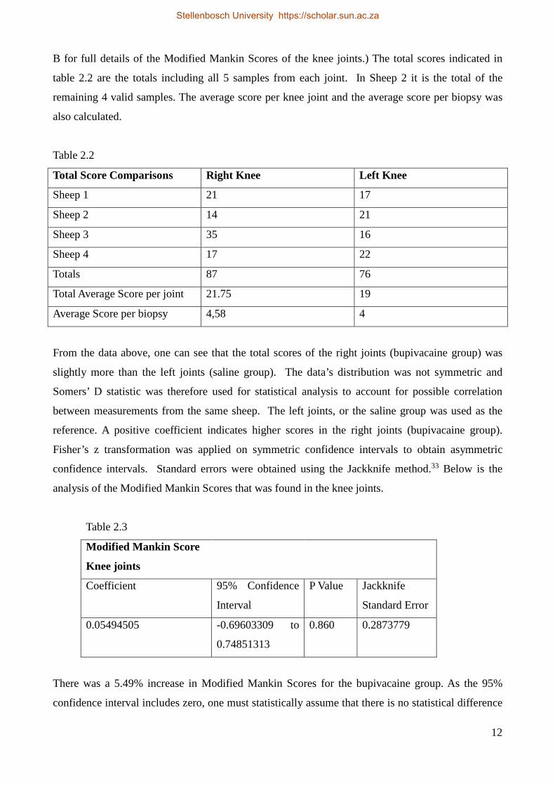
B for full details of the Modified Mankin Scores of the knee joints.) The total scores indicated in
table 2.2 are the totals including all 5 samples from each joint. In Sheep 2 it is the total of the
remaining 4 valid samples. The average score per knee joint and the average score per biopsy was
also calculated.
Table 2.2
Total Score Comparisons Right Knee Left Knee
Sheep 1 21 17
Sheep 2 14 21
Sheep 3 35 16
Sheep 4 17 22
Totals 87 76
Total Average Score per joint 21.75 19
Average Score per biopsy 4,58 4
From the data above, one can see that the total scores of the right joints (bupivacaine group) was
slightly more than the left joints (saline group). The data’s distribution was not symmetric and
Somers’ D statistic was therefore used for statistical analysis to account for possible correlation
between measurements from the same sheep. The left joints, or the saline group was used as the
reference. A positive coefficient indicates higher scores in the right joints (bupivacaine group).
Fisher’s z transformation was applied on symmetric confidence intervals to obtain asymmetric
confidence intervals. Standard errors were obtained using the Jackknife method.33 Below is the
analysis of the Modified Mankin Scores that was found in the knee joints.
Table 2.3
Modified Mankin Score
Knee joints
Coefficient 95% Confidence
Interval
P Value Jackknife
Standard Error
0.05494505 -0.69603309 to
0.74851313
0.860 0.2873779
There was a 5.49% increase in Modified Mankin Scores for the bupivacaine group. As the 95%
confidence interval includes zero, one must statistically assume that there is no statistical difference
Stellenbosch University https://scholar.sun.ac.za

13
between the groups.
Figure 2.5 is a photo taken from a biopsy of a saline injected joint, illustrating normal surface
integrity, normal cellularity with only minimal cloning present in the form of doublets:
Fig. 2.5: Sheep 3; os carpale tertium, left knee joint
In contrast, figure 2.6 is a biopsy from a bupivacaine injected joint, with severe degenerative
changes. Note the surface irregularity, decrease in cellularity and cloning formation in the form of
doublets and triplets.
Fig. 2.6: Sheep 3; os carpi ulnare, right knee joint
Differences in safranin-O staining were also observed. Safranin-O normally stains the nuclei black,
the cytoplasma blue to green, and the cartilage, mucin and mast cell granules orange to red. Figure
2.7 is an example of normal safranin-O staining:
Stellenbosch University https://scholar.sun.ac.za

14
Fig. 2.7: Sheep 3; os carpi ulnare, left knee joint
In contrast, figure 2.8 shows a picture of the contralateral, bupivacaine injected, knee of the same
sheep. The safranin-O staining is visibly reduced.
Fig. 2.8: Sheep 3; os carpi ulnare, right knee joint
The different parameters of the Modified Mankin Score were also analysed individually. Tables 2.4,
2.5, 2.6 and 2.7 include the results that were found concerning the articular surface integrity,
cellularity, cloning formation and safranin-O staining respectively.
Table 2.4
Articular surface
Knee joints
Coefficient 95% Confidence
Interval
P Value Jackknife
Standard Error
0.2967033 -0.36444782 to
0.75898096
0.252 0.2161564
Stellenbosch University https://scholar.sun.ac.za

15
Table 2.5
Cellularity
Knee joints
Coefficient 95% Confidence
Interval
P Value Jackknife
Standard Error
0.10989011 -0.79646316 to
0.86415781
0.789 0.376817
Table 2.6
Clone formation
Knee joints
Coefficient 95% Confidence
Interval
P Value Jackknife
Standard Error
0.07692308 -0.54032866 to
0.6403521
0.743 0.2142047
Table 2.7
Safranin-O staining
Knee joints
Coefficient 95% Confidence
Interval
P Value Jackknife
Standard Error
-0.05494505 -0.71882294 to
0.66134718
0.850 0.2671545
From the data above, the greatest difference was seen with articular surface integrity, with an almost
30% reduction in articular surface integrity in the bupivacaine group. The p value was also the
lowest at 0.252 for this parameter, but as the 95% confidence interval still includes 0, a significant
difference cannot be concluded. Interestingly, analysis of the safranin-O staining resulted in a
negative coefficient, indicating a 5% decreased safranin-O staining in the saline group.
2.3.2 THERAPEUTIC SINGLE DOSE IN STIFLE JOINTS
As described in the methodology, 15 samples were taken from each stifle joint, namely;
Stellenbosch University https://scholar.sun.ac.za

16
1. anterior patella
2. posterior patella
3. anterior femoral groove
4. central femoral groove
5. posterior femoral groove
6. anterior lateral femoral condyle
7. posterior lateral femoral condyle
8. anterior medial femoral condyle
9. posterior medial femoral condyle
10. anterior lateral tibial plateau
11. posterior lateral tibial plateau
12. central lateral tibial plateau
13. anterior medial tibial plateau
14. posterior medial tibial plateau
15. central medial tibial plateau
Each one of these biopsies was graded individually by the Modified Mankin Score. From Sheep 1,
the biopsy taken from the right posterior lateral tibial plateau was insufficient for histological
analysis. The contralateral (left) posterior lateral tibial plateau sample from Sheep 1 was therefore
also excluded from the data.
Table 2.8 summarizes the Modified Mankin Scores that were found in the stifle joints. (See
Appendix C for full details.) The total scores are the totals including all 15 samples from each joint.
In Sheep 1 it is the total of the remaining 14 valid samples. The average scores per joint and sample
were also calculated.
Table 2.8
Total Score Comparisons Right Stifle joint Left Stifle joint
Sheep 1 31 33
Sheep 2 57 78
Sheep 3 81 45
Sheep 4 68 65
Totals 237 221
Total Average Score per joint 59.25 55.25
Average Score per biopsy 4.02 3.75
Stellenbosch University https://scholar.sun.ac.za

17
As seen with the supra-normal dosage of intra-articular bupivacaine in the knee joints, we also
found higher total Modified Mankin Scores in the bupivacaine group as compared to the normal
saline group, with a therapeutic dosage of intra-articular bupivacaine. Somers’ D statistic was again
used to analyse the Modified Mankin Scores as well as the individual parameters. Tables 2.9 – 2.13
contain the statistical analysis of the data.
Table 2.9
Modified Mankin Score
Stifle joints
Coefficient 95% Confidence
Interval
P Value Jackknife
Standard Error
0.07233065 -0.49063693 to
0.59269692
0.73 0.1914741
Table 2.10
Articular surface
Stifle joints
Coefficient 95% Confidence
Interval
P Value Jackknife
Standard Error
-0.08610792 -0.57329343 to
0.44606062
0.661 0.1778799
Table 2.11
Cellularity
Stifle joints
Coefficient 95% Confidence
Interval
P Value Jackknife
Standard Error
-.01722158 -0.32036783 to
0.28912427
0.873 0.0989282
Stellenbosch University https://scholar.sun.ac.za
18
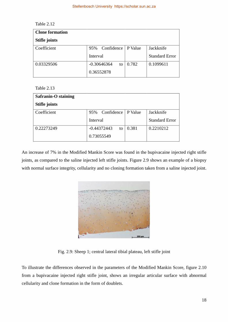
Table 2.12
Clone formation
Stifle joints
Coefficient 95% Confidence
Interval
P Value Jackknife
Standard Error
0.03329506 -0.30646364 to
0.36552878
0.782 0.1099611
Table 2.13
Safranin-O staining
Stifle joints
Coefficient 95% Confidence
Interval
P Value Jackknife
Standard Error
0.22273249 -0.44372443 to
0.73055549
0.381 0.2210212
An increase of 7% in the Modified Mankin Score was found in the bupivacaine injected right stifle
joints, as compared to the saline injected left stifle joints. Figure 2.9 shows an example of a biopsy
with normal surface integrity, cellularity and no cloning formation taken from a saline injected joint.
Fig. 2.9: Sheep 1; central lateral tibial plateau, left stifle joint
To illustrate the differences observed in the parameters of the Modified Mankin Score, figure 2.10
from a bupivacaine injected right stifle joint, shows an irregular articular surface with abnormal
cellularity and clone formation in the form of doublets.
Stellenbosch University https://scholar.sun.ac.za

19
Fig. 2.10: Sheep 2; central medial tibial plateau, right stifle joint
These degenerative changes were not only limited to our bupivacaine group. The sample illustrated
in figure 2.11 below, was taken from the left stifle joint in sheep 2 (saline group) and shows severe
surface irregularities, cloning formation in the form of doublets and focal decrease in cellularity.
Fig. 2.11: Sheep 2; posterior lateral tibial plateau, left stifle joint
From the individual parameters assessed, the biggest difference between the bupivacaine injected
joints and the saline control joints, was found in safranin-O staining, with a 22% reduction in
staining in the bupivacaine group (p = 0.381). Figure 2.12 is an example of normal safranin-O
staining from the control, left stifle joint.
Stellenbosch University https://scholar.sun.ac.za

20
Fig. 2.12: Sheep 1; anterior lateral femoral condyle, left stifle joint
Complete loss of safranin-O staining was found in a bupivacaine injected right stifle joint, as shown
in figure 2.13.
Fig. 2.13: Sheep 3; posterior lateral tibial plateau, right stifle joint
Stellenbosch University https://scholar.sun.ac.za

21
CHAPTER 3
DISCUSSION
3.1 LITERATURE REVIEW
Numerous in vitro and in vivo studies confirmed the chondrotoxic effect of bupivacaine.
Ropivacaine may be the least toxic local anesthetic agent for intra-articular use.18 One in vivo study
suggested that bupivacaine’s enantiomer, levobupivacaine, may be safe.20 In this study the time
from intra-articular levobupivacaine administration and sampling of the cartilage for histological
analysis was only 21 days, which could falsely increase the likelihood of negative results as
changes become more prevalent at longer intervals after administration.11 One in vitro study even
found levobupivacaine to be more toxic to chondrocytes than bupivacaine.19 The chondrotoxic
effect of bupivacaine is a uniform finding over numerous species, as investigated in dogs, rats and
humans.13-15
The addition of epinephrine to intra-articular bupivacaine did not increase the risk of
chondrotoxicity in an in vivo rat model.12 In contrast, in an in vitro study on human chondrocytes,
the addition of epinephrine did cause more chondrolysis.13 Cartilage cells in joint cavities have a
very limited blood supply. Epinephrine is a very strong vasoconstrictor. This should caution against
its use as additive to intra-articular bupivacaine.
The duration of exposure of chondrocytes to bupivacaine has proved to increase cartilage damage.14
One in vitro study suggested that an infusion time of less than 48 hours may be safe.13
Chondrotoxicity due to bupivacaine is also dependent on the intra-articular dosage used.16 The total
dosage of bupivacaine is a product of the concentration and volume used. By reducing either the
volume or the concentration, one can therefore limit the total dosage administered.
3.2 SINGLE DOSE INTRA-ARTICULAR BUPIVACAINE PILOT STUDY
Total Modified Mankin Scores were calculated in both the knee and stifle joints, after
administration of the supra-normal and therapeutic dosages of 0.5% bupivacaine respectively. These
were compared to 0.9% saline administration in the contralateral joints, which served as controls.
With both dosages, the total scores were higher in the bupivacaine group as compared to the saline
group. Statistical analysis of the different parameters, namely, articular surface integrity, cloning
formation, cellularity and safranin-O staining, also proved to be higher in the bupivacaine groups,
Stellenbosch University https://scholar.sun.ac.za

22
except for the safranin-O staining in the knee joints (-5,5%). Articular surface integrity (-8.6%) and
cellularity (-1.7%) in the stifle joints were also higher in the saline groups.
Statistical analysis could not conclude a significant difference between the groups, as the 95%
confidence intervals always included 0. This was only a pilot study, with a very small sample size,
limited by the costs involved. A power analysis of the pilot study is also not applicable. It is
therefore difficult to draw conclusions with the limited data available. Bigger sample sizes will
narrow the 95% confidence interval and improve interpretation.
If we are to assume that equal anatomical samples from different legs are uncorrelated, according to
Wilcoxon signed-rank test, there was a significant difference between the bupivacaine and saline
groups in safranin-O staining (p=0.014) as illustrated in table 3.1. Higher scores were observed in
the right stifle joints (bupivacaine group) when compared to the left stifle joints (saline group).
There were no differences found in the front knee joints as mentioned above, concerning the
safranin-O staining.
Table 3.1
Wilcoxin signed-rank test
Safranin-O Staining: Stifle joints
Sign Observed
Positive 28
Negative 13
Zero 18
Total 59
We therefore conclude that single dose intra-articular bupivacaine may be chondrotoxic, but we
need a bigger sample size to confirm our strong suspicion.
3.3 PATHOPHYSIOLOGY OF BUPIVACAINE CHONDROTOXCICITY
The exact mechanism of bupivacaine chondrotoxicity is unclear. A proposed mechanism is
disruption of the cell membrane that might cause acute necrosis. This phenomenon increases with
increased fat solubility, which is a known characteristic of bupivacaine. Mitochondrial function may
also be affected, by disrupting the mitochondrial transmembrane potential, leading to less ATP
Stellenbosch University https://scholar.sun.ac.za

23
production. Alterations in mitochondrial DNA then lead to chondrocyte apoptosis.11 The agent must
diffuse through the intercellular matrix to exert its chondrotoxic effect. Disruption of the superficial
layer of cartilage, by example inserting anchor sutures by the surgeon, can increase the risk of
chondrotoxicity by damaging the protective barrier of chondrocytes. An intra-articular injectate can
then infiltrate the intercellular proteoglycan matrix of the cartilage, with less resistance, and cause
its toxic effects to chondrocytes.
Cartilage, compared to other tissues, has limited ability to heal and regenerate itself. Once an insult
has occurred, a long term effect becomes possible. Loss of functional cells will result in loss of
extracellular matrix and collagen production, which plays a vital role in the integrity of the cartilage
tissue.17
Because of uncertainty concerning the mechanism of action of bupivacaine chondrotoxicity, can
one safely assume cause and effect? In a meta-analysis by Matsen, et al.11, it was shown that there is
indeed “strong association, consistency, specificity, temporal relationship, biological gradient and
plausibility, overall coherence of evidence, good experimental evidence and analogy”. Matsen
therefore concluded that bupivacaine does cause chondrocyte damage.11
3.4 ALTERNATIVES TO INTRA-ARTICULAR BUPIVACAINE
All local anesthetics have been implicated in chondrocyte death. Ropivacaine seems to be the least
toxic.18 Recently, magnesium, a N-methyl-D-aspartate (NMDA) receptor antagonist, has been used
as part of a multimodal analgesic regime for joint pain. NMDA receptors have been found on intra-
articular surfaces and Bondok et al.26 showed that intra-articular magnesium is an effective
alternative to intra-articular bupivacaine.26 Magnesium not only has analgesic properties but also
has chondro-proliferative properties.27 Interestingly, Baker et al.16 found that 10% magnesium
attenuated the chondrotoxic effects of local anesthetics.16
Intra-articular steroids have proven to increase the risk of septic arthritis and may even have a
synergistic toxic effect on chondrocytes when combined with local anesthetics.3, 28
The data on the use of intra-articular morphine has been conflicting. In a qualitative review of 46
randomized controlled trials done by Rosseland et al.29 in 2005, the conclusion was that intra-
articular morphine was an ineffective analgesic for joint surgery.29 Previous studies used small
dosages (example 1 mg) of intra-articular morphine, so Garcia et al.30 undertook a randomized
Stellenbosch University https://scholar.sun.ac.za

24
controlled trial, in 2010, on 50 patients comparing 10 mg intra-articular morphine with intra-
articular normal saline. A significant reduction in rescue analgesia was observed in the morphine
group (p = 0.0001). The time to request additional analgesia was also longer in the morphine group
(p = 0.0166). Post-operative pain scores were also reduced at 2 and 6 hours in the morphine
group.30
Intra-articular alpha 2 agonists such as clonidine have also been used with good effect, but more
data is needed for safe conclusions.3
Stellenbosch University https://scholar.sun.ac.za

25
CHAPTER 4
CONCLUSION
Intra-articular bupivacaine is common practice in joint analgesia. It's efficacy as analgesic shows
good response, but the risk involved is undoubtedly irreversible, and potentially disabling,
chondrolysis. Bupivacaine’s intra-articular chondrotoxic effect is time, concentration and dose
dependent. Using intra-articular bupivacaine infusions increases the risk of chondrocyte toxicity.
Single dose intra-articular bupivacaine may also be significantly chondrotoxic. Chu et al.23 showed
a statistically significant 50% reduction in chondrocyte density after a single dose of intra-articular
bupivacaine in rats. From our pilot study done on 4 sheep, the overall impression was increased
chondrotoxicity in the bupivacaine groups, but statistical significance could not be proven due to a
small sample size.
The addition of intra-articular magnesium sulphate appears to attenuate the chondrotoxic effect of
local anesthetics and magnesium has a proliferative effect on chondrocytes. High dose intra-
articular morphine and intra-articular clonidine may prove beneficial as analgesics for joint surgery,
but more data is needed. Poor integrity of the articular surface increases the risk of chondrocyte
death from intra-articular medications, which opens the discussion of optimal surgical technique.
Stellenbosch University https://scholar.sun.ac.za

26
APPENDICES
1. APPENDIX A: Monitoring sheet for sheep wellbeing
A case controlled experiment to evaluate the in vivo effects of single dose intra-articular 0.5% bupivacaine on cartilage of sheep, 2014
MONITORING SHEET Date : Sheep number (1-4) : Physical Indicators Acute complications Normal Swelling Effusion Induration Tenderness Right knee joint Left knee joint Right stifle joint Left stifle joint
Chronic complications
Normal Antalgic
Gait
Psycological indicators
Normal Refusal to eat
Appetite
Reviewed by: .......................... ................................
Signature
Stellenbosch University https://scholar.sun.ac.za

27
2. APPENDIX B: Modified Mankin Scores of the Front Knees
SampleSide
Articular surface integrity
Cellularity Clone formation
Safranin-O Staining Total
Sheep 1 : Front Knee Os Carpi Ulnare Right 1 1 1 2 5Os carpi intermedium Right 1 1 1 2 5Os Carpi Radiale Right 1 1 0 1 3Os Carpale quartum Right 2 1 1 1 5Os Carpale tertium Right 0 1 1 1 3
Total 21Os Carpi Ulnare Left 0 1 3 1 5Os carpi intermedium Left 0 1 3 0 4Os Carpi Radiale Left 0 1 1 1 3Os Carpale quartum Left 1 1 0 3 5Os Carpale tertium Left 0 0 0 0 0
Total 17
Sheep 2 : Front Knee Os Carpi Ulnare Right 3 1 1 0 5Os carpi intermedium Right 0 1 1 1 3Os Carpi Radiale Right 1 1 1 0 3Os Carpale quartum Right Sample not suitable for analysisOs Carpale tertium Right 1 1 1 0 3
Total 14Os Carpi Ulnare Left 3 1 2 1 7Os carpi intermedium Left 0 1 2 2 5Os Carpi Radiale Left 3 1 1 0 5Os Carpale quartum LeftOs Carpale tertium Left 2 0 1 1 4
Total 21
Sheep 3 : Front Knee Os Carpi Ulnare Right 4 2 2 2 10Os carpi intermedium Right 3 1 2 4 10Os Carpi Radiale Right 0 1 0 2 3Os Carpale quartum Right 2 1 2 2 7Os Carpale tertium Right 1 1 2 1 5
Total 35Os Carpi Ulnare Left 0 0 1 1 2Os carpi intermedium Left 1 0 1 2 4Os Carpi Radiale Left 2 0 1 2 5Os Carpale quartum Left 0 1 1 2 4Os Carpale tertium Left 0 0 1 0 1
Total 16
Sheep 4 : Front Knee Os Carpi Ulnare Right 3 0 1 1 5Os carpi intermedium Right 0 0 0 0 0Os Carpi Radiale Right 0 0 0 0 0Os Carpale quartum Right 0 0 3 2 5Os Carpale tertium Right 3 0 3 1 7
Total 17Os Carpi Ulnare Left 2 1 1 1 5Os carpi intermedium Left 0 0 0 2 2Os Carpi Radiale Left 0 1 3 2 6Os Carpale quartum Left 0 1 0 1 2Os Carpale tertium Left 3 1 1 2 7
Total 22
Stellenbosch University https://scholar.sun.ac.za
28
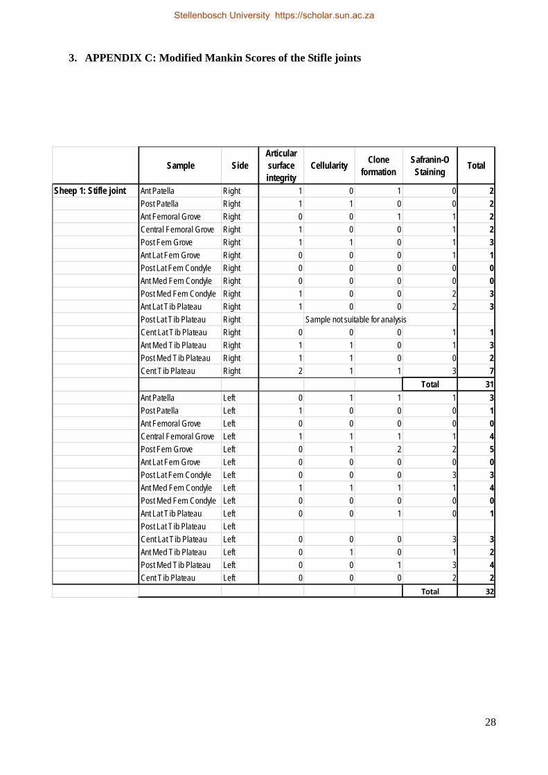
3. APPENDIX C: Modified Mankin Scores of the Stifle joints
Sample SideArticular surface integrity
Cellularity Clone formation
Safranin-O Staining Total
Sheep 1: Stifle joint Ant Patella Right 1 0 1 0 2Post Patella Right 1 1 0 0 2Ant Femoral Grove Right 0 0 1 1 2Central Femoral Grove Right 1 0 0 1 2Post Fem Grove Right 1 1 0 1 3Ant Lat Fem Grove Right 0 0 0 1 1Post Lat Fem Condyle Right 0 0 0 0 0Ant Med Fem Condyle Right 0 0 0 0 0Post Med Fem Condyle Right 1 0 0 2 3Ant Lat T ib Plateau Right 1 0 0 2 3Post Lat T ib Plateau Right Sample not suitable for analysisCent Lat T ib Plateau Right 0 0 0 1 1Ant Med T ib Plateau Right 1 1 0 1 3Post Med T ib Plateau Right 1 1 0 0 2Cent T ib Plateau Right 2 1 1 3 7
Total 31Ant Patella Left 0 1 1 1 3Post Patella Left 1 0 0 0 1Ant Femoral Grove Left 0 0 0 0 0Central Femoral Grove Left 1 1 1 1 4Post Fem Grove Left 0 1 2 2 5Ant Lat Fem Grove Left 0 0 0 0 0Post Lat Fem Condyle Left 0 0 0 3 3Ant Med Fem Condyle Left 1 1 1 1 4Post Med Fem Condyle Left 0 0 0 0 0Ant Lat T ib Plateau Left 0 0 1 0 1Post Lat T ib Plateau LeftCent Lat T ib Plateau Left 0 0 0 3 3Ant Med T ib Plateau Left 0 1 0 1 2Post Med T ib Plateau Left 0 0 1 3 4Cent T ib Plateau Left 0 0 0 2 2
Total 32
Stellenbosch University https://scholar.sun.ac.za

29
Sample SideArticular surface integrity
Cellularity Clone formation
Safranin-O Staining Total
Sheep 2: Stifle joint Ant Patella Right 0 0 0 1 1Post Patella Right 1 0 0 2 3Ant Femoral Grove Right 2 0 1 0 3Central Femoral Grove Right 0 1 3 1 5Post Fem Grove Right 0 0 0 1 1Ant Lat Fem Grove Right 1 0 0 1 2Post Lat Fem Condyle Right 0 0 0 0 0Ant Med Fem Condyle Right 1 1 1 2 5Post Med Fem Condyle Right 0 0 0 2 2Ant Lat T ib Plateau Right 1 1 1 3 6Post Lat T ib Plateau Right 3 1 1 5 10Cent Lat T ib Plateau Right 0 1 1 0 2Ant Med T ib Plateau Right 0 1 0 2 3Post Med T ib Plateau Right 3 1 1 0 5Cent T ib Plateau Right 4 1 2 2 9
Total 57Ant Patella Left 0 1 0 2 3Post Patella Left 3 1 1 2 7Ant Femoral Grove Left 1 1 1 0 3Central Femoral Grove Left 1 1 3 1 6Post Fem Grove Left 0 1 0 1 2Ant Lat Fem Grove Left 2 1 1 5 9Post Lat Fem Condyle Left 0 0 0 0 0Ant Med Fem Condyle Left 2 0 0 1 3Post Med Fem Condyle Left 4 1 0 3 8Ant Lat T ib Plateau Left 0 0 0 0 0Post Lat T ib Plateau Left 3 1 2 5 11Cent Lat T ib Plateau Left 3 1 1 2 7Ant Med T ib Plateau Left 2 1 1 2 6Post Med T ib Plateau Left 2 1 3 1 7Cent T ib Plateau Left 3 1 0 3 7
Total 79
Stellenbosch University https://scholar.sun.ac.za

30
Sample SideArticular surface integrity
Cellularity Clone formation
Safranin-O Staining Total
Sheep 3: Stifle joint Ant Patella Right 1 0 0 3 4Post Patella Right 0 1 3 3 7Ant Femoral Grove Right 0 1 3 2 6Central Femoral Grove Right 0 0 1 5 6Post Fem Grove Right 0 0 0 4 4Ant Lat Fem Grove Right 0 0 1 2 3Post Lat Fem Condyle Right 1 0 2 3 6Ant Med Fem Condyle Right 2 0 1 2 5Post Med Fem Condyle Right 2 1 0 2 5Ant Lat T ib Plateau Right 2 1 1 3 7Post Lat T ib Plateau Right 2 1 0 5 8Cent Lat T ib Plateau Right 2 0 0 5 7Ant Med T ib Plateau Right 1 0 1 1 3Post Med T ib Plateau Right 2 1 1 5 9Cent T ib Plateau Right 0 0 0 1 1
Total 81Ant Patella Left 0 0 0 1 1Post Patella Left 0 0 0 1 1Ant Femoral Grove Left 1 1 2 2 6Central Femoral Grove Left 1 0 3 1 5Post Fem Grove Left 0 0 0 2 2Ant Lat Fem Grove Left 3 0 0 0 3Post Lat Fem Condyle Left 3 1 0 2 6Ant Med Fem Condyle Left 1 0 0 1 2Post Med Fem Condyle Left 2 0 0 1 3Ant Lat T ib Plateau Left 1 1 0 0 2Post Lat T ib Plateau Left 1 1 0 5 7Cent Lat T ib Plateau Left 0 0 1 0 1Ant Med T ib Plateau Left 1 0 0 0 1Post Med T ib Plateau Left 0 1 1 0 2Cent T ib Plateau Left 3 1 0 0 4
Total 46
Stellenbosch University https://scholar.sun.ac.za

31
Sample SideArticular surface integrity
Cellularity Clone formation
Safranin-O Staining Total
Sheep 4: Stifle joint Ant Patella Right 1 0 0 5 6Post Patella Right 0 0 3 1 4Ant Femoral Grove Right 0 1 1 1 3Central Femoral Grove Right 0 1 1 2 4Post Fem Grove Right 1 0 0 2 3Ant Lat Fem Grove Right 2 1 1 2 6Post Lat Fem Condyle Right 0 1 1 0 2Ant Med Fem Condyle Right 3 1 3 0 7Post Med Fem Condyle Right 1 1 1 1 4Ant Lat T ib Plateau Right 1 1 0 2 4Post Lat T ib Plateau Right 3 2 0 2 7Cent Lat T ib Plateau Right 0 2 0 2 4Ant Med T ib Plateau Right 1 1 0 2 4Post Med T ib Plateau Right 1 1 0 5 7Cent T ib Plateau Right 0 3 0 0 3
Total 68Ant Patella Left 1 0 1 2 4Post Patella Left 3 1 0 0 4Ant Femoral Grove Left 1 1 3 0 5Central Femoral Grove Left 3 0 0 1 4Post Fem Grove Left 0 0 1 0 1Ant Lat Fem Grove Left 1 0 0 1 2Post Lat Fem Condyle Left 2 0 0 0 2Ant Med Fem Condyle Left 1 0 0 0 1Post Med Fem Condyle Left 1 1 3 2 7Ant Lat T ib Plateau Left 3 1 0 0 4Post Lat T ib Plateau Left 5 3 0 2 10Cent Lat T ib Plateau Left 0 0 1 2 3Ant Med T ib Plateau Left 1 2 0 2 5Post Med T ib Plateau Left 5 3 0 1 9Cent T ib Plateau Left 2 1 1 0 4
Total 65
Stellenbosch University https://scholar.sun.ac.za

32
BIBLIOGRAPHY
1. Middleton F, Coakes J, Umarji S, Palmer S, Venn R, Panayiotou S. The efficacy of intra-articular
bupivacaine for the relief of pain following arthroscopy of the ankle. J Bone Joint Surg Br
2006;88-B(12);1603-5
2. Georgopoulos G, Carry P, Pan Z, Chang F, Heare T, Rhodes J, Hotchkiss M, Miller NH, Erickson
M. The efficacy of intra-articular injections for pain control following the closed reduction and
percutaneous pinning of pediatric supracondylar humeral fractures. A randomized controlled trial. J
Bone Joint Surg Am 2012;94:1633-42
3. Lavelle W, Lavelle ED, Lavelle L. Intra-articular injections. Anesthesiology Clinics
2007;25:853-62
4. Sullivan SG, Abbott PJ, Jr. Cardiovascular toxicity associated with intra-articular bupivacaine.
Anesth Analg 1994;79:591-3
5. Linguori GA, Chimento GF, Borow L, Figgie M. Possible bupivacaine toxicity after intra-
articular injection for postarthroscopic analgesia of the knee: Implications of the surgical procedure.
Anesth Analg 2002;94:1010-3
6. Meinig RP, Holtgrewe JL, Wiedel JD, et al. Plasma bupivacaine levels following single dose
intra-articular instillation for arthroscopy. Am J Sports Med 1988;16:295-300
7. Petty D, Jazrawi LM, Estrada LS, Andrews JR. Glenohumoral chrondrolysis after shoulder
arthroscopy: Case reports and review of the literature. Am J of Sports Med 2004;32(2):509-15
8. Greis PE, LeGrand A, Burks RT. Bilateral shoulder chondrolysis following arthroscopy. A
report of two cases. J Bone Joint Surg Am. 2008;90:1338-44
9. Anakwenze OA, Hosalkar H, Huffman GR. Two cases of glenohumoral chondrolysis after intra-
articular pain pumps. Clin Orthop Relat Res. 2010;468(9):2545-9
10. Hansen BP, Beck CL, Beck EP, Townsley RW. Postarthroscopic glenohumeral chondrolysis.
Stellenbosch University https://scholar.sun.ac.za

33
Am J Sports Med. 2007;35:1628-34
11. Matsen FA, Papadonikolakis A. Published evidence demonstrating the causation of
glenohumoral chondrolysis by postoperative infusion of local anesthetic via a pain pump. J Bone
Joint Surg Am. 2013;95:1126-34
12. Gomoll AH, Kang RW, Williams JM, Rach BR, Cole BJ. Chrondrolysis after continuous intra-
articular bupivacaine infusion: an experimental model investigating chondrotoxcicity in the rabbit
shoulder. Arthroscopy: The Journal of Arthroscopic and Related Surgery. 2006;22(8):813-19
13. Dragoo JL, Korotkova T, Kanwar R, Wood B. The effect of local anesthetics administered via
pain pump on chondrocyte viability. American Journal of Sports Medicine. 2008;36(8):1484-8
14. Lo IK, Sciore P, Chung M, Liang S, Boorman RB, Thornton GM, Rattner JB, Muldrew K.
Local anesthetics induce chondrocyte death in bovine articular cartilage disks in a dose- and
duration-dependent manner. Arthroscopy 2009;25(7):707-15
15. Hennig GS, Hosgood G, Bubenik-Angapen LJ, Lauer SK, Morgan TW. Evaluation of
chondrocyte death in canine osteochondral explants exposed to a 0.5% solution of bupivacaine. Am
J Vet Res. 2010;71(8):875-83
16. Baker JF, Walsh PM, Byrne DP, Mulhall KJ. In vitro assessment of human chondrocyte
viability after treatment with local anesthetic, magnesium sulphate or normal saline. Knee Surg
Sports Traumatol Arthrosc. 2001;19:1043-46.
17. Wang D, Vo N, Sowa G, Hartman R, Ngo K, Ra Choe S, Witt W, Dong Q, Lee J, Niedernhofer
L, Kang J. Bupivacaine decreases cell viability and matrix protein synthesis in an intervertebral
disc organ model system. Spine J. 2011;11(2):139-46.
18. Breu A, Eckl S, Zink W, Kujat R, Angele P. Cytotoxicity of local anesthetics on human
mesenchymal stem cells in vitro. Arthroscopy. 2013;29(10):1676-84
19. Beyzadeoğlu T, Torun KG, Ekinci ID, Bekler H, Yilmaz C. Cytotoxicity of local anesthetics to
rats’ articular cartilage: an experimental study. Acta Orthop Traumatol Turc. 2012;46(3):201-7
Stellenbosch University https://scholar.sun.ac.za

34
20. Erden IA, Altinel S, Saricaoglu F, Zeybek ND, Akinci SB, Asan E, Ayper U. Effect of intra-
articular injection of levobupivacaine on articular cartilage and synovium in rats. Der Anaesthesist.
2012;61(5):420-3
21. Gungor I, Yilmaz A, Ozturk AM, Ergun MA, Menevse S, Kaya K. Bupivacaine and
levobupivacaine induce apoptosis in rat chondrocyte cell cultures at ultra-low doses. Eur J Orthop
Surg Traumatol. 2014;24(3):291-95
22. Gomoll AH, Yanke AB, Kang RW, Chubinskaya S, Williams JM, Bach BR, Cole BJ. Long-
term effects of bupivacaine on cartilage in a rabbit shoulder model. Am J Sports Med.
2009;37(1):72-7
23. Chu CR, Coyle CH, Chu CT, Szczodry M, Seshadri V, Karpie JC, Cieslak KM, Pringle EK. In
vivo effects of single intra-articular injection of 0,5% bupivacaine on articular cartilage. J Bone
Joint Surg Am. 2010;92(3):599-608
24. Osterhoff G, Löffler S, Steinke H, Feja C, Josten C, Hepp P. Comparative anatomical
measurements of osseous structures in the ovine and human knee. The Knee. 2011;18(2):98-103
25. Moody HR, Heard BJ, Frank CB, Shrive NG, Oloyede AO. Investigating the potential value of
individual parameters of histological grading systems in a sheep model of cartilage damage: the
Modified Mankin Method. J Anat. 2012;221:47-54
26. Bondok RS, Abd El-Hady AM. Intra-articular magnesium is effective for postoperative
analgesia in arthroscopic knee surgery. Br J Anesth. 2006;97:389-392.
27. Egerbacher M, Wolfesberger B, Gabler C. In vitro evidence for the effects of magnesium
supplementation on quinolone treated horse and dog chondrocytes. Vet Pathol 2001;38(2):143-148
28. Seshadri V, Coyle CH, Chu CR. Lidocaine potentiates the chondrotoxicity of
methylprednisolone. Arthroscopy. 2009;25:337-47
29. Rosseland LA. No evidence for analgesic effect of intra-articular morphine after knee
arthroscopy: a qualitative systematic review. Regional Anesthesia and Pain Medicine.
2005;30(1):83-98
Stellenbosch University https://scholar.sun.ac.za

35
30. Garcia JBS, Neto JOB, Vasconcelos JW, Ferro LSG, e Silva RC. Analgesic efficacy of the
intra-articular administration of high doses of morphine in patients undergoing total knee
arthroplasty. Revista Brasileira de Anestesiologia. 2010;60(1):7-12
31. Piper SL, Kramer JD, Kim HT, Feeley BT. Effects of local Anesthetics on Articular Cartilage.
The American Journal of Sports Medicine. 2011;39(10):2245-53
32. Feher G. The functional anatomy of domestic animals. [Online] Hungry: Mezõgazda Kiadó
Publishing; 1980. Available from: http://www.tankonyvtar.hu/en/tartalom/tkt/haziallatok/ch02.html
[Accessed 21 July 2015]
33. Newson R. Confidence intervals for rank statistics. Somers’ D and Extensions. 2006;6(3):309-
34
Stellenbosch University https://scholar.sun.ac.za