intestinal nematodes
TRANSCRIPT

Intestinal Nematodes
Aman UllahB.Sc. MLT
M. Phil MicrobiologyMaster in Health Research
Certificate in Health and Physical Education

Introduction• The medically important nematodes can be divided into two categories according to their
primary location in the body, namely, intestinal and tissue nematodes• The intestinal nematodes include Enterobius (pinworm), Trichuris (whipworm), Ascaris (giant
roundworm), Necator and Ancylostoma (the two hookworms), Strongyloides (small roundworm), and Trichinella
• Enterobius, Trichuris, and Ascaris are transmitted by ingestion of eggs; the others are transmitted as larvae
• There are two larval forms: the first- and second-stage (rhabditiform) larvae are noninfectious, feeding forms; the third-stage (filariform) larvae are the infectious, nonfeeding forms
• As adults, these nematodes live within the human body, except for Strongyloides, which can also exist in the soil
• The important tissue nematodes Wuchereria, Onchocerca, and Loa are called the "filarial worms," because they produce motile embryos called microfilariae in blood and tissue fluids
• These organisms are transmitted from person to person by bloodsucking mosquitoes or flies• A fourth species is the guinea worm, Dracunculus, whose larvae inhabit tiny crustaceans
(copepods) and are ingested in drinking water

ENTEROBIUSDisease: Enterobius vermicularis causes pinworm infection EnterobiasisLife cycle: • The life cycle is confined to humans• The infection is acquired by ingesting the worm eggs• The eggs hatch in the small intestine, where the larvae differentiate
into adults and migrate to the colon• The adult male and female worms live in the colon, where mating
occurs • At night, the female migrates from the anus and releases thousands of
fertilized eggs on the perianal skin and into the environment• Within 6 hours, the eggs develop into embryonated eggs and become
infectious• Reinfection can occur if they are carried to the mouth by fingers after
scratching the itching skin

ENTEROBIUSPathogenesis & Clinical Findings• Perianal pruritus is the most prominent symptom• Pruritus is thought to be an allergic reaction to the presence of
either the adult female or the eggs• Scratching predisposes to secondary bacterial infectionLaboratory Diagnosis• The eggs are recovered from perianal skin by using the Scotch
tape technique and can be observed microscopically • Unlike those of other intestinal nematodes, these eggs are not
found in the stools• The small, whitish adult worms can be found in the stools or
near the anus of diapered children

Egg of E. vermicularis• Left: Adhesive tape preparation showing
eggs ofE.vermicularis recovered from anal skin
• Right: E.vermicularis egg in faeces
• It is colourless.• Oval in shape and
usually flattened on one side
• Contains a larva

TRICHURISDisease:• Trichuris trichiura causes whipworm infection trichuriasisLife cycle: • Humans are infected by ingesting worm eggs in food or water
contaminated with human feces• The eggs hatch in the small intestine, where the larvae
differentiate into immature adults• These forms migrate to the colon, where they mature, mate, and
produce thousands of fertilized eggs daily, which are passed in the feces
• Eggs deposited in warm, moist soil form embryos• When the embryonated eggs are ingested, the cycle is completed

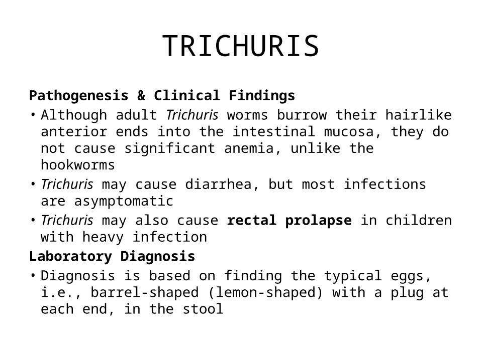
TRICHURISPathogenesis & Clinical Findings• Although adult Trichuris worms burrow their hairlike anterior
ends into the intestinal mucosa, they do not cause significant anemia, unlike the hookworms
• Trichuris may cause diarrhea, but most infections are asymptomatic
• Trichuris may also cause rectal prolapse in children with heavy infection
Laboratory Diagnosis• Diagnosis is based on finding the typical eggs, i.e., barrel-
shaped (lemon-shaped) with a plug at each end, in the stool

Egg of T. trichiura
• It is yellow-brown • Has a characteristic
barrel shape with a colourless protruding mucoid plug at each end
• Contains a central granular mass which is the unsegmented ovum

ASCARISDisease: Ascaris lumbricoides causes ascariasisLife cycle:• Humans are infected by ingesting worm eggs in food or water contaminated
with human feces• The eggs hatch in the small intestine, and the larvae migrate through the gut
wall into the bloodstream and then to the lungs• They enter the alveoli, pass up the bronchi and trachea, and are swallowed• Within the small intestine, they become adults• They live in the lumen, do not attach to the wall, and derive their sustenance
from ingested food• The adults are the largest intestinal nematodes, often growing to 25 cm or more• A. lumbricoides is known as the "giant roundworm." Thousands of eggs are laid
daily, are passed in the feces, and differentiate into embryonated eggs in warm, moist soil
• Ingestion of the embryonated eggs completes the cycle

ASCARISPathogenesis & Clinical Findings:• The major damage occurs during larval migration rather than from the presence
of the adult worm in the intestine• The principal sites of tissue reaction are the lungs, where inflammation with an
eosinophilic exudate occurs in response to larval antigens• Because the adults derive their nourishment from ingested food, a heavy worm
burden may contribute to malnutrition, especially in children in developing countries
• Most infections are asymptomatic• Ascaris pneumonia with fever, cough, and eosinophilia can occur with a heavy
larval burden• Abdominal pain and even obstruction can result from the presence of adult
worms in the intestineLaboratory Diagnosis:• Diagnosis is usually made microscopically by detecting eggs in the stools• Occasionally, the patient sees adult worms in the stools.

Eggs of A. lumbricoides• Usually fertilized eggs are found in faeces but
occasionally infertile eggs are produced by unfertilized female worms.
FERTILIZED EGG• Yellow-brown, oval or round• Shell is often covered by an uneven albuminous
coat • Contains a central granular mass which is the
unsegmented fertilized ovum.Decorticated egg: • This term is used to describe an eggthat has no
albuminous coat. A decorticated egg has a smooth shell and appears pale yellow or colourless.
INFERTILE EGG• It is darker in colour and has a thinner wall• and more granular albuminous covering.• More elongated than a fertilized egg• Contains a central mass of large granules.

ANCYLOSTOMA & NECATORDisease:• Ancylostoma duodenale (Old World hookworm) and Necator americanus (New
World hookworm) cause hookworm infectionLife cycle:• Humans are infected when filariform larvae in moist soil penetrate the skin,
usually of the feet or legs• They are carried by the blood to the lungs, migrate into the alveoli and up the
bronchi and trachea, and then are swallowed• They develop into adults in the small intestine, attaching to the wall with either
cutting plates (Necator) or teeth (Ancylostoma)• They feed on blood from the capillaries of the intestinal villi• Thousands of eggs per day are passed in the feces • Eggs develop first into noninfectious, feeding (rhabditiform) larvae and then
into third-stage, infectious, nonfeeding (filariform) larvae • which penetrate the skin to complete the cycle


ANCYLOSTOMA & NECATORPathogenesis & Clinical Findings• The major damage is due to the loss of blood at the site of attachment in
the small intestine. Up to 0.1 to 0.3 mL per worm can be lost per day• Weakness and pallor accompany the microcytic anemia caused by blood
loss• These symptoms occur in patients whose nutrition cannot compensate for
the blood loss• "Ground itch," a pruritic papule or vesicle, can occur at the site of entry of
the larvae into the skin• Pneumonia with eosinophilia can be seen during larval migration through
the lungsLaboratory Diagnosis• Diagnosis is made microscopically by observing the eggs in the stools• Occult blood in the stools is frequent• Eosinophilia is typical

Egg of hookworm (N. americanus orA. duodenale)
• Left:Segmented. • Right: Embryonated
• In faecal specimens less than 12 hours old, a
• hookworm egg has the following appearance:
• It is colourless with a thin shell which appears microscopically as a black line around the ovum.
• Oval in shape, contains an ovum which appears segmented (usually 4–8 cell stage).
• Note: If the specimen is more than 12 hours old, a larva may be seen inside the egg
• If the faeces is more than 24 hours old, the larva may hatch and must then be differentiated from a Strongyloides larva

STRONGYLOIDESDisease:• Strongyloides stercoralis causes strongyloidiasisLife cycle:• S. stercoralis has two distinct life cycles, one within the human body and
the other free-living in the soil• The life cycle in the human body begins with the penetration of the skin,
usually of the feet, by infectious (filariform) larvae and their migration to the lungs
• They enter the alveoli, pass up the bronchi and trachea, and then are swallowed
• In the small intestine, the larvae molt into adults that enter the mucosa and produce eggs
• The eggs usually hatch within the mucosa, forming rhabditiform larvae that are passed in the feces
• Some larvae molt to form filarial larvae, which penetrate the intestinal wall directly without leaving the host and migrate to the lungs (autoinfection)

STRONGYLOIDES• In immunocompetent patients, this is an infrequent, clinically
unimportant event, but in immunocompromised patients, e.g., those who have AIDS or are taking high-dose corticosteroids, or patients who are severely malnourished, autoinfection can lead to massive reinfection, with larvae passing to many organs and with severe, sometimes fatal consequences
• If larvae are passed in the feces and enter warm, moist soil, they molt through successive stages to form adult male and female worms
• After mating, the entire life cycle of egg, larva, and adult can occur in the soil
• After several free-living cycles, filarial larvae are formed• When they contact skin, they penetrate and again initiate the
parasitic cycle within humans


STRONGYLOIDESPathogenesis & Clinical Findings• Most patients are asymptomatic, especially those with a low worm burden• Adult female worms in the wall of the small intestine can cause
inflammation, resulting in watery diarrhea• In autoinfection, the penetrating larvae may cause sufficient damage to the
intestinal mucosa that sepsis caused by enteric bacteria can occur• Larvae in the lungs can produce a pneumonitis similar to that caused by
Ascaris• Pruritus can occur at the site of larval penetration of the skin, as with
hookwormLaboratory Diagnosis:• Diagnosis depends on finding larvae, rather than eggs, in the stool• As with many nematode infections in which larvae migrate through tissue,
eosinophilia can be striking

Larva of S.stercoralis
• Larva of S.stercoralis as seen with 10x objective

Questions/[email protected]