interventional studies on immunological balance in … · immunological tolerance mirrored by down...
TRANSCRIPT

From Department of Medicine Solna
Clinical Immunology and Allergy Unit Karolinska Institutet, Stockholm, Sweden
INTERVENTIONAL STUDIES ON IMMUNOLOGICAL BALANCE IN IBD AND ALLERGIC ASTHMA
Vladislaw Muratov
Stockholm 2011

2010
Gårdsvägen 4, 169 70 Solna
Printed by
All previously published papers were reproduced with permission from the publisher. Published by Karolinska Institutet. Printed by Reproprint AB © Vladislaw Muratov, 2011 ISBN 978-91-7457-191-2

“… les étoiles sont éclairées afin que chacun puisse un jour retrouver la sienne.” (”…the stars are lit so everyone may one day find their own”.)
A. de Saint-Exupéry


ABSTRACT We have studied a role of immunological balance in connection to allergen challenge in asthma patients and in connection to apheresis treatment in inflammatory bowel disease (IBD) patients. It has been suggested that T-helper type 2 cells become recruited into the bronchial mucosa and regulate allergic asthma reaction. Patients who mounted a late-phase reaction in connection to allergen challenge were designated dual responders opposite to single responders. Our finding that IL4+CD4+ cells decreased in the patient group and IFN-γ + CD4+ cells decreased in the single responders after allergen challenge suggests the active traffic of both Th1 and Th2 cells into bronchial mucosa. A diminished capacity to down-regulate the Th2 response by recruitment of sufficient number of IFN-γ positive CD4+ lymphocytes was suggested as explanation to the late phase symptoms in the dual responders. A previously proposed mechanism of granulocyte- and monocyte adsorbing apheresis (GMA) is that in removing activated granulocytes and monocytes the production of pro-inflammatory cytokines, predominantly TNF-α will be reduced. Following GMA in patients with chronic, active IBD, IFN-γ-positive lymphocytes decreased in post-treatment biopsies in responders and appeared to predict the maintenance of long-term remission or response after 12 months. The finding of down regulation of IFN-γ+CD4+ cells in post-treatment blood samples and IFN-γ+ cells in post-treatment biopsies indicates that the mechanisms of GMA are complex and may influence the Th-balance. Given that FoxP3+ T regulatory cells and Toll Like Receptor (TLR) expression are key actors in mucosal immunoregulation we extended the previous study to identify the dynamics of these actors in the intestinal mucosa in relation to clinical improvement following GMA. The number of FoxP3+ cells and TLR-2 expression significantly decreased in post-treatment biopsies. The down regulation of FoxP3+ cells and TLR-2 expression mirrored clinical improvement in patients with active IBD after GMA. The results suggest a potential role of these cells in the pathogenesis of IBD and the induction of immunological tolerance in the mucosa. The effect of apheresis system lines on soluble regulatory molecules (which are important for the immunological balance) has not been studied before, but was assessed on selected regulatory molecules during a safety study on a modified Cellsorba (device for leukacytapheresis (LCAP)). An important observation was that LL-37 increased at all apheresis sessions within the apheresis plastic lines. LL-37 is a major constituent of neutrophil granules. The peptide mediates a wide range of immunomodulatory actions (microbicidal and chemokine for granulocytes, monocytes, mastcells and T lymphocytes, it suppresses TLR-induced secretion of proinflammatory cytokines) and may therefore have a positive impact on the immunological tolerance and which may contribute to LCAP efficacy on UC. In summary: We have suggested that differences in response to allergen can depend on different capacity to maintain and restore the immunological balance in bronchial mucosa, the Th-balance. Diminished capacity to recruit IFN-γ+ CD4+ lymphocytes is associated with development of an additional so called late-phase reaction. We have demonstrated that one plausible mechanism of GMA are immunomodulating involving down regulation of IFN-γ+ lymphocytes hereby influencing the Th-balance. The clinical improvement in IBD after GMA was associated with improved immunological tolerance mirrored by down regulation of FoxP3+ cells and TLR-2 expression. And finally, we have described generation of LL-37 in the plastic lines of apheresis system, which may also have a positive effect on the immunologic tolerance. The results emphasize that the restoration of the immunological tolerance can be a key to future successful therapeutic strategy.

LIST OF PUBLICATIONS
I. Muratov V, Barck C, Bylin G, Källström E, Halldén G, van Hage M, Elvin K,
Lundahl J. Allergen challenge alters intracellular cytokine expression.
Scand J Immunol. 2005; 62(2):161-7.
II. Muratov V, Lundahl J, Ulfgren AK, Elvin K, Fehrman I, Öst Å, Hittel N,
Saniabadi A, Löfberg R. Downregulation of Interferon-γ Parallels Clinical
Response to Selective Leukocyte Apheresis in Patients with Inflammatory
Bowel Disease. A 12-Month Follow-up Study. Int J Colorectal Dis 2006; 21:
493-504.
III. Muratov V, Ulfgren A-K, Engström M, Elvin K, Winqvist O, Löfberg R,
Lundahl J. Decreased numbers of FoxP3 positive and TLR-2 positive cells in
intestinal mucosa are associated with improvement in patients with active
inflammatory bowel disease following selective leukocyte apheresis. J
Gastroenterology 2008; 43:277-282.
IV. Muratov V, Lundahl J, Mandic-Havelka A, Elvin K, Öst Å, Shizume Y,
Furuya K, Löfberg R. Safety and tolerability of a modified filter-type device
for leukocytapheresis using ACD-A as anticoagulant in patients with mild to
moderately active ulcerative colitis. Results of a pilot study. J Clin Apheresis
2010; 25(5):287-293.

CONTENTS Abstract List of publications included in the thesis 1 Introduction.................................................................................................1
1.1 Allergic asthma and IBD...................................................................1 1.2 Immunological tolerance, T helper cells, Th1/Th2 concept..............1 1.3 T regulatory cells...............................................................................4 1.4 Toll Like Receptors (TLRs), impact on Th1/Th2 .............................5 1.5 Immuno regulatory molecules and immunological balance..............6
2 Interventions................................................................................................8 2.1 Background for intervention by allergen challenge in asthma..........8 2.2 Background for intervention by apheresis treatment in IBD.............9
2.2.1 Disease activity scores in clinical studies on IBD.................9 2.2.2 Leukocyte adsorption apheresis techniques GMA and LCAP, adsorbed cells ..................................................................................10 2.2.3 Specific background for paper 2: ........................................12 A safety study on GMA. Th1/Th2...................................................12 2.2.4 Specific background for paper 3: ........................................12 An extension of the previous study. T regs, TLRs. .........................12 2.2.5 Specific background for paper 4: ........................................13 A safety study on modified LCAP. Regulatory molecules. ............13
3 Aims of the study ......................................................................................14 4 Methods.....................................................................................................15 5 Results and Discussion..............................................................................25 6 Future perspectives....................................................................................32 7 Acknowledgements...................................................................................33 8 References.................................................................................................34 9 Sammanfattning på svenska......................................................................45

LIST OF ABBREVIATIONS ACD-A acid citate dextrose solution A (citrate based anticoagulant) APC antigen presenting cell CD Crohn Disease DAB 3,3´diaminobenzidinetetrahydrochloride DC dendritic cell FcγR receptor binding to Fc (fragment crystallizable) region of IgG FEV1 Forced expiratory volume in 1 sec GMA granulocyte- and monocyte adsorbing apheresis (ADA®-
column) IBD inflammatory bowel disease LCAP leukocyte adsorbing apheresis (CellsorbaTM) MAb monoclonal antibody MNC mononuclear cells NM nafamostat mesilate PBMC peripheral blood mononuclear cells SRaw Specific Airway resistance (reflects the overall dimensions of the
airway) T regs T regulatory cells TLR Toll Like Receptors TNF-R TNF-receptor UC Ulcerative Colitis

1
1 INTRODUCTION 1.1 ALLERGIC ASTHMA AND IBD Allergic asthma and inflammatory bowel diseases (IBD) are inflammatory conditions
where a loss of the normal immunological tolerance is seen. In allergic asthma the
tolerance to airborne normal environmental particles such as pollen (allergens) is lost
and in IBD the tolerance to the normal gut flora is lost. The prevalence of these diseases
has increased significantly since the 1970s, especially among the young and children.
Allergic asthma is a chronic respiratory disease, arising from allergic inflammation, and
characterized by recurring attacks of laboured breathing, reversible bronchoobstruction,
airway remodelling and mucus hypersecretion. Inflammatory bowel disease (IBD) is a
chronic inflammation of the intestine, which includes two major diagnoses, Crohn´s
disease (CD) and ulcerative colitis (UC). IBD symptoms are like allergic asthma
recurring in “flares”. The dominating symptoms are diarrhoea and abdominal pain,
while weight loss and rectal bleeding are common.
The inflammatory processes behind theses diseases are complex and involve a number
of known and unknown mechanisms. By elucidating the underlying mechanisms
involved, new therapeutic principles can be explored resulting in more effective and
possibly individualized regimens.
1.2 IMMUNOLOGICAL TOLERANCE, T HELPER CELLS, TH1/TH2 CONCEPT
Under normal conditions, the regulatory processes maintain immunological tolerance.
A loss of immunological tolerance (or skewing of immunological balance) seen in
chronic inflammatory diseases can occur on different levels such as, impaired tolerance
to normal gut flora [Okhusa et al, 2004; Duchman et al, 1996], breakdown of balance
between effector cells and regulatory T cells [Maul et al, 2005], aberrant immune
responses to the luminal antigens by activated helper T cells [Ibbotson et al, 1992] and
defects in epithelial and leukocyte antimicrobial defence barrier [Wehkamp et al,
2007].
Inflammatory conditions are generally characterised by recruitment of inflammatory
cells such as granulocytes, monocytes, lymphocytes and maintenance of the
inflammatory processes. Chemokines and their receptors are key factors in attraction of
leukocytes to sites of inflammation [Zhong et al, 2008]. Activated leukocytes express

2
specialized molecules for adhesion to vascular endothelium and transmigration into the
inflammatory site. Mucosal macrophages produce cytokines (e.g. TNF-α), which
stimulate endothelial cells to express adhesion molecules. Interaction of adhesion
molecules on activated leukocytes and on endothelial cells mediates leukocyte
recruitment from the blood into the inflamatory site. The infiltrating leukocytes produce
pro-inflammatory cytokines and chemokines that facilitate the persistence of the
disease, see image below.
Image 1. Break-down of immunological tolerance in the mucosa and maintenance of
the chronic inflammatory process. This image illustrates chemokine release, leukocyte
recruitment, skewed Th1/Th2 and T regulatory cells balance. Multiple adhesion
molecules include different selectins (e.g. L-selectin) and integrins (e.g. VLA-4), which
support rolling and adhesion respectively. APC- antigen presenting cell. TLR - Toll
Like Receptor. Antigens (macromolecules or microorganisms) - marked as red stars.
T helper cells are necessary for activation of both B cells and T killer cells and produce
different cytokines for these purposes. Mosman and Coffman have shown that there are
Th1/Th2
NaiveT-cells
cytokines
TLRs
Treg
APC
chemokinesSelectins Integrins
Blood
Epithelium

3
different groups of T helper cells involved in the activation of T killer cells - T helper
cells type 1(Th1) and B cells - T helper cells type 2(Th2). These groups suppress each
other. For example, Th2 cells are suppressed by IFN-γ produced by Th1 cells, while
Th1 cells are down regulated by IL-10 produced by Th2 cells. Immune deviation
towards Th1 or Th2 is characterized mainly by production of the cytokines IFN-γ and
IL4 respectively- “the signature” cytokines [Glimcher and Murphy, 2000].
Mosmann and Coffman postulated a concept of Th1/Th2 in 1986, where two different
subsets of T helper cells are distinguished by different subsets of cytokines and
possessing different effector functions [Mosmann et al, 1986; Mosmann and Coffman,
1989].
Th1/Th2 imbalance is considered pivotal in the pathophysiology in both allergic
asthma and inflammatory bowel diseases (IBD ( Image, see below).
Image 2. Th1/Th2 concept, balance and dysregulation.
Regarding allergic asthma there is a generally accepted concept of deviation towards a
Th2-response [Truyen et al, 2006].
A recruitment of eosinophils and lymphocytes into the bronchial mucosa is

4
characteristic for allergic asthma. The Th2 hypothesis indicates a relative IFN-γ
deficiency, but at the same time the numbers of IFN-γ positive cells and IFN-γ levels in
serum of asthmatics are increased and correlate to asthma activity [Sahid El-Radgi et al,
2000].
Imbalance towards a Th1-response pertains to Crohn Disease (CD), whereas the
circumstances are more complex for ulcerative colitis (UC). A general overproduction
of inflammatory cytokines is seen in both CD and UC [Fiocchi,1998, MacDonald et al,
2000]. This disease group is also characterized by the recruitment of white blood cells
into intestinal mucosa.
The Th1 / Th2 concept became up-dated after the recent discovery of Th17 and T regs,
where T regs can suppress / regulate the effector cells.
There are three types of effector T helper cells, Th1 which are protective against
intracellular bacteria, Th2 which play a role in protection against nematodes, but are
also responsible for allergic reactions and the more recently discovered Th17 cells
which are probably protective against extracellular bacteria, but are also involved in
autoimmune disorders [Harrington et al, 2005].
Kugathasan et al have showed that production of IL-4 increases in late phases of CD,
which indicates that the regulation at the T helper cell level may be changing depending
on the disease phases. At onset of CD mucosal T cells appear to mount a typical Th1
response that resembles an acute infectious process, but this response is lost with
progression of CD [Kugathasan et al, 2007].
Nowadays it is considered that both Th1 and Th17 are important mediators of
inflammation in Crohn Disease (CD) [Brand et al, 2009].
Indeed, the Th-concept became more complicated with the discovery of Th17 and T
regulatory cells, but the concept gave the notion that the subsets of T cells could
negatively regulate the functions of each other [Steinman, 2007].
1.3 T REGULATORY CELLS
Regulatory T cells (T regs) are a subset of T cells with a capacity to balance immune
responses and maintain tolerance. [Sakaguchi et al, 1995; Suri-Payer et al, 2006].

5
T regs are divided into naturally occurring thymically derived and peripherally
induced. The induced regulatory T cells originate from CD25 negative T
lymphocytes. The natural T regs are defined as CD4+CD25+high. Both types express
X-linked forkhead /winged-helix transcription factor box P3 (FoxP3) [Rondacor et al,
2005], which is important for their development and function. FoxP3 is now
recognized as the most reliable marker of regulatory T-cell lineage irrespective of
CD25 expression [Fontenot et al, 2005], and even transient expression of FoxP3 is
associated with regulatory properties [Pillai et al, 2007]. Mutations in FoxP3 gene are
associated with a severe immunodeficiency syndrome IPEX (immune dysregulation,
polyendocrinopathy, enteropathy, X-linked) [McMurchy et al, 2010].
T regs use various mechanisms to affect effector cells. Some mechanisms are unknown.
To mention a few known mechanisms T regs can by secretion of IL-10 and TGFβ
inhibit T cell function. By a granzyme mediated way T regs can induce apoptosis of
effector cells [Grossman et al, 2004 and Grossman et al, 2004], they can even suppress
the function of antigen presenting cells (when their CTLA-4 interacts with co-
stimulatory molecules on APCs) and hereby prevent activation of naïve T cells [Misra
et al, 2004; Onishi et al, 2008], T regs may also inhibit induction of inflammatory
cytokines through TNF-R shedding [van Mierlo et al, 2008].
1.4 TOLL LIKE RECEPTORS (TLRS), IMPACT ON TH1/TH2 Toll Like Receptors (TLRs) were first described in the end of the 90-ies. These
molecules play a front line role in recognition of microbial structures and for tolerance
maintenance.
TLR are expressed mainly on antigen presenting cells (such as dendritic cells and
macrophages) and epithelial cells. Bacterial lipoproteins and Gram-positive bacterial
peptidoglycan are activators of TLR2, while TLR4 is the main receptor for Gram-
negative bacterial lipopolysaccharide (LPS)[Furrie et al, 2005; Erridge et al, 2004;
Opitz et al, 2001]. Stimulation of different TLRs leads to different cytokine and
chemokine gene transcription [Re et al, 2001] and different cytokine secretion with
impact on Th1/Th2 balance [Schaub et al, 2004]. For example, Sieling et al
demonstrated that IFN-γ (but not IL-4) production was mediated via TLR2 and not
TLR4 [Sieling et al, 2003]. Inhalation of LPS, a TLR4 agonist, leads to increased TNF-
α, IL-1β, IL-6 and IL-8 expression [Mookherjee et al, 2006].
The composition of microbial flora affects mucosal immunological balance [Ivanov et
al, 2008].

6
A breakdown in the appropriate regulation of the TLR pathway can cause chronic
inflammatory disorders [Mookherjee et al, 2006; Pierik et al, 2006].
1.5 IMMUNO REGULATORY MOLECULES AND IMMUNOLOGICAL BALANCE.
Some mediators are stored in cell granules and can be released within seconds, i.e.
histamine in mast cells. Another group of molecules will be generated during minutes,
i.e. free oxygen radicals and NO. Prostaglandins, leukotriens, tromboxanes on the other
hand are mediators generated from lipids of the cell membrane. Cytokines need a time
to be synthesized and secreted by the cells. Membrane associated structures may
become soluble forms after shedding as for example CD30 ligand (see below).
It appears that the balance between proinflammatory cytokines and their endogenous
inhibitors, such as IL-1 and IL-1-receptor-antagonist (IL-1Ra) is disturbed in
inflammatory bowel disease [Casini-Raggi et al, 1995; Noguchi et al, 1998]. IL-1Ra is
a member of the interleukin 1 cytokine family. IL-1Ra blocks the biologic activity of
IL-1 and since two years is availabe as a medication (in recombinant form, Anakinra®)
in rheumatoid arthritis.
CD30 ligand (CD30L, CD153) and its receptor CD30 are members of the TNF-α /
TNF-receptor superfamily. CD30 is expressed on subpopulations of T and B cells,
while CD30L is expressed on activated T cells and dendritic cells. CD30 has a function
to regulate lymphocyte survival and differentiation. Most members of the TNF-
superfamily expressed on membranes can be cleaved by specific proteases and exist in
a soluble form, exerting its activity on the corresponding receptor. Both CD30 and
CD30L(CD153) exist in soluble forms and may prevent interaction between CD30+
cell and CD30L+ cell [Kennedy et al, 2006]. It has been demonstrated in murine
models that blocking of CD30/CD30L signaling reduces inflammation in allergic
asthma [Polte et al, 2006] and in IBD [Sun et al, 2008]. Sun and co-workers have
suggested recently that CD30L is involved in pathogenesis of intestinal inflammation
and may prevent development of UC [Sun et al, 2008].
The sole cathelicidin peptide in humans, hCAP-18/LL-37 is found at high
concentrations in its unprocessed form in azurophilic granules of neutrophils and is
7
processed to the antimicrobial peptide LL-37 upon degranulation. hCAP-18/LL-37 is
also produced by epithelial cells and keratocytes. LL-37 gets into active configuration
first in contact with its target membrane [Johansson et al, 1998; Oren et al, 1999]. LL-
37 mediates a wide range of immunomodulatory actions [Davidson et al, 2004; Akashi-
Takimura and Miyake, 2008]. It is microbicidal [Ouhara et al, 2005] and a chemokine
for granulocytes, monocytes, mast cells and T lymphocytes [Nijnik and Hancock,
2009] and suppresses TLR-induced (TLR-2, 4 and 9) secretion of proinflammatory
cytokines [Mookherje et al, 2006]. Low expression of LL-37 is believed to be
associated with infections and low concentration of LL-37 in neutrophil granules was
recently described in patients with acute myeloid leukemia [An et al, 2005].
Neutrophilic granulocytes (60 % of all circulating white blood cells) have four types of
granules and can release one or all type granules depending on the received signal.
The idea to use some compounds of the blood cells as therapeutic agents was
described in the 90`s after the finding that transfusions of non-filtered blood had some
immunomodulatory effects. These effects were connected to apoptosis-inducing
molecules, such as FasL and soluble HLA [Ghio et al, 1999]. This finding is
interesting in the context of IBD, since this condition can be associated with
prolonged survival of activated intestinal T -cells [Souza et al, 2005]. FasL and
soluble HLA were described as released by the white blood cells during blood storage
[Puppo et al, 2001; Ottonello et al, 2007].
A way to stimulate generation of regulatory agents is the exposure of white blood cells to
foreign materials. One example of earlier experiments is how MNC in vitro were
stimulated for production of IL-1Ra in presence of human IgG adhered to plastic
[Andersen et al 1995]. Later, it was reported that IL-1Ra [Saniabadi et al 2005, Tozawa et
al, 2008] and soluble TNF- RI/RII were generated during GMA in the apheresis device
[Hanai H et al, 2004] by the cells adhered on the cellulose acetate beads. The apheresis
system is closed, and once generated these molecules return back to the blood circulation
of the patient. It has been suggested that IL-1Ra and soluble TNF-RI/II have anti-
inflammatory effects when returned into the patient circulation.

8
2 INTERVENTIONS
2.1 BACKGROUND FOR INTERVENTION BY ALLERGEN CHALLENGE IN ASTHMA
Allergic asthma is the most common type of asthma, affecting over 50% of asthma
patients. Patients with allergic asthma develop airway inflammation and broncho
obstruction depending on specific airborne allergens. Birch, mugworth and timothy are
the most common allergens in Sweden [Rönmark et al, 2003].
When exposed to allergens to which they are sensitive one half of allergic asthmatics
develop only one allergic reaction (early reaction) with broncho constriction and
another half develops an additional late-phase reaction [Matsumoto et al, 2002]. The
early reaction occurs when mast cells and basophils release histamin from the
granules and produce leukotrienes and prostoglandines after IgE molecules on the
surface of these cells have bound to the corresponding allergens. Both histamine and
leukotrienes cause the bronchoconstriction [Roquet et al, 1997]. Moreover, the
degranulation initiates inflammatory cells recruitment. The early reaction begins
within 30 minutes and usually ends by two hours. The late-phase reaction can begin
three to seven hours after exposure to the allergen [Matsumoto et al, 2002].
Image 3. Summary image for allergen challenge.

9
High numbers of IL-4+ and IL-5 + cells in bronchial submucosa in allergic asthma
patients reflect conditions with a Th-balance deregulated towards Th2 compared to
healthy controls [Brightling et al, 2002]. Relative IFN-γ deficiency was suggested in
the Th2 hypothesis of allergic asthma, but the role of IFN-γ is believed to be more
complex [Smart et al, 2002; Wong et al, 2001; Cho et al, 2002]. Earlier described
differences in circulating T cell populations were mostly based on comparison
between healthy individuals and asthmatic patients. An active trafficking between
circulating blood and the lungs was suggested as an explanation for different results
in stable asthma and its exacerbation. [Hamid et al, 2000].
To study the role of balance between IFN-γ+ and IL4+ T lymphocytes we have
suggested a model when the subsets of the lymphocytes are measured before and after
allergen challenge.
Allergen challenge is a well-recognized instrument in allergic asthma research [Scott,
1989]. An allergen challenge is performed until a certain degree of an asthmatic
reaction is reached, measured by increasing airway resistance and diminished forced
expiratory volume.
We hypothesized that an allergen challenge induces an active recruitment of
lymphocytes into the bronchial mucosa, which is mirrored by a decrease in
corresponding circulating population.
2.2 BACKGROUND FOR INTERVENTION BY APHERESIS TREATMENT IN IBD
2.2.1 Disease activity scores in clinical studies on IBD
Separate scoring systems were developed for UC and CD to use during clinical trials
- clinical disease activity indices (see Methods, paper II, IV). The clinical outcomes
are classified as “worsening”, “no response” or “response” depending on how many
points the score is changed. If the score is lower than the value for remission, the
outcome will be called “remission”.

10
2.2.2 Leukocyte adsorption apheresis techniques GMA and LCAP, adsorbed cells
The word “apheresis” originates from Greek “to remove”. There are many different
apheresis treatment techniques used to remove or replace different blood components.
To remove any blood components the blood of a patient or donor is collected
continuously under procedure time, passed through an apparatus that separates the
desired constituent and returnes the remainder to the circulation, see image below.
Image 4. This image illustrates how the apheresis system is connected to the patient’s
blood circulation. The GMA apheresis device (Adacolumn) consists of a plastic
column with cellulose acetate beads. The LCAP apheresis device (CellSorba) consists
of a plastic column with polyester filter.
More than 10 years ago, two leukocyte adsorption apheresis devices became available
for treatment of inflammatory conditions in Japan. Granulocyte and monocyte
apheresis (GMA) device, ADA®-column, contents cellulose acetate beads, while
leukocytapheresis (LCAP) device CellsorbaTM contents nonwoven polyester fabric.
Previous case studies showed beneficial effects of removal granolocytes and
monocytes using non-pharmacological treatment by GMA [Kashiwagi N et al, 1998,

11
Shimoyama T et al, 2001; Hanai et al, 2004] and LCAP [Sawada et al, 1995; Sawada
et al, 2003; Sawada et al, 2005; Emmrich et al, 2007; Matsumoto et al, 2008].
Both GMA and LCAP devices are used to remove white blood cells, which are
considered as a major source of inflammatory cytokines and appropriate target for
therapies [Hiraishi K et al, 2003; Ohara M et al, 1997].
GMA adsorbs on its cellulose acetate beads cells expressing both Fcγ receptor and
complement receptors [Saniabadi et al, 2005]. These features belongs to granulocytes
and monocytes [Hanai, et al, 2004], see image below. Neutrophils and monocytes
constitute a majority of circulating leukocytes, 60 respectively 10 % (eosinophils –1-
3%, basophils -<1%). Totally one column adsorbs 65% of circulating granulocytes and
55% of circulating monocytes. Thus, a mechanism of GMA suggested earlier was that
by removal of activated granulocytes and monocytes, the production of pro-
inflammatory cytokines, predominantly TNF-α was reduced[Hiraishi et al, 2003;
Mitsuyama et al, 2005].
Image 5. This image reflects a hypothesis of adsorption of granulocytes and monocytes
on cellulose acetate beads. CR-complement receptor (e.g. CR3), C-complement
fragment (e.g. C3b). FcγR -Fcγ receptor.
LCAP adsorbs on its polyester filter higher cell numbers compared to GMA, nearly
100% of circulating granulocytes and monocytes and 64% of circulating lymphocytes,
even platelets are influenced significantly [Abreu et al, 2007]. It may lead to some
differences regarding immunological effects between GMA and LCAP.

12
There are no publications at the moment about which adhesion molecules mediate the
adsorption in the LCAP filter. Common mechanisms of leukocyte removal by filters
were suggested earlier to be associated with complement and adhesion molecules
[Fehr, 1977; Dzik, 1993; Kohgo et al, 2003].
We suggest that similarities in adsorption mechanisms of GMA and LCAP exist. This
suggestion is based on studies, which show that both treatments reduce numbers of
CD14+CD16+DR+ monocytes [Hanai et al, 2008; Kanai et al, 2007]. These
CD14+CD16+DR+ cells are FcγRIII (=CD16) positive activated monocytes and a
major source of TNF-α [Belge et al, 2002]. The cell removal changes neutrophil
populations to immature profile both after GMA (CD10 negative [Kashiwagi et al,
2002]; down regulated L-selectin [Saniabadi et al, 2005]) and LCAP (down regulated
VLA-4 [Okawa-Takatsuji et al, 2007]).
GMA and LCAP appear to be safe, and the depletion of cells does not lead to fall in
total white blood cells counts below the normal range [Saniabadi et al, 2003; Pineda,
2006].
2.2.3 Specific background for paper 2: A safety study on GMA. Th1/Th2. There was a need for a pilot safety study in European setting before GMA could be
introduced. Primarily our study was aimed to provide information on safety, tolerability
and impact on clinical symptoms when using GMA in patients with chronic active IBD.
We hypothesized that in addition to the removal granolocytes and monocytes
(mentioned in the previous part) some immunoregulatory mechanisms involving other
cell groups such as the lymphocyte populations exist and may contribute to restore of
the immunological tolerance.
2.2.4 Specific background for paper 3: An extension of the previous study. T regs, TLRs. Human inflammatory bowel disease (IBD) is driven by expansion of effector T cells
(Teff) that overwhelm regulatory T cells (Tregs) and propagate innate immune
responses [Maul et al, 2005; Hart et al, 2005; Kelsen et al, 2006; Holmen et al, 2006].
Toll Like Receptors are widely distributed on and inside the cells of immune system in
order to recognize and respond to diverse molecules, and were shown be responsible
for bacterial recognition and tolerance to normal gut flora [Hart et al, 2005]. Szebeni et

13
al, 2008 described elevated expression of TLR-2 and TLR-4 in inflamed colonic
mucosa of children with IBD. To further elucidate immunological response on GMA
we extended the second study by analysing FoxP3+ T regulatory cells and expression
of Toll Like Receptors in the colonic biopsies.
2.2.5 Specific background for paper 4: A safety study on modified LCAP. Regulatory molecules. Nafamostat mesilate (NM) is used as anticoagulant in previous LCAP devices (in
Japan). NM suppresses bradykinin generation during apheresis [Kojima et al, 1991] but
accumulates in the kidney and induces hyperkalemia [Muto et al, 1994; Li et al, 2004].
In the present study ACD-A is introduced as anticoagulant in LCAP for the first time,
as well the filter material was treated to diminish the risks for bradykinin generation
[Hild et al, 1998, Schaefer et al, 1993, Iwama, 2001]. Therefore a safety study on this
new device was performed.
There are no earlier studies on how the apheresis system lines could impact the
immunoregulatory molecules, which are potentially important for restoration of the
immunological balance.
During the apheresis treatments co-interactions of blood components and the foreign
materials of the apheresis system occur, these co-interactions are of interest for future
investigations.
We hypothesized that contact of blood with the apheresis system lines or filter might be
modulating the immune system by influencing (selected) immunoregulatory molecules.
14
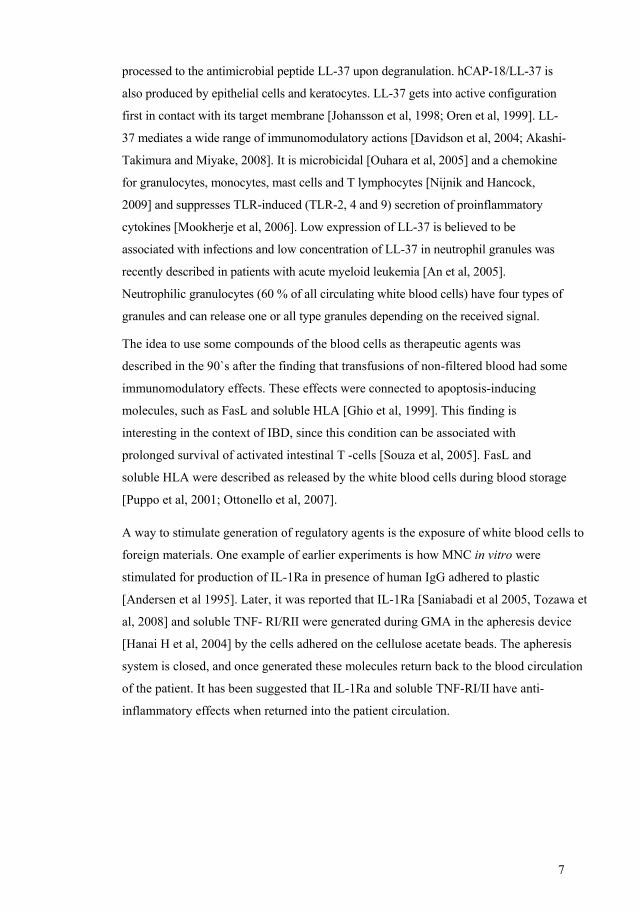
3 AIMS OF THE STUDY
The general objective of this thesis was to elucidate the role of immunological balance
in connection to interventions by allergen challenge in asthma patients and apheresis
treatment in inflammatory bowel disease patients.
Paper I. The aim was to investigate the impact of allergen challenge on the cytokine
profile of circulating lymphocyte populations (in allergic asthma) and thereby study
role of Th1/Th2 balance in relation to the clinic.
Paper II. The objective of this study was to provide information on safety, tolerability,
feasibility, and impact on clinical symptoms when using a selective granulocyte- and
monocyte apheresis device in patients with chronic active IBD in a European setting.
Aims in my project were to study correlation between clinical response to granulocyte-
and monocyte- apheresis and cytokine profile changes in peripheral blood and in
colonic mucosal tissue in patients with IBD.
Paper III. Given that regulatory T cells and TLR expression constitute additional key
actors in mucosal immune regulation this study was aimed to identify the dynamics of
these actors in the intestinal mucosa in relation to clinical improvement following
GMA treatment.
Paper IV. The primary aim of this pilot trial was to evaluate safety and tolerability of
the Cellsorba™ EX filter in combination with ACD-A in the treatment of UC patients
by monitoring clinical adverse effects, whole blood cell counts, bradykinin as well as
IL-6. Potential therapeutic efficacy was evaluated using the composed clinical and
endoscopical disease activity index (Mayo score). The aim of my project was to
evaluate the effect of the LCAP filter and apheresis system lines on selected regulatory
molecules.

15
4 METHODS Methods paper 1
Subject characterization and sampling procedure
Ten subjects underwent a bronchial allergen challenge and peripheral blood samples
were collected before and 24 hours after allergen provocation (Schedule 1, below).
Schedule 1. This schedule illustrates the study design and shows the time points for sampling. Heparinized blood from 10 healthy blood donors served as control. The patients used
no per os or inhaled steroids during the study period and no patient had an on-going
infection. All patients inhaled β2- agonists as needed.
Bronchial allergen challenge
The allergen challenge was done with a dosimeter jet nebulizer. Standardized and
freeze-dried
birch or timothy allergen extracts were diluted and used at a maximum of four
concentrations: 1000, 4000, 16000 and 64000 SQ (Standardized Quality) allergen units
x mL-1. The nebulizer was set to nebulize for 0.5 s giving an output of 7.1 µL/breath.
At each concentration, first 2 and then 4 breaths could be taken, and if needed followed
by 8 and 16 breaths at the highest concentration providing doses from 14 to 7040 SQ
allergen units. Specific airway resistance (SRaw ) and thoracic gas volume were
measured in a constant-volume whole-body plethysmograph 15 min after each dose of
allergen. The challenge proceeded until a 100 % increase in SRaw was reached.
Blood‐ samples
Day 2 24 h after allergen challenge
Day 1 before allergen challenge
Allergen challenge

16
Median provocative dose of allergen required to cause 100% increase of SRaw
(PDSRaw100%) was calculated by linear interpolation on a logarithmic scale. Forced
expiratory volume in one second (FEV1) and SRaw were recorded immediately before
and 15 min after each single dose of the allergen.
Spirometry
The measurements of FEV1 were made hourly using a portable computerized
spirometer.
The maximal fall in FEV1 from allergen challenge to 3-10 hours after allergen
challenge, as well as the average fall in FEV1 during this period, was used to measure
the asthmatic reaction during the late phase.
Definition of single- and dual-responders
Single responders had only an early reaction 15 min after the allergen challenge with a
100% increase in SRaw. Late phase reaction was defined as at least 15% decline in
FEV1 3-10 hours after the last dose of allergen challenge. Dual responders had a late
phase reaction in addition to the initial 100% increase in SRaw.
Stimulation of peripheral blood lymphocytes
Whole blood samples were incubated for 4 hours with mitogen Phorbol 12-Myristate-
13-Acetate (PMA) and Ionomycin in presence of Brefeldin (BFA) and Hepes-RPMI.
Unstimulated cells, which served as controls, remained in BFA and medium only for 4
hours. BFA was used to interrupt the intracellular Golgi mediated transport and to
allow the cytokines to be accumulated inside the cells.
Surface immunostaining
In vitro activation of CD3+ cells was assessed by CD69 up-regulation, and 90%
CD69+CD3+ was set as the lowest acceptable level. To identify the CD4 and CD8
positive lymphocytes by flow cytometry the cells were stained with specific
fluorescent-conjugated antibodies.
17
Cell membrane fixation, permeabilization and immunofluorescence staining of
intracellular IFN-γ and IL-4
After cell membrane fixation and permeabilization (using a commercial kit)
fluorescent-conjugated antibodies to IFN-γ respectively IL-4 were added. Samples were
finally analysed in an EPICS XL flow cytometer.
Flow cytometric analysis
Discrimination gates were set around the respective cell population and a minimum of
3000 lymphocytes were accumulated during analysis. The cells were identified both as
CD3, CD69, CD4, CD8-positive and positive for the respective cytokine.
Statistical methods
Results are given as median and range values or Δ -values which represent the
percentage change between two observations within the same individual. The Wilcoxon
test was used to analyse the differences between paired measurements before and after
the allergen provocation. And the Mann-Whitney U Test was used to analyse the
differences of Δ -values between single and dual responders.
Methods paper 2
Study design
An initial assessment included colonoscopy with biopsies and blood samples. The
active treatment period was five weeks with one treatment per week and a new overall
evaluation, including endoscopy with biopsies, was made two weeks after the last
apheresis had been carried. Following the active part of the study, the patients were
then followed as out-patients on a bi-monthly schedule with clinical assessment,
recording of possible adverse events and side effects until week 52, when a final
clinical assessment of tolerability and response was made. (Schedule 2, below)

18
Schedule 2. This schedule illustrates the study design and shows the time
points for sampling.
Patients
All 10 patients (7 with Crohn Disease; 3 with Ulcerative Colitis, median age:
31 yrs, 3 male and 7 female) had a diagnosis of mild to moderately active IBD.
Apheresis was then performed with a perfusion rate of 30 mL/minute, the
duration of each session was 60 minutes and anticoagulation was performed
using a bolus dose of heparin of 4000 units. Altogether, each patient received
once weekly apheresis treatments for five consecutive weeks. The dosages of
all anti-inflammatory drugs were kept constant during the study period, with
the exception of oral prednisolone. The dose of prednisolone was decreased
after the second apheresis if the patient had improved. Tapering was then
gradually performed with 2.5 mg per week.
Clinical assessment
The patients’ symptoms were assessed using the modified Clinical Activity
Index (CAI) [Rachmilewitz et al, 1989], and for the CD-patients the CD
activity index-score (CDAI) [Best et al, 1979] was also calculated. A reduction
of the CAI by three points or more compared with baseline (week 0) indicated
clinical response, and a score of four or less was considered as clinical
remission. A reduction of the CDAI by 70 points or more was indicating
response and a reduction to less than 150 was considered as clinical remission.
Clinical follow up
1 year
Blood‐ sample
5:th treatment
1:st treatment
Biopsy at start
Biopsy 2 weeks after the last treatment
19
Intracellular cytokines
Monoclonal antibodies suitable for intracellular staining and flow cytometry
were used for detection of intracellular human IL-4, TNF-α, IL-10 and IFN-γ
as described in the first paper.
ELISpot assay for detection of cells producing IFN-γ, IL-10, TNFα, IL-12, IL-
4, IL-6
Ninety six-well membrane plates were coated with mAb to IFN-γ; IL-10;
TNF-α; IL-12; IL-4 and IL-6. PBMC were separated from blood samples by
gradient centrifuging. A total of 30,000 and 100,000 PBMC were added to
each well in culture medium containing mitogen PHA. ELISpot plates were
incubated for 21-23 h. Plates were then washed and biotinylated detection
mAb to IFN-γ; IL-10; TNF-α ; total IL-12; IL-4; IL-6 was respectively added.
Plates were washed and streptavidin-ALP was added before incubation for 1 h
at RT. The plates were washed and substrate was added. After development of
spots for 15 min at RT, the plates were washed with tap water and dried. The
number of spots per well was counted by an automated ELISpot reader.
ELISA determination of IFN-γ, IL-6 and TNF-α plasma levels
Analyses were performed with commercial ELISA kits according to the
manufacturer's instructions. The lowest detectable concentration of cytokine
was 8, 0.7 and 0.1 pg/mL for the IFN-γ, IL-6 and TNF-α assays, respectively.
Immunohistochemistry staining of biopsies
Sections for CD3 and CD8 staining were fixed with acetone and sections for
cytokine staining (TNF-α, IL-1β, IFN-γ, IL-4, IL-15) were fixed with
formaldehyde.
A mixture of H2O2 and NaN3 in PBS for one hour in the dark in room
temperature was used for blocking endogenous peroxidase activity. Primary
monoclonal antibodies (mAbs) where in a mixture of PBS, BSA, NaN3 and
saponin for the cytokine staining. Saponin was used as a detergent to make
intracellular compartments available for antibodies. The primary antibodies
were incubated over night in a humid chamber. Incubation with normal horse
serum (NHS) was used as blocking procedure. The secondary antibody was

20
diluted in a mixture of PBS and NHS for incubation. An avidin-biotin-
horseradish peroxidase was diluted in PBS or PBS-saponin. Sections were
incubated in the solution. A staining reaction was developed with a Peroxidase
Substrate Kit containing 3,3´diaminobenzidinetetrahydrochloride (DAB).
The sections were assessed on two different occasions by two independent
observers in all cases with concordant results.
Examinations were performed in a blind manner with regard to patient
response, biopsy site and time point. The assessment of CD3, CD8 as well as
for all cytokines was performed by semiquantative evaluation. For CD3 and
CD8 assessment a grading scale ranging from four (massive infiltration: >
1000 positive cells per view), three (between 100 and 1000 cells), two
(between 10 and 100), one (between 1 and 10) to zero (no visible staining) was
used. An almost similar, but four graded scale was used for all cytokine
assessments; three (massive infiltration, > 100 positive cells per view), two
(between 10 and 100), one (between 1 and 10), zero (no visible staining).
Negative controls were processed on each slide.
Statistics
Statistical analyses were done by using the Wilcoxon non-parametric tests. A
P-value <0.05 was considered significant.
Methods paper 3
Patients
Ten patients (7 with Crohn Disease; 3 with Ulcerative Colitis, median age:
31 yrs, 3 male and 7 female) with mild to moderately chronic active IBD
were studied. Eight out of 10 patients were improved clinically with good
long-term effect after GMA , which was done once weekly over five
consecutive weeks.

21
Immunohistochemistry
Immunohistochemical staining of biopsy materials taken before start of treatment and
two weeks after last treatment was performed on acetone fixed sections (CD25 and
CD163 (macrophage marker)) and on formaline fixed sections (FoxP3, BDCA-2, -3, -4
(dendritic cells marker), TLR-2, TLR-4) as described in the second paper.
All examinations were done in a blinded manner with regard to patient
response, biopsy site and time point. The assessments were done by semi-
quantitative evaluation with a grading scale ranging from 3 (> 50 cells), 2 (10 -
50), 1 (1 -10) to 0 (no visible staining).
Statistics
Statistical analyses were done by using the Wilcoxon non-parametric tests.
P-values <0.05 were considered significant.
Methods paper 4
Patients
Patients with mild to moderate active UC (Mayo Score: 6 – 10) despite treatment
with 5-ASA equivalent to minimum of 4 g sulfasalazine / day for at least six weeks
were eligible to enter this study (Table I). Aminosalicylate treatment was continued
without any change during the current study. Analgesic or spasmolytic drugs were
allowed as concomitant medication, while, long-term antibiotic treatment or bowel
motility modifying drugs (e.g. loperamide) were not permitted during the study.
Treatment course
Two sessions of LCAP each week were carried out for the first three weeks, followed
by one session a week for the subsequent four weeks (10 sessions totally). A follow- up
visit was held in week 16 (Schedule 3, below).

22
Schedule 3. This schedule illustrates the study design and shows the time
points for sampling.
Safety and tolerability.
Vital signs including pulse rate and blood pressure were recorded before, during and
after each LCAP session. Measurements of differential blood flow pressure between
the inlet and outlet lines of the CellsorbaTM EX device were also done. Any adverse
event and/or device or procedure related incidents were registered.
Clinical follow up and efficacy
Clinical activity was evaluated by means of disease activity index – the Mayo
score [Lewis et al, 2008] before start of LCAP, at week 8 (after 1 week of the
last LCAP) and at week 16. Videocolonoscopy with pictures and biopsies
were performed at entry and at weeks 8 and 16. A reduction of Mayo score to
≤ 2 was taken as a clinical remission, while a drop in Mayo score ≥ 3 points
was considered as a clinical response. Histological assessment of biopsies was
performed in a blinded fashion using the grading by Geboes et al [Geboes et
al, 2000]. The worst score was used for the evaluation.
Laboratory analyses
Peripheral venous samples for whole blood counts were collected before and directly
after each LCAP, designated “Before” and “After” respectively. The samples taken
before LCAP # 1, 3 and 10 were in addition subjected to analysis of CRP and ESR.
Samples were also collected from the inlet and outlet lines of the CellsorbaTM EX
Clinical follow up
16 weeks
Blood- samples at LCAP sessions
10:th treatment, 8 weeks
1:st treatment
Biopsy at start
Biopsies, 8 weeks and 16 after the first Treatment, as well blood samles.

23
device at two occasions during a treatment session, namely at time of first blood return
(I), and at before rinse back (II). The inlet samples were taken from taps on plastic lines
located 190 cm after vein puncture and 180 cm before the column, while the outlet
samples were taken from taps located 49 cm after the column. These samples were
designated “In-I”, “Out-I”, “In-II” and “Out-II”, respectively and subjected to analysis
of WBC and platelets as well as soluble markers. IL-6 and bradykinin were analysed at
the first LCAP. (Schedule 4, below).
Schedule 4. The image illustrates where and when the samples were taken. Samples
were also collected from the inlet (In) and outlet (Out) lines device at two occasions
during a treatment session, namely at time of first blood return (I), and at before rinse
back (II).
Commercial ELISA kits were used and the analysis procedures were done according to
manufacturers´ instructions for assessment of bradykinin, IL-6, IL-1ra, LL-37, CD30L
(CD153).

24
Statistics
The Wilcoxon non-parametric tests were used for the statistical analyses,
P- values <0.05 were considered as significant.

25
5 RESULTS AND DISCUSSION
First paper
Allergen challenge was used as a way to study allergic inflammatory responses. The
patients got symptoms of allergic asthma and lymphocyte profiles became changed. IL-
4 positive CD4+ cells decreased in peripheral blood after the allergen challenge. IFN-γ
positive CD4+ cells decreased in peripheral blood in single responders, but had a
tendency to increase in dual responders. There was a significant difference between
single and dual responders regarding changes in numbers of IFN-γ positive CD4+ cells.
Image 6. Recruitment of Th2 and Th1 lymphocytes into bronchial mucosa in response
to allergen challenge. Reduced recruitment of CD4+IFN-γ+ (Th1) cells in dual
responders is illustrated by a dotted line.
Existence of an active traffic of T lymphocytes into bronchial mucosa in order to
participate in the immune response was suggested earlier [Durham et al. 2000]. It has
been hypothesized that an active recruitment of lymphocytes into the bronchial mucosa
is reflected by a decrease of the corresponding circulating population.
The allergic inflammatory response is associated with IgE-mediated mast cell

26
activation and followed by recruitment of leucocytes into the airway tissue. It has been
suggested that T-helper type 2 cells become recruited into the lungs and regulate
allergic asthma reaction. [Durham et al, 2000]. Our finding that IL4+CD4+ cells
decreased in peripheral blood in the patient group and IFN-γ + CD4+ cells decreased in
the single responders after allergen challenge suggests the active traffic of both Th1 and
Th2 cells.
We suggest that a difference in recruitment of IFN-γ positive CD4+ lymphocytes can
explain differences between clinical response in patients with late-phase reaction and
patients without late-phase reaction. A diminished capacity to down-regulate the Th2
response by recruitment of sufficient number of IFN-γ positive CD4+ lymphocytes was
suggested as explanation to the late phase symptoms in the dual responders. Matsumoto
and co-workers [Matsumoto et al, 2002] have reported that IL-10+CD4+ lymphocytes
increase in single responders and decrease in dual responders after an allergen
challenge. This finding goes in line with our IFN-γ-data, because an existing
antagonistic balance between IFN-γ and IL-10 has been suggested [Romagnani, 1991].
In the present study we also compared T lymphocyte subpopulations in patients with
allergic asthma and in healthy blood donors. Our findings that the patients have lower
IFN-γ + and higher IL-4+ lymphocyte counts compared to the healthy controls were
consistent with results reported by other research groups. This confirms the accuracy of
the applied laboratory method in this study and encourages us to apply this method
again in other patient group.
Second paper
Patients with inflammatory bowel disease (IBD), both Crohn´s disease and ulcerative
colitis underwent a series of five consequent one per week treatments by selective
granulocyte- and monocyte- apheresis (GMA). Eighth of ten patients reached good
clinical response and even maintained a long-term response.
A significant decrease of IFN-γ-producing CD4+ cells after five apheresis sessions
(P=0.046) was found by flow cytometric analyses. This result was further supported by
the ELISpot analyses from eight cases where IFN-γ-producing cells also decreased
significantly between week 0 and 7 (P=0.037).
The median IFN-γ-positive cell staining score decreased from 2.0 to 0.0 for all patients.
However, when biopsies from the patients attaining a significant clinical response at
week 7 (7/9) were analyzed separately, the median IFN-γ-positive cell staining score

27
significantly decreased from 3.0 to 0.0 (P=0.027). At week 52, eight patients had
attained clinical response and when the reduction in IFN-γ-positive cells measured at
week 7 was then reassessed, the result fell just short of significance (P=0.052). The
result was influenced by one patient (#10) who, at week seven, had only achieved
limited clinical benefit and interestingly had a slight increase of IFN-γ-positive staining
in the biopsy from week 7 compared to baseline. However, this patient still had clinical
improvement at week 52 (CDAI score drop by almost 70 points).
The immunohistochemistry staining for other cytokines (i.e., TNF-α, IL-1 β, Il-4, and
IL-15) did not show an statistically significant change, although some patients had a
marked reduction of TNF-α staining. There was a tendency towards a decrease of IL-
15-positive cells at week 7 (P=ns). The analyses of CD3+ and CD8+ did not show
significant changes at week 7 (NK cells were few in pre- and post-treatment biopsies
and there were no decrease in their numbers, unpublished data). Apart from IFN-γ, no
significant changes were measured by ELISpot (IL-10, TNF-α, IL-12, IL-4, and IL-6)
or by flow cytometry (IL-4, IL-10, and TNF-α).
A significant increase of soluble (measured by ELISA) TNF-α in the serum after the
apheresis treatment were noted for the whole group of patients (P=0.007), but there was
no appreciable difference between median values before and after 4.0 (range: 1.3–51.1)
vs 5.3 (1.4–76.9). In eight out of ten patients, TNF-α levels were almost unchanged. No
significant changes were observed for IL-6 and IFN-γ when measured by ELISA.
The clinical response was parallel to down regulation of IFN-γ+ CD4+ cells in
peripheral blood and IFN-γ+ cells in colonic mucosa biopsies.
Common feature for the first and second trials was that lymphocyte subpopulations
were influenced. Tendency to normal balance would lead to symptom relief. In the
asthma trial, IFN-γ+ (Th1) are involved in limiting of IL-4+ (Th2) response. And the
scarcity in this capacity deteriorates the inflammatory response. In this case late-phase
reaction will be observed.
In the second study the finding of down regulation of IFN-γ+ T helper cells in the blood
and IFN-γ+ cells in the biopsies confirmed our suggestions that mechanisms of column
apheresis were complex and immunomodulatory involving more cells types in addition
to the absorbed cell type.
In the IBD patients who maintained a long-term response (for 12 months) IFN-γ
positive lymphocytes were decreased already in the post-treatment biopsies at week 7.
28
week 7. Therefore we suggested that down regulation of these cells might be a
predictive marker for long-term response.
IFN-γ can be both pro-inflammatory and regulatory, but if the immunological balance
has turned to the tolerance state it might be predictive for a better clinical outcome. Our
opinion is shared in recent review by Bosani et al, 2009, who have written that with
anti-IFN-γ agents “we are moving closer to the epicentre of immune reaction. Only
agents restoring T cell tolerance long term or repairing the basic dysfunction in the
innate immune system may provide the perspective of cure in chronically remitting and
flaring diseases…”
Third paper
When we expanded the second study by analysing T regulatory cells and expression of
Toll Like Receptors in the colonic biopsies the findings became following:
The number of FoxP3-positive cells decreased significantly in the mucosa.
Furthermore, the number of TLR-2 positive cells decreased significantly in the lamina
propria, but not in the epithelium. TLR-4 positive cells were unchanged. No significant
differences were noticed for BDCA-2, -3, -4 neither for CD3 and CD25. In the patients
who experienced a long-term effect of the treatment (n=8), the number of CD163
positive cells decreased significantly (P=0.046), but not significantly when all patients
were included (n=9). UC and CD groups were also analysed separately showing equal
response in measured parameters.
We did not find a strict relationship between FoxP3 and CD25 staining, and FoxP3+
cells were slightly more numerous than CD25+ cells at the start of treatment. FoxP3
expression is not exclusively linked to CD25 expression, which is supported by recent
studies [Pillai et al, 2007; Fontenot et al, 2005].

29
IFN-γ FoxP3 TLR-2
Image 7. Summary immunohistochemistry image from second and third paper
It was a novel message that the number of FoxP3+ T regs can diminish parallel to
clinical improvement, because of belief that the higher number of T regs the better
protection against inflammation [Maul et al, 2005; Denning et al, 2003; Uhlig et al,
2006]. Our finding suggests existence of an ongoing balance process with dynamic
tendencies. According to our suggestion, diminished numbers of T regs reflects a state
of improved tolerance (T regs vs effector cells).
The selective decrease in TLR-2 expression may reflect differences in regulation of
TLR-2 and TLR-4. TLR-2 expression is induced in bacterial infections, while TLR-4 is
constantly expressed [Matsuguchi et al, 2000]. According to previous reports induction
of tolerance to bacterial lipoprotein might be associated with down-regulation of TLR-2
expression [Wang et al, 2002]. Thus, down regulated expression of TLR-2 in our study
may reflect a state of improved tolerance to intestinal flora.
It has been speculated that in spite of increased numbers T regs suppressive capacity is
down regulated in inflamed mucosa [Yu et al, 2007]. Expression of TLRs on T regs
was described recently [Caramalho et al, 2003]. Sutmuller et al have demonstrated in
experiments on mice T regs that TLR-2 signalling directly stimulates T regs expansion

30
and T regs stimulated through TLR-2 have diminished capacity to suppress
proliferation of T helper cells in vitro [Sutmuller et al, 2006]. Their results are in line
with simultaneous decrease of TLR-2 expression and T regs number in our study.
Further research is required to elucidate the role of TLR triggering on T regs [van
Maren et al, 2008].
Fourth paper
The modified LCAP using ACD-A as anticoagulant was found to be a safe and well-
tolerated procedure, regarding both the vital signs and IL-6/ bradykinin generation.
Four patients were responders, of whom two patients went into remission. Median
histological scores decreased from 3.5 to 2.0 in these four patients.
The analyses of CD30L, IL-1Ra and LL-37 have shown that the LCAP filter does not
hinder the passage of these important soluble molecules. Some absorbance of IL-1Ra in
the plastic lines could not be excluded.
There was no increase of IL-1Ra during LCAP, which is indicative of a difference
between LCAP and GMA in this aspect.
An important observation regarding the analyzed molecules was that LL-37 increased
at all sessions within the apheresis plastic lines.
Disturbed tolerance to the intestinal bacteria is considered important in the pathogenesis
of IBD [Ohkusa et al, 2004]. Since LL-37 affects the cellular response to bacteria and is
known for its antibacterial properties it plays an important role for maintaining the
tolerance to the bacteria [Mookherje et al, 2006].
Wehlin et al reported earlier that Mac-1 (CD11b/CD18) up-regulation occurs when
blood samples are kept in plastic tubing [Wehlin et al, 1998]. Mac-1 up-regulation is
associated with neutrophil degranulation and is complement dependent [Borregaard et
al, 1994]. Thus, we assume that LL-37 (contained in the granules of neutrophil
granulocytes [Sörensen et al, 1997]) is released from neutrophils within the plastic
apheresis lines and may perform its regulatory functions when returned back into the
patient.
Nafamostat mesilate used in earlier types of CellSorba (LCAP) and heparin used in
GMA inhibit complement activation [Keck et al, 2001; Weiler et al, 1992]. While,
significant complement activation was described in connection to apheresis with ACD-
A (the most widely used in the apheresis systems citrate based anticoagulant) [Ptak et
al, 2005]. Lever et al have reported that neutrophil degranulation is inhibited by heparin
31
[Lever et al, 2007]. Thus, the anticoagulants have different properties regarding their
capacity to inhibit the complement activation and the neutrophil degranulation. Our
own unpublished data indicates that heparin (used as anticoagulant) inhibits
degranulation of neutrophils and generation of LL-37. The anticoagulant (ACD-A)
used in the present study did not prevent the neutrophil degranulation.
LL-37 was recently shown to have potential therapeutic properties against cancers, HIV
[Chuang et al, 2009; Bergman et al, 2007], for wound healing, against bacterial biofilm
and methicillin-resistant bacteria [Nijnik and Hancock, 2009; Komatsuzawa et al,
2006]. There is a need of a method to stimulate the endogenous production of LL-37
which may find therapeutic applications [Nijnik and Hancock, 2009]. Mookherje et al,
2006, showed that even small increase in concentration of LL-37 enhances the cellular
tolerance to bacterial components and suppresses TLR-induced secretion of
proinflammatory cytokines, which supports our suggestion that generation of LL-37 in
the lines might have a positive impact on the immunologic tolerance and contribute to
the efficacy of the apheresis treatment.
Conclusions
We have suggested that differences in response to allergen can depend on different
capacity to maintain immunologic balance (Th-balance) – diminished capacity to
recruit IFN-γ+ CD4+ lymphocytes seems to be associated with development of an
additional so called late-phase reaction. We have shown that mechanisms of leukocyte
adsorbing apheresis were immunomodulating involving down regulation of IFN-γ+
lymphocytes hereby influencing the Th-balance. The clinical improvement in IBD was
associated with improved immunologic tolerance mirrored by down regulation of
FoxP3+ cells and TLR-2 expression in the gut mucosa. And finally, we have described
generation of LL-37 in plastic lines of apheresis system, which may have a positive
impact on the immunologic tolerance.
The restoration of the immunologic tolerance can be the key to successful therapeutic
strategies.

32
6 FUTURE PERSPECTIVES GMA and LCAP were reported to have promising clinical results in many prospective
studies. The absence of serious side effects is an advantage of the apheresis treatments.
However, their role in the treatment of IBD is still debated. Only two sham-controlled
studies have been published. One smaller study on LCAP showed a better outcome in
active moderate UC [Sawada et al, 2005]. More recently, a larger study on GMA was
published [Sands et al, 2008]. This study did not show any significant difference in the
effect on UC between the sham-control and GMA. In the later study however, blood of
sham-control patients was circulating through plastic tubing and then returned back into
the patient circulation. This approach can be challenged since different cascade systems
can be activated when in contact with foreign surfaces as the complement and the
coagulation systems. We have also shown a generation of LL-37 in the plastic lines.
Therefore, there is a need to further investigate the role of complement system
activation as well as other immunomodulators for the treatment outcomes.
We do not know why some patients are treatment responders and others are not. We
speculate that different phases of the disease might be of importance for induction of
response to the treatment [Kugathasan et al, 2007]. We also suggest that treatment
frequency and interval between the sessions must be adjusted to each patient.
Today there are various treatments for inflammatory conditions, but one cannot know
in advance whether the patient will benefit from a certain treatment or develop serious
side effects. One example is the use of biologics that are expensive and can cause
serious side effects. Moreover, not all patients respond to these treatments. It is
important to be able to choose the right treatment and spare the patients from the
treatments with serious adverse reactions. Identification of immunological predictive
markers for treatment response would help to give the right individual treatment and
even save a lot of money. At best, predictors could help to choose tailored [Scaldaferri
and Fiocchi, 2007] treatment in the early phase of inflammatory diseases. The future
challenge is thus to find such predictors.

33
7 ACKNOWLEDGEMENTS
I would like to thank everyone who has supported me through this study and especially:
My supervisor, professor Joachim Lundahl, for sharing your profound knowledge, your
enthusiasm, for teaching me a lot, for generous help whenever needed.
My co-suprvosor, professor Robert Löfberg, for sharing your profound knowledge,
your positive spirit and ideas, for valuable and stimulating discussions.
My co-supervisor, Dr Kerstin Elvin, for sharing your profound knowledge, for valuable
and stimulating discussions, availability and support.
My mentor, Ann-Charlotte Wikström for your advices.
The whole staff at the Department of Clinical immunology and Transfusion medicine
for all kinds of help.
All members of the “Inflammatory unit” for your warm support.
Titti Nieminen for teaching me flow cytometry and many more. Josefin Paulsson for
being a wonderful roommate. Michaela Cassmer for your help.
My co-authors, especially Marianne van Hage, Ann-Kristin Ulfgren, Ola Winqvist,
Aleksandra Mandic-Havelka for fruitful collaboration.
Abraham Mamo, Eva Lindroos, Marianne Malmström, Petra Jones for helping me in
labs.
My family for love and support.
34
8 REFERENCES Abreu MT, Plevy S, Sands BE, Weinstein R. Selective leukocyte apheresis for the treatment of inflammatory bowel disease. J Clin Gastroenterol 2007;41(10):874-888. An LL, Ma XT, Yang YH, Lin YM, Song YH, Wu KF. Marked Reduction of LL-37/hCAP-18, an Antimicrobial Peptide, in Patients with Acute Myeloid Leukemia. Int J Hematol 2005;81(1):45-47. Andersen LS, Petersen J, Svenson M, Bendtzen K. IgG for intravenous use, autologus serum and plasma induce comparable interleukin-1 receptor antagonist liberation from human mononuclear cells: an in vitro phenomenon upon plastic adherence. Autoimmunity 1995;22(2):127-133. Belge KU, Dayyani F, Horelt A Siedlar M, Frankenberger M, Frankenberger B, Espevik T, Ziegler-Heitbrock L. The proinflammatory CD14+CD16+DR++ monocytes are a major source of TNF. J Immunol 2002;168(7):3536-3542. Bergman P, Walter-Jallow L, Broliden K, Agerberth B, Söderlund J. The antimicrobial peptide LL-37 inhibits HIV-1 replication. Curr HIV Res 2007;5(4):410-415. Best WR, Becktel JM, Singleton JW. Rederived values of eight coefficients of the Crohn’s Disease Activity Index (CDAI). Gastroenterology 1979;77(4):843–846. Borregaard N, Kjeldsen L, Sengløv H, Diamond MS, Springer TA, Anderson HC, Kishimoto TK, Bainton DF. Changes in subcellular localization and surface expression of L-selectin, alkaline phosphatase, and Mac-1 in human neutrophils during stimulation with inflammatory mediators. J Leukoc Biol 1994;56:80-87. Bosani M, Ardizzone S, Porro GB. Biologic targeting in the treatment of inflammatory bowel diseases. Biologics 2009;3:77-97. Brand S. Crohn’s disease: Th1, Th17 or both? The change of a paradigm: new immunological and genetic insights implicate Th17 cells in the pathogenesis of Crohn’s disease. Gut 2009;58(8):1152-1167. Review. Brightling CE, Symon Fa, Birring SS, Bradding P, Pavord ID, Wardlaw AJ.Th2 cytokine expression in bronchoalveolar lavage fluid T-lymphocytes and bronchial submucosa is a feature of asthma and eosinophilic bronchitis. J Allergy Clin Immunol 2002;110(6):899-905. Caramalho I, Lopes-Carvalho T, Ostler D, Zelenay S, Haury M, Demengeot J. Regulatory T cells selectively express toll like receptors and are activated by lipopolysaccharide. J Exp Med 2003;197(4):403-411.
35
Casini-Raggi V, Kam L, Chong YJ, Fiocchi C, Pizarro TT, Cominelli F. Mucosal imbalance of IL-1 and IL-1 receptor antagonist in inflammatory bowel disease. A novel mechanism of chronic intestinal inflammation.J Immunol 1995;154(5):2434-2440. Cho SH, Stanciu LA, Begishivili T, Bates PJ, Holgate ST, Johnston SL. Peripheral blood CD4+ and CD8+ T cell type cytokine production in atopic asthmatic and normal subjects. Clin Exp Allergy 2002;32(3):427-433. Chuang CM, Monie A, Wu A, Mao CP, Hung CF. Treatment with LL-37 peptide enhances antitumor effects induced by CpG oligodeoxynucleotides against ovarian cancer. Hum Gene Ther 2009;20(4):303-313. Davidson DJ, Currie AJ, Reid GS, Bowdish DM, MacDonald KL, Ma RC, Hancock RE, Speert DP. The cationic antimicrobial peptide LL-37 modulates dendritic cell differentiation and dendritic cell-induced T cell polarization. J Immunol 2004;172:1146-1156. Denning TL , Qi H, König R, Scott KG, Naganuma M and Ernst PB. CD4+ T cells resembling regulatory T cells that inhibit chronic colitis differentiate in the absence of interactions between CD4 and class II MHC. J Immunol 2003;71:2279-2286. Duchmann R, Schmitt E, Knolle P, Meyer zum Buschenfelde KH, Neurath M. Tolerance towards resident intestinal flora in mice is abrogated in experimental colitis and restored by treatment with interleukin-10 or antibodies to interleukin-12. Eur J Immunol 1996;26:934-938. Durham SR. Mechanisms of mucosal inflammation in the nose and lungs. Clin Exp Allergy 1998;28(2):11-16. Review. Dzik S. Leukodepletion blood filters: filter design and mechanisms of leukocyte removal. Transfus Med Rev 1993;7(2):65-77. Review. Emmrich J, Petermann S, Nowak D, Beutner I, Brock P, Klingel R, Mausfeld-Lafdhiya P, Liebe S, Ramlow W. Leukocytapheresis (LCAP) in the management of chronic active ulcerative colitis--results of a randomized pilot trial. Dig Dis Sci 2007;52(9):2044-2053. Erridge C, Pridmore A, Eley A, Stewart J and Poxton IR. Lipopolysaccharides of Bacteroides fragilis, Chlamydia trachomatis and Pseudomonas aeruginosa signal via toll-like receptor 2. J Med Microbiol 2004;53:735-740. Fehr J. Complement as a mediator of granulocyte adherence and migration: studies based on the acute neutropenia of filtration leukapheresis. Prog Clin Biol Res 1977;13:243-258. Review. Filaci G, Bacilieri S, Fravega M, Monetti M, Contini P, Ghio M, Setti M, Puppo F, Indiveri F. Fas, Fas ligand,and transfusion immunomodulation. J Immunol 2001;166(10):6452-6457.
36
Fiocchi C. Inflammatory bowel disease. Etiology and pathogenesis. Gastroenterology 1998; 115(1):182-205. Fontenot JD, Rasmussen JP, Williams LM, Dooley JL, Farr AG, Rudensky AY. Regulatory T cell lineage specification by the forkhead transcription factor foxp3. Immunity 2005; 22:329-341. Furrie E, Macfarlane S, Thomson G, Macfarlane GT. Toll-like receptors -2, -3 and -4 and expression patterns on human colon and their regulation by mucosal-associated bacteria. Immunology 2005;115;565-574. Geboes K, Riddel R. Öst Å, Jensfelt B, Persson T, Löfberg R. A reproducible grading for histological assisment of inflammation in ulcerative colotis. Gut 2000;47:404-409. Ghio M, Contini P, Mazzei C, Brenci S, Barberis G, Filaci G, Indiveri F, Puppo F. Soluble HLA Class I, HLA Class II, and Fas Ligand in Blood Components: A Possible Key to Explain the Immunomodulatory Effects of Allogeneic Blood Transfusions. Blood 1999;93(5):1770-1777. Gionchetti P, Rizzello F, Lammers KM, Morselli C, Tambasco R, Campieri M. Antimicrobials in the management of inflammatory bowel disease. Digestion 2006;73(1):77-85. Glimcher LH, Murphy KM. Lineage commitment in the immune system: the T helper lymphocyte grows up. Genes Dev 2000;14(14):1693-1711. Grossman WJ, Verbsky JW, Barchet W, Colonna M, Atkinson JP, Ley TJ. Human T regulatory cells can use the perforin pathway to cause autologus target cell death. Immunity 2004, 21(4):589-601. Grossman WJ, Verbsky JW, Tollefsen BL, Kemper C, Atkinson JP, Ley TJ. Differential expression of granzymes A and B in human cytotoxic lymphocyte subsets and T regulatory cells. Blood 2004,104(9):2840-2848. Hamid QA, Cameron LA. Recruitment of T cells to the lung in response to allergen challenge, J Allergy Clin Immunol 2000;106(5):227-234. Hanai H, Iida T, Takeuchi K, Watanabe F, Yamada M, Kikuyama M, Maruyama Y, Iwaoka Y, Hirayama K, Nagata S, Takai K. Adsorptive depletion of elevated proinflammatory CD14+CD16+DR++ monocytes in patients with inflammatory bowel disease. Am J Gastroenterol 2008;103(5):1210-1216. Hanai H, Watanabe F, Yamada M, Sato Y, Takeuchi K, Iida T, Tozawa K, Tanaka T, Maruyama Y, Matsishita I, Ivaoka Y, Saniabadi A. Correlation of serum soluble TNF-alpha receptors I and II levels with disease activity in patients with ulcerative colitis.Am J Gastroenterol 2004;99:1532- 1538.

37
Hanai H, Watanabe F, Yamada M, Sato Y, Takeuchi K, Iida T, Tozawa K, Tanaka T, Maruyama Y, Matsushita I, Iwaoka Y, Kikuch K, Saniabadi AR. Adsorptive granulocyte and monocyte apheresis versus prednisolone in patients with corticosteroid-dependent moderately severe ulcerative colitis. Digestion 2004;70(1):36-44. Harrington LE, Hatton RD, Mangan PR, Turner H, Murphy TL, Murphy KM, Weaver CT. Interleukin 17-producing CD4+ effector T cells develop via a lineage distinct from the T helper type 1 and 2 lineages. Nat Immunol 2005;6(11):1123-1132. Hart AL, Al-Hassi HO, Rigby RJ, Bell SJ, Emmanuel AV, Knight SC, Kamm MA, Stagg AJ. Characteristics of intestinal dendritic cells in inflammatory bowel diseases. Gastoenterology 2005;129:50-65. Hild M, Söderström T, Egberg N, Lundahl J. Kinetics of bradykinin levels during and after leucocyte filtration of platelet concentrates. Vox Sang 1998;75:18-25. Hiraishi K, Takeda Y, Shiobara N, Shibusawa H, Jimma F, Kashiwagi N, Saniabadi AR, Adachi M. Studies on the mechanisms of leukocyte adhesion to cellulose acetate beads: an in vitro model to assess the efficacy of cellulose acetate carrier-based granulocyte and monocyte adsorptive apheresis. Therap Apher Dial 2003;7:334-340. Holmen N, Lundgren A, Lundin S, Bergin AM, Rudin A, Sjövall H, Ohman L. Functional CD4+CD25+high regulatory T cells are enriched in the colonic mucosa of patients with active ulcerative colitis and increase with disease activity. Ibbotson JP, Lowes JR, Chahal H, Gaston JS, Life P, Kumararatne DS, Sharif H, Alexander-Williams J, Allan RN.Mucosal cell-mediated immunity to mycobacterial, enterobacterial and other microbial antigens in inflammatory bowel disease. Cin Exp Immunol 1992;87(2):224-230. Ivanov II, Frutos Rde L, Manel N, Yoshinaga K, Rifkin DB, Sartor RB, Finlay BB, Littman DR. Specific microbiota direct the differentiation of IL-17-producing cells in the mucosa of the small intestine. Cell Host Microbe 2008;4:337-349. Iwama H. Bradykinin-associated reactions in white cell-reduction filter. J Crit Care 2001; 16:74-81. Johansson J, Gudmundsson GH, Rottenberg ME, Berndt KD, Agerberth B. Conformation-dependent antibacterial activity of the naturally occurring human peptide LL-37. J Biol Chem 1998;273(6):3718-3724. Kanai T, Makita S, Kawamura T, Nemoto Y, Kubota D, Nagayama K, Totsuka T, Watanabe M. Extracorporeal elimination of TNF-α-producing CD14dullCD16+ monocytes in leukocytapheresis therapy for ulcerative colitis. Inflamm Bowel Dis 2007;13:284-290.

38
Kashiwagi N, Hirata I, Kasukawa R. A role for granulocyte and monocyte apheresis in the treatment of rheumatoid arthritis. Ther Apher 1998;2:134-141. Kashiwagi N, Sugimura K, Koiwai H, Yamamoto H, Yoshikawa T, Saniabadi AR, Adachi M, Shimoyama T. Immunomodulatory effects of granulocyte and monocyte adsorption apheresis as a treatment for patients with ulcerative colitis. Dig Dis Sci 2002;47:1334-1341. Keck T, Balcom JH, Antoniu BA, Lewandrowski K, Warshaw AL, Fernández-del Castillo CF. Regional effects of nafamostat, a novel potent protease and complement inhibitor, on severe necrotizing pancreatitis. Surgery 2001;130(2):175-181. Kelsen J, Agnholt J, Hoffmann HJ, Rømer JL, Hvas CL and Dahlerup JF. FoxP3+CD4+CD25+ T cells with regulatory properties can be cultured from colonic mucosa of patients with Crohn´s disease. Clin Exp Immunol 2005;141:549-557. Kennedy MK, Willis CR, Armitage RJ. Deciphering CD30 igand biology and its role in humoral immunity. Immunology 2006;118(2):143-152. Review. Kohgo Y, Ashida T, Maemoto A, Ayabe T. Leukocytapheresis for treatment of IBD. J Gastroenterol 2003;38(15):51–54. Review. Kojima S, Harada-Shiba M, Nomura S, Kimura G, Tsushima M, Kuramochi M, Yamamoto A, Omae T. Effect of nafamostat mesilate on bradykinin generation during low-density lipoprotein apheresis using a dextran sulfate cellulose column. ASAIO Trans 1991;37(4):644-648. Komatsuzawa H, Ouhara K, Yamada S, Fujiwara T, Sayama K, Hashimoto K, Sugai M.J Innate defences against methicillin-resistant Staphylococcus aureus (MRSA) infection. Pathol 2006;208:249-260. Review. Kugathasan S, Saubermann LJ, Smith L, Kou D, Itoh J, Binion DG, Levine AD, Blumberg RS, Fiocchi C. Mucosal T-cell immunoregulation varies in early and late inflammatory bowel disease. GUT 2007;56:1696-1705. Lever R, Lo WT, Faraidoun M, Amin V, Brown RA, Gallagher J, Page CP. Size-fractionated heparins have differential effects on human neutrophil function in vitro. Br J Pharmacol 2007;151(6):837-843. Lewis JD, Chuai S, Nessel L, Lichtenstein GR, Aberra FN, Ellenberg JH. Use of the non-invasive components of the mayo score to assess clinical response in ulcerative colitis. Inflamm Bowel Dis 2008;14:1660-1666. Li Q, Sai Y, Kato Y, Muraoka H, Tamai I, Tsuji A. Transporter-mediated renal handling of nafamostat mesilate. J Pharm Sci 2004;93(2):262-272. MacDonald TT, Monteleone G, Pender SL. Recent developments in the immunology of inflammatory bowel disease. Scand J Immunol 2000;51(1):2-9. Review.

39
Matsuguchi T, Musikacharoen T, Ogawa T, Yoshikai Y. Gene expressions of Toll-like receptor 2, but not Toll-like receptor 4, is induced by LPS and inflammatory cytokines in mouse macrophages. J Immunol 2000;165:5767–5772. Matsumoto K, Gauvreau GM, Rerecich T, Watson RM, Wood LJ, O’Byrne PM. IL-10 production in circulating T cells differs between allergen-induced isolated early and dual asthmatic responders. J Allergy Clin Immunol 2002;109(2):281-286. Matsumoto T, Andoh A, Okawa K, Ito H, Torii A, Yoshikawa S, Nakaoka R, Okuyama Y, Oshitani N, Nishishita M, Watanabe K, Fukunaga K, Ohnishi K, Kusaka T, Yokoyama Y, Sasaki M, Tsujikawa T, Aoki T, Kusaka T, Takeda Y, Umehara Y, Nakamura S, Fujiyama Y. Multivariate Analysis for Factors Predicting Rapid Response of Leukocytapheresis in Patients With Steroid-resistant Ulcerative Colitis: A Multicenter Prospective Open-label Study Ther Apher Dial 2008;12:484-490. Maul J, Loddenkemper C, Mundt P, Berg E, Giese T, Stallmach A, Zeitz M, Duchmann R. Peripheral and intestinal regulatory CD4+CD25high T cells in inflammatory bowel disease. Gastroenterology 2005;128:1868-1878. McMurchy AN, Gillies J, Allan SE, Passerini L, Gambineri E, Roncarolo MG, Bacchetta R, Levings MK.Point mutants of forkhead box P3 that cause immune dysregulation, polyendocrinopathy, enteropathy, X-linked have diverse abilities to reprogram T cells into regulatory T cells. J Allergy Clin Immunol. 2010 [Epub ahead of print] Misra N, Bayry J, Lacroix-Desmazes S, Kazatchkine MD, Kavery SV. Cutting edge: human CD4+CD25+ T cells restrain the maturation and antigen-presenting function of dendritic cells. J Immunol 2004;172(8):4676-4680. Mitsuyama K, Suzuki A, Matsumoto S, Tomiyasu N, Takaki K, Takedatsu H, Masuda J, Handa K, Harada K, Nishida H, Toyonaga A, Sata M. Diminished cytokine signalling against bacterial components in mononuclear leukocytes from ulcerative colitis patients after leukocytapheresis. Clin Exp Immunol 2005;141:130-140. Mookherjee N, Brown KL, Bowdish DM, Doria S, Falsafi R, Hokamp K, Roche FM, Mu R, Doho GH, Pistolic J, Powers JP, Bryan J, Brinkman FS, Hancock RE. Modulation of the TLR-mediated inflammatory response by the endogenous human host defense peptide LL-37. J Immunol 2006;176:2455-2464. Mosmann TR, Cherwinski H, Bond MW, Giedlin MA, Coffman RL. Two types of murine helper T cell clone. I. Definition according to profiles of lymphokine activities and secreted proteins. J Immunol 1986;136(7):2348-2357. Mosmann TR, Coffman RL. Th1 and Th2 cells: different patterns of lymphokine secretion lead to different functional properties. Annu Rev Immunol 1989;7:145-173. Review.
40
Muto S, Imai M, Asano Y. Mechanisms of hyperkalemia caused by nafamostat mesilate. Gen Pharmacol 1995;26(8):1627-16532. Review. Nijnik A, Hancock RE. The roles of cathelicidin LL-37 in immune defences and novel clinical applications. Curr Opin Hematol 2009;16(1):41-7. Noguchi M, Hiwatashi N, Liu Z, Toyota T. Secretion imbalance between tumour necrosis factor and its inhibitor in inflammatory bowel disease. Gut 1998;43(2):203-209. Ohara M, Saniabadi AR, Hirata I, Kasukawa R. Granulocytapheresis in the treatment of patients with rheumatoid arthritis. Artif Organs 1997;21:989-994. Ohkusa T, Nomura T, Sato N. The role of bacterial infection in the pathogenesis of inflammatory bowel disease. Intern Med 2004; 43:534-539. Okawa-Takatsuji M, Nagatani K, Nakajima K, Itoh K, Kano T, Nagashio C, Takahashi Y, Aotsuka S, Mimori A. Recruitment of immature neutrophils in peripheral blood following leukocytapheresis therapy for rheumatoid arthritis. J Clin Apher 2007;22(6):323-329. Onishi Y, Fehervari Z, Yamaguchi T, Sakaguchi S. FoxP3+ natural regulatory T cells preferentially form aggregates on dendritic cells in vitro and actively inhibit their maturation. Proc Natl Acad Sci USA 2008;105(29):10113-10118. Optitz B, Schröder NWJ, Spreitzer I, Michelsen KS, Kirschning CJ, Hallatschek W, Zähringer U, Hartung T, Göbel UB, Schumann RS. Toll like receptor 2 mediates Treponema glycolipid and lipoteichoic acid-induced NF-κB translocation. J Biol Chem 2001;276(25):22041-22047. Oren Z, Lerman JC, Gudmundsson GH, Agerberth B, Shai Y. Structure and organization of the human antimicrobial peptide LL-37 in phospholipid membranes: relevance to the molecular basis for its non-cell-selective activity. Biochem J 1999;341(3):501-513. Osada T, Ohkusa T, Okayasu I, Yoshida T, Hirai S, Beppu K, Shibuya T, Sakamoto N, Kobayashi O, Nagahara A, Terai T, Watanabe S. Correlations among total colonoscopic findings, clinical symptoms, and laboratory markers in ulcerative colitis. J Gastroenterol Hepatol 2008;23(2):262-267. Ottonello L, Ghio M, Contini P, Bertolotto M, Bianchi G, Montecucco F, Colonna M, Mazzei C, Dallegri F, Indiveri F. Nonleukoreduced red blood cell transfusion induces a sustained inhibition of neutrophil chemotaxis by stimulating in vivo production of transforming growth factor-b1 by neutrophils: role of the immunoglobulinlike transcript 1, sFasL, and sHLA-I. Transfusion. 2007;47(8):1395-1404. Ouhara K, Komatsuzawa H, Yamada S, Shiba H, Fujiwara T, Ohara M, Sayama K, Hashimoto K, Kurihara H, Sugai M. Susceptibilities of periodontopathogenic and

41
cariogenic bacteria to antibacterial peptides, ß-defensins and LL37, produced by human epithelial cells. J Antimicrob Chemother 2005;55:888-896. Pierik M, Joossens S, Van Steen K, Van Schuerbeek N, Vlietink R, Rutgeers P, Vermeire S. Toll-like receptor-1, -2, and -6 polymorphisms influence disease extension in inflammatory bowel diseases. Inflamm Bowel Dis 2006;12:1-8. Pillai V, Ortega SB, Wang CK, Karandikar NJ. Transient regulatory T-cells: a state attained by all activated human T-cells. Clin Immunol 2007;123(1):18-29. Pineda AA. Developments in the apheresis procedure for the treatment of inflammatory bowel disease. Inflamm Bowel Dis 2006;12(1):10-14. Polte T, Behrendt AK, Hansen G. Direct evidence for a critical role of CD30 in the development of allergic asthma. J Allergy Clin Immunol 2006;118(4):942-948. Pták J, Lochman J. Immunoadsorption therapy and complement activation. Transfus Apher Sci 2005;32(3):263-267. Puppo F, Ghio M, Contini P, Mazzei C, Indiveri F. Fas, Fas ligand, and transfusion immunomodulation. Transfusion 2001;41(3):416-418. Rachmilewitz D. Coated mesalazine (5-amino-salicilic acid) versus sulphasalazine in treatment of active ulcerative colitis: a randomized trial. BMJ 1989;298:82–86. Re F, Strominger JL. Toll-like receptor 2 (TLR2) and TLR4 differentially activate human dendritic cells. J Biol Chem 2001;276(40):37692-37699. Romagnani S. Type 1 and Type 2 helper cells: functions, regulation and role in protection and disease. Int J Clin Lab Res 1991;21(2):152-158. Roncador G, Brown PJ, Maestre L, Hue S, Martinez-Torrecuadrada JL, Ling KL, Pratap S, Toms C, Fox BC, Cerundolo V, Powrie F, Banham AH. Analysis of FoxP3 protein expression in human CD4+CD25+ regulatory T cells at the single-cell level. Eur J Immunol 2005;35:1681-1691. Roquet A, Dahlén B, Kumlin M, Ihre E, Anstrén G, Binks S, Dahlén SE. Combined antagonism of leukotrienes and histamine produces predominant inhibition of allergen-induced early and late phase airway obstruction in asthmatics. Am J Respir Crit Care Med 1997;155(6):1856-1863. Rönmark E, Perzanowski M, Platts-Mills T, Lundbäck B.Different sensitization profile for asthma, rhinitis, and eczema among 7-8-year-old children: report from the Obstructive Lung Disease in Northern Sweden studies. Pediatr Allergy Immunol 2003;14(2):91-99.

42
Sahid El-Radhi A, Hogg CL, Bungre JK, Bush A, Corrigan CJ. Effect of glucocorticoid treatment on serum inflammatory markers in acute asthma. Arch Dis Child 2000;83(2):158-162. Sakaguchi S, Sakaguchi N, Asano M, Itoh M, Toda M. Immunologic self-tolerance maintained by activated T cell expressing IL-2 receptor alpha chains (CD25). Breakdown of a single mechanism of self-tolerance causes various autoimmune diseases.J Immunol 1995;155(3):1151-1164. Sands BE, Sandborn WJ, Feagan B, Löfberg R, Hibi T, Wang T, Gustofson LM, Wong CJ, Vandervoort MK, Hanauer S; Adacolumn Study Group. A randomized, double-blind, sham-controlled study of granulocyte/monocyte apheresis for active ulcerative colitis. Gastroenterology 2008;135(2):400-409. Saniabadi AR, Hanai H, Suzuki Y, Ohmori T, Sawada K, Yoshimura N, Saito Y, Takeda Y, Umemura K, Kondo K, Ikeda Y, Fukunaga K, Nakashima M, Beretta A, Bjarnason I, Löfberg R. Adacolumn for selective leukocytapheresis as a non-pharmacological treatment for patients with disorders of the immune system: an adjunct or an alternative to drug therapy? J Clin Apher 2005;20:171-184. Review. Saniabadi AR, Hanai H, Takeuchi K, Umemura K, Nakashima M, Adachi T, Shima C, Bjarnason I, Lofberg R.Adacolumn, an adsorptive carrier based granulocyte and monocyte apheresis device for the treatment of inflammatory and refractory diseases associated with leukocytes. Ther Apher Dial. 2003;7(1):48-59. Review. Sawada K, Kusugami K, Suzuki Y, Bamba T, Munakata A, Hibi T, Shimoyama T. Leukocytapheresis in ulcerative colitis: Results of a multicenter double-blind prospective case-control study with sham apheresis as placebo treatment. Am J Gastroenterology 2005; 100:1362-1369. Sawada K, Muto T, Shimoyama T, Satomi M, Sawada T, Nagawa H, Hiwatashi N, Asakura H, Hibi T. Multicenter randomized controlled trial for the treatment of ulcerative colitis with a leukocytapheresis column. Curr Pharm Des 2003;9(4):307-321. Sawada K, Ohnishi K, Kosaka T, Fukui S, Yamamura M, Amano K, Satomi M, Shimoyama T. Leukocytapheresis therapy with leukocyte removal filter for inflammatory bowel disease. J Gastroenterol 1995;30(8):124-127. Scaldaferri F and Fiocchi C. Inflammatory bowel disease: progress and current concepts of etiopathogenesis. J Dig Dis 2007; 8; 171-178. Schaefer RM, Fink E, Schaefer L, Barkhausen R, Kulzer P, Heidland A. Role of bradykinin in anaphilactoid reactions during hemodialysis with AN69 dialyzers. Am J Nephrol 1993;13:473-477. Schaub B, Bellou A, Gibbons FK, Velasco G, Campo M, He H, Liang Y, Gillman MW, Gold D, Weiss ST, Perkins DL, Finn PW. TLR2 and TLR4 stimulation
43
differentially induce cytokine secretion in human neonatal, adult, and murine mononuclear cells. J Interferon Cytokine Res 2004;24(9):543-552. Scott GC . Bronchoprovocational challenges in the assessment of the efficacy of therapeutic agents. South Med J 1989;82(7):868-72. Review. Shimoyama T, Sawada K, Hiwatashi N, Sawada T, Saniabadi AR. Safety and efficacy of granulocyte and monocyte adsorption apheresis in patients with active ulcerative colitis: a multicenter study. J Clin Apheresis 2001;16:1-9. Sieling PA, Chung W, Duong BT, et al. Toll-like receptor 2 ligands as adjuvants for human Th1 responses. J Immunol 2003;170:194-200. Smart JM, Horak E, Kemp AS, Robertson CF and Tang MLK. Polyclonal and allergen-induced cytokine responses in adults with asthma: resolution of asthma is associated with normalization of IFN-gamma responses. J Allergy Clin Immunol 2002;110(3):450-456. Souza HS, Tortori CJ, Castelo-Branco MT, Carvalho AT, Margallo VS, Delgado CF, Dines I, Elia CC. Apoptosis in the intestinal mucosa of patients with inflammatory bowel disease: evidence of altered expression of FasL and perforin cytotoxic pathway Int J Colorectal Dis 2005;20(3):277-286. Steinman L. A brief history of Th17, the first major revision in the Th1/Th2 hypothesis of T cell-mediated tissue damage. Nat Med 2007;13(2):139-45. Sun X, Somada S, Shibata K, Muta H, Yamada H, Yoshihara H, Honda K, Nakamura K, Takayanaqi R, Tani K, Podack R, Yoshikai Y. A critical role of CD30 ligand/CD30 in controlling inflammatory bowel diseases in mice. Gastroenterology 2008;134:447-58. Suri-Payer E, Fitzsching B. Regulatory T cells in experimental autoimmune disease. Springer Semin Immunopathol 2006; 28:3-16. Sutmuller RP, den Brok MH, Kramer M, Bennink EJ, Toonen LW, Kullberg BJ, Joosten LA, Akira S, Netea MG, Adema GJ. Toll-like receptor 2 controls expansion and function of regulatory T cells. J Clin Invest 2006;116:485–494. Szebeni B, Veres G, Dezsõfi A, Rusai K, Vannay A, Mraz M, Majorova E, Arató A. Increased expression of Toll-like receptor (TLR) 2 and TLR4 in the colonic mucosa of children with inflammatory bowel disease. Clin Exp Immunol 2008;151(1):34-41. Sørensen O, Arnljots K, Cowland JB, Bainton DF, Borregaard N. The Human Antibacterial Cathelicidin, hCAP-18, is Synthesized in Myelocytes and Metamyelocytes and Localized to Specific Granulesin Neutrophils. Blood 1997;90:2796-2803.

44
Tozawa K, Fukunaga K, Kamikozuru K, Ohnishi K, Hida N, Ohda Y, Kusaka T, Yoshida K, Jinno Y, Nagase K, Nakamura S, Miwa H, Matsumoto T. Hyperthermic stimulation of blood increases the immunological effects of granulocyte and monocyte adsorbtion in vitro: relevance to extracorporeal immunomodulation. Transfus Apher Sci 2008;39(2):129-135. Truyen E, Coteur L, DilissenE , Overbergh L, Dupont LJ, Ceuppens JL, Bullens DMA. Evaluation of airway inflammation by quantitative Th1/Th2 cytokine mRNA measurement in sputum of asthma patients. Thorax 2006;61:202-208. Uhlig HH, Coombes J, Mottet C, Izcue A, Thompson C, Fanger A, Tannapfel A, Fontenot JD, Ramsdell F, and Powrie F. Characterization of FoxP3+ CD4+ CD25+ and IL-10-secreting CD4+ CD25+ T cells during cure of colitis. J Immunol 2006;177:5852-5860. van Maren WW, Jacobs JF, de Vries IJ, Nierkens S, Adema GJ. Toll-like receptor signalling on Tregs: to suppress or not to suppress? Immunology 2008;124(4):445-452. van Mierlo GJ, Scherer HU, Hameetman M, Morgan ME, Flierman R, Huizinga TW, Toes RE. Cutting Edge: TNF-R-Shedding by CD4+CD25+ Regulatory T Cells Inhibits the Induction of Inflammatory Mediators.J Immunol. 2008;180(5):2747-2751. Wang JH, Doyle M Brian J. Manning, Qiong Di Wu, Siobhan Blankson, and H. Paul Redmond. Induction of bacterial lipoprotein tolerance is associated with suppression of toll-like receptor 2 expression. J Biol Chem 2002;277(39):36068-36075. Wehkamp J, Schmid M, Stange EF Defensins and other antimicrobial peptides in inflammatory bowel disease. Curr Opin Gastroenterol 2007;23(4):370-378. Review. Wehlin L, Gustavsson K, Halldén G, Emilson A, Svensson A, Hild M, Lundahl J. Complement activation during blood sampling procedures alters the expression of CD11b/CD18 on human neutrophils. Vox Sang 1998;74:21-26. Weiler JM, Edens RE, Linhardt RJ, Kapelanski DP. Heparin and modified heparin inhibit complement activation in vivo. J Immunol 1992;148(10):3210-5. Wong CK, Ho CY, Ko FW, Chan CH, Ho AS, Hui DS, Lam CW. Proinflammatory cytokines (IL-17, IL-6, IL-18 and IL-12) and Th cytokines (IFN-gamma, IL-4, Il-10 and IL-13) in patients with allergic asthma. Clin Exp Immunol 2001;125(2):177-183. Yu QT, Satura M, Avanesyan A, Fleshner PR, Banham AH, Papadikis KA. Expression and functional characterization of FOXP3+CD4+ regulatory T cells in ulcerative colitis. Inflamm Bowel Dis 2007;13(2):191-199. Zhong W, Kolls JK, Chen H, McAllister F, Oliver PD, Zhang Z. Chemokines orchestrate leukocyte trafficking in inflammatory bowel disease. Front Biosci 2008;13:1654-1664. Review.

45
9 SAMMANFATTNING PÅ SVENSKA
Allt fler drabbas av inflammatoriska sjukdomar som allergisk astma och
inflammatoriska tarmsjukdomar. Denna ökning är märkbar speciellt bland de yngre.
Vi har studerat hur den immunologiska balansen förändras vid exponering för allergen
hos patienter med allergisk astma och i samband med aferesbehandling som avlägsnar
en del av vita blodkroppar ur blodet hos patienter med inflammatorisk tarmsjukdom.
Mellan undergrupper av T hjälparceller, Th1 och Th2, existerar en balans. Denna
balans är förskjuten vid inflammatoriska sjukdomar som allergisk astma och
inflammatorisk tarmsjukdom. Man vet att vid allergisk reaktion alternativt vid allergisk
provokation hos allergiska astmatiker Th2 celler går in i bronkvävnad och styr
inflammatoriska processer. Vi har påvisat att även Th1 celler rekryteras in i
bronkvävnad och om denna rekrytering inte är tillräcklig utvecklar patienterna en
ytterliggare allergisk reaktion med bronkoobstruktion.
Antal Th1 celler (IFN-γ+) minskar i tarmslemhinna och i blod hos patienter med
inflammatorisk tarmsjukdom efter aferesbehandling som avlägsnar vita blodkroppar,
vilket tyder på att behandlingsmekanismerna påverkar immunologisk balans. Även
minskning av uttryck av molekyler som känner igen bakteriella strukturer och minskat
antal T celler som har förmåga att nedreglera inflammatoriska celler har samband med
förbättrad immunologisk tolerans hos patienter med inflammatorisk tarmsjukdom som
genomgått denna behandling.
Vi har upptäckt generering av LL-37 i aferesslangarna, vilket kan ha positiv inverkan
på immunologisk tolerans och verka därmed mot inflammatorisk tarmsjukdom.
Resultaten tyder på att återställandet och upprätthållandet av immunologisk
tolerans/balans kan vara en nyckel till framtida framgångsrika behandlingsstrategier.