interpreting deep learning based cerebral palsy prediction
TRANSCRIPT
Interpreting Deep Learning based Cerebral PalsyPrediction with Channel Attention
Manli Zhu1, Qianhui Men2, Edmond S. L. Ho1, Howard Leung2, and Hubert P. H. Shum3*
1Department of Computer and Information Sciences, Northumbria University, Newcastle upon Tyne, UK2Department of Computer Science, City University of Hong Kong, Kowloon, Hong Kong
3Department of Computer Science, Durham University, Durham, UK
Abstract—Early prediction of cerebral palsy is essential asit leads to early treatment and monitoring. Deep learning hasshown promising results in biomedical engineering thanks toits capacity of modelling complicated data with its non-lineararchitecture. However, due to their complex structure, deeplearning models are generally not interpretable by humans,making it difficult for clinicians to rely on the findings. In thispaper, we propose a channel attention module for deep learningmodels to predict cerebral palsy from infants’ body movements,which highlights the key features (i.e. body joints) the modelidentifies as important, thereby indicating why certain diagnosticresults are found. To highlight the capacity of the deep network inmodelling input features, we utilize raw joint positions instead ofhand-crafted features. We validate our system with a real-worldinfant movement dataset. Our proposed channel attention moduleenables the visualization of the vital joints to this disease thatthe network considers. Our system achieves 91.67% accuracy,suppressing other state-of-the-art deep learning methods.
Index Terms—Cerebral palsy, deep learning, artificial neuralnetwork, channel attention
I. INTRODUCTION
Cerebral palsy (CP) is one of the most common childhoodneurodevelopmental disorders in the United States [1]. Itaffects people’s movements, and has an impact on growthprogression and life quality. It is estimated that between 3to 10 out of 1,000 children develop some form of cerebralpalsy depending on gestational age at birth [2]. Therefore,early diagnosis is essential as it leads to taking the treatment asearly as possible for preventing bigger health issues. However,a definitive diagnosis can be challenging for clinicians as yearsof practical experience and training is required.
Early diagnosis of CP has been investigated by the diag-nostic tool General Movements Assessment (GMA), however,significant time and resource investment is needed to train anassessor. Automated GMA has been studied in some works.Stahl et al. [3] applied optical flow and statistical patternrecognition on early video recordings of infants’ spontaneousmovements for later CP prediction. Wu et al. [4] extractedthe infant body 2D key points from RGB images. From thedepth information in RGB-D videos, they obtained the 3D
This project is supported in part by the Royal Society (Ref: IES/R2/181024and IES/R1/191147).
Emails: [email protected], [email protected],[email protected], [email protected], [email protected]
* Corresponding author
movements of infants in the supine position, and extracted thelimb angle features for infant CP prediction.
Machine learning and deep learning-based automated sys-tems for CP diagnosis have been proposed. McCay et al. [5]proposed the histograms of joint orientation 2D (HOJO2D)and joint displacement 2D (HOJD2D) features, for CP predic-tion by using machine learning methods such as K-nearestneighbors (KNN) and linear discriminant analysis (LDA).They further proposed five deep learning models [6] to demon-strate the prospect of using deep learning in disease prediction.As manually crafted features require considerable domainexpertise and may not be transferable to another domain, weopt for raw features, i.e. the joint positions, and demonstratethe same level of accuracy with our model.
Although deep learning systems achieved impressive perfor-mance in disease diagnosis, it is hard for humans to understandhow the decisions are made due to their black-box nature [7].A medical diagnostic system needs to be interpretable thatallowing physicians and regulators to have the confidence onthe diagnostic result.
To this end, we propose a channel attention module thattells what features have been considered important in our deeplearning model during predictions. Attention mechanisms [8]have been widely used in deep learning for feature modelling,but there are limited researches on using them as an interfacefor interpreting deep neural networks. We implement ourattention module with the squeeze-and-excitation framework[8], which allows a single attention value to be learned for eachchannel. We propose a novel design to represent each joint asa channel, such that the attention value can be interpreted byhumans.
We validate our system with an infant movement dataset[9] targeting for CP prediction. We show that the inclusion ofour module not only allows users to interpret the network ata feature level, but also achieves state-of-the-art performancecomparing to existing deep learning methods that use hand-crafted features, demonstrating a reliable framework to analyzeand predict CP on infants. Our source code can be downloadedat bit.ly/InterpretableDLforCP.
II. METHODOLOGY
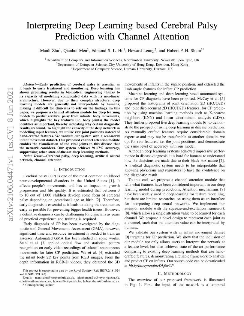
The overview of our proposed framework is illustratedin Fig. 1. First, the input of the network is a temporal
arX
iv:2
106.
0447
1v1
[cs
.CV
] 8
Jun
202
1

Label···
1×1×J
Squeeze
···
AttentionExcitation
J
T
C
Joint Position
JT CJ
1×1×J.
.
.
Conv2D
Conv2D
Conv2DConv2D
Conv2D
S S'z Az𝜎
FC
Softmax
Filter: (3, 1) Stride: (3, 1)
Fig. 1. The overview of our proposed framework.
sequence of joint positions represented as a tensor (i.e. a multi-dimensional matrix). It is then fed into an attention moduleincluding Squeeze and Excitation blocks, and the importanceof each joint is represented by its respective channel attentionvalue. The attention value is multiplied with the input feature,forming an attended feature tensor. Such a tensor is fed intofive 2D convolutional layers (Conv2D) to further extract thehigh-level information. Finally, a fully-connected layer (FC)and a softmax layer are applied to perform the classification.
A. The Dataset
We conduct experiments on the public dataset MovingInfants In RGB-D (MINI-RGBD) [9], in which twelve se-quences of real infant movements were captured. To protect theprivacy of recorded infants, such movements are representedby synthetic infant characters generated using the SkinnedMulti-Infant Linear (SMIL) model [10]. In this research, weuse the provided 3D joint positions of the sequences as ourinput.
McCay et al. [5] further annotated the dataset, and eachsequence was labeled as either normal or abnormal by anindependent expert using the GMA method. We use suchannotations as ground-truth.
To ensure that the classifier is not affected by the globaltranslation of the infant, we normalize the joint positions frameby frame. This is done by translating the spine joint of eachframe to the global origin, and representing other joints as arelative value to the spine.
B. Channel Attention
We propose an attention module based on the channel-wiseattention mechanism [8] to provide insight into the network’sdecision process. As shown in our overall framework in Fig.1, we first propose a squeeze step that aims at compressingthe joint information, then an excitation operation is appliedto identify the importance of each joint to the network, suchthat different weights are assigned to different joints.
Unlike traditional research to use channel attention formodelling features, we model the joints dimension intuitivelyas channels as a novel way for interpreting the network. Onthe one hand, the joint dimension provides a more meaningfulinterpretation to the users compared to the coordinate dimen-sion. On the other hand, using a single attention value forthe whole 3D trajectory of a joint in the entire video results
in a much more compact representation, making it easier forhumans to follow and more stable to train the network. As aresult, the designed attention module helps humans understandwhich joints contribute more to the diagnostic outcomes.
More specifically, the system input is represented as afeature tensor S ∈ RT×C×J , in which T is the total number offrames, C = 3 represents the 3 dimensions of a joint position,J is the total number of joints. In S, each feature value isrepresented as S(t, c, j), where t ∈ T , c ∈ C and j ∈ J .
In the squeeze operation, the tensor from each joint iscompressed into a representative joint descriptor using GlobalAverage Pooling. For a specific joint j′ ∈ J , its descriptor zj′is calculated as:
zj′ =1
T × C
T∑t=1
C∑c=1
S(t, c, j′) (1)
These descriptors form a joint-level embedding z =[z1, . . . , zJ ] from the whole movements. The obtained globalembedding enables the aggregation of the local joint featureswhich provides expressive joint-level statistics that triggers thefollowing excitation steps.
The excitation operation calculates the attention value thatindicates the importance of each channel (i.e. joint) by encod-ing the correlations from the embedded output z in the squeezestep. This is done by allocating the inner dependencies amongjoints with a nonlinear gating mechanism:
Az = σ (W2δ (W1z)) (2)
where δ is the ReLU activation, σ is the sigmoid activationfunction, W1 and W2 are parameters of the fully-connectedlayers.
The obtained Az ∈ R1×1×J from the squeeze-and-excitation steps provides the joints attention, and each value inAz indicates the attention weight of the corresponding joint.The attended feature tensor S′ is obtained by multiplying Az
with the original feature tensor S.
C. The CP Prediction Network
We propose a 2D convolutional neural network to encodethe tensor S′ for CP prediction. The network consists of fiveconvolutional layers, one fully-connected layer and one soft-max layer, as shown in Fig. 1. We found that having multipleconvolutional layers help to encode high-level features with

a suitable receptive field, and achieve the best classificationaccuracy. The fully-connected layer maps the learned featurewith the CP labels (i.e. normal or abnormal ), and the Softmaxlayer is to generate probability distributions for classification.
The filter K in the convolutional layers has the dimensionof FT×FC×J , where FT = 3 and FC = 1 in our experiment.This means that the filter covers all joints at once, and moves inthe frame and coordinate dimensions. This design ensures thatthe intrinsic correlations among joints can be discovered, andthat the temporal correlations of nearby frames are modelled.
D. Loss Functions
We propose a novel loss function known as attention loss.Unlike the traditional usage of attention, we apply attention fordeep neural network interpretation. To facilitate better humanunderstanding, the number of attended features should be min-imized, while keeping the classification accuracy unchanged.We define the attention loss as:
Latt =
J∑j=1
Azj (3)
The final loss function consists of three parts: the cross-entropy loss Lcep to encourage classification accuracy, theattention loss Latt to minimize the number of attended features,and the regularization loss ‖w‖2 (where w is the networkparameters) to discourage overfitting:
L = Lcep + γLatt + λ‖w‖2 (4)
where γ and λ are the loss weights set as 0.0005 and 0.0001respectively. We deliberately chose a small γ such to minimizethe attention loss effect to the classification accuracy.
As the data is biased with fewer abnormal samples (i.e. 4abnormal, 8 normal), we introduce a class balancing weightin Lcep. The idea is to give a higher weight to the abnormalsamples to facilitate training. Without the weights, the modelwould have poor predictive performance for the minority class(i.e. abnormal) when an imbalanced dataset is given, resultingin more false-negatives. In particular, we assign a weight αi
to the class i by the following equation:
αi =
√n
nc × ni(5)
where n is the total number of the samples, ni is the numberof samples of class i, and nc = 2 is the number of classes.
In our implementation, the whole network was trained inan end-to-end manner using the PyTorch platform with theAdams optimizer. The hyper-parameters epoch, learning rate,and batch size were set as 400, 0.0003 and 3 respectively forall cross-validations.
III. EXPERIMENTAL RESULTS
A. Quantitative Evaluation
We perform the leave-one-out cross-validation on the MINI-RGBD dataset [9], which is also used in the baseline works[4], [6], and the averaged result for all cross-validations ispresented as the final accuracy.
As deep learning has become the mainstream in patternrecognition due to its capacity to modelling complicated data,we compare the classification performance of our proposeddeep learning method with the existing deep learning methods[6] as well as the most recent GMA-based method [4] thatis also based on 3D skeletal data as input. The results arereported in Table I. It can be seen that our full system (i.e. thebottom row) achieves the same state-of-the-art performance,i.e. 91.67% prediction accuracy, in addition to the interpretabil-ity of our network which tells users which of the joints of aninfant body are the most important for its diagnostic outcome.Compared with [6], we utilize raw features instead of hand-crafted ones (i.e. HOJO2D + HOJD2D that generates the bestaccuracy), which makes our method easy to be transferred toanother domain as there is no feature engineering involved.Compared with [4], which does not require any training forthe model but a threshold has to be defined manually toseparate the positive and negative samples. In contrast, ourmodel is trained end-to-end without the need for any humanintervention.
TABLE ICLASSIFICATION ACCURACY COMPARISON BETWEEN OUR PROPOSED
METHOD AND OTHER BASELINE METHODS.
Feature Method Accuracy (%)HOJO2D FCNet [6] 83.33HOJD2D FCNet [6] 91.67
HOJO2D / HOJD2D Conv1D-1 [6] 83.33HOJO2D / HOJD2D Conv1D-2 [6] 83.33HOJO2D / HOJD2D Conv2D-1 [6] 83.33HOJO2D / HOJD2D Conv2D-2 [6] 83.33HOJO2D + HOJD2D Conv1D-1 [6] 91.67HOJO2D + HOJD2D Conv1D-2 [6] 91.67
Limb Angle GMA-based method [4] 91.67Joint Position Ours - Without Attention 91.67Joint Position Ours - Full System 91.67
We conducted an ablation study to evaluate if the attentionmodule, which was responsible for providing the interpretationfunctionality, had any negative effect on classification accu-racy. The results are shown at the bottom two rows of TableI. The attention module does not affect the overall classifi-cation accuracy, and it also provides us with an interface tointerpret the outcome of the deep learning model in a human-understandable way. Notice that while traditional attentionmodules tend to improve classification accuracy, the targetof ours is for interpretation. We tailor the way a channel isdefined such that the results can be as human-understandableas possible. This limits the module’s potential to fully optimizethe attention for accuracy improvement.
We further studied the effectiveness of the proposed atten-tion loss (see Table II). The present numbers are averagedby the total number of cross-validations. We find that the lossterm effectively reduced the average per-joint attention values.The average number of joints with high attention values is alsoreduced. This allows the results to be more easily understoodby humans, while maintaining the same prediction accuracy.

TABLE IITHE INFLUENCE OF THE ATTENTION LOSS IN OUR FRAMEWORK.
Method Without Latt With Latt
Avg. Per-joint Attention Value 0.492 0.439Avg. # of Joints with Attention ≥ 0.5 11.039 9.910Avg. # of Joints with Attention ≥ 0.6 10.856 9.861Avg. # of Joints with Attention ≥ 0.7 10.750 9.702Avg. # of Joints with Attention ≥ 0.8 10.566 9.583Avg. # of Joints with Attention ≥ 0.9 10.074 9.084Avg. # of Joints with Attention = 1.0 6.583 6.250
Accuracy (%) 91.67 91.67
B. Quantitative Evaluation
Fig. 2 presents a box plot to visualize the distributionsof joint attention values across the twelve cross-validations.For each joint, the interquartile range (IQR) is represented bythe blue bar, and the yellow line denotes the median value.The generally high median lines indicate most of the cross-validations tend to assign a higher attention value to the joint.
From the median lines, we can see that the joints ‘LeftThigh’, ‘Right Hand’, ‘Thoracic Spine’, ‘Right Shoulder’ and‘Left Upper Arm’ have the highest attention values in almostall the cross-validations. Most of these joints are located in theupper body, including those on the arms and shoulders. Thisaligns with the results reported in [5] that the features extractedfrom the arms are more discriminative and resulted in higheraccuracy in CP prediction on the MINI-RGBD dataset.
Fig. 2. The visualization of attention values of all joints across twelvevalidations.
We also visualize the joint attention values in two cross-validations, one with a normal sample (Fig. 3 upper) and onewith an abnormal sample (Fig. 3 lower) as validation. Thesizes of the red circles visualize the attention values of thecorresponding joints. It can be seen that the example motionsfrom different classes (i.e. normal and abnormal) resulted indifferent sets of attention values. This highlights the networkhas different focuses on the body joints to differentiate motionsfrom the two classes. Compared with the previous works [4],[6], in which manually select some joints for analysis, ourattention module is able to identify the more important jointsautomatically.
IV. CONCLUSION
We have presented an interpretable deep learning frameworkwith channel attention for CP prediction. The attention moduledesigned in this paper has shown how the network analyzes
Fig. 3. The visualization of joints attention in two cross-validations with anormal sample (upper row) and an abnormal sample (lower row) as validation.
the movements by looking at the body joints that are more im-portant to the final diagnostic results. We further demonstratedthe effectiveness of the proposed attention loss for minimizingthe number of attended joints such that the user can focus ona smaller set of joints for more in-depth analysis. Comparedwith the state-of-the-art methods, our proposed model achievesthe same level of classification accuracy, even though we donot use hand-crafted features nor any manual interventions.
A future direction is to incorporate both spatial and temporalattention into our channel attention module, which will allowinterpreting when an infant performs abnormal movementswith which joints. This can facilitate a user to focus theanalysis on the video segment with high attention. A keychallenge is the large number of frames, which results in alarge number of temporal attention values. Further researchon how to effectively visualize such information is needed.
REFERENCES
[1] I. Novak, C. Morgan, L. Adde, J. Blackman, R. N. Boyd, J. Brunstrom-Hernandez, G. Cioni, D. Damiano, J. Darrah, A.-C. Eliasson et al.,“Early, accurate diagnosis and early intervention in cerebral palsy:advances in diagnosis and treatment,” JAMA pediatrics, vol. 171, no. 9,pp. 897–907, 2017.
[2] P. Anderson, L. W. Doyle, V. I. C. S. Group, V. I. C. S. Group et al.,“Neurobehavioral outcomes of school-age children born extremely lowbirth weight or very preterm in the 1990s,” jama, vol. 289, no. 24, pp.3264–3272, 2003.
[3] A. Stahl, C. Schellewald, Ø. Stavdahl, O. M. Aamo, L. Adde, andH. Kirkerod, “An optical flow-based method to predict infantile cere-bral palsy,” IEEE Transactions on Neural Systems and RehabilitationEngineering, vol. 20, no. 4, pp. 605–614, 2012.
[4] Q. Wu, G. Xu, F. Wei, L. Chen, and S. Zhang, “Rgb-d videos-based earlyprediction of infant cerebral palsy via general movements complexity,”IEEE Access, vol. 9, pp. 42 314–42 324, 2021.
[5] K. D. McCay, E. S. L. Ho, C. Marcroft, and N. D. Embleton, “Establish-ing pose based features using histograms for the detection of abnormalinfant movements,” in IEEE EMBC, July 2019, pp. 5469–5472.
[6] K. D. McCay, E. S. L. Ho, H. P. H. Shum, G. Fehringer, C. Marcroft,and N. D. Embleton, “Abnormal infant movements classification withdeep learning on pose-based features,” IEEE Access, vol. 8, pp. 51 582–51 592, 2020.
[7] W. Samek, T. Wiegand, and K.-R. Muller, “Explainable artificial in-telligence: Understanding, visualizing and interpreting deep learningmodels,” arXiv preprint arXiv:1708.08296, 2017.
[8] J. Hu, L. Shen, and G. Sun, “Squeeze-and-excitation networks,” in 2018IEEE/CVF Conference on Computer Vision and Pattern Recognition(CVPR), June 2018, pp. 7132–7141.
[9] N. Hesse, C. Bodensteiner, M. Arens, U. G. Hofmann, R. Weinberger,and A. Sebastian Schroeder, “Computer vision for medical infant motionanalysis: State of the art and rgb-d data set,” in Proceedings of ECCVWorkshops, 2018, pp. 0–0.
[10] N. Hesse, S. Pujades, J. Romero, M. J. Black, C. Bodensteiner, M. Arens,U. G. Hofmann, U. Tacke, M. Hadders-Algra, R. Weinberger et al.,“Learning an infant body model from rgb-d data for accurate full bodymotion analysis,” in Proceedings of MICCAI. Springer, 2018, pp. 792–800.