international journal of pharmaceutical research &...
TRANSCRIPT

Available online www.ijpras.com
International Journal of Pharmaceutical Research & Allied Sciences, 2017, 6(1):107-112
Research Article ISSN : 2277-3657
CODEN(USA) : IJPRPM
107
Research on the Clinical Phenotype of Coronary Heart Disease with Retinol
Binding Protein 4, Lipoprotein-related Phospholipase A2, and the Severity
of Coronary Artery Lesion
Liu-qiang Lv, Yang-zhang Tang, Shi-qiang Wang, Yi-min Xie, Ling Ge and Xun-min
Cheng
Department of Cardiovascular Medicine, Huaibei People’s Hospital, Huaibei 235000, China
Corresponding author
Liu-qiang Lv
_________________________________________________________________________________
ABSTRACT
This study has been done to investigate the correlation between retinol binding protein 4 (RBP4), lipoprotein
associated phospholipase A2 (Lp-PLA2) and the clinical phenotype of coronary heart disease (CHD) and the severity
of coronary artery disease. 160 cases of patients were suspected with coronary heart disease underwent coronary
angiography, 112 cases of patients with coronary heart as treatment group, and 48 cases of patients with non CHD
as control group. According to the clinical characteristics, the coronary heart disease group was divided into acute
myocardial infarction (AMI) group with 15 cases, unstable angina pectoris (UAP) group with 71 cases, and stable
angina pectoris (SAP) group with 26 cases; according to the results of coronary angiography, it was divided into
single-branch lesion group with 42 cases, double-branch lesion group with 47 cases, and three-branch lesion group
with 23 cases. Fasting serum RBP4 and LP-PLA2 levels were detected. (1) Serum RBP4 concentrations and the
clinical phenotype of coronary artery disease: AMI group and UAP group were significantly higher than those in SAP
and control group (P<0.05); the difference was not statistically significant between the comparison of AMI, UAP
group, SAP and control group (P>0.05). (2) Serum LP-PLA2 concentration in patients with coronary heart disease
were significantly higher than those in the control group (P<0.01), in LP-PLA2 concentration comparison between
subgroup, AMI group was significantly higher than UAP and SAP group (P<0.01), UAP group was higher than SAP
group (P<0.05). (3) RBP4, LP-PLA2 concentration and coronary angiography results: three groups of lesions, RBP4,
LP-PLA2 concentration of the three groups pf lesions were significantly higher than the control group, the differences
were statistically significant (P<0.05), the group comparison in three groups, differences in RBP4 concentration have
no statistical significance (P>0.05); differences in LP-PLA2 concentration have statistical significance (P<0.05). The
changes of retinol binding protein 4, LP-PLA2of serum concentration has relation with the severity of coronary artery
disease progression, but no clear relationship with the degree of coronary artery lesion and count, LP-PLA2
concentration increases with the increase of the severity of coronary artery disease and the number of disease.
Keywords: Retinol Binding Protein 4; Lipoprotein Associated Phospholipase A2; Coronary Heart Disease;
Coronary Artery Lesion Degree
_____________________________________________________________________________________________
Liu-qiang Lv et al Int. J. Pharm. Res. Allied Sci., 2017, 6(1):107-112
______________________________________________________________________________
108
INTRODUCTION
Coronary heart disease is one of the common diseases in cardiology. Many risk factors and chronic inflammatory
reaction can lead to the formation of coronary heart disease. One of the common risk factors is dyslipidemia. Lipid
deposition in the vascular wall, causing atherosclerosis and plaque formation lumen narrowing, results in loss of
oxygen and oxygen consumption out of balance. Plaque rupture, can lead to a series of coronary events, in which of
inflammatory reaction in the arteriosclerosis process plays a certain role in triggering. RBP4 is a novel adipocyte-
derived cytokine that mediates chronic inflammatory response and promotes insulin resistance (IR), leading to further
metabolic disorders of glucose and lipid [1-2]. These metabolic disorders are the major risk factors for coronary heart
disease. LP-PLA2 is a newly discovered inflammatory factor, and has shown an important role in the promotion of
atherosclerosis formation and development process [3][6]. In this paper, the concentrations of RBP4 and LP-PLA2
in different populations were evaluated, and the values of RBP4 and LP-PLA2 in predicting the different types of
coronary artery disease and the degree of coronary artery disease were assessed.
MATERIALS AND METHODS
From September 2013 to March 2014, 160 patients with coronary heart disease were selected from our hospital,
including 102 males and 58 females with an average age of (65.37 ± 7.52) years [4-5], [7-8]. Selection criteria: refer
to the 2012 Chinese Medical Association coronary atherosclerotic heart disease diagnosis and classification criteria
[9-10]. All the selected patients underwent coronary angiography. According to the results of coronary angiography,
112 patients were divided into two groups: the coronary artery disease group (treatment group), the coronary artery
disease group, and the control group of 48 cases. Exclusion criteria: primary and secondary cardiomyopathy,
rheumatic heart disease, senile valve disease, infectious diseases, severe hepatitis, severe renal insufficiency and so
on. There was no significant difference between the two groups in age, gender differences, duration of disease, history
of smoking, blood pressure and body mass ratio (P > 0.05).
Coronary angiography and coronary heart disease diagnostic criteria and the determination of the number of
coronary artery disease
Coronary angiography was done using Judkins method, the radial artery approach; in the DSA room (catheter room)
coronary angiography, there are at least two qualified physicians in our division of our hospital to complete the
acquisition of orthotopic, liver, right shoulder position, head bit, left shoulder position, spider bit irradiation image.
Acute myocardial infarction, unstable angina, stable angina diagnosis consistent with the 2012 Chinese Medical
Association of coronary atherosclerotic heart disease diagnosis and classification criteria was done [5]. Criteria for
determining the number of lesions: single branch disease: refers to the anterior descending artery, circumflex artery,
right coronary artery in any one involvement, diameter stenosis greater than 50%; double-vessel disease: refers to the
anterior descending artery, circumflex artery, right coronary artery in the Diameter stenosis greater than 50%; three
lesions: anterior descending artery, circumflex artery, right coronary artery involvement at the same time, the diameter
stenosis greater than 50%, or the degree of stenosis is greater than 50%; left main plus any one of the vessels, the
diameter stenosis greater than 50%.
Liu-qiang Lv et al Int. J. Pharm. Res. Allied Sci., 2017, 6(1):107-112
______________________________________________________________________________
109
Specimen collection and testing methods
Specimen collection: Before coronary angiography, early morning fasting venous blood 5ml, 1000r / min Centrifuge
5min, collected serum, stored in -70 ℃ refrigerator, to be measured. Detection methods: The enzyme-linked
immunosorbent assay (ELISA) quantitative determination of RBP4, LP-PLA2 concentration levels, kit were provided
by Wuhan Huamei Biotechnology Co., Ltd., and microplate reader was provided by Beijing Tianshi Technology Co.
Statistical methods
Statistical analysis was performed with SPSS 16.0 software. Paired t-test was used to measure the data, andχ2 test was
used for the count data. The difference was statistically significant (P <0.05), P <0.01 was significant difference (χx
± S) and had statistical significance.
RESULT Results & Discussion
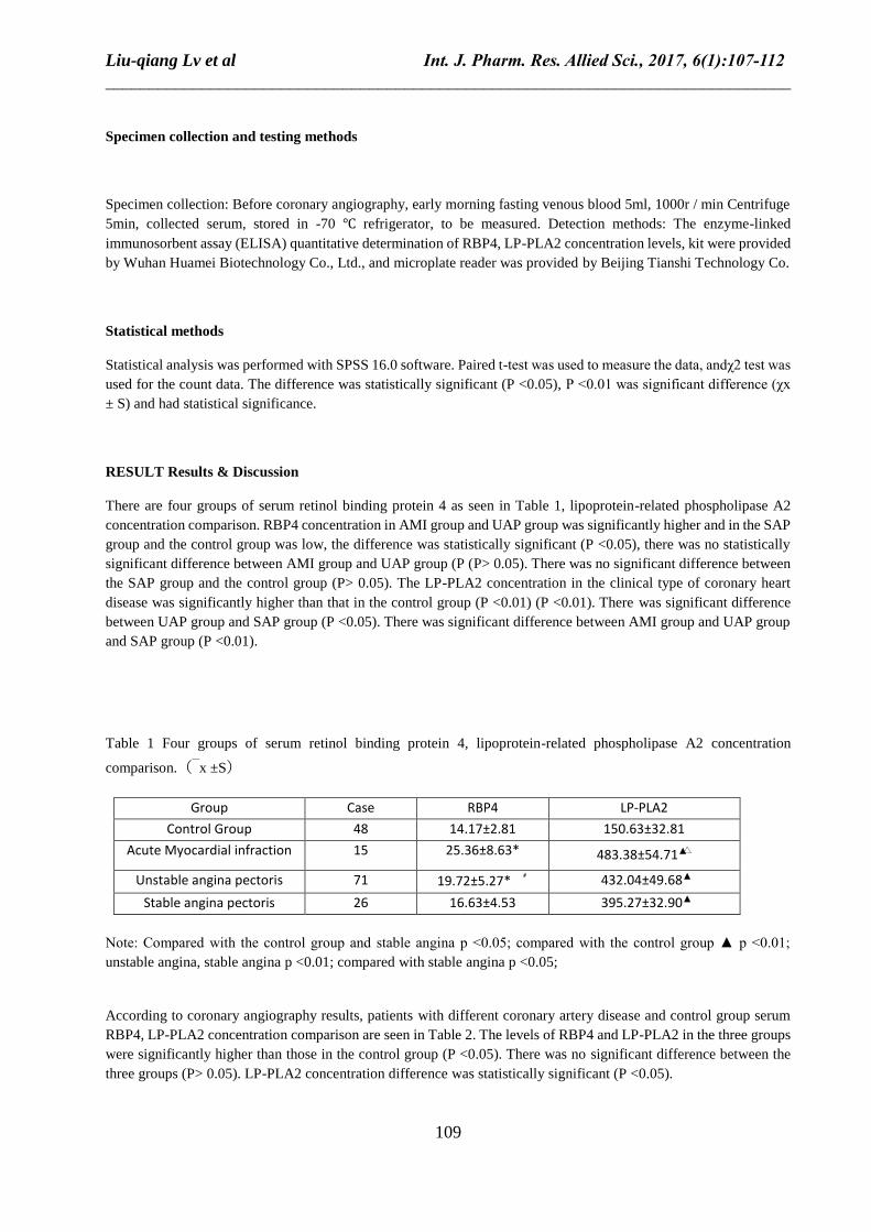
There are four groups of serum retinol binding protein 4 as seen in Table 1, lipoprotein-related phospholipase A2
concentration comparison. RBP4 concentration in AMI group and UAP group was significantly higher and in the SAP
group and the control group was low, the difference was statistically significant (P <0.05), there was no statistically
significant difference between AMI group and UAP group (P (P> 0.05). There was no significant difference between
the SAP group and the control group (P> 0.05). The LP-PLA2 concentration in the clinical type of coronary heart
disease was significantly higher than that in the control group (P <0.01) (P <0.01). There was significant difference
between UAP group and SAP group (P <0.05). There was significant difference between AMI group and UAP group
and SAP group (P <0.01).
Table 1 Four groups of serum retinol binding protein 4, lipoprotein-related phospholipase A2 concentration
comparison.(x ±S)
Group Case RBP4 LP-PLA2
Control Group 48 14.17±2.81 150.63±32.81
Acute Myocardial infraction 15 25.36±8.63* 483.38±54.71▲△
Unstable angina pectoris 71 19.72±5.27* ﹟ 432.04±49.68▲
Stable angina pectoris 26 16.63±4.53 395.27±32.90▲
Note: Compared with the control group and stable angina p <0.05; compared with the control group ▲ p <0.01;
unstable angina, stable angina p <0.01; compared with stable angina p <0.05;
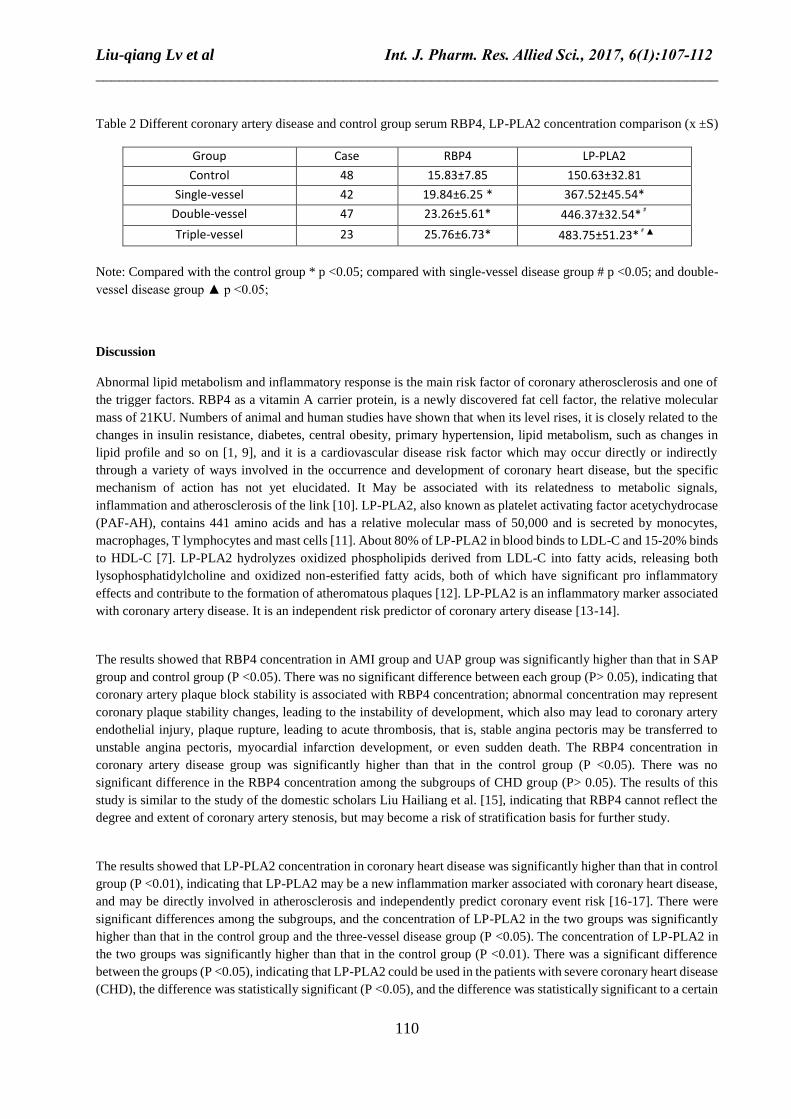
According to coronary angiography results, patients with different coronary artery disease and control group serum
RBP4, LP-PLA2 concentration comparison are seen in Table 2. The levels of RBP4 and LP-PLA2 in the three groups
were significantly higher than those in the control group (P <0.05). There was no significant difference between the
three groups (P> 0.05). LP-PLA2 concentration difference was statistically significant (P <0.05).
Liu-qiang Lv et al Int. J. Pharm. Res. Allied Sci., 2017, 6(1):107-112
______________________________________________________________________________
110
Table 2 Different coronary artery disease and control group serum RBP4, LP-PLA2 concentration comparison (x ±S)
Group Case RBP4 LP-PLA2
Control 48 15.83±7.85 150.63±32.81
Single-vessel 42 19.84±6.25 * 367.52±45.54*
Double-vessel 47 23.26±5.61* 446.37±32.54*﹟
Triple-vessel 23 25.76±6.73* 483.75±51.23*﹟▲
Note: Compared with the control group * p <0.05; compared with single-vessel disease group # p <0.05; and double-
vessel disease group ▲ p <0.05;
Discussion
Abnormal lipid metabolism and inflammatory response is the main risk factor of coronary atherosclerosis and one of
the trigger factors. RBP4 as a vitamin A carrier protein, is a newly discovered fat cell factor, the relative molecular
mass of 21KU. Numbers of animal and human studies have shown that when its level rises, it is closely related to the
changes in insulin resistance, diabetes, central obesity, primary hypertension, lipid metabolism, such as changes in
lipid profile and so on [1, 9], and it is a cardiovascular disease risk factor which may occur directly or indirectly
through a variety of ways involved in the occurrence and development of coronary heart disease, but the specific
mechanism of action has not yet elucidated. It May be associated with its relatedness to metabolic signals,
inflammation and atherosclerosis of the link [10]. LP-PLA2, also known as platelet activating factor acetychydrocase
(PAF-AH), contains 441 amino acids and has a relative molecular mass of 50,000 and is secreted by monocytes,
macrophages, T lymphocytes and mast cells [11]. About 80% of LP-PLA2 in blood binds to LDL-C and 15-20% binds
to HDL-C [7]. LP-PLA2 hydrolyzes oxidized phospholipids derived from LDL-C into fatty acids, releasing both
lysophosphatidylcholine and oxidized non-esterified fatty acids, both of which have significant pro inflammatory
effects and contribute to the formation of atheromatous plaques [12]. LP-PLA2 is an inflammatory marker associated
with coronary artery disease. It is an independent risk predictor of coronary artery disease [13-14].
The results showed that RBP4 concentration in AMI group and UAP group was significantly higher than that in SAP
group and control group (P <0.05). There was no significant difference between each group (P> 0.05), indicating that
coronary artery plaque block stability is associated with RBP4 concentration; abnormal concentration may represent
coronary plaque stability changes, leading to the instability of development, which also may lead to coronary artery
endothelial injury, plaque rupture, leading to acute thrombosis, that is, stable angina pectoris may be transferred to
unstable angina pectoris, myocardial infarction development, or even sudden death. The RBP4 concentration in
coronary artery disease group was significantly higher than that in the control group (P <0.05). There was no
significant difference in the RBP4 concentration among the subgroups of CHD group (P> 0.05). The results of this
study is similar to the study of the domestic scholars Liu Hailiang et al. [15], indicating that RBP4 cannot reflect the
degree and extent of coronary artery stenosis, but may become a risk of stratification basis for further study.
The results showed that LP-PLA2 concentration in coronary heart disease was significantly higher than that in control
group (P <0.01), indicating that LP-PLA2 may be a new inflammation marker associated with coronary heart disease,
and may be directly involved in atherosclerosis and independently predict coronary event risk [16-17]. There were
significant differences among the subgroups, and the concentration of LP-PLA2 in the two groups was significantly
higher than that in the control group and the three-vessel disease group (P <0.05). The concentration of LP-PLA2 in
the two groups was significantly higher than that in the control group (P <0.01). There was a significant difference
between the groups (P <0.05), indicating that LP-PLA2 could be used in the patients with severe coronary heart disease
(CHD), the difference was statistically significant (P <0.05), and the difference was statistically significant to a certain

Liu-qiang Lv et al Int. J. Pharm. Res. Allied Sci., 2017, 6(1):107-112
______________________________________________________________________________
111
extent, that can reflect the extent of coronary lesions or stenosis [18]. The study of the domestic scholars Pan Chenliang
et al. [19] has shown that plasma LP-PLA2 levels can be used to indirectly assess the scope of coronary lesions.
CONCLUSION
In summary, the level of RBP4 is associated with the progression of coronary artery disease, and has nothing to do
with the severity of coronary artery disease. The level of LP-PLA2 in patients with coronary heart disease increases
with the progression and severity of coronary lesion. So, the changes in LP-PLA2 levels can determine the
deterioration of coronary heart disease, progress and degree of severity of coronary artery disease. Due to the small
sample size of this study, the future studies still are needed to increase the sample size and more clinical centers to
participate in further studies to confirm the value from the response to the clinical phenotype of coronary heart disease
and coronary lesions[20].
REFERENCES
1. Guo Jifang, Kong Fanhe, Retinol binding protein 4 and coronary heart disease correlation, Mudanjiang
Medical College, 2013, 34(2), 12-14.
2. Wei Cai, Xuelin Cui, Xiangrong Zhou, Optimization of a GPU Implementation of Multi-dimensional RF
Pulse Design Algorithm, International Conference on Bioinformatics and Biomedical Engineering, 2011.
3. Wei Cai, Cheng Li, Heng Gu, Low Power SI Based Power Amplifier for Healthcare Application,
International Journal of Pharmacy and Pharmaceutical Sciences, 2016, 8(9), 171-178.
4. Maiolino G, Pedon L,Cesari M, et al, Lipoprotein-associated phospholipase A2 activity predicts
cardiovascular events in high rick coronary artery disease patients, PLOS One, 2012, 7(10), e48171.
5. Han Yaling, 2012 Chang'an International Cardiovascular Forum. Chinese percutaneous coronary
intervention guidelines 2012 (Simplified Chinese), Chinese Journal of Cardiology, 2012, 40(4), 252-254.
6. Wei Cai, Liang Huang and WuJie Wen, Low Power Class AB SI Power Amplifier for Wireless Medical
Sensor Network, Bioscience & Engineering: An International Journal (BIOEJ), 2016, 3(3).
7. Wei Cai, Frank Shi, 2.4 GHz Heterodyne Receiver for Healthcare Application, International Journal of
Pharmacy and Pharmaceutical Sciences, 2016, 8 (2), 22-25.
8. Wei Cai, Liang Huang, WuJie Wen, 2.4GHZ Class AB Power Amplifier for Wireless Medical Sensor
Network, International Journal of Enhanced Research in Science, Technology & Engineering, 2016, 5(4),
94-98.
9. Li F,Xia K,Sheikh MS,et al. Retinol binding protein 4 promotes hyperinsulinism-induced proliferation
of rat aortic smooth muscle cells, Mol Med Rep, 2014, 9(5), 1634-1640.
10. Ge Ling, Cheng Xunmin, YANG Song, et al. Retinol binding protein 4 and lipoprotein-associated
phospholipase A2 levels and coronary heart disease and coronary artery disease characteristics of the
correlation analysis, Bengbu Medical College, 40 (8), 1102-1104.

Liu-qiang Lv et al Int. J. Pharm. Res. Allied Sci., 2017, 6(1):107-112
______________________________________________________________________________
112
11. Fortunato J, Bláha V,Bis J,et al. Lipoprotein-associated phospholipase A2 mass level is increased in
elderly subjects with type 2 diabetes mellitus, J Diabetes Res, 2014, 278063.
12. Caslake M J,Packard C J.Lipoprotein–associated phospholipase A2 (platelet-activating factor
acetylhydrolase) and cardiovascular disease, Curr Opin Lipidol, 2003, 14(4), 347-352.
13. Zhao Yong, Guo Zhibin, the value of lipoprotein-associated phospholipase A2 in predicting the risk of
coronary heart disease, Journal of Applied Clinical Medicine, 2014,15(12), 1-3.
14. Ferguson J F, Hinkle CC, Mehta NN, et al, Translational studies of lipoprotein-associated phospholipase
A2 in inflammation and atherosclerosis, J AM Coll Cardiol, 2012,59(8), 764-772.
15. Liu Hai-liang, LI Guo-qing, Relationship between serum retinol-binding protein-4 and high-sensitivity C-
reactive protein in patients with coronary heart disease, Journal of Clinical Internal Medicine, 2011, 28(2),
92-94.
16. Liu Xingjia, Zheng Xing, Qin Yongwen, et al, Lipoprotein-related phospholipase A2 activity in response
to coronary angiography sclerosis degree, Journal of the Second Military Medical University, 2006, 27,
391-395.
17. Cai A, Li G, Chen J, et al, Increased serum level of LP-PLA2 is independentiy associatated with the severity
of coronary artery diseases:a cross-sectional study of Chinese population, BMC Cardiovasc Disord,
2015,15(1), 14.
18. Wang Lili, Lei Changcheng, Lipoprotein-related phospholipase A2 and coronary heart disease related
research progress, Modern Medicine and Health, 2015, 31(1), 57-60.
19. PAN Chen-liang, PENG Yu, HU Xue-ting, et al, Correlation between lipoprotein-associated phospholipase
A2 and acute syndromes, J of Clinical Cardiovascular Diseases, 2014, 30 (11), 962-965.
20. Pu, Chao, and Yanfei Gao, Crystal plasticity analysis of stress partitioning mechanisms and their
microstructural dependence in advanced steels, Journal of Applied Mechanics, 2015, 82(3), 031003.
21. Huang, Lu, et al, A Zr-based bulk metallic glass for future stent applications: Materials properties, finite
element modeling, and in vitro human vascular cell response, Acta biomaterialia, 2015, 25, 356-368.