intern a tional journal of leprosy - ilslila.ilsl.br/pdfs/v25n1a01.pdf · intern a tional journal...
TRANSCRIPT

INTERN A TIONAL JOURNAL OF LEPROSY
VOLUME 25, NUMBER 1 JANUARy-MARCH, 1957
THE PLACE OF RECONSTRUCTIVE SURGERY IN THE PREVENTION OF FOOT ULCERATION IN LEPROSY
ERNEST P. FRITSCHI, M.B.B.S. AND PAUL W. BRAND, F.R.C.S. (EDIN.) 1
Orthopaedic Research Unit, Leprosy Department Christian Medical College, Vellore, South India
In almost any outpatient department in this country it is a familiar sight to see the same patient coming day after day, week after week, for dressing of the chronic perforating ulcer of his foot. Too often the patient finally ceases to come, not because his ulcer has healed but because he has concluded that what cannot be cured must be endured. He has resigned himself to his ulcer. Then starts the story of neglect: bone involvement, loss of digits, and, finally, foot amputation as a last resort. The tragedy of the situation is that often the doctor comes to the same conclusion as the patient, and, with an impatient sigh, contents himself with writing, "Re-peat dressing" on the outpatient card. .
It is, therefore, of the utmost importance to consider the etiological factors involved in ulceration with a view to preventing the condition. Probably one of the most easily preventable factors is that of paralytic deformity. When the foot is not presented to the ground in its normal pattern we get stresses on areas which are not adapted to withstand them. Hence the importance of making the weight-bearing mechanism as nearly normal as possible.
The two motor nerves of the lower limb which are commonly affected are the lateral popliteal nerve, and the lateral and medial plantar branches of the posterior tibial nerve.
THE LATERAL POPLITEAL NERVE
The site of the lesion in this nerve is the three or four inches just proximal to the point at which it -crosses the neck of the fibula. Most commonly the whole of the nerve is paralyzed. Sometimes, however, the musculocutaneous component is spared, and only the anterior tibial nerve is paralyzed. In complete paralysis a drop foot results, where all the dorsi-
lWorking under a grant received from the Indian Council of Medical Research.
1

2 International Journal of Leprosy 1957
flexors are involved and also the peroneus longus and brevis. The foot retains the power of active plantar flexion and inversion. When the anterior tibial nerve only is involved, the peronei are still present. The result, then, is a drop foot which is capable of plantar flexion, inversion, and eversion. Incomplete lesions of the lateral popliteal nerve may also be seen where certain muscles are spared, resulting in weak dorsiflexion.
Complete paralysis of the lateral popliteal nerve is the commonest form. The two functions which are lost are dorsiflexion and eversion. The tibialis posterior provides inversion. With this affection the patient walks with the typical high-stepping gait of foot-drop, and also has a tendency to walk on the outer (lateral) border of the foot. This frequently gives rise to an ulcer under the head or base of the fifth metatarsal.
Three operations have been performed in our department for this condition: (1) the tibialis posterior transplant; (2) triple arthrodesis, or subtalar fusion; (3) anterior transposition of the tibialis posterior and peroneus brevis and longus (when these latter muscles are still unaffected).
The first and usual procedure for complete paralysis is an active tendon transplant, first described by Ober in 1933 (5). This consists of the transfer of the tibialis posterior tendon to the dorsum of the foot so that it acts as a dorsiflexor.
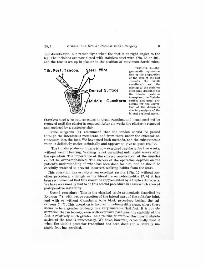
Operations.-First procedure: The tibialis posterior tendon is cut from its insertion at the tubercle of the navicular. It is then withdrawn through an incision about four inches above the medial malleolus at about the musculotendinous junction. A curved incision is made over the mid-dorsum of the foot and a flap raised in such a way as to expose the superior surfaces of the cuneiform bones. The dorsal ligaments are grasped with toothed forceps and pulled in the direction of the proposed transplant. The position of insertion of the tendon which would give truest dorsiflexion, with neither inversion nor eversion, is determined. This is usually in the middle cuneiform.
A hole big enough to take the end of the tendon and three-eights of an inch deep is reamed out through the superior cortex of this bone. The tibialis posterior tendon is now passed subcutaneously into the incision on the dorsum of the foot. Two small drill holes are made between the superior surface of the bone and the bottom of the large hole. A 30-gauge stainless steel wire is now introduced downward through one of the drill holes and drawn out from the bottom of the large hole. It is then threaded into a stout, round-bodied needle and passed into the end of the tendon, after which it is threaded in and out two or three times up the tendon and then down in the same way, to emerge from the end of the tendon. This end of the wire is then passed upward through the other drill hole. Thus the two ends of the suture emerge on the dorsum of the bone (Text-fig. 1).
The end of the tibialis posterior tendon is now pushed firmly into the large hole, and the two suture ends are pulled tight and tied over the dorsum of the bone. The tension on the tendon should be such that it is slack in
25, 1 Fritschi and Brand: Reconstructive Surgery 3
full dorsiflexion, but rather tight when the foot is at right angles to the leg. The incisions are now closed with stainless steel wire (No. 35 or 40), and the foot is set up in plaster in the position of maximum dorsiflexion.
T ib. Post . T endon. Wire
Surface
Cunelform
TEXT-FIG. I.-Diagrammatic representation of the preparation of the bone of the foot (usually the middle cuneiform) and the placing of the stainless steel wire, described for the tibialis posterior transplant, the first-described and usual procedure for the correction of the deformity due to paralysis of the lateral popliteal nerve.
Stainless steel wire sutures cause no tissue reaction, and hence need not be removed until the plaster is removed. After six weeks the plaster is removed and replaced by a posterior slab.
Some surgeons ( 4) recommend that the tendon should be passed through the interosseus membrane and from there under the extensor retinaculum into the foot. We have used both methods, and the subcutaneous route is definitely easier technically and appears to give as good results.
The tibialis posterior muscle is now exercised regularly for two weeks, without weight bearing. Walking is not permitted until eight weeks after the operation. The importance of the correct re-education of the muscles cannot be over-emphasized. The success of the operation depends on the patient's understanding of what has been done for him, and he should be carefully watched to prevent incorrect walking habits from the start.
This operation has usually given excellent results (Fig. 1) without any other procedure, although in the literature on poliomyelitis (2, 5) it has been recommended that this should be supplemented by a triple arthrodesis. We have occasionally had to do this second procedure in cases which showed postoperative instability.
Second procedure: This is the classical triple arthrodesis described by Ryerson (7), with wedge resection of the lateral part of the subtalar joint, and with or without Campbell's bone block procedure behind the calcaneous (1,2). This operation is favored in poliomyelitis cases, where there seems to be a greater tendency to a very unstable flail foot. It is our observation that in leprosy, even with extensive paralysis, the stability of the foot is relatively much greater. As a routine, therefore, this drastic stabilization of the foot is unnecessary. We have, however, occasionally used it when the tibialis posterior transplant has been done and a laterally unstable foot has resulted.

4 International Journal of Leprosy 1957
Third procedure: In the case of a paralysis which does not affect the musculocutaneous branch of the lateral popliteal nerve, the function of the peronei is still present. Here another operation has been performed. This is the anterior transposition of the tendons of the peroneous longus and brevis, and, on the other side, the tibialis posterior, to a position in front of the respective malleoli. Thus the action of the muscles are converted from eversion and inversion in plantar flexion to eversion and inversion in dorsiflexion. It is usually necessary to shorten the tendons after transposing them. The foot is then set up in dorsiflexion, and the same postoperative regime of mobilization and weight-bearing is carried out as for the tibialis posterior transplant.
This operation, again, has given good results with as much as thirty degrees of active dorsiflexion. It should be noted, however, that we are making use of muscles (the peronei) which may become paralyzed at a later date and leave the patient with only the tibialis posterior tendon acting, and, therefore, with a marked tendency to varus deformity. We have not yet seen this complication. If it should occur, it would require immediate attention to prevent ulceration of the lateral border of the foot.
Occasionally one sees a case where only the musculocutaneous nerve is involved, giving a paralysis of the peronei only. This ·results in marked inversion of the foot and ulceration of the lateral border (Fig. 2). Here a simple tenodesis is probably preferable to the active tendon transplant such as that described by Peabody (6).
The tendon of the peroneus brevis is cut at about the musculotendinous junction in the leg. This tendon is withdrawn through a small incision just above the tuberclB of the fifth metatarsal. It is then threaded subcutaneously anterior to the lateral malleolus and passed through a drill hole in the lower end of the shaft of the fibula. The tendon is fixed at a tension sufficient to maintain the foot at approximately a right angle. The foot is immobilized in the right-angle position for at least six weeks. This operation has given good results in preventing the lateral-border ulcers of the foot.
INVOLVEMENT OF THE POSTERIOR TIBIAL NERVE
The lesion in this case is just proximal to the flexor retinaculum (lacinate ligament), where the nerve is in company with the tendons of the tibialis posterior, flexor hallucis longus, and flexor digitorum longus, and the posterior tibial artery. Here the nerve lies superficially and is perhaps subjected to trauma and pressure, and we find it to be thickened in the two or three inches proximal to the point at which it disappears under the retinaculum.
This lesion results in the paralysis of all the small muscles of the foot, with the possible exception of the first dorsal interosseous which is partially supplied by the anterior tibial nerve. This usually leads to a departure from the normal relations of the bones of the toes (Text-fig. 2A) to a

25,1 Fritschi and Brand: Reconstructive Surgery 5
typical claw toe deformity, especially if the extensors of the toes are not paralyzed. If these are active, there is a strong pull on the proximal phalanx of the toe, while the flexor digitorum longus pulls on the terminal phalanx. The early result, which we have termed the first stage of clawing (Textfig. 2B), is a hyperextended metatarsophalangeal point and increased flexion of the tip of the toe. Thus, instead of the ·pulp of the toe coming in contact with the ground, the tip of the toe is the most dependent part. This exposes the tip of the toe to a friction trauma to which it is not adapted, and hence there is, first, flattening of the tip, and then ulceration, which may go on to osteomyelitis of the terminal phalanx.
Fig. 2 A
NORMAL 1ST DEGREE
2ND DEGREE 3RD DEGREE TEXT-FIG. 2-Diagrammatic representation of the relations of the bones of the toes
in the claw toe deformity resulting from involvement of the posterior tibial nerve. A, normal; B, first stage or degree of deformity; C, second stage; D, third stage.
The second stage of clawing (Text-fig. 2C) is when the capsule of the metatarsophalangeal joint begins to give way to the unequal strains imposed upon it and the base of the proximal phalanx begins to slip dorsally, leaving the head of the metatarsal unduly projecting towards the sole of the foot (Fig. 3). This means that the soft tissue of the sole will be thinned out under the projecting head, and the skin underlying the heads will be exposed to excessive "thrust" trauma during walking, with the result that there is ulceration under the metatarsal heads. (Fig. 4) .
In the third stage of clawing (Text-fig. 2D) there is a complete subluxation of the proximal phalanx, so that its base actually lies sometimes

6 International Journal of L eprosy 1957
as much as a half-inch proximal to the head of the metatarsal (Fig. 5). Very often the ulceration under the metatarsal, which is the result of the exposure of the area to excessive pressure during the second stage, has already destroyed the heads of the metatarsals. The x-ray shows a shortening of all the metatarsals, and the bases of the proximal phalanges overriding their pointed ends.
Clawing of the toes is well known in conditions other than leprosy, and a number of operations have been described for the correction of this deformity. Of these we have tried the interphalangeal fusion, and the tendon transplant (3). The latter is technically simpler, and also theoretically more satisfactory. In our present small series the results have been encouraging (Figs. 6 and 7.)
Operation.-The toe is opened through a mid-medial .side incision along the middle proximal phalanges. The tendon of the flexor digitorum longus is cut at its insertion. The tendon of the extensor digitorum is exposed in the proximal segment through the same incision. The fibrous flexor sheath of the flexor tendons is cut as far as the base of the toe, and the flexor digitorum longus tendon is sutured to the extensor tendon in the proximal segment of the toe.
This procedure results in extension of the interphalangeal joints of the toes due to the pull of the flexor digitorum longus on the extensor expansion and the release of its flexor action on the terminal phalanx.
DISCUSSION
A long-term assessment of the value of these operative procedures in the prevention of trophic ulceration has yet to be made. It may, however, be said at this stage that the early results are very promising. The correction of the "drop foot" by the tibialis posterior transplant gives on the average around thirty degrees of active dorsiflexion and corrects completely the "high-stepping gait." The recurrence of ulceration in postoperative cases has been very low. However, it must be said that this may also be due to the fact that patients who had a history of chronic ulceration were also provided with moulded shoes, and were instructed in leading as far as possible sedentary lives. It is therefore difficult to attribute the improvement to anyone of these factors.
It may be considered that some of these procedures are very elaborate and difficult, involving long periods of bed rest in plaster casts. Most of our cases, however, are sent home within a fortnight of operation with a wooden "walking heel" incorporated in the plaster (Fig. 8). With this they can lead almost normal lives at home while they are being immobilized. It will, however, be readily conceded that if these procedures are a factor in the prevention of foot ulceration, as we believe to be the case, the trouble taken and the hospitalization time will be less than that used in curing chronic ulceration.
As is suggested above, no single factor can be regarded as preventing

25,1 Fritschi and Bmnd: Reconstructive Surgery 7
foot ulceration in a denervated extremity. In addition to reconstructive operative procedures it is of the utmost importance to instruct patients in foot hygiene, and especially in modifying their lives- so that they need to do as little walking as possible. We are of the opinion that a denervated foot is incapable of withstanding "physiological trauma" which would not affect a normal limb. The use of moulded shoes is helpful, since the weightbearing surface on the sole is increased and weight is evenly distributed.
SUMMARY
1. Paralysis of the lateral popliteal nerve in relation to lateral border ulceration is discussed, and three operations used for its correction are described.
2. Paralysis of the posterior tibial nerve in relation to metatarsal-head ulceration is discussed. An operation for the correction of the claw toe deformity is described.
3. Trophic ulceration can only be prevented by careful attention to several factors, namely, (a) reduction of walking and standing to the minimum, (b) reconstruction of paralytic deformities, and (c) use of moulded soles to distribute the weight evenly over the foot.
RESUMEN
1. Se discute la panilisis del nervio popliteo lateral en relacion con la uIceraci6n del borde lateral, describiendose tres operaciones usadas para rectificar esta situacion.
2. Se discute la panilisis del nervio tibial posterior en relacion con la ulceracion de la cabeza del metatarso. Describese una operacion para la correccion de la deformidad representada POI' el pie bot.
3. Puede impedirse la ulceracion trOiica unicamente mediante la cuidadosa atencion a varios factores, a saber: (a) reduccion al minimo de las caminatas y del permanecer de pie, (b) reconstruccion de las deformidades paraliticas y (c) uso de suelas moldeadas que distribuyan el peso uniformemente POl' to do el pie.
REFERENCES
1. CAMPBELL, W. C. An operation for the correction of "drop foot." J. Bone & Joint Surg. 5 (1923) 815-825.
2. CAMPBELL, W. C. Campbell's Operative Orthopedics. St. Louis: C. V. Mosby Co., 2nd ed., 1949, Vol. 2, p. 1308.
3. CATHCART, E. P. and others. Feet of the industrial worker. Lancet 2 (1938) 1480-1486.
4. JAMES, J. I. P. Personal communication. 5. OBER, F. R. Tendon transplantation in the lower extremity. New England J. Med.
209 (1933) 52-59. 6. PEABODY, C. W. Tendon transposition; an end-result study. J. Bone & Joint Surg.
20 (1938) 193-205. . . 7. RYERSON, E. W. Arthrodesing operations on the fee't. J. Bone & Joint Surg. 6
(1923) 453-471.
Supplemental References
(For readers who may wish to go farther into the literature of this subject, the following supplementary list is provided.)
8 International Journal of L eprosy 1957
BASTOS ANSART, M. Pan-arthrodesis for paralytic flail foot. J. Bone & Joint Surg. 33·B (1951) 503-507.
BRAND, P. W. The orthopaedic care of leprosy patients. J . Christian Med. Assoc. (Vellore) 25 (1950) 1-9.
BRAND, ' P. W. The place of physical medicine and orthopedic surgery in leprosy. Leprosy Rev. 25 (1954) 5-10.
BRAND, P. W. The value of surgical and physiotherapeutic measures in leprosy. Lep. India 27 (1955 ) 131-137.
BRAND, P. W. Treatment of leprosy. II. The role of surgery. New England J. Med. 254 (1956 ) 64-67.
BURMAN, M. S. Spastic intrinsic-muscle imbalance of the foot ; resection of the motor branch of the lateral plantar nerve for intrinsic-muscle contracture. J. Bone &
Joint Surg. 20 (1938) 145-148. DREW, A. J. Late results of arthrodesis of the foot. J. Bone & Joint Surg. 33·B
(1951) 496-502. FORRESTER-BROWN, M. F. Tendon transplantation for clawing of the great toe. J. Bone
& Joint Surg. 20 (1938) 57-60. INGRAM, A. J. and HUNDLEY, J. M. Posterior Bone-block of the Ankle for Paralytic
Equinus. J. Bone & Joint Surg. 33·A (1951) 679-691. MCCARROLL, H. R. Foot deformities resulting from irreparable nerve lesions. Recon
structive Surgery of the Extremities, Am. Acad. of Or. Surg. Lectures on Reconstruction Surgery 1944, p. 149-168.
MORTON, D. J. Biomechanics of the human foot. American Acad. Orthop. Surgeons, Lect., 1944, pp. 92-99. .
PAUL, M. Surgical measures in leprosy. Internant. J. Leprosy 4 (1936) 29-34. TAYLOR, R. G. The treatment of claw toes by multiple transfers of flexor into ex
tensor 'tendons. J. Bone & Joint Surg. 33·B (1951) 539-542. WATKINS, M. B .. JONES, J. B., RYDER, C. T., JR .. and BROWN, T. H., JR., Transplan
tation of the posterior tibial tendon. J. Bone & Joint Surg. 36·A (1954) 1181-1189.
DESCRIPTION OF PLATES
PLATE 1
FIG. 1. Results of the first-described operation, the active tendon transplantation of the tibialis pos'terior for paralysis of the lateral popliteal nerve.
FIG. 2. An example of lateral border ulceration resulting from "inversion of the foot due to isolated paralysis of the peronei, when only the musculocutaneous nerve is involved.
FIG. 3. X-ray demonstration of the arrangement of the bones of the toes in the second stage of clawing due to paralysis of the posterior tibial nerve. The heads of the metatarsals project unduly toward the sole of the foot.
FIG. 4. Ulcerations under the heads of the metatarsals in the second stage of claw toes.

PLATE 1

PLATE 2
FIG. 5. X-ray demonstration of the bones of the toes in the third stage of clawing, with destruction of bone.
Figs. 6 and 7. Results of the described operation of tendon tmnsplant for the correction of clawing of the toes.
FIG. 8. Illustrating the wooden "walking heel" incorporated in the plaster cast, to permit early release of patients from the hospital.

PLATE 2