interferon-γ acts as a regulator in the trade-off between
TRANSCRIPT
RESEARCH Open Access
Interferon-γ acts as a regulator in thetrade-off between phagocytosis andproduction performance in dwarf chickensYitong Yuan1, Shunqi Liu2, Yue Zhao2, Ling Lian1* and Zhengxing Lian1*
Abstract
Background: Interferon-γ (IFN-γ) is critical for innate and adaptive immunity against viral and bacterialinfections. IFN-γ reportedly affects the phagocytic ability of monocytes and macrophages as well as regulatespituitary function in humans and mice. The present study analyzed the impact of IFN-γ on monocyte andmacrophage phagocytosis, production performance, and pituitary function in vivo and in vitro (in dwarfchickens). IFN-γ was injected into dwarf chickens through a vein, and then, the laying rate, average eggweight, and levels of follicle-stimulating hormone (FSH) and IFN-γ were measured in treatment and controlgroups. For the in vitro experiment, the pituitary tissues were supplemented with IFN-γ, and the mRNAexpression levels of follicle-stimulating hormone beta subunit (FSH-β), interferon gamma receptor 1 (IFNGR1),and interferon gamma receptor 2 (IFNGR2) in the pituitary were assessed.
Results: Monocyte and macrophage phagocytosis product (PP) was decreased by IFN-γ treatment in adose-dependent manner in vitro. In the in vivo experiment, the level of IFN-γ in the treatment group washigher than that in the control group at 7 d (P < 0.05), 14 d (P < 0.01), and 21 d (P < 0.01) post-injection.Compared with the control group, monocyte and macrophage PP was lower in the treatment group afterinjection (P < 0.01). The laying rate was higher in the treatment group than in the control group at 2 and3 wk post-injection (P < 0.05). There was a significant difference between the treatment and control groupsin the levels of FSH at 1, 3, 7, and 14 d post-injection (P < 0.01). In the in vitro experiment, increased mRNAexpression levels of FSH-β, IFNGR1, and IFNGR2 were observed in the treatment group after stimulation with100 U/mL IFN-γ for 24 h compared to those in the control group (P < 0.05).
Conclusions: IFN-γ inhibited the phagocytosis of monocytes and macrophages; up-regulated the mRNAexpression levels of the FSH-β, IFNGR1, and IFNGR2; enhanced the secretion of FSH; and improved thelaying rate. IFN-γ might be an important regulator in the trade-off between the immune effect andproduction performance in dwarf chickens.
Keywords: Dwarf chicken, Interferon-γ, Macrophage, Monocyte, Phagocytosis product, Productionperformance
* Correspondence: [email protected]; [email protected] Laboratory of Animal Genetics and Breeding of the Ministry ofAgriculture, Beijing Key Laboratory for Animal Genetic Improvement,Department of Animal Genetics and Breeding, College of Animal Scienceand Technology, China Agricultural University, Beijing 100193, ChinaFull list of author information is available at the end of the article
© The Author(s). 2018 Open Access This article is distributed under the terms of the Creative Commons Attribution 4.0International License (http://creativecommons.org/licenses/by/4.0/), which permits unrestricted use, distribution, andreproduction in any medium, provided you give appropriate credit to the original author(s) and the source, provide a link tothe Creative Commons license, and indicate if changes were made. The Creative Commons Public Domain Dedication waiver(http://creativecommons.org/publicdomain/zero/1.0/) applies to the data made available in this article, unless otherwise stated.
Yuan et al. Journal of Animal Science and Biotechnology (2018) 9:40 https://doi.org/10.1186/s40104-018-0256-y

BackgroundProduction performance has a profound and lastingsignificance for poultry in terms of both evolutionaryand economic perspectives [1, 2]. In commercial set-tings, improvements in production traits are alwaysmore profitable, but they also put the birds at in-creased risk of stress and disease because of unbal-anced resource allocation [3]. Selection for highproduction efficiency in broilers can have a detrimen-tal impact on physiological and immunological func-tion, such as higher mortality and susceptibility todisease [4, 5]. For example, infections caused by vari-ous pathogens were observed in turkeys artificially se-lected for high egg production [6]. Other researchersshowed that White Leghorn chickens with low anti-bodies to sheep red blood cells showed increased eggproduction after artificial selection for 24 generations[3]. Our previous study indicated that dwarf chickenswith low levels of monocyte and macrophage phago-cytosis exhibited low antibody titers to avian influenzaH9 but greater laying rates during the early layingperiod (unpublished observations). Therefore, strongerproduction performance is typically accompanied byweaker immune responses. Thus, the trade-off be-tween the immune effect and production performanceremains to be clarified.Interferon-γ (IFN-γ), the only member of the type II
IFNs, is a pro-inflammatory cytokine mainly producedby T helper type 1 cells, CD8+ cytotoxic lymphocytes,natural killer cells, B cells, natural killer T cells, and pro-fessional antigen-presenting cells [7, 8]. IFN-γ plays avital role in innate and adaptive immunity that activatesthe host defense against viral and bacterial infection [8–10]. IFN-γ exerts its immunological function by activat-ing various host cells, especially monocytes and macro-phages, and also can regulate the expression of the classII major histocompatibility complex [11]. As professionalphagocytic cells, monocytes and macrophages are indis-pensable in the host defense mechanism, enhancing theimmune response by releasing pro-inflammatory and in-flammatory cytokines [9, 12, 13]. IFN-γ has an inhibitoryeffect on the nonopsonized phagocytosis of macrophagesin mice and humans. Monocyte- and macrophage-mediated microbial phagocytosis was shown to be de-creased by IFN-γ in viral infections in mice [14, 15].Peritoneal macrophages from mice treated with recom-binant mouse IFN-γ exhibited reduced phagocytosis ofchicken red blood cells and Escherichia coli [16]. Inhumans, this inhibition was also found in the phagocyticprocess of monocyte-derived macrophages [17, 18].However, few studies have been conducted on the effectof IFN-γ on phagocytic activity in chickens.Previous studies have reported that IFN-γ can also
regulate pituitary function by influencing hormone
secretion. For example, inhibitory effects of IFN-γ on ad-renocorticotropic hormone, prolactin, and growth hor-mone were observed in anterior pituitary cells from 3-month-old female Wistar rats [10]. In another study, ex-ogenous IFN-γ increased the expression of growth hor-mone in rat anterior pituitary cells in a dose-dependentmanner [19]. Furthermore, subcutaneous IFN-γ injec-tions in humans led to increased cortisol levels [20].These findings demonstrated that IFN-γ may mediatethe production of pituitary hormone. However, in dwarfchickens, the effect of IFN-γ on the secretion of pituitaryhormone has not been studied yet.The immune effect and production performance are
both essential aspects during long-term breeding, and anegative relationship between them has been found;however, how to maintain this balance have not been re-solved. Here, we hypothesized that IFN-γ maintains orconverts the allocation of resources between productionand immune function. Accordingly, the objective of thepresent investigation was to clarify the effect of IFN-γon monocyte and macrophage phagocytosis, pituitaryfunction, and performance in dwarf chickens. This studywill provide novel insights into the impact of IFN-γ onboth immune function and production traits. IFN-γ canpotentially be utilized in chicken breeding as a regulatorof this balance.
MethodsExperimental animalsAll animal procedures were approved by the Experimen-tal Animal Care and Use Committee at China Agricul-tural University (Beijing, China), and the experimentswere performed according to regulations and guidelinesestablished by this committee. One flock of 160 dwarfhens and another flock of 786 dwarf hens at 12 wk ofage were obtained from the Genetic Resource Center ofChina Agricultural University (Beijing, China). All chick-ens were fed ad libitum and reared under standard man-agement and environmental conditions.
Isolation and culture of chicken monocytes andmacrophagesOne milliliter of peripheral blood was collected in hep-arin (15 U/mL) (China National Pharmaceutical GroupCorporation, Beijing, China) from the brachial wingveins of 160 dwarf hens at 12 wk of age. Peripheralblood mononuclear cells (PBMCs) were isolated usingFicoll-Paque (Sigma, St. Louis, MO, USA) gradient cen-trifugation as previously described [21, 22]. Isolated cellswere washed twice in 1 mL of phosphate buffered saline(PBS, Hyclone, Logan, UT, USA) by centrifugation at800×g for 10 min. Trypan blue solution (Sigma) wasused to assess the PBMC number and viability. PBMCswere resuspended in RPMI 1640 medium (Gibco,
Yuan et al. Journal of Animal Science and Biotechnology (2018) 9:40 Page 2 of 10

Invitrogen, La Jolla, CA, USA) supplemented with 10%fetal bovine serum (Gibco, Invitrogen) and 1% penicillin-streptomycin (Gibco, Invitrogen). Afterward, 1 × 10⁶
cells/well were seeded in 96-well plates (Corning, Inc.,Corning, NY, USA) (6 wells per sample) and incubatedat 37 °C with 5% CO2. Non-adherentcells (lymphocytes)were removed 24 h and 48 h after incubation, and ap-proximately 90% of the adherent cells were monocytesand macrophages [21, 23].
Preparation of dyed-tumor cells using thiazolyl bluetetrazolium bromide (MTT)Human intestinal epithelial adenocarcinoma cells (PekingUnion Medical College Hospital, Beijing, China, HCT-8)(1 × 10⁵ cells/mL) were cultured in 100-mm culture plates(Corning) containing RPMI 1640 medium (Gibco, Invitro-gen) supplemented with 10% fetal bovine serum (Gibco,Invitrogen) and 1% penicillin-streptomycin (Gibco,Invitrogen) at 37 °C with 5% CO2. When the cells reached70 to 80% confluence (1 × 10⁷ cells per plate), MTT(Amresco, Radnor, PA, USA) solution (5 mg/mL) wasadded to the plate, and the cells were dyed for 10 h. Thecells were washed three times with PBS (Hyclone). Thecell suspension was collected in a sterile tube and thenstored at 4 °C. The dyed HCT-8 cells were used as phago-cytic antigens as previously described [24].
Monocyte and macrophage phagocytosis product assayThe detection method of monocyte and macrophagephagocytosis was according to previous publications[24]. After 72 h of culture, the density of monocytes andmacrophages was approximately 2 × 10⁴ cells/well. In thetreatment wells (three wells), dyed HCT-8 cells (1 × 10⁶
cells) were added to the monocytes and macrophages ineach well followed by incubation for 10 h. In the controlwells (three wells), MTT (Amresco) solution (5 mg/mL)was added to the monocytes and macrophages followedby incubation for 4 h. Then, the cells were washed threetimes with PBS (Hyclone), and 150 μL of dimethylsulfoxide (Sigma) was added. The experiments wereperformed in triplicate. The absorbance of each well wasdetected at 575 nm and 630 nm (blank wells) with aMicroplate Reader (BioTek, Winooski, VT, USA).Phagocytosis product (PP) was used as an indicator of thephagocytic ability of monocytes and macrophages and wascalculated as follows: PP = absorbance (treatment wells) /absorbance (control wells).
Detection of monocyte and macrophage PP after IFN-γstimulation in vitroEight chickens with higher PP levels were selected fromamong 160 dwarf hens. Peripheral blood (3 mL) was col-lected from the wing veins of each chicken at 12 wk ofage. PBMCs were isolated by Ficoll-Hypaque density
gradient centrifugation. After 24 h of culture, monocytesand macrophages were washed three times with PBS(Hyclone), and non-adherent cells (mostly lymphocytes)were removed. The density of monocytes and macro-phages was approximately 2 × 10⁴ cells/well. Recombinantchicken IFN-γ (Shanghai Medicine’nest PharmaceuticalCo. Ltd., Shanghai, Beijing) was prepared at differentconcentrations in PBS (Hyclone). In the treatmentwells, monocytes and macrophages were incubatedwith IFN-γ at concentrations of 0 ng/mL, 1 ng/mL,3 ng/mL, 30 ng/mL, 300 ng/mL, and 1,000 ng/mLper sample. The medium was changed after 48 h oftreatment. Dyed HCT-8 cells were added to monocytesand macrophages in the treatment wells, and MTT(Amresco) solution (5 mg/mL) was added to monocytesand macrophages in the control wells. PP was evaluated asdescribed earlier.
Determination of serum IFN-γ in chickens after IFN-γinjection in vivoSixty dwarf chickens with higher PP levels and similarbody weights were selected from among 160 dwarf hensat 23 wk of age. The chickens were randomly dividedinto treatment and control groups. The chickens in thetreatment group were intravenously injected with re-combinant chicken IFN-γ (14,000 U/kg) (Shanghai Med-icine’nest Pharmaceutical Co. Ltd) for 7 consecutivedays. Chickens in the control group were simultaneouslyintravenously injected with 1 mL of PBS (Hyclone). Theserum IFN-γ levels in the treatment and control groupswere determined using a commercial chicken specificELISA kit (Elabscience Biotechnology Co., Ltd., Wuhan,Hubei, China) at 0, 1, 3, 7, 14, 21, and 28 d post-injection.
Detection of monocyte and macrophage PP after IFN-γinjection in vivoAt 0, 1, 3, 7, 10, 14, 21, 28, and 35 d post-injection, per-ipheral blood was collected from the wing veins, andPBMCs were isolated. After 72 h of culture, the PP ofmonocyte and macrophage was evaluated as describedearlier.
Measurement of production performance after IFN-γinjection in vivoThe egg production of the treatment and control groupswas recorded, and all the eggs laid by each hen wereweighed successively for 7 wk after injection with IFN-γ.The individual laying rate was calculated as the total num-ber of laying eggs divided by the total number of chickensand multiplied by 100%. Average egg weight was calcu-lated the total weight of laying eggs divided by the totalnumber of eggs collected and multiplied by 100%.
Yuan et al. Journal of Animal Science and Biotechnology (2018) 9:40 Page 3 of 10

The serum follicle-stimulating hormone (FSH) levelsof chickens in the treatment and control groups wasdetected using a radioimmunoassay kit (Beijing NorthInstitute of Biological Technology, Beijing, China) at0, 1, 3, 7, 14, 21 and 28 d post-injection. The experi-ments were performed in triplicate.
Pituitary gland stimulated by chicken IFN-γ in vitro andFSH concentration measurementThe monocyte and macrophage PP levels of 786 dwarfhens were detected at 12 wk of age as described earlier.Forty dwarf hens with higher PP levels were selected at25 wk of age and euthanized. The pituitary was immedi-ately removed and weighed. The pituitary was washedtwice with PBS (Hyclone) and finely sliced. The pituitarytissues were placed in 24-well plates (Corning) contain-ing 500 μL of serum-free RPMI 1640 medium (Gibco,Invitrogen) supplemented with 1% penicillin-streptomycin (Gibco, Invitrogen) and pre-incubated for1 h with unceasing shaking followed by manual disper-sion under 37 °C and 5% CO2. One hour later, themedium was changed. The pituitary tissues were cul-tured for 24 h or 48 h without cytokines or with differ-ent concentrations of IFN-γ (100, 500, and 1,000 U/mL)in serum-free RPMI 1640 medium (Gibco, Invitrogen).After incubation, the supernatants and the pituitary tis-sues were separately collected and stored at − 80 °C forsubsequent assay. The concentrations of FSH in culturesupernatants were measured with the commercial radio-immunoassay kits (Beijing North Institute of BiologicalTechnology, Beijing, China).
RNA extraction and real-time PCRTotal RNA was extracted with TRIzol Reagent (Invitro-gen, Gibco-BRL, Bethesda, MD, USA). The yield of RNAwas determined using a spectrophotometer (DS-11,DeNovix, Inc., Wilmington, DE, USA), and the integritywas evaluated by RNA electrophoresis. Total RNA wasreverse transcribed into cDNA using a QuantScript
Reverse Transcription Kit (Takara Bio, Dalian, China) ina total volume of 10 μL containing 0.5 μg RNA, 0.5 μLPrimerScript RT Enzyme Mix I, 0.5 μL RT Primer Mix,and 2 μL Primer Script Buffer, followed by incubation at37 °C for 15 min and 85 °C for 5 s.The specific sequences of the primers were designed
and synthesized by Sangon Biotech Co., Ltd. (Shanghai,China) and are shown in Table 1. Real-time PCR wasperformed using a real-time QPCR System (Mx3000P,Agilent technologies, Santa Clara, CA, USA) with a20-μL reaction mixture including 1 μL cDNA, 0.4 μmol/Lof forward and reverse primer, 0.4 μL ROX reference dye,and 10 μL SYBR Premix Ex Taq II (Takara Bio). Theamplification conditions were as follows: 95 °C for 30 sand 40 cycles of 95 °C for 5 s, 60 °C for 30 s, 95 °C for15 s, 60 °C for 1 min, and 95 °C for 15 s. Each sample wasrun in triplicate. The mRNA expression level of targetgenes was calculated using the 2−ΔΔCt method aspreviously described [25].
Statistical analysisThe difference between two means was evaluatedusing Student’s t-test. Data are presented as the mean± standard error (SE). Statistical analyses were per-formed using SPSS17.0 software (IBM Co. Ltd.,Armonk, NY, USA). Differences were considered sta-tistically significant at P < 0.05 and highly significantat P < 0.01.
ResultsThe effect of IFN-γ on monocyte and macrophage PP invitroThe results showed that monocyte and macrophage PPdecreased in a dose-dependent manner after stimulationwith chicken IFN-γ for 48 h (Fig. 1). The difference wassignificant at concentrations of 3 ng/mL (P < 0.05),30 ng/mL (P < 0.05), 300 ng/mL (P < 0.01), and 1,000 ng/mL(P < 0.01).
Table 1 Real-time PCR primers sequences for the target genes
Genea Accession No. Primer Sequencesb (5′→3′) Product size, bp Temperature, °C
GAPDH NM_204305.1 F: CTGAGAACGGGAAACTTGTG 105 60
R: CCACAACATACTCAGCACCTG
FSH-β NM_204257.1 F: AGCAGTGGAAAGAGAAGAATGTGA 151 60
R: TGTTTCATACACAACCTCCTTGAAG
IFNGR1 NM_001130387.1 F: AACCTGAGCATCCCAGTTCC 135 60
R: ACTCCAAGCCTGCGTGATAG
IFNGR2 NM_001008676.2 F: GCACCGGAGGATGTAATGGT 145 60
R: GGGTGCAGTCCATCTCACTCaGAPDH glyceraldehyde-3-phosphate dehydrogenase, FSH-β follicle stimulating hormone beta subunit, IFNGR1 interferon gamma receptor 1, IFNGR2 interferongamma receptor 2bF, forward; R, reverse
Yuan et al. Journal of Animal Science and Biotechnology (2018) 9:40 Page 4 of 10

The level of serum IFN-γ in dwarf chicken after IFN-γinjection in vivoAfter injection with exogenous IFN-γ, the serum IFN-γlevel in chickens increased gradually in the treatment group(Fig. 2). The difference was significant at 7 d (P < 0.05), 14 d(P < 0.01), and 21 d (P < 0.01) post-injection.
The effect of IFN-γ on monocyte and macrophage PP invivoMonocyte and macrophage PP in the treatment groupdecreased gradually after IFN-γ injection, with the low-est value at 14 d post-injection (Fig. 3). The PP waslower in the treatment group than in the control groupat 1, 3, 7, 10, 14, 21, and 28 d post-injection, and thesedifferences were significant at 1, 3, 7, 10, 14, and 21 dpost-injection (P < 0.01).
The production performance of dwarf chickens after IFN-γinjection in vivoThe laying rate of chickens in the treatment group washigher than that in the control group during the periodfrom 1 to 7 wk post-injection (Fig. 4). The difference
Fig. 1 Monocyte and macrophage phagocytosis product (PP) afterinterferon-γ (IFN-γ) stimulation for 48 h in vitro (n = 8). Bars representthe mean ± standard error (SE). * indicates P < 0.05, ** indicates P < 0.01
Fig. 2 Levels of interferon-γ (IFN-γ) in dwarf chickens after IFN-γ injectionin vivo (n= 30). Bars represent the mean ± standard error (SE). * indicatesP< 0.05, ** indicates P< 0.01
Fig. 3 Monocyte and macrophage phagocytosis product (PP) afterinterferon-γ (IFN-γ) injection in vivo (n = 30). Bars represent the mean ±standard error (SE). ** indicates P < 0.01
Fig. 4 Laying rate of dwarf chickens after interferon-γ (IFN-γ) injectionin vivo (n = 30). Bars represent the mean ± standard error (SE). *indicates P < 0.05
Yuan et al. Journal of Animal Science and Biotechnology (2018) 9:40 Page 5 of 10
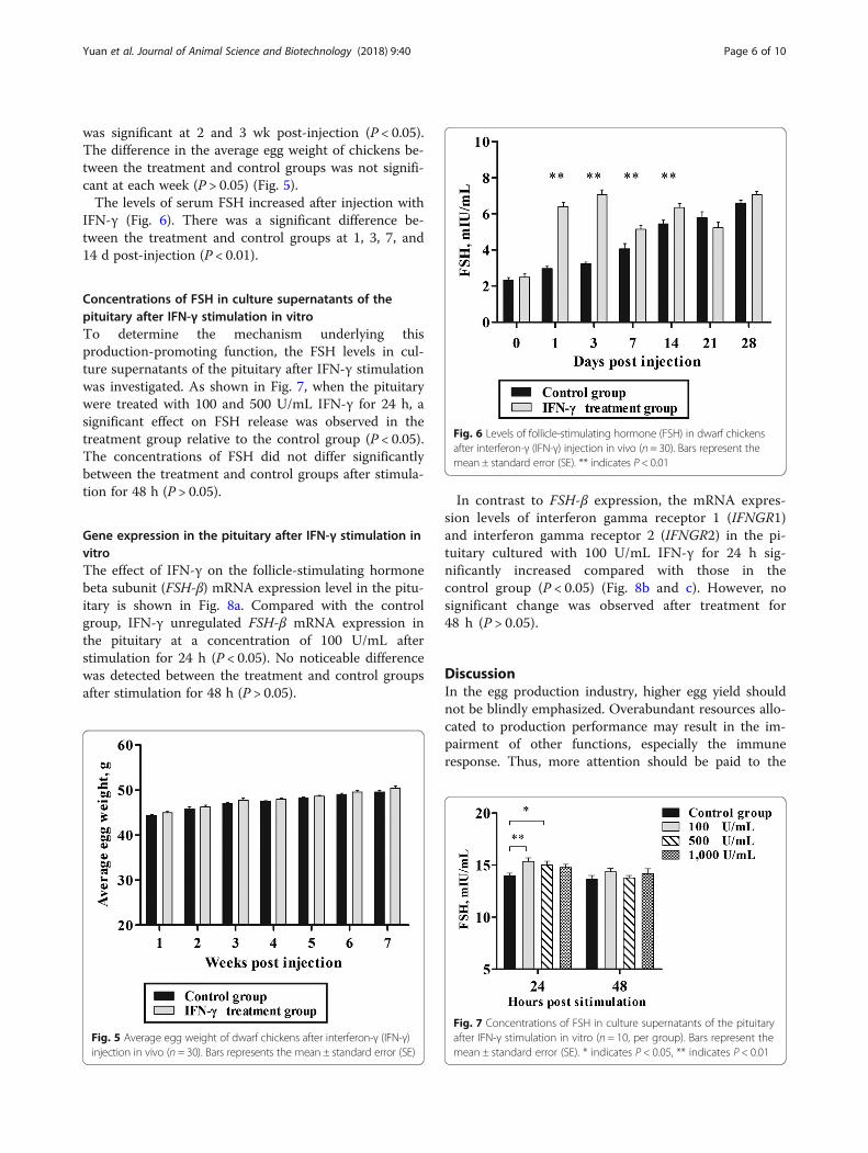
was significant at 2 and 3 wk post-injection (P < 0.05).The difference in the average egg weight of chickens be-tween the treatment and control groups was not signifi-cant at each week (P > 0.05) (Fig. 5).The levels of serum FSH increased after injection with
IFN-γ (Fig. 6). There was a significant difference be-tween the treatment and control groups at 1, 3, 7, and14 d post-injection (P < 0.01).
Concentrations of FSH in culture supernatants of thepituitary after IFN-γ stimulation in vitroTo determine the mechanism underlying thisproduction-promoting function, the FSH levels in cul-ture supernatants of the pituitary after IFN-γ stimulationwas investigated. As shown in Fig. 7, when the pituitarywere treated with 100 and 500 U/mL IFN-γ for 24 h, asignificant effect on FSH release was observed in thetreatment group relative to the control group (P < 0.05).The concentrations of FSH did not differ significantlybetween the treatment and control groups after stimula-tion for 48 h (P > 0.05).
Gene expression in the pituitary after IFN-γ stimulation invitroThe effect of IFN-γ on the follicle-stimulating hormonebeta subunit (FSH-β) mRNA expression level in the pitu-itary is shown in Fig. 8a. Compared with the controlgroup, IFN-γ unregulated FSH-β mRNA expression inthe pituitary at a concentration of 100 U/mL afterstimulation for 24 h (P < 0.05). No noticeable differencewas detected between the treatment and control groupsafter stimulation for 48 h (P > 0.05).
In contrast to FSH-β expression, the mRNA expres-sion levels of interferon gamma receptor 1 (IFNGR1)and interferon gamma receptor 2 (IFNGR2) in the pi-tuitary cultured with 100 U/mL IFN-γ for 24 h sig-nificantly increased compared with those in thecontrol group (P < 0.05) (Fig. 8b and c). However, nosignificant change was observed after treatment for48 h (P > 0.05).
DiscussionIn the egg production industry, higher egg yield shouldnot be blindly emphasized. Overabundant resources allo-cated to production performance may result in the im-pairment of other functions, especially the immuneresponse. Thus, more attention should be paid to the
Fig. 5 Average egg weight of dwarf chickens after interferon-γ (IFN-γ)injection in vivo (n = 30). Bars represents the mean ± standard error (SE)
Fig. 6 Levels of follicle-stimulating hormone (FSH) in dwarf chickensafter interferon-γ (IFN-γ) injection in vivo (n = 30). Bars represent themean ± standard error (SE). ** indicates P < 0.01
Fig. 7 Concentrations of FSH in culture supernatants of the pituitaryafter IFN-γ stimulation in vitro (n = 10, per group). Bars represent themean ± standard error (SE). * indicates P < 0.05, ** indicates P < 0.01
Yuan et al. Journal of Animal Science and Biotechnology (2018) 9:40 Page 6 of 10

balance between greater production performance and aneffective immune function.The present study demonstrated that IFN-γ has an in-
hibitory effect on the phagocytosis of monocytes andmacrophages in a dose-dependent manner in vitro. Inthe in vivo experiment, the inhibition of PP was immedi-ate at 1 d post-injection. The maximum inhibiting effecton PP appeared at 14 d and persisted until 28 d post-injection. Our results are consistent with a study report-ing that the phagocytic percentage of mouse peritonealmacrophages to chicken erythrocytes decreased by morethan one-half and that the phagocytic percentage of E.coli decreased by one-third after treatment with IFN-γ[16]. In addition, the ability of human PBMCs tophagocytize sheep red blood cells was reported to sig-nificantly decrease after being cultured for 48 h withIFN-γ at different concentrations, which is in agreementwith our results [17, 26]. IFN-γ has a dual effect onmonocyte and macrophage function that enhances therespiratory burst via up-regulating the class II majorhistocompatibility complex and suppresses the capacityof nonopsonic receptors on the surface of macrophagesto bind the ligand [27]. The results of our study indicatethat IFN-γ may reduce the binding function of
nonopsonic receptors and then interfere with the phago-cytosis of monocytes and macrophages. In the innateimmune response, IFN-γ enhanced killing of opsonizedpathogens, antigen presentation, and production of in-flammatory mediators. In the other side, IFN-γ dimin-ished phagocytosis of nonopsonized particles, includedbacteria and sheep red blood cells. Both opsonized andnonopsonized phagocytosis were important which wereworthy to be further investigated. The mechanism in-volved in opsonized phagocytosis would be consideredas the research fields of our future study.To confirm whether the decreased PP caused by IFN-γ
is accompanied by changes in production performance,egg production traits were investigated. The data indi-cate that the laying rate remarkably improved at 2and 3 wk following IFN-γ administration, at a timewhen the PP was inhibited. This result agreed withthat in our previous study, in which chickens withlow PP had a higher laying rate in the early period.Furthermore, it was reported that Taiwan countrychicken with lower γ-globulin concentrations showedsubdued carbon clearance to the supernatant fractionof ink and higher egg production during the periodfrom 16 to 52 wk of age [28].
Fig. 8 Gene expression in the pituitary after interferon-γ (IFN-γ) stimulation in vitro (n = 10, per group). a The mRNA expression of follicle-stimulatinghormone-β (FSH-β). b The mRNA expression of interferon gamma receptor 1 (IFNGR1). c The mRNA expression of interferon gamma receptor 2(IFNGR2). Bars represent the mean ± standard error (SE). * indicates P < 0.05
Yuan et al. Journal of Animal Science and Biotechnology (2018) 9:40 Page 7 of 10

The reproductive endocrinology of chickens is closelylinked with their production performance [29]. Previousresearch has confirmed that FSH promotes follicular de-velopment and maturation and ultimately affects eggproduction [30–34]. In line with the laying rate results,chickens in the treatment group had higher FSH levelsat 14 d post-infection and higher IFN-γ levels at 14 and21 d post-infection, at a time when monocyte andmacrophage phagocytosis was at its lower value. Fur-thermore, the FSH level increased two times at 1 and 3d with a rapid decrease in phagocytosis caused by IFN-γ.Thus, chicken IFN-γ caused a decrease in monocyte andmacrophage phagocytosis and simultaneously improvedFSH levels, leading to an increased laying rate. Thesefindings indicated that IFN-γ may have a direct or indir-ect influence on the synthesis or secretion of FSH.The synthesis and release of FSH are regulated by the
pituitary, which is the master gland and crucial bridgebetween the central nervous system and endocrine sys-tem [35–38]. FSH consists of a common alpha subunitnoncovalently attached to a distinct beta subunit thatdetermines the species specificity [39–41]. Studies havefound that IFN-γ might affect hormone secretion bythe pituitary. In female Wistar rats, a significant in-crease in prolactin (PRL) in anterior pituitary cells wasobserved after stimulation with IFN-γ at concentrations of0.1 ng/mL, 1 ng/mL, and 10 ng/mL [42]. In this study, ourdata showed that IFN-γ has a direct influence on FSHsynthesis and secretion by the pituitary, providing anexplanation for the increased FSH levels at 1, 3, 7, and14 d after injection with IFN-γ. It was reported thatthe transcription of GH in the dairy cow anterior pituit-ary was obviously improved after treatment with IFN-γfor 24 h at 10 ng/mL, 20 ng/mL, 40 ng/mL, and 80 ng/mL[43]. In addition, treatment with IFN-γ at 102 and103 U/mL resulted in increased activity of hGH in ratpituitary tumor cells [19]. Therefore, the significantup-regulation of the FSH-β transcriptional level in thepituitary induced more secretion of FSH, which suggeststhat the IFN-γ has the ability to regulate pituitary functionin dwarf chickens.IFN-γ performs regulatory functions through combin-
ation with a specific hormone-receptor complex on thesurface of cells formed by IFNGR1 (IFN-γ receptor α-chain) and IFNGR2 (IFN-γ receptor β-chain). IFNGR1and IFNGR2 are mutually interdependent in their func-tions [8, 44–46]. Impaired IFN-γ-induced signaling andreduced IFN-γ activity were observed in IFNGR1- orIFNGR2- deficient mouse embryonic fibroblasts [9]. Inthe current study, increased mRNA expression levels ofIFNGR1 and IFNGR2 were observed in the pituitary fol-lowing treatment with IFN-γ. The IFN-γ ligand is adimmer and binds to a heterodimer receptor, whichactivates the signal transduction cascades and results in
the dimerization of the signal transducers and activatorsof gene transcription [7, 8, 47]. In our research, the com-bination of endogenous IFN-γ ligand and the pre-assembled up-regulated receptor complex activated therelevant signaling pathways and led to increased FSH-βexpression at the transcription level, promoting the syn-thesis and secretion of FSH.Collectively, IFN-γ has an effect on monocyte and
macrophage PP and the secretion of pituitary hormones.We found that IFN-γ suppressed monocyte and macro-phage phagocytosis and improved the production per-formance in dwarf chickens. In future breedingprograms, IFN-γ may be considered as a regulator tomaintain the dynamic balance between the immune ef-fect and production traits.
ConclusionsIFN-γ inhibited monocyte and macrophage phagocyt-osis; enhanced the secretion of FSH via up-regulatingthe expression of IFNGR1, IFNGR2, and FSH-β; andaccordingly improved the laying rate. This study canprovide a practical and theoretical basis for the links be-tween the immune effect and production performance,and it offers a novel approach for selective breeding.
AbbreviationsFSH: Follicle-stimulating hormone; FSH-β: Follicle stimulating hormone betasubunit; GAPDH: Glyceraldehyde-3-phosphate dehydrogenase;IFNGR1: Interferon gamma receptor 1; IFNGR2: Interferon gamma receptor 2;IFN-γ: Interferon-γ; MTT: Thiazolyl blue tetrazolium bromide;PBMCs: Peripheral blood mononuclear cells; PBS: Phosphate buffered saline;PP: Phagocytosis product
AcknowledgementsThe authors gratefully thank all of the staff at the Genetic Resource Center ofChina Agricultural University (Beijing, China) for taking care of the animals.
FundingThis work was supported by grants from National Natural ScienceFoundation of China (3157130570), Young Scientist Supporting Project,Program for Changjiang Scholars and Innovative Research Team in University(IRT_15R62), Farm Animals Germplasm Resource Platform, NationalTransgenic Creature Breeding Grand Project (2016ZX08008-003), InnovationBase Cultivation and Development Project-research on precise geneticmodify in sheep (Z171100002217072).
Availability of data and materialsThe data analyzed during the current study are available from thecorresponding author on reasonable request.
Authors’ contributionsZL, LL, and YY designed the study. YY, SL, and YZ participated in samplecollection. YY performed all experiments, analyzed the data, and drafted themanuscript. YY and LL revised the manuscript. All authors read andapproved the final manuscript.
Ethics approvalAll animal procedures were approved by the Experimental Animal Care andUse Committee at China Agricultural University (Beijing, China). Theexperiments were performed according to the regulations and guidelinesestablished by this committee.
Competing interestsThe authors declare that they have no competing interests.
Yuan et al. Journal of Animal Science and Biotechnology (2018) 9:40 Page 8 of 10

Author details1Key Laboratory of Animal Genetics and Breeding of the Ministry ofAgriculture, Beijing Key Laboratory for Animal Genetic Improvement,Department of Animal Genetics and Breeding, College of Animal Scienceand Technology, China Agricultural University, Beijing 100193, China.2Department of Animal Genetics and Breeding, College of Animal Scienceand Technology, China Agricultural University, Beijing 100193, China.
Received: 20 October 2017 Accepted: 19 April 2018
References1. Kulkarni G, Zhang H. Evaluation of reproductive characteristics of 21 highly
inbred lines of white leghorns divergently selected for or segregating intumor resistance. Open J Animal Sci. 2015;5:59–70.
2. Blendea A, Cazimir I, Cornila N, Irimescu I, Damian A. Anatomohistologicalstudy regarding the ovary and oviduct in different age groups in thechicken (Gallus domesticus). Sci Works C Series V. 2012;58:21–30.
3. Zhao XL, Honaker CF, Siegel PB. Phenotypic responses of chickens tolong-term selection for high or low antibody titers to sheep red blood cells.Poult Sci. 2012;91:1047–56.
4. Rauw WM, Kanis E, Noordhuizen-Stassen EN, Grommers FJ. Undesirable sideeffects of selection for high production efficiency in farm animals: a review.Livest Prod Sci. 1998;56:15–33.
5. van der Most PJ, de Jong B, Parmentier HK, Verhulst S. Trade-off betweengrowth and immune function: a meta-analysis of selection experiments.Funct Ecol. 2011;25:74–80.
6. Mangel M, Stamps J. Trade-offs between growth and mortality and themaintenance of individual variation in growth. Evol Ecol Res. 2001;3:583–93.
7. Pestka S, Krause CD, Walter MR. Interferons, interferon-like cytokines, andtheir receptors. Immunol Rev. 2004;202:8–32.
8. Goossens KE, Ward AC, Lowenthal JW, Bean AGD. Chicken interferons,their receptors and interferon-stimulated genes. Dev Comp Immunol.2013;41:370–6.
9. Schroder K, Hertzog PJ, Ravasi T, Hume DA. Interferon-γ: an overview ofsignals, mechanisms and functions. J Leukoc Biol. 2004;75:163–89.
10. Vankelecom H, Carmeliet P, Heremans H, Van Damme J, Dijkmans R, BilliauA, et al. Interferon-gamma inhibits stimulated adrenocorticotropin, prolactin,and growth-hormone secretion in normal rat anterior-pituitary cell-cultures.Endocrinology. 1990;126:2919–26.
11. Fratti RA, Ghannoum MA, Edwards JE Jr, Filler SG. Gamma interferonprotects endothelial cells from damage by Candida albicans by inhibitingendothelial cell phagocytosis. Infect Immun. 1996;64:4714–8.
12. Yong SB, Song Y, Kim HJ, Ain QU, Kim YH. Mononuclear phagocytes as atarget, not a barrier, for drug delivery. J Control Release. 2017;259:53–61.
13. Djaldetti M, Salman H, Bergman M, Djaldetti R, Bessler H. Phagocytosis-themighty weapon of the silent warriors. Microsc Res Tech. 2002;57:421–31.
14. Sun K, Metzger DW. Inhibition of pulmonary antibacterial defense byinterferon-γ during recovery from influenza infection. Nat Med. 2008;14:558–64.
15. Arimori Y, Nakamura R, Yamada H, Shibata K, Maeda N, Kase T, et al. Type Iinterferon plays opposing roles in cytotoxicity and interferon-γ productionby natural killer and CD8+ T cells after influenza a virus infection in mice.J Innate Immun. 2014;6:456–66.
16. Wang Z, Zhou S, Sun C, Lei T, Peng J, Li W, et al. Interferon-gamma inhibitsnonopsonized phagocytosis of macrophages via an mTORC1-c/EBPbetapathway. J Innate Immun. 2015;7:165–76.
17. Frausto-Del-Río D, Soto-Cruz I, Garay-Canales C, Ambriz X, Soldevila G,Carretero-Ortega J, et al. Interferon gamma induces actin polymerization,Rac1 activation and down regulates phagocytosis in human monocyticcells. Cytokine. 2012;57:158–68.
18. Capsoni F, Minonzio F, Ongari AM, Bonara P, Pinto G, Carbonelli V,et al. Fc receptors expression and function in mononuclear phagocytesfrom AIDS patients: modulation by IFN-gamma. Scand J Immunol.1994;39:45–50.
19. Gong FY, Deng JY, Shi YF. IFN-gamma increases the hGH gene promoteractivity in rat GH3 cells. Horm Res. 2003;60:14–20.
20. Späth-Schwalbe E, Porzsolt F, Digel W, Born J, Kloss B, Fehm HL. Elevatedplasma cortisol levels during interferon-gamma treatment.Immunopharmacology. 1989;17:141–5.
21. Li H, Zhang Y, Ning ZH, Deng XM, Lian ZX, Li N. Effect of selection forphagocytosis in dwarf chickens on immune and reproductivecharacters. Poult Sci. 2008;87:41–9.
22. Sun SF, Pan QZ, Hui X, Zhang BL, Wu HM, Li H, et al. Stronger in vitrophagocytosis by monocytes-macrophages is indicative of greaterpathogen clearance and antibody levels in vivo. Poult Sci. 2008;87:1725–33.
23. Ma H, Ning ZH, Lu Y, Han HB, Wang SH, Mu JF, et al. Monocytes-macrophages phagocytosis as a potential marker for disease resistancein generation 1 of dwarf chickens. Poult Sci. 2010;89(9):2022.
24. Ma H, Lian ZX, Liu WB, Han HB, Yuan YT, Ning ZH. Salmonella Pullorumresistance in dwarf chickens selected for high macrophage phagocytosis.J Appl Poult Res. 2017;26:437–48.
25. Schmittgen TD, Livak KJ. Analyzing real-time PCR data by the comparativeCT method. Nat Protoc. 2008;3:1101–8.
26. Rüegg SJ, Jungi TW. Antibody-mediated erythrolysis anderythrophagocytosis by human monocytes, macrophages and activatedmacrophages. Evidence for distinction between involvement of high-affinityand low-affinity receptors for IgG by using different erythroid target cells.Immunology. 1988;63:513–20.
27. Speert DP, Thorson L. Suppression by human recombinant gammainterferon of in vitro macrophage nonopsonic and opsonic phagocytosisand killing. Infect Immun. 1991;59:1893–8.
28. Chao CH, Lee YP. Relationship between reproductive performance andimmunity in Taiwan country chickens. Poult Sci. 2001;80:535–40.
29. Long L, Wu SG, Yuan F, Zhang HJ, Wang J, Qi GH. Effects of dietaryoctacosanol supplementation on laying performance, egg quality,serum hormone levels, and expression of genes related to thereproductive axis in laying hens. Poult Sci. 2017;96:894–903.
30. Li Z, Johnson AL. Regulation of P450 cholesterol side-chain cleavagemessenger ribonucleic acid expression and progesterone production in hengranulosa cells. Biol Reprod. 1993;49:463–9.
31. Christians JK, Williams TD. Effects of porcine follicle-stimulating hormone onthe reproductive performance of female zebra finches (Taeniopygia guttata).Gen Comp Endocrinol. 2002;125:121–31.
32. Nataraja SG, Yu HN, Palmer SS. Discovery and development of smallmolecule allosteric modulators of glycoprotein hormone receptors.Front Endocrinol (Lausanne). 2015;6:142.
33. Johnson AL, Lee J. Granulosa cell responsiveness to follicle stimulating hormoneduring early growth of hen ovarian follicles. Poult Sci. 2016;95:108–14.
34. Yamamoto Y, Adam Luckenbach J, Goetz FW, Young G, Swanson P.Disruption of the salmon reproductive endocrine axis through prolongednutritional stress: changes in circulating hormone levels and transcripts forovarian genes involved in steroidogenesis and apoptosis. Gen CompEndocrinol. 2011;172:331–43.
35. Yamamura N, Takeishi M, Goto H, Tagami M, Mizutani T, Miyamoto K,et al. Expression of messenger RNA for gonadotropin receptor in thegranulosa layer during the ovulatory cycle of hens. Comp BiochemPhysiol A Mol Integr Physiol. 2001;129:327–37.
36. Uhm SJ, Gupta MK, Yang JH, Chung HJ, Min TS, Lee HT. Epidermalgrowth factor can be used in lieu of follicle-stimulating hormonefor nuclear maturation of porcine oocytes in vitro. Theriogenology.2010;73:1024–36.
37. Lin JX, Jia YD, Zhang CQ. Effect of epidermal growth factor on follicle-stimulating hormone-induced proliferation of granulosa cells from chickenprehierarchical follicles. J Zhejiang Univ Sci B. 2011;12:875–83.
38. Yoshimura Y, Okamoto T, Tamura T. Effects of luteinizing-hormone andfollicle-stimulating-hormone on the progesterone-receptor induction inchicken granulosa cells in vivo. Poult Sci. 1995;74:147–51.
39. Grzegorzewska AK, Sechman A, Paczoska-Eliasiewicz HE, Rzasa J. Theexpression of pituitary FSHbeta and LHbeta mRNA and gonadal FSH and LHreceptor mRNA in the chicken embryo. Reprod Biol. 2009;9:253–69.
40. Krishnan KA, Proudman JA, Bahr JM. Purification and characterization of chickenfollicle-stimulating hormone. Comp Biochem Physiol B. 1992;102:67–75.
41. Shen ST, Yu JY. Cloning and gene expression of a cDNA for the chickenfollicle-stimulating hormone (FSH)-beta-subunit. Gen Comp Endocrinol.2002;125:375–86.
42. Yamaguchi M, Koike K, Matsuzaki N, Yoshimoto Y, Taniguchi T, MiyakeA, et al. The interferon family stimulates the secretions of prolactinand interleukin-6 by the pituitary-gland in vitro. J Endocrinol Investig.1991;14:457–61.
Yuan et al. Journal of Animal Science and Biotechnology (2018) 9:40 Page 9 of 10

43. Wang JF, Fu SP, Li SN, Yang ZQ, Xue WJ, Li ZQ, et al. Establishment andcharacterization of dairy cow growth hormone secreting anterior pituitarycell model. In Vitro Cell Dev Biol Anim. 2014;50:103–10.
44. Han X, Chen T, Wang M. Molecular cloning and characterization of chickeninterferon-gamma receptor alpha-chain. J Interf Cytokine Res. 2008;28:445–54.
45. Bach EA, Aguet M, Schreiber RD. The IFN gamma receptor: a paradigm forcytokine receptor signaling. Annu Rev Immunol. 1997;15:563–91.
46. Han CL, Zhang W, Dong HT, Han X, Wang M. A novel gene of beta chain ofthe IFN-gamma receptor of Huiyang chicken: cloning, distribution, and CDassay. J Interf Cytokine Res. 2006;26:441–8.
47. Lasfar A, Cook JR, Cohen Solal KA, Reuhl K, Kotenko SV, Langer JA, et al.Critical role of the endogenous interferon ligand-receptors in type I andtype II interferons response. Immunology. 2014;142:442–52.
Yuan et al. Journal of Animal Science and Biotechnology (2018) 9:40 Page 10 of 10