integumentary system dwelling organisms from …rmoen/mammalogy_4764_2009/handouts/integument… ·...
TRANSCRIPT

Skull_Skeleton_Lab2.doc 10/07/08 page 39 of 56
INTEGUMENTARY SYSTEM
The skin and associated structures make up the integumentary system. The skin protects land-dwelling organisms from desiccation and from loss of heat. Skin is a mammal's largest organ. Its protects the body against physical, chemical, and biological attacks, it helps to regulate body temperature, it is used to communicate to other individuals, and a skin derivative provides nourishment for the young.
Like the integuments of other vertebrates, mammalian skin is composed of two layers, the dermis and the epidermis. Identify and locate the structures underlined in the following text in Fig. 47.
Figure 47. Cross-sectional diagram of skin surface (Martin and DeBlaise 1981).

Skull_Skeleton_Lab2.doc 10/07/08 page 40 of 56
Epidermis.--The epidermis consists of several layers, representing successive stages of development. The oldest part of the skin is the outer layer of tough, protective, cells. The cells, which are dead, are continually worn off at the surface and replaced from below. As the cells age and mature, they eventually lose their nuclei and most of the cell contents are converted to keratin. Keratin is a protein that makes up the protective layer of skin, and also such structures as nails, hooves, hair, and horns--evolutionary and developmental derivatives of skin.
The outermost layer of epidermis is the stratum corneum. The epidermis on the soles of feet and the palms of hands is thick. Elsewhere on the body, the epidermis may be quite thin. Thickened portions of the epidermis form the pads on the feet of most mammals and the friction ridges on the digits and palm of primates. Fingerprints are the impression of these friction ridges. Calluses are also products of the epidermis. Hair, horn, claws, and epidermal scales are all made of modified keratinized cells of epidermal origin.
Figure 48. Fingerprint of human (Homo sapiens). We may have an opportunistic sample (e.g., last year had a pine marten (Martes americana).

Skull_Skeleton_Lab2.doc 10/07/08 page 41 of 56
Dermis.--The dermis lies below the epidermis. It is a thick layer of connective tissue with associated muscles, nerves, and blood vessels. The connective tissue consists largely of collagen. Collagen may be up to 6 per cent of body weight in humans, and is the most abundant protein in the body, being present in skin, bones, tendons, cartilage, and ligaments. Collagen is from a Greek word meaning “glue-maker”. Collagen and the other fibers in the dermis become toughened and hardened during the process of tanning when a skin is transformed into leather. Unlike the epidermis, the dermis is well-supplied with blood vessels and nerves for sensation of touch, pressure, temperature, and pain. Beneath the dermis is a layer of fatty tissue, variably thick, that provides insulation and energy storage. In many species, the extent of subcutaneous fat varies dramatically with season (Fig. 49).
Figure 49. Subcutaneous fat in a deer in the winter. This picture is of the subcutaneous fat of a yearling doe (nearly 2 years old) that was hit by a car on 2/8/04. On the left is in image from just anterior to the tail cut through the tissue, and on the right is much of the back with the skin peeled back.
Skin Glands.--Associated with the skin are two kinds of glands, sweat glands and sebaceous glands. The epidermal sebaceous glands lubricate the hair and are described below. Sweat glands (sudoriferous glands) are coiled tubes in the dermis connected with the surface by narrow ducts. They are well supplied with blood vessels, secrete mostly water and salts, and function largely in thermoregulation. In humans and some ungulates, sweat glands are distributed over much of the body. Some mammals such as rodents and lagomorphs (rabbits) do not have sweat glands. Cats (Felidae) and dogs (Canidae), and perhaps other carnivores, have sweat glands in the pads of the feet. It is thought that mammary glands evolved from sweat glands as discussed below.

Skull_Skeleton_Lab2.doc 10/07/08 page 42 of 56
Hair
General.--Hair is a uniquely mammalian feature. The developing epidermis invaginates into the dermis to form a follicle. At the deepest point of the follicle, the dermis pushes back and forms a small structure called the papilla. The papilla is well supplied with blood vessels. Epidermal cells on top of the papilla multiply and are pushed towards the surface by those growing beneath them, keratinizing (as epidermal cells also do to form the outer layer of skin) and forming the hair.
Each hair consists of three parts (Fig. 47). The center is the medulla (Latin, “marrow”). This is surrounded by a denser cortex (Latin, “bark”) containing most of the pigment granules that give each hair its characteristic color. The cortex is covered by a thin layer called the cuticle (Latin, “little skin”). Its cells often overlap like the shingles or tiles on a roof. Cuticular scales are often characteristic of particular genera or even species of mammal. They are never pigmented.
Glands.--Sebaceous glands open into each follicle. They secrete oily substances (sebum) that continually lubricate and condition skin and hair. Cells inside these glands gradually fill with grease and then break away, becoming part of the secretion themselves. Sebum makes beavers waterproof and prevents undue drying of the pelage of terrestrial mammals. Glands that secrete cellular debris as well as molecular products are termed apocrine glands. These glands empty into or near a hair follicle.
There are many examples of skin glands that have moved beyond their roles in lubrication to serve other functions. In skunks protective and communicative functions are both present. We have a skunk study skin in the laboratory but do not have the anal sac from which the skunk sprays. Note the warning coloration of the skunk. Some shrews have glands on their sides that advertise reproductive condition. Many species use glandular scents to mark individual territories, particularly the carnivores (order Carnivora).
Skull_Skeleton_Lab2.doc 10/07/08 page 43 of 56
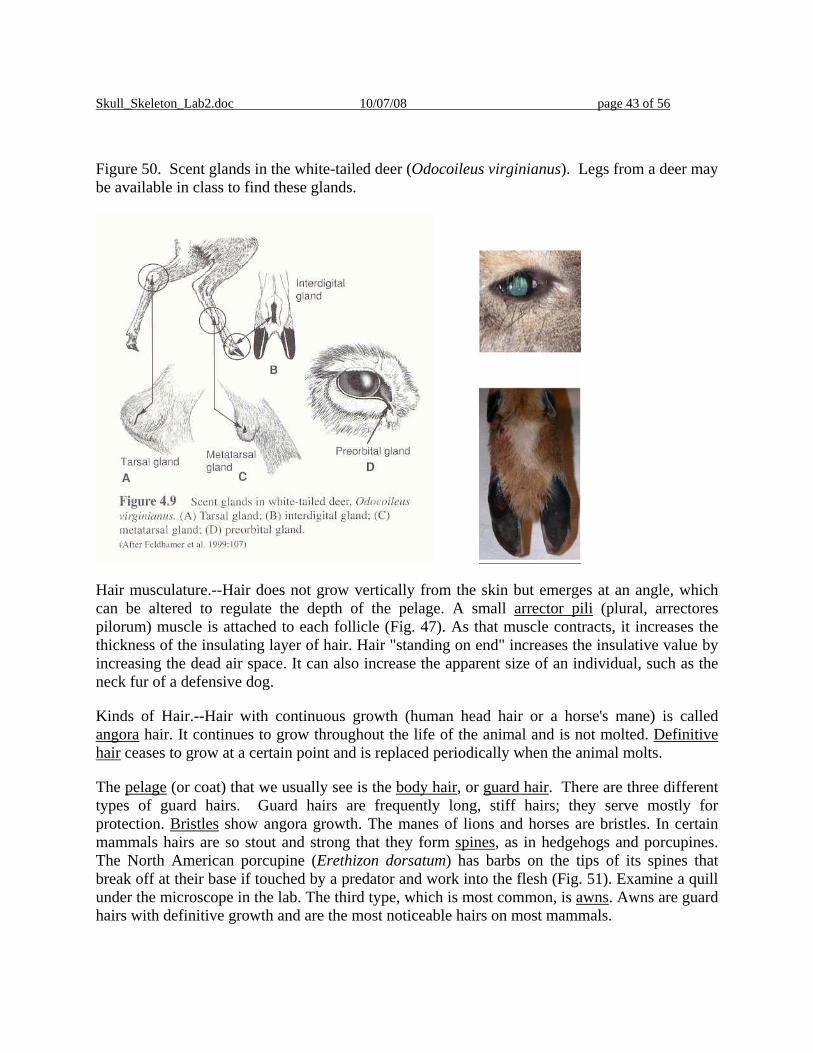
Figure 50. Scent glands in the white-tailed deer (Odocoileus virginianus). Legs from a deer may be available in class to find these glands.
Hair musculature.--Hair does not grow vertically from the skin but emerges at an angle, which can be altered to regulate the depth of the pelage. A small arrector pili (plural, arrectores pilorum) muscle is attached to each follicle (Fig. 47). As that muscle contracts, it increases the thickness of the insulating layer of hair. Hair "standing on end" increases the insulative value by increasing the dead air space. It can also increase the apparent size of an individual, such as the neck fur of a defensive dog.
Kinds of Hair.--Hair with continuous growth (human head hair or a horse's mane) is called angora hair. It continues to grow throughout the life of the animal and is not molted. Definitive hair ceases to grow at a certain point and is replaced periodically when the animal molts.
The pelage (or coat) that we usually see is the body hair, or guard hair. There are three different types of guard hairs. Guard hairs are frequently long, stiff hairs; they serve mostly for protection. Bristles show angora growth. The manes of lions and horses are bristles. In certain mammals hairs are so stout and strong that they form spines, as in hedgehogs and porcupines. The North American porcupine (Erethizon dorsatum) has barbs on the tips of its spines that break off at their base if touched by a predator and work into the flesh (Fig. 51). Examine a quill under the microscope in the lab. The third type, which is most common, is awns. Awns are guard hairs with definitive growth and are the most noticeable hairs on most mammals.

Skull_Skeleton_Lab2.doc 10/07/08 page 44 of 56
Figure 51. Tip of spine of porcupine quill.
Examine skins of a variety of mammals—such as the porcupine, hare (Lepus americanus), ermine (Mustela erminea), otter (Lutra canadensis), and moose (Alces alces). Identify the types of hair found on each. What is the function of each kind of hair? What differences do you see among these different animals that might be associated with the habitats they normally live in? Why might moose or deer hair have the character that it does, while snowshoe hare hair is different in nature?
Underhairs are shorter and finer hairs growing around the guard hairs often in much greater numbers. Their function is to insulate. Underhairs with angora growth are called wool. In domestic sheep, guard hairs have been eliminated through selective breeding and the growth rate and density of the wool has been increased.
The pelage of an animal is the combination of longer guard hairs and the underfur (underhairs), fine and relatively short hair with definitive growth that densely covers most mammals.
Special tactile hairs, the vibrissae, are found not only on a mammal's face (mystacial vibrissae) but may occur also on the legs or elsewhere on the body (Fig. 52). Nerves at the base of vibrissae communicate response to the brain. Vibrissae are especially prominent on the muzzles of nocturnal and burrowing mammals. Humans do not have vibrissae.
Skull_Skeleton_Lab2.doc 10/07/08 page 45 of 56
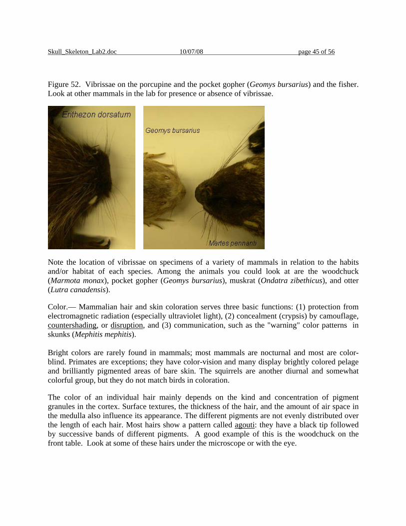
Figure 52. Vibrissae on the porcupine and the pocket gopher (Geomys bursarius) and the fisher. Look at other mammals in the lab for presence or absence of vibrissae.
Note the location of vibrissae on specimens of a variety of mammals in relation to the habits and/or habitat of each species. Among the animals you could look at are the woodchuck (Marmota monax), pocket gopher (Geomys bursarius), muskrat (Ondatra zibethicus), and otter (Lutra canadensis).
Color.— Mammalian hair and skin coloration serves three basic functions: (1) protection from electromagnetic radiation (especially ultraviolet light), (2) concealment (crypsis) by camouflage, countershading, or disruption, and (3) communication, such as the "warning" color patterns in skunks (Mephitis mephitis). Bright colors are rarely found in mammals; most mammals are nocturnal and most are color-blind. Primates are exceptions; they have color-vision and many display brightly colored pelage and brilliantly pigmented areas of bare skin. The squirrels are another diurnal and somewhat colorful group, but they do not match birds in coloration.
The color of an individual hair mainly depends on the kind and concentration of pigment granules in the cortex. Surface textures, the thickness of the hair, and the amount of air space in the medulla also influence its appearance. The different pigments are not evenly distributed over the length of each hair. Most hairs show a pattern called agouti: they have a black tip followed by successive bands of different pigments. A good example of this is the woodchuck on the front table. Look at some of these hairs under the microscope or with the eye.

Skull_Skeleton_Lab2.doc 10/07/08 page 46 of 56
Hair Replacement.—The pelage must be maintained to maintain its functionality. Hair cannot be repaired when damaged because it is nonliving. Most hair is of definitive growth and is replaced periodically. This process is called molting. Two kinds of molts are recognized: maturational molt (from juvenile to subadult to adult pelage) and seasonal molt, which usually occurs once or twice a year and often follows a regular spatial pattern within a particular species. Species subjected to seasonal changes generally have a longer pelage with good insulating abilities in winter. Some northern species have white coats for winter and brown coats for summer (Figure 53).
Figure 53. Compare winter and summer pelts of the ermine (Mustela erminea) and the snowshoe hare (Lepus americanus) that are present in the lab.
In many mammals there is a distinctly juvenile pelage that distinguishes young animals from adults (in addition to other clues such as body size). Members of the deer family are a good example of this, moose calves are reddish in color, for example. There is the skin of a white-tailed deer fawn in the laboratory, compare its hair to that of the adult deer (see the legs).
Scales.—The scales on the more or less naked tails of rats, mice, and beavers are protective, epidermal thickenings of the skin made of keratinized cells. The thinner skin between these scales allows flexibility. The pangolin (Order Pholidota) is covered with epidermal scales of a different kind. They consist of keratinized cells and are in structure and development basically equivalent to hairs (Fig. 54). However, they do not grow from follicles in the skin but from raised papillae protruding from the surface.

Skull_Skeleton_Lab2.doc 10/07/08 page 47 of 56
Figure 54. Examine the scaly tail of a beaver (Castor canadensis) or rat (Rattus norvegicus) or opposum (Didelphis virginiana) and note the placement of hairs in relation to the placement of scales.
The armadillo (Edentata: Dasypodidae) has both epidermal scales and dermal bone. The epidermal scales resemble the scales on the tails of the rats and beavers. The dermal bone is unique among mammals. It is true bone within the dermis, forming a shell constructed somewhat like the armor of a medieval knight. Dermal bone also arose in ancient fishes, the ostracoderms. Today dermal bones are found in some modern fish, in the shells of turtles, and in the skin of many lizards and crocodilians.
Examine the nine-banded armadillo (Dasypus novemcinctus). Note the arrangements of dermal bone and epidermal scales (Figure 55). How do the size and the shape of the two layers compare? How is the shell constructed to allow for flexibility?
Figure 55. Shell of a nine-banded armadillo (Dasypus novemcinctus).

Skull_Skeleton_Lab2.doc 10/07/08 page 48 of 56
Horns and Antlers
Horns and antlers are found today only in two mammalian orders, Artiodactyla and Perissodactyla. Extinct mammals from other orders (including the Rodentia) also had cranial ornamentations. Five different kinds of head ornamentations are recognized, each occurring in a different family. These kinds can be distinguished by their location on the head and their mode of development. You should be able to distinguish true horns, pronghorns, and antlers.
Horns.--True horns are found only in the family Bovidae (Order Artiodactyla). True horns are always unbranched and permanent and are composed of two parts: the bony horncore and the horn itself (Fig. 56). The cores extend upward or outward from the frontal bones. Horns are covered by a sheathing layer of keratinized epidermis, the horn. The horn grows from its base throughout the adult life of the animal.
Figure 56. Diagram of horn (DeBlase and Martin 1981) on the left, and the horncore and horn of a bison on the right. This specimen is available in the laboratory
.
Examine horns and horn cores of the available bovids (cow, Bos taurus, and on wall bighorn sheep, Ovis canadensis and mountain goat, Oreamnos americana). There are no cross-sections of horns in the UMD collection. Size, length, and curvature of horns varies among species.
Pronghorns.--The pronghorn (Antilocapra americana) of western North America is the only living species of the family Antilocapridae (Artiodactyla). As in the Bovidae the horn (properly called a pronghorn) has a bony core covered by a keratinized sheath and serves a similar function (Fig. 57). However, unlike other horns, in pronghorns the sheaths are (1) branched and (2) deciduous. They are shed annually after the breeding season. The new sheath grows while the old one is still in place and only pushes it off when its development is much advanced. Both sexes have pronghorns, but they are more prominent in the males. In females, the pronghorns

Skull_Skeleton_Lab2.doc 10/07/08 page 49 of 56
sometimes are unbranched or absent altogether. Compare the diagrams of the pronghorn (Fig. 57) with the diagram of the bovid horn (Fig. 56). How do the horn cores differ?
Figure 57. Cross-sectional diagram of a pronghorn (DeBlase and Martin 1981).
Antlers.--Antlers are only found in the family Cervidae (Artiodactyla). Antlers are present only in males, except for female caribou (reindeer) in the genus Rangifer. Fully developed antlers are made completely of bone. They arise from bony stumps (pedicels) on the frontal bones (Fig. 58 and Fig. 59). The pedicels are covered with skin. The antlers themselves are shed after the mating season. The point of separation between pedicel and antler is the burr. In spring a new set of antlers begins to grow. The developing antlers are covered with a layer of skin and short hairs. This “velvet” carries blood vessels and nerves supplying the growing bone. When growth is complete the blood supply ceases and the velvet is shed or rubbed off. Antlers are usually used only during sparring matches and in displays to potential mates and rivals. Antlered animals tend to use their hooves for defense when attacked by predators.
Figure 58. Diagrammatic cross-section of a developing antler (DeBlase and Martin 1981).

Skull_Skeleton_Lab2.doc 10/07/08 page 50 of 56
Figure 59. Sample skulls of deer (Cervidae) in the laboratory showing different aspects of antler growth.
Be able to distinguish to species the antlers of available cervids (mule deer Odocoileus hemionus and white-tailed deer Odocoileus virginianus, wapiti (elk) Cervus elaphus, moose, Alces alces, and caribou Rangifer tarandus (Fig. 60). Moose antlers are palmated, while caribou antlers have a small amount of palmation and the brow tine (on at least one of the antlers). Wapiti antlers are larger than deer antlers and have a single beam that branches. White-tailed deer antlers typically curve forward and around, while mule deer antlers appear to branch rather than curve around. The mule deer antlers present in the laboratory are not the best example of this type of branching (as opposed to curving around).

Skull_Skeleton_Lab2.doc 10/07/08 page 51 of 56
Figure 60. Drawing of different antler shapes and body sizes of extant members of the deer family in North and South America (from Geist 1999). Note the curvature on the white-tailed deer antler compared to the branching on the mule deer antler.
Look at the skulls of male and female moose, and also at the skulls of male and female deer. Note the abscission line and/or the pedicel.

Skull_Skeleton_Lab2.doc 10/07/08 page 52 of 56
Claws, Hooves, Nails
Objectives.--In this section we learn about claws, hooves, and nails of mammals. You should be able to identify and apply correctly all underlined terms.
The ends of most digits of mammals other than whales and most sirenians are protected by hardened plates of the protein keratin (also present in hair). These plates take the form of claws, nails, or hooves. They are formed by the epidermis in a process similar to the growth of hair.
Claws.--The claw is the ancestral form of digital covering. Mammalian claws are similar to claws of reptiles and birds. A claw is composed of a harder dorsal plate called the unguis and a softer ventral plate termed the subunguis (Fig. 61). The subunguis is continued by the cushion-like pad. Mammals like dogs and cats walk on these pads. In cross-section unguis and subunguis form a U-shaped structure with the unguis enclosing the subunguis. The downward curve is caused by a higher growth rate of the upper surface of the unguis. A claw is thicker in the median line than at the sides. The sides wear more quickly than the center, producing a more or less sharp point. In addition to the protection of the digits, claws are used in many ways for climbing, digging, hanging, or grasping and even killing prey.
Figure 61. Diagram of claws and nails (Romer 1977 for the horizontal figure, Feldhamer 2004 (your text) for the vertical figure).

Skull_Skeleton_Lab2.doc 10/07/08 page 53 of 56
Examine claws of an arboreal squirrel (e.g., gray squirrel), cat, dog, and a badger or mole (Fig. 62). Locate the unguis and subunguis on each. What is the principal function of the claws in each of these mammals?
Figure 62. Pictures of claws of several species that are present in the lab.
Nails.--A nail is a simplified derivative of a claw covering only the dorsal surface of the digit. Compared to a claw, the nail's wide unguis is thinner and less rigid and the subunguis is very much reduced (Fig. 61). A nail offers less protection than does a claw but exposes the end of the digit to permit more precise manipulation of objects.
Examine your own fingernail and locate the unguis and subunguis. Compare with the nail of other primates. Contrast with the claws observed above.

Skull_Skeleton_Lab2.doc 10/07/08 page 54 of 56
Figure 63. Fingernail of a human (Homo sapiens) and the hand skeleton of a monkey, species unknown. Compare your fingernail to the monkey present in lab.
Hooves.—Well-developed hooves are found among extant mammals only in ungulates (Artiodactyla and Perissodactyla). They are further modified claws in which the unguis encloses both the end of the digit and the subunguis (Fig. 61). The softer subunguis wears away more quickly than the unguis, thus forming a sharp edge. The pad lies just behind the hoof and is called the frog. In ungulates normally only the hoof, not the frog, is in contact with the ground.
Examine the hooves of the cow, white-tailed deer, and horse hooves in the laboratory (Figs. 63-65). Locate the unguis, subunguis, and frog.
Figure 63. Picture of Bos taurus hoof in laboratory.

Skull_Skeleton_Lab2.doc 10/07/08 page 55 of 56
Figure 64. Pictures of Odocoileus virginianus hooves that are available in the laboratory.
Figure 65. Picture of horse hoof (Equus caballus) in laboratory. First row, left to right is the hoof on mounted leg, side view of hoof, and cross-section. Bottom row is a view of the cross-section from the bottom.

Skull_Skeleton_Lab2.doc 10/07/08 page 56 of 56
The following specimens are available in the lab today, other species will be available in the next labs. Order Family Species Common name
Artiodactyla Cervidae Odocoileus virginianus White-tailed deer
Carnivora Mephitidae Mephitis mephitis Striped skunk
Rodentia Erethizontidae Erethizon dorsatum Porcupine
Rodentia Geomyidae Geomys bursarius pocket gopher
Rodentia Castoridae Castor canadensis Beaver
Artiodactyla Cervidae Cervus elaphus Elk
Artiodactyla Cervidae Alces alces Moose
Artiodactyla Cervidae Odocoileus virginianus White-tailed deer
Artiodactyla Cervidae Odocoileus hemionus Mule deer
Artiodactyla Cervidae Rangifer tarandus Caribou, Reindeer
Artiodactyla Bovidae Bos taurus Cow
Artiodactyla Ovidae Ovis canadensis Bighorn sheep
Artiodactyla Ovidae Oreamnos americana Mountain goat
Artiodactyla Antilocapridae Antilocapra americana Pronghorn antelope
Perissodactyla Equidae Equus caballus Horse
Rodentia Sciuridae Sciurus carolinensis Gray squirrel Carnivora Mustelidae Lutra canadensis Otter Carnivora Mustelidae Taxidea taxus Badger Carnivora Mustelidae Mustela erminea Ermine Artiodactyla Cervidae Alces alces Moose Lagomorpha Leporidae Sylvilagus floridanus Eastern cottontail Lagomorpha Leporidae Lepus americanus Snowshoe hare Rodentia Sciuridae Marmota monax Woodchuck Xenarthra Dasypodidae Dasypus novemcinctus Armadillo Rodentia Muridae Ondatra zibethicus Muskrat