integrative and comparative biology doi:10.1093/icb/icx081
TRANSCRIPT

SYMPOSIUM
A Hypothesis for the Composition of the Tardigrade Brainand its Implications for Panarthropod Brain EvolutionFrank W Smith1 Paul J Bartelsdagger and Bob Goldstein
Department of Biology University of North Carolina at Chapel Hill Chapel Hill NC 27599 USA daggerDepartment of
Biology Warren Wilson College PO Box 28815 Asheville NC 9000 USA
From the symposium ldquoThe Evolution of Arthropod Body PlansndashIntegrating Phylogeny Fossils and Developmentrdquo
presented at the annual meeting of the Society for Integrative and Comparative Biology January 4ndash8 2017 at New
Orleans Louisiana
1E-mail smithfwliveuncedu
Synopsis Incredibly disparate brain types are found in Metazoa which raises the question of how this disparity evolved
Ecdysozoa includes representatives that exhibit ring-like brainsmdashthe Cycloneuraliamdashand representatives that exhibit
ganglionic brainsmdashthe Panarthropoda (Euarthropoda Onychophora and Tardigrada) The evolutionary steps leading
to these distinct brain types are unclear Phylogenomic analyses suggest that the enigmatic Tardigrada is a closely related
outgroup of a EuarthropodathornOnychophora clade as such the brains of tardigrades may provide insight into the
evolution of ecdysozoan brains Recently evolutionarily salient questions have arisen regarding the composition of
the tardigrade brain To address these questions we investigated brain anatomy in four tardigrade speciesmdashHypsibius
dujardini Milnesium n sp Echiniscus n sp and Batillipes n spmdashthat together span Tardigrada Our results suggest
that general brain morphology is conserved across Tardigrada Based on our results we present a hypothesis that
proposes direct parallels between the tardigrade brain and the segmental trunk ganglia of the tardigrade ventral nervous
system In this hypothesis brain neuropil nearly circumscribes the tardigrade foregut We suggest that the tardigrade
brain retains aspects of an ancestral cycloneuralian brain while exhibiting ganglionic structure characteristic of euar-
thropods and onychophorans
Introduction
The evolutionary steps that connect disparate meta-
zoan brains remain unclear (Schmidt-Rhaesa 2007
Hejnol and Lowe 2015) Ecdysozoa includes lineages
characterized by ring-like brainsmdashthe Cycloneuralia
(Nematoda Nematomorpha Priapulida
Kinorhyncha and Loricifera)mdashand lineages character-
ized by ganglionic brainsmdashthe Panarthropoda
(Euarthropoda Onychophora and Tardigrada) The
cycloneuralian brain typically consists of anterior and
posterior clusters of neuronal somata with a ring of
circumesophageal neuropil positioned between them
(Schmidt-Rhaesa 19971998 Richter et al 2010
Rothe and Schmidt-Rhaesa 2010 Martın-Duran et al
2016 Henne et al 2017b reviewed in Hejnol and Lowe
2015 Schafer 2016) The brains characteristic of
Nematomorpha have diverged from this architecture
(Schmidt-Rhaesa 1996) but retain evidence of
cycloneuralian ancestry (Henne et al 2017a)
Although often referred to as ganglia the brain clusters
of cycloneuralians are not true ganglia because they do
not contain neuropil (Richter et al 2010 Schafer 2016)
In contrast to cycloneuralian brains panarthropods ex-
hibit ganglionic brains (Scholtz and Edgecombe 2006
Mayer 2016 Mayer et al 2013a) Intriguingly
Panarthropoda appears to be nested within
Cycloneuralia (Campbell et al 2011 Rota-Stabelli
et al 2013 Borner et al 2014) suggesting that the gan-
glionic brains of panarthropods evolved from a ring-
like cycloneuralian brain (Hejnol and Lowe 2015)
The relatively simple tardigrade brain may hold im-
portant clues regarding the transition from a cycloneura-
lian brain to a ganglionic brain in the panarthropod stem
lineage Unlike the brains of euarthropods and onycho-
phorans which are composed of multiple segmental gan-
glia (Scholtz and Edgecombe 2006 Strausfeld et al 2006a
The Author 2017 Published by Oxford University Press on behalf of the Society for Integrative and Comparative Biology
All rights reserved For permissions please email journalspermissionsoupcom
Integrative and Comparative BiologyIntegrative and Comparative Biology pp 1ndash14
doi101093icbicx081 Society for Integrative and Comparative Biology
Mayer et al 2010 Whitington and Mayer 2011 Martin
and Mayer 2015) the tardigrade brain is composed of a
single segmental ganglion (Hejnol and Schnabel 2005
Gabriel et al 2007 Mayer et al 2013a Gross and Mayer
2015 Smith et al 2016 Gross et al 2017 see Zantke
et al 2008 and Persson et al 2012 for alternative
views)mdasha condition that is thought to be plesiomor-
phic for Panarthropoda (reviewed in Strausfeld 2012
Smith and Goldstein 2017 Ortega-Hernandez et al
2017) Furthermore Tardigrada is most likely the sister
group of a EuarthropodathornOnychophora clade
(Campbell et al 2011 Rota-Stabelli et al 2013 Dunn
et al 2014 Giribet 2016 for an alternative view see
Borner et al 2014 and Laumer et al 2015 reviewed in
Edgecombe and Giribet this volume) Under this hy-
pothesis the most recent common ancestor of
Panarthropoda exhibited a brain composed of a single
segmental ganglion referred to as the protocerebrum
tardigrades retain a protocerebral brain while more
posterior segmental ganglia were incorporated into
the brains of euarthropods and onychophorans after
the euarthropodthorn onychophoran lineage diverged
from Tardigrada (Martin and Mayer 2015 reviewed
in Strausfeld 2012 Hejnol and Lowe 2015 Scholtz
2016 Strausfeld et al 2016 Ortega-Hernandez et al
2017 Smith and Goldstein 2017) Additionally in
terms of its position along the body axis the entire
brain of tardigrades is most likely homologous to the
entire brain of non-panarthropod invertebrate animals
(Steinmetz et al 2010 Smith et al 2016)
Unlike the circumesophageal brains of cycloneura-
lians the protocerebrum of euarthropods and ony-
chophorans is restricted to a supraesophageal
position (Eriksson and Budd 2000 Strausfeld et al
2006a 2006b Martin and Mayer 2014 Ortega-
Hernandez et al 2017) which raises the question
of when during panarthropod evolution the proto-
cerebrum became restricted to a supraesophageal
position The position of the tardigrade brain relative
to the foregut clearly has important implications for
addressing this question but recent investigations of
tardigrade brain anatomy have led to several oppos-
ing views regarding the composition and position of
the tardigrade brain (Fig 1A Zantke et al 2008
Persson et al 2012 Mayer et al 2013a 2013b
Schulze and Schmidt-Rhaesa 2013 Persson et al
2014 Schulze et al 2014 Smith and Jockusch
2014) In one model tardigrades exhibit a circum-
esophageal brain (Zantke et al 2008) the circum-
esophageal component of the tardigrade brainmdashthe
dorsal commissure (dco)thorn the circumbuccal connec-
tives (cco)thorn the post-oral commissure (poc)mdashis
suggested to be directly homologous to the ring of
neuropil characteristic of cycloneuralian brains
(Fig 1B top panel) Tardigrades also exhibit a cir-
cumesophageal brain in a second model (Fig 1B
middle panel) but in this model the tardigrade brain
is composed of three paired brain lobesmdashthe outer
lobes (ol) inner lobes (il) and ventrolateral lobes
(vll)mdashthat are not clearly relatable to cycloneuralian
brains (Persson et al 2012) In a third model tardi-
grades exhibit a supraesophageal brain (Mayer et al
2013a) This model includes a circumesophageal ner-
vous system componentmdashthe nerve ring (nr) but
the nerve ring is predicted to be part of the stomo-
deal nervous system rather than part of the brain
(Fig 1B bottom panel)
Distinguishing between different models of tardi-
grade brain anatomy is difficult Different tardigrade
species were investigated in different studies
(Fig 1A) raising the possibility that reported differ-
ences represent true taxonomic variation
Furthermore different immunohistochemical and
imaging approaches were used in different studies
Both of these factors may have contributed to different
results and ultimately different conclusions Here we
investigate brain morphology in four tardigrade spe-
cies using identical immunohistochemical and imag-
ing approaches This includes the first study of a
member of the tardigrade lineage Apochela using these
techniques Our results suggest that brain neuropil
wraps around the tardigrade foregut Based on our
results we present a hypothesis for the evolution of
the ganglionic brains of panarthropods from an ances-
trally cycloneuralian state
Materials and methods
Specimen collection
Hypsibius dujardini (Eutardigrada Parachela) speci-
mens were collected from a laboratory culture (Gabriel
et al 2007) We collected three wild tardigrade species
from habitats in North Carolina Based on taxonomic
keys these appear to be three new species We refer to
them as Milnesium n sp Echiniscus n sp and Batillipes
n sp We collected Milnesium n sp specimens
(Eutardigrada Apochela) from lichen samples originat-
ing from Durham County North Carolina (location
3559047500N 7852034100W) Echiniscus n sp
(Heterotardigrada Echiniscoidea) specimens were
collected from lichen originating in Orange
County North Carolina (location 3554046400N
7903021200W) Batillipes n sp (Heterotardigrada
Echiniscoidea) specimens were collected from intertidal
sand in Brunswick County North Carolina (location
3351058800N 7830020100W) When we refer to hatch-
lings we are referring to specimens that were collected as
embryos and fixed within 24 h after they hatched
2 F W Smith et al
Specimen preparation
We prepared specimens to visualize neurites nuclei
and muscles Specimens were stretched in carbonated
water They were then transferred to 4 formaldehyde
in 05 PB-Triton (05 phosphate-buffered saline
01 Triton X-100 pH 74) They were stored in this
solution at 4 C for between 1 day and 1 week
Specimens were then washed five times with 05PB-Triton To permeabilize specimens we bisected
them transversely with a 25-gauge needle Specimens
were then washed two times for 1 h in 02 bovine
serum albumin in 05X PB-Triton followed by a 15 h
wash in 5 normal goal serum (NGS in 05 PB-
Triton) Specimens were incubated overnight in a
1100 dilution of a b-tubulin antibody (E7
Developmental Studies Hybridoma Bank) which
stains the tardigrade nervous system (Smith and
Jockusch 2014) They were then washed three times
for 5 min and two times for 1 h in 05 PB-Triton
This was followed by two 1=2 h washes in NGS
Specimens were then incubated overnight at 4 C in a
1200 dilution of a goat anti-mouse Cy3-conjugated
secondary antibody (Jackson ImmunoResearch) in
NGS Next specimens were washed two times for 1 h
and three times quickly in 05 PB-Triton They were
then incubated in a 140 dilution of Oregon Green 488
phalloidin (Molecular Probes) which stains tardigrade
muscles (Smith and Jockusch 2014) for at least 16 h at
4 C We then washed the specimens three times
quickly in 05 PB-Triton We mounted specimens
on slides in DAPI Fluoromount-G (SouthernBiotech)
to visualize nuclei
Imaging and analysis
Z-series of specimens were collected on a Zeiss 710
laser scanning confocal microscope To help visualize
Fig 1 Summary of recent studies that investigated tardigrade brain morphology (A) Diversity of tardigrade species investigated with a
combination of immunohistochemistry and laser scanning confocal microscopy The numbers in parentheses refer to the models in (B)
that the results of the studies most closely match in terms of neuronal connectivity and the regions in the head that were considered
to be part of the brain The model for Echiniscus testudo nervous system anatomy was shown in Schulze et al (2014) based on data
from Schulze and Schmidt-Rhaesa (2013) The phylogeny is based on Joslashrgensen et al (2010) Guil and Giribet (2012) and Bertolani
et al (2014) (B) Models of tardigrade brain anatomy based on Zantke et al (2008 top panel) Persson et al (2012 middle panel) and
Mayer et al (2013a bottom panel) Gray structures are considered to be part of the brain black structures are not considered to be
part of the brain The dashed line demarcates the boundary between the head and the trunk Heads are modeled in cross section
trunks are modeled in frontal view For simplicity the outer region of the brain that connects to the first trunk ganglion via the outer
connectivesmdashthe outer lobe in the middle panelmdashis not diagrammed in the top and bottom panels Abbreviations cco circumbuccal
connective co commissure dco dorsal commissure g0 subesophageal ganglion g1 first trunk ganglion ic inner connective il inner
lobe mo mouth ne neurites np neuropil oc outer connective ol outer lobe nr nerve ring poc post-oral commissure vll
ventrolateral lobe
Panarthropod brain evolution 3
brain morphology we show the Cy3 excitation chan-
nel (neurons) using the Glow look up table (LUT)
and the DAPI excitation channel (nuclei) using the
Cyan Hot LUT available in ImageJ Maximum pro-
jections were produced using the Zprojection tool in
ImageJ Virtual slices were produced using the
Volume Viewer plugin in ImageJ Levels were
adjusted in either ImageJ or Adobe Photoshop CS4
Results
We investigated brain morphology in a suite of species
that span tardigrade phylogeny (Joslashrgensen et al 2010
Guil and Giribet 2012 Bertolani et al 2014) Milnesium
n sp is similar to undescribed Milnesium specimens
from the Great Smoky Mountains National Park with a
[3-3]ndash[3-3] claw structure smooth cuticle and cylin-
drical buccal tube Echiniscus n sp is most similar to
members of the Echiniscus bigranulatus species group
but differs from these species by lacking basal spurs on
claws Batillipes n sp is most similar to Batillipes mirus
but has smaller body size and lacks lateral processes
between legs III and IV We follow the guidelines set
by Richter et al (2010) for labeling nervous system
morphology
Hypsibius dujardini brain morphology
We started this project by focusing on H dujardini hatch-
lings because hatchlings are small and do not contain
autofluorescent algae that H dujardini eat in culture
both traits facilitate investigations with laser scanning
confocal microscopy In H dujardini inner connectives
(ic) extended between the first trunk ganglion and the
inner head (Figs 2A and 3C) At a ventral position in
the head each inner connective met a neurite cluster
(nc Figs 2A and 3B) Neurite clusters were in regions
of low nucleus density (lower panels of Figs 2A and
3B) A post-oral commissure (poc) extended between
the neurite clusters at this position (Fig 2A) From this
position the neurite clusters traveled dorsally along the
internal head cavity around the foregut we identified the
foregut based on the positions of neurites extending
posteriorly from the mouth (mo) that is the stomodeal
nervous system (Dewel et al 1999) stytlet muscles (st)mdash
which lie directly above below and along the buccal
tubemdashand the muscular pharynx (ph) (top panels of
Fig 2BndashD) Nuclei were still absent within the neurite
clusters in this region (lower panels of Figs 2BC and
3B) Near the top of the mouth a preoral commissure
(prc) extended between the neurite clusters (Fig 2C)
Immediately dorsal to this position a thick band of neu-
rites extended across the dorsal mid-line of the brain (Figs
2D and 3C) this band of neurites was located in a region
that lacked cell nuclei (lower panels of Figs 2D and 3C)
Therefore we refer to this structure as the dorsal neuropil
(dnp) In virtual cross sections made using the Volume
Viewer plugin in ImageJ part of the dorsal neuropil
appeared to be a continuation of the neurite clusters
(Fig 2E) Additional neurites extending from the outer
brain region (ob)mdashthe region that connects to the
first trunk ganglion via the outer connective (oc)mdash
contributed to the dorsal neuropil (Figs 2E and 3AB)
Brain morphology in other tardigrade species
Next we analyzed brain morphology in the other
species of this study (Figs 4ndash6) Given the large
size of the Milnesium n sp specimens that we col-
lected we opted to analyze morphology of hatchlings
that came from embryos that adult Milnesium n sp
laid after we collected them As in H dujardini the
inner connectives met neurite clusters in the ventral
head of Milnesium n sp specimens (Fig 4AB)
Echiniscus n sp (Fig 5AB) and Batillipes n sp
(Fig 6B) A post-oral commissure extended between
the neurite clusters at this position in these species
(Figs 4A 5A and 6B) The neurite clusters extended
dorsally around either side of the internal head cav-
ity in regions of low nucleus density (lower panels of
Figs 4BC 5BC and 6BC) A preoral commissure
extended between the neurite clusters above the fore-
gut (Figs 4C 5C and 6B) From here the neurite
clusters continued to extend dorsally ultimately
reaching the dorsal neuropil (Figs 4DE 5DE and
6C E) As with H dujardini neurites emanating
from the outer brain region also contributed to the
dorsal neuropil in the other species of this study
(Figs 4E 5DE and 6DE) We identified anterior
(ac) and posterior (pc) neurite clusters in
Milnesium n sp in positions similar to those seen
in other eutardigrades (Supplementary Fig S1)
Trunk ganglion neuropil
We were surprised by how ganglion-like the ventral part
of the neurite clusters appeared in H dujardini hatch-
lings To test for serial homology between the ventral
neurite clusters and trunk ganglia we compared the
structure of these regions across species (Fig 7) In
both H dujardini and Milnesium n sp ganglionic
neuropil and ventral neurite clusters were composed of
densely distributed thin neuronal fibers (Fig 7AB) In
Echiniscus n sp ganglionic neuropil and ventral neurite
clusters were composed of thicker and less densely dis-
tributed neuronal fibers relative to H dujardini and
Milnesium n sp (Fig 7C) Both thick and thin neuronal
fibers were present in ganglionic neuropil and ventral
neurite clusters of Batillipes n sp (Fig 7D)
4 F W Smith et al
Discussion
General brain anatomy is conserved across
Tardigrada
We have investigated anterior nervous system
morphology in members of Parachela Apochela
Echiniscoidea and Arthrotardigrada These lineages
span Tardigrada allowing us to make inferences
about anterior nervous system anatomy in the
most recent common ancestor of Tardigrada
Across species the inner connectives meet the head
at ventral neurite clusters From this position the
neurite clusters wrap around the foregut Pre- and
post-oral commissures connect the neurite clusters
Part of the dorsal brain neuropil appears to be a
continuation of the neurite clusters in confocal Z-
series that we analyzed and in virtual cross sections
that we produced the remaining part of the dorsal
neuropil appears to emanate from the outer brain
region (Figs 2ndash6)mdashthe region that connects to the
anteriormost trunk ganglion via the outer connective
(Zantke et al 2008 Persson et al 2012 Mayer et al
2013a 2013b Schulze and Schmidt-Rhaesa 2013
Persson et al 2014 Schulze et al 2014 Smith and
Jockusch 2014) Based on our comparisons we pre-
dict that this general nervous system architecture was
present in the most recent common ancestor of
Tardigrada (Fig 8A)
A new hypothesis for the composition
of the tardigrade brain
Here we present a hypothesis for the composition of
the tardigrade brain that relates the anterior nervous
Fig 2 Frontal views of Hypsibius dujardini brain morphology (AndashD) Confocal Z series slices from ventral (left) to dorsal (right)
Anterior is to the top Neurites are stained with a b-tubulin antibody (red) muscles are stained with phalloidin (green) and nuclei are
stained with DAPI (blue) The top panels show neurites nuclei and muscles Lower panels show a subset of these structures We use
the top panels to label structures associated with the mouth and foregut The scale bar in the lower left hand corner equals 10 lm
(A) Dashed line represents the boundary between the head and the trunk (E) Virtual cross-sections through the brain of the specimen
shown in AndashD from anterior (top) to posterior (bottom) Neurons are rendered red and nuclei are rendered blue Images are not to
scale with each other in E Abbreviations dnp dorsal neuropil ic inner connective mo mouth that is the stomodeal nervous system
nc neurite clusters ob outer brain ph pharynx poc post-oral commissure prc preoral commissure st stylet muscles
Panarthropod brain evolution 5
system to the trunk ganglia In our hypothesis the
neurite clusters that wrap around the foregut and
give rise to part of the dorsal brain neuropil are
serially homologous to the neuropil of trunk gan-
glia (Fig 8A) Our hypothesis is based on structural
and positional similarities between trunk ganglion
neuropil and the neurite clusters (Fig 7) The
morphology of trunk ganglion neuropil and the
neurite clusters appears to covary between species
Trunk ganglion neuropil and the neurite clusters
are in regions of low nucleus density a defining
characteristic of ganglionic neuropil (Richter et al
2010) Trunk ganglion neuropil and the ventral
part of the neurite clusters are wider than the
paired connectives that they give rise to
Furthermore paired connectives extend directly be-
tween the ventral parts of the neurite clusters and
the first trunk ganglion (the inner connectives in
Figs 2ndash6) paired connectives also extend directly
between adjacent trunk ganglia (Zantke et al 2008
Persson et al 2012 Mayer et al 2013a 2013b
Schulze and Schmidt-Rhaesa 2013 Persson et al
2014 Schulze et al 2014 Smith and Jockusch
2014) Based on their structural and positional sim-
ilarities to neuropil of trunk ganglia and their ap-
parent direct connection to the dorsal brain
neuropil (Figs 2ndash6) we predict that the neurite
clusters represent neuropil of the brain therefore
Fig 3 Lateral views of H dujardini brain morphology (AndashC) Maximum projections of confocal Z series from outer (left) to inner
(right) Anterior is to the left Dorsal is to the top We use the top panels to label structures associated with the mouth and the
foregut Neurites are stained with a b-tubulin antibody (red) muscles are stained with phalloidin (green) and nuclei are stained with
DAPI (blue) The top panels show neurites nuclei and muscles Lower panels show a subset of these structures The scale bar in the
lower left hand corner equals 10 lm Abbreviations dnp dorsal neuropil ic inner connective mo mouth that is the stomodeal
nervous system nc neurite clusters ob outer brain oc outer connective ph pharynx
6 F W Smith et al
in our model the tardigrade brain nearly circum-
scribes the foregut (Fig 8A)
Our hypothesis could be tested by investigating the
expression patterns of genes that regulate trunk gan-
glion development during embryogenesis in
H dujardini The conserved set of genes that regulates
neurogenesis in euarthropods and other metazoans
provides good candidates for this study (Stollewerk
2016) We predict that genes that pattern trunk gan-
glia in H dujardini will also be expressed in the re-
gion of the developing head where neurite clusters
will ultimately be positioned Intriguingly during H
dujardini embryogenesis a Paired box antibody marks
nuclei in presumptive trunk ganglia and nuclei near
where neurite clusters will be positioned (Gabriel and
Goldstein 2007 Smith and Goldstein 2017) which
supports our hypothesis Alternatively the neurite
clusters we identified might not represent ganglionic
neuropil In this case expression of genes that pattern
ganglia in the trunk may be restricted to a dorsal
region in the head if the brain is restricted to a
supraesophageal position which has been suggested
(Mayer et al 2013a Schulze et al 2014) We consider
this alternative to be unlikely given that our results
suggest that the structure of the neurite clusters and
the structure of trunk ganglion neuropil appear to be
evolving in concert in Tardigrada (Fig 7) in accord-
ance with serial homology
Comparison with previous reports
Our results support the conclusions of previous
studies while expanding on them in interesting
Fig 4 Milnesium n sp brain morphology (AndashD) Ventral (left) to dorsal (right) Anterior is to the top (AndashC) Virtual slices
(D) Maximum projection (A B) Dashed line represents the boundary between the head and the trunk Neurites are stained with a
b-tubulin antibody (red) muscles are stained with phalloidin (green) and nuclei are stained with DAPI (blue) The top panels show
neurites nuclei and muscles Lower panels show a subset of these structures The buccal tube was also visible in the blue channel We
use the top panels to label structures associated with the mouth and the foregut The scale bar in the lower left hand corner equals
10 lm (E) Virtual cross-section renderings of the specimen shown in AndashD from anterior (top) to posterior (bottom) Neurons are
rendered red and nuclei are rendered blue Images are not to scale with each other in E Abbreviations bt buccal tube dnp dorsal
neuropil ic inner connective nc neurite clusters ob outer brain ph pharynx poc post-oral commissure prc preoral commissure
st stylet muscles
Panarthropod brain evolution 7
ways Zantke et al (2008) suggested that the tardi-
grade brain exhibits circumesophageal morphology
Results of our study support this conclusion The
circumesophageal component of the brain in the
model of Zantke et al (2008) consists of paired cir-
cumbuccal connectives (cco) that are connected by a
dorsal commissure (dco) above and a post-oral com-
missure (poc) below the foregut (modeled in Fig 1B
top panel) We predict that the dorsal commissure
circumbuccal connectives and post-oral commissure
in the model presented by Zantke et al (2008) cor-
respond to the dorsal neuropil neurite clusters and
post-oral commissure respectively of our model
(compare Fig 1B top panel to Fig 8A) In our
model we refer to the dorsal most part of the brain
as the dorsal neuropil because it includes neurites
extending from two different positionsmdashthe outer
and inner brain that is it represents outer and inner
brain commissures that are not easily distinguish-
able We refer to the circumbuccal connectives iden-
tified by Zantke et al (2008) as neurite clusters
because we predict that they represent a part of a
ganglionmdashthe neuropil connectives refer to neurite
bundles that connect ganglia rather than referring to
parts of ganglia (Richter et al 2010) Unlike the
model of Zantke et al (2008) we identified a preoral
commissure that directly connects to the neurite
clusters in all species of our study The model of
Zantke et al (2008) includes a preoral commissure
but it is not directly connected to the circumbuccal
connectives in their model (the neurite clusters in
our model)
Fig 5 Echiniscus n sp brain morphology (AndashD) Maximum projections of ventral (left) to dorsal (right) Z-series slices Anterior is to
the top (AndashB) Dashed line represents the boundary between the head and the trunk In the upper panels neurites are stained with a
b-tubulin antibody (red) muscles are stained with phalloidin (green) and nuclei are stained with DAPI (blue) The top panels show
neurites nuclei and muscles Lower panels show a subset of these structures We use the top panels to label structures associated
with the mouth and the foregut The scale bar in the lower left hand corner equals 10 lm (E) Virtual cross-section renderings of the
specimen shown in AndashD from anterior (top) to posterior (bottom) Neurons are rendered red and nuclei are rendered blue Images
are not to scale with each other in E Abbreviations dnp dorsal neuropil ic inner connective mo mouth that is the stomodeal
nervous system nc neurite clusters ob outer brain ph pharynx poc post-oral commissure prc preoral commissure st stylet
muscles
8 F W Smith et al
Persson et al (2012) predicted that the inner part
of the tardigrade brainmdashthe part adjacent to the
foregut in their modelmdashwas composed of several
lobes (modeled in Fig 1B middle panel) By con-
trast we did not identify independent lobes in the
presumptive inner brain region We predict that the
subesophageal ganglion (g0) ventrolateral lobes
(vll) and the inner lobes (il) of Persson et al
(2012) represent neuronal somata that lie adjacent
to the neurite clusters in our model (compare
Fig 1B middle panel to Fig 8A) Furthermore
we predict that the commissures connecting the
paired inner lobes and paired outer lobes in the
model of Persson et al (2012) represent the dorsal
neuropil of our model It is unclear what parts of
their model correspond to the pre- and post-oral
commissures identified in our study In agreement
with the model of Persson et al (2012 2014) early
descriptions of tardigrade nervous systems typically
included a subesophageal ganglion (Marcus 1929
Kristensen 1983 Dewel and Dewel 1996 Dewel
et al 1999 Nielsen 2001) However several recent
studies that utilized immunohistochemical methods
and confocal laser scanning microscopy could not
identify a subesophageal ganglion (Zantke et al
2008 Mayer et al 2013a 2013b Schulze and
Schmidt-Rhaesa 2013 Schulze et al 2014 Smith and
Jockusch 2014) Our model reconciles these different
interpretations by suggesting that the subesophageal
ganglion identified in earlier studies is actually paired
ventrolateral extensions of the brain as previously
proposed (Mayer et al 2013a)
Fig 6 Batillipes n sp brain morphology The mouth faces ventrally in this species (AndashD) Maximum projections of ventral (left) to
dorsal (right) Z-series slices Anterior is to the top In the upper panels neurites are stained with a b-tubulin antibody (red) muscles
are stained with phalloidin (green) and nuclei are stained with DAPI (blue) The top panels show neurites nuclei and muscles Lower
panels show a subset of these structures We use the top panels to label structures associated with the mouth and the foregut The
scale bar in the lower left hand corner equals 10 lm (B) Dashed line demarcates posterior boundary of the head (E) Virtual cross-
section renderings of the specimen shown in AndashD from anterior (top) to posterior (bottom) Neurons are rendered red and nuclei are
rendered blue Images are not to scale with each other in E Abbreviations dnp dorsal commissure ic inner connective mo mouth
that is the stomodeal nervous system nc neurite clusters ob outer brain ph pharynx poc post-oral commissure prc preoral
commissure st stylet muscles
Panarthropod brain evolution 9
Mayer et al (2013a) suggested that the circumeso-
phageal component of the tardigrade nervous systemmdash
referred to as the nerve ring (nr) in their modelmdashis part
of the stomodeal nervous system rather than part of the
brain (Fig 1B bottom panel) In their model the tar-
digrade brain is restricted to a dorsal position We sug-
gest that the nerve ring identified by Mayer et al
corresponds to the preoral commissure neurite clus-
ters and post-oral commissure of our model (compare
Fig 1B bottom panel to Fig 8A) We agree with Mayer
et al (2013a) that these components innervate the sto-
modeal nervous system However in our model the
neurite clusters and the neuronal somata that give
rise to them are part of the brain This is based on
our interpretation of the neurite clusters as ganglionic
neuropil and the fact that part of the dorsal brain
neuropil appears to be a direct extension of the neurite
clusters Therefore we conclude that the tardigrade
brain nearly circumscribes the foregut rather than
being restricted to a dorsal position
Fig 7 Comparison between the ventral parts of the inner neurite clusters (top panels) to trunk ganglion neuropil (bottom panels)
(AndashD) b-Tubulin antibody stained neurites All panels show 15 lm thick maximum projections besides the upper panel of B which is a
virtual slice Neuropil is outlined in the bottom panel and predicted neuropil is outlined in the top panel Within columns images are
to scale (A) Hypsibius dujardini Second trunk ganglion (B) Milnesium n sp Second trunk ganglion (C) Echiniscus n sp First trunk
ganglion (D) Batillipes n sp First trunk ganglion Abbreviation co commissure
Fig 8 Hypothesis for the structure of the tardigrade brain and its implications for panarthropod brain evolution (A) Hypothesis for
homology between the tardigrade inner brain (top) and trunk ganglia (bottom) We show models of cross-sections through the brain
and a trunk ganglion The dashed lines demarcate the boundaries between what we refer to as neurite clusters and what we refer to
as the dorsal neuropil in the ldquoResultsrdquo section We only model the inner brain Neurites extending from the outer brain also contribute
to the dorsal brain neuropil (B) Model of the evolution of the panarthropod protocerebrum The phylogeny is based on Campbell
et al (2011) with the addition of Loricifera based on Laumer et al (2015) Abbreviations co commissure dnp dorsal neuropil mo
mouth that is the stomodeal nervous system nc neurite clusters poc post-oral commissure prc preoral commissure
10 F W Smith et al
Our model expands on the models of previous
workers by suggesting that the neurite clusters within
the head represent ganglionic neuropil Other studies
have not recognized the ganglion-like morphology of
the neurite clusters in the head It is possible that
our combination of b-tubulin antibody and Cy3
labeled secondary antibody is more effective for
labeling ganglion-like structures compared with the
combinations of a-tubulin antibodies and secondary
antibodies used in other studies It also seems likely
that the general morphology of the tardigrade brain
is easiest to interpret in smaller specimens For ex-
ample the boundary between the presumptive inner
brain and the stomodeal nervous system is much
clearer in H dujardini hatchlings (Fig 2) than it is
at later stages (Smith and Jockusch 2014)
A model for the evolution of the
panarthropod protocerebrum
In our interpretation of tardigrade brain morphology
brain neuropil nearly wraps around the foregut
(Fig 8A) Based on our interpretation and recent
hypotheses regarding ecdysozoan phylogeny
(Campbell et al 2011 Rota-Stabelli et al 2013 Dunn
et al 2014 Giribet 2016) we propose a model for the
evolution of the protocerebral brain ganglion of
Panarthropoda (Fig 8B) In this model the most recent
common ancestor of Ecdysozoa exhibited a
cycloneuralian-like brain (Fig 8B transition 1) A gan-
glionated protocerebrum evolved after the panarthro-
pods diverged from their cycloneuralian relatives
(Fig 8B transition 2) This step would have required
two major changes in nervous system morphology
First the clusters of neuronal somata that typically sit
in anterior and posterior positions relative to the
neuropil ring in cycloneuralian brains would have to
have moved to primarily lateral positions immediately
adjacent to the neuropil ring Second the ventromedial
nerve cord that extends from the nerve ring in cyclo-
neuralians would have to have split into two independ-
ent nerve cords that sit in ventrolateral positions
Interestingly Schmidt-Rhaesa (19971998) suggested
that the ventromedial nerve cord characteristic of
Nematoda and Nematomorphamdashwhich together
form the proposed sister lineage of Panarthropoda
(Campbell et al 2011 Rota-Stabelli et al 2013)mdashhas
a paired origin Therefore the evolution of paired
ventrolateral nerve cords in Panarthropoda could
have simply involved the repositioning of ventromedial
nerve cords that were already paired in the most recent
common ancestor of Panarthropoda and
NematodathornNematomorpha If the ventrolateral
paired nerve cords of tardigrades were forced to sit
immediately adjacent to each other in a ventromedial
position neuropil would completely circumscribe the
foregut in our model of the tardigrade brain as it does
in cycloneuralians Lastly in our model dorsal restric-
tion of protocerebral neuropil evolved in the euarthro-
podthorn onychophoran lineage after it split from
Tardigrada (Fig 8B transition 3) In this sense a dor-
sally restricted protocerebral ganglion can be viewed as
a synapomorphy of the euarthropodonychophoran
clade
Our model is contingent on two conditions First the
neurite clusters we identified must be part of the brain
Evidence supporting or refuting this condition could be
collected via additional studies of brain development in
tardigrades (see above) Second our model is contingent
on a particular view of ecdysozoan phylogeny In this
view Tardigrada forms the sister group of a euarthro-
podthorn onychophoran clade (Campbell et al 2011 Rota-
Stabelli et al 2013 Dunn et al 2014 Giribet 2016) By
contrast some recent molecular analyses recover
Tardigrada as a sister group to either Nematoda or
NematodathornNematomorpha (Borner et al 2014
Laumer et al 2015 reviewed in Edgecombe and Giribet
this volume) Additionally recent morphological analyses
recover Tardigrada as the sister group of Euarthropoda
(Smith and Ortega-Hernandez 2014 Smith and Caron
2015 Murdock et al 2016Yang et al 2016) Under these
alternative phylogenetic hypotheses when and how many
times brains evolved ganglionated architecture or shifted
to a supraesophageal position in ecdysozoan phylogeny
would be less clear However the grouping of Tardigrada
with Nematoda has been suggested to result from system-
atic error due to long branch attraction (Dunn et al 2008
Campbell et al 2011 Giribet and Edgecombe 2012)
Furthermore the TardigradathornEuarthropoda clade
recovered by recent morphological analyses is based on
only a small number of similarities in nervous system
architecture between these two lineages (Edgecombe
and Giribet this volume) Additional analyses are
required to resolve ecdysozoan phylogeny
Conclusions
While our hypothesis requires additional tests (see
previous section) we find it interesting to speculate
on its ramifications Our hypothesis predicts that the
most recent common ancestor of Panarthropoda
exhibited a circumesophageal brain If the tardigrade
brain is homologous to the protocerebrum of euar-
thropods and onychophorans as evidence suggests
(Mayer et al 2013a Smith et al 2016) then it
may be possible to identify the regions of the euar-
thropod and onychophoran protocerebrum that
evolved from the ancestral circumesophageal
Panarthropod brain evolution 11
component The protocerebrum of euarthropods and
onychophorans is composed of two regionsmdashthe
archicerebrum which includes the optic lobes and
the prosocerebrum which includes neurosecretory
centers (Siewing 1963 Scholtz and Edgecombe
2006 Steinmetz et al 2010 Strausfeld 2012
Ortega-Hernandez et al 2017) It is possible that
the circumesophageal component of the tardigrade
brain includes cells from either or both of these
regions By contrast the tardigrade brain may not
be divided into regions with clear homology to either
the archicerebrum or prosocerebrum Any of these
possibilities would be informative with regards to the
evolution of the panarthropod protocerebrum These
possibilities could be tested by investigating the em-
bryonic expression patterns of orthodenticle and
six3mdashmarkers of the archi- and prosocerebrum re-
spectively (Steinmetz et al 2010 Ortega-Hernandez
et al 2017)mdashduring tardigrade brain development
The evolutionary origins of the protocerebrum of
Panarthropoda remain mysterious (Ortega-
Hernandez et al 2017) Future studies of the tardi-
grade brain may contribute to unlocking this
mystery
Acknowledgments
We would like to thank Ariel Chipman and Doug
Erwin for organizing the Evolution of Arthropod
Body Plans Symposium and inviting us to partici-
pate We thank Paulo Fontoura Lukasz Kaczmarek
and Diane Nelson for help in identifying tardigrades
We thank Elizabeth L Jockusch and two anonymous
reviewers for helpful comments on our manuscript
Goldstein laboratory members Kira Glynn Jenny
Heppert Ari Pani and Mark Slabodnick helped to
collect wild tardigrades Thomas Boothby provided
advice on collecting Batillipes n sp tardigrades
Jackie Meier helped collect Batillipes n sp tardi-
grades and provided helpful comments on the
manuscript
Funding
This work was supported by an National Science
Foundation grant [1557432 to BG]
Supplementary data
Supplementary Data available at ICB online
References
Bertolani R Guidetti R Marchioro T Altiero T Rebecchi L
Cesari M 2014 Phylogeny of Eutardigrada new molecular
data and their morphological support lead to the
identification of new evolutionary lineages Mol
Phylogenet Evol 76110ndash26
Borner J Rehm P Schill RO Ebersberger I Burmester T
2014 A transcriptome approach to ecdysozoan phylogeny
Mol Phylogenet Evol 8079ndash87
Campbell LI Rota-Stabelli O Edgecombe GD Marchioro T
Longhorn SJ Telford MJ Philippe H Rebecchi L Peterson
KJ Pisani D 2011 MicroRNAs and phylogenomics resolve
the relationships of Tardigrada and suggest that velvet
worms are the sister group of Arthropoda Proc Natl
Acad Sci U S A 10815920ndash4
Dewel RA Dewel WC 1996 The brain of Echiniscus viridis-
simus Peterfi 1956 (Heterotardigrada) a key to under-
standing the phylogenetic position of tardigrades and the
evolution of the arthropod head Zool J Linn Soc
11635ndash49
Dewel R Budd G Castano D Dewel W 1999 The organiza-
tion of the subesophageal nervous system in tardigrades
insights into the evolution of the arthropod hypostome
and tritocerebrum Zool Anz 238191ndash203
Dunn CW Hejnol A Matus DQ Pang K Browne WE Smith
SA Seaver E Rouse GW Obst M Edgecombe GD et al
2008 Broad phylogenomic sampling improves resolution
of the animal tree of life Nature 452745ndash9
Dunn CW Giribet G Edgecombe GD Hejnol A 2014
Animal phylogeny and its evolutionary implications
Annu Rev Ecol Evol Syst 45371ndash95
Eriksson BJ Budd G 2000 Onychophoran cephalic nerves
and their bearing on our understanding of head segmen-
tation and stem-group evolution of Arthropoda Arthropod
Struct Dev 29197ndash209
Gabriel WN Goldstein B 2007 Segmental expression of
Pax37 and engrailed homologs in tardigrade development
Dev Genes Evol 217421ndash33
Gabriel WN McNuff R Patel SK Gregory TR Jeck WR
Jones CD Goldstein B 2007 The tardigrade Hypsibius
dujardini a new model for studying the evolution of de-
velopment Dev Biol 312545ndash59
Giribet G Edgecombe GD 2012 Reevaluating the arthropod
tree of life Annu Rev Entomol 57167ndash86
Giribet G 2016 Genomics and the animal tree of life con-
flicts and future prospects Zool Scr 4514ndash21
Gross V Mayer G 2015 Neural development in the tardi-
grade Hypsibius dujardini based on anti-acetylated a-tubu-
lin immunolabeling Evodevo 612
Gross V Minich I Mayer G 2017 External morphogenesis of
the tardigrade Hypsibius dujardini as revealed by scanning
electron microscopy J Morphol 278563ndash73
Guil N Giribet G 2012 A comprehensive molecular
phylogeny of tardigradesmdashadding genes and taxa to a
poorly resolved phylum-level phylogeny Cladistics
2821ndash49
Hejnol A Schnabel R 2005 The eutardigrade Thulinia ste-
phaniae has an indeterminate development and the poten-
tial to regulate early blastomere ablations Development
1321349ndash61
Hejnol A Lowe CJ 2015 Embracing the comparative ap-
proach how robust phylogenies and broader developmen-
tal sampling impacts the understanding of nervous system
evolution Philos Trans R Soc Lond B Biol Sci 37010 pub-
lished online (doi1098rstb20150045)
12 F W Smith et al
Henne S Friedrich F Hammel JU Sombke A Schmidt-
Rhaesa A 2017a Reconstructing the anterior part of the
nervous system of Gordius aquaticus (Nematomorpha
Cycloneuralia) by a multimethodological approach J
Morphol 278106ndash18
Henne S Sombke A Schmidt-Rhaesa A 2017b
Immunohistochemical analysis of the anterior nervous sys-
tem of the free-living nematode Plectus spp (Nematoda
Plectidae) Zoomorphology 20171ndash16 published online
(doi101007s00435-017-0347-x)
Joslashrgensen A Faurby S Hansen JG Moslashbjerg N Kristensen
RM 2010 Molecular phylogeny of Arthrotardigrada
(Tardigrada) Mol Phylogenet Evol 541006ndash15
Kristensen RM 1983 The first record of cyclomorphosis in
Tardigrada based on a new genus and species from arctic
meiobenthos J Zool Syst Evol Res 20249ndash70
Laumer CE Bekkouche N Kerbl A Goetz F Neves RC
Soslashrensen MV Kristensen RM Hejnol A Dunn CW
Giribet G et al 2015 Spiralian phylogeny informs the evo-
lution of microscopic lineages Curr Biol 252000ndash6
Marcus E 1929 Tardigrada In Bronn HG editor Klassen
und Ordnungen des Tier-reichs Vol 5 Section 4 Leipzig
Akademische Verlagsgesellschaft Part 3 1ndash609
Martin C Mayer G 2014 Neuronal tracing of oral nerves in
a velvet wormmdashimplications for the evolution of the ecdy-
sozoan brain Front Neuroanat 87 published online
(doi103389fnana201400007)
Martin C Mayer G 2015 Insights into the segmental identity
of post-oral commissures and pharyngeal nerves in
Onychophora based on retrograde fills BMC Neurosci
1653 published online (doi101186s12868-015-0191-1)
Martın-Duran JM Wolff GH Strausfeld NJ Hejnol A 2016
The larval nervous system of the penis worm Priapulus
caudatus (Ecdysozoa) Philos Trans R Soc Lond B Biol
Sci 37120150050
Mayer G Whitington PM Sunnucks P Pfluger H 2010 A
revision of brain composition in Onychophora (velvet
worms) suggests that the tritocerebrum evolved in arthro-
pods BMC Evol Biol 101 published online (doi101186
1471-2148-10-255)
Mayer G Kauschke S Rudiger J Stevenson PA 2013a Neural
markers reveal a one-segmented head in tardigrades (water
bears) PLoS One 8e59090
Mayer G Martin C Rudiger J Kauschke S Stevenson PA
Poprawa I Hohberg K Schill RO Pfluger HJ Schlegel
M 2013b Selective neuronal staining in tardigrades and
onychophorans provides insights into the evolution of
segmental ganglia in panarthropods BMC Evol Biol
13230
Mayer G 2016 Onychophora In Schmidt-Rhaesa A Harzsch S
Purschke G editors Structure and evolution of invertebrate
nervous systems London Oxford University Press p 390ndash401
Murdock DJ Gabbott SE Purnell MA 2016 The impact of
taphonomic data on phylogenetic resolution Helenodora
inopinata (carboniferous mazon creek lagersteuroatte) and the
onychophoran stem lineage BMC Evol Biol 1622ndash19
Nielsen C 2001 Animal evolution interrelationships of the
living phyla London Oxford University Press
Ortega-Hernandez J Janssen R Budd GE 2017 Origin and
evolution of the panarthropod headmdasha palaeobiological
and developmental perspective Arthropod Struct Dev
46354ndash79
Persson DK Halberg KA Joslashrgensen A Mobjerg N Kristensen
RM 2012 Neuroanatomy of Halobiotus crispae
(Eutardigrada Hypsibiidae) tardigrade brain structure
supports the clade Panarthropoda J Morphol 2731227ndash45
Persson DK Halberg KA Joslashrgensen A Moslashbjerg N Kristensen
RM 2014 Brain anatomy of the marine tardigrade
Actinarctus doryphorus (Arthrotardigrada) J Morphol
275173ndash90
Richter S Loesel R Purschke G Schmidt-Rhaesa A Scholtz
G Stach T Vogt L Wanninger A Brenneis G Doring C
2010 Invertebrate neurophylogeny suggested terms and
definitions for a neuroanatomical glossary Front Zool
729 published online (doi1011861742-9994-7-29)
Rota-Stabelli O Daley AC Pisani D 2013 Molecular time-
trees reveal a Cambrian colonization of land and a new
scenario for ecdysozoan evolution Curr Biol 23392ndash8
Rothe BH Schmidt-Rhaesa A 2010 Structure of the nervous
system in Tubiluchus troglodytes (Priapulida) Invertebr Biol
12939ndash58
Schafer W 2016 Nematode nervous systems Curr Biol
26R955ndash9
Schmidt-Rhaesa A 1996 The nervous system of Nectonema
munidae and Gordius aquaticus with implications for the
ground pattern of the Nematomorpha Zoomorphology
116133ndash42
Schmidt-Rhaesa A 19971998 A phylogenetic relationships of
the Nematomorphamdasha discussion of current hypotheses
Zool Anz 236203ndash16
Schmidt-Rhaesa A 2007 The evolution of organ systems
London Oxford University Press
Scholtz G Edgecombe GD 2006 The evolution of arthropod
heads reconciling morphological developmental and
palaeontological evidence Dev Genes Evol 216395ndash415
Scholtz G 2016 Perspectivemdashheads and brains in arthropods
40 years after the lsquoendless disputersquo In Schmidt-Rhaesa A
Harzsch S Purschke G editors Structure and evolution of
invertebrate nervous systems London Oxford University
Press p 402ndash10
Schulze C Schmidt-Rhaesa A 2013 The architecture of the
nervous system of Echiniscus testudo (Echiniscoidea
Heterotardigrada) J Limnol 7244ndash53
Schulze C Neves RC Schmidt-Rhaesa A 2014 Comparative
immunohistochemical investigation on the nervous system
of two species of Arthrotardigrada (Heterotardigrada
Tardigrada) Zool Anz 253225ndash35
Siewing R 1963 Zum Problem der Arthropodenkopfseg-
mentierung Zool Anz 170429ndash68
Smith FW Jockusch EL 2014 The metameric pattern of
Hypsibius dujardini (Eutardigrada) and its relationship to
that of other panarthropods Front Zool 1166 published
online (doi101186s12983-014-0066-9)
Smith FW Boothby TC Giovannini I Rebecchi L Jockusch
EL Goldstein B 2016 The compact body plan of tardi-
grades evolved by the loss of a large body region Curr Biol
26224ndash9
Smith FW Goldstein B 2017 Segmentation in Tardigrada
and diversification of segmental patterns in
Panarthropoda Arthropod Struct Dev 46328ndash40
Panarthropod brain evolution 13
Smith MR Ortega-Hernandez J 2014 Hallucigeniarsquos
onychophoran-like claws and the case for Tactopoda
Nature 514363ndash6
Smith MR Caron J 2015 Hallucigeniarsquos head and the pha-
ryngeal armature of early ecdysozoans Nature 52375ndash8
Steinmetz PR Urbach R Posnien N Eriksson J
Kostyuchenko RP Brena C Guy K Akam M Bucher G
Arendt D 2010 Six3 demarcates the anterior-most devel-
oping brain region in bilaterian animals EvoDevo 114
Stollewerk A 2016 A flexible genetic toolkit for arthropod
neurogenesis Philos Trans R Soc Lond B Biol Sci 371
published online (doi 101098rstb20150044)
Strausfeld NJ Ma X Edgecombe GD 2016 Fossils and the
evolution of the arthropod brain Curr Biol 26R989ndash1000
Strausfeld NJ Strausfeld CM Stowe S Rowell D Loesel R
2006a The organization and evolutionary implications of
neuropils and their neurons in the brain of the onychoph-
oran Euperipatoides rowelli Arthropod Struct Dev
35169ndash96
Strausfeld NJ Strausfeld CM Loesel R Rowell D Stowe S
2006b Arthropod phylogeny onychophoran brain or-
ganization suggests an archaic relationship with a cheli-
cerate stem lineage Proc R Soc Lond B Biol Sci
2731857ndash66
Strausfeld NJ 2012 Arthropod brains evolution functional
elegance and historical significance Cambridge (MA)
Belknap Press of Harvard University Press
Whitington PM Mayer G 2011 The origins of the arthropod
nervous system insights from the Onychophora
Arthropod Struct Dev 40193ndash209
Yang J Ortega-Hernandez J Butterfield NJ Liu Y Boyan GS
Hou JB Lan T Zhang XG 2016 Fuxianhuiid ventral nerve
cord and early nervous system evolution in Panarthropoda
Proc Natl Acad Sci U S A 1132988ndash93
Zantke J Wolff C Scholtz G 2008 Three-dimensional reconstruc-
tion of the central nervous system of Macrobiotus hufelandi
(Eutardigrada Parachela) implications for the phylogenetic
position of Tardigrada Zoomorphology 12721ndash36
14 F W Smith et al
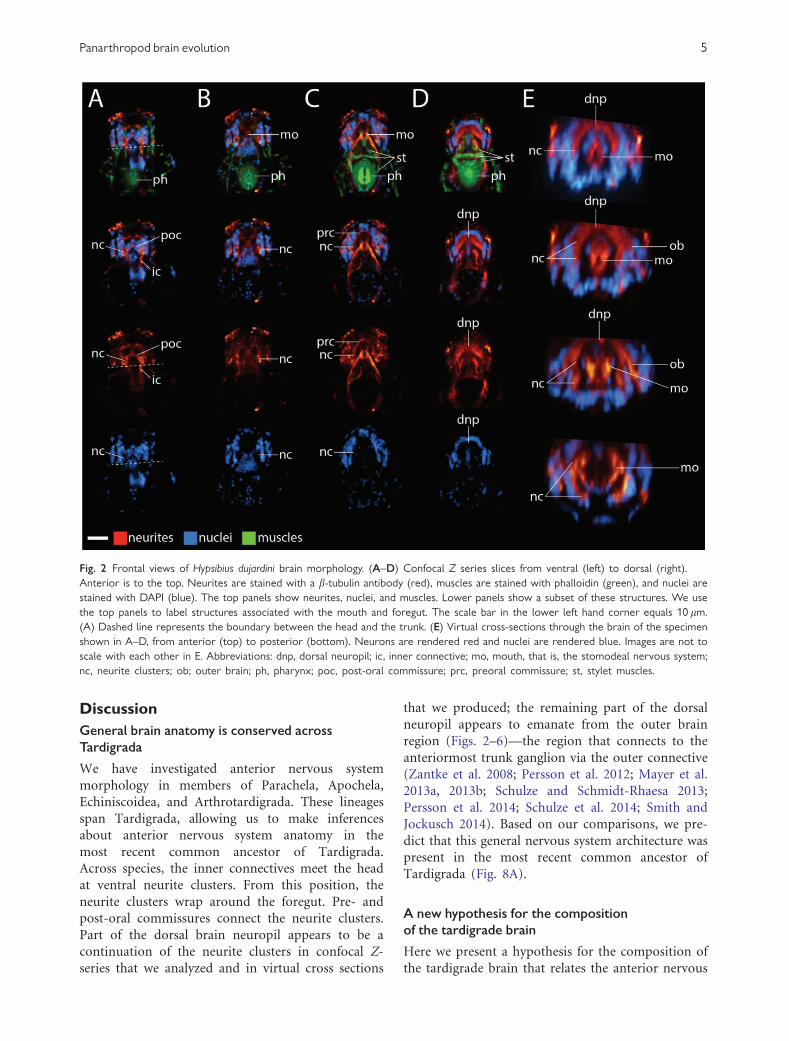