integrated labelling, dissociation, electrokinetic … · 2010-08-27 · and gel-based...
TRANSCRIPT

INTEGRATED LABELLING, DISSOCIATION, ELECTROKINETIC TRANSPORT AND DETECTION OF PRIMARY TUMOUR CELLS
Jane Woods,1 Peter T. Docker,1 Charlotte E. Dyer,2 Stephen. J. Haswell1 and John Greenman*2 1Department of Chemistry, University of Hull, UK, 2Centre for Biomedical Research, University of Hull, UK
ABSTRACT
Microfluidics, an enabling and emerging technology in cancer metastasis research, has yet to be applied in the analysis of individual cells taken directly from patient carcinomas. Accordingly, we have developed an integrated microfluidic methodology to label, dissociate, transport and detect patient-derived tumour cells. Following on-chip cold perfusion of tissue with antibody and enzymes, fluorescently-labelled cells were liberated and electrokinetically transported in a HEPES / sucrose agarose gel for downstream detection. Observed electrokinetic mobilities comprised dominant anodic electroosmotic flow (EOF) and opposing cathodic cell electrophoresis.
KEYWORDS: Microfluidic, metastasis, electroosmotic flow, cell electrophoresis, microflow cytometry, μFACS.
INTRODUCTION Metastasis research often employs flow cytometric analysis of cultured cell lines. However, results cannot be
extrapolated to explain in vivo tumour behaviour; even 3D co-cultures do not represent tissue complexity in which multiple cell populations interact with one another and with the extracellular matrix (ECM). An ideal starting point for flow cytometric assays would be patient biopsies, but conventional cell dissociation and labelling protocols involve multiple steps, each necessitating cell washing and centrifugation leading to cell losses and the potential introduction of methodological artefacts. Microfluidics lends itself to the integration of preanalytical sample processing, and furthermore electrokinetically-driven systems have the advantages of few moving parts and simple macro-to-micro interfacing. Early work on EOF cytometry explored the manipulation of bacteria, yeast and red blood cells [1]. Subsequently, bacteria have been stained on-chip with fluorescent dyes prior to EOF-enabled sorting [2], but for human cancer cell manipulation the convenience of EOF has yet to be exploited. We present here an integrated methodology that allows antibody-labelling of cells prior to dissociation from the tumour mass, followed by electrokinetic cell transport and detection (Figure 1).
Figure 1: Schematic of integrated microfluidic processes from biopsy to fluorescent cell detection.
THEORY
Diffusion time is governed by Fick’s law:
D2xt
2� (1)
where t is time, x is distance, and D is diffusion coefficient. For IgG1 and collagenase in tumour tissue, D is typically 10 μm2 s-1, and theoretical diffusion time to the centre of 1 mm3 of tumour tissue at 20 °C is ≈ 3.5 h. However, the effective rate of antibody diffusion is also influenced by antibody-antigen binding, decreasing inversely with antigen density [3]. Collagenase activity at 4 °C is relatively low; incubation at 4 °C for 24 h therefore preserves the tissue, enzymes and antibodies whilst permitting extensive perfusion of the ECM with enzymes and saturation of antibody-binding.
Apparent cellular electrokinetic velocity, vapp, is governed by the equation:
� ����� E
v cwapp
���
(2)
where ε is fluid dielectric constant, ζw zeta potential in the electric double layer (EDL) at the microchannel-fluid interface,
978-0-9798064-3-8/µTAS 2010/$20©2010 CBMS 1643 14th International Conference onMiniaturized Systems for Chemistry and Life Sciences
3 - 7 October 2010, Groningen, The Netherlands

ζc cellular zeta potential, η viscosity, and E electric field strength [4]. Cells in an electrolyte solution possess their own EDL; the negatively-charged membrane attracts a “screening cloud” of cations, causing cathodic electrophoretic flow (EPF) [4, 5, 6]. Cellular μapp equates empirically to vapp divided by E, and comprises both EOF bulk flow and EPF. Zwitterionic buffers increase ε with no concomitant increase in conductivity, thereby enhancing electrkinesis [7]. Similarly, low ionic strength solutions favour electrokinesis; EDL thickness is inversely proportional to square root of ionic strength, a requirement that precludes the conventional use of salts for cell isotonicity [5]. A further issue is buffer temperature sensitivity because of Joule heating. HEPES [4-(2-hydroxyethyl)-1-piperazineethanesulfonic acid] is a zwitterionic Good’s buffer with an apparent pKa of 7.4 equivalent to physiological pH, and a low temperature coefficient (dpKa / dT) of -0.014 / °C [8]. Consequently, a low ionic strength HEPES / sucrose mixture was selected for electrokinetic cell manipulation, and used as agarose gel both to provide some resistance against hydrodynamic back flow, and also for its physical similararity to ECM.
EXPERIMENTAL Glass microfluidic devices were fabricated using standard photolithography and wet etching techniques to produce the
design shown in Figure 2. Channels 70 μm deep and 150 μm wide were etched in 1 mm borofloat glass. A 3 mm diameter tissue chamber and 1.5 mm diameter holes for sample, reagent and electrode access were drilled in a 3 mm glass top plate before thermally bonding it to the etched base plate to form the device.
Figure 2: Microfluidic device. a) Scaled schematic showing channel layout, tissue chamber & access ports A – D.
b) Photograph of device with reservoirs attached. c) Micrograph of 50 μm mesh cell strainer in tissue chamber.
Channels were filled with 0.25 % (w / v) low melting point (LMP) agarose gel [A9419 Sigma-Aldrich, UK], which was made using 0.025 M HEPES / 0.25 M sucrose. Anonymised tumour samples were obtained from patients undergoing surgery, having gained ethical approval from Hull and East Yorkshire Research Ethics Committee (07 / H1304) and the Hull and East Yorkshire Hospitals NHS Trust (RO568). Samples were stored in Dulbecco's modified Eagle's medium [DMEM; E15-009, PAA Laboratories Ltd, UK] at 4 °C, and if not used immediately, snap frozen and cryopreserved in liquid nitrogen at -196 °C. Tumour tissue (~1 mm3) was placed upon a 50 μm cell strainer [340632, BD Falcon, UK] secured within the tissue chamber (as shown in Figure 2c) and submerged in 200 μl DMEM containing collagenases together with antibody against integrin subunit β1 (implicated in metastasis). The device was incubated at 4 °C for 24 h. Excess enzyme / antibody mixture was then replaced with buffer, and the device incubated at 37 °C for 30 min, before being placed upon the stage of an inverted fluorescence microscope [Zeiss Axiovert S-100]. Once cell motion had subsided, an electric field was applied via 0.5 mm Pt electrodes submerged in reservoirs A and B (Figure 2a & 2b) and gel-based electrokinesis employed to transport the dissociated labelled cells along the microchannel. Image sequences were captured by monochrome CCD camera [Hamamatsu Orca ER] and analysed in ImageJ to derive vapp and μapp for tumour cells labelled and dissociated as above, and also (in separate experiments) for unlabelled K562 cells.
RESULTS AND DISCUSSION
Following removal of the perfusate, incubation at 37 °C increased enzyme activity within the tissue; ECM fibres were digested and labelled cells released, whereupon the flow of dissociated cells through the cell strainer was assisted by gravity and hydrostatic pressure. Concurrently, gel in the channel began to melt, permitting cells to enter the macropores. Antibody-labelling of cell membrane-expressed cancer-associated targets was thus achieved in situ during interaction with neighbouring cells and tumour-specific ECM, providing a more accurate reflection of in vivo tumour cell phenotypes compared with conventional protocols. Furthermore, Figure 3 shows that cells in the channel remain fluorescently-labelled and detectable above the background antibody during electrokinesis.
HEPES / sucrose gel facilitated minimal hydrodynamic back pressure during EOF, and also consistently generated anodic EOF conditions. Cell velocities comprised both dominant EOF bulk flow and opposing cell electrophoresis. Labelled fragments – with greater charge-to-size ratios – were observed moving against the bulk flow by dominant electrophoresis. For intact labelled cells, mean μapp was -0.8 x 10⁻⁸ m2 (V s)-1. In contrast, experiments using unlabelled cells resulted in mean μapp > -2.0 x 10⁻⁸ m2 (V s)-1
Cells with bound conjugated antibody have increased cellular zeta potentials and greater charge to size ratios than unlabelled cells [6]. Labelled cells are consequently more electrophoretically mobile than unlabelled cells and, in anodic EOF, are impeded to a greater extent.
1644
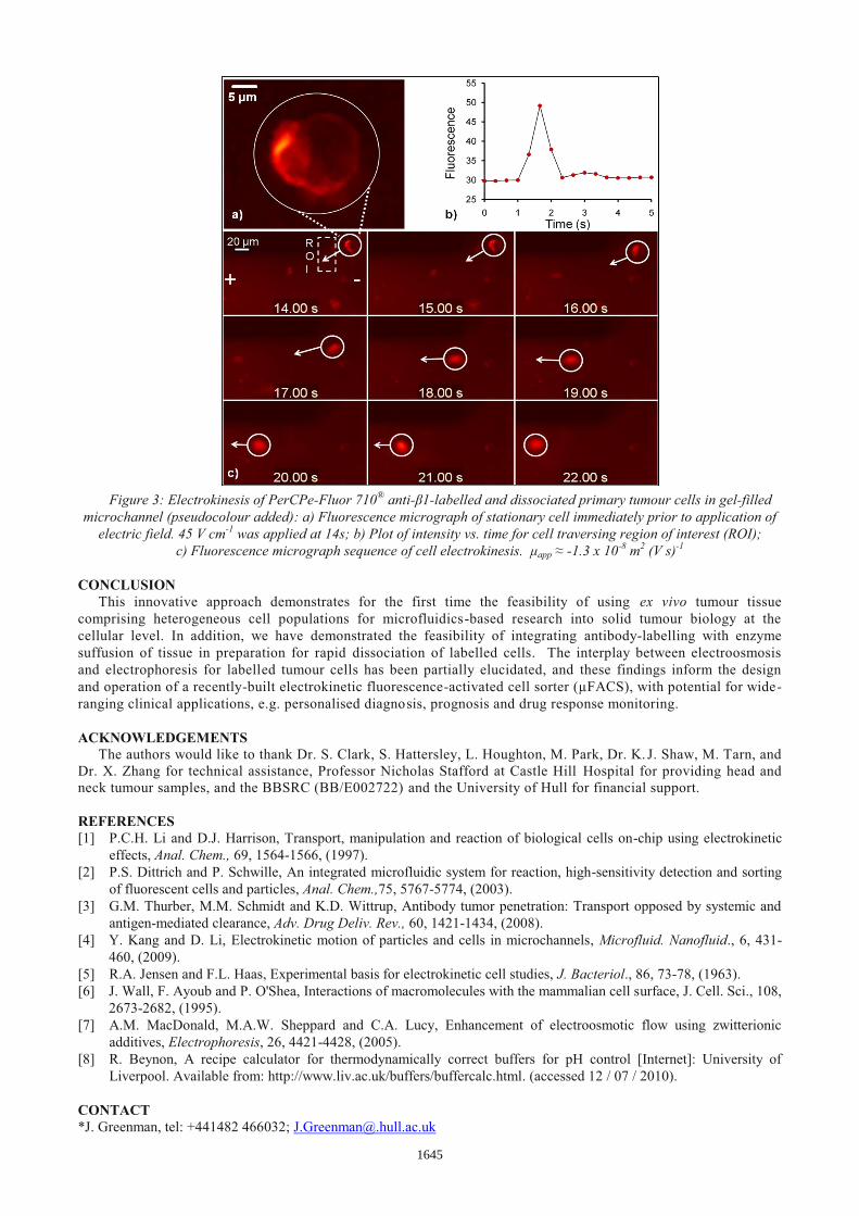
Figure 3: Electrokinesis of PerCPe-Fluor 710® anti-β1-labelled and dissociated primary tumour cells in gel-filled
microchannel (pseudocolour added): a) Fluorescence micrograph of stationary cell immediately prior to application of electric field. 45 V cm-1 was applied at 14s; b) Plot of intensity vs. time for cell traversing region of interest (ROI);
c) Fluorescence micrograph sequence of cell electrokinesis. μapp ≈ -1.3 x 10⁻⁸ m2 (V s)-1
CONCLUSION This innovative approach demonstrates for the first time the feasibility of using ex vivo tumour tissue
comprising heterogeneous cell populations for microfluidics-based research into solid tumour biology at the cellular level. In addition, we have demonstrated the feasibility of integrating antibody-labelling with enzyme suffusion of tissue in preparation for rapid dissociation of labelled cells. The interplay between electroosmosis and electrophoresis for labelled tumour cells has been partially elucidated, and these findings inform the design and operation of a recently-built electrokinetic fluorescence-activated cell sorter (µFACS), with potential for wide-ranging clinical applications, e.g. personalised diagnosis, prognosis and drug response monitoring.
ACKNOWLEDGEMENTS
The authors would like to thank Dr. S. Clark, S. Hattersley, L. Houghton, M. Park, Dr. K. J. Shaw, M. Tarn, and Dr. X. Zhang for technical assistance, Professor Nicholas Stafford at Castle Hill Hospital for providing head and neck tumour samples, and the BBSRC (BB/E002722) and the University of Hull for financial support.
REFERENCES [1] P.C.H. Li and D.J. Harrison, Transport, manipulation and reaction of biological cells on-chip using electrokinetic
effects, Anal. Chem., 69, 1564-1566, (1997). [2] P.S. Dittrich and P. Schwille, An integrated microfluidic system for reaction, high-sensitivity detection and sorting
of fluorescent cells and particles, Anal. Chem.,75, 5767-5774, (2003). [3] G.M. Thurber, M.M. Schmidt and K.D. Wittrup, Antibody tumor penetration: Transport opposed by systemic and
antigen-mediated clearance, Adv. Drug Deliv. Rev., 60, 1421-1434, (2008). [4] Y. Kang and D. Li, Electrokinetic motion of particles and cells in microchannels, Microfluid. Nanofluid., 6, 431-
460, (2009). [5] R.A. Jensen and F.L. Haas, Experimental basis for electrokinetic cell studies, J. Bacteriol., 86, 73-78, (1963). [6] J. Wall, F. Ayoub and P. O'Shea, Interactions of macromolecules with the mammalian cell surface, J. Cell. Sci., 108,
2673-2682, (1995). [7] A.M. MacDonald, M.A.W. Sheppard and C.A. Lucy, Enhancement of electroosmotic flow using zwitterionic
additives, Electrophoresis, 26, 4421-4428, (2005). [8] R. Beynon, A recipe calculator for thermodynamically correct buffers for pH control [Internet]: University of
Liverpool. Available from: http://www.liv.ac.uk/buffers/buffercalc.html. (accessed 12 / 07 / 2010).
CONTACT *J. Greenman, tel: +441482 466032; [email protected]
1645