insulin receptor substrate-l (irs-i) distribution in the ... receptor substrate-l (irs-i)...
TRANSCRIPT
The Journal of Neuroscience, November 1994, 14(11): 6412-6422
Insulin Receptor Substrate-l (IRS-I) Distribution in the Rat Central Nervous System
Franc0 FoIli,’ Luca Bonfanti,* Eric Renard,’ C. Ronald Kahn,’ and Adalbetto Merighi’
‘Research Division, Joslin Diabetes Center, Department of Medicine, Brigham and Women’s Hospital, and Harvard Medical School, Boston, Massachusetts 02215 and *Dipartimento di Morfofisiologia Veterinaria, UniversitA di Torino, l-10126 Torino, Italy
Insulin receptor substrate 1 (IRS-l) is the primary cytosolic substrate of the insulin and insulin-like growth factor-l (IGF- I) receptors. Following tyrosine phosphorylation IRS-1 binds to and activates specific proteins containing SH2 domains. Using biochemical and immunocytochemical techniques, we have mapped the distribution of IRS-1 in the CNS of the adult rat and compared it with that of insulin and IGF-I receptors and phosphatidylinositol 3-kinase (PI-3 kinase), a signaling molecule functionally related to IRS-l. lmmunoprecipitation and Western blotting experiments demonstrate the pres- ence of substantial amounts of IRS-l, insulin receptor, and PI-3 kinase in the brain.
IRS-1 immunoreactivity is widely distributed in neurons from several areas of the brain and spinal cord. The cerebral cortex, the hippocampus, many hypothalamic and thalamic nuclei, the basal ganglia, the cerebellar cortex, the brainstem nuclei, and the lamina X of the spinal cord are particularly rich of immunopositive nerve cells. In these areas most of the neurons immunoreactive for IRS-1 are also stained by either anti-insulin receptor or anti-IGF-I receptor antibodies as well as PI-3 kinase antiserum. IRS-1 immunostaining was very weak or totally absent in neurons of the olfactory bulb, the supraoptic and paraventricular nuclei, the mesence- phalic trigeminal nucleus, and the granule cell layer of the cerebellum, despite the fact that these areas were immu- nolabeled with antibodies against insulin or IGF-I receptors and/or PI-3 kinase. These results show that neurons in the adult rat CNS are endowed with some of the components of the early signaling pathway for growth factors of the insulin/ IGF-I family, although IRS-1 has a distribution distinct from that of the two receptors.
[Key words: insulin receptor substrate 1, insulin-like growth factor I, insulin receptor kinase, insulin-like growth factor I receptor, phosphatidylinositol 3-kinase, CM, immunocyto- chemistry]
Received Dec. 6, 1993; revised Mar. 21, 1994; accepted Apr. 13, 1994. This work was partly supported by grants of the Italian Consiglio Nazionale
delle Ricerche and Minister0 dell’Universitll e della Ricerca Scientifica e Tecnol- ogica and by NIH Grants DK 3 1036 and 3320 1. F.F. was supported by Dottorato di Ricerca (Fisiopatologia Clinica), Universita di Milano, Milano, Italy. We are greatly indebted to Drs. Wang and Liu for anti-IGF-I receptor antibodies, and Dr. Gammeltofi for stimulating discussion.
Correspondence should be addressed to Dr. Adalberto Merighi, Dipartimento di Morfofisiologia Veterinaria, Via Nizza 52, I-10126 Torino, Italy.
Copyright 0 1994 Society for Neuroscience 0270-6474/94/146412-l 1$05.00/O
Insulin effects at the cellular level are initiated by ligand binding to the a-subunit of the insulin receptor and activation of the tyrosine kinase present in the P-subunit (Massague et al., 198 1; Kasuga et al., 1982). Insulin receptor activation leads to tyrosyl phosphorylation of a cytosolic protein with an apparent molec- ular weight of 160-185 kDa named insulin receptor substrate 1 (IRS- 1) both in vivo and in vitro (White et al., 1985; Rothenberg et al., 1991; Sun et al., 199 1, 1992; Hadari et al., 1992; Saad et al., 1992). IRS-l is highly serine phosphorylated in the basal state and undergoes rapid tyrosine phosphorylation in response to insulin, insulin-like growth factor I (IGF-I), and, in some cells, interleukin 4 (IL-4) (White et al., 1985; Sun et al, 1991; Myers et al., 1993; Wang et al., 1993a,b). One mechanism by which IRS- 1 transmits insulin signaling inside the cells is bind- ing of the molecule to SH2-SH3 containing proteins via specific YMXM/YXXM motifs (Cantley et al., 199 1; Koch et al., 199 1; Folli et al., 1992; Myers and White., 1993; Sun et al., 1993). Among these SH2 domain proteins, one of the better charac- terized is the regulatory subunit of phosphatidylinositol-3 kinase (PI-3 kinase), also referred to as ~85. PI-3 kinase activity is stimulated by a variety of growth factors, including insulin, IGF- I, NGF, epidermal growth factor (EGF), and following cellular transformation (Whitman et al., 1985; Endeman et al., 1990; Soltoff et al., 1992; Cantley et al., 1991; Hiles et al., 1992; Lapetina et al., 1992; Obermeier et al., 1993). After insulin stimulation either in vivo or in vitro, PI-3 kinase associates with specific YMXM/YXXM motifs in IRS-l and is activated by this association (Sun et al., 199 1, 1992; Backer et al., 1992a; Folli et al., 1992; Hadari et al., 1992).
Insulin, IGF-I and IGF-II receptors, are widely expressed in both the developing and adult mammalian CNS, as demon- strated by autoradiography and immunocytochemistry (Posner et al., 1974; Havrankova et al., 1978; Gammeltoft et al., 1985, 1990; Hill et al., 1986; Bohannon et al., 1988; Lesniak et al., 1988; Ocrant et al., 1988; Unger et al., 1989, 1991a,b; Moss et al., 1990; King and Baskin, 199 1). Likewise, biochemical and Northern blot analyses indicate that IRS- 1 and PI-3 kinase are also present in the CNS at high concentrations (Cohen et al., 1990; Skolnik et al., 1991; Sun et al., 1992; Araki et al., 1993; this study). However, the topographic and cellular distribution of these molecules in the brain has not yet been investigated. To shed some light on the possible role of these early compo- nents of the insulin/IGF-I signaling pathway in the CNS, we have mapped the distribution of IRS-l in the rat brain and spinal cord and compared it with the immunocytochemical lo- calization of PI-3 kinase, insulin receptors, and IGF-I receptors.

The Journal of Neuroscience, November 1994, 14(11) 6413
Materials and Methods A4uteriuls. Reagents for sodium dodecyl sulfate-polyacrylamide gel elec- trophoresis (SDS-PAGE) and immunoblotting were from Bio-Rad (Richmond, CA). Protein A-Sepharose 6MB was purchased from Phar- macia (Upsala, Sweden), 1251-protein A from ICN Biomedicals (Costa Mesa, CA); nitrocellulose paper (BA 85, 0.2 pm) from Schleicher and Schuell (Keene, NH), sodium amobarbital (Amytal) and human recom- binant insulin (Humulin R) from Lilly (Indianapolis, IN). All other reagents were from Sigma Chemical Co. (St. Louis,, MO).
Animals. Male rats (130-180 gm; n = 15) and mice (6-8 weeks; n = 4) were obtained from Charles River Breeding Laboratories Inc. (Wil- mington, MA). They were fed standard rodent chow and water ad li- bitum. All mice and five rats were used in insulin stimulation experi- ments in vivo, the remaining rats were employed for immunocytochemical analysis.
Antibodies. Anti-IRS-l antibodies were raised using two synthetic peptides (CT and NT) derived from the carboxy-terminal (TY- ASINFQKQPEDRQ) and the amino-terminal (MASPPDTDGF- SDVRKVGY) amino acid sequence of the rat liver IRS- 1 protein (Sun et al., 1991). They were partially purified either with a protein A- Sepharose column (anti-CT-IRS- 1 antibodies; Folli et al., 1993) or on a peptide affinity column (anti-NT-IRS-l antibodies; Cheatham et al., 1993). Anti-insulin receptor (a-subunit) antibodies (Rosenzweig et al., 1990) were from UBI (Lake Placid, NY). Anti-insulin receptor (P-sub- unit) antibodies were raised in rabbits using a synthetic peptide derived from the carboxy-terminal amino acid sequence (KKNGRILTLPRSNPS) of the P-subunit of the rat insulin receptor. Anti-IGF-I receptor (@- subunit) antibodies (a kind sift of Drs. Wane and Liu. Mount Sinai Medical Center, New York, -NY) have been previously described and characterized (Liu et al., 1992). Polyclonal anti-phosphotyrosine anti- bodies were raised in rabbits and affinity purified on phosphotyramine columns, as previously described (Pang et al., 1985). The anti-rat PI-3 kinase polyclonal antiserum (raised against the 85 kDa subunit of the enzyme) was from UBI (Lake Placid, NY).
Biochemistry: insulin stimulation in vivo. Food was withdrawn 12- 14 hr before experiments; mice and rats were anesthetized with sodium amobarbital (15 m&kg of body weight, i.p) and were used 10-15 min later. The abdominal cavitv was onened and 1 ml ofnormal saline (0.9% NaCl) with or without 205 pg of insulin was injected into the portal vein. The hindlimb skeletal muscles and the brain were excised re- spectively 1.5 and 2.5 min after insulin injection and frozen in liquid nitrogen. Frozen tissues were ground into a fine powder and immediately homogenized in ice cold solubilization buffer (1:6 w/v) with a Polytron PTA 20s generator (Brinkmann Instruments), as previously described (Folli et al., 1992). Insoluble material was removed by centrifugation at 50,000 rpm in a 70Ti rotor (Beckman) for 50 min. One milliliter aliquots of the supernatants (protein concentration, 10 mg/ml) were used as a sample for immunoprecipitation with anti-insulin receptor o-subunit (0.5 &ml), anti-NT-IRS-l (0.2 &ml), and anti-PI-3 kinase (whole antiserum, 1:200 final dilution) antibodies and protein A-Se- pharose. Samples were then processed for SDS-PAGE and Western blotting as previously reported (Folli et al., 1992). Blots were finally incubated with anti-phosphotyrosine (0.3 &ml), anti-insulin receptor P-subunit (0.5 rug/ml), anti-IRS-l (anti-CT-IRS-l, 9 &ml), or anti- PI-3 kinase antibodies (whole antiserum, 1:200 final dilution), ‘Z51-pro- tein A and subjected to autoradiography. Protein determination was performed by the Bradford dye method (Bio-Rad, UK) with bovine serum albumin as standard (Bradford, 1976).
Immunocytochemistry. Rats were anesthetized with sodium pento- barbitone (> 60 mg/ 100 gm body weight, i.p.) and perfused through the aorta with Bouin’s solution. The brain and segments of cervical, tho- racic, lumbar, and sacral spinal cord were dissected out, cut in pieces, and placed in the same fixative for 6 additional hours. Following de- hydration and wax embedding, coronal sections (8 pm) representative of the entire CNS were mounted onto poly-L-lysine-coated slides. After inhibition of endogenous peroxidase activity and preincubation in 0.1 M phosphate-buffered saline pH 7.4 (PBS) containing 1% egg albumin (Sigma Chemicals Co., St. Louis, MO), sections were incubated over- night at room temperature with anti-NT-IRS- 1 (1:500 affinity-purified antiserum), anti-insulin receptor (a-subunit, 1:50), or anti-IGF-I re- ceptor (@-subunit, 1: 100) antibodies. They were subsequently processed according to the avidin-biotin-peroxidase procedure (ABC. Vector, UK), and the peroxidase reaction was developed using the glucose oxidase nickel-DAB method (Shu et al., 1988). Immunocytochemical controls consisted of (1) liquid phase adsorption of the anti-NT-IRS- 1 peptide
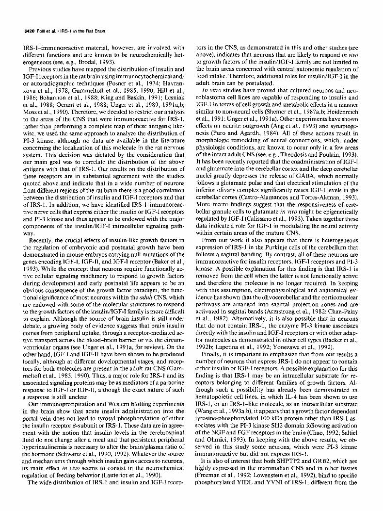
Brain Muscle Liver A. I--
116 Mr
IR +
1 2 3
B.
IRS-1 -)
C.
PI 3-K -)
205 Mr
116
.116 Mr
77
6 7 8
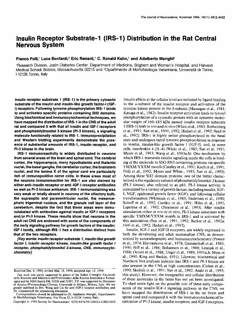
Figure I. Western blot analysis of insulin receptor P-subunit in anti- insulin receptor antibody (IR-CT) immunoprecipitates (A), IRS- 1 pro- tein in anti-NT-IRS- 1 antibody immunoprecipitates (B), and PI-3 ki- nase (85 kDa subunit) in anti-PI-3 kinase antiserum immunoprecipi- tates (c). Proteins from rat brain. skeletal muscle and liver were extracted as described in Materials and Methods. After centrifugation, aliquots containing the same amount of proteins were incubated with anti-in- sulin receptor (IR-CT), anti-NT-IRS- 1 or anti-PI-3 kinase antibodies and protein A-Sepharose. Immunoprecipitated proteins were analyzed by Western blotting with either anti-insulin receptor antibodies (IR- CT), anti-CT-IRS- 1 antibodies, or anti-PI-3 kinase antiserum and 12sI- protein A and subjected to autoradiography. Lanes I, 4, and 6, brain; lanes 2, 5, and 7, muscle; lanes 3 and 8, liver. IR, insulin receptor; PI 3-K, PI-3 kinase.
affinity-purified antibody with different concentrations of the synthetic peptide used to raise the antibody; (2) liquid phase adsorption of the anti-NT-IRS-l peptide affinitv-ourified antihodv with different con- centrations of the synthetic CT-IRS- 1 peptide (Folli et al., 1993) and synthetic Pep-80 peptide (Rothenberg et al., 1991); (3) omission of primary antisera or their substitution with normal serum; and (4) omis- sion of second-layer biotinylated antibodies or avidin-biotin-peroxi- dase complex in the ABC procedure.
Results Biochemistry In order to examine the relative level of expression of insulin receptor, IRS-l, and PI-3 kinase in rat brain as compared to two classical targets of the metabolic actions of insulin, that is, skeletal muscle and liver, immunoprecipitation was followed
6414 Folli et al. * IRS-1 in the Rat Brain
by Western blotting employing antibodies directed against the P-subunit of the insulin receptor (Fig. lA), the IRS-l protein (Fig. lB), and the 85 kDa subunit of PI-3 kinase (Fig. 1 C). This experiment suggests that all three proteins are present in adult rat CNS at similar concentrations as compared to muscle and liver. Western blot analysis of tyrosyl-phosphorylated proteins in anti-insulin receptor P-subunit immunoprecipitates from rat brain and muscle extracts confirmed that following intraportal insulin administration, there was no increase in the phosphor- ylation of the insulin receptor or IRS- 1 in the brain, while there was a clear stimulation of the tyrosyl-phosphorylation of these proteins in skeletal muscle (data not shown).
lmmunocytochemistry General considerations on IRS- 1 immunolocalization IRS-I immunoreactivity was detected in most nerve cells throughout the CNS, but not in all neurons of the brain and spinal cord. IRS- 1 immunostaining was usually restricted to the neuronal perikarya (Figs. 2-4). However, in certain brain regions, such as the cerebellar cortex and many brainstem nuclei, con- siderable amounts of reaction product were observed in the initial portion of the primary dendrites. This was particularly true for large neurons, in which immunostaining was often stronger in the dendrites than in the cell body (Fig. 4A). Although quantitative evaluations could not be performed, the staining intensity was different between the various IRS- 1 -positive neu- ronal populations; for example, it was consistently high in hip- pocampal neurons and very faint in magnocellular supraoptic nerve cells.
Except the ependymal cells lining the third ventricle and cells of the choroid plexus, neurons were the only cells in the brain and spinal cord expressing immunoreactive IRS-l. Glial cells and other non-neuronal elements had no detectable IRS- 1 pro- tein. The specific labeling of neurons by the anti-IRS-l anti- serum was completely abolished after liquid phase adsorption with its corresponding synthetic peptide.
Distribution of IRS-I immunoreactivity in the rat CNS Olfactory bulb. Neuronal cell bodies were usually immunone- gative in the olfactory bulb. Faint IRS-l staining was occasion- ally observed in the mitral cell perikarya and in neurons of the anterior olfactory nucleus. In the glomerular layer a fine punctate reaction was present, likely due to the olfactory nerve terminals.
Cerebral cortex. IRS- 1 immunoreactivity was detected in all the cortical areas, but it was particularly prominent in the pir- iform cortex. Immunoreactive nerve cells were found in all cor- tical layers, and there was no obvious difference between layers
in the density of positive cells (Fig. 2C). The reaction product was generally restricted to the cell bodies of the pyramidal neu- rons but, occasionally, it was also found in the initial part of their apical dendrites.
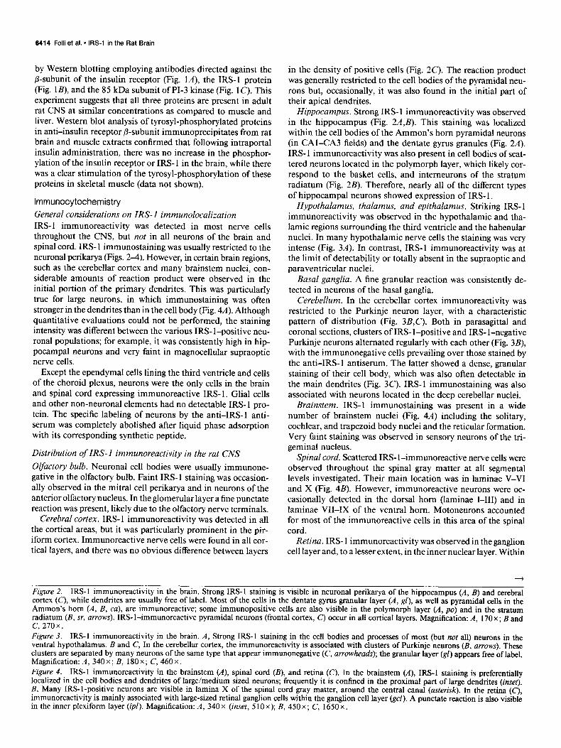
Hippocampus. Strong IRS-l immunoreactivity was observed in the hippocampus (Fig. 2AJ). This staining was localized within the cell bodies of the Ammon’s horn pyramidal neurons (in CAI-CA3 fields) and the dentate gyrus granules (Fig. 2A). IRS- 1 immunoreactivity was also present in cell bodies of scat- tered neurons located in the polymorph layer, which likely cor- respond to the basket cells, and interneurons of the stratum radiatum (Fig. 2B). Therefore, nearly all of the different types of hippocampal neurons showed expression of IRS- 1.
Hypothalamus, thalamus, and epithalamus. Striking IRS-l immunoreactivity was observed in the hypothalamic and tha- lamic regions surrounding the third ventricle and the habenular nuclei. In many hypothalamic nerve cells the staining was very intense (Fig. 3A). In contrast, IRS-l immunoreactivity was at the limit of detectability or totally absent in the supraoptic and paraventricular nuclei.
Basal ganglia. A fine granular reaction was consistently de- tected in neurons of the basal ganglia.
Cerebellum, In the cerebellar cortex immunoreactivity was restricted to the Purkinje neuron layer, with a characteristic pattern of distribution (Fig. 3B,C). Both in parasagittal and coronal sections, clusters of IRS- 1 -positive and IRS- l-negative Purkinje neurons alternated regularly with each other (Fig. 3B), with the immunonegative cells prevailing over those stained by the anti-IRS-l antiserum. The latter showed a dense, granular staining of their cell body, which was also often detectable in the main dendrites (Fig. 3C). IRS-l immunostaining was also associated with neurons located in the deep cerebellar nuclei.
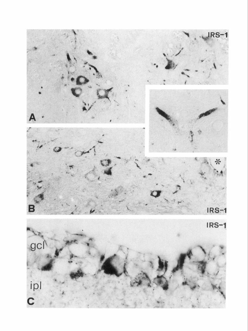
Brainstem. IRS-l immunostaining was present in a wide number of brainstem nuclei (Fig. 4A) including the solitary, cochlear, and trapezoid body nuclei and the reticular formation. Very faint staining was observed in sensory neurons of the tri- geminal nucleus.
Spinal cord. Scattered IRS- 1-immunoreactive nerve cells were observed throughout the spinal gray matter at all segmental levels investigated. Their main location was in laminae V-VI and X (Fig. 4B). However, immunoreactive neurons were oc- casionally detected in the dorsal horn (laminae I-III) and in laminae VII-IX of the ventral horn. Motoneurons accounted for most of the immunoreactive cells in this area of the spinal cord.
Retina. IRS- 1 immunoreactivity was observed in the ganglion cell layer and, to a lesser extent, in the inner nuclear layer. Within
Figure 2. IRS-l immunoreactivity in the brain. Strong IRS-l staining is visible in neuronal perikarya of the hippocampus (A, B) and cerebral cortex (C), while dendrites are usually free of label. Most of the cells in the dentate gyrus granular layer (A, gl), as well as pyramidal cells in the Ammon’s horn (A, B, ca), are immunoreactive; some immunopositive cells are also visible in the polymorph layer (A, po) and in the stratum radiatum (B, sr, arrows). IRS- I-immunoreactive pyramidal neurons (frontal cortex, c) occur in all cortical layers. Magnification: A, 170 x; B and C, 2.70 x Figure 3. IRS-I immunoreactivity in the brain. A, Strong IRS-l staining in the cell bodies and processes of most (but not all) neurons in the ventral hypothalamus. B and C, In the cerebellar cortex, the immunoreactivity is associated with clusters of Purkinje neurons (B, arrows). These clusters are separated by many neurons of the same type that appear immunonegative (C, arrowheads); the granular layer (gl) appears free oflabel. Magnification: A, 340 x ; B, 180 x ; C, 460 x . Figure 4. IRS-l immunoreactivity in the brainstem (A), spinal cord (B), and retina (C). In the brainstem (A), IRS-l staining is preferentially localized in the cell bodies and dendrites of large/medium sized neurons; frequently it is confined in the proximal part of large dendrites (inret). B, Many IRS-l-positive neurons are visible in lamina X of the spinal cord gray matter, around the central canal (asterisk). In the retina (C), immunoreactivity is mainly associated with large-sized retinal ganglion cells within the ganglion cell layer (gel). A punctate reaction is also visible in the inner plexiform layer (ipl). Magnification: A, 340x (inset, 510x); B, 450x; C, 1650x.


The Journal of Neuroscience, November 1994, 14(11) 6419
the former, staining was associated with the cell bodies of retinal ganglion cells, mainly of large diameter (Fig. 4C). In the inner nuclear layer a faint IRS- 1 positivity was present in some ama- crine and bipolar cells.
Table 1. Correlation between the immunocytochemical distribution of IRS-l and other components of the insulin/IGF-I signaling pathway, in the rat CNS
Distribution of insulin receptor, IGF-I receptor, and PI-3 kinase in the rat CNS
CNS regions/nuclei IRS- 1
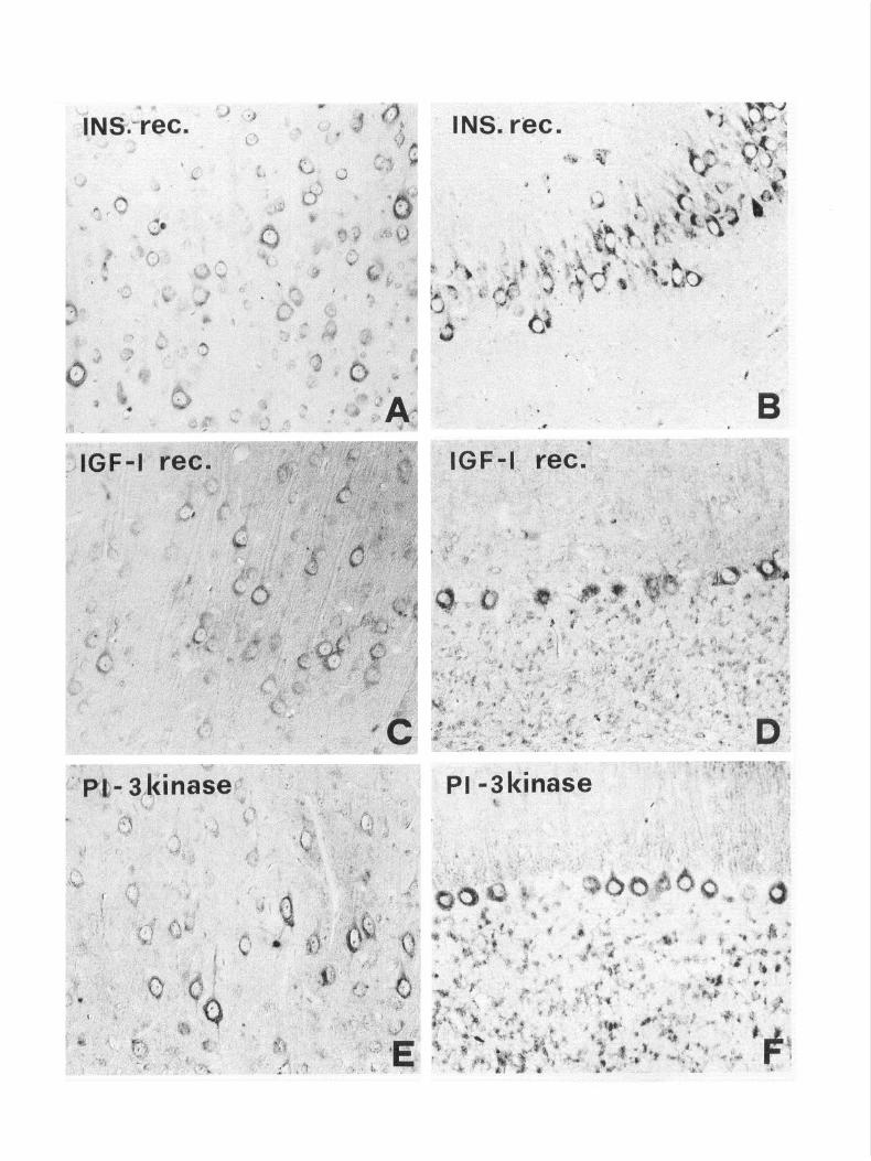
In most of the areas investigated, a good correlation between the distribution of IRS- 1 and insulin receptor immunoreactiv- ities was found, with almost overlapping patterns within the cerebral cortex, hippocampus, and brainstem (Fig. 5). Only mi- nor differences in the distribution of insulin receptor immu- nostaining were observed, such as, for example, in the cerebral cortex, where staining for the insulin receptor was particularly evident within layer IV and the hippocampus, where insulin receptor immunoreactivity was very weak in the dentate gyrus. In other regions of the brain, on the other hand, significant differences in the localization of IRS-l and insulin receptor immunoreactivities were observed. The latter was very strong in the olfactory bulb and the hypothalamic supraoptic nucleus, which, as described above, contained little or no detectable IRS- 1. In the thalamus, hypothalamic nuclei and septal nuclei, in- sulin receptor immunostaining was preferentially associated with axons and terminals, rather than with the neuronal perikarya. In the cerebellar cortex, Purkinje neurons were only weakly reactive after incubation with anti-insulin receptor antibodies.
The distributions of IGF-I receptor and PI-3 kinase immu- noreactivities were very similar and overlapped that of IRS-l in the cerebral cortex (Fig. 5CJ) and hippocampus. As for the insulin receptor, staining was particularly prominent in cortical layer IV. In the hippocampus, IGFl -receptor immunoreactivity was stronger in the Ammon’s horn than in the dentate gyrus. In the cerebellar cortex, IGF-I receptor and PI-3 kinase im- munoreactivities were detected in almost all Purkinje neurons, without the characteristic pattern observed after incubation with the anti-IRS-l antiserum (Fig. 5DJ). Moreover, weak IGF-I receptor staining was detected in the granular layer (Fig. 5D). Differences in the pattern of distribution of IGF-I receptor, PI-3 kinase and IRS-l immunoreactivities were not obviously ap- parent in the brainstem and spinal cord. These results are sum- marized in Table 1.
Olfactory bulb No Cerebral cortex” Yes Hypothalam<hala-
mus Yes SON/PVN magnocellu-
lar neurons No Basal ganglia Yes Cerebellar cortex
Purkinje cell layer Yes Granule cell layer No
Deep cerebellar nuclei Yes Brainstem’ Yes Mesencephalic trigemi-
nal nucleus No Spinal cord Yes Retina Yes
Insulin IGF-I PI-3 receptor receptor kinase
Yes No No Yes Yes Yes
Yes No Yes
No Yes
Yes Yes
Yes Yes
Yes* No Yes Yes
Yes+ Yes No Yes
Ye+ Yes Yes Yes
No Yes ND
Yes Yes ND
Yes Yes Yes
Yes, presence of immunoreactivity; no, absence of immunoreactivity or very weak immunolabeling; ND, not done. IRS- 1, insulin receptor substrate- 1; IGF-I, in- sulin-like growth factor I; PI-3 kinase, phosphatidylinositol-3 kinase; SON, su- praoptic nucleus; PVN, paraventricular nucleus.
a Differences were observed in layer IV (see text). b Other than SON/PVN magnocellular neurons. c Immunostaining was associated with fibers rather than cell bodies.
d Staining was observed in all Purkinje neurons. * Other than the mesencephalic trigeminal nucleus.
Discussion
lular localization of IRS- 1 and PI-3 kinase in the rat brain and spinal cord and confirms the existence of insulin and IGF-I receptors in the CNS (Posner et al., 1974; Havrankova et al., 1978; Gammeltoft et al., 1985, 1990; Hill et al., 1986; Bohannon et al., 1988; Lesniak et al., 1988; Ocrant et al., 1988; Unger et al., 1989, 1991a,b; Moss et al., 1990; King and Baskin, 1991). The unique nature of the distribution of IRS-l in the rat CNS and the possible functional implications of our findings, which suggest that several neuronal populations are capable of re- sponding in vivo to insulin, IGF-I, and, possibly, other growth factor stimulation, deserve further consideration.
Protein tyrosine kinases can be subdivided into two large fam- Our biochemical and immunocytochemical results demon- ilies: the transmembrane growth factor receptors family and the strate that IRS- 1 is widely distributed in the rat brain and spinal cytosolic nonreceptor family. These proteins are commonly re- cord. In particular IRS-l protein expression data are in agree- garded as fundamental components of mitogenic pathways and ment with the results obtained from quantitative PCR of IRS- 1 are highly expressed in non-nervous tissues where they appear mRNA from human fetal tissues (Araki et al., 1993). High levels to play a crucial role in the control of cell growth and metabolism of immunoreactivity are found in the cerebral and cerebellar in both physiologic and pathologic states (Cantley et al., 199 1). cortices, the hippocampus and several brainstem nuclei. It is of Nevertheless, many observations indicate the presence of sub- interest that within some of these areas, such as the cerebral stantial amount of tyrosine kinase activity in the CNS, which cortex, IRS- 1 seems to be detectable, although at different levels, is typically composed of nonproliferating cells (Naim et al., in virtually all neurons. Conversely, in other regions of the CNS, 1983; Hunter, 1987). Using the biochemical and immunocy- such as the cerebellum, the molecule is clearly limited to certain tochemical approach, this study describes the presence and cel- neuronal subpopulations. The nerve cells that are positive for
t
Figure 5. Immunoreactivity for the insulin (4, B) and IGF-I (C, D) receptors, and PI-3 kinase (E, F) in some of the brain regions that are highly immunoreactive for IRS- 1. In the cerebral cortex (A, C, E), immunoreactivity to the three antigens is visible in the cytoplasm of pyramidal neurons. In the hippocampus (B), a strong staining for insulin receptor is present in the cell bodies of the Ammon’s horn pyramidal neurons. In the cerebellar cortex, the perikarya of Purkinje neurons and granule cells are immunoreactive for IGF-I (0) and PI-3 kinase (0. Magnification: A, C, D, F, 280 x ; B, 235x; E, 335x.
6420 Folli et al. l IRS-l in the Rat Brain
IRS- I-immunoreactive material, however, are involved with different functions and are known to be neurochemically het- erogeneous (see, e.g., Brodal, 1993).
Previous studies have mapped the distribution of insulin and IGF-I receptors in the rat brain using immunocytochemical and/ or autoradiographic techniques (Posner et al., 1974; Havran- kova et al., 1978; Gammeltoft et al., 1985, 1990; Hill et al., 1986; Bohannon et al., 1988; Ring and Baskin, 1991; Lesniak et al., 1988; Ocrant et al., 1988; Unger et al., 1989, 1991a,b, Moss et al., 1990). Therefore, we decided to restrict our analysis to the areas of the CNS that were immunoreactive for IRS-l, rather than performing a complete map of these antigens; like- wise, we used the same approach to analyze the distribution of PI-3 kinase, although no data are available in the literature concerning the localization of this molecule in the rat nervous system. This decision was dictated by the consideration that our main goal was to correlate the distribution of the above antigens with that of IRS-l. Our results on the distribution of these receptors are in substantial agreement with the studies quoted above and indicate that in a wide number of neurons from different regions of the rat brain there is a good correlation between the distribution of insulin and IGF-I receptors and that of IRS- 1. In addition, we have identified IRS- l-immunoreac- tive nerve cells that express either the insulin or IGF-I receptors and PI-3 kinase and thus appear to be endowed with the major components of the insulin/IGF-I intracellular signaling path- way.
Recently, the crucial effects of insulin-like growth factors in the regulation of embryonic and postnatal growth have been demonstrated in mouse embryos carrying null mutations of the genes encoding IGF-I, IGF-II, and IGF-I receptor (Baker et al., 1993). While the concept that neurons require functionally ac- tive cellular signaling machinery to respond to growth factors during development and early postnatal life appears to be an obvious consequence of the growth factor paradigm, the func- tional significance of most neurons within the adult CNS, which are endowed with some of the molecular structures to respond to the growth factors of the insulin/IGF-I family is more difficult to explain. Although the source of brain insulin is still under debate, a growing body of evidence suggests that brain insulin comes from peripheral uptake, through a receptor-mediated ac- tive transport across the blood-brain barrier or via the circum- ventricular organs (see Unger et al., 199 1 a, for review). On the other hand, IGF-I and IGF-II have been shown to be produced locally, although at different developmental stages, and recep- tors for both molecules are present in the adult rat CNS (Gam- meltoft et al., 1985, 1990). Thus, a major role for IRS- 1 and its associated signaling proteins may be as mediators of a paracrine response to IGF-I or IGF-II, although the exact nature of such a response is still unclear.
Our immunoprecipitation and Western blotting experiments in the brain show that acute insulin administration into the portal vein does not lead to tyrosyl phosphorylation of either the insulin receptor p-subunit or IRS- 1. These data are in agree- ment with the notion that insulin levels in the cerebrospinal fluid do not change after a meal and that persistent peripheral hyperinsulinemia is necessary to alter the brain/plasma ratio of the hormone (Schwartz et al., 1990, 1992). Whatever the source and mechanisms through which insulin gains access to neurons, its main effect in vivo seems to consist in the neurochemical regulation of feeding behavior (Lauteriot et al., 1990).
The wide distribution of IRS- 1 and insulin and IGF-1 recep-
tors in the CNS, as demonstrated in this and other studies (see above), indicates that neurons that are likely to respond in vivo to growth factors of the insulin/IGF-I family are not limited to the brain areas concerned with central autonomic regulation of food intake. Therefore, additional roles for insulin/IGF-I in the adult brain can be postulated.
In vitro studies have proved that cultured neurons and neu- roblastoma cell lines are capable of responding to insulin and IGF-I in terms of cell growth and metabolic effects in a manner similar to non-neural cells (Shemer et al., 1987a,b; Heidenreich et al., 199 1; Unger et al., 199 1 a). Other experiments have shown effects on neurite outgrowth (Ang et al., 1993) and synaptoge- nesis (Puro and Agardh, 1984). All of these actions result in morphologic remodeling of neural connections, which, under physiologic conditions, are known to occur only in a few areas of the intact adult CNS (see, e.g., Theodosis and Poulain, 1993). It has been recently reported that the coadministration of IGF-I and glutamate into the cerebellar cortex and the deep cerebellar nuclei greatly depresses the release of GABA, which normally follows a glutamate pulse and that electrical stimulation of the inferior olivary complex significantly raises IGF-I levels in the cerebellar cortex (Castro-Alamancos and Torres-Aleman, 1993). More recent findings suggest that the responsiveness of cere- bellar granule cells to glutamate in vivo might be epigenetically regulated by IGF-I (Calissano et al., 1993). Taken together these data indicate a role for IGF-I in modulating the neural activity within certain areas of the mature CNS.
From our work it also appears that there is heterogeneous expression of IRS- 1 in the Purkinje cells of the cerebellum that follows a sagittal banding. By contrast, all of these neurons are immunoreactive for insulin receptors, IGF-I receptors and PI-3 kinase. A possible explanation for this finding is that IRS-l is removed from the cell when the latter is not functionally active and therefore the molecule is no longer required. In keeping with this assumption, electrophysiological and anatomical ev- idence has shown that the olivocerebellar and the corticonuclear pathways are arranged into sagittal projection zones and are activated in sagittal bands (Armstrong et al., 1982; Chan-Palay et al., 1982). Alternatively, it is also possible that in neurons that do not contain IRS-l, the enzyme PI-3 kinase associates directly with the insulin and IGF-I receptors or with other adap- tor molecules as demonstrated in other cell types (Backer et al., 1992b; Lapetina et al., 1992; Yonezawa et al., 1992).
Finally, it is important to emphasize that from our results a number of neurons that express IRS- 1 do not appear to contain either insulin or IGF-I receptors. A possible explanation for this finding is that IRS-l may be an intracellular substrate for re- ceptors belonging to different families of growth factors. Al- though such a possibility has already been demonstrated in hematopoietic cell lines, in which IL-4 has been shown to use IRS-l, or an IRS-l-like molecule, as an intracellular substrate (Wang et al., 1993a,b), it appears that a growth factor dependent tyrosine-phosphorylated 100 kDa protein other than IRS- 1 as- sociates with the PI-3 kinase SH2 domain following activation of the NGF and FGF receptors in the brain (Chao, 1992; Saltiel and Ohmici, 1993). In keeping with the above results, we ob- served in this study some neurons, which were PI-3 kinase immunoreactive but did not express IRS- 1.
It is also of interest that both SHPTP2 and GRB2, which are highly expressed in the mammalian CNS and in other tissues (Freeman et al., 1992; Lowenstein et al., 1992), bind to specific phosphorylated YIDL and YVNI of IRS-l, different from the
The Journal of Neuroscience, November 1994, 74(11) 6421
motifs that bind to PI-3 kinase (Sun et al., 1993). Thus, it is tempting to hypothesize that intracellular signaling pathways activated by phosphorylated IRS-1 may be different between the various subpopulations of immunoreactive neurons.
In conclusion, this study demonstrates that a large number of neurons in the adult CNS are endowed with many of the early components of the signal transduction pathway for recep- tor tyrosine kinases of the insulin/IGF-I growth factor family, although in a few areas of the brain the expression of IRS- 1 and PI-3 kinase does not parallel the distribution of the insulin and/ or IGF-I receptors. These results highlight the importance of correctly understanding the functional significance of receptor tyrosine kinases in cells that are postmitotic and undergoing terminal differentiation. Protein tyrosine kinases have been tra- ditionally regarded as cell-transforming agents, but a growing body of evidence suggests that in the adult brain they are more likely to be involved in sculpting and maintaining the synaptic circuitry and architecture of the mature nervous system (Chao, 1992; Grant et al., 1992; Saltiel and Ohmici, 1993).
References Ang LC, Bhaumick B, Jurlink BHJ (1993) Neurite promoting activity
of insulin, insulin-like growth factor I and nerve growth factor on spinal motor neurons is astrocyte dependent. Dev Brain Res 74:83- 88.
Araki E, Sun X-J, Haag BL, Chuang L-M, Zhang Y, Yang-Feng TL, White MF, Kahn CR (1993) Human skeletal muscle insulin receptor substrate-l. Characterization of the cDNA, gene, and chromosomal localization. Diabetes 42:1041-1054.
Armstrone DM. Camobell NC. Edalev SA. Schild RF. Trott JR (1982) Investigitions of the olivockreb&ar and spinocerebellar pathways. In: The cerebellum: new vistas (Palay SL, Chan-Palay V, eds), pp 195-232. Berlin: Springer.
Backer JM, Myers MC Jr, Shoelson SE, Chin DJ, Sun XJ, Miralpeix M, Hu P, Margolis B, Skolnik EY, Schlessinger J, White MF (1992a) Phosphatidylinositol3kinase is activated by association with IRS- 1 durine insulin stimulation. EMBO J 9:3469-3479.
Backer YM, Schroeder GG, Kahn CR, Myers MG Jr, Wilden PA, Cahill DA, White MF (1992b) Insulin stimulation of phosphatidylinositol 3-kinase activity maps to insulin receptor regions required for en- doaenous substrate phosphorylation. J Biol Chem 267: 1367-l 374.
Baker J, Liu J-P, Robertson EJ,Efstradiatis A (1993) Role of insulin- like arowth factor in embrvonic and postnatal growth. Cell 76:73-82.
Bohan&n NJ, Corp ES, Wilcox BJ, F&lewicz DP, Dorsa DM, Baskin DG (1988) Localization of binding sites for insulin-like growth factor-I (IGF-I) in the rat brain by quantitative autoradiography. Brain Res 444:205-2 13.
Bradford MM (1976) A rapid and sensitive method for the quanti- tation of microgram quantities of protein utilizing the principle of urotein dve bindins. Anal Biochem 72~248-254.
Brodal P (1993) Thi central nervous system: structure and function. London: Oxford UP.
Calissano P, Ciotti MT, Battistini L, Zona C, Angelini A, Merlo D, Mercanti D (1993) Recombinant human insulin-like growth factor I exerts a trophic action and confers glutamate sensitivity on gluta- mate-resistant cerebellar granule cells. Proc Nat1 Acad Sci USA 90: 8752-8756.
Cantley LC, Auger KR, Carpenter CL, Duckworth B, Graziani A, Ka- peller R, Soltoff S (1991) Oncogenes and signal transduction. Cell 64128 l-302.
Castro-Alamancos MA, Torres-Aleman I (1993) Long-term depres- sion of glutamate-induced r-aminobutyric acid release in cerebellum by insulin-like growth factor I. Proc Nat1 Acad Sci USA 90:7386- 7390.
Chan-Palay V, Palay SL, Wu J-Y (1982) Sagittal cerebellar microbands of taurine neurons: immunocytochemical demonstration by using an- tibodies against the taurine synthesizing enzyme cysteine sulfinic acid decarboxylase. Proc Nat1 Acad Sci USA 79:4221-4225.
Chao MV (1992) Neurotrophin receptors: a window into neuronal differentiation. Neuron 9:583-593.
Cheatham B, Shoelson SE, Yamada K, Goncalves E, Kahn R (1993)
Substitution of the erbB-2 oncoprotein transmembrane domain ac- tivates the insulin receptor and modulates the action of insulin and insulin-receptor substrate 1. Proc Nat1 Acad Sci USA 90:7336-7340.
Cohen B, Liu Y, Druker B, Roberts TM, Schallhausen BS (1990) Char- acterization of pp85, a target of oncogenes and growth factor receptors. Mol Cell Biol 10:2909-29 15.
Endemann G, Yonezawa KK, Roth R (1990) Phosphatidylinositol kinase or an associated protein is a substrate for the insulin receptor tyrosine kinase. J Biol Chem 265:396-400.
Folli F, Saad MJA, Baker JM, Kahn CR (1992) Insulin stimulation of phosphatidylinositol3-kinase and association with insulin receptor substrate 1 in liver and muscle of the intact rat. J Biol Chem 267: 22171-22177.
Folli F, Saad MJA, Baker JM, Kahn CR (1993) Regulation of phos- phatidylinositol3-kinase activity in liver and muscle of animal mod- els of insulin-resistant and insulin-deficient diabetes mellitus. J Clin Invest 92~1787-1794.
Freeman RM Jr, Plutzky J, Neel BG (1992). Identification of a human src homology 2-containing protein-tyrosine-phosphatase: a putative homolog ofDrosophi/acorkscrew. Proc Nat1 Acad Sci USA 89: 11239- 11243.
Gammeltoft S, Haselbacher GK, Humbel RE, Fehlmann M, Van Ob- berghen E (1985) Two types of receptor for insulin-like growth fact&s in mammalian brain: EMBO J 4:3407-34 12.
Gammeltoft S. Auletta M. Danielsen A. Larsen E. Nielsen FC. Nilsson C, Senen D,’ Wang E (1990) Insulin-like growth factors in’ the ner- vous system. Gene expression, receptor binding and actions. In: Growth factors in health and disease (Westermark B, Betsholtz C, Hokfelt T, eds), pp 227-241. Amsterdam: Elsevier.
Grant SGN, O’Dell TJ, Karl KA, Stein PL, Soriano P, Kandell ER (1992) Impaired long-term potentiation, spatial learning, and hip- oocamoal development in fin mutant mice. Science 258: 1903-l 9 10.
Hadari Y-R, Tzahar-E, Nadivb, Rothenberg P, Roberts CT Jr, LeRoith D, Yarden Y, Zick Y (1992) Insulin and insulinomimetic agents induce activation of phosphatidylinositol 3’-kinase upon its associ- ation with pp 185 (IRS- 1) in intact rat livers. J Biol Chem 267: 17483- 17486.
Havrankova J, Roth J, Bowstein M (1978) Insulin receptors are widely distributed in the central nervous system of the rat. Nature 272:827- 829.
Heidenreich KA, Toledo SP, Kenner KA (199 1) Regulation of protein phosphorylation by insulin and insulin-like growth factors in cultured fetal neurons. Adv Exp Med Biol 293:379-384.
Hiles, ID, Otsu M, Volinia S, Fry MJ, Gout I, Dhand R, Panayotou G, Ruiz-Larrea F, Thompson A, Totty NF, Hsuan JJ, Courtneidge SA, Parker PJ, Waterfield MD (1992) Phosphatidylinositol3kinase: structure and expression of the 110 kd catalytic subunit. Cell 70:419- 429.
Hill JM, Lesniak MA, Pert CB, Roth J (1986) Autoradiographic lo- calization of insulin receptors in rat brain: prominence in olfactory and limbic areas. Neuroscience 17: 1127-l 138.
Hunter T (1987) A thousand and one protein kinases. Cell 50:823- 829.
Kasuga M, Karlsson FA, Kahn CR (1982) Insulin stimulates the phos- phorylation of the 95,000-dalton subunit of its own receptor. Science 215:185-187.
King MG, Baskin DG (199 1) Effect of paraformaldehyde fixation on localization and characterization of insulin-like growth factor-I (IGF- I) receptors in the rat brain. Anat Ret 23 1:467-472.
Koch CA, Anderson D, Moran MF, Ellis C, Pawson T (1991) SH2 and SH3 domains: elements that control interactions of cytoplasmic signaling proteins. Science 252:668-673.
Lapetina EG, Altschuler D, Wood E, Horlick K, Jacobs S (1992) As- sociation of phosphorylated insulin-like growth factor-I receptor with the SH2 domains of ohosnhatidvlinositol3kinase ~85. J Biol Chem 267:11337-11343. - - -
Lauteriot TJ, Avarich PF, Rotwein P (1990) Divergent effects of in- sulin on insulin-like growth factor II gene expression in the rat hy- pothalamus. Endocrinology 126:392-398.
Lesniak MA, Hill JA, Kiess W, Rojeski M, Pert CB, Roth J (1988) Receptors for insulin-like growth factors I and II: autoradiographic localization in rat brain and comparison to receptors for insulin. Endocrinology 123:2089-2099.
Liu D, Rutter WJ, Wang LH (1992) Enhancement of transforming potential of human insulin-like growth factor I receptor by N-terminal

6422 Folli et al. l IRS-l in the Rat Brain
truncation and fusion to avian sarcoma virus UR2 gag sequence. J Virol 66:374-385.
Lowenstein EJ, Daly RJ, Batzer AG, Li W, Margolis B, Lammers R, Uhich A, Skolnik EY, Bar-Sagi D, Schlessinger J (1992) The SH2 and SH3 domain-containing protein GRB2 links receptor tyrosine kinases to ras signaling. Cell 70:43 l-442.
Massague J, Pilch PF, Czech MP (198 1) Electrophoretic resolution of three major insulin receptor structures with unique subunit stoichi- ometries. Proc Nat1 Acad Sci USA 77:7 137-7 14 1.
Moss AM, Unger JW, Moxley RT, Livingston JN (1990) Location of phosphotyrosine-containing proteins by immunocytochemistry in the rat forebrain corresponds to the distribution of the insulin receptor. Proc Nat1 Acad Sci USA 87:44534457.
Myers MG Jr, White MF (1993) The new elements of insulin signaling. Insulin receotor substrate-l and nroteins with SH2 domains. Diabetes 42~643-656.
Myers MG Jr, Sun XJ, Cheatham B, Jachna BR, Glasheen EM, Backer JM, White MF (1993) IRS-1 is a common element of insulin and IGF- 1 signaling to the phosphatidylinositol3’kinase. Endocrinology 132:1421-1430
Naim AC, Hemmings HC, Greengard P (1985) Protein kinases in the brain. Annu Rev Biochem 54:93 l-976.
Obermeier A, Lammers R, Wiesmuller K-H, Jung G, Schlessinger J, Ullrich A (1993) Identification of the trk binding sites for SHC and phosphatidyl inositol 3’kinase and formation of a multimeric sig- naling complex. J Biol Chem 268:22963-22966
Ocrant I, Valentino KL, Eng LF, Hintz RL, Wilson DM, Rosenfeld RG (1988) Structural and immunohistochemical characterization of in- sulin-like growth factor I and II receptor in the murine central nervous system. Endocrinology 123: 1023-1533.
Pane. DT. Sharma BR. Shafer JA f 1985) Purification of the catalvt- ically active phosphorylated form ofinsulin receptor kinase by affinity chromatography with 0-phosphotyrosil-binding antibodies. Arch Biochem Biophys 242: 176-l 86.
Posner BI, Kelly PA, Shiu RPC, Friesen HG (1974) Studies of insulin, growth hormone and prolactine binding: tissue distribution, species variations and characterization. Endocrinology 95:52 1-53 1.
Puro DG, Agardh E (1984) Insulin-mediated regulation of neuronal maturation. Science 225: 1170-I 172.
Rosenzweig SA, Z’etterstrom C, Benjamin A (1990) Identification of retinal insulin receptors using site-specific antibodies to a carboxy- terminal peptide of the human insulin receptor (Y subunit. Upregu- lation of neuronal insulin receptor in diabetes. J Biol Chem 265: 18030-l 8034.
Rothenberg PL, Lane WS, Karasik A, Backer JM, White MF, Kahn CR (199 1) Purification and partial sequence analysis of ppl85, the major cellular substrate of the insulin receptor tyrosine kinase. J Biol Chem 266:8302-8311.
Saad MJA, Araki E, Mirapleix M, Rothenberg PL, White MF, Kahn CR (1992) Regulation of insulin receptor substrate-l in liver and muscle of animal models of insulin resistance. J Clin Invest 90: 1839- 1849.
Saltiel AR, Ohmichi M (1993) Pleiotropic signaling from receptor tyrosine kinases. Curr Opin Neurobiol 3:352-359.
Schemer J, Raizada MK, Masters BA, Ota A, Lerdith D (1987a) In- sulin-like growth factor-I receptors in neuronal and glial cells. J Biol Chem 262:7693-7699.
Schemer J, Adamo M, Wilson GL, Heffez D, Zick Y, Leroith D (1987b) Insulin and insulin-like growth factor-I stimulate a common endog- enous phosphoprotein substrate (pp 185) in intact neuroblastoma cells. J Biol Chem 262: 15476-l 5482.
Schwartz MW, Sipols AJ, Kahn SE, Latterman DP, Taborsky G Jr, Berchan RN. Wood SC. Porte D Jr (1990) Kinetics and snecificitv of insulin uptake from plasma into cerebrospinal fluid. Am j Physidl 259:E378-E383.
Schwartz MW, Ficlewicz DP, Baskin DG, Wood SC, Porte D Jr (1992) Insulin in the brain: a hormonal regulator of energy balance. Endocr Rev 13:3874114.
Shu S, Ju G, Fan L (1988) The glucose oxidase-DAB-nickel method in peroxidase histochemistrv of the nervous system. Neurosci Lett 85:.169-171.
Skolnik EY, Margolis B, Mohammadi M, Lowenstein E, Fischer R, Drepps A, Ulhich A, Schlessinaer J ( 199 1) Cloning of PI-3 kinase- associated ~85 utilizing a novel method for expre&ion/cloning of target proteins for receptor tyrosine kinases. Cell 65:83-90.
Soltoff SP, Rabin SL, Cantley LC, Kaplan DR (1992) Nerve growth factor promotes the activation of phosphatidylinositol 3-kinase and its association with the trk tyrosine kinase. J Biol Chem 267: 17472- 17477.
Sun XJ, Rothenberg P, Kahn CR, Backer JM, Araki E, Wilden P, Cahill DA, Goldstein BJ, White MF (199 1) Structure ofthe insulin receptor substrate IRS-l defines a unique signal transduction protein. Nature 352173-77.
Sun XJ, Miralpeix M, Myers MG Jr, Glasheen EM, Backer JM, Kahn CR. White MF (1992) Exoression and function of IRS- 1 in insulin signal transmission. J Biol Chem 267~22662-22672.
Sun XJ, Crimmins DL, Myers MG Jr, Miralpeix M, White MF (1993) Pleiotropic insulin signals are engaged by multisite phosphorylation of IRS-l. Mol Cell Biol, in press.
Theodosis DT, Poulain DA (1993) Activity-dependent neuronal-glial and synaptic plasticity in the adult mammalian hypothalamus. Neu- roscience, in press.
Unger JW, McNeil1 TH, Moxley RT, White MF, Moss AM, Livingston JN (1989) Distribution of insulin receptor-like immunoreactivity in the rat forebrain. Neuroscience 3 1:143-l 57.
Unger JW, Livingston JN, Moss AM (199 1 a) Insulin receptors in the central nervous system: localization, signaling mechanisms and func- tional aspects. Prog Neurobiol 36:343-362.
Unger JW, Moss AM, Livingston JN (199 1 b) Immunohistochemical localization of insulin receptors and phosphotyrosine in the brainstem of the adult rat. Neuroscience 42:853-861.
Wang L, Keegan AD, Li W, Lienhard GE, Pacini S, Gutkind JS, Myers MG, Sun XJ, White MF, Aaronson SA, Paul WE, Pierce JH (1993a) Common elements in IL4 and insulin signaling pathways in factor dependent hematopoietic cells. Proc Nat1 Acad Sci USA 90:4032- 4036.
Wang LM, Myers MG, Sun XJ, Aaronson SA, White MF, Pierce JH (1993b) Expression of the princiual phosphotvrosine substrate of the insulin’ receptor, IRS- 1, restores insulin- and IL-4-mediated mito- genesis. Science, in press.
White MF, Maron R, Kahn CR (1985) Insulin rapidly stimulates tyrosine phosphorylation of a M, 185,000 protein in intact cells. Na- ture 318:183-186.
Whitman M, Kaplan DR, Schaffausen B, Cantley L, Roberts TM (1985) Association of phosphatidylinositol kinase activity with polyoma middle-T competent for transformation. Nature 3 15:239-242.
Yonezawa K, Yokono K, Shii K, Ogawa W, Ando A, Hara K, Baba S, Kaburagi Y, Yamamoto-Honda R, Momomura K, Kadowaki T, Ka- suga M (1992) In vitro association of phosphatidylinositol3kinase activity with the activated insulin receptor tyrosine kinase. J Biol Chem 267~440-446.