institute of medical biochemistry and laboratory ...ulbld.lf1.cuni.cz/file/720/tumor markers -...
TRANSCRIPT
Tumour markersTumour markers
Marta KalousováInstitute of Medical Biochemistry and Laboratory
Diagnostics,
1st Faculty of Medicine and General University Hospital, Charles University, Prague

Laboratory examination in Laboratory examination in
patients with tumourspatients with tumours
• Blood count
• Basic biochemical parameters – various
changes (inflammatory markers, nutrition, changes (inflammatory markers, nutrition,
metastases – liver, bones – calcium,
expansion of tumour - ureter, tumour
degradation – uric acid etc.)
• Tumour markers – no universal marker
• …

Tumour markersTumour markers
• Substance present in the tumour, produced by
the tumour or by the organism as a response to
the presence of the tumour
• Provide information about biological
characteristics of the tumourcharacteristics of the tumour
• Qualitative determination – histopathologic, in
the tunour tissue
• Quantitative determination – in the serum or
biological fluids, dynamic follow-up

Tumour markers Tumour markers -- historyhistory
• 30-ies of the 20th century – hCG (physiologically
produced by placenta) discovered in young men
with testicular tumours (Zondek)
• 70-ies of the 20th century - αααα1-fetoprotein• 70-ies of the 20th century - αααα1-fetoprotein
discovered in liver tumours in mice (Tatarinov),
later on described in human hepatomas (Abelev)
• Further intensive research and their practical usage
of markers in oncology and prenatal diagnostics
• EGTM – European Group on Tumour Markers

Tumour markersTumour markers
• Soluble markers – classical tumour markers ,
various chemical substances
• Circulating cellular elements – circulating
tumour cells, circulating endothelial cells and their tumour cells, circulating endothelial cells and their
precursors
• Genetic abnormalities – detection of mutations in
oncogenes and tumour supressor genes, protein
products of oncogenes, further changes

Chemical characteristics of Chemical characteristics of
TU markers TU markers
• Enzymes – PSA, NSE,TK, LDH
• Immunoglobulins – IgG, IgM, IgA, β2-microglobulin, free light chains
• Hormones – growth hormon, ACTH, TG, PRL, calcitonin, PTH, hCGcalcitonin, PTH, hCG
• Cytokeratines (soluble derivatives) – tissue polypeptide antigen (TPA), tissue polypeptide specific antigen (TPS), fragment of cytokeratine 19 (CYFRA 21-1)
• Glycoproteins, glycolipids and saccharides –AFP, hCG, CEA, squamous cell carcinoma antigen (SCC), CA 19-9, CA 125, CA 15-3, CA 549, CA 72-4
• Receptors – estrogen and progesteron receptors, HER2/neu, EGF

Tumour markers Tumour markers ––
clinicalclinical--chemical divisionchemical division
• Oncofetal antigens
• Tissue and organ specific antigens• Tissue and organ specific antigens
• Non-specific antigens

Oncofetal antigensOncofetal antigens
• Substances produced during the fetal period or by placenta, postnatally low concentration and increase in connection with some disease, mainly tumours.
Antigens that appear soon in the ontogenesis and Antigens that appear soon in the ontogenesis and postnatally characteristic for less differentiated (i.e. more malignant) tumours.
α1-fetoprotein (AFP)
human chorionic gonadotrophin (hCG)
carcinoembryonic antigen (CEA)
placental alkaline phosphatase (PLAP)

Tissue and organ specific antigensTissue and organ specific antigens
• Physiologically present in healthy tissue or organ, outside released only in minimal amounts
• Pathological states (tumours, inflammation, injury) –increased release increased release
prostatic specific antigen (PSA), neuron specific enolase (NSE), protein S-100, soluble fragments of cytokeratins (TPA, TPS, CYFRA 21-1), CA antigen defined by monoclonal antibodies, squamous cells carcinoma antigen (SCC), thyreoglobulin (TG), hormones and their precursors in tumours from glands which produce them physiologically (e.g. C-peptid in insulinoma)

NonNon--specific antigensspecific antigens
• enzymes and hormones produced by tumours (tumours from organs which do not produce them physiologically – paraneoplastic production), reaction to the presence of tumourreaction to the presence of tumour
ferritin, lactate dehydrogenase (LDH), thymidinkinase (TK), β2-microglobulin,
some acute phase reactants,
lipid associated sialic acid (LASA)
lung tumours – ACTH, ADH, parathormon etc.

•• AFP (AFP (αααααααα11--fetoprotein)fetoprotein) – glycoprotein structurally similar to albumin, physiologically produced by yolk sack, later by fetal liver. Used for dg and monitoring of hepatocellular carcinoma and germ cells testicular and ovarian, also in prenatal screening of Down syndrome in the 2nd trimester of pregnancy.
•• CEACEA – glycoproteins with high saccharides content, MW 180 kDa, present in fetal intestine, used for monitoring of colorectal CA, event. other CA (breast, lung), higher levels colorectal CA, event. other CA (breast, lung), higher levels in smokers.
•• Human chorionic gonadotrophin (hCG)Human chorionic gonadotrophin (hCG) –glycoprotein, α and β subunits non-covalently bound, αsubunit identical with LH, FSH and TSH.
Indication of examination: dg of pregnancy (hCG), prenatal screening of Down syndrome (free β hCG), monitoring and prognosis of germ cell tumours, trophoblastic disease (β hCG – specific hCG)

•• CA 125CA 125 – monitoring of ovarian CA
•• CA 15CA 15--33 – monitoring of breast CA
•• CA 72CA 72--44 – monitoring of gastric CA
•• CA 19CA 19--99 – glycolipid, determinant of blood group Lewis a (5% of population does not produce it), for monitoring of pancreas CA (and bile ducts), CAVE – contamination by salivasaliva
•• CYFRA 21CYFRA 21--11 – soluble fragment of cytokeratine 19, for lung CA (non-small cell) and urinary bladder
•• NSENSE – for monitoring of small cell lung cancer, neuroblastoma, apudoma, CAVE – hemolysis
•• PSAPSA – serin protease, glycoprotein, monitoring of prostata CA, CAVE – preanalytical phase
ratio fPSA/PSA, velocity, density

•• SCCSCC – squamous cell carcinoma antigen, monitoring of head and neck tumours, genital tumours and oesophagus tumour
•• TPATPA – tissue polypeptide antigen, mixture of soluble cytokeratines 8, 18 and 19, monitoring of CA of urinary bladder
•• TPSTPS – tissue polypeptide specific antigen, soluble fragment of cytokeratine 18, monitoring of metastasing breast CAbreast CA
•• TKTK – thymidinkinase, marker of proliferation, leukemias
•• ββββββββ22--microglobulinmicroglobulin – hematological malignancies (NHL), influenced by renal function
•• FerritinFerritin – hematological malignancies
•• Paraprotein, free light chains Paraprotein, free light chains – monoclonal gamapathy (urine – Bence-Jones protein, not determined by the dip stick test)

•• S100BS100B – malignant melanoma
•• Chromogranin AChromogranin A – neuroendocrine tumours
•• Isoenzyme of pyruvate kinaseIsoenzyme of pyruvate kinase – kidney cancer
•• Estrogen receptorsEstrogen receptors – prediction of the effect of hormonal therapy in breast cancer, determination in the tumour tissue
•• Progesteron receptorProgesteron receptor – prediction of the effect of hormonal therapy in breast cancer, determination in the tumour tissue
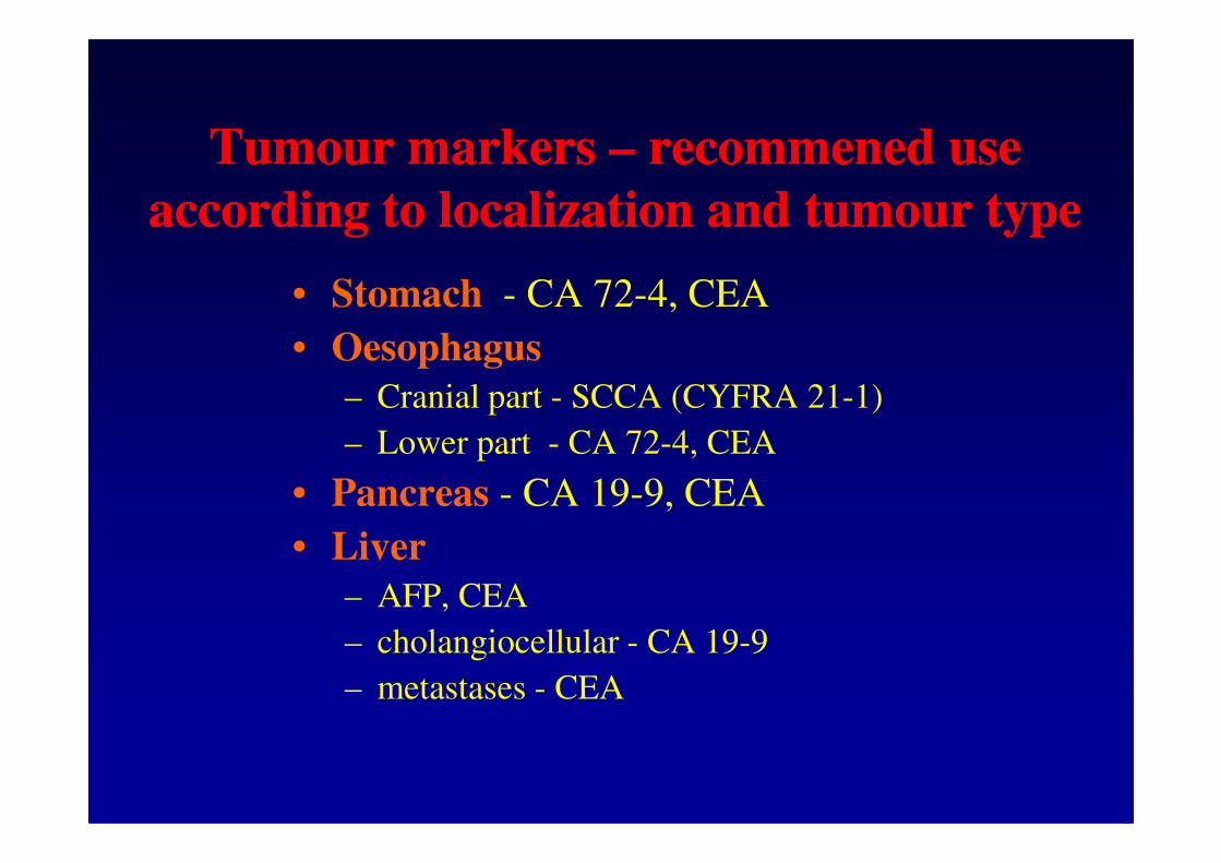
Tumour markers Tumour markers –– recommened use recommened use
according to localization and tumour typeaccording to localization and tumour type
• Stomach - CA 72-4, CEA
• Oesophagus
– Cranial part - SCCA (CYFRA 21-1)
– Lower part - CA 72-4, CEA– Lower part - CA 72-4, CEA
• Pancreas - CA 19-9, CEA
• Liver
– AFP, CEA
– cholangiocellular - CA 19-9
– metastases - CEA

• Breast - CA 15-3, CEA (TPA/S)
• Lung
– SCLC - CEA, NSE (TPA/S)
Tumour markers Tumour markers –– recommened use recommened use
according to localization and tumour typeaccording to localization and tumour type
– SCLC - CEA, NSE (TPA/S)
– NSCLC - CYFRA 21-1, CEA (SCC)

• Ovary
– non-mucinous - CA 125 (TPA/S)
– mucinous - CA 19-9, CA 72-4 (CEA)
Tumour markers Tumour markers –– recommened use recommened use
according to localization and tumour typeaccording to localization and tumour type
– germinative - AFP, hCG
• Cervix
– epidermoid - SCCA (CYFRA 21-1, CEA)
– adenocarcinomas - CEA
• Corpus uteri - CA 125 (CEA)
• Vulva - SCCA

• Kidney - TPA/S, CEA (NSE)
• Urinary bladder - TPA/S (CYFRA 21-1)
• Prostate - PSA, fPSA (ChgA)
Tumour markers Tumour markers –– recommened use recommened use
according to localization and tumour typeaccording to localization and tumour type
• Prostate - PSA, fPSA (ChgA)
• Testes
– seminomas - hCG, AFP (NSE)
– non-seminomas - hCG, AFP

• Karcinoid - 5-hydroxy, 3-indolylacetic acid, NSE
• Thyroid gland
– medullar CT, CEA (NSE)
– anaplastic TPA/S
Tumour markers Tumour markers –– recommened use recommened use
according to localization and tumour typeaccording to localization and tumour type
– anaplastic TPA/S
• Melanoma - NSE, S100beta (TK)
• Head, neck - SCCA (CYFRA 21-1)
• CNS
– neuroblastomas - NSE
– gliomas - CEA
– astrocytomas - TK

• Leukemia - TK, FER, LD
• Lymfoma
– Hodgkin - B2M, FER, LD
Tumour markers Tumour markers –– recommened use recommened use
according to localization and tumour typeaccording to localization and tumour type
– non-Hodgkin - TK, B2M, LD
• Multiple myeloma - B2M, paraproteins

Determination of tumour markersDetermination of tumour markers
• Indication
• Preanalytical phase
• Determination – metods, interferences• Determination – metods, interferences
• Interpretation

Determination of tumour markersDetermination of tumour markers
• Immunochemistry
- radio immune assay – RIA, IRMA
- enzyme immune assay - ELISA, EIA, MEIA - enzyme immune assay - ELISA, EIA, MEIA
- fluorescence assay - FPIA, TRACE
- chemiluminiscence assay - CLIA
• Use the same diagnostic kit from the same
company!!! (or rebaselining)
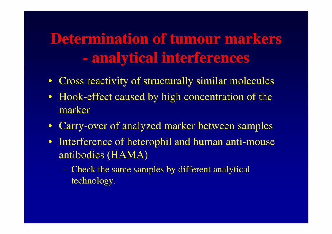
Determination of tumour markersDetermination of tumour markers
-- analytical interferencesanalytical interferences
• Cross reactivity of structurally similar molecules
• Hook-effect caused by high concentration of the
marker
• Carry-over of analyzed marker between samples
• Interference of heterophil and human anti-mouse
antibodies (HAMA)
– Check the same samples by different analytical
technology.

Indication and interpretation Indication and interpretation
of tumour markersof tumour markers• Not for diagnostics but for monitoring.
They can help in the diagnostic process.
• Positive finding of tumour markers is of diagnostic value, negative finding does not exclude a tumour!!!tumour!!!For diagnosis, histopathological examination and additional TU markers determination is decisive.
Transient elevation of a tumour marker – inflammation, non-malignant tumour, trauma, after efficient therapy, in decreased renal or liver function for markers which are eliminated this way
• Screening – faecal blood test, discussed PSA – not yet
u specific populations – calcitonin in familes with medullar CA of the thyroid gland, CA 15-3 in BRCA mutations

Indication and interpretation Indication and interpretation
of tumour markersof tumour markers
• Dynamics of changes (increase, although in reference range may indicate a recidive sooner than visualization by CT, US, PET)
increase in 3 consecutive blood collections or increase by more than 25% is significantincrease by more than 25% is significant
TU marker may detect a tumour of 1 mg (106 malignant
cells), clinical diagnosis is possible for 109 malignant cells
• Systematic examination – repeated determination after operation, at the beginning shorter intervals, later cca 3-6 months)
• Follow up of more tumour markers – higher probability of detection of a tumour
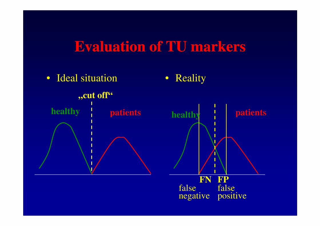
Evaluation of TU markersEvaluation of TU markers
• Ideal situation • Reality
healthy healthypatients patients
„cut off“
healthy healthypatients patients
FN FPfalse negative
false positive
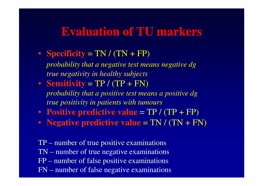
Evaluation of TU markersEvaluation of TU markers
• Specificity = TN / (TN + FP)
probability that a negative test means negative dg
true negativity in healthy subjects
• Sensitivity = TP / (TP + FN)probability that a positive test means a positive dgprobability that a positive test means a positive dg
true positivity in patients with tumours
• Positive predictive value = TP / (TP + FP)
• Negative predictive value = TN / (TN + FN)
TP – number of true positive examinations
TN – number of true negative examinations
FP – number of false positive examinations
FN – number of false negative examinations

Evaluation of TU markers Evaluation of TU markers
using ROC curvesusing ROC curves(ROC = receiver operating characteristic)
sensitivity (%)
100 %
suitable
TU marker
0 %100 % 0 % specificity (%)
TU marker
no
discrimination
among healthy
subjects and
patients
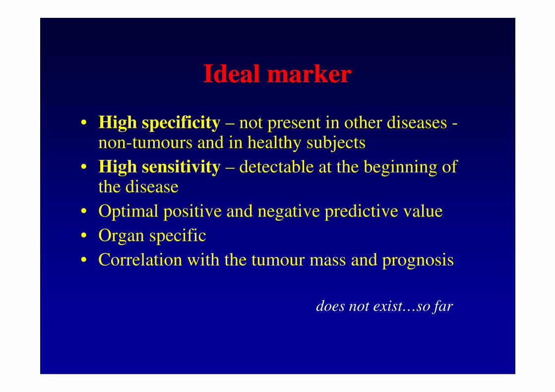
Ideal markerIdeal marker
• High specificity – not present in other diseases -non-tumours and in healthy subjects
• High sensitivity – detectable at the beginning of the diseasethe disease
• Optimal positive and negative predictive value
• Organ specific
• Correlation with the tumour mass and prognosis
does not exist…so far

Example
CEA for colorectal CA
• 95% specificity – i.e. 5% of healthy subjects are
falsly regarded as patients with tumours
• 70% sensitivity – i.e. does not detect 30% of
patients with tumours

Interpretation of results ofInterpretation of results of
TU markersTU markers
• In the past – comparison with reference range(might be suitable for a unique determination of unknown
patient)
• Today recommended determination of • Today recommended determination of
individual baseline values (concentration of a TU
marker in „stabilized“ status, i.e. after operation –
extraction of the tumour mass) and systematic
dynamic follow up

Dynamic follow upDynamic follow upconcentration
of TU marker
in blood
time of follow up
„cut off“

Dynamic follow upDynamic follow upconcentration
of TU marker
in blood
time of follow up
„cut off“

Molecular biology Molecular biology
in diagnostics of tumoursin diagnostics of tumours
• Tumours – mutations of genes which
products regulate cell proliferation, products regulate cell proliferation,
development, differentiation and cell death
• Oncogenes and antioncogenes
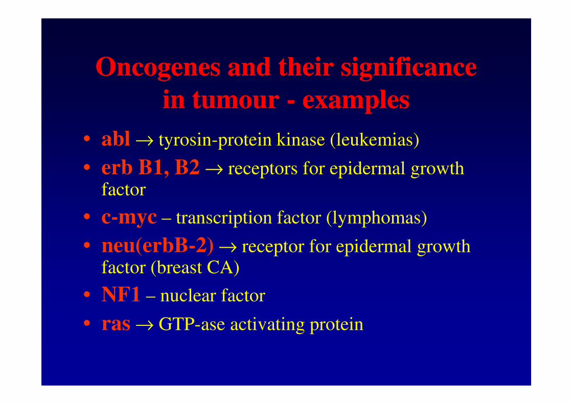
Oncogenes and their significance Oncogenes and their significance
in tumour in tumour -- examplesexamples
• abl → tyrosin-protein kinase (leukemias)
• erb B1, B2 → receptors for epidermal growth factor
• c-myc – transcription factor (lymphomas)
• neu(erbB-2) → receptor for epidermal growth factor (breast CA)
• NF1 – nuclear factor
• ras → GTP-ase activating protein

Antioncogenes and their Antioncogenes and their
significance in tumour significance in tumour -- examplesexamples
• BRCA 1 and BRCA 2 – reparation of DNA
defects (breast and ovarian CA)
• p53 – regulation of the cell cycle • p53 – regulation of the cell cycle
• RB1 a RB2 – regulation of the cell cycle
(retinoblastoma)

Potential new tumour markersPotential new tumour markers
Proteins and oncoproteins – products of mutated
genes which play a role in cell life, their division,
differentiation and metastasing
�Regulation of the cell cycle - cyclins
�Apoptosis – Bcl-2 protein, sFas, protein-product of mutated gene p53gene p53
�Signal transduction - c-erbB-2 (Her-2/neu), EGRF, IGF, TNF-α
�Adhesion - ICAM-1, VCAM-1
�Angiogenesis – inhibitors of angiogenesis - angiostatin, angiogenin, trombospondin
�Markers associated with specific characteristics of tumour cells – matrix metalloproteinases, urokinase plasminogen activator (uPA) and its inhibitor (PAI-1)

New and potential New and potential
tumour markerstumour markers
• free DNA in plasma (and microsatelite changes)
• free mRNA in plasma
• enzymes of DNA synthesis in tissue samples• enzymes of DNA synthesis in tissue samples
• mammaglobin - breast cancer
• heparanase
• …

LiteratureLiterature
• Guidelines of the Czech Society of Clinical Chemistry – www.cskb.cz
• Guidelines of the European Group for Tumour • Guidelines of the European Group for Tumour Markers (EGTM) – www.egtm.eu