instability of the subtalar joint - semantic scholar · 2017-10-17 · management of foot problems...
TRANSCRIPT
•arison of Keller's il osteotomy in the . J Bone Joint Surg
odeling with com-miedRes 19:535-
m IX, et al: Three-•om computerized 239-248, 1986 •atiD.etal: Three-' le from analysis lata. Orthopedics
Management of Foot Problems 0030-5898/89 $0.00 + .20
Instability of the Subtalar Joint
Thomas O. Clanton, MD*
jllege of Medicine al Medical Center 0 Wabash Avenue ., OH 44307-2433
Chronic lateral ligamentous instability of the ankle has received substantial attention in the orthopedic literature,1-5'9'12'13'15-1:7-30'33'35-40
y e t there has been littje discussion of the relationship of subtalar instability to this problem. Reconstructive procedures for lateral ankle instability have varied in complexity from late repairs5-15 to complicated fascial12 or tendon grafts.9-13-17-30-33'35-40 Subtalar instability is not corrected equally well by these procedures.2-40 Therefore, objective assessment of the patient with symptoms of instability to determine a possible subtalar component seems warranted. This would allow more specific tailoring of-the treatment to fit the problem.
HISTORICAL REVIEW
In 1962, Rubin and Witten32 were the first to suggest a clinical significance to subtalar instability and to propose a method for evaluating this instability. Their method employed a foot-holding device to produce varus stress while tomograms were taken. This allowed the authors to calculate a tibio-calcaneal angle that they said was normally between 40 and 50 degrees. Although the authors used this method to.test 27 patients, including 17 with symptoms of "excessive turning over," they found no patient with obvious subtalar instability.
Laurin et al,24 6 years later, correlated the results of cadaver ligament sectioning with stress roentgenograms demonstrating talar tilt and subtalar tilt. The findings were used to recommend a special radiographic technique for evaluating subtalar instability. The important contributions of this study included the. following.
(1) Cutting the calcaneofibular ligament caused abnormal opening between the posterior articular surfaces of the talus and the calcaneus with inversion stress.
(2) The mechanism for the isolated calcaneofibular ligament tear (inversion of a dorsiflexed ankle) was described.
(3) The amount of tilt was similar when testing was performed manually or when a special apparatus was used in stress production.
Brantigan et al,2 in 1977, employed the method of Rubin and Witten to demonstrate subtalar instability in three patients. They suggested that the normal tibiocalcaneal angle is 38 ± 6 degrees, whereas their unstable patients had angles of 53, 62, and 55 degrees. The authors advocated the tomographic technique for diagnosing subtalar instability because they believed that one of the procedures in vogue for correction of lateral ankle instability (the Watson-Jones technique) ofjen failed because of its inability to reconstruct the function of the calcaneofibular ligament.
The existence of subtalar instability was recognized by Chrisman and Snook9 in their article describing a modification of the Elmslie procedure for treating lateral ankle instability. Among seven patients so treated, three had subtalar instability noted at the time of surgery. Although no attempt was made to diagnose this preoperatively, it was corrected with the surgical technique by reconstructing both the anterior talofibular ligament and the calcaneofibular ligament. Indeed, its effectiveness in accomplishing this was confirmed at follow-up by the restriction in subtalar motion displayed by all patients.
Despite the effectiveness of the Chrisman-Snook procedure in correcting lateral ankle instability, it has some morbidity. Horstman et al17 have looked critically at lateral ligament reconstructions of the ankle and discovered a relatively high complication rate. This was confirmed in the late follow-up study of the Chrisman-Snook procedure reported by Snook et al in 1985.3 5 Disenchantment with tendon transfers at the ankle led me to a modification of the Brostrom technique5 for the treatment of chronic ankle instability. Because of this, I began to take a more critical look at the lesions present in both acute and chronic ankle and hindfoot injuries, related cadaver studies, and the biomechanics of the tibiotalocalcaneal joints.
* Clinical Assistant Professor, Division of Orthopaedic Surgery, University of Texas Medical School at Houston, Houston, Texas
Orthopedic Clinics of North America—Vol. 20, No. 4, October 1989 583

8aaa»"
584 Thomas O. Clanton
S3.
MECHANISM OF INJURY AND INJURY PATTERNS
It is well accepted that inversion is the primary mechanism by which lateral ankle sprains occur.1,11-26 Varying patterns of damage to the lateral ligaments have been attributed to the degree of dorsiflexion or plantar flexion present at the time of injury.26-33 In plantar flexion, the anterior talofibular ligament is more vulnerable to inversion stress, whereas in dorsiflexion, the calcaneofibular ligament assumes a more important role in resisting such stress. The anatomic divergence of these two ligaments (Fig. 1A) accounts for this difference, and the individual variability in the exact degree of divergence explains some of the alteration seen in the injury pattern with the same mechanism of injury.
Inversion also is the mechanism that produces the medial subtalar dislocation.6-22 This injury also occurs more commonly in the plantar-flexed foot, yet there are obvious differences in the end result. The calcaneofibular ligament is torn, but this appears secondary to tearing of the talonavicular ligament and the talonavicular joint capsule. The sustentaculum tali then acts as a fulcrum and the head of the talus dislocates laterally while the calcaneus moves medially tearing the lateral talocalcaneal ligament, the talocalcaneal joint capsule, and the calcaneofibular ligament. J£ the mechanism continues, and tearing of the cervical ligament and the ligament of the tarsal canal occurs in addition to disruption of the deltoid ligament, then we see the rare entity: total dislocation of the talus.25 Somewhere in between this extreme and the severe grade of lateral ankle sprain is an injury that results in instability of the subtalar joint as an isolated problem or as a component of the other injuries.
EXPERIMENTAL INVESTIGATIONS
Multiple cadaver studies have been performed with selective ligament cutting and analysis of in
stability. Leonard26 showed the relationship between the anterior talofibular and calcaneofibular ligaments to injuries in ankle plantar flexion and neutral positions, respectively. He also demonstrated an increase in subtalar motion with division of the calcaneofibular ligament. Dias11 produced various injury patterns in 39 amputation specimens with supination-inversion, supination-inter-nal rotation, and supination-plantar flexion. Three medial subtalar dislocations, were produced by either inversion or internal rotation force on the supinated foot. In two of these three cases, the dislocation occurred with rupture of the calcaneofibular ligament as an isolated injury among the
.lateral ankle ligaments. Following their cadaver research, Laurin et al24
emphasized the critical role of the calcaneofibular ligament in subtalar stability. This was demonstrated anatomically as well as radiographically. Talar tilt was related to anterior talofibular ligament injury with or without associated calcaneofibular ligament rupture, whereas subtalar tilt was produced when the latter ligament was damaged. Routine stress roentgenograms could be interpreted as negative in the face of an isolated calcaneofibular ligament rupture. Therefore, Laurin et al24 proposed a modified stress radiograph to document subtalar instability.
Further investigation of the role of the calcaneofibular ligament in talocalcaneal stability has been performed by Kjaersgaard-Andersen et al.19-21 An increase in talocalcaneal adduction of 3.1 to 4.6 degrees was found in ten freshly frozen cadaver specimens after section of the calcaneofibular ligament. This constituted 61 to 77 per cent of the total increment of adduction between the leg and the hindfoot, with the maximum increment being at 5 degrees dorsiflexion. External rotation at the tibiotalocalcaneal articulation also was affected by calcaneofibular ligament division but to a lesser extent: it ranged from 2.9 degrees maximum in dorsiflexion to a minimum of 1.4 degrees in plantar flexion for the talocalcarieal joint, and from 5.4 to 2.2 degrees for the tibiotalocalcaneal joints.
FIBULA
Post. Talofibular
CALCANEUS
Lateral Talocalcaneal
Calcaneofibular
TALUS
MEDIAL
Interosseous Talocalcaneal
B
Retinacula
LATERAL
Cervical
CALCANEUS
Figure 1. A, Diagrammatic representation of the lateral ligament complex of the tibiotalar and talocalcaneal joints. B, Diagrammatic representation of ligaments in the tarsal canal and sinus tarsi on frontal section. (Drawings by Bob Boeye.)

up be-ifibular on and lemon-livision aduced
speci-i-inter-. Three ced by on the
ies, the lcaneo-mg the
n et al24
ofibular demon-ihically. lar liga-dcaneo-• tilt was imaged, e inter-id calca-,aurin et to docu-
alcaneo-las been I.19-21 An 1 to 4.6 cadaver
ular liga-:it of the 3 leg and snt being on at the •ectedby < a lesser dmum in n plantar )m 5.4 to d joints.
- Retinacula
TERAL
~" Cervical
id talocal-on frontal
Instability of the Subtalar Joint 585
Though small, this incremental increase represents a 10 to 30 per cent increase from the intact specimen, according to the authors, and further supports the role of the calcaneofibular ligament in ankle-hindfoot stability. Despite this evidence for the role of the calcaneofibular ligament in talocalcaneal joint stability, Cass et al18 have questioned this role in a separate study using a different experimental method.
Anatomic studies of the talocalcaneal articulation and its stability must examine other structures in-addition to the calcaneofibular ligament. These structures include not only the bony anatomy but also the inferior extensor retinaculum, the lateral talocalcaneal ligament, the cervical ligament, and the interosseous talocalcaneal ligament (Figs. 1A and B). The retinaculum tenses with inversion stress but plays a minimal role in stability because of its elasticity.39 The contribution of the interosseous talocalcaneal ligament (or ligament of the tarsal canal) in resisting inversion stress has been questioned by some authors because it lies near the axis of rotation for the subtalar joint.7'23'34 Smith,34
Cahill,7 and Viladot et al39 stressed the importance of the cervical ligament in allowing controlled inversion while preventing excessive motion. Few studies have looked at the relative biomechanical. contribution of the ligaments of the sinus tarsi and tarsal canal and subtalar stability until the recent work of Kjaersgaard-Andersen et al20 and Heil-mann et al.-16
The work of Kjaersgaard-Andersen and his group20 involved selective cutting of the ligaments of the sinus and canalis tarsi, followed by measurement of increases in range of motion for both the tibiotalocalcaneal joints and the talocalcaneal joint. Increases in movement were calculated for rotation, adduction-abduction, and plantar flexion-dorsiflexion. While the numerical values for these measurements showed only small increases ranging from 0.4 to 2.6 degrees for the maximum median increase, the authors believed that this constituted a sufficient percentage increase to allow correlation of injury to these ligaments with clinical instability in the hindfoot complex (tibiotalocalcaneal or talocalcaneal joints).
Heilmann et al16 have performed a similar study looking at the relative contribution to subtalar stability provided by the various lateral ligamentous structures. Selective cutting of the calcaneofibular and interosseous talocalcaneal ligaments was performed on seven fresh cadaver ankles, and instability analysis was made by radiographic measurement. The authors used a stress Broden's view,3
which is a slight modification of the subtalar tilt view recommended by Laurin et al.24 Using this view, they found an average increase of 5 and 7 mm with sequential cutting of the calcaneofibular and interosseous talocalcaneal ligaments.
CLINICAL INVESTIGATIONS
A key deficit in the analysis of subtalar instability and its relationship to lateral ankle instability has
been the lack of sufficient correlation between cadaver studies and clinical examination of the pathologic lesions noted in the acute injury situation. Isolated injury to the calcaneofibular ligament has been mentioned by several authors,4'11'14,24-40-44
yet it is obviously infrequent in comparison to the more common anterior talofibular ligament tear or the combined injury to both structures. No published study has documented the extent of injury to the structures of the sinus tarsi or tarsal canal seen with inversion injuries to the ankle and hind-foot, although Ruth32 does mention-six cases of lateral talocalcaneal ligament rupture in his series of acute lateral ankle repairs.
Meyer et al28 presented a study of this subject in 1988 wherein he used anterior drawer and talar tilt stress radiographs in combination with subtalar arthrograms to classify the extent of injury. He divided the patients on the basis of a positive subtalar arthrogram. Four types of injury pattern were noted in the 32 patients with contrast leakage from their subtalar arthrogram. Of these patients, 12 had their pathology confirmed at surgical exploration.
This study lends credence to the aforementioned cadaver studies and attempts to correlate outcome with extent of injury and type of treatment. It gives clinical meaning to the relationship between inversion mechanism ankle sprains and subtalar instability. The question remains whether this will be a practical method for evaluation of severe ankle sprains when a majority of practitioners treat this injury without stress radiographs and without surgery.18 Certainly, it confirms the necessity for evaluating the chronically painful or unstable ankle for subtalar pathology.
DIAGNOSTIC TECHNIQUES
Stress Radiographs
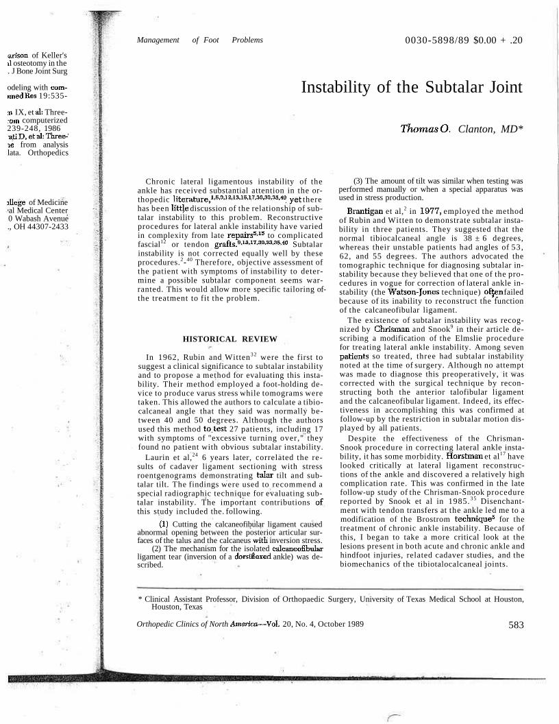
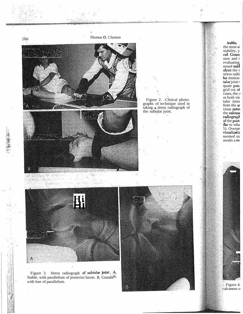
Subtalar. The method proposed by Laurin et al24 and used by others in a slightly modified fash-ion10-16-44 has proved effective in evaluating subtalar instability. I have incorporated this view into my own practice and found it to be easily performed and reproducible. This view is a 40-degree Broden (projection I) view3 with inversion stress applied to the calcaneus and forefoot (Fig. 2A). The patient is positioned obliquely on the table and the foot is rotated internally 45 degrees. The beam then is directed 40 degrees away from the vertical and centered on the sinus tarsi. Stress is applied with the thumb on the lateral heel and the index finger on the lateral forefoot (Fig. 2B). A normal radiograph demonstrates parallelism of the posterior articular surfaces of the subtalar joint (Fig. 3A), whereas an abnormal study shows divergence (Fig. 3B). Because of the small increases in subtalar motion noted by Kjaersgaard-Andersen19-21 after ligament sectioning, it would appear that any loss of parallelism is indicative of instability, as previously hypothesized by Laurin et al.24
... mffifl
f ' l ,
" H i
586
HHHB$
Thomas O. Clanton
P.:
syn nek
HB3
iWaYMBWAVjhHMri
Figure 2. Clinical photographs of technique used in taking a stress radiograph of the subtalar joint.
Ankle, the most o stability, y cal. Conce sure and i evaluating nosed ankl alyze the t stress radii lar motion talar joint i quate pem grid cut oi cases, the ,• or both vie talar insta from the ai vious antei the calcanc radiograpr. of the post, ilar to wha 5). Overpe visualizatic seemed us awaits a mi
..I! mm
iiiiil
: !1
IllfSl « 1
Hi HP Hi
Figure 3. Stress radiograph of subtalar joint i Stable, with parallelism of posterior facets. B, Unstabl with loss of parallelism.
m
i?Mm
Figure 4. calcaneus o

inical photo-ique used in adiograph of f.
Instability of the Subtalar Joint 587
Ankle. Specific views of the subtalar joint are the most obvious method of evaluating subtalar instability, yet this method is not necessarily practical. Concern over the amount of radiation exposure and a search for a retrospective method of evaluating subtalar instability in previously diagnosed ankle instability cases prompted me to reanalyze the traditional anterior drawer and talar tilt stress radiographs for any sign of pathologic subtalar motion. In a large percentage of films, the subtalar joint was not well visualized because of inadequate penetration (on the inversion stress view), grid cut off, or blockage by a lead glove. In a few cases, the subtalar joint was well depicted on one or both views and allowed analysis of possible subtalar instability. This instability was diagnosed from the anterior drawer view when there was obvious anterior translation of the posterior facet of the calcaneus on the talus (Fig. 4). On the talar tilt radiograph, the diagnosis was made when opening of the posterior subtalar joint was seen. This is similar to what one sees with a stress tomogram (Fig. 5). Overpenetration of the talar tilt view improves • visualization ofthe subtalar joint. This method has seemed useful'to me clinically, but its true value awaits a" more scientific clinical or cadaver study.
Stress Tomograms
Rubin and Witten31 believed that instability of the talocalcaneal joint could not be evaluated properly by routine radiographic techniques. They pioneered the use of a special device for holding the feet and applying an inversion stress while tomograms were taken. A frontal section view, cut through the sinus tarsi, was used to calculate a tibiocalcaneal angle, measuring from the inferior articular surface of the tibia to the most prominent areas of the superior calcaneal surface. Because these authors had no documented cases of subtalar instability in their series, the clinical usefulness of this method remained theoretical until the report of Brantigan et al.2
The applicability of the tomographic technique was confirmed by Brantigan and his co-workers2 in their study of three patients with subtalar instability. Two of these patients had instability following
^Watson-Jones reconstructions, and their residuial symptoms were traced to the subtalar joint by this method. Although the authors advocate the method, a review of the figures contained in the original article gives one the impression of lack of clarity and resulting imprecision in angle calcula-

IpF"
tMJ-
588 Thomas O. Clanton
Wsmism
11111111111
wmmmmm :|;Mjj$ip;5ii Inversion stress radiograph of subtalar joint. A, Stable, with normal posterior facet congruity. B, Unstable, with loss of congruity.
tion. Furthermore, no follow-up study of the technique has appeared in the English literature in the past 12 years, and one of the authors (Lippert FG: personal communication) has acknowledged abandoning the technique.
Although I have had no personal experience with the tomographic method, I would agree that it can demonstrated subtalarJnstability quite well. This demonstration does not require the calculation of a talocalcaneal angle, however, since loss of congruity in the subtalar joint is a clear indication' of instability (Fig. 6). This method has been used by Zollinger (Zollinger H: personal communication) from whom this example was borrowed. Nevertheless, I believe the method to be impractical and unnecessary in most situations because of the time required and expense, involved in tomographic studies compared with alternative methods for diagnosing subtalar instability.
Fluoroscopy
The fluoroscopic technique has been described in only one case of combined ankle and subtalar instability.41 It can include videotaping and spot films but is obviously impractical in the office set
ting. I have used an image intensifier in the operating room to visualize the tibiotalar and talocalca-neal joint motion in both acute and chronic surgical cases. It can provide a dynamic picture that is unavailable with other methods.
Subtalar Arthrogram
The subtalar arthrogram was mentioned in the original work of Laurin et al24 on subtalar instability, yet he showed no examples of it and did not describe the technique. It was Meyer et al28 who eventually described the technique and performed an extensive investigation using arthrography of the joints of the hindfoot. This work was continued in the clinical setting and described in two subsequent articles that discussed subtalar pathology.29-37 As previously mentioned, Meyer et al28 have used the subtalar arthrogram as a means of differentiating between the simple anterior talofibular ligament sprain and more severe injuries to the hindfoot complex.
Stimulated by the work of Meyer et al27-29- 37 I have performed subtalar arthrograms on normal paid volunteers as well as in patients with acute and chronic lateral ankle symptoms. In the acute
yl
i '*•
'-•i
••) %
M •i l ••
''''% , :-'-M
4 ;.;;*># :£$£
'"$ r f '%
•••«
-'
;'
IS"
"' y
r'if'fi ?#:£- " :&&i|i^1'.;
£*v i'J *V
>' j - " *
^?SI&i??;.y ǤsSj4i0*fe " - j \ ft.
WSiMtt-'.-C.ilfSifc';^','
^SKKi& I7;,
| l §y H|fe ife-^'••-
Figure grams o Stable. Zollinge: Waldisl terenSpi tat mit raphie. heilkund delberg, 1983,pp mission.)
setting,! drawera firm a tei caneofibi tieht wb combina are exam examinai
Figure graphs arthrogrs mentiriji talar joir Anterior

Instability of the Subtalar Joint 589
•nana Hi *?H Itllll ;yyii-
fflM.
i lilbi HH
HRi WMwi HBgpasM
IslllifliiS
congruity.
the operat-1 talocalca-•onic;surgi-ture-that is
v -A
•mmi>
mm:
it-yi
Figure 6. Stress tomograms of subtalar joint. A, Stable. B, Unstable. (From Zollinger H, Meier CH, Waldis M: Diagnostik der un-teren Sprunggelenksiristabili-tat mittels Stress-Tomog-raphie. Hefte zur Unfall-heilkunde, Hefte 165. Heidelberg, Springer-Veriag, 1983, pp 175-177; with permission.)
setting, it is ideally combined with routine anterior drawer and talar tilt stress roentgenograms to confirm a tear of both the anterior talofibular and calcaneofibular ligaments inffhe young athletic patient who may be a candidate for repair. This combination! of tests has beeri used in patients who are examined under anesthesia-when their clinical examination reveals gross instability (Fig. 7).
The technique of the subtalar arthrogram is most easily performed with the patientprone, but may be done with the patient tilted away from the affected side. A 22-gauge spinal needle is inserted just lateral to the Achilles tendon and directed 15 degrees caudal to the plantar surface of the foot (Fig. 8A). The needle enters the back of the ankle approximately 1 to 2 cm above the posterior supe-
3ned in the larinstabil-and did hot' et al28 who ( and per-g arthrogra-is^workwas •'•. lescribediii subtalar pa-1, Meyer et l as a; means anterior ta-'ereinjuries
t al27'29' 3 71 -s oh normal, i with acute [n the acute
t . l f
felt/ Figure 7. Stress radiographs following subtalar arthrogram in lateral ligament injury to ankle and subtalar joints. A, Inversion. B,. Anterior drawer.
wmmm
"His
H B H H H i WsMmSm iHnBa |*fe£J

%*\]
590 Thomas O. Clanton
I sj^pg
l k :
M
D H1
Figure 8. A, Photograph of needle insertion technique for subtalar arthrogram with patient prone. B, Radiograph demonstrating proper needle placement.
rior tuberosity of the calcaneus and is directed into the posterior subtalar joint (Fig. 8B). This is easily accomplished in the operating room with image intensifier control, but may be done almost as easily in the office using local anesthesia and radiographic confirmation of needle position. Radiographic contrast medium (2 to 4 ml) is injected and the subtalar joint is visualized with the image intensifier or with routine radiographs. <-~
The normal subtalar joint demonstrates free movement of dye with variation in foot position, welf-defined medial and lateral talocalcaneal joint recesses, and numerous microrecesses surrounding the interosseous talocalcaneal ligament (Fig. g) 29,37 Acute cases may show a bloody aspiration on penetration of the joint and contrast leakage into surrounding soft tissues, the ankle joint, or the sinus tarsi (see Fig. 7). Chronic lesions are characterized by two distinguishing features. The lateral recess does not fill normally with contrast medium, and the dye assumes a completely flat appearance along the anterior margin of the posterior subtalar joint with absence of the normal microrecesses along the talocalcaneal ligament (Fig. 10).
Iii my opinion, the subtalar arthrogram has potential for aiding in the classification of injuries to the ankle and hindfoot; however, any time an additional investigative study is added, there is the potential for increased morbidity, particularly when the investigation is invasive. Therefore, I believe that the subtalar arthrogram should be used on a limited basis until further study confirms the utility shown by Meyer and his colleagues.
CONCLUSIONS
The topic of subtalar instability seems to be gaining attention in the orthopedic community.36
' This topic has been demonstrated as a clinical entity as well asan experimental one. Subtalar instability can be one of the causes of instability symptoms in the patient who sustains an inversion-type injury to the ankle and hindfoot. If peroneal mus-
- cle weakness, Achilles tendon tightening, and pro-
Si atfSrliaS
" • S l i p
Figure 9. Normal subtalar arthrogram showing microrecesses along talocalcaneal ligament.
Figure ] showing flat ligament wi
prioceptive other causes
In seekinj the "clinician which to ch joint, as visi probably off least- alterati ankle instab quire educaf tional radiat form them, Stress tomo| and are mor patient, witr. methods. Fl gives a dynai generally in operating ro the subtalar sive study tl in diagnosinj lar arthrogra setting force chronic une pain withoui
I£ subtalar .above meth mount. Norn muscle strei propriocepti cient imprc
^symptoms. C effective incl to limit invei bilize the sul fective or no tervention rr 'L Few long-
Sport the surf The study b

tographpf technique gram with ladiograph per needle
. clinical en-btalar instability symp-/ersion-type ironeal musing, and pro-
11111111111
9£HSS8 flHHH II fifBmr&ZSMBS
ogram showing ligament. ••_
mm
"'•v;:
m
•••••.?.
•'••'WW. '" 3
r
._...:
Instability of the Subtalar Joint 591
s^^^^Sr *--":' ' '-''•"
HBBH
M S
HHi
mm
Figure 10. Abnormal subtalar arthrogram showing flattening of dye adjacent to talocalcaneal ligament with loss of microrecesses.
prioceptive dysfunction have been excluded, then other causes for instability must be sought.
In seeking confirmation of subtalar instability, the clinician has an armamentarium of studies from which to choose. Critical analysis of the subtalar joint, as visualized on routine stress radiographs,. probably offers the most practical method with the least alteration in a standard approach for lateral ankle instability. Specific subtalar stress views require education of the radiology technician, additional radiation exposure, and more time to perform them, but they are probably more specific. Stress tomograms demand a foot-holding device and are more expensive and inconvenient for the patient, with little apparent benefit over the other methods. Fluoroscopy with an image intensifief gives a dynamic view of talocalcaneal motion but is generally impractical until one is already in the operating room with the diagnosis made. Finally, the subtalar arthrogram is an invasive and expen-sive study that offers little additional information in diagnosing chronic subtalar instability. A subtalar arthrogram may be of value in the acute" injury setting for certain select patients28 or in the case of chronic unexplained lateral ankle or sinus .tarsi pain without instability.29'37
I£ subtalar instability is diagnosed by one" of the above methods, then treatment becomes paramount. Nonoperative treatment methods such as muscle strengthening, Achilles stretching, and proprioceptive re-education may provide sufficient improvement to eliminate bothersome symptoms. Other treatment modalities that can be effective include lateral heel wedges, ankle braces to limit inversion, and more specific bracing to stabilize the subtalar joint. If these methods are ineffective or not tolerated by the patient, surgical intervention may be considered.
Few long-term follow-up studies exist that report the surgical treatment of subtalar instability. The study by Chrisman and Snook9 reflects that
their procedure is capable of correcting subtalar instability. The procedure rebuilds both the anterior talofibular and calcaneofibular ligaments, thereby restraining motion at the talocalcaneal joint and the tibiotalar joint. The critical need for anatomic reconstruction of these ligaments has been pointed out by the experimental work of Wirth and. Artmann.40 Further confirmation of the applicability of the modified Elmslie procedure has come from the work of Vidal et al38 and Zwipp et al,42-43 who have used it in cases of isolated subtalar instability and combined instability.
Although Brostrom5 specifically described a late repair of the anterior talofibular ligament, I have often isolated and repaired the previously torn calcaneofibular ligament when performing an anatomic reconstruction. As an extra-articular structure, the calcaneofibular ligament does not resorb and therefore can be isolated and often reattached to the calcaneus. Further resistance to inversion is provided by tightening the inferior extensor retinaculum as described by Gould.15 Because of its anatomic location, this tightening would add some stability to the talocalcaneal articulation in the area of the sinus tarsi and further reinforces the ability of this graftless procedure to correct both tibiotalar and talocalcaneal instability.
SUMMARY
Instability of the subtalar joint can follow inversion stress .injuries to the lateral ankle and hindfoot. Ligaments involved in the injury include the calcaneofibular, lateral talocalcaneal, cervical, interosseous talocalcaneal, or a combination. Although the problem most often occurs in conjunction, with lateral ankle instability, it may be an isolated problem and it is not corrected equally well by the various lateral ankle ligament reconstructions. Techniques available for diagnosis include routine stress radiographs of the ankle, which also include the subtalar joint, specific subtalar stress-radiographs, stress tomograms, fluoroscopy, or subtalar arthrograms. Once diagnosed, subtalar instability symptoms can be treated by either nonoperative or operative means.
,REFERENCES
1. Anderson KJ, Lecocq JF, Lecocq EA: Recurrent anterior subluxation of the ankle joint: A" report of two cases and an experimental study. J Bone Joint Surg 34A:853-860, 1952
2. Brantigan JW, Pedegana LR, Lippert FG: Instability of the subtalar joint: Diagnosis by stress tomography in three cases. J Bone Joint Surg 59A:321-324,1977
3. Broden B: Roentgen examination of the subtaloid joint in fractures of the calcaneus. Acta Radiol 31:85-91, 1949
4. Brostrom L: Sprained ankles. I. Anatomic lesions in recent sprains. Acta Chir Scand 128:483-495, 1964

592 Thomas O. Clanton
5. Brostrom L: Sprained ankles. VI. Surgical treatment of "chronic" ligament ruptures. Acta Chir Scand 132:551-565,1966
6. Buckingham WW Jr: Subtalar dislocation of the foot. J Trauma 13:753-765, 1973
7. Cahill DR: The anatomy and function of the contents of the human tarsal sinus and canal. Anat Rec 153:1-17, 1965
8. Cass JR, Morrey BF, Chao EYS: Three-dimensional kinematics of ankle instability following serial sectioning of lateral collateral ligaments. Foot Ankle 5:142-149, 1984
9. Chrisman OD, Snook GA: Reconstruction of lateral ligament tears of the ankle: An experimental study and clinical evaluation of seven patients treated by a new modification of the Elmslie procedure. J Bone Joint Surg 51A-.904-912, 1969
10. Clanton TO: Assessment and classification of subtalar instability. Presented at the Annual Meeting of the American Orthopaedic Foot and Ankle Society, St. Paul, Minnesota, July 1988
11. Dias LS: The lateral ankle sprain: An experimental study. J Trauma 19:266-269,1979
12. Elmslie RC: Recurrent subluxation of the ankle-joint. Ann Surg 100:364-367, 1934
13. Evans DL: Recurrent instability of the ankle—a method of surgical treatment. Proc Roy Soc Med 46:343-344, 1953
14. Francillon MR: Distorsio pedis with an isolated lesion of the ligamentum calcaneo-fibulare. Acta Orthop Scand 32:469-475, 1962
Gould N, Selligson D, Gassman J: Early and late repair of lateral ligament of the ankle. Foot Ankle 1:84-89, 1980
Heilman A, Bishop J, Braly WG, et al: Anatomic study of subtalar instability. Presented at the Annual Meeting of the American Orthopaedic Foot and Ankle Society, St. Paul, Minnesota, July 1988
17. Horstman JK, Kantor GS, Samuelson KM: Investigation of lateral ankle ligament reconstruction. Foot Ankle 1:338-342, 1981
18. Kay DB: The sprained ankle: Current therapy. Foot Ankle 6:22-28, 1985
19. Kjaersgaard-Andersen P, Wethelund J, Helmig P, et al: Effect of the calcaneofibular ligament on hindfoot rotation in amputation specimens. Acta Orthop Scand 58:135-138, 1987
20. Kjaersgaard-Andersen P, Wethelund J, Helmig P, et al: The stabilizing effect of the ligamentous structures in the sinus and eanalis tarsi on movements in the hindfoot: An experimental study. Am J Sports Med 16:512-516, 1988
21. Kjaersgaard-Andersen P, Wethelund J, Nielsen S: Lateral talocalcaneal instability following section of the calcaneofibular ligament: A kmesiologie study. Foot Ankle 7:355-361, 1987
22. Lancaster S, Horowitz M, Alonso J: Subtalar dislocations: A prognosticating classification. Orthop 8:1234-1240,1985
23. Last RJ: Specimens from the Hunterian Collection. 7. The subtalar joint (specimens S 100 1 and S 100 2). J Bone Joint Surg 34B:116-119, 1952
24. Laurin CA, Ouellet R, St-Jacques R: Talar and subtalar tilt: An experimental investigation. Can J Surg 11:270-279, 1968
25. Leitner B: The mechanism of total dislocation of the talus. J Bone Joint Surg 37A:89-95, 1955
26. Leonard MH: Injuries of the lateral ligaments of the
15
16
ankle: A clinical and experimental study. J Bone Joint Surg 31A:373-377, 1949
27. Meyer JM: L'arthrographie de l'articulation sous-astragalienne posterieure et de l'articulation de Chopart. These med Geneve, No. 3318, 1973
28. Meyer JM, Garcia J, HoffmeyerP: Subtalar sprain: A radiological study. Presented at the Annual Meeting of the American Orthopaedic Foot and Ankle Society, Atlanta, Georgia, February 1988
29. Meyer JM, Lagier R: Post-traumatic sinus tarsi syndrome: An anatomical and radiological study. Acta Orthop Scand 48:121-128,1977
30. Nilsonne H: Making a new ligament in ankle sprain. J Bone Joint Surg 31A:380-381, 1949
31. Rubin G, Witten M: The subtalar joint and the symptom of turning over on the ankle: A new method of evaluation utilizing tomography. Am J Orthop 4:16-19, 1962
32. Ruth CJ: The surgical treatment of injuries of the fibular collateral ligaments of the ankle. J Bone Joint Surg 43A-.229-239, 1961
33. St. Pierre R, Allman F Jr, Bassett FH m , et al: A review of lateral ankle ligamentous reconstructions. Foot Ankle 3:114-123, 1982
34. Smith JW: The ligamentous structures in the eanalis , and sinus tarsi. J Anat 92:616-620, 1958
35. Snook GA, Chrisman OD, Wilson TC: Long-term results of the Chrisman-Snook operation for reconstruction of the lateral ligaments of the ankle. J Bone Joint Surg 67A-.1-7, 1985
36. Symposium on Subtalar Instability. Presented at the Annual Meeting of the American Orthopaedic Foot and Ankle Society, St. Paul, Minnesota, July 1988
37. Taillard W, Meyer JM, Garcia J, et al: The sinus tarsi syndrome. Int Orthop 5:117-130, 1981
38. Vidal J, Fassio B, Buscayret C, et al: Instabilite ex-terne de la cheville. Importance de l'articulation sous-astragalienne: Nouvelle technique de reparation. Rev Chir Orthop 60:635-642, 1974
39. Viladot A, Lorenzo JC, Salazar J, et al: The subtalar joint: Embryology and morphology. Foot Ankle 5:54-66, 1984
40. Wirth CJ, Artmann M: Chronische fibulare sprung-gelenksinstabilitat-untersuchungen zur Rontgen-diagnostik und bandplastik. Arch Orthop TJnfall-chir 88:313-320, 1977
41. Zell BK, Shereff MJ, Greenspan A, et al: Combined ankle and subtalar instability. Bull Hosp Joint Dis Orthop Inst 46:37-46, 1986
42. Zwipp H, Krettek C: Diagnostik und therapie der akuten und chronischen bandinstabilitat des un-teren sprunggelenkes. Orthopade 15:472-478, 1986
43. Zwipp H, Tscherne H: Zur behandlung der chronischen rotationsinstabilitat im hinteren unteren sprunggelenk. Unfallheilkunde 87:196-200, 1984
44. Zwipp H, Tscherne H: Die radiologische diagnostik der rotationsinstabilitat im hinteren unteren sprunggelenk. Unfallheilkunde 85:494-498, 1982
Division of Orthopaedic Surgery University of Texas Medical School at Houston
6410 Fannin, Suite 1100 Houston, TX 77030
Manageme
Rheumatc disorder of flammation among antig mune comp arthritis is 0 commonly i: ease is often either sex, often than n at any age, sixth decade
, by intermix course. As t ticular tissii also may be ments, tend' deformities the inflamm. lar bone. E: come a maj course of th dominant, ai develop.
The disea cent of the that the pain torn in 28 p. cases at soir fected. Of £ Vainio57 foui Others have 100 per cei cally.
The hindfi the three ac (talocalcane: disease invo quently,57 t fected.4-26 V males and 5 arthritis had reported sig
uLLi