insights into the mechanisms of catalysis and heterotropic regulation of escherichia coli aspartate...
TRANSCRIPT
Insights Into the Mechanisms of Catalysis and HeterotropicRegulation of Escherichia coli AspartateTranscarbamoylase Based Upon a Structure of the EnzymeComplexed With the Bisubstrate AnalogueN-phosphonacetyl-L-aspartate at 2.1 ÅLei Jin,1 Boguslaw Stec,2 William N. Lipscomb,3 and Evan R. Kantrowitz1*1Department of Chemistry, Boston College, Merkert Chemistry Center, Chestnut Hill, Massachusetts2Department of Biochemistry and Cell Biology, Rice University, Houston, Texas3Department of Chemistry, Harvard University, Gibbs Laboratory, Cambridge, Massachusetts
ABSTRACT Ahigh-resolution structure of Esch-erichia coli aspartate transcarbamoylase has beendetermined to 2.1 Å; resolution in the presence ofthe bisubstrate analog N-phosphonacetyl-L-aspar-tate (PALA). The structure was refined to a freeR-factor of 23.4% and a working R-factor of 20.3%.The PALA molecule is completely saturated withinteractions to side chain and backbone groups inthe active site, including two interactions that arecontributed from the 80s loop of the adjacent cata-lytic chain. The charge neutralization of the boundPALA molecule (and presumably the substrates aswell) induced by the electrostatic field of the highlypositively charged active site is an important factorin the high binding affinity of PALA and must beimportant for catalysis. The higher-resolution struc-ture reported here departs in a number of waysfrom the previously determined structure at lowerresolution. These modifications include alterationsin the backbone conformation of the C-terminal ofthe catalytic chains, the N- and C-termini of theregulatory chains, and two loops of the regulatorychain. The high-resolution of this structure hasallowed a more detailed description of the bindingof PALA to the active site of the enzyme and hasallowed a detailed model of the tetrahedral interme-diate to be constructed. This model becomes thebasis of a description of the catalytic mechanism ofthe transcarbamoylase reaction. The R-structuralstate of the enzyme-PALA complex is an excellentrepresentation of the form of the enzyme that oc-curs at the moment in the catalytic cycle when thetetrahedral intermediate is formed. Finally, im-proved electron density in the N-terminal region ofthe regulatory chain (residues 1 to 7) has allowedtracing of the entire regulatory chain. TheN-terminal segments of the R1 and R6 chains arelocated in close proximity to each other and to theregulatory site. This portion of the molecule may beinvolved in the observed asymmetry between theregulatory binding sites as well as in the hetero-
tropic response of the enzyme. Protein 1999;37:729–742. r 1999 Wiley-Liss, Inc.
Key words: allosteric regulatory; enzyme-inhibitorcomplex; cooperativity; tetrahedral in-termediate
INTRODUCTION
Aspartate transcarbamoylase (E.C.2.1.3.2) catalyzes theformation of N-carbamoyl-L-aspartate and inorganic phos-phate from carbamoyl phosphate and L-aspartate. Inmany prokaryotes such as Escherichia coli this reaction isthe committed step in pyrimidine biosynthesis. To regulatethe entire pyrimidine pathway, the activity of aspartatetranscarbamoylase is regulated by the relative concentra-tion of its substrate aspartate (homotropic effect) and theend products of both the pyrimidine and purine biosynthe-sis pathways (heterotropic effect).1 Cytidine 58-triphos-phate (CTP) and uridine 5-triphosphate (UTP), the endproducts of pyrimidine biosynthesis, inhibit the enzyme;1
however, UTP only inhibits the enzyme in the presence ofCTP.2 Adenosine 5-triphosphate (ATP), the end product ofthe parallel purine biosynthesis pathway, activates theenzyme.1 The opposing regulation by the purine andpyrimidine nucleotides provides the cell a mechanism forbalancing the levels of these nucleotides for nucleic acidbiosynthesis.
In eukaryotes, aspartate transcarbamoylase is oftenfound in a multienzyme complex covalently associatedwith one or more enzymes involved in pyrimidine biosyn-
Abbreviations: PALA, N-phosphonacetyl-L-aspartate; CP, car-bamoyl phosphate; Al, allosteric; rms, root mean square; PDB,Brookhaven Protein Data Bank; 8atc, the R-state structure of aspar-tate transcarbamoylase with PALA bound deposited in the ProteinData Bank determined to 2.5 Å; resolution; 6at1, the T-state structureof aspartate transcarbamoylase deposited in the Protein Data Bankdetermined to 2.6 Å; resolution.
Grant sponsor: National Institutes of Health; Grant numbers GM26237 and GM06920.
*Correspondence to: Evan R. Kantrowitz, Department of Chemistry,Merkert Chemistry Center, Boston College, Chestnut Hill, MA 02467.E-mail: [email protected]
Received 27 April 1999; Accepted 6 July 1999
PROTEINS: Structure, Function, and Genetics 37:729–742 (1999)
r 1999 WILEY-LISS, INC.

thesis.3,4 A common feature of all aspartate transcarbam-oylases is that they are inhibited by N-phosphonacetyl-L-aspartate (PALA), an analogue of both natural substrates,carbamoyl phosphate and L-aspartate. PALA combinesinto one molecule most of the important binding featuresof both substrates. This compound has been proposed as atransition-state analogue;5 however, this notion has beenquestioned because the tetrahedral structure of the ex-pected transition state is not mimicked in this compound.6
Although other analogues with structures more similar tothe proposed transition state of the aspartate transcarbam-oylase reaction have been synthesized, no compound hasyet been found that inhibits the enzyme better than doesPALA, Ki 5 27 nM.5 Because pyrimidine biosynthesis is arequirement for cell division, PALA has been tested as apotential anticancer drug. In fact, Swyryd et al.7 foundthat PALA blocked proliferation of mammalian cells inculture, and Tsuboi et al.8 found that PALA inhibited thegrowth of colonic cancer cells. These results have led toclinical trials of PALA as an anticancer drug.9–14
The aspartate transcarbamoylase from E. coli is adodecamer composed of two catalytic trimers and threeregulatory dimers. Both the catalytic and regulatory chainshave two structural domains, the aspartate (Asp) andcarbamoyl phosphate (CP) domains in the catalytic chainand the allosteric (Al) and zinc (Zn) domains in theregulatory chain.15 Based upon the structure of the E. colienzyme with PALA bound,16 determined to 2.5 A resolu-tion, the binding of PALA induces tertiary conformationalchanges in both catalytic and regulatory chains. The twodomains of the catalytic chain undergo domain closure,whereas the two domains of the regulatory chain undergodomain opening. The binding of PALA also induces aquaternary conformational change involving a rotationand expansion of the molecule by 11 A along the threefoldaxis with simultaneous rotations about the three twofoldaxes, converting the enzyme from the low-activity low-affinity T state to the high-activity high-affinity R state.16
A modeling study of the low-angle X-ray scattering data insolution suggests that the expansion and rotations areeven larger than those observed in the crystal.17
Here we report a significantly higher resolution struc-ture of E. coli aspartate transcarbamoylase with PALAbound. The high resolution of this structure has allowed amore detailed description of the binding of PALA to theactive site of the enzyme and has allowed a detailed modelof the tetrahedral intermediate to be constructed. Thismodel becomes the basis of a description of the catalyticmechanism of the transcarbamoylase reaction. In addi-tion, sufficient electron density was observed to trace theN-terminal of the regulatory chains, which was not ob-served in the previous structure.16 This N-terminal loop,which is in close proximity to the regulatory binding site,may play a role in the regulatory properties of the enzyme.
MATERIALS AND METHODSMaterials
Q-Sepharose Fast Flow resin was purchased from Phar-macia. Tris, ampicillin, potassium dihydrogen phosphate,
2-mercaptoethanol, sodium EDTA, sodium azide, and ura-cil were purchased from Sigma Chemical Company. Aga-rose, electrophoresis-grade acrylamide, and enzyme-gradeammonium sulfate were purchased from ICN Biomedicals.Casamino acids, yeast extract, and tryptone were obtainedfrom Difco. PALA was a gift of the National CancerInstitute.
MethodsEnzyme purification and concentrationdetermination
The holoenzyme of E. coli aspartate transcarbamoylasewas isolated as described by Nowlan and Kantrowitz18
from E. coli strain EK1104 containing the plasmid pEK2.18
The concentration of pure holoenzyme was determined byabsorbance measurements at 280 nm with an extinctioncoefficient of 0.59 cm2/mg.19
Crystallization and data collection
The enzyme was crystallized in the presence of PALA bymicrodialysis20 using the previously reported conditions.21
The crystals, which were hexagonal rods, appeared afterapproximately 1 week in a microdialysis chamber wherethe enzyme concentration was 18 mg/mL, and the bufferpH was 5.9. Crystals were mounted in glass capillariesbefore data collection.
The data were collected at the Crystallographic Facilityin the Chemistry Department of Boston College with Cu Kradiation on the Area Detector Systems MARK II systemmounted on Rigaku RU-200 rotating anode generatoroperated at 50 kV and 150 mA. A DEC-Alpha 3300computer controlled the data collection. Diffraction datawere collected to 2.1 A with Rmerge 5 7.0%. Of the uniquereflections possible at 2.1 A resolution, 94% were collectedwith an average redundancy of 3.8. The reflections aftercorrection were scaled and merged by using the softwareprovided by Area Detector Systems.
The space group (P321) was the same as reportedpreviously,21 and the unit cell dimensions (a 5 b 5 122.24A, c 5 156.36 A) were similar to those reported previously(a 5 b 5 122.11 A, c 5 156.17 A). The asymmetric unit ofthe crystal is composed of two catalytic and two regulatorychains, which are labeled C1, C6, R1, and R6, respectively.The remainder of the molecule can be generated byrotations about the threefold axis.
Structure refinement
The initial model used for the structure refinement wasan unpublished structure of a mutant version of aspartatetranscarbamoylase (Asp-162=Ala) with PALA bound,which was derived from the Brookhaven Protein DataBank (PDB) file 8atc16 and refined in our laboratory. Thealanine side chain at position 162 was replaced withaspartate using the program O.22 The entire process ofrefinement utilized IMPLOR (PolyVision, Inc. Hopedale,MA), a Korn shell script that automates the XPLOR23
refinement steps. The system works by setting the desirednumber of test sets (in our case four) to monitor Rfree at allstages of the refinement. One data set was used for oneparticular step to avoid overfitting, and the step is ac-
730 L. JIN ET AL.

cepted only if the Rfreedecreases and the difference of R 2Rfree does not increase.
At each stage of the refinement, water molecules wereautomatically added using IMPLOR at positions indicatedby their density in omit maps (Fo-Fc). New water moleculepositions are accepted only if they are within reasonablehydrogen bonding distance with other atoms in the model.After each cycle of refinement, coordinates of the watermolecules with temperature factors greater than 79.9 A2
were deleted from the model and checked against the omitdensity after the next refinement cycle. At the end of therefinement 617 water molecules had been added to thestructure (926 amino acids).
The refinements was carried out using the SiliconGraphics Indigo II computers at Boston College. Therefined model was visualized and modified against 2Fo-Fc
and Fo-Fc maps using O. The stereochemical properties ofthe intermediate and the final structures were checked byPROCHECK.24 Refinement was considered complete wheneach of the available procedures, including the waterbuilding, failed to reduce further the Rfree. The finalrefinement statistics are summarized in Table I. Thecoordinates from the final refinement have been depositedin the PDB.
Structure comparisons
The method for comparing this structure with otherdeposited T (PDB code 6at1) and R (PDB code 8atc)structures of aspartate transcarbamoylase is a modifica-tion of the method described by Williams et al.25 Only thea-carbon atom coordinates of the a-helices and b-strandsof the regulatory and catalytic chains were used forsuperposition and calculation of root mean square (rms)deviations and planar angles. The loop and turn regionswere excluded because of their flexible nature and signifi-cant positional variations in different structures. Thea-carbon atom coordinates selected were 5c-32c,† 37c-74c,87c-140c, and 292c-305c for the carbamoyl phosphatedomain; 141c-231c, 248c-269c, and 273c-291c for the aspar-tate domain; 15r-48r and 56r-100r for the allosteric do-main; and 101r-128r and 134r-149r for the zinc domain.The geometric center for each domain was calculated byadding the x, y, and z coordinates of all the selecteda-carbon atoms, respectively, and then obtaining the aver-aged x, y, and z coordinates. The hinges between domainswere chosen carefully by examining the structure using O.The hinges chosen were the a-carbon atom of 140c betweenthe CP and Asp domains, 115r between the CP and zincdomains, 100r between the allosteric and zinc domains,and the average coordinate of the two 44r a-carbon atomsfrom the R1 and R6 chains between the allosteric domainsof R1 and R6.
Superposition of structures and subsequent calculationsof the rms deviations as well as calculations of the planarangles were performed in QUANTA (Biosym/MSI). For theplanar angle calculation, the coordinates of the geographic
centers of the domains and the hinges were input intoQUANTA. The planar angles are as defined by Williams etal.25 The planar angle X is the angle between two centers ofgravity and the hinge between the two domains in thesame molecule. When one of the domains in two structureswas superimposed, the Y angle was defined as the angle
†A ‘‘c’’ or ‘‘r’’ is appended to the residue number to indicate thecatalytic or regulatory chain, respectively.
TABLE I. Data Collection and Refinement Summary oftheAspartate Transcarbamoylase With PALABound
Data collectionSpace group P321dmin (Å) 2.1Unique reflections 74,685Redundancy 3.8Completeness (%) 94Unit cella (Å) a 5 b 5 122.24
c 5 156.36Final Rmerge
b (%) 7.0Refinement
Resolution range (Å) 8–2.1Sigma cutoff (s) 2Reflections 73,113Non-hydrogen protein atoms 7,418
Water molecules 617Working R-factor
Beginning 31.3End 20.3
Free R-factorBeginning 31.3End 23.4
Average B factors (main chain/side chain)C1 25.1/27.1R1 52.6/56.1C6 20.3/22.7R6 54.3/56.4
Refinement (highest resolution shell)Resolution range (Å) ,2.2–2.1Completeness (%) 91Rmerge
b (%) 43Working R-factor 34.0Free R-factor 36.4
Statistics (rms deviations)Bonds (Å) 0.015Angles (degrees) 1.74Dihedrals (degrees) 25.35Impropers (degrees) 1.74
Average B factors for each domain
Chain C1 C6Domain CP Asp CP AspB-factor 21.9 30.2 19.1 23.9
Chain R1 R6Domain Al Zn Al ZnB-factor 62.4 39.1 63.9 39.1aUnit cell dimensions of the 8atc structure: a 5 b 5 122.11, c 5156.17.
bRmerge 5
ohkl
oi
0Imean 2 Ii 0
ohkl
oi
Ii
Al, allosteric; Asp, aspartate; CP, carbamoyl phosphate; Zn, zinc.
731PALA-ASPARTATE TRANSCARBAMOYLASE STRUCTURE
between the centers of gravity of the same comparingdomain of the two structures at the average coordinate ofthe common hinge.
RESULTSPALA Binding Site
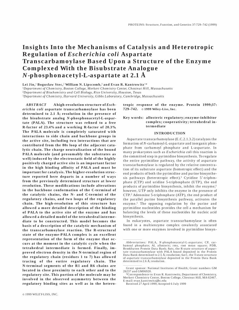
The active sites of aspartate transcarbamoylase are atthe interfaces between two adjacent catalytic chains.Furthermore, as has been demonstrated by biochemicalstudies,26 residues from both chains are required forenzymatic activity. Residues that interact with PALAinclude Ser52, Thr53, Arg54, Thr55, Arg105, His134,Arg167, Arg229, Gln231, and Leu267 (backbone) alongwith Ser80 and Lys84 from the adjacent catalytic chain.Shown in Fig. 1 are the interactions between these resi-dues and the PALA molecule in the C1 active site. Theinteractions are essentially identical in the C6 active siteexcept for a reorientation of the guanidinium group ofArg54. Noteworthy is the number of interactions to thePALA molecule and the number of these that involvepositively charged amino acid side chains (see Table II).Clearly, the electrostatic neutralization of PALA is ex-tremely important for its binding to the enzyme.
Main Chain Comparison Between the 8atc and theNew Structure
Overall, the new structure is very similar to the previ-ously reported structure of the enzyme with PALA boundat 2.5 A resolution (PDB code 8atc; the 8atc structure). Tocompare these two structures in more detail, each of thedomains in the two independently determined catalyticand regulatory chains (C1:R1 and C6:R6) were superim-posed separately between the two structures (Table III).For the superposition the flexible loop regions and terminiwere omitted in the calculation. The CP, Asp, and Zndomains are very similar, with rms deviations of less than0.3 A; however, there are twofold larger deviations in theallosteric domains, with rms deviations of approximately0.5 A. The larger rms deviations observed for the allostericdomains can be explained by a mechanical uncouplingfrom the rest of the molecule, as previously proposed.27
The major differences of the backbone conformation arelocated at the C-terminal of the catalytic chain, residue307 to 310; the N- and C-termini of the regulatory chain,residues 1r to 11r and residues 151r-153r, respectively; aswell as two loops of the regulatory chain, the 50s loop,
Fig. 1. Schematic diagram of the PALA bindingsite in the C1 chain of aspartate transcarbam-oylase. Shown are all of the residues that havehydrogen bonding interactions (dashed lines) withPALA. The only substantial difference between theC1 and C6 active sites is a reorientation of the sidechain of Arg54. This figure was drawn with theprogram LIGPLOT.45
732 L. JIN ET AL.
residues 48r to 55r, in the allosteric domain and the 130sloop, residues 129r to 133r, in the zinc domain.
Regulatory Chain 50s and 130s Loops
When the new and 8atc structures are compared, twoloop regions, the 50s and 130s loops, of the regulatorychain exhibit differences corresponding to slightly differ-ent conformations. The altered conformation of the 50sloop does result in significant reorientations of the sidechains of Glu52r and Met53r. This altered conformationmay reflect different conformational substates presentupon binding of the regulatory effectors. The conforma-tional change at the end of the loop would influence Lys56r,which has been directly implicated in altering the bindingaffinities of the regulatory effectors.28
Interfaces Between the Chains
Critical for the function of aspartate transcarbamoylaseare the interactions between the catalytic and regulatorychains of the enzyme.29 During the conversion of theenzyme from the T to the R state, numerous interactions
that stabilize the T state are broken while numerous otherinteractions that stabilize the R state are formed. Criticalfor stabilization of the T state of the enzyme are interac-tions between the 240s loop of one catalytic chain (C1) toeither a catalytic chain (C4) or a regulatory chain (R4) inthe opposite half of the molecule, C1-C4 and C1-R4 interac-tions. In the new structure of the enzyme with PALAbound reported here, no polar C1-C4 or C1-R4 interactionsare observed (see Table IV). The polar interactions be-tween the adjacent catalytic chains (C1-C2), regulatorychains of a dimer (R1-R6), and adjacent catalytic andregulatory chains (C1-R1) are provided in Table IV. Note-worthy is the asymmetric nature of the interactions be-tween the catalytic and regulatory chains in the twoindependent determined interfaces (C1-R1 and C6-R6).Substantially more interactions are observed in the C1-R1interface than in the C6-R6 interface. These data furthersupport the asymmetric nature of the upper versus lowerhalves of the enzyme that may be responsible for theobserved biphasic binding of the regulatory nucleotides.
Side Chain Conformation Changes
Because of the higher resolution of the new structuraldata, side chain conformations have been significantlyimproved in the new structure compared with the 8atcstructure. Most of the side chains in the new structure areclose to their ideal rotamer positions. A few important sidechain corrections were made in the active site of the C1and C6 catalytic chains (see Fig. 2). The configurations ofthe guanidinium groups of Arg54 in C6 and Arg229 in bothC1 and C6 were reversed, thus allowing them to form idealand saturated hydrogen bonds to PALA. This is the firsttime that any asymmetry between configurations of Arg54in the C1 and C6 chains has been observed.
Geometrical and Conformational Analysis of New,8atc, and 6at1 Structures
The higher than expected rms deviations for allostericdomains of the regulatory chains (see Table III) may
TABLE II. Interactions with PALAin theActive Site ofAspartate Transcarbamoylase
Residue AtomPALAatoma
C1 distanceb
(Å)C6 distanceb
(Å)
Ser52 OG O3P 2.47 2.46Thr53 N O2P 2.88 3.01Arg54 N O2P 3.14 2.75Arg54 NE O2P — 3.06Arg54 NH1 O2P 3.12 —Arg54 NH2 O2P — 2.89Thr55 N O3P 2.82 2.76Thr55 OG1 O3P 2.82 2.63Arg105 NH1 O1 2.64 2.98Arg105 NH2 O1P 2.63 3.35His134 NE2 O1 2.57 2.90Arg167 NE O2 2.73 2.75Arg167 NH2 O3 2.97 3.16Arg229 NE O5 2.71 2.86Arg229 NH2 O4 2.87 2.94Gln231 NE2 O5 3.12 —Leu267 O N2 3.06 2.93Ser80c OG O1P 3.30 2.97Ser80c OG O2P 2.77 3.16Lys84c NZ O3 3.02 2.91Lys84c NZ O4 2.67 2.99Lys84c NZ O1P 2.90 2.77aThe notations used for the atoms in the PALA molecule are as follows:
bC1 and C6 refer to the two independent catalytic chains in theasymmetric unit.cSer80 and Lys84 are contributed into the active site from the adjacentcatalytic chain.
TABLE III. Comparison of DomainsBetween This Structure and 8atc When
the Carbamoyl Phosphate Domains of C1areAligned†
Chaina Domain rms deviation (Å)
C1 Aspartate 0.269R1 Zinc 0.312R1 Allosteric 0.568C6 Carbamoyl 0.269C6 Aspartate 0.275R6 Zinc 0.291R6 Allosteric 0.529
†The rms deviation of the C1 CP domains in the twostructures was 0.201 Å.aThe chain designations used for 8atc are differentfrom all other deposited aspartate transcarbam-oylase coordinates. For these comparisons, C1 wascompared with the 8atc chain labeled C, R1 withthe 8atc chain labeled D, C6 with the 8atc chainlabeled A, and R6 with the 8atc chain labeled B.
733PALA-ASPARTATE TRANSCARBAMOYLASE STRUCTURE

indicate the existence of the protein in different conforma-tional substates because of a mechanical uncoupling of theallosteric domains from the rest of the molecule.27 To
evaluate the substates more quantitatively we have per-formed a conformational analysis using the method ofWilliams et al.25 This procedure was used to compare the
TABLE IV. Polar Interface Interactions in the New Structure†
C1-R1 C6-R6 C1-C2 R1-R6
Ser11-Glu142 Ser11-Glu142 Glu37-Lys40 Val9*-Val9*Leu88*-Glu119 Leu88*-Glu119 Gly37-His41 Glu10*-Gln8Ala89*-Glu119 Ala89*-Glu119 His41*-Arg65 Ala11*-Gln8Pro107*-Asn113 Pro107*-Asn113 Val43*-Arg65 Thr36*-Gln24
Gln108-Asn113 Ser52*-Asn78* Thr38*-Gln24Gln108-Cys114* Gln108-Cys114* Thr53-Ser80 Thr38*-Asn47Gln109-Asn113 Gln109-Asn113 Arg54-Ser80 Asp39-Arg55Glu109-Asn111 Glu109-Asn111 Arg54-Glu86 Asp39*-Asn47
Glu109-Ile115* Arg54*-Tyr98 Gln40*-Asn47Glu110*-Tyr140* Arg56-Gly72* Arg41-Asn47*Arg113-Lys139* Arg113-Lys139 His64-Ser69 Ile42*-Leu46*
Arg113-Glu142 His64-Val70* Ile42*-Ile42*Glu117-Lys139 Glu117-Lys139 Arg65-Tyr98* Ile44*-Ile44*Glu117-Tyr140 Glu117-Tyr140 Arg65-Asp100 Leu46*-Ile42*Ser131-Lys143 Ser74-Asn78 Arg55-Asp39Asn132-Cys141* Asn132-Cys141* Asp75-Asn78Asn132-Tyr140* Asp90-Arg269Asn132-Glu142 Thr97-Gly290Gln133-Glu142Glu196-Arg130Tyr197-Lys143Asp200-Arg128Asp200-Glu144Asp200-Arg130Glu204-Arg128 Glu204-Arg128†All interactions with distances less than 3.8 Å between nitrogen and oxygen are shown. Nopolar interactions were observed between the C1-R4 and C1-C4 interfaces. Asterisksindicate backbone atoms.
Fig. 2. Stereoview of the C6 active site of aspartate transcarbam-oylase with PALA bound. The portion of the 2 Fo-Fc electron density mapfor PALA is shown (contoured at 2.0 s) along with a comparison of theside chain conformations of the active site residues in the new (thick
lines) and 8atc (thin lines) structures. Water molecules are shown asasterisks. Residue numbers followed by a number sign (#) indicate thatthese residues are from the adjacent polypeptide chain. Hydrogen bondsare shown as dashed lines.
734 L. JIN ET AL.

new structure of the enzyme with PALA bound to the 8atcand 6at1 structures by calculating the angle and twistamong the various domains in the structure. The X-anglerepresents the degree to which two domains are openwhereas the Y-angle represents the relative twist of thetwo domains. The X-angles between all pairs of domainshave been calculated as described in Materials and Meth-ods and are presented in Table V. For both the C1 and C6chains, the angles between the CP and Asp domains in thenew structure are very similar to those in the 8atcstructure, but they are smaller than those in the 6at1structure. The angle between the two allosteric domains ofthe R1 and R6 chains (Al1-Al6) in the new structure wasvery similar to that in the 8atc structure, but it was largerthan that in the 6at1 structure.
A comparison of the angles between the Zn and Aldomains in the R1 and R6 chains of the new structureindicates that they are more symmetric than those of 8atcstructure, as opposed to the CP and Asp domains of thecatalytic chains, which show a more open and differenti-ated state (Table V). In the new structure, the anglesbetween CP1 and Zn1 are more open compared with thosebetween CP6 and Zn6 and between Zn1 and Al1 comparedwith Zn6 and Al6. The angle differences between the CPand Zn domains are smaller than those between the Znand Al domains.
A comparison of the twist (Y-angle) between the Zn andAl domains in the new and 8atc structures relative to theposition of these domains in the 6at1 structure was alsoperformed. Using 6at1, the T-state structure, as a refer-ence, the allosteric domain in the R1 domain of the newstructure twisted 0.87 degrees more than it did in the 8atcstructure, whereas the R6 allosteric domain of the newstructure twisted 0.42 degrees less than it did in the8atc structure. Furthermore, even between the new and8atc structures, the R1 and R6 chains exhibited slightly
different twist angles (approximately 1°), suggesting thatthere is a domain motion that cannot be represented bythis kind of measurement.
Conformational Change of the 240s Loop in theT=R Transition
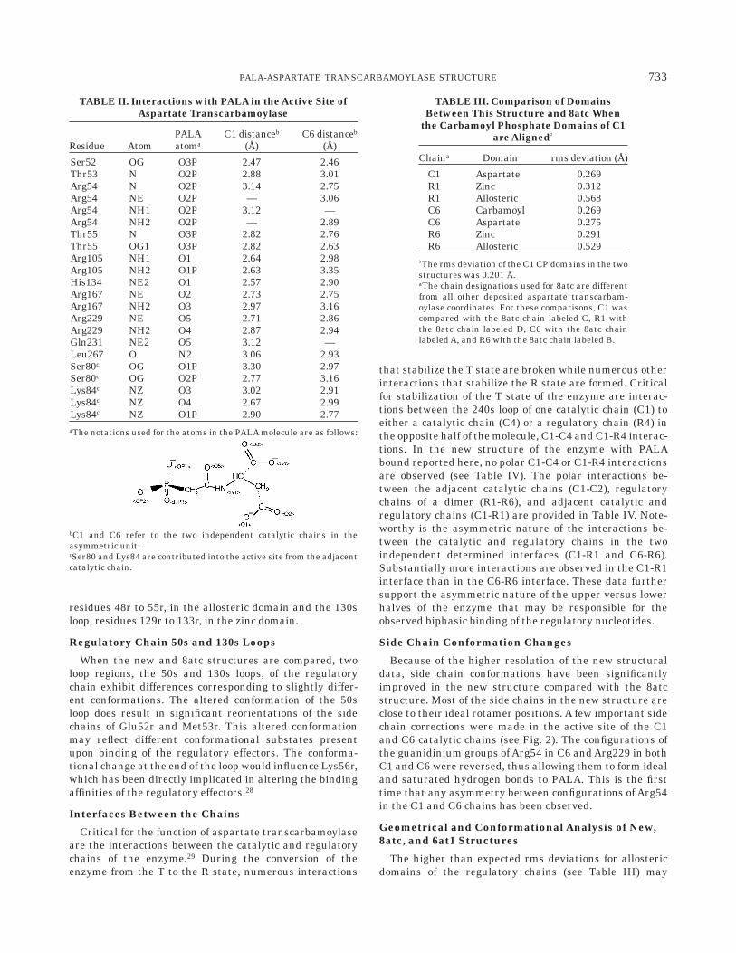
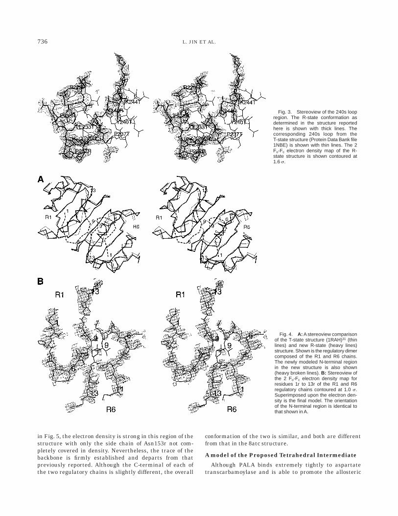
Critical for the conversion of the enzyme from the T tothe R state is the conformational change of the 240s loop.In the T-state structure, residues of the 240s loop interac-tion across the C1:C4 and C1:R4 interfaces, formingimportant T-state stabilizing interactions;29 however, theseinteractions are broken during the transition to the R-statewhen the 240s loop undergoes a significant conformationalchange. Fig. 3 shows a comparison of the 240s loop in the Tand R structures. For this figure we have used the T-statecoordinates of the 240s loop from the Thr82r=Ala mutantstructure at 2.6 A (PDB code 1NBE), which was rebuiltduring the course of its refinement.25 The Thr82r=Alamutation on the regulatory chains of the enzyme shouldnot directly influence the T-state conformation of this loop.When the T- and R-state structures are compared, thealteration in the conformation of the 240s loop results froma very simple rotation of only two peptide bonds (244 and245), which causes an unwinding of a helical segmentbetween residues 236 and 243 into a more extendedconformation. This extended R-state conformation of the240s loop allows side chains to form intrachain interac-tions stabilizing the loop’s conformation and thereby posi-tion side chains to create the high-activity high-affinityactive site.
N-termini of the Regulatory Chains
In early structures of aspartate transcarbamoylase,insufficient density was observed for the N-terminal regionof the regulatory chains to trace the polypeptide chain,presumably because of the mobility of this area of thestructure.21,30 In the T-state structure of the enzyme withCTP bound, Kosman et al.31 were able to model theN-terminal of one of the regulatory chains (R1). In thenew structure of the enzyme with PALA bound, theelectron density associated with the N-terminal region ismuch weaker than that in other parts of the molecule(compare Fig. 3 and Fig. 4B). However, there was sufficientdensity to build a tentative model for all the residues.After a number of rebuilding steps, a new model isproposed for the position of the first seven residues alongwith modifications to the position of residues eight to ten.As seen in Fig. 4, the N-terminal of the R1 chain and thatof the R6 chain in the new structure are different fromeach other, and both are different from the trace deter-mined for the R1 chain in the T-state structure with CTPbound.31
C-termini of the Regulatory Chains
Another area of the new structure that is different fromthe 8atc structure of the enzyme is the C-terminal regionof the two regulatory chains. In particular, residues 150r-153r unwind, forming a more extended structure. As seen
TABLE V. Comparison of the New PALA-LigatedStructure with the T and R Structures
ofAspartate Transcarbamoylase†
Domains incomparison1
X-planar angles between domainsa (degrees)
6at1 8atcbNew PALAstructure
Asp1-CP1 134.11 127.41 127.71Asp6-CP6 135.69 127.64 128.35CP1-Zn1 111.17 110.71 111.15CP6-Zn6 111.26 110.54 110.80Zn1-Al1 105.56 105.56 104.63Zn6-Al6 105.62 102.62 103.18Al1-Al6 153.28 155.52 155.83
†The number after the domain name corresponds to the particularchain in the asymmetric unit.aSee Materials and Methods for definition of the X-planar angle.bThe chain designations used for 8atc are different from all otherdeposited aspartate transcarbamoylase coordinates. For these compari-sons, CP1 and Asp1 were compared with the 8atc chain labeled C, Zn1and Al1 with the 8atc chain labeled D, CP6 and Asp6 with the 8atcchain labeled A, and Zn6 and Al6 with the 8atc chain labeled B.
735PALA-ASPARTATE TRANSCARBAMOYLASE STRUCTURE
in Fig. 5, the electron density is strong in this region of thestructure with only the side chain of Asn153r not com-pletely covered in density. Nevertheless, the trace of thebackbone is firmly established and departs from thatpreviously reported. Although the C-terminal of each ofthe two regulatory chains is slightly different, the overall
conformation of the two is similar, and both are differentfrom that in the 8atc structure.
A model of the Proposed Tetrahedral Intermediate
Although PALA binds extremely tightly to aspartatetranscarbamoylase and is able to promote the allosteric
Fig. 3. Stereoview of the 240s loopregion. The R-state conformation asdetermined in the structure reportedhere is shown with thick lines. Thecorresponding 240s loop from theT-state structure (Protein Data Bank file1NBE) is shown with thin lines. The 2Fo-Fc electron density map of the R-state structure is shown contoured at1.6 s.
Fig. 4. A: A stereoview comparisonof the T-state structure (1RAH)31 (thinlines) and new R-state (heavy lines)structure. Shown is the regulatory dimercomposed of the R1 and R6 chains.The newly modeled N-terminal regionin the new structure is also shown(heavy broken lines). B: Stereoview ofthe 2 Fo-Fc electron density map forresidues 1r to 13r of the R1 and R6regulatory chains contoured at 1.0 s.Superimposed upon the electron den-sity is the final model. The orientationof the N-terminal region is identical tothat shown in A.
736 L. JIN ET AL.

transition to the R-structural state, it is not an exactanalogue of the transition state because the carbonylcarbon does not have tetrahedral geometry, the aminogroup of carbamoyl phosphate is lacking, and the esteroxygen between the carbonyl and the phosphorus hasbeen replaced by a methylene bridge. Based upon thestructure reported here, we have modeled the proposedtetrahedral intermediate into the active site by replacingPALA and by keeping atoms that are the same in bothPALA and the tetrahedral intermediate in similar posi-tions. Necessary minor adjustments were then made tothe conformation of the tetrahedral intermediate tooptimize interactions. In addition, the conformations ofthe side chains of Lys84 and Gln137 were altered toimprove stereochemistry or contacts. As seen in Fig. 6,the intermediate fits into the position previously occupiedby PALA with almost no alteration in the conformation ofthe protein (see Fig. 2). All the charges of the intermediate
are electrostatically neutralized by the protein, and theorientation of the intermediate is positioned with elabo-rate precision by a variety of hydrogen-bonding interac-tions.
In this model there are numerous interactions to groupsattached to the tetrahedral carbon. For example, theamino group of the intermediate forms interactions withthe side chain of Gln137 and the backbone carbonyl ofPro266. The negatively charged oxygen is neutralized byside chain interactions with Arg105 and His134. Further-more, the ester oxygen interacts with Arg54, and theamide forms hydrogen bonds with Lys84 and the backbonecarbonyl of Pro268. The a-carboxylate is neutralized by abidentate interaction withArg167, whereas the b-carboxyl-ate is neutralized by interactions with Lys84 and a biden-tate interaction with Arg229. Finally, the phosphate isstabilized and neutralized by interactions with Ser52,Thr55, Ser80, Lys84, Arg54, and Arg105.
Fig. 5. Stereo view of the electrondensity map (2 Fo-Fc) for the C-termi-nal residues (145r to 153r) of the R1regulatory chain contoured at 1.0 s.Superimposed upon the electron den-sity is the final refined model (thicklines) as well as the correspondingresidues from the 8atc structure(dashed lines). A similar unwinding ofthe C-terminal is also observed in theR6 chain. This figure was drawn withthe program SETOR.46
Fig. 6. Stereo view of the C1 catalyticchain of aspartate transcarbamoylase withthe tetrahedral intermediate modeled intothe active site. Shown are all the side chainsthat make direct contact with the intermedi-ate from the C1 chain as well as Ser80 andLys84 from the adjacent catalytic chain.Hydrogen bonds are shown as dashed lines.
737PALA-ASPARTATE TRANSCARBAMOYLASE STRUCTURE

Our ability to replace the bisubstrate analogue PALAwith the structure of the tetrahedral intermediate withalmost no alterations in the structure of the enzyme-PALAcomplex indicates that the structure of the enzyme-PALAcomplex is an excellent model of the enzyme in its catalyti-cally active conformation.
DISCUSSION
Aspartate transcarbamoylase with PALA bound is con-sidered the R state or high-activity high-affinity state ofthe enzyme. Given the fact that PALA is also a potentialanticancer drug, the structure of the enzyme with PALAbound was reinvestigated, at significantly higher resolu-tion, in the work reported here. The overall quaternaryconformational change induced by PALA was similar tothat previously reported;16 however, the new higher-resolution structure provides a significantly improveddescription of the active site of the enzyme and new detailsof interactions at the domain interfaces and the N- andC-terminal regions of both the catalytic and the regulatorychains. Those regions were implicated in the homotropicand the heterotropic responses; therefore, the better struc-tural definition will allow us to better understand theirfunction in both catalysis and allosteric control.
Active Site of the Enzyme
As seen in Figs. 1 and 2, the PALA molecule is com-pletely saturated with interactions to side chain andbackbone groups in the active site. In fact, every hydrogenbonding donor or acceptor atom on the PALA molecule isinvolved in at least one interaction. Although most of theseinteractions come from side chain or backbone residues ofthe same polypeptide chain that contains the active site,two interactions are contributed from the 80s loop of theadjacent catalytic chain. A comparison of the T- andR-state structures of the enzyme in the vicinity of theactive site provides a rationale for the difference in activityand affinity between the T and R states of the enzyme. Inthe T-state structure, many of the positions of groupsinteracting with PALA are in alternate conformations,often pointing out of the active site (e.g., Lys84 andArg229). The free energy associated with the binding ofPALA, and presumably the natural substrates as well,induces the closure of the Asp and CP domains, therebymoving critical catalytic groups into their R-state positions(see Fig. 7). In addition, because of steric interference thedomain closure requires the associated quaternary confor-mational change, which also requires a new conformationof the 80s loop, allowing Ser80 and Lys84 to attain theircritical conformations that promote catalysis. The chargeneutralization of the bound PALA molecule (and presum-ably substrates as well) induced by the electrostatic field ofthe highly positively charged active site is an importantfactor in the high binding affinity of PALA (see Fig. 8).
Implications of the Structure for theCatalytic Mechanism
The high-resolution structure of the complex of aspar-tate transcarbamoylase and PALA reported here has pro-
vided a starting point for the development of a model of theproposed tetrahedral intermediate bound to the active siteof enzyme (see Fig. 6), which extends significantly theaccuracy attainable in the previous model.32 These modelsof the tetrahedral intermediate along with extensive func-tional data from site-specific mutagenesis allow us tobetter detail the events that take place during the catalyticcycle of aspartate transcarbamoylase.
As has been well established, the catalytic mechanismfor the transcarbamoylase reaction is ordered with car-bamoyl phosphate binding before aspartate and N-car-bamoyl-L-aspartate leaving before phosphate.33 The en-zyme exhibits little affinity for aspartate in the absence ofcarbamoyl phosphate, and thus the binding of aspartateoccurs only to the enzyme–carbamoyl phosphate complex.Evidence from X-ray structural analysis of the enzyme-phosphonoacetamide structure,34 low-angle X-ray solutionscattering,35 and ultraviolet36 and circular dichroism37
spectroscopy indicate that the binding of carbamoyl phos-phate induces conformational changes; however, theseconformational changes are local and distinct from thelarge quaternary conformational change that occurs uponaspartate binding. Thus, the binding of carbamoyl phos-phate significantly enhances the affinity of the enzyme foraspartate. It is noteworthy that modification of residuesinvolved in carbamoyl phosphate binding such as Thr55,His134, Arg105, and Gln137 has the most dramatic influ-ence on aspartate affinity.38 Clearly, if carbamoyl phos-phate cannot bind or binds in an incorrect orientation,because of alterations in side chain contacts, the bindingsite of aspartate is not formed correctly.
The mode of PALAbinding suggests thatArg105, His134,and Thr55 all interact with the carbonyl oxygen of car-bamoyl phosphate thereby enhancing the electrophilicityof the carbonyl carbon for attack by the amino group ofaspartate. However, for attack the protonated amino groupof aspartate must lose one proton, and a second protonmust be lost upon formation of the covalent bond betweenthe amino nitrogen and the carbonyl carbon. The structureof the PALA complex and a model of the proposed tetrahe-dral intermediate (see Fig. 6) suggest how the protons areremoved from the amino group of aspartate. As seen in Fig.8, the active site pocket is lined with many positivelycharged side chains forming an extremely electropositivemicroenvironment. The positive electrostatic field in theactive site pocket should lower the pKa of the amino groupof aspartate sufficiently so that the 2NH3
1 group loses aproton to solvent as aspartate binds. Two routes arepossible for the loss of the next proton from the aminogroup of aspartate in conjunction with the formation of thetetrahedral intermediate. One possibility, as suggested byGouaux et al.,32 is that there is a direct transfer of theproton to the leaving phosphate group. The other possibil-ity is that when the 80s loop of the adjacent catalytic chainswings into the active site, positioning Ser80 and Lys84,the electrostatic environment again is utilized to lower thepKa of the e-amino group of Lys84, allowing it to act as abase capturing the proton from the amino group of aspar-tate as the tetrahedral intermediate is formed. In support
738 L. JIN ET AL.

of this role for Lys84 is the direct interaction between thee-amino group of Lys84 and the nitrogen of the aminogroup of aspartate in the tetrahedral intermediate, as wellas recent site-specific mutagenesis experiments in whichLys84 was replaced by Asn. The K84N enzyme exhibitsmore than a 1,200-fold reduction in activity compared with
the wild-type enzyme.39 Upon departure of N-carbamoyl-L-aspartate from the active site, the domain-closed conforma-tion, characteristic of the active form of the enzyme, becomesunstable. This results in a possible repositioning of the 80sloop. Because Ser80 and Lys84 both interact with the phos-phate-leaving group, this repositioning of the 80s loop may
Fig. 7. Stereo view of the active site ofaspartate transcarbamoylase in the T (toppanel ) and R (bottom panel ) active sites.The side chains that interact with PALA inthe R state are shown in both panels. The80s loop of the adjacent catalytic chain,which contributes Ser80 and Lys84 in theactive site, is shown in yellow. Note thelarge number of interactions (dashed lines)with the PALA molecule in the R state(lower panel), many of which are involvedin neutralization of the negative chargeson the PALA molecule. For clarity Ser52 isnot labeled. The data used for the T stateof the enzyme (top panel) were from Pro-tein Data Bank file 1NBE. This figure wasdrawn with the program SETOR.46
Fig. 8. Stereo view of the C1 cata-lytic chain of aspartate transcarbam-oylase represented as a molecule sur-face as generated by the programGRASP.47 Mapped onto the surface isthe calculated electrostatic potential.The PALA molecule was placed in theactive site after generation of the sur-face; thus, the blue active site pocketindicates the positive electrostatic po-tential available to neutralize the highlynegatively charged PALA molecule.
739PALA-ASPARTATE TRANSCARBAMOYLASE STRUCTURE

provide assistance for the release of phosphate from the highlypositively charged active site.
The model of the tetrahedral intermediate also suggestsan explanation for the role of Arg54. When Arg54 isreplaced by Ala, the activity of the mutant enzyme isreduced by approximately 20,000-fold, with little changein substrate affinity.40 However, the X-ray structure of themutant enzyme in the presence of PALA showed that theactive site of the enzyme was virtually identical to that ofthe wild-type enzyme except for the loss of the side chain ofArg54 and the addition of three water molecules in theactive site.41 In the model of the tetrahedral intermediate(see Fig. 6), the side chain of Arg54 interacts with thebridging phosphate oxygen. The neutralization of thedeveloping negative charge on this oxygen as phosphate isexpelled would make the phosphate a significantly betterleaving group, thereby substantially lowering the activa-tion energy of the reaction. Also observed in the model aresome strained hydrogen bonds, which upon phosphate releasewould straighten, thereby promoting the chemical reactionand the spatial separation of the reaction products.
Conformation of the N-terminal of theRegulatory Chain
In most of the previous structures of aspartate transcar-bamoylase, the N-terminal region of the regulatory chains(residues 1r-7r) has not been modeled because of lack ofsufficient density in the maps. However, the N-terminal ofthe R1 chain was modeled in the structure of the enzymewith CTP bound.31 The location of the N-terminal near theregulatory binding site suggested that it might be involvedin the heterotropic interactions of the enzyme. Based uponthis structural data, mutagenesis experiments42,43 were
carried out that supported the notion that this region ofthe regulatory chain is involved in regulation.
In the recently published T-state structure of the unli-ganded Thr82r=Ala enzyme, a tentative model for theN-terminal fragment of the regulatory chains was pro-posed,25 which varied from that reported for the CTP-liganded enzyme.31 The new model was characterized byplacement of the first few residues of the regulatory chainat the nucleotide binding site, and a similar motif ispresent in the new structure of the PALA-ligated enzymereported here (see Fig. 9).
In the structure of the enzyme reported here, theelectron density in both the R1 and R6 chains was suffi-cient to model the first seven residues of the N-terminaland to allow a remodeling of residues 8r-10r. As seen inFig. 9, The new fold, despite being similar to that of theT-state structure, shows some unwinding and opens up thenucleotide binding site more than is observed in theT-state structure.25 In addition, the trace of the N-terminalresidues (1r-7r) was somewhat different in the R1 and R6chains, suggesting some inherent asymmetry (see Fig. 4).Furthermore, the location of the N-terminal residue itselfis extremely close to the nucleotide binding site. Thus, aconformational change in the N-terminal region may berequired for the nucleotides to bind. This notion is sup-ported by mutagenesis data that indicate that the bindingof CTP is enhanced by approximately fourfold in a mutantenzyme in which the N-terminal of the regulatory chainshas been deleted. Furthermore, the observed structuralasymmetry of the N-terminal region may be partiallyresponsible for the high- and low-affinity nucleotide bind-ing sites observed by equilibrium dialysis.44 Thus, theN-terminal region of the regulatory chain is important for
Fig. 9. Stereoview of the allosteric domain of three aspartate transcar-bamoylase structures. The open rope corresponds to the R-state struc-ture of the enzyme in the presence of phosphonoacetamide, malonate,and adenosine 58-triphosphate48 (PDB code 7at1). In this structure thefirst seven residues of the regulatory chain could not be resolved becauseof poor electron density. The striped rope corresponds to the R-statestructure of the Thr82=Ala mutant of aspartate transcarbamoylase in the
absence of heterotropic effects25 (PDB code 1NBE). In this structure theentire N-terminal region was fit. Note that the position of the N-terminalresidue precludes the binding of the nucleotide. The filled rope corre-sponds to the R-state structure of the enzyme in the presence of PALAreported here. In the R state, the position of the N-terminal does notpreclude the binding of the nucleotide. This figure was drawn with theprogram SETOR.46
740 L. JIN ET AL.
both nucleotide binding and for the heterotropic interac-tions of aspartate transcarbamoylase.
ACKNOWLEDGMENTS
This work was supported by Grants GM26237 (E.R.K.)and GM06920 (W.N.L.) from the National Institutes ofHealth.
REFERENCES1. Gerhart JC, Pardee AB. Enzymology of control by feedback
inhibition. J Biol Chem 1962;237:891–896.2. Wild JR, Loughrey-Chen SJ, Corder TS. In the presence of CTP,
UTP becomes an allosteric inhibitor of aspartate transcarbamy-lase. Proc Natl Acad Sci USA 1989;86:46–50.
3. Coleman PF, Suttle DP, Stark GR. Purification from hamster cellsof the multifunctional protein that initiates de novo synthesis ofpyrimidine nucleotides. J Biol Chem 1977;252:6379–6385.
4. Lue PF, Kaplan JG. Aspartate transcarbamylase and carbamylphosphate synthetase of yeast: a multifunctional enzyme com-plex. Biochem Biophys Res Commun 1969;34:426–433.
5. Collins KD, Stark GR. Aspartate transcarbamylase: interactionwith the transition state analogue N-(phosphonacetyl)-L-aspar-tate. J Biol Chem 1971;246:6599–6605.
6. Laing N, Chan WWC, Hutchinson DW, Oberg, B. Phosphorus-containing inhibitors of aspartate transcarbamoylase from Esch-erichia coli. FEBS Lett 1990;260:206–208.
7. Swyryd EA, Seaver SS, Stark GR. N-(phosphonacetyl)-L-aspar-tate, a potent transition state analog of aspartate transcarbamy-lase, blocks proliferation of mammalian cells in culture. J BiolChem 1974;249:6945–6950.
8. Tsuboi KK, Edmunds HN, Linda K. Selective inhibition of pyrimi-dine biosynthesis and effect on proliferative growth of coloniccancer cells. Cancer Res 1977;37:3080–3087.
9. Kensler TW, Erlichman C, Jayaram HN, Tyagi AK, Ardalan B,Cooney DA. Peripheral leukocytes as indicators of the enzymiceffects of N-(phosphonacetyl)-L-aspartic acid (PALA) on humanL-aspartate transcarbamoylase (ATCase) activity. Cancer TreatRep 1980;64:967–973.
10. Madani S, Baillon J, Fries J, et al. Pyrimidine pathway enzymesin human tumors of brain and associated tissues: potentialities forthe therapeutic use of N-(phosphonacetyl)-L-aspartate and 1-b-D-arabinofuranosylcytsine. Eur J Cancer Clin Oncol 1987;23:1485–1490.
11. Morton RF, Creagan ET, Cullinan SA, et al. Phase II studies ofsingle-agent cimetidine and the combination N-phosphonacetyl-L-aspartate (NSC-224131) plus L-alanosine (NSC-153353) in ad-vanced malignant melanoma. J Clin Oncol 1987;5:1078–82.
12. Cohen RE, Schachman HK. Kinetics of the interaction ofN-(phosphonacetyl)-L-aspartate with the catalytic subunit of as-partate transcarbamoylase. J Biol Chem 1986;261:2623–2631.
13. Ahluwalia GS, Grem JL, Hao Z, Cooney DA. Metabolism andaction of amino acid analog anti-cancer agents. Pharmacol Ther1990;46:243–71.
14. Livingstone LR, White A, Sprouse J, Livanos E, Jacks T, Tisty TD.Altered cell cycle arrest and gene amplication potential accom-pany loss of wild-type p53. Cell 1992;70:923–935.
15. Honzatko RB, Crawford JL, Monaco HL, et al. Crystal andmolecular structures of native and CTP-liganded aspartate car-bamoyltransferase from Escherichia coli. J Mol Biol 1982;160:219–263.
16. Ke HM, Lipscomb WN, Cho Y, Honzatko RB. Complex ofN-phosphonacetyl-L-aspartate with aspartate carbamoyltransfer-ase: X-ray refinement, analysis of conformational changes andcatalytic and allosteric mechanisms. J Mol Biol 1988;204:725–747.
17. Svergun DI, Barberato C, Koch MHJ, Fetler L, Vachette P. Largedifferences are observed between the crystal and solution quater-nary structures of allosteric aspartate transcarbamylase in theR-state. Proteins 1997;27:110–117.
18. Nowlan SF, Kantrowitz ER. Superproduction and rapid purifica-tion of E. coli aspartate transcarbamoylase and its catalyticsubunit under extreme derepression of the pyrimidine pathway. JBiol Chem 1985;260:14712–14716.
19. Gerhart JC, Holoubek H. The purification of aspartate transcarba-mylase of Escherichia coli and separation of its protein subunits. JBiol Chem 1967;242:2886–2892.
20. Ke HM, Honzatko RB, Lipscomb WN. Structure of unligatedaspartate carbamoyltransferase of Escherichia coli at 2.6-A reso-lution. Proc Natl Acad Sci USA 1984;81:4027–4040.
21. Krause KL, Voltz KW, Lipscomb WN. 2.5 A structure of aspartatecarbamoyltransferase complexed with the bisubstrate analogN-(phosphonacetyl)-L-aspartate. J Mol Biol 1987;193:527–553.
22. Jones TA, Zou JY, Cowan SW, Kjeldgaard M. Improved proteinmodels in electron density maps and the location of errors in thesemodels. Acta Crystallogr 1991;A47:110–119.
23. Brunger AT. X-PLOR, Version 3.1. A system for crystallographyand NMR New Haven: Yale University Press; 1992.
24. Laskowski RA, MacArthur MW, Moss DS, Thornton JM. PRO-CHECK: A program to check the stereochemical quality of proteinstructures. J Appl Crystallogr 1993;26:283–291.
25. Williams MK, Stec B, Kantrowitz ER. A single mutation in theregulatory chain of Escherichia coli aspartate transcarbamoylaseis an extreme T-state structure. J Mol Biol 1998;281:121–134.
26. Wente SR, Schachman HK. Shared active sites in oligomericenzymes: model studies with defective mutants of aspartatetranscarbamoylase produced by site-directed mutagenesis. ProcNatl Acad Sci USA 1987;84:31–35.
27. Tanner JJ, Smith PE, Krause KL. Molecular dynamics simula-tions and rigid body (TLS) analysis of aspartate carbamoyltrans-ferase: evidence for an uncoupled R state. Protein Sci 1993;2:927–935.
28. Corder TS, Wild JR. Discrimination between nucleotide effectorresponses of aspartate transcarbamoylase due to a singe sitesubstitution in the allosteric binding site. J Biol Chem 1989;264:7425–7430.
29. Kantrowitz ER, Lipscomb WN. Escherichia coli aspartate transcar-bamoylase: the molecular basis for a concerted allosteric transi-tion. Trends Biochem Sci 1990;15:53–59.
30. Kim KH, Pan Z, Honzatko RB, Ke HM, Lipscomb WN. Structuralasymmetry in the CTP-liganded form of aspartate carbamoyltrans-ferase from Escherichia coli. J Mol Biol 1987;196:853–875.
31. Kosman RP, Gouaux JE, Lipscomb WN. Crystal structure ofCTP-ligated T state aspartate transcarbamoylase at 2.5 A resolu-tion: implications for aspartate transcarbamoylase mutants andthe mechanism of negative cooperativity. Proteins 1993;15:147–176.
32. Gouaux JE, Krause KL, Lipscomb WN. The catalytic mechanismof Escherichia coli aspartate carbamoyltransferase: a molecularmodeling study. Biochem Biophys Res Commun 1987;142:893–897.
33. Wedler FC, Gasser FJ. Ordered substrate binding and evidencefor a thermally induced change in mechanism for E. coli aspartatetranscarbamylase. Arch Biochem Biophys 1974;163:57–68.
34. Gouaux JE, Lipscomb WN. Crystal structures of phosphonoacet-amide ligated T and phosphonoacetamide and malonate ligated Rstates of aspartate carbamoyltransferase at 2.8 A resolution andneutral pH. Biochemistry 1990;29:389–402.
35. Fetler L, Tauc P, Vachette P. Carbamyl phosphate modifies the Tquaternary structure of aspartate transcarbamylase, therebyfacilitating the structural transition associated with cooperativity.J Appl Crystallogr 1997;30:781–786.
36. Collins KD, Stark GR. Aspartate transcarbamylase: studies of thecatalytic subunit by ultraviolet difference spectroscopy. J BiolChem 1969;244:1869–1877.
37. Griffin JH, Rosenbusch JP, Weber KK, Blout ER. Conformationalchanges in aspartate transcarbamylase. I. Studies of ligandbinding and of subunit interactions by circular dichroism spectros-copy. J Biol Chem 1972;247:6482–6490.
38. Stevens RC, Chook YM, Cho CY, Lipscomb WN, Kantrowitz ER.Escherichia coli aspartate carbamoyltransferase: the probing ofcrystal structure analysis via site-specific mutagenesis. ProteinEng 1991;4:391–408.
39. Macol C, Dutta M, Stec B, Kantrowitz ER. The 80s loop of thecatalytic chain of Escherichia coli aspartate transcarbamoylase iscritical for catalysis and homotropic cooperativity. Protein Sci1999;8:1305–1313.
40. Stebbins JW, Xu W, Kantrowitz ER. Three residues involved inbinding and catalysis in the carbamyl phosphate binding site of
741PALA-ASPARTATE TRANSCARBAMOYLASE STRUCTURE

Escherichia coli aspartate transcarbamylase. Biochemistry 1989;28:2592–2600.
41. Stebbins JW, Robertson DE, Roberts MF, Stevens RC, LipscombWN, Kantrowitz ER. Arginine 54 in the active site of Escherichiacoli aspartate transcarbamoylase is critical for catalysis: a site-specific mutagenesis, NMR and X-ray crystallographic study.Protein Sci 1992;1:1435–1446.
42. Dembowski NJ, Kantrowitz ER. The use of alanine scanningmutagenesis to determine the role of the amino terminus of theregulatory chain in the heterotropic mechanism of Escherichiacoli aspartate transcarbamoylase. Protein Eng 1994;7:673–679.
43. Sakash JB, Kantrowitz ER. The N-terminus of the regulatorychain of Escherichia coli aspartate transcarbamoylase is impor-tant for nucleotide binding and heterotropic effects. Biochemistry1998;37:281–288.
44. Winlund CC, Chamberlin MJ. Binding of cytidine triphosphate toaspartate transcarbamylase. Biochem Biophys Res Commun 1970;40:43–49.
45. Wallace AC, Laskowski RA, Thornton JM. LIGPLOT: a program togenerate schematic diagrams of protein-ligand interactions. ProtEng 1995;8:127–134.
46. Evans SV. SETOR: Hardware-lighted three-dimensional solidrepresentations of macromolecules. J Mol Graph 1993;11:134–138.
47. Nicholls A, Sharp KA, Honig B. Protein folding and association:insights from the interfacial and thermodynamic properties ofhydrocarbons. Proteins 1991;11:281–296.
48. Gouaux JE, Stevens RC, Lipscomb WN. Crystal structures ofaspartate carbamoyltransferase ligated with phosphonoacet-amide, malonate and CTP or ATP at 2.8 A resolution and neutralpH. Biochemistry 1990;29:7702–7715.
742 L. JIN ET AL.