injury in septic patients abstract index as an indicator ... lung water index as an indicator of...
TRANSCRIPT

Extravascular lung waterindex as an indicator of lunginjury in septic patientsBY ŽELJKO DRVAR, VIŠNJA MAJERIĆ KOGLER, DINKO TONKOVIĆ , MIRJANA MIRIĆ , MARIO
PAVLEK, MLADEN PERIĆ
Abstract
Introduction. Transpulmonary thermodilution using PiCCO (Pulse-induced
Contour Cardiac Output) is a standard minimally invasive method used for
haemodynamic monitoring. Objectives. The goal of this paper is to examine the
correlation and dynamics of the ExtraVascular Lung Water Index (EVLWI) as an
indicator of acute lung injury in septic patients who underwent major abdominal
surgery. Two groups of patients were selected: the ones with ALI (Acute Lung
Injury): ALI patient group, and the ones without ALI: non-ALI patient group. A
correlation between EVLWI and other haemodynamic and respiratory data in both
groups were analyzed.
Materials and methods. The study included 48 patients. Throughout the seven-day
period EVLWI, GEDVI (Global End-Diastolic Volume Index), ITBVI (IntraThoracic
Blood Volume Index), CI (Cardiac Index), SVRI (Systemic Vascular Resistance
Index) were measured in both groups using PiCCO monitoring over 8-hour
intervals as well as heart rate, mean arterial pressure, serum albumin
concentration, PaCO2 (arterial partial pressure of carbon dioxide), PaO2 (arterial
partial pressure of oxygen), PaO2/FiO2 (arterial partial pressure of
oxygen/fraction of inspired oxygen) ratio, lung compliance, lung resistance and
ScvO2 (central venous oxygen saturation). All patients were analgosedated,
intubated, mechanically ventilated, in sinus cardiac rhythm. Circulatory unstable
patients had vasoactive support and Sequential Organ Failure Assessment (SOFA)
scores calculated. Ventilator settings and dosage of vasoactive drugs were kept
SIGNA VITAE 2015; 10(1): 74 - 92
74
constant during the study.
Results. EVLWI was significantly higher in ALI patients group compared to non-
ALI patients group. In patients with ALI group 11/22 patients died (50%), in the
non-ALI patients group 6/26 patients died (23%). EVLWI was significantly higher in
patients that died compared to ones who survived.
Conclusion. EVLWI is a good indicator of early acute lung injury in surgical
patients with sepsis.
Key words: extravascular lung water index, acute lung injury, PiCCO monitoring,
sepsis
Introduction
It is possible to measure EVLWI (ExtraVascular Lung Water Index) by TPTD
(TransPulmonary ThermoDilution) technique using PiCCO (Pulse-induced
Contour Cardiac Output) monitoring. It is a standard minimally invasive method
used for continuous monitoring of haemodynamic and volumetric parameters
combining TPTD method and pulse curve analysis. (1,2)
EVLWI is defined as the difference between ITTV (IntraThoracic Thermal Volume),
which is measured by transpulmonary thermodilution, and ITBV (IntraThoracic
Blood Volume), see formula 1. By indexing the retreived data according to body
mass we can extrapolate EVLWI, see formula 2. As it is not possible to get ITBV by
transpulmonary thermodilution, instead an indirect indicator is measured, that is
GEDV (Global End-Diastolic Volume), see formula 3. EVLWI is indicative of the
volume in interstitial lung tissue and intraalveolar space. (3,4) Indirect acute lung
injury is a common consequence of sepsis. According to the literature, Acute Lung
Injury (ALI) occurs in 37-41% of all sepsis patients. Depending on the diagnostic
criteria, age groups and therapeutic protocols, the death rate is 29-42%. (5-12) ALI
is characterized by hypoxemia, non-cardiogenic lung oedema, poor lung
compliance and increased alveolar capillary permeability. (13)
Increased alveolar capillary permeability is a result of bacterial endotoxins,
migration and adherence of polymorphonuclear leukocytes as well as a number of
inflammatory factors: neutrophil elastase, colagenase A and B, protease and nitric
oxide. Other pro-inflammatory factors transmit the systemic inflammatory
response to the lungs: TNF-α (tumour necrosis factor α), IL-1β (interleukin 1β), IL-
SIGNA VITAE 2015; 10(1): 74 - 92
75

6 (interleukin 6), IL-8 (interleukin 8), leukotriene and cyclooxygenase pathways
without fibrinolysis, the C5a complement system, along with numerous
chemokines and cytokines. (14)
Along with the aforementioned, the intensity of alveolar injury is dependent on
the activity of thrombin, oxygen radicals, proteolytic enzymes and adhesion
molecules. As a result surfactant formation is insufficient and its action is
inadequate. Consequently, destruction of capillary endothelium and alveolar
epithelium causes increased vascular permeability and quickly results in fluid
collection in the interstitial space and in alveoli. (14)
The EVLWI reference range in healthy adults is 3.0-7.0ml/kg. EVLWI values above
10ml/kg indicate development of pulmonary oedema and correlate with morbidity
and mortality. A 100% increase in EVLWI values are not clinically evident on chest
x-rays, while those of a 200-300% increase are correlated to significant
hypoxemia. (15)
The goal of this paper is to examine the correlation and dynamics of the EVLWI as
an indicator of acute lung injury in septic patients who have undergone major
abdominal surgery.
Materials and methods
This prospective observational study was conducted from May 2009 until October
2011, at the Department of Anaesthesiology, Reanimatology and Intensive care at
the University Hospital of Zagreb. It included 48 septic patients who had
undergone major abdominal surgery. Of the 48 patients, 25 were male aged 47
(SD=8) and 23 were female aged 43 (SD=7). They were divided into two groups. The
first consisted of 22 patients who had ALI (ALI patients group), the second group,
consisted of 26 patients, who were without ALI (non-ALI patients group). All of the
participants had signed informed consent.
Diagnosis of both sepsis and ALI was based upon internationally defined criteria,
clinical signs and symptoms, as well as laboratory results. (16-20)
Exclusion criteria are listed in table 1.
Upon admission to the intensive care unit, post-operatively, all of the patients had
endotracheal tubes and were mechanically ventilated. They were all
SIGNA VITAE 2015; 10(1): 74 - 92
76

analgosedated with midazolam (0.08-0.2mg/kg/h) and sufentanil (0.3-0.5µg/kg).
Both drugs were applied intravenously by a syringe pump system.
Patients who were haemodynamically unstable (mean arterial pressure <
60mmHg) were given vasoactive drugs (norepinephrine 0.6-1.8µg/kg/min), and
had their SOFA (Sequential Organ Failure Assessment) score calculated.
The ventilation mode was BIPAP (BiPhasic Positive Airway Pressure) and was set
to FiO2 (fraction of inspired oxygen) 0.4-0.8, respiratory rate of 12-18/min, Phigh
(pressure high) 15-25cmH2O, Plow (pressure low) 5-8cmH2O, Thigh (time high) 2-
5s, Tlow (time low) 0.5-1.0s. All patients had a dual-lumen central venous catheter
(Arrow International, 7F, 20cm, Athlore, Ireland) placed in the internal jugular
vein. Each patient had a chest x-ray done in order to confirm that the catheter
was in place. Next, an intraarterial PiCCO catheter (Pulsiocath PV2014L16N, 4F)
was placed into the femoral artery using Seldinger’s method. Primary monitor
used was Dräger Infinity Delta XL, Germany, with secondary input from PiCCO
Dräger Infinity R PiCCO Smart Pod TM.
All of the measured variables, EVLWI, GEDVI (Global End-Diastolic Volume
Index), ITBVI, CI (Cardiac Index) and SVRI (Systemic Vascular Resistance Index)
were taken in both study groups using PiCCO in the same 8-hour intervals, three
times a day throughout the study.
Each measurement was done using a thermodilution indicator, 15ml of cold
(<8%C) 5% glucose solution, which was injected through the central venous
catheter. The final value was the mean of three consecutive measurements, each
taken within 5-minute intervals. PiCCO monitoring was used in both study groups
within a seven-day time period until they achieved optimal haemodynamic and
TPTD values. During this time period HR (Heart Rate), MAP (Mean Arterial
Pressure), serum albumin concentration (as an indirect indicator of oncotic
pressure within vascular system), PaCO2 (arterial partial pressure of carbon
dioxide), PaO2 (arterial partial pressure of oxygen), PaO2/FiO2 ratio (arterial
partial pressure of oxygen/fraction of inspired oxygen), lung compliance and
resistance, ScvO2 (central venous oxygen saturation) were all regulary measured.
Both study groups had chest x-rays, ECGs and transthoracic heart ultrasound
(Siemens P4-2 probe) done in order to measure their left ventricular ejection
fraction and diastolic heart dysfunction.
Both groups of patients were given crystalloids (30ml/kg) up to the point at which
SIGNA VITAE 2015; 10(1): 74 - 92
77
their optimal preload was achieved (MAP>60mmHg, EVLWI<7ml/kg, ITBVI 850-
1000ml/m2, GEDVI 680-800ml/m2). Patients with haemoglobin concentration
<70g/l and/or CI≤2.5l/min/m2 were given packed red cell transfusion.
All of the data measured or calculated was statistically processed using the
independent t-test, along with ANOVA (ANalysis Of VAriance) applied for
repeating measurements. Values of p<0.05 were considered statistically
significant.
Results
The study included 48 septic patients who had undergone major abdominal
surgery. Of those, 22 had ALI and 26 did not. There were 14 male and 8 female
patients in ALI group versus 11 male and 15 female patients in non-ALI group.
Table 2 displays the results according to age, APACHE II (Acute Physiology and
Chronic Health Evaluation II) score and SOFA score in ALI and non-ALI group.
Patients with positive haemocultures received antibiotics according to the
antibiogram (table 3). Microorganisms isolated from bronchoalveolar lavage
samples in patients with ALI group are displayed in table 4.
EVLWI was markedly increased in the ALI patient group between the 3rd and 7th
day. Also, a statistically significant difference in EVLWI values was found between
the two groups within the 1st and 7th day (table 5). Furthermore, there was an
evident positive growth trend of EVLWI with each passing day (table 5).
Figure 1 depicts the trends and differences of EVLWI in the two groups over a
period of seven days. There is a discrepancy between the median of EVLWI in the
two groups from 4th to 7th day. That is, the EVLWI in the non-ALI group of
patients reaches a plateau, while in the ALI group it continues to rise.
Figure 2 depicts AUROC (Area Under Receiver Operating Characteristic) curve for
EVLWI (0.943; 95% confidence interval 0.849-0.987) with p<0.001.
Optimal discrimination threshold between the groups was 14% for EVLWI
(sensitivity 72.2%, specificity 97.5%) which indicates that this diagnostic method is
efficient.
There was also a statistically significant difference throughout the rest of the
values measured in both groups: GEDVI, ITBVI, SVRI and CVP. Mean values of
SIGNA VITAE 2015; 10(1): 74 - 92
78
GEDVI as well as ITBVI in ALI group of patients were higher than reference values
(table 6). CI values did not show a statistically significant difference between the
two groups (table 6).
In the ALI patient group, there was a sharp rise in PaCO2 from the 3rd day, while
the non-ALI patient group showed a trend towards a decrease in PaCO2 from the
1st day onwards (table 5, figure 3). On the other hand, the measured difference in
PaO2 in both patient groups showed a constant drop in PaO2 in the ALI patient
group during a week-long observation that was greater than the decrease in PaO2
in the non-ALI patient group (table 5, figure 4). Albumin values were also tracked
during the seven-day period. The results show that serum albumin concentration
in the non-ALI patient group was greater in comparison to ALI patient group
(table 5, figure 5). Patients with ALI had increased EVLWI values and reduced
serum albumin concentrations.
The following lung function indices were monitored: PaO2/FiO2 ratio, compliance,
lung resistance and LIS (Lung Injury Score). The ALI patient group had a
significantly higher resistance of the respiratory system and reduced compliance
and PaO2/FiO2 ratio in comparison to non-ALI patient group (table 7).
Total seven-day mortality in both research groups was 17/48 patients (35%). In the
ALI patient group 11/22 patients died (50%), while 6/26 (23%) patients died in the
non-ALI patient group. Measured EVLWI values on the 7th day of monitoring were
significantly increased in the group of deceased patients in comparison to those
who survived: 16.5ml/kg vs 12.4ml/kg (figure 6).
Discussion
The results of this research study show that the EVLWI can be used in the
assessment of the appearance and dynamics of acute lung injury in surgical
patients with sepsis.
This is shown in the significantly higher EVLWI values in the ALI patient group
when compared to the non-ALI patient group. EVLWI values in the ALI patient
group were higher in patients who died than in those who survived. The initially
measured EVLWI values in the ALI patient group were higher than in the non-ALI
patient group, however it was still within the reference interval spread. A
significant increase in measured EVLWI values in the ALI patient group began on
SIGNA VITAE 2015; 10(1): 74 - 92
79

the 3rd day and maximum values were attained on the 7th day of measurement.
Simultaneously with the increase in EVLWI, the ALI patient group presented a
reduced serum albumin concentration. In the ALI patient group an increase in
EVLWI resulted in significant lung function disorder, which could be seen through
changes in relations between PaO2/FiO2 ratio, lung compliance and lung
resistance. The resulting hypoxemia was the result of hindered oxygen diffusion
through the alveocapillary membrane and reduced lung compliance as well as
increased lung resistance that stemmed from oedema and lung colapse.
The measured EVLWI values and the dynamics of the increase in the ALI patient
group is in accordance with the stated goal of this research, as well as with the
current knowledge on the subject. Recent data from the literature supports the
effectiveness of EVLWI as an indicator for early detection of patients with ALI. (21,
22)
Cordemans et al. confirm the significance of the connection between EVLWI
dynamics, intraabdominal hypertension, preload optimisation with fluid therapy
in mechanically ventilated patients and poor treatment results in patients with
ALI. (23) The meta-analysis of Zhongheng et al investigated EVLWI as an indicator
of poor outcome in non-surgical patients with burns, sepsis and ALI. It included 11
studies carried out in 9 countries with a total of 670 patients between 1999 and
2010. EVLWI was significantly increased in the group of deceased patients in
comparison to those who survived, with a median of 5.06ml/kg. (24) Our research
showed a median of 6.6ml/kg.
The meta-regression analysis carried out by Eichhorn et al. researched the
connection between EVLWI and GEDVI in the optimisation of preload in two
different patient groups with sepsis, surgical and non-surgical. The surgical group
consisted of patients after cardiac surgery, neurosurgery and vascular surgery. It
analyzed a total of 138 studies with a total of 4682 patients published on PubMed
with the keywords EVLWI, GEDVI and TPTD between 1990 and 2010. EVLWI and
GEDVI were significantly higher in the patient group with sepsis after major
surgery. (25)
Maharaj’s overview consisted of research of PubMed using EVLWI, ALI and ARDS
(Acute Respiratory Distress Syndrome) as keywords. It examined the usefulness of
exact EVLWI quantification in diagnosis, treatment and prognosis of both ALI and
ARDS.
SIGNA VITAE 2015; 10(1): 74 - 92
80

In conclusion, EVLWI is a useful indicator of clinical behaviour in mechanically
ventilated patients, especially during PEEP (Positive End-Expiratory Pressure)
titration, optimisation of volume therapy and poor patient outcome. (26)
The level of PEEP applied during mechanical ventilation can influence the
measured EVLWI values, therefore the ventilator settings were constant
throughout the research period. Of course, as in all research, this study has
drawbacks: specifics of the observed population – surgical patients after
laparotomy. Furthermore, the reliability of EVLWI measurements is reduced
through vascular lung obstruction along with a change in the
ventilation/perfusion ratio, including hypoxic lung vasoconstriction and a high
PEEP level. An additional drawback is a small patient sample included in the
research. Finally, the usefulness of applying TPTD technology in patients with
heart shunts (recirculation of the indicator) is questionable. Both the first and the
second group represented critically ill patients in need of haemodnaymic
monitoring that is minimally invasive, with a goal of preventing possible
complications.
The results of this research show that EVLWI can be useful for early detection of
patients with ALI.
This is in accordance with expectations and assumptions built upon current
literature. Insofar as all standards and prerequisites for the application of PiCCO
monitors have been met, the obtained data on EVLWI make it a good indicator of
the early onset of acute lung injury in patients with sepsis. EVLWI can also be an
indicator of the effectiveness of the therapy in treating ALI, especially fluid
therapy, mechanical ventilation strategies and treatment outcomes. However, for
definitive confirmation of EVLWI value in an assessment of the onset of acute lung
injury in patients with sepsis after major abdominal surgery, further research on a
larger number of patients is necessary.
Formula 1.
EVLW=ITTV-ITBV
EVLW, ExtraVascular Lung Water; ITBV, IntraThoracic Blood Volume; ITTV,
IntraThoracic Thermal Volume.
Formula 2.
EVLWI=EVLW/(predicted body weight)
SIGNA VITAE 2015; 10(1): 74 - 92
81
EVLW, ExtraVascular Lung Water; EVLWI, ExtraVascular Lung Water Index.
Formula 3.
ITBV=1.25×GEDV-28.4
GEDV, Global End-Diastolic Volume; ITBV, IntraThoracic Blood Volume.
Table 1. Exclusion criteria.
EXCLUSION CRITERIA
Coagulation disorders
Cardiomyopathy
Aortic valve insufficiency
Left ventricular ejection fraction <45%
Chronic obstructive lung disease
Children
Pregnant women
Patients who did not sign informed consent
Patients with contraindication for femoral artery puncture
Allergic diathesis
Disseminated malignancy
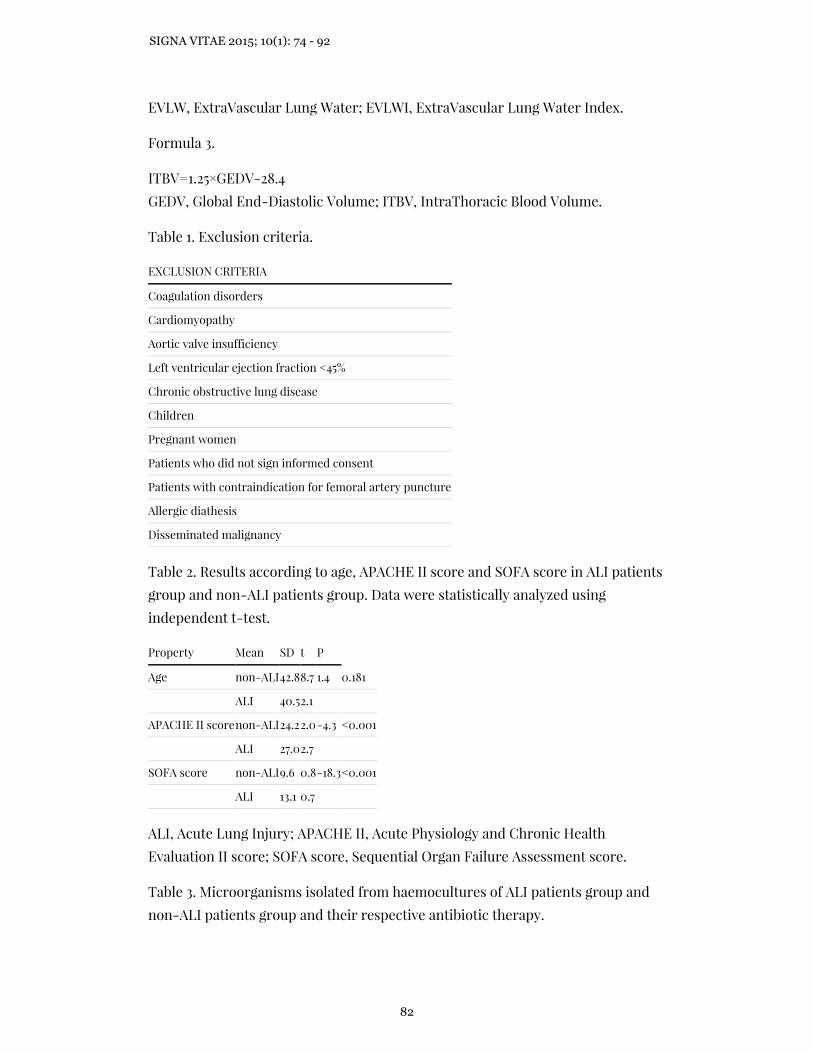
Table 2. Results according to age, APACHE II score and SOFA score in ALI patients
group and non-ALI patients group. Data were statistically analyzed using
independent t-test.
Property Mean SD t P
Age non-ALI42.88.7 1.4 0.181
ALI 40.52.1
APACHE II scorenon-ALI24.22.0-4.3 <0.001
ALI 27.02.7
SOFA score non-ALI9.6 0.8-18.3<0.001
ALI 13.1 0.7
ALI, Acute Lung Injury; APACHE II, Acute Physiology and Chronic Health
Evaluation II score; SOFA score, Sequential Organ Failure Assessment score.
Table 3. Microorganisms isolated from haemocultures of ALI patients group and
non-ALI patients group and their respective antibiotic therapy.
SIGNA VITAE 2015; 10(1): 74 - 92
82

Microorganism Number of positive
haemocultures
Antibiotic(s)
Patients with ALIn(%) Patients without
ALIn(%)
S. aureus 2(6.9) 3(15.8) cloxacillin,
clindamycin
S. aureus (MRSA) 6(20.7) 4(21.1) teicoplanin,
vancomycin
Coagulase negative
staphylococcus
4(13.8) 2(10.5) teicoplanin,
vancomycin
P. aeruginosa 8(27.6) 6(31.6) meropenem, colistin
K. pneumoniae ESBL 5(17.2) 2(10.5) meropenem,
cefepime
A. baumanii 4(13.8) 2(10.5) meropenem
ALI, Acute Lung Injury; ESBL, Extended Spectrum Beta Lactamase; MRSA,
Methicillin Resistant Staphylococcus Aureus.
Table 4. Microorganisms isolated from bronchoalveolar lavage samples in ALI
patient group.
Microorganism Number of positive culturesn(%)
S. aureus (MRSA) 4(18.2)
P. aeruginosa 6(27.3)
K. pneumoniae ESBL3(13.6)
C. albicans 5(22.7)
C. dubliniensis 1(4.5)
C. krusei 3(13.6)
ALI, Acute Lung Injury; ESBL, Extended Spectrum Beta Lactamase; MRSA,
Methicillin Resistant Staphylococcus Aureus
Table 5. Transpulmonary thermodilution and laboratories data in ALI patient
group and non-ALI patient group during the seven-day period. Data were
statistically analyzed using independent t-test.
Variable Time ALIn=22non-ALIn=26t P
Mean SD MeanSD
EVLWI(ml/kg)Day 1 7.09 0.29 5.27 0.35 19.37 <0.001
SIGNA VITAE 2015; 10(1): 74 - 92
83
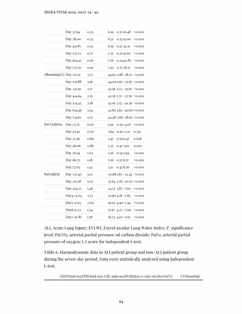
Day 27.64 0.23 6.19 0.35 16.48 <0.001
Day 38.00 0.35 6.71 0.25 15.00 <0.001
Day 411.85 0.53 6.91 0.17 45.31 <0.001
Day 513.53 0.71 7.35 0.25 41.62 <0.001
Day 614.42 0.79 7.76 0.2440.82 <0.001
Day 7 15.30 0.95 7.93 0.25 38.15 <0.001
Albumin(g/L) Day 1 17.11 3.73 44.92 2.98 -28.71 <0.001
Day 215.88 3.16 44.09 2.65 -33.67 <0.001
Day 315.19 2.77 43.59 2.73 -35.67 <0.001
Day 414.64 2.55 43.36 2.71 -37.56 <0.001
Day 514.42 2.18 43.19 2.57 -41.36 <0.001
Day 614.46 2.14 42.82 2.61 -40.60<0.001
Day 7 14.61 2.27 42.48 2.66 -38.62 <0.001
PaCO2(kPa) Day 1 5.37 0.50 5.95 0.50-4.01 <0.001
Day 25.43 0.70 5.64 0.50-1.21 0.231
Day 35.56 0.80 5.47 0.500.47 0.638
Day 46.06 0.86 5.35 0.47 3.62 0.001
Day 56.54 1.03 5.16 0.54 5.94 <0.001
Day 66.75 1.18 5.16 0.55 6.17 <0.001
Day 77.05 1.42 5.11 0.47 6.56 <0.001
PaO2(kPa) Day 1 17.40 5.13 30.68 1.83 -12.34 <0.001
Day 215.58 5.03 27.64 2.78 -10.50 <0.001
Day 314.23 5.46 24.53 3.87 -7.62 <0.001
Day4 13.04 3.53 21.90 4.18 -7.85 <0.001
Day5 11.93 2.00 19.50 4.40-7.44 <0.001
Day6 11.23 1.34 17.97 4.27 -7.09 <0.001
Day7 10.87 1.76 16.33 4.01 -5.91 <0.001
ALI, Acute Lung Injury; EVLWI, ExtraVascular Lung Water Index; P, significance
level; PaCO2, arterial partial pressure od carbon dioxide; PaO2, arterial partial
pressure of oxygen; t, t score for independent t-test.
Table 6. Haemodynamic data in ALI patient group and non-ALI patient group
during the seven-day period. Data were statistically analyzed using independent
t-test.
GEDVI(ml/m2)ITBVI(ml/m2) CI(L/min/m2)SVRI(dyn×s×cm2×m2)ScvO2(%) CVP(mmHg)
SIGNA VITAE 2015; 10(1): 74 - 92
84
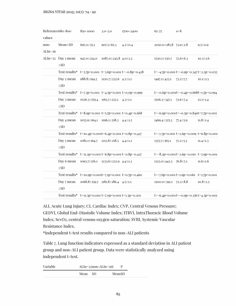
Reference
values
680-800 850-1000 3.0-5.0 1700-2400 65-75 0-8
non-
ALIn=26
Mean±SD 697.0±55.1 907.5±65.3 4.2±0.4 2050.0±185.8 73.9±2.8 9.5±0.9
ALIn=22 Day 1 mean
±SD
947.0±214.0 1087.0±245.8 4.0±1.2 1530.1±530.7 72.6±6.3 10.3±1.6
Test results* t=5.7p<0.001 t=3.6p<0.001 t=-0.8p=0.428 t=-4.7p<0.001 t=-0.9p=0.347 t=2.2p=0.035
Day 2 mean
±SD
988.8±194.5 1120.7±232.6 4.2±1.1 1497.1±423.1 73.2±7.7 10.1±1.5
Test results* t=7.3p<0.001 t=4.5p<0.001 t=0.0p=0.999 t=-0.6p<0.001t=-0.4p=0.668t=1.7p=0.094
Day 3 mean
±SD
1026.3±179.4 1165.7±222.1 4.3±1.1 1506.3±347.1 73.6±7.4 12.1±1.4
Test results* t=8.9p<0.001 t=5.7p<0.001 t=0.4p=0.668 t=-6.9p<0.001t=-0.2p=0.849t=7.7p<0.001
Day 4 mean
±SD
1053.9±164.1 1196.1±218.3 4.4±1.2 1469.4±325.2 77.4±5.9 11.8±1.4
Test results* t=10.4p<0.001t=6.4p<0.001 t=0.8p=0.427 t=-7.7p<0.001 t=2.6p=0.009 t=6.8p<0.001
Day 5 mean
±SD
1081.1±164.7 1213.8±218.5 4.4±1.2 1375.7±365.1 77.2±5.5 11.4±1.3
Test results* t=11.2p<0.001 t=6.8p<0.001 t=0.8p=0.427 t=-8.2p<0.001 t=2.6p=0.010 t=5.9p<0.001
Day 6 mean
±SD
1093.7±176.1 1233.6±222.9 4.4±1.3 1323.0±443.3 76.8±7.1 11.6±1.6
Test results* t=10.9p<0.001t=7.1p<0.001 t=0.7p=0.460 t=-7.6p<0.001 t=1.9p=0.061 t=5.7p<0.001
Day 7 mean
±SD
1068.8±159.7 1181.8±186.4 4.5±1.1 1500.0±391.1 72.2±8.8 10.8±1.2
Test results* t=11.3p<0.001 t=7.0p<0.001 t=1.3p=0.201 t=-6.4p<0.001 t=-0.9p=0.356 t=4.3p<0.001
ALI, Acute Lung Injury; CI, Cardiac Index; CVP, Central Venous Pressure;
GEDVI, Global End-Diastolic Volume Index; ITBVI, IntraThoracic Blood Volume
Index; ScvO2, central venous oxygen saturation; SVRI, Systemic Vascular
Resistance Index.
*independent t-test results compared to non-ALI patients
Table 7. Lung function indicators expressed as a standard deviation in ALI patient
group and non-ALI patient group. Data were statistically analyzed using
independent t-test.
Variable ALIn=22non-ALIn=26t P
Mean SD MeanSD
SIGNA VITAE 2015; 10(1): 74 - 92
85

PaO2/FiO2 19.33 0.75 42.77 1.16 -81.31 <0.001
Compliance(ml/cmH2O)13.56 0.62 22.19 1.43 -26.28<0.001
Resistance(cmH2O/L/s) 3.33 0.46 2.33 0.50 7.14 <0.001
LIS score 1.60 0.36 0.00 0.0022.88 <0.001
ALI, Acute Lung Injury; LIS, Lung Injury severity Score; P, significance level;
PaO2/FiO2, arterial pressure of oxygen/fraction of inspired oxygen; SD, standard
deviation; t, t score for independent t-test.
Figure 1. EVLWI (ExtraVascular Lung Water Index) in ALI (Acute Lung Injury)
patient group and non-ALI patient group during the seven-day period. Data were
analyzed using independent t-test and ANOVA (ANalysis Of VAriance).
Figure 2. AUROC (Area Under Receiver Operating Characteristic) curve for EVLWI
(ExtraVascular Lung Water Index); (0.943; 95% confidence interval 0.849-0.987)
with p<0.001.
SIGNA VITAE 2015; 10(1): 74 - 92
86

Figure 3. PaCO2 (arterial partial pressure of carbon dioxide) in ALI (Acute Lung
Injury) patient group and non-ALI patient group during the seven-day period.
Data were analyzed using independent t-test and ANOVA (ANalysis Of VAriance).
Figure 4. PaO2 (arterial partial pressure of oxygen) in ALI (Acute Lung Injury)
patient group and non-ALI patient group during the seven-day period. Data were
analyzed using independent t-test and ANOVA (ANalysis Of VAriance).
SIGNA VITAE 2015; 10(1): 74 - 92
87

Figure 5. Serum albumin concentration in ALI (Acute Lung Injury) patient group
and non-ALI patient group during the seven-day period. Data were analyzed
using independent t-test and ANOVA (ANalysis Of VAriance).
SIGNA VITAE 2015; 10(1): 74 - 92
88
Figure 6. EVLWI (ExtraVascular Lung Water Index) in patients who survived and
patients who did not survive sepsis after major abdominal surgery in this study.
Data were analyzed using independent t-test and ANOVA (ANalysis Of VAriance).
SIGNA VITAE 2015; 10(1): 74 - 92
89
References
1. Chung FT, Lin SM, Lin SY, Lin HC. Impact of extravascular lung water index on
outcomes of severe sepsis patients in medical intensive care unit. Respir Med
2008;102(7):956-61.
2. Craig TR, Duffy MJ, Shyamsundar M, McDowell C, McLaughlin B, Elborn JS, et al.
Extravascular water indexed to predicted body weight is a novel predictor of
intensive care unit mortality in patients with acute lung injury. Crit Care Med
2010;38:114-20.
3. Chew MS,Ihrman L, During J, Bergenzaun L, Ersson A, Unden J, et al.
Extravascular lung water index improves the diagnostic accuracy of lung injury in
patients with shock. Crit Care 2012;16:R1.
4. Vish M, Shanley PT. Acute Lung Injury and Acute Respiratory Distress Syndrome.
In: Wheeler DS, Wong HR, Shanley TP, editors. The respiratory tract in pediatric
critical illness and injury. London: Springer-Verlag; 2009. p. 49-67.
5. Ware LB, Matthay MA. The acute respiratory distress syndrome. N Engl J Med
2000;342(18):1334-49.
6. Piantadosi CA, Schwartz DA. The acute respiratory distress syndrome. Ann Intern
Med 2004;141(6):460-70.
7. Martin GS, Eaton S, Mealer M, Moss M. Extravascular lung water in patients with
severe sepsis: a prospective cohort study. Crit Care 2005;9:74-82.
8. Kuzkov V, Kirov M, Sovershaev A. Extravascular lung water deterimned with
single transpulmonary thermodilution correlates with the severity of sepsis-
induced acute lung injury. Crit Care Med 2006;122(6):1647-53.
9. Schmidt S, Westhoff TH, Hofmann C, Schaefer JH, Zidek W, Compton F, et al.
Effect of the venous catheter site on transpulmonary thermodilution
measurement variables. Crit Care Med 2007;35:783-6.
10. Monnet X, Anguel N, Osman D, Hamzaoui O, Richard C, Teboul JL. Assessing
pulmonary permeability by transpulmonary thermodilution allows differentiation
of hydrostatic pulmonary edema from ALI/ARDS. Int Care Med 2007;33(3):448-53.
11. Kumar A. Sepsis and septic shock. In: Gabrielli A, Layon AJ, Yu M, editors. Critical
Care. Philadelphia: Lippincott & Wilkins; 2009. p. 855-92.
12. Peters CW, Yu M, Sladen RN, Gabrielli A, Layon AJ. Acute lung injury and acute
respiratory distress syndrome. In: Gabrielli A, Layon AJ, Yu M, editors. Critical
Care. Philadelphia: Lippincott & Wilkins; 2009. p. 2061-80.
13. Johnson ER, Matthay MA. Acute lung injury: epidemiology, pathogenesis and
treatment. J Aerosol Med Pulm Drug Deliv 2010;23(4):234-52.
SIGNA VITAE 2015; 10(1): 74 - 92
90

14. Ware LB, Koyama T, Billheimer DD, Wu W, Bernard GR, Thompson BT, et al.
Prognostic and pathogenetic value of combining clinical and biochemical indices
in patients with acute lung injury. Chest 2010;137(2):288-96.
15. Fernandez-Mondjar E, Guerrero-Lopez F, Colomenero M. How important is the
measurement of extravascular lung water? Crit Care 2007;13:79-83.
16. Dellinger P, Mitchell M, Rhodes A, Annane D, Gerlach H, Opal SM, et al. Surviving
Sepsis Campaign: International Guidelines for Management of Severe Sepsis and
Septic Shock. JAMA 2012;308(16):1641-50.
17. Levy MM, Dellinger P, Townsend SR, Linde-Zwirble WT, Marshall JC, Bion J, et al.
Surviving sepsis campaign. Crit Care Med 2010;38(2):367-74.
18. Levy MM, Fink MP, Marshall JC, Abraham E, Angus D, Cook D, et al. 2001
SCCM/ESICM/ACCP/ATS/SIS International Sepsis Definition Cnoference. Crit
Care Med 2003;31(4):1250-6.
19. Bernard GR, Srticas A, Gordon R, Carlet J, Dreyfuss D, Gattinoni L, et al.The
American-European Consensus Conference on ARDS. AM J Respir Crit Care Med
1998;157:1332-47.
20. Ranieri VM, Rubenfeled GD, Thompson BT, Ferguson ND, Caldwell E, Fan E, et al.
Acute Respiratory Distress Syndrome: The Berlin Definition. JAMA 2012;307:2526-
33.
21. Kushimoto S, Tomoyuki E, Satoshi Y, Sakamoto T, Ishikura H, Kitazawa Y, et al.
Relationship between extravascular lung water and severity categories of acute
respiratory distress syndrome by the Berlin Definition. Crit Care 2013;17(4):R132.
22. Perel A. Extravascular lung water and the pulmonary vascular permeability index
may improve the definition of ARDS. Crit Care 2013;17:108.
23. Cordemans C, De Laet I, Van Regenmortel N, Schoonheydt K, Dits H, Huber W, et
al. Fluid management in critically ill patients: the role of extravascular lung water,
abdominal hypertension, capillary leak and fluid balance. Annals of Intensive Care
2012;2(1):S1.
24. Zhongheng Z, Balong L, Hongying N. Prognostic value of extravascular lung water
index in critically ill patients: a systematice review of the literature. J Crit Care
2012;27(4):420.e1-8.
25. Eichhorn V, Goepfert MS, Eulenburg C, Malbrain M, Reuter DA. Comparison of
values in critically ill patients for global end-diastolic volume and extravascular
lung water measured by transcardiopulmonary thermodilution: A metaanalysis of
the literature. Med Intensiva 2012;36(7):467-74.
26. Maharaj R. Extravascular lung water and acute lung injury. Cardiol Res Pract
2012;Article ID 407035.
SIGNA VITAE 2015; 10(1): 74 - 92
91

Željko Drvar, Višnja Majerić Kogler, Dinko Tonković, Mirjana Mirić, Mario Pavlek, Mladen PerićDepartment of Anaesthesiology, Reanimatology and Intensive Care, University Hospital Zagreb , Zagreb, CroatiaVišnja Majerić Kogler, Mladen PerićUniversity of Zagreb School of Medicine, Zagreb, CroatiaCorresponding authorŽeljko DrvarDepartment of Anaesthesiology, Reanimatology and Intensive careUniversity Hospital ZagrebKišpatićeva 12, 1000 ZagrebCroatiaPhone: +385 098 902 02 41E-mail: [email protected]
SIGNA VITAE 2015; 10(1): 74 - 92
92