inhibitory effects of “greenâ€additives on the crystal growth of
TRANSCRIPT
In: Green Chemistry Research Trends ISBN: 978-1-60692-092-3 Editor: Jeffrey T. Pearlman, pp. 265-287 © 2009 Nova Science Publishers, Inc.
Chapter 8
INHIBITORY EFFECTS OF “GREEN” ADDITIVES ON THE CRYSTAL GROWTH OF SPARINGLY
SOLUBLE SALTS
Konstantinos D. Demadis1* and Mualla Öner2 1 Crystal Engineering, Growth & Design Laboratory, Department of Chemistry,
University of Crete, Heraklion, Crete, GR-71003, Greece 2 Department of Chemical Engineering, Yildiz Technical University,
Davutpasa, Istanbul, 34210, Turkey
ABSTRACT
In this chapter the effects of environmentally friendly additives on formation and growth of sparingly soluble salts are presented in a concise way. Specifically, the influence of a biodegradable, environmentally friendly polysaccharide-based polycarboxylate, carboxymethyl inulin (CMI), on the crystal growth kinetics of calcium oxalate has been studied. The spontaneous crystallization method was used to delineate the crystallization kinetics of calcium oxalate (CaC2O4, CaOx). The results demonstrate that the retardation in crystal growth is controlled by the carboxylation degree of the CMI and its concentration. These studies also show that CMI additives direct calcium oxalate crystallization from calcium oxalate monohydrate (COM) to calcium oxalate dihydrate (COD). Colloidal silica is known to be a substantial problem in silica-laden process waters. Although silica “technically” is not a salt, it is often categorized with them, as it brings about the same problems as mineral scale deposits (reduced heat transfer, etc.). We also report a strategy to retard silicic acid condensation in supersaturated aqueous solutions by using non-toxic, “green”, zwitter-ionic phosphonomethylayed chitosan (PCH). An overview of the use of green additives in water treatment is also presented.
Keywords: green chemistry, environmentally friendly, scale inhibition, calcium oxalate, additives, water treatment, polymers, silica
* Email: [email protected], Web Page: http://www.chemistry.uoc.gr/demadis
Konstantinos D. Demadis and Mualla Öner 266
INTRODUCTION The crystal growth of calcium oxalate (CaC2O4, CaOx) and its chemical control have
been at the epicenter of intense study by engineers and urologists for several decades [1]. Its formation is of particular importance not only to researchers working in the biomineralization field [2], but also to engineers/chemists who are active in the industrial crystallization applications’ field [3]. Calcium oxalate (CaC2O4, CaOx) is a naturally occurring mineral found in fossils, plants, mammal urinary calculi [4] and is a by-product in some processes such as paper, food and beverage processing [5]. Calcium oxalate crystallization yields different hydrates. The thermodynamically most stable is the monoclinic monohydrate COM (CaC2O4.H2O, whewellite [6]). Under certain conditions, the metastable tetragonal dihydrate (CaC2O4.(2+x)H2O, x<0.5), weddelite [7]) and triclinic trihydrate (CaC2O4.xH2O, 3>x>2.5; COT [8]) can form. An understanding of CaOx crystallization processes is essential to urologists for possible development of new therapeutic agents, but also to water chemists who seek its inhibition as an undesirable salt in the process industries.
Among water-formed deposits colloidal silica (SiO2·nH2O, n is variable and dependent on hydration) is especially troublesome because it can cause serious materials failure and operational shut-downs. Silica scale prevention, in principle, can be achieved by use of scale inhibitors, key components of any chemical water treatment [9]. Unfortunately, traditional scale control methods (inhibition and crystal modification) applied to crystalline mineral salt precipitates, do not apply to silica because of its amorphous state. Therefore, much more well-designed inhibition approaches have to be applied for controlling silica deposition. Increasing environmental concerns and discharge limitations have imposed additional challenges in treating process waters. Therefore, the discovery and successful application of chemical additives that have mild environmental impact has been the focus of several researchers [10, 11]. In this paper we also focus on use of “green” inhibitors silica scale inhibition. This research is part of our on-going investigation on the discovery and application of scale inhibitors, with an emphasis on “green” additives, in industrial process waters [12-20].
OVERVIEW OF TWO “GREEN” SCALE INHIBITORS: CMI AND PCH
In this investigation we present a study of the effect of carboxymethylinulin (CMI, figure
1) on the crystallization kinetics and phase transformation of CaC2O4 using a plethora of experimental techniques (FTIR, XRD and SEM). CMI is produced from a chemical reaction of the biopolymer inulin and select reagents [21]. Inulin [22] is extracted from the roots of the chicory plant (dry matter content: 20-25%, inulin content: 14.9-18.3 %). Inulin is a polydisperse polysaccharide consisting mainly, if not exclusively, of β(2→1) fructosyl fructose units with normally, but not necessarily, one glucopyranose unit at the reducing end. It is also known that the fructose molecules are all present in the furanose form. Inulin is used as dietary fiber, fat substitute, sweetener (fructose syrups). Carboxymethylinulin (CMI) has been investigated in a series of acute toxicity (oral rat, > 2000 mg/kg B.W.), sub-acute toxicity (28 days, rat 1000 mg/kg B.W.), mutagenicity (Ames Test, in vitro cytogenetics, no

Inhibitory Effects of “Green” Additives on the Crystal Growth… 267
effect) and dermal sensitization studies (guinea pigs, no effect) to evaluate its toxicological profile [23]. All studies followed accepted testing guidelines as recommended by international regulatory agencies (OECD, EEC and US EPA). No significant toxicological findings were evident. Results of the present toxicity studies with CMI, all conforming to internationally-accepted testing standards, show that the toxicological profile of CMI is consistent with other polycarboxylates used in foods. Among other attractive attributes of CMI, its inherent biodegradability and non-toxicity are most prominent. Data that support these conclusions include: toxicity (ppm) EC10 > 10000, bacteria (ppm) EC0 = 2000, Daphnia (ppm) EC50(24h) = 5500, fish (ppm) LC0 > 10000. These results should be contrasted to those obtained for a polyacrylate polymer (M.W. = 1500) commonly used as precipitation inhibitor in industrial waters: toxicity (ppm) EC10 = 180, bacteria (ppm) EC0 = 200, Daphnia (ppm) EC50(24h) = 240, fish (ppm) LC0 = 200.
Figure 1. The building blocks in the CMI polymer backbone. The carboxymethyl groups (highlighted in red) are introduced by the reaction of monochloroacetic acid with the ring –OH groups. Up to three –CH2COOH groups can be introduced per ring. The presence of acidic –CH2COOH groups on the CMI backbone makes it an anionic polyelectrolyte (at appropriate pH regions).
We have selected to utilize chitosan-based biopolymers, figures 2 and 3, as additives that may enhance silicic acid solubility. More specifically, in the context of this chapter we studied the polymer chitosan on which animomethylenephosphonate groups have been grafted by a Mannich-type reaction. We selected this particular polymer because it possesses attractive similarities to naturally-found, silica controlling proteins called silaffins [24], among other reasons outlined below. In particular: (a) PCH possesses cationic charge by virtue of its protonated (at pH 7) –NH2 groups, (b) PCH possesses anionic phosphonate groups (deprotonated -PO3H2 moieties) that resemble the phosphate groups in silaffins, (c) PCH contains tertiary and secondary, protonated amine groups (from the aminomethylenephosphonate moiety), also present in silaffins. Furthermore, PCH is synthesized from chitosan (a degradation product of chitin, a renewable material) in an efficient and low-cost manner. PCH also has low aquatic toxicity (vide infra). An additional

Konstantinos D. Demadis and Mualla Öner 268
reason that prompted us to study the effect of PCH on silicic acid condensation is the report that the diatom cell wall (frustulum) is made of nanostructured amorphous silica that is associated with polysaccharides and proteins. The link that may exist between polysaccharides and the formation of biosilica seems to be an interesting area to explore.
Figure 2. Schematic structures of the polymers chitin (CHT), chitosan (CHS), phosphonomethylated chitosan (PCH). Added groups are clearly shown on the PCH polymer backbone.
A facile model system to study human disease and drug responses are zebrafish, Danio rerio. They are particularly suited for this purpose because they represent a vertebrate species, their genome is sequenced, and a large number of synchronously developing, transparent embryos can be produced. Zebrafish embryos are permeable to drugs and can easily be manipulated using well-established genetic and molecular approaches. LC50 values (50 % lethal concentration) is the concentration at which 50% of test organisms are killed. Adult wild-type zebrafishes were maintained at 26 ºC and fed with ZF Biofood. Fertilized zebrafish embryos were obtained by hormonally induced treatment to spawners and in vitro fertilization. Three assays with a variable number (4-7) of water soluble-N-methylene phosphonic chitosan concentrations between 99 and 1000 mg/l and a control without N-methylene phosphonic chitosan were conducted in 96-well multiplates at 26ºC. Spawns and eggs were selected at 3-4 hpf (hours post-fertilization) according to standard criteria. Lethal effects were observed in 48 hpf embryos, based on four morphological and functional endpoints, specific for lethality studies. The LC50 of N-methylene phosphonic chitosan was determined in our laboratory using the Probit 1.5 software as 279.6 ± 29.7 mg/L. The LC50 of
Inhibitory Effects of “Green” Additives on the Crystal Growth… 269
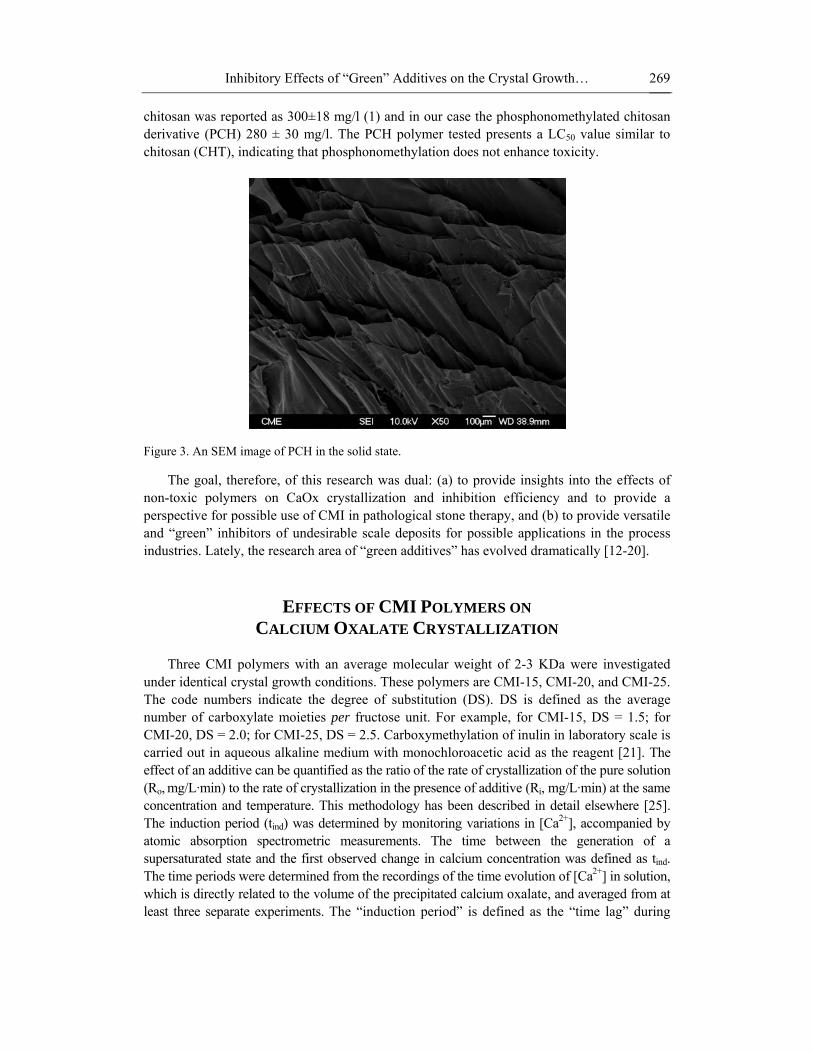
chitosan was reported as 300±18 mg/l (1) and in our case the phosphonomethylated chitosan derivative (PCH) 280 ± 30 mg/l. The PCH polymer tested presents a LC50 value similar to chitosan (CHT), indicating that phosphonomethylation does not enhance toxicity.
Figure 3. An SEM image of PCH in the solid state.
The goal, therefore, of this research was dual: (a) to provide insights into the effects of non-toxic polymers on CaOx crystallization and inhibition efficiency and to provide a perspective for possible use of CMI in pathological stone therapy, and (b) to provide versatile and “green” inhibitors of undesirable scale deposits for possible applications in the process industries. Lately, the research area of “green additives” has evolved dramatically [12-20].
EFFECTS OF CMI POLYMERS ON CALCIUM OXALATE CRYSTALLIZATION
Three CMI polymers with an average molecular weight of 2-3 KDa were investigated
under identical crystal growth conditions. These polymers are CMI-15, CMI-20, and CMI-25. The code numbers indicate the degree of substitution (DS). DS is defined as the average number of carboxylate moieties per fructose unit. For example, for CMI-15, DS = 1.5; for CMI-20, DS = 2.0; for CMI-25, DS = 2.5. Carboxymethylation of inulin in laboratory scale is carried out in aqueous alkaline medium with monochloroacetic acid as the reagent [21]. The effect of an additive can be quantified as the ratio of the rate of crystallization of the pure solution (Ro, mg/L·min) to the rate of crystallization in the presence of additive (Ri, mg/L·min) at the same concentration and temperature. This methodology has been described in detail elsewhere [25]. The induction period (tind) was determined by monitoring variations in [Ca2+], accompanied by atomic absorption spectrometric measurements. The time between the generation of a supersaturated state and the first observed change in calcium concentration was defined as tind. The time periods were determined from the recordings of the time evolution of [Ca2+] in solution, which is directly related to the volume of the precipitated calcium oxalate, and averaged from at least three separate experiments. The “induction period” is defined as the “time lag” during
Konstantinos D. Demadis and Mualla Öner 270
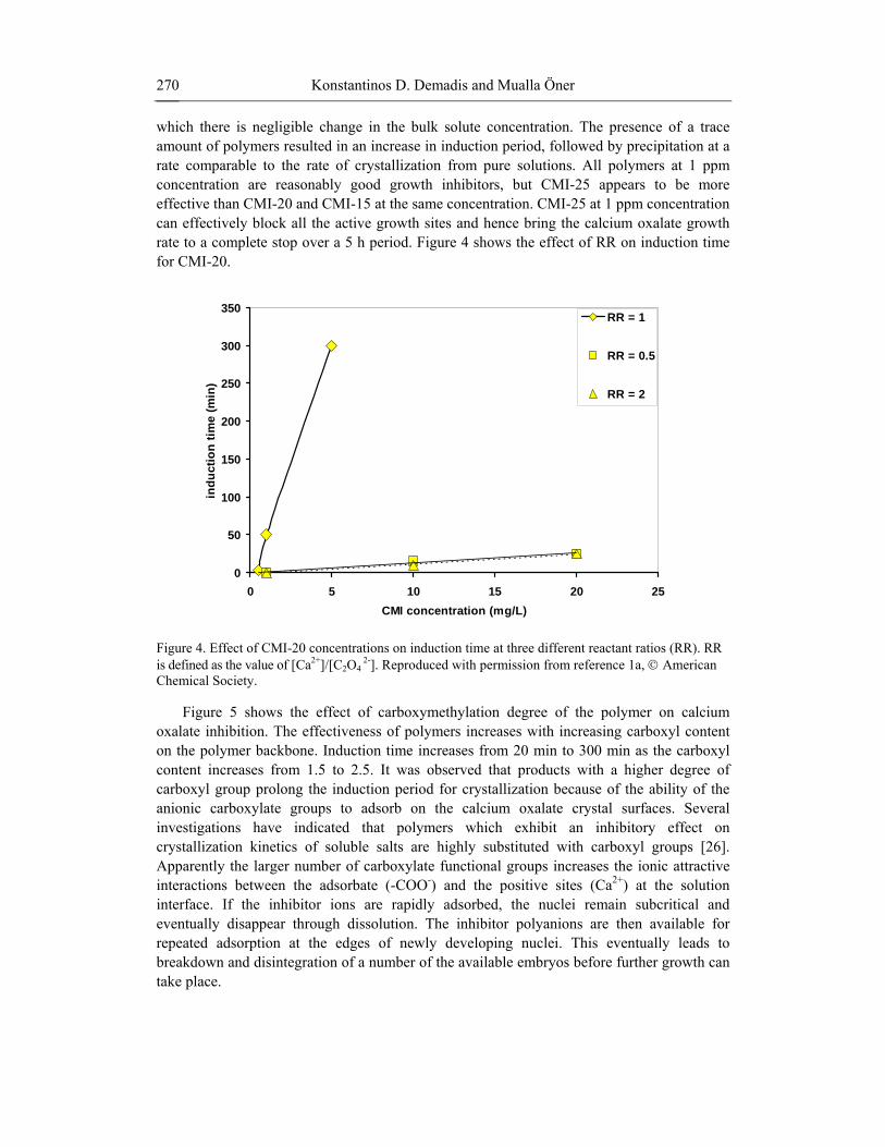
which there is negligible change in the bulk solute concentration. The presence of a trace amount of polymers resulted in an increase in induction period, followed by precipitation at a rate comparable to the rate of crystallization from pure solutions. All polymers at 1 ppm concentration are reasonably good growth inhibitors, but CMI-25 appears to be more effective than CMI-20 and CMI-15 at the same concentration. CMI-25 at 1 ppm concentration can effectively block all the active growth sites and hence bring the calcium oxalate growth rate to a complete stop over a 5 h period. Figure 4 shows the effect of RR on induction time for CMI-20.
0
50
100
150
200
250
300
350
0 5 10 15 20 25CMI concentration (mg/L)
indu
ctio
n tim
e (m
in)
RR = 1
RR = 0.5
RR = 2
Figure 4. Effect of CMI-20 concentrations on induction time at three different reactant ratios (RR). RR is defined as the value of [Ca2+]/[C2O4
2-]. Reproduced with permission from reference 1a, © American Chemical Society.
Figure 5 shows the effect of carboxymethylation degree of the polymer on calcium oxalate inhibition. The effectiveness of polymers increases with increasing carboxyl content on the polymer backbone. Induction time increases from 20 min to 300 min as the carboxyl content increases from 1.5 to 2.5. It was observed that products with a higher degree of carboxyl group prolong the induction period for crystallization because of the ability of the anionic carboxylate groups to adsorb on the calcium oxalate crystal surfaces. Several investigations have indicated that polymers which exhibit an inhibitory effect on crystallization kinetics of soluble salts are highly substituted with carboxyl groups [26]. Apparently the larger number of carboxylate functional groups increases the ionic attractive interactions between the adsorbate (-COO-) and the positive sites (Ca2+) at the solution interface. If the inhibitor ions are rapidly adsorbed, the nuclei remain subcritical and eventually disappear through dissolution. The inhibitor polyanions are then available for repeated adsorption at the edges of newly developing nuclei. This eventually leads to breakdown and disintegration of a number of the available embryos before further growth can take place.

Inhibitory Effects of “Green” Additives on the Crystal Growth… 271
0
50
100
150
200
250
300
350
1 1.5 2 2.5 3
degree of substitution
indu
ctio
n tim
e (m
in)
Figure 5. The effect of DS increase on induction time. The line is added to aid the reader. Reproduced with permission from reference 1a, © American Chemical Society.
In this way outgrowth of the nuclei beyond their critical value/size is hampered; in due course, because of their thermodynamic instability, most nuclei will re-dissolve, thus freeing the polymer for interaction with other embryos. The polymers, by effectively reducing the number of active growth sites through adsorption on the crystal surface, will prolong induction periods. During the induction period, most of the active growth sites are “poisoned” by the adsorbed macromolecule additives. However, some of the growth sites of lower energy may still be free to grow, and thus, the crystallization proceeds at a very slow rate. The rate of precipitation/deposition of calcium oxalate following the induction period is dependent on polymer concentration.
The marked effect of polymers on the crystal growth of calcium oxalate from supersaturated solutions have been explained in terms of the following factors: (a) Polymers may change the ionic strength of the solution; (b) Polymers may form stable complexes with calcium ion; (c) Adsorption of the polymer on the crystal surfaces either indiscriminately or at specific growth sites (d) polymer incorporation into the calcium oxalate lattice. Under the experimental conditions employed herein, the presence of the induction period is believed to originate from inhibitor surface adsorption. The polymer concentration is too low to modify the ionic strength of the solution and the calcium ion binding is small enough to be ignored. Polymer insertion/incorporation into the lattice can be ruled out because these macromolecules are much larger in size compared to the oxalate ion.
Since the amounts of additive in solution are small, the growth inhibition is most likely caused by biopolymer adsorption of the active growth sites on crystal surfaces rather than binding to solution Ca2+ ions. This assumption was tested by fitting the kinetic results in a Langmuir type kinetic isotherm. When a polyelectrolyte poisons active growth sites on the crystal faces, the coverage by polymers, θ, for this adsorption model is described by Equation 1.
Konstantinos D. Demadis and Mualla Öner 272
θ= KCi /(1+KCi) (1)
where K is the adsorption or affinity constant which is the ratio of the rate constants for adsorption and desorption, kads/kdes, and can be considered as a measure for the adsorption affinity of the polymer onto the crystal surface and Ci is the total equilibrium concentration of the polymer. The rate in the absence of polymer, R0, is reduced to a slower rate, Ri, according to the relationship
Ri= R0(1-θ) (2) Combination of Equations 1 and 2 gives
iads
des
i0
0
C1
kk
1RR
R+=
− (3)
Equation 3 shows that this model predicts a linear relationship between R0/(R0-Ri) and
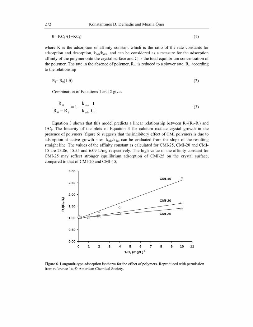
1/Ci. The linearity of the plots of Equation 3 for calcium oxalate crystal growth in the presence of polymers (figure 6) suggests that the inhibitory effect of CMI polymers is due to adsorption at active growth sites. kads/kdes can be evaluated from the slope of the resulting straight line. The values of the affinity constant as calculated for CMI-25, CMI-20 and CMI-15 are 23.86, 15.55 and 6.09 L/mg respectively. The high value of the affinity constant for CMI-25 may reflect stronger equilibrium adsorption of CMI-25 on the crystal surface, compared to that of CMI-20 and CMI-15.
0.00
0.50
1.00
1.50
2.00
2.50
3.00
0 1 2 3 4 5 6 7 8 9 10 11
R0/(
R0-
Ri)
1/C, (mg/L)-1
CMI-15
CMI-20
CMI-25
Figure 6. Langmuir-type adsorption isotherm for the effect of polymers. Reproduced with permission from reference 1a, © American Chemical Society.
Inhibitory Effects of “Green” Additives on the Crystal Growth… 273
MORPHOLOGICAL CHANGES OF CALCIUM OXALATE CRYSTAL SURFACES DUE TO CMI POLYMERS
It has been known that very low concentrations of additives can greatly affect rates of
crystallization and the habit of the growing crystals. Even if the additives are present in such low concentrations their effect may still be significant. It is almost always the case that the presence of additives reduces the growth rate of a crystal, and various index faces may be affected differently and leads to a modification of habit. The shape or morphology of a crystal is determined by the relative growth rate along different crystallographic directions. The crystal face that is perpendicular to the fast growing direction is usually not expressed in the final crystal form while the face that is parallel to the fast growing direction is usually expressed. Thus modification of the crystal morphology can be achieved by altering the relative crystal growth rate with inhibitor capable of specific inhibition [27].
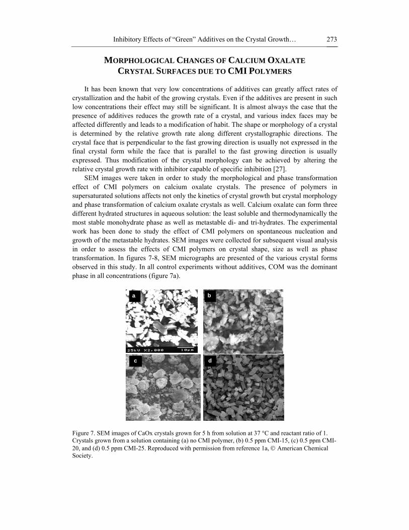
SEM images were taken in order to study the morphological and phase transformation effect of CMI polymers on calcium oxalate crystals. The presence of polymers in supersaturated solutions affects not only the kinetics of crystal growth but crystal morphology and phase transformation of calcium oxalate crystals as well. Calcium oxalate can form three different hydrated structures in aqueous solution: the least soluble and thermodynamically the most stable monohydrate phase as well as metastable di- and tri-hydrates. The experimental work has been done to study the effect of CMI polymers on spontaneous nucleation and growth of the metastable hydrates. SEM images were collected for subsequent visual analysis in order to assess the effects of CMI polymers on crystal shape, size as well as phase transformation. In figures 7-8, SEM micrographs are presented of the various crystal forms observed in this study. In all control experiments without additives, COM was the dominant phase in all concentrations (figure 7a).
a
d
b
c
Figure 7. SEM images of CaOx crystals grown for 5 h from solution at 37 °C and reactant ratio of 1. Crystals grown from a solution containing (a) no CMI polymer, (b) 0.5 ppm CMI-15, (c) 0.5 ppm CMI-20, and (d) 0.5 ppm CMI-25. Reproduced with permission from reference 1a, © American Chemical Society.
Konstantinos D. Demadis and Mualla Öner 274
a
b
c
d
e
f
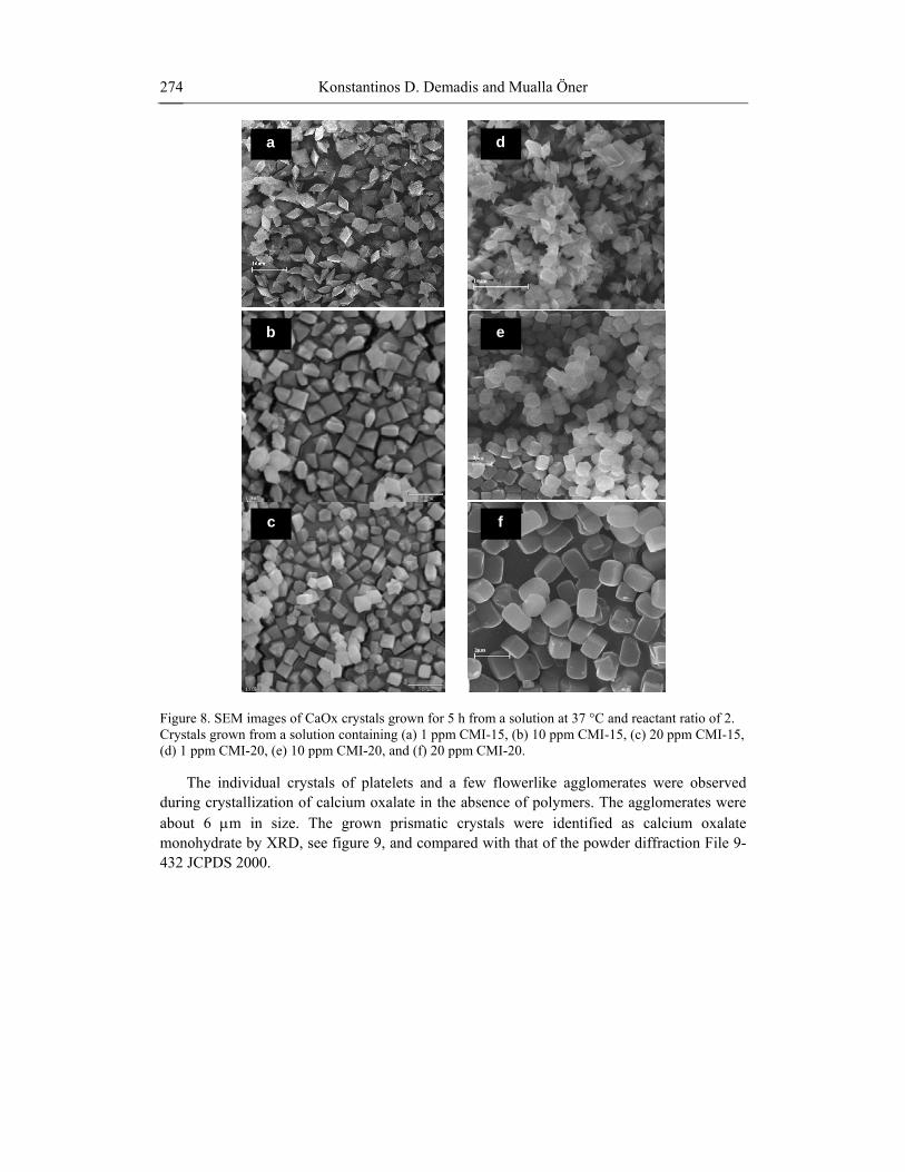
Figure 8. SEM images of CaOx crystals grown for 5 h from a solution at 37 °C and reactant ratio of 2. Crystals grown from a solution containing (a) 1 ppm CMI-15, (b) 10 ppm CMI-15, (c) 20 ppm CMI-15, (d) 1 ppm CMI-20, (e) 10 ppm CMI-20, and (f) 20 ppm CMI-20.
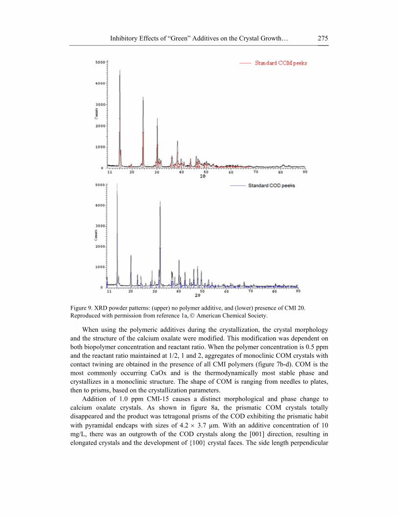
The individual crystals of platelets and a few flowerlike agglomerates were observed during crystallization of calcium oxalate in the absence of polymers. The agglomerates were about 6 µm in size. The grown prismatic crystals were identified as calcium oxalate monohydrate by XRD, see figure 9, and compared with that of the powder diffraction File 9-432 JCPDS 2000.
Inhibitory Effects of “Green” Additives on the Crystal Growth… 275
Figure 9. XRD powder patterns: (upper) no polymer additive, and (lower) presence of CMI 20. Reproduced with permission from reference 1a, © American Chemical Society.
When using the polymeric additives during the crystallization, the crystal morphology and the structure of the calcium oxalate were modified. This modification was dependent on both biopolymer concentration and reactant ratio. When the polymer concentration is 0.5 ppm and the reactant ratio maintained at 1/2, 1 and 2, aggregates of monoclinic COM crystals with contact twining are obtained in the presence of all CMI polymers (figure 7b-d). COM is the most commonly occurring CaOx and is the thermodynamically most stable phase and crystallizes in a monoclinic structure. The shape of COM is ranging from needles to plates, then to prisms, based on the crystallization parameters.
Addition of 1.0 ppm CMI-15 causes a distinct morphological and phase change to calcium oxalate crystals. As shown in figure 8a, the prismatic COM crystals totally disappeared and the product was tetragonal prisms of the COD exhibiting the prismatic habit with pyramidal endcaps with sizes of 4.2 × 3.7 µm. With an additive concentration of 10 mg/L, there was an outgrowth of the COD crystals along the [001] direction, resulting in elongated crystals and the development of {100} crystal faces. The side length perpendicular
Konstantinos D. Demadis and Mualla Öner 276
to the tetragonal axis increases from 0.80 µm to 0.93 µm when the CMI-15 concentration increases from 10 to 20 ppm. The same behavior was observed with the addition of CMI-20 (figure 8d-f). COD has a tetragonal structure with a space group of I4/m and a crystal habit that can be best described as tetragonal bipyramidal [28]. The calcium surface concentration of the COD on the (100) face (0.0439 ions/A2) is greater than that on the (101) face (0.0225 ions/A2). This result implies that the (100) face can adsorb a higher number of carboxylate moieties from the polymers. Therefore the interactions between polymer and the COD (100) faces resulted in a gradual morphological transition of COD crystals from tetragonal bipyramids to elongated tetragonal prisms with increasing polymer concentration.
A large number of reports have appeared in the literature on the effects of various additives on calcium oxalate crystallization. These include polyphosphate [29], sodium cholate [30], maleic acid copolymers [31], polyaspartic acids [32] poly-(styrene-alt-maleic acid) [33], various polyhydroxycarboxylic acids [34], acrylic polymers [35], tartarates [36], diisooctyl sulfosuccinate [37], mucin [38], Tamm-Horsfall proteins [39], herbal extracts [40], uric acid [41], poly(sodium 4-styrene-sulfonate) [42], liposome solutions of different carboxylates [43], fluorescent molecules [44], algae-derived sulfated polysaccharides [45], osteopontin [46], aspartic-rich synthetic peptides [47], synthetic osteopontin phosphopeptides [48], uropontin [49], pyrophosphate [50], citrate and isocitrate [51], tryptophan [52], dipalmitoylphosphatidylcholine [53], inositol hexaphosphate (phytate) [54], glycosaminoglycans [55], fibronectin [56], glycoproteins [57], unidentified macromolecules from whole human urine [58], α-ketoglutaric acid [59] and adenosine phosphates [60].
A number of important observations point to the way carboxylate and phosphate containing macromolecules may effect CaOx crystallization. The precipitation of calcium oxalate hydrates was investigated in the presence of polyphosphate by Nancollas and Tomazic [61]. It was reported that there is a greater adsorption of polyelectrolyte by COM than COD or COT. The greater adsorption of polymer by COM results in greater inhibition of COM nucleation and growth than for COD and COT. The effect of anionic polyelectrolytes on the crystallization of calcium oxalate hydrates was studied by Manne et al. [62]. It was found that only the monohydrate was formed in the presence of low concentrations of polyelectrolyte. At intermediate concentration (1-50 ppm) trihydrate was the predominant product and at high concentrations (above 50 ppm) the dihydrate was the dominant product. Brecevic and Kralj [63] were studied the influence of some amino acids on calcium oxalate dihydrate transformation. They found that tryptophan and histidine promote the formation of COD over COM.
The studies report habit changes, aggregations and attachment in calcium oxalate crystals, under the influence of glucosaminoglycans [64], polyglutamic [65] and polyaspartic acid [32] and anionic lipids [66]. Each of these macromolecular structures contains carboxyl groups, which are ionized at normal physiological pH values.
Recently, real time in situ Atomic Force Microscopy (AFM) was used to determine the activity and selectivity of macromolecular binding profiles to specific calcium oxalate crystal faces (monohydrate and dihydrate). AFM can visualize the crystal growth and directly measure the adhesion force between molecules and crystal surfaces. Elegant work by Ward et al. (with AFM) [67] and by Jung et al. (with SEM and XRD) [68] has focused on studying the interactions of polymers with pendant carboxylate groups (polyacrylate, polyaspartate and polyglutamate) with various crystallographic planes of the CaOx monohydrate crystals. It was found that polyglutamic acid inhibited growth more strongly along [010] direction than along

Inhibitory Effects of “Green” Additives on the Crystal Growth… 277
[001], consistent with preferential binding of polyglutamic acid to the (010) planes. In contrast, polyaspartic acid was more effective in inhibiting growth along the [001] direction. As a result it was concluded that CaOx formation was regulated by anionic molecules-crystal interaction that depend on molecular level recognition between anionic subunits of the macromolecules and lattice ions in the CaOx crystal.
Negative “charge density” (the number of bonds separating the negative charge located on the carboxylate groups) must be invoked when examining inhibitory activity. Bonds separating the deprotonated oxygen in the –COO- groups in the polymers’ backbone are: polyacrylate 6, polyaspartate 12, polyglutamate 12, CMI 16 (for CMI-15, containing 1 carboxylate group per D-fructofuranose ring), or 9 (for CMI-20, containing 2 carboxylate groups per D-fructofuranose ring) see figure 10. Based on the work of Ward et al. [67] there is a inversely proportional relationship between inhibitory activity and the number of bonds separating the carboxylate units. Our results confirm this significant observation, as the inhibitory activity ranking is CMI-15 < CMI-20 < CMI-25 ~ PAA. The effects of charge density on inhibitory activity may be a wider theme in inhibition chemistry.
Figure 10. Energy-minimized fragment of the CMI polymeric backbone. Carboxylic acid groups that can be deprotonated at appropriate pH regions are shown by arrows.
EFFECTS OF PCH POLYMER ON SILICIC ACID CONDENSATION Phosphonated chitosan (PCH) was tested for its ability to enhance silicate solubility and
affect silica formation in “short-term” experiments (8 h) at dosages 40, 150 and 200 ppm. The results are presented in figure 11. Silicate condensation appears to be independent of additive dosage within the first 8 h. Soluble silicate reaches a value of ~ 350 ppm 8 h. Compared to control solutions not containing this results in ~ 150 ppm additional silicate stabilization.

Konstantinos D. Demadis and Mualla Öner 278
0
50
100
150
200
250
300
350
400
450
500
0 1 2 3 4 5 6 7 8
time (h)
solu
ble
silic
ate
(ppm
as
SiO
2)
control
40 ppm
Figure 11. Colloidal silica formation demonstrated by monitoring soluble silicic acid level decrease. In the absence of PCH additives rapid reduction of soluble silicate is observed (control). In the presence of 40 ppm PCH substantial retardation of this process is observed.
The long-term silicate stabilization by PCH was studied over a 72 h time period. The effect of various dosages of PCH (10, 20, 40, 60, 100, 150 and 200 ppm) on silica formation is shown in figure 12.
176
151
146
175
122 13
9
180
141
147
187
113 13
8
226
133 15
1
253
190
164
296
246
206
297
246
217
0
50
100
150
200
250
300
350
solu
ble
silic
ate
(as
ppm
SiO
2)
control 10 20 40 60 100 150 200PCH dosage (ppm)
24h48h72h
Figure 12. Long-term (24 h – 48 h – 72 h) enhancement of silicic acid solubility in the presence of various levels of PCH (from 10 ppm to 200 ppm).
Inhibitory Effects of “Green” Additives on the Crystal Growth… 279
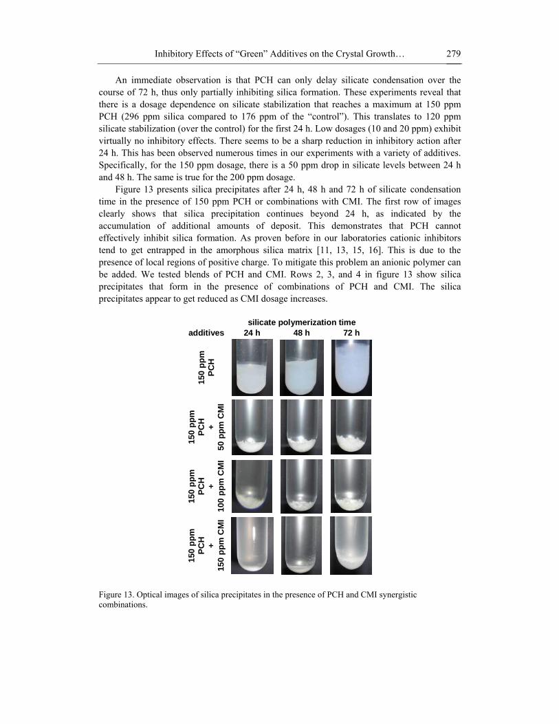
An immediate observation is that PCH can only delay silicate condensation over the course of 72 h, thus only partially inhibiting silica formation. These experiments reveal that there is a dosage dependence on silicate stabilization that reaches a maximum at 150 ppm PCH (296 ppm silica compared to 176 ppm of the “control”). This translates to 120 ppm silicate stabilization (over the control) for the first 24 h. Low dosages (10 and 20 ppm) exhibit virtually no inhibitory effects. There seems to be a sharp reduction in inhibitory action after 24 h. This has been observed numerous times in our experiments with a variety of additives. Specifically, for the 150 ppm dosage, there is a 50 ppm drop in silicate levels between 24 h and 48 h. The same is true for the 200 ppm dosage.
Figure 13 presents silica precipitates after 24 h, 48 h and 72 h of silicate condensation time in the presence of 150 ppm PCH or combinations with CMI. The first row of images clearly shows that silica precipitation continues beyond 24 h, as indicated by the accumulation of additional amounts of deposit. This demonstrates that PCH cannot effectively inhibit silica formation. As proven before in our laboratories cationic inhibitors tend to get entrapped in the amorphous silica matrix [11, 13, 15, 16]. This is due to the presence of local regions of positive charge. To mitigate this problem an anionic polymer can be added. We tested blends of PCH and CMI. Rows 2, 3, and 4 in figure 13 show silica precipitates that form in the presence of combinations of PCH and CMI. The silica precipitates appear to get reduced as CMI dosage increases.
silicate polymerization time additives 24 h 48 h 72 h
150
ppm
PC
H
150
ppm
PC
H
+
50 p
pm C
MI
150
ppm
PC
H
+
100
ppm
CM
I
150
ppm
PC
H
+
150
ppm
CM
I
Figure 13. Optical images of silica precipitates in the presence of PCH and CMI synergistic combinations.

Konstantinos D. Demadis and Mualla Öner 280
Silica precipitates were also studied by SEM, and the results are shown in figure 14. Generally, silica particles are spherical in shape. Upon PCH dosage increase they tend to agglomerate and form larger aggregates. In addition, these agglomerates grow larger as silicic polymerization time increases. After 600 h of polymerization the formation of large particles (> 2 µm) is obvious, see figure 14.
additives 8 h 72 h 600 h
40 p
pm
PCH
150
ppm
PC
H
Figure 14. SEM images of silica deposits in the presence of 40 ppm (upper row) and 150 ppm (lower row) PCH, after silicic acid polymerization times of 8 h (1st column), 72 h (2nd column) and 600 h (3rd column). All scale bars are 200 nm, except the 3rd column, 2 µm.
POSSIBLE MECHANISMS OF SILICA INHIBITION
BY THE PCH POLYMER Silicic acid condensation is a complex process whose mechanistic details have been the
subject of a number of literature reports [69]. Silicic acid condensation in the presence of threshold amounts of scale inhibitor is even more complicated because of added reaction pathways. When silicate polymerization occurs in the presence of a cationic polymeric additive, the following competing events occur:
1) Polymerization of silicic acid/silicate anions. This occurs through an SN2-like
mechanism that involves attack of a monodeprotonated silicic acid molecule on a fully protonated silicic acid molecule. This pathway generates at first short-lived silicate dimers, which in turn continue to polymerize in a random way to eventually yield colloidal silica particles.
2) Silicic acid/silicate ion stabilization by the cationic additive. This is the actual inhibition step and occurs presumably through cation-anion and/or hydrogen bonding interactions.
3) Flocculation between the polycationic inhibitor and the produced negatively charged colloidal silica particles (at pH 7) that are formed by the uninhibited silicic acid polymerization.
Inhibitory Effects of “Green” Additives on the Crystal Growth… 281
Cationic inhibitor is trapped within the colloidal silica matrix, based on reaction (3). This is demonstrated by the appearance of a light flocculent precipitates (or stable dispersions at times). Inhibitor entrapment causes its depletion from solution and its deactivation. Therefore, only a portion of the inhibitor is available to continue inhibition, albeit at much lower levels than initially added to the polymerization medium. Thus, soluble silicate levels continue to decrease because eventually there is insufficient inhibitor to perform inhibition. Inhibitor entrapment is directly proportional to cationic charge density.
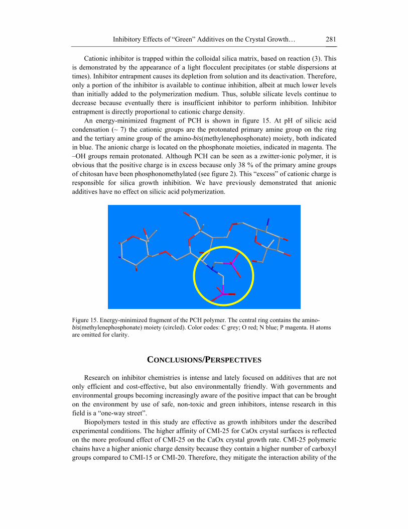
An energy-minimized fragment of PCH is shown in figure 15. At pH of silicic acid condensation (~ 7) the cationic groups are the protonated primary amine group on the ring and the tertiary amine group of the amino-bis(methylenephosphonate) moiety, both indicated in blue. The anionic charge is located on the phosphonate moieties, indicated in magenta. The –OH groups remain protonated. Although PCH can be seen as a zwitter-ionic polymer, it is obvious that the positive charge is in excess because only 38 % of the primary amine groups of chitosan have been phosphonomethylated (see figure 2). This “excess” of cationic charge is responsible for silica growth inhibition. We have previously demonstrated that anionic additives have no effect on silicic acid polymerization.
Figure 15. Energy-minimized fragment of the PCH polymer. The central ring contains the amino-bis(methylenephosphonate) moiety (circled). Color codes: C grey; O red; N blue; P magenta. H atoms are omitted for clarity.
CONCLUSIONS/PERSPECTIVES
Research on inhibitor chemistries is intense and lately focused on additives that are not
only efficient and cost-effective, but also environmentally friendly. With governments and environmental groups becoming increasingly aware of the positive impact that can be brought on the environment by use of safe, non-toxic and green inhibitors, intense research in this field is a “one-way street”.
Biopolymers tested in this study are effective as growth inhibitors under the described experimental conditions. The higher affinity of CMI-25 for CaOx crystal surfaces is reflected on the more profound effect of CMI-25 on the CaOx crystal growth rate. CMI-25 polymeric chains have a higher anionic charge density because they contain a higher number of carboxyl groups compared to CMI-15 or CMI-20. Therefore, they mitigate the interaction ability of the

Konstantinos D. Demadis and Mualla Öner 282
appended anionic –COO- groups with the solid phase. The polymer backbone can then act as a “fence” on the crystal surface, thus forming an obstacle for propagating steps that lead to further crystal growth. The results described herein indicate that anionic biopolymers can inhibit calcium oxalate crystal growth and that such inhibition is directly linked to fractional coverage of adsorption sites. The degree of inhibition of calcium oxalate crystallization by CMI biopolymers is related to the maximum surface charge density due to adsorbed polymer. The polymer concentration and the [Ca2+]/[C2O4
2-] ratio are found to be important parameters for the control of morphologies and phase transformation of CaOx crystals. Agglomerated and twinned COM crystals formed in control experiments. Presence of CMI polymers favors the transition of COM to COD crystals. The high binding affinity of the CMI polymer molecules resulted in morphological transition of COD crystals from tetragonal bipyramids dominated by the (101) faces to elongated tetragonal prisms dominated by the (100) faces.
Zwitter-ionic polymers based on the chitosan backbone can be colloidal silica growth inhibitors. PCH, with phosphonate groups chemically introduced, can substantially retard silicic acid condensation and maintain high levels of soluble silicate for an extended period of time. Silicic acid/silicate is thought to be stabilized by a combination of hydrogen bonding and ionic interactions that perturb the dimerization/polymerization process.
Inhibition of sparingly soluble salts is an active area of research and intense industrial interest [70]. Progress in this field of technology is fueled by the continuous need for inhibitors in a plethora of disciplines, from medicine to water treatment. An added need for non-toxic, green, biodegradable and eco-friendly inhibitors is expected to drive research forward.
ACKNOWLEDGMENTS M.Ö. thanks the Scientific and Technological Research Council of Turkey (TUBİTAK,
Project No: 105M329)) and K.D.D. thanks the General Secretariat of Science and Technology (Greece, under contract # GSRT 2007-202e) for funding this work under a Bilateral Cooperation Program between Turkey and Greece, Dr. Viviana Ramos for a sample of PCH and Antonia Ketsetzi for technical assistance.
REFERENCES
[1] (a) B. Akın, M. Öner, Y. Bayram, K. D. Demadis, ‘Effects of Carboxylate-Modified,
“Green” Inulin Biopolymers on the Crystal Growth of Calcium Oxalate’ Cryst. Growth Des. 8 (2008) 1997–2005. (b) L. Ajayi, P. Jaeger, W. Robertson, R. Unwin, ‘Renal stone disease’ Medicine, 35 (2007) 415-419. (c) A. Hesse, H.-G. Tiselius, A. Jahnen, ‘Urinary Stones: Diagnosis, Treatment and Prevention of Recurrence’, 2nd ed.; Karger: Switzerland, 2002.
[2] M.D. Sikiric, H. Furedi-Milhofer, ‘The influence of surface active molecules on the crystallization of biominerals in solution’, Adv. Coll. Int. Sci. 128-130 (2006) 135-158.
[3] W.O.S. Doherty, ‘Effect of calcium and magnesium ions on calcium oxalate formation in sugar solutions’ Ind. Eng. Chem. Res. 45 (2006) 642-647.

Inhibitory Effects of “Green” Additives on the Crystal Growth… 283
[4] V.R. Franceschi, ‘Developmental features of calcium oxalate crystal sand deposition in Beta vulgaris L. Leaves’ Protoplasma. 120 (1984) 216-223.
[5] S. Potter, S. Reath, A. Hussein, W. Gee, V. Lawrence, V. Drummond, ‘Calcium accumulation in the wood of short-rotation cottonwood species: Effects on pulp properties’ J. Wood Sci. Technol. 37 (2003) 321–329.
[6] L.U. Ogbuji, C.D. Batich, ‘Ultrastructure of whewellite kidney stones: Electron-analytical investigation’ J. Ultrastruct. Res. 90 (1985) 1–8.
[7] J. Kaloustian, T.F. El-Moselhy, T. F.; Portugal, ‘Determination of calcium oxalate (mono- and dihydrate) in mixtures with magnesium ammonium phosphate or uric acid: The use of simultaneous thermal analysis in urinary calculi’ Clin. Chim. Acta. 334 (2003) 117-129.
[8] F.J. Opalko, J.H. Adair, S.R. Khan, ‘Heterogeneous nucleation of calcium oxalate trihydrate in artificial urine by constant composition’ J. Cryst. Growth. 181 (1997) 410–417.
[9] K.D. Demadis, ‘Combating Heat Exchanger Fouling and Corrosion Phenomena in Process Waters’, In Compact Heat Exchangers and Enhancement Technology for the Process Industries; Shah, R. K., Ed.; Begell House Inc.: New York, 2003; pp 483–490.
[10] M.A. Quraishi, I.H. Farooqi, P.A. Saini, ‘Investigation of Some Green Compounds as Corrosion and Scale Inhibitors for Cooling Systems’ Corrosion. 55 (1999) 493-497.
[11] K.D. Demadis, E. Neofotistou, E. Mavredaki, M. Tsiknakis, E.-M. Sarigiannidou, S.D. Katarachia, ‘Inorganic Foulants in Membrane Systems: Chemical Control Strategies and the Contribution of “Green Chemistry”’, Desalination. 179 (2005) 281-295.
[12] K.D. Demadis, E. Neofotistou, ‘Inhibition and Growth Control of Colloidal Silica: Designed Chemical Approaches’ Mater. Performance. 43(4) (2004) 38-42.
[13] E. Neofotistou, K.D. Demadis, ‘Silica Scale Growth Inhibition By Polyaminoamide STARBURST Dendrimers’ Coll. Surf. A: Physicochem. Eng. Asp. 242 (2004) 213-216.
[14] E. Neofotistou, K.D. Demadis, ‘Use of Antiscalants for Mitigation of Silica (SiO2) Fouling and Deposition: Fundamentals and Applications in Desalination Systems.’ Desalination. 167 (2004) 257-272.
[15] K.D. Demadis, ‘Focus on Operation & Maintenance: Scale Formation and Removal’ Power. 148(6) (2004) 19-23.
[16] K.D. Demadis, ‘A Structure/Function Study of Polyaminoamide (PAMAM) Dendrimers As Silica Scale Growth Inhibitors’ J. Chem. Technol. Biotechnol. 80 (2005) 630-640.
[17] E. Mavredaki, E. Neofotistou, K.D. Demadis, ‘Inhibition and Dissolution as Dual Mitigation Approaches for Colloidal Silica Fouling and Deposition in Process Water Systems: Functional Synergies’ Ind. Engin. Chem. Res. 44 (2005) 7019-7026.
[18] K.D. Demadis, E. Mavredaki, ‘Dissolution Enhancement of Colloidal Silica By Environmentally Benign Additives. Potential Applications in Silica-Laden Water Systems’ Env. Chem. Lett. 3 (2005) 127-131.
[19] K.D. Demadis, A. Stathoulopoulou, ‘Novel, Multifunctional, Environmentally Friendly Additives for Effective Control of Inorganic Foulants in Industrial Water and Process Applications’ Mater. Performance. 45(1) (2005) 40-44.
[20] K.D. Demadis, E. Mavredaki, A. Stathoulopoulou, E. Neofotistou, C. Mantzaridis, Industrial Water Systems: Problems, Challenges and Solutions for the Process Industries, Desalination, 213 (2007) 38.

Konstantinos D. Demadis and Mualla Öner 284
[21] D.L. Verraest, J.A. Peters, J.G. Batelaan, H. van Bekkum, ‘Carboxymethylation of inulin’ Carbohydrate Res. 271 (1995) 101-112.
[22] C.-Y. Won, C.-C. Chu, ‘Inulin polysaccharide having pendant amino acids: Synthesis and characterization’ J. Appl. Polym. Sci. 70 (1998) 953–963.
[23] F.R. Johannsen, ‘Toxicological profile of carboxymethyl inulin’ Food Chem. Toxicol. 41 (2003) 49-59.
[24] T. Coradin, P.J. Lopez, ‘Biogenic silica patterning: simple chemistry or subtle biology?’ Chem. Bio. Chem. 4 (2003) 251-259.
[25] E. Akyol, A. Bozkurt, M. Öner, ‘The effects of polyelectrolytes on the inhibition and aggregation of calcium oxalate crystallization’ Polym. Adv. Technol. 17 (2006) 58–65.
[26] M.G. Lioliou, C.A. Paraskeva, P.G. Koutsoukos, A.C. Payatakes, ‘Calcium sulfate precipitation in the presence of water-soluble polymers’ J. Colloid Interf. Sci. 303 (2006) 164–170.
[27] S. Weiner, L. Addadi, ‘Acidic macromolecules of mineralized tissues: the controllers of crystal formation’ Trends Biochem. Sci. 16 (1991) 252-256.
[28] C. Sterling, ‘Crystal structure analysis of weddellite, CaC2O4.(2+x)H2O’. Acta Cryst. 18, (1965) 917-921.
[29] Y. Shirane, S. Kagawa, ‘Scanning Electron-Microscopic Study of the Effect of Citrate and Pyrophosphate on Calcium-Oxalate Crystal Morphology’ J. Urol. 150 (1993) 1980-1983.
[30] D. Skrtic, N. Filipovi-Vincekovi, V. Babi-Ivani, Lj. Tuek-Boi, ‘Influence of Sodium Cholate on the Crystallization of Calcium Oxalate’ J. Cryst. Growth. 133 (1993) 189-195.
[31] K. Bouropoulos, N. Bouropoulos, M. Melekos et al., ‘The Inhibition of Calcium Oxalate Monohydrate Crystal Growth by Maleic Acid Copolymers’ J. Urol. 159 (1998) 1755-1761.
[32] J. A. Wesson, E. M. Worcester, ‘Formation of Hydrated Calcium Oxalates in the Presence of Poly-L-Aspartic Acid’ Scanning Microscopy. 10 (1996) 415-424.
[33] J. Yu, H. Tang, B. Cheng, X. Zhao, ‘Morphological Control of Calcium Oxalate Particles in the Presence of Poly-(styrene-alt-maleic acid)’ J. Solid State Chem. 177 (2004) 3368-3374.
[34] F. Grases, A. Millan, A. Garcia-Raso, ‘Polyhydroxycarboxylic Acids as Inhibitors of Calcium Oxalate Crystal Growth; Relation Between Inhibitory Capacity and Chemical Structure’ J. Cryst. Growth. 89 (1988) 496-500.
[35] (a) M. Öner, P. Calvert, ‘The Effect of Architecture of Acrylic Polyelectrolytes on Inhibition of Oxalate Crystallization” Mater. Sci. Engin.: C 2 (1994) 93-101. (b) E. Akyol, M. Öner, J. Cryst. Growth. 307, 137-144 (2007).
[36] J. M. Ouyang, S. –P. Deng, N. Zhou, B. Tieke, ‘Effect of Tartrates with Various Counterions on the Precipitation of Calcium Oxalate in Vesicle Solutions’ Coll. Surf. A: Physicochem. Engin. Asp. 256 (2005) 21-27.
[37] L. Tunik, H. Furedi-Milhofer, N. Garti, ‘Adsorption of Sodium Diisooctyl Sulfosuccinate onto Calcium Oxalate Crystals’ Langmuir. 14 (1998) 3351-3355.
[38] M. Akbarieh, R. Tawashi, ‘Calcium-Oxalate Crystal-Growth in the Presence of Mucin’ Scanning Microscopy. 5 (1991) 1019-1027.
[39] S. Deganello, ‘The Interaction Between Nephrocalcin and Tamm-Horsfall Proteins with Calcium-Oxalate Dihydrate’ Scanning Microscopy. 7 (1993) 1111-1118.
Inhibitory Effects of “Green” Additives on the Crystal Growth… 285
[40] V. S. Joshi, B. B. Parekh, M. J. Joshi, A. B. Vaidya, ‘Herbal Extracts of Tribulus Terrestris and Bergenia Ligulata Inhibit Growth of Calcium Oxalate Monohydrate Crystals in Vitro’ J. Cryst. Growth. 275 (2005) e1403-e1408.
[41] M. C. Frincu, C. E. Fogarty, J. A. Swift, ‘Epitaxial Relationships Between Uric Acid Crystals and Mineral Surfaces: A Factor in Urinary Stone Formation” Langmuir. 20 (2004) 6524-6529.
[42] J. Yu, H. Tang, B. Cheng, ‘Influence of PSSS Additive and Temperature on Morphology and Phase Structures of Calcium Oxalate’ J. Colloid Interf. Sci. 288 (2005) 407-411.
[43] J. M. Ouyang, L. Duan, J. H. He, B. Tieke, ‘Crystallization of Calcium Oxalate in Liposome Solutions of Different Carboxylates’ Chem. Lett. 32 (2003) 268-269.
[44] L. A. Touryan, R. H. Clark, R. W. Gurney, P. S. Stayton, B. Kahr, V. Vogel, ‘Incorporation of Fluorescent Molecules and Proteins Into Calcium Oxalate Monohydrate Single Crystals’ J. Cryst. Growth. 233 (2001) 380-388.
[45] X. M. Wu, J. M. Ouyang, S. P. Deng, Y. Z. Cen, ‘Inhibition of the Crystal Growth and Aggregation of Calcium Oxalate by Algae Sulfated Polysaccharide In-Vitro’ Chin. Chem. Lett. 17 (2006) 97-100.
[46] (a) A. Taller, B. Grohe, K. A. Rogers et al., ‘Specific Adsorption of Osteopontin and Synthetic Polypeptides to Calcium Oxalate Monohydrate Crystals’ Biophys. J. 93 (2007) 1768-1777. (b) S. R. Qui, A. Wierzbicki, C. A. Orme, A. M. Cody, J. R. Hoyer, G. H. Nancollas, J. J. De Yoreo, ‘Molecular Modulation of Calcium Oxalate Crystallization by Osteopontin and Citrate’ Proc. Natl. Acad. Sci. 101 (2004) 1811-1815. (c) S. Sorensen, S. J. Justesen, A. H. Johnsen, ‘Identification of A Macromolecular Crystal-Growth Inhibitor in Human Urine as Osteopontin’ Urol. Res. 23 (1995) 327-334.
[47] L. J. Wang, S. R. Qiu, W. Zachowicz, ‘Modulation of Calcium Oxalate Crystallization by Linear Aspartic Acid-Rich Peptides’ Langmuir. 22 (2006) 7279-7285.
[48] B. Grohe, J. O'Young, D. A. Ionescu, G. Lajoie, K. A. Rogers, M. Karttunen, H. A. Goldberg, G. K. Hunter, ‘Control of Calcium Oxalate Crystal Growth by Face-Specific Adsorption of an Osteopontin Phosphopeptide’ J. Am. Chem. Soc. 129 (2007) 14946-14951.
[49] H. Shiraga, W. Min, W. J. VanDusen, M. D. Clayman, D. Miner, C. H. Terrell, J. R. Sherbotie, J. W. Foreman, C. Przysiecki, E. G. Neilson, J. R. Hoyer, ‘Inhibition of Calcium-Oxalate Crystal-Growth Invitro by Uropontin - Another Member of the Aspartic Acid-Rich Protein Superfamily’ Proc. Natl. Acad. Sci. 89 (1992) 426-430.
[50] R. L. Ryall, C. M. Hibberd, V. R. Marshall ‘A Method for Studying Inhibitory Activity in Whole Urine’ Urol. Res. 13 (1985) 285-289.
[51] (a) J. M. Baumann, M. Wacker, ‘Experiences With the Measurement of Inhibitory Activity of Urine and Crystallisation Inhibitors by Different Techniques’ Urol. Res. 7 (1979) 183-188. (b) C. Hennequin, V. Lalanne, M. Daudon, B. Lacour, T. Drueke, ‘A New Approach to Studying Inhibitors of Calcium-Oxalate Crystal-Growth’ Urol. Res. 21 (1993) 101-108.
[52] Y. Shen, S. Li, A. Xie, W. Xu, L. Qiu, H. Yao, X. Yu, Z. Chen, ‘Controlled growth of calcium oxalate crystal in bicontinuous microemulsions containing amino acids’ Coll. Surf. B: Biointerfaces. 58 (2007) 298-304.
Konstantinos D. Demadis and Mualla Öner 286
[53] S. P. Deng, J. M. Ouyang, ‘Effects of Dipalmitoylphosphatidylcholine Monolayers to the Crystallization of Calcium Oxalate Monohydrate From the Solution Containing Chondroitin Sulfate C’ Coll. Surf. A: Physicochem. Engin. Asp. 257-58 (2005) 47-50.
[54] N. K. Saw, K. Chow, P. N. Rao, J. P. Kavanagh, ‘Effects of Inositol Hexaphosphate (Phytate) on Calcium Binding, Calcium Oxalate Crystallization and In Vitro Stone Growth’ J. Urol. 177 (2007) 2366-2370.
[55] M. D. I. Gohel, D. K.Y. Shum, P. C. Tam, ‘Electrophoretic Separation and Characterization of Urinary Glycosaminoglycans and Their Roles in Urolithiasis’ Carbohydrate Research. 342 (2007) 79-86.
[56] M. Tsujihat, O. Miyke, K. Yoshimura, K. -I. Kakimoto, S. Takahara, A. Okuyama, ‘Fibronectin as a Potent Inhibitor of Calcium Oxalate Urolithiasis’ J. Urol. 164 (2000) 1718-1723.
[57] (a) F. Atmani, B. Lacour, T. Drueke, M. Daudon, ‘Isolation and Purification of a New Glycoprotein from Human Urine Inhibiting Calcium-Oxalate Crystallization’ Urol. Res. 21 (1993) 61-66. (b) T. Umekawa, M. Iguchi, E. Konya, T. Yamate, N. Amasaki, T. Kurita, ‘Localization and Inhibitory Activity of Alpha 2HS-Glycoprotein in the Kidney’ Urol. Res. 27 (1999) 315-318.
[58] (a) K. A. Edyvane, C. M. Hibberd, R. M. Harnett, V. R. Marshall, R. L. Ryall, ‘Macromolecules Inhibit Calcium Oxalate Crystal Growth and Aggregation in Whole Human Urine’ Clin. Chim. Acta 167 (1987) 329-338. (b) O. Miyake, K. Kakimoto, M. Tsujihata, K. Yoshimura, S. Takahara, A. Okuyama, ‘Strong Inhibition of Crystal-Cell Attachment by Pediatric Urinary Macromolecules: A Close Relationship with High Urinary Citrate Secretion’ Urology. 58 (2001) 493-497.
[59] S. Atanassova, K. Neykov, I. Gutzow, ‘Solubility, Inhibited Growth and Dissolution Kinetics of Calcium Oxalate Crystals in Solutions, Containing α-Ketoglutaric Acid’ J. Cryst. Growth. 160 (1996) 148-153.
[60] F. Grases, J. G. March, A. Costa-Bauza, ‘The Crystallization of Calcium Oxalate at Different pH Values and in the Presence of Various Adenosine Phosphates’ J. Coll. Interf. Sci. 128 (1989) 382-387.
[61] B. Tomazic, G. H. Nancollas, ‘Crystal Growth of Calcium Oxalate Hydrates: A Comparative Kinetics Study’ J. Coll. Interf. Sci. 75 (1980) 149-160.
[62] J. S. Manne, N. Biala, A. D. Smith, C. C. Gryte, ‘The Effect of Anionic Polyelectrolytes on the Crystallization of Calcium Oxalate Hydrates’ J. Cryst. Growth. 100 (1990) 627-634.
[63] L. Breevi, D. Kralj, ‘The Influence of Some Amino Acids on Calcium Oxalate Dihydrate Transformation’ J. Cryst. Growth. 79 (1986) 178-184.
[64] Y. Shirane, Y. Kurokawa, Y. Sumiyoshi et al., ‘Morphological Effects of Glycosaminoglycans on Calcium Oxalate Monohydrate Crystals’ Scanning Microscopy. 9 (1995) 1081-1088.
[65] J. A. Wesson, M. E. Worcester, J. G. Kleinman, ‘Role of Anionic Proteins in Kidney Stone Formation: Interactiodel Polypeptides and Calcium Oxalate Crystals’ J. Urol. 163 (2000) 1343-1348.
[66] R. Backov, S. R. Khan, C. Mingotaud, ‘Precipitation of Calcium Oxalate Monohydrate at Phospholipid Monolayers’ J. Am. Soc. Nephrol. 10 (1999) S359-S363.
[67] (a) T. Jung, X. Sheng, C. K. Choi, W.S. Kim, J.A. Wesson, M.D. Ward ‘Probing Crystallization of Calcium Oxalate Monohydrate and the Role of Macromolecule
Inhibitory Effects of “Green” Additives on the Crystal Growth… 287
Additives with in situ Atomic Force Microscopy’ Langmuir. 20 (2004) 8587-8596. (b) S. W. Guo, M. D. Ward, J. A. Wesson, ‘Direct Visualization of Calcium Oxalate Monohydrate Crystallization and Dissolution with Atomic Force Microscopy and the Role of Polymeric Additives’ Langmuir 18 (2002) 4284-4291. (c) X. Sheng, T. Jung, J. A. Wesson, M. D. Ward, ‘Adhesion at calcium oxalate crystal surfaces and the effect of urinary constituents’ Proc. Natl. Acad. Sci. 102 (2005) 267–272.
[68] T. Jung, W.–S. Kim, K. C. Chang, ‘Crystal Structure and Morphology Control of Calcium Oxalate Using Biopolymeric Additives in Crystallization’ J. Cryst. Growth. 279 (2005) 154-162.
[69] (a) R.K. Iler, ‘The Chemistry of Silica’, Wiley-Interscience: New York; 1979. (b) K.D. Demadis, E. Neofotistou, E. Mavredaki and A. Stathoulopoulou, ‘Silica scale growth, fouling and deposition: can chemists solve water treatment’s “Gordian Knot”?’ Proceedings of the 10th European Symposium on Corrosion and Scale Inhibitors. (10 SEIC), Ann. Univ. Ferrara, N.S., Sez. V, Suppl. N. 12, 2005, p. 451. (c) K.D. Demadis and A. Stathoulopoulou, ‘Solubility enhancement of amorphous silica with polyamine/polyammonium cationic macromolecules: relevance to silica laden process waters’ Ind. Eng. Chem. Res., 45 (2006) 4436-4440.
[70] (a) Z. Amjad, Editor ‘Mineral Scale Formation and Inhibition’, Plenum Press: New York, 1995 and references therein. (b) Z. Amjad, Editor, ‘Advances in Crystal Growth Inhibition Technologies’, Plenum Press, New York, 2000 and references therein.