inhibitory effect of nigella sativa seeds and curcumin on
TRANSCRIPT

SCVMJ, XX (2) 2015 367
Inhibitory Effect of Nigella Sativa Seeds and Curcumin on
Hepatotoxicity Induced By Aflatoxin in Japanese Quails
Associated With DNA Damage Reham A. Abd-Elwahab
*1, Saleh S.Y.
2, Ibrahim A. I
3, Elramady R. A.
4
1,4 Department of Biochemistry, Animal Health Research Institute (AHRI),
Mansoura, Egypt. 2,3
Department of Biochemistry, Faculty of Vet. Medicine, Suez Canal
University, Ismailia, Egypt.
Abstract
This study is aimed to investigate the adverse effects of
Aflatoxin (AFs) on performances, immunity and some
biochemical parameters in serum and tissues of Japanese quails
as well as to evaluate the possible protective effects of Nigella
sativa (NS) crushed seeds and Curcumin powders to overcome
the adverse effects of AFs. A total 120 unsexed Japanese quails
chicks were divided equally into 4 groups; control group (G1),
AFs group 2.5 ppm (G2), AFs 2.5 ppm plus Nigella sativa 1.5%
(G3) and AFs 2.5 ppm plus Curcumin powders 400 mg/ kg diet
(G4) for one month of experiment. AFs induced hepatotoxicity
and immunosuppressive revealed by significant decrease in B.W,
total leukocytes count and lymphocytes percent, while increase
in heterophils percent, it also induced a significant decrease in
serum total protein, albumin and globulin, on the other hand
significant increase in alanine aminotransferase (ALT) and
aspartate aminotransferase (AST), significant decrease in
superoxide dismutase (SOD) and reduced glutathione (GSH)
activity, while significant increase in malonaladahyde activity
(MDA) and also in DNA fragmentation. Treatment with NS and
Curcumin produce significant increase in B.W, TLC and
lymphocytes percent, while decrease in heterophils percent,
significant increase in total protein, albumin and globulin, on the
other hand significant decrease in ALT and AST, significant
increase in SOD, GSH activity and significant decrease in MDA
activity and significance decrease in DNA fragmentation. This
study indicated that treatment with Nigella sativa and Curcumin
improved the adverse effects of AFs on performances, immunity,
some biochemical parameters in serum and tissues and also gave
more protection for DNA from fragmentation. Key words: quails, AFs, Nigella sativa, Curcumin, immunity.

368 Reham Abd-Elwahab et al.
Introduction
Quails are considered very
important that are used for eggs and
meat (DAFF, 2013). The quail egg
contains vitamins and antioxidants
(Sahin et al., 2008), it has 3 to 4
times multi-nutritional content more
than chicken eggs (Tunsaringkarn
et al., 2013). Mycotoxins are
poisonous substances produced by
fungi, it’s important to know that
not every toxic substance
synthesized by fungi is considered
mycotoxin (Bennett and
Klich, 2003). Mycotoxicosis is a
current problem faces the poultry
farmers (Madheswaran et al.,
2004; Ortatatli et al., 2005). The
factors affecting mycotoxin
production are including fungal
species potential, substrate
composition, the duration of fungal
growth, moisture percent,
temperature degree, and storage
enviroment. Stress factors such as
shortage of water, insect infestation,
and other pests’ attack can also
enhance toxin production (Sanchis
and Magan 2004; Milani, 2013).
Consumption of contaminated diet
with AFs causes serious problems
to animal's health because it has
severed toxic, carcinogenic and
mutagenic effects on it (Ferreira et
al., 2013). Reduction of AFs in
contaminated diet applies by many
methods. Although, none of them
has any value (Diaz et al., 2002).
AFs makes significant changes in
serum biochemistry parameters are
generally regarded as indicative of
aflatoxicosis (Basmacioglu et al.,
2005). From the ancient times the
medicinal plants and herbal
remedies used in prevention and
treatment of many diseases and
disorders cure (Ashiq et al., 2014).
Nigella sativa is an annual
herbaceous plant; seeds are black in
color and taste slightly bitter
(Aljabre et al., 2005). The seeds of
the plant are extensively used in
traditional medicine in some
countries, for the treatment of
several diseases (Nostro et al.,
2000).
NS seeds have many
pharmacological properities as anti-
carcinogenic (Rooney and Ryan,
2005), antiulcer (Kanter et al.,
2005), antibacterial (Morsi, 2000),
antifungal (Khan et al., 2003), anti-
inflammatory, antipyretic and
analgesic (Al-Ghamdi, 2001).
Curcumin structure (1, 7-Bis 4-
hydroxy-3-methoxy phenyl-1-6-
heptadiene-3, 5-dione) like
diferuloylmethane was known
Kazimierz Kostanecki, J.
Miłobędzka and Wiktor Lampe
(Shishodia et al., 2005).
The Aim of The present Work:
This study was aimed to investigate
the effect of AF on Japanese quail
as follows: (1) Preparation of AFs
using standard toxigenic strain of
Aspergillus parasiticus. (2) Study
the effect of AFs on the body
weight and performance of
aflatoxicated Japanese quails. (3)
Estimation of the
immunosuppressive effect on the

SCVMJ, XX (2) 2015 369
cellular immunity. (4) Evaluation
the effect of NS and Curcumin as
anti-mycotoxin on Japanese quails
treated with AF.
Materials and methods
Experimental birds and
management:
A total number of 120, one- week-
old apparent healthy unsexed
Japanese quails obtained from
Agricultural Technological Center,
Faculty of Agriculture, Cairo
University, Giza, Egypt. Chicks
one-week- old were kept for one
week for acclimatization at the
Animal House of Veterinary
Medicine then for 30 days of
experiment. The chicks were
randomly divided into four groups
of average body weight (30.5g).
Each treatment group contained 30
birds which were subdivided into
three replicates, each of 10 chicks.
According to (Hassan et al., 2003),
chicks were housed in wire battery
cages of 86 L×50 W×25 H cm were
equally partitioned into 3 pens
(29x50x25cm). The batteries were
provided with feeders and drinker
equipments, the chicks were
allowed ad libitum access to feed
and water. Ventilation and
temperature (22oC-31
oC). Room
were electrically heated and
conditions of constant light were
employed, provided 24 hours of
lighting and checked three times
daily for food, water and mortality.
Basal experimental diet:
Diet was formulated to meet the
nutritional requirements as
suggested by the (NRC, 1994), to
contain 24% CP and 2900 kcal
ME/kg. Fresh feed were mixed
weekly.
Standred toxigenic strains:
Toxigenic strain of Aspergillus
parasiticus NRRL 2999 (ATCC)
from Animal Health Research
Institute Aldoki (AHRI) (Mycology
department. On Fresh potato
dextrose agar medium (FPDA): It
used to stimulate spore formation of
the mould strain, it was prepared
according to shotwell et al., (1966).
Moldy rice was autoclaved, dried
and ground to fine powder. AFs
levels in rice powder were
measured by HPLC method in the
Mycotoxins Central Lab and Food
Safety of the National Research
Center. Milled rice was added to the
basal diet to provide 2.5 mg AF /Kg
diet (2500 ppb) according to
Eraslan et al ., (2004).
Treatment
Nigella sativa seeds: it was
purchased from Isis company,
registration No: COAE-Proc. /Exp.
111. COAE: Accreditation Certificate
No.: DAKKs.ZE-3307.00. It was
added freshly crushed with blender
every week to the feed in a dose rate
1.5% according to Tahan and
Bayram, (2011). Curcumin powder (C21H20O6). It
was purchased from research lab
company imported from India
Batch No.557A110713. It was
added to the diet in a dose rate 400
mg/kg diet according to Tarasub et
al., (2012).
Sampling:-

370 Reham Abd-Elwahab et al.
Every 10 days samples were taken
from all groups by slaughtering 10
birds from each group for the
measurement of cellular immunity,
biochemical parameters and DNA
fragmentation.
Blood sampling:- Two blood
samples were collected in each
slaughter; the first one was
collected in EDTA tubes to prevent
blood coagulation and used for
determination of total leucocytic
count according to the method
described by Natt and Herrick,
(1952). And differential leucocytic
count was performed using the
method described by Hoyer, (1993).
The second blood sample was
collected into a clean and dry screw
capped centrifuge tubes without
anticoagulant and left to clot at
room temperature, then
centrifugated at 3000 r.p.m for
collection of clear serum. Sample
used for the biochemical analysis of
serum total protein, serum albumin,
serum globulin and serum liver
enzymes ALT and AST by
commercial kits (Diamond, Egypt)
according to methods described by
Young, (1995) ; Rodkey, (1965) ;
Doumas and Biggs, (1972) ;
Murray, (1984), respectively.
Tissues sampling:-
Biochemical analysis: - After the
quails were slaughtered liver tissues
were washed with normal saline
then divided into 2 parts and
preserved at -20ₒ
C. The first part
for determination of Superoxide
dismutase (SOD), Reduced
glutathione (GSH), and
Malondialdhehyde (MDA) by
commercial kits (Bio diagnostic,
Egypt) according to methods
described by Nishikimi et al.,
(1972) ; Beutler et al., (1963) ;
Satoh, (1978) respectively, the
second part of tissues were sent to
Animal Health Research Institute
(AHRI). Egypt. for determination of
percentage of DNA fragmentation
according to Gibb et al., (1997). As
in this formula:
[OD(S)/ OD(S) + OD (P)] X 100 =
%Fragmented DNA
Statistical analysis:
All the data were expressed as
Means ±S.E. The statistical
significance was evaluated by one-
way analysis of variance (ANOVA)
using SPSS, 18.0 software, 2011
and the individual comparisons
were obtained by Duncan’s multiple
range test (DMRT). Values were
considered statistically significant
when p≤0.05.

SCVMJ, XX (2) 2015 371
Table (A): the components of Basal experimental diet
Ingredient Concentration (kg/100kg diet)
Ground yellow corn 55.780
Soya bean meal 31.960
Fish meal 1.000
Corn gluten 7.450
Bran 1.000
DicalciumPhosphate(22%Ca&19%P) 0.710
Limeston (38% Ca) 1.300
Lysine (purity 98%) 0.170
DL – Methionine (purity 98%) 0.070
Iodized sodium chloride 0.300
Mineral & Vitamin premix 0.300
Table (B): HPLC results by National Research Center: according to
Nabney and Nesbit (1965)
Sample AFs (mg/kg)
B1 B2 G1 G2 Total
AFs level in rice 4.65 0.02 0.08 0.06 4.81 ppm
Table (c): Experimental Design:
Group No of quails
Age of quails/week
AFs 2.5
ppm
NS 1.5%
Curcumin 400mg/kg/diet
Time of experimen
t
G1
10
2 - - -
10 days
10 20 days
10 30 days
G2
10
2 + - -
10 days
10 20 days
10 30 days
G3
10
2 + + -
10 days
10 20 days
10 30 days
G4
10
2 + - +
10 days
10 20 days
10 30 days
Total 120 30 days

372 Reham Abd-Elwahab et al.
Table keys: + = treated - = non treated
G1: control –ve group G2: AFs group G3: AFs+ NS group G4: AFs +
Curcumin group
Results:
Table (1) showed that G2 which
received AF in a dose 2.5 ppm was
significant (P≤ 0.05) decrease in
body weight when compared to G1
after 10, 20 & 30 days, Also total
leucocytic counts was significantly
(P≤ 0.05) decreased lymphocyte
percent, while heterophils percent
was significantly(P≤ 0.05)
increased. Table (1) showed that G3
and G4 were statistically significant
(P≤ 0.05) increase in body weight
when compared to G2 after 10, 20
& 30 days, G3 significantly
increased TLC and lymphocyte
percent, while decrease heterophils
percent than G|2, also G4 induced
non significance changes in TLC
and lymphocytes percent after 10
days but, showed significant (P≤
0.05) increase when compared to
G2 after 20 & 30 days, on the other
hand G4 showed non significance
changes after 20 days but, shows
significant (P≤ 0.05) decrease
heterophils percent at when
compared to G2 after 10 & 30 days. Table (2) demonstrated that G2 was
significant (P≤ 0.05) decrease in
total protein, albumin and globulin
when compared to G1 after 10, 20
& 30 days, while ALT and AST
was significant (P≤ 0.05) increase
when compared to G1 after 10, 20
& 30 days. Also Table (2) showed
that G3 was significant (P≤ 0.05)
increase in total protein and albumin
when compared to G2 after 10, 20
& 30 days. But G3 was non
significance altered after 10 & 20
days although showed significant
(P≤ 0.05) increase in globulin when
compared to G2 after 30 days.
However G3 had significant (P≤
0.05) decrease in ALT and AST
when compared to G2 after 10, 20
& 30 days. Moreover Table (2)
showed that G4 was non
significance changes in total protein
and albumin after 10 & 30 days but,
shows significant (P≤ 0.05) increase
in total protein and albumin than G2
after 20 days, however G4 induced
non significance changes in
globulin when compared to G2 after
10, 20 & 30 days. However G4 was
significant (P≤ 0.05) decrease in
ALT when compared to G2 after
10, 20 & 30 days, while G4 had non
significance changes after 10 days
in AST but, shows significant (P≤
0.05) decrease in AST when
compared to G2 after 20 & 30 days. Table (3) demonstrated that the G2
was significant (P≤ 0.05) decrease
in SOD and GSH in liver tissues
when compared to G1 after 10, 20
& 30 days, However the G2 was
significant (P≤ 0.05) increase in
MDA activity in liver tissues
homogenate when compared with
G1. Table (3) showed that G3 and
G4 were significant (P≤ 0.05)
increase in SOD and GSH activity
when compared to G2 after 10, 20

SCVMJ, XX (2) 2015 373
& 30 days, while that G3 and G4
were significant (P≤ 0.05) decrease
in MDA when compared to G2 after
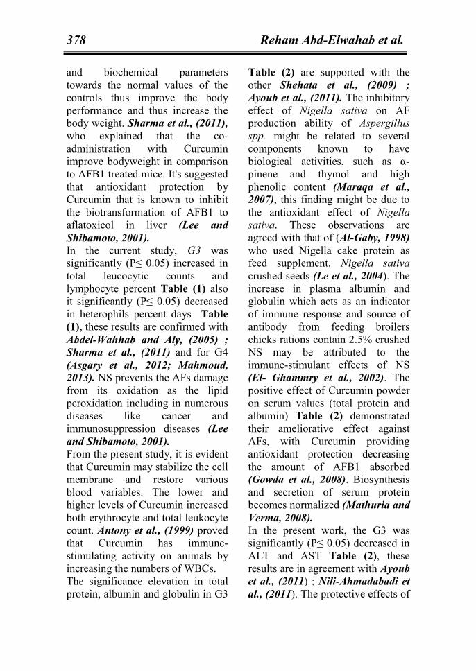
10, 20 & 30 days Table (3) & Fig.
(1) showed that there were no
significance changes in DNA
fragmentation appeared in G1, G2,
G3 and G4. DNA fragmentation
percentage when compared to G1
induced significant (P≤ 0.05)
decrease in DNA fragmentation
when compared to G2 after 20 & 30
days of experiment.
Table (1): Effect of dietary AFs on body weight changes (in grams), TLC,
Lymphocytes percent and Heterophils percent of quails in control and
experimental quails:
Values are means ± standard error (SE). Means within the same row with
different superscripts are significantly different (P≤0.05).
Groups Control -ve
(G1) AFs (G2)
AFs+ NS
(G3)
AFs+ Cur
(G4)
Liv
e b
od
y
wei
gh
t (g
)
Initial 80.564.38a 80.003.75
a 80.803.91
a 80.744.22
a
Final 264.007.65a 180.006.52
d 240.004.74b 212.006.04
c
TL
C (
103
x m
m3)
10 days 24.600.92a 17.201.50
b 21.600.81
a 18.000.71
b
20 days 32.000.70a 21.001.18
c 31.801.15
a 25.601.40
b
30 days 35.401.80a 18.202.00
c 36.201.20
a 27.000.83
b
Ly
mp
ho
cyte
% 10 days 57.001.14
a 47.401.07
b 55.402.15
a 47.401.88
b
20 days 58.001.26a 47.600.74
c 57.800.86
a 51.200.85
b
30 days 60.200.37a 46.000.89
d 56.800.96
b 52.801.28
c
Het
ero
ph
ils
%
10 days 34.401.22c 42.200.86
a 35.000.71
bc 37.100.71
b
20 days 35.181.32b 46.800.97
a 36.400.93
b 43.201.66
a
30 days 32.600.81d 52.001.41
a 37.000.71
c 41.001.16
b

374 Reham Abd-Elwahab et al.
Table (2): Effect of dietary AFs on [Tp, Albumin, Globulin, ALT and AST] in
control and experimental quails:
Values are means ± standard error (SE).
Means within the same row with different superscripts are significantly
different (P≤0.05). Table (3): Effect of dietary AF on [tissue concentration of SOD activity, GSH activity, MDA
and DNA fragmentation] of quails in control and experimental quails:
Values are means ± standard error (SE).
Means within the same row with different superscripts are significantly different (P≤0.05).
Groups Control -ve (G1) AFs (G2) AFs+ NS
(G3) AFs+ Cur
(G4)
TP
(g
/dl)
10 days 3.170.15a 2.060.01
c 2.500.20b 2.26 0.09
bc
20 days 4.500.16a 3.230.06
c 3.890.22b 3.700.08
b 30 days 4.990.23
a 3.010.11c 3.700.08
b 3.270.25c
Alb
um
in
(g/d
l) 10 days 1.740.06
a 1.220.08b 1.670.05
a 1.180.92b
20 days 1.880.03a 1.460.02
d 1.740.02b 1.560.02
c 30 days 2.050.05
a 1.530.15c 1.900.06
ab 1.690.05bc
Glo
bu
lin
(g
/dl)
10 days 1.440.21a 0.840.07
b 0.820.04b 1.080.37
ab
20 days 2.60.18a 1.780.07
b 2.000.21b 2.140.06
b
30 days 2.940.23a 1.480.21
b 2.400.08a 1.580.24
b
AL
T
(U/
l) 10 days 15.820.34
c 22.531.07a 16.800.28
bc 18.310.97b
20 days 21.764.42c 40.920.46
a 23.691.4c 32.492.40
b
30 days 18.840.56c 65.805.43
a 33.981.83b 41.810.81
b
AST (U/ l)
10 days 165.952.08b 187.692.64
a 169.233.18b 181.582.78
a 20 days 186.190.60
d 264.395.07a 218.302.85
c 229.434.02b
30 days 193.062.03d 320.165.25
a 230.011.19c 241.763.65
b
Control -ve (G1) AF (G2) AF+ NS (G3) AF+ Cur (G4)
SO
D
U/
g.t
issu
e
10 days 12.250.26a 7.50.08
d 10.260.08b 8.920.16
c
20 days 14.290.40a 6.300.10
d 11.700.32b 10.590.31
c
30 days 15.380.24a 4.360.48
d 13.270.47b 11.590.24
c
GS
H
ng
/g.
tiss
ue
10 days 4.650.07a 1.710.12
d 3.490.12b 2.850.17
c
20 days 8.340.51a 0.990.02
d 6.100.08b 4.730.24
c
30 days 8.880.15a 0.910.03
d 7.590.39b 5.640.11
c
MD
A
Mm
ol
/g.t
issu
e 10 days 27.491.06c 58.255.46
a 33.871.6bc 42.262.06
b
20 days 35.621.25d 109.313.11
a 61.493.17c 86.133.9
b
30 days 40.951.10d 144.294.91
a 69.174.71c 94.054.64
a
DN
A
fra
gm
enta
tio
n %
10 days 0.000.00a 0.000.00
a 0.000.00a 0.000.00
a
20 days 4.230.03d 19.770.63
a 7.370.60c 11.130.16
b
30 days 5.500.28d 46.321.38
a 11.130.16c 18.330.66
b

SCVMJ, XX (2) 2015 375
Fig. (1): Measurement of DNA fragmentation percentage in the liver tissues
of quails.
Disscussion AFs eradication from animal diets
and feed stuffs considers a major
problem worldwide so there is a
great need to create new
technological techniques for its
removal from diets (Williams et al.,
2004). Serious diseases caused by
AFs in animals lead to severe
economic losses and death
sometimes (Mariam et al., 2013).
Many methods were used for
detoxification of AFs include
physical, chemical and biological
methods. In our experiments we
used medicinal plants like Nigella
sativa and Curcumin for control of
AFs.
Concerning this study, G2 was
significant (P≤ 0.05) decrease in
body weight at after 10, 20 & 30
days Table (1), these results were in
agreement with the findings of
Shehata et al., (2009). It might be
due to that glutathione enzymes are
utilized in process of detoxification
to form methionine and cysteine, so
the metabolic availability of
methionine decrease and lead to
poor feed efficiency and low growth
rate (Devegowda et al., 1998).
According to our study which
showed that G2 was significant (P≤
0.05) decrease in total leucocytic
count (TLC) and lymphocyte
percent, while significant (P≤ 0.05)
increase in heterophils percent after
10, 20 & 30 days Table (1), these
results come in accordance with
results of Mohapatra et al., (2011);
Selim et al., (2014). Chamanza et
al., (1999) and Bals, (2000).
0.00
10.00
20.00
30.00
40.00
50.00
60.00
DNA fragmentat
ion %
Groups
After 10 days After 20 days After 30 days

376 Reham Abd-Elwahab et al.
Reported that the recruitment and
migration mechanisms involved in
cellular immunity are affected by
AFs. Communication between cells
of the immune system and innate
responses frequently require
different proteins in blood,
depression of cellular immunity in
AFs contaminated animals may be
due to decrease of protein level in
serum in affected poultry, especially
the globulins level which antibodies
are belonged (Nazar et al., 2012).
In the present study, G2 was
significant (P≤ 0.05) decrease in
total protein, albumin and globulin
after 10, 20 & 30 days Table (2),
these finding is supported by Rosa
et al., (2001) ; El-Sayed and Khalil,
(2009) ; Mohapatra et al., (2011).
AFs adducts binds to
macromolecules of liver cells which
in turn causes alterations in protein
synthesis and cellular integrity, also
lead to decrease of serum total
protein and albumin (Jindal et al.,
1994; Abo-Norag et al., 1995).
However, depletion of globulin is
due to the toxicity of hemopoietic
plus lymphocytolysis (Sahoo et al.,
2001). For the present study, G2 was
significant (P≤ 0.05) increase in
ALT and AST after 10, 20 & 30
days Table (2), these results are in
agreement with Amiridumari et al.,
(2013) ; Selim et al., (2014). AST
and ALT are present in the cytosol
of the hepatocytes, their elevation in
the blood stream means that AFs
cause severe destruction inside the
hepatocytes and mitochondrial
(Mathuria and Verma, 2008). Concerning this study, the G2 was
significant (P≤ 0.05) decrease in
SOD, GSH activity in liver tissues
after 10, 20 & 30 days Table (3),
these results correlate well with
(Mohamed et al., 2014; Naaz et al.,
2007; Karabacak et al., 2015), with
significant increase in hepatic MDA
activity in liver tissues homogenate
when compared with control group
Table (3), these results are in
accordance with (Naaz et al., 2007;
Wang et al., 2013; Eraslan et al.,
2005). During the metabolism of
AFs by cytochrome p450 inside
cells the reactive oxygen species
(ROS) generate as super oxide
anion, hydroxyl radical and H2O2
(Josephy, 1997; Preetha et al.,
2006). SOD, an antioxidant enzyme
is considered as the first and most
important defense line against
oxidative stress by catalyzing the
conversion of superoxide anions or
the active oxygen radical, produced
in different stages of aerobic
metabolism (Yamakura and
Fujimura, 1998), to 2 (O2) + H2O2
then catalase and glutathione
peroxidase turn it into H2O (Afonso
et al., 2007). GSH plays an
important role in the antioxidant
system of the body. It maintains the
normal structure and function of the
cells via a redox and detoxification
reaction (El-Bahr, 2014). Eraslan
et al., (2005) explained that the
decline in SOD enzyme activity
observed upon administration of
high doses of AFs may be related to

SCVMJ, XX (2) 2015 377
the consumption of highly active
components during conversion into
H2O2 due to the effect of AFs. That
indicated the failure of antioxidant
defense system to overcome the
influx of ROS induced by AFs
toxicity (Piner and Üner, 2012).
Determination of MDA by
thiobarbituric acid is used as in
index of the extend of lipid
peroxidation (Andallu and
Varadacharyulu, 2003). The
cytotoxic nature of AFs may be the
underlying reason for the increase
in MDA level (Eraslan et al.,
2005). The significant reduction in
the activities of antioxidant enzyme
SOD and non-enzymatic
antioxidant system (GSH) in
aflatoxin treated group as compared
to the control group could be
responsible for increased TBARS
levels observed during aflatoxin-
induced hepatic damage
(Choudhary and Verma, 2005).
In this study, non-significant
changes in DNA fragmentation
appeared in G1, G2, G3 or G4 after
10 days of experiment Table (3)
and Fig. (1), in the other hand, G2
was significant (P≤ 0.05) increase
in DNA fragmentation percentage
than G1 after 20 & 30 days of
experiment Table (3) and Fig. (1),
these results were also confirmed by
(Eshak et al., 2013). AFs oxidase
by the oxidase system to many
hydroxylated metabolites and to
AFBs 8, 9 epoxide that binds to
mitochondrial DNA lead to
formation of AFB1-DNA adducts
which indicated its genotoxicity
(Busby and Wogan, 1984) and
disturbs DNA replication causing
genetic alteration (Preston and
Williams, 2005). The favorable
binding formation is with guanine
lead to formation of AFB1-N-7
guanine adduct which is the cause
of mutagenesis in AF quails (Kallio
and Lahdetie, 1997). This
interaction of AFs can be with the
total genomic DNA (Choy, 1993).
This can result in small to large
changes in genomic DNA including
micronuclei and DNA
fragmentation (Faridha et al.,
2006).
The result of these study showed
that G3 and G4 were significant (P≤
0.05) increase in body weight Table
(1), these results correlate well with
other reports Zaki et al., (2011);
Sharma et al., (2011). Lee et al.,
(2003) reported that NS stimulate
the enzymes of digestive as lipase
and amylase beside mucosal
secretion of intestine, these
secretion are needed to enhance the
digestion, to remove the pathogens
adhesion and to establish the
environmental media in intestine
which the microorganisms balance
are needed, so it improve the
broilers performance by increasing
the absorption and digestion of
intestinal nutrients. Phenolic
contents present in NS decrease the
numbers of gut pathogens, so
decreasing the loss of nutrients
(Nasir and Grashorn, 2010).
Abdel-Wahhab and Aly, (2005) Reported in NS groups showed
improvement of all hematological
378 Reham Abd-Elwahab et al.
and biochemical parameters
towards the normal values of the
controls thus improve the body
performance and thus increase the
body weight. Sharma et al., (2011),
who explained that the co-
administration with Curcumin
improve bodyweight in comparison
to AFB1 treated mice. It's suggested
that antioxidant protection by
Curcumin that is known to inhibit
the biotransformation of AFB1 to
aflatoxicol in liver (Lee and
Shibamoto, 2001). In the current study, G3 was
significantly (P≤ 0.05) increased in
total leucocytic counts and
lymphocyte percent Table (1) also
it significantly (P≤ 0.05) decreased
in heterophils percent days Table
(1), these results are confirmed with
Abdel-Wahhab and Aly, (2005) ;
Sharma et al., (2011) and for G4
(Asgary et al., 2012; Mahmoud,
2013). NS prevents the AFs damage
from its oxidation as the lipid
peroxidation including in numerous
diseases like cancer and
immunosuppression diseases (Lee
and Shibamoto, 2001).
From the present study, it is evident
that Curcumin may stabilize the cell
membrane and restore various
blood variables. The lower and
higher levels of Curcumin increased
both erythrocyte and total leukocyte
count. Antony et al., (1999) proved
that Curcumin has immune-
stimulating activity on animals by
increasing the numbers of WBCs.
The significance elevation in total
protein, albumin and globulin in G3
Table (2) are supported with the
other Shehata et al., (2009) ;
Ayoub et al., (2011). The inhibitory
effect of Nigella sativa on AF
production ability of Aspergillus
spp. might be related to several
components known to have
biological activities, such as α-
pinene and thymol and high
phenolic content (Maraqa et al.,
2007), this finding might be due to
the antioxidant effect of Nigella
sativa. These observations are
agreed with that of (Al-Gaby, 1998)
who used Nigella cake protein as
feed supplement. Nigella sativa
crushed seeds (Le et al., 2004). The
increase in plasma albumin and
globulin which acts as an indicator
of immune response and source of
antibody from feeding broilers
chicks rations contain 2.5% crushed
NS may be attributed to the
immune-stimulant effects of NS
(El- Ghammry et al., 2002). The
positive effect of Curcumin powder
on serum values (total protein and
albumin) Table (2) demonstrated
their ameliorative effect against
AFs, with Curcumin providing
antioxidant protection decreasing
the amount of AFB1 absorbed
(Gowda et al., 2008). Biosynthesis
and secretion of serum protein
becomes normalized (Mathuria and
Verma, 2008). In the present work, the G3 was
significantly (P≤ 0.05) decreased in
ALT and AST Table (2), these
results are in agreement with Ayoub
et al., (2011) ; Nili-Ahmadabadi et
al., (2011). The protective effects of

SCVMJ, XX (2) 2015 379
NS oil may be due to the radical
scavenging activity of its
components, whereas the protective
effects of S. aromaticum oil may be
due to the phenolic compounds
present in the oil that decrease the
formation of aflatoxin B1 epoxides
by the inhibition of CYP450
enzymes and increase the ability of
liver microsomes to catalyse
aflatoxin–glutathione conjugation.
Consequently, NS is quite useful
and reasonable in the treatment of
aflatoxicosis (Abdel-Wahhab and
Aly, 2005). Concerning the present
study, the positive results of
Curcumin treated group G4 on
decreasing ALT and AST, Similar
results have been reported by
(Trivedi, 1999). El-Agamy, (2010)
Curcumin showed significant
hepatoprotective activity by
lowering the levels of serum marker
enzymes ALT & AST. Treatment
with Curcumin almost completely
abolished the increase of serum
ALT activity and decreased AST
activity suggesting that Curcumin
could effectively inhibit AFB1-
induced liver cell injury.
the present study showed that G3
significantly (P≤ 0.05) increased
SOD and GSH Table (3) and
significantly (P≤ 0.05) decreased
MDA Table (3), these finding are
in agreement with Kanter et al.,
(2006) ; Nili-Ahmadabadi et al.,
(2011). Thymoquinone, has a strong
antioxidant effect that inhibite lipid
peroxidation, beside the presences
of p-cymene, m-cymene, α-thujene
and carvacrol as a consititeuent of
NS have also very powerful
antioxidants activities and free
radicals scavenging effects (Burits
and Bucar, 2000). Current study
show that G4 was significant
increase in SOD activity and GSH
activity Table (3), these results are
in agreement with Mahmoud,
(2013), and significant decrease in
MDA Table (3), these results are in
agreement with Naik et al., (2004) ;
Mahmoud, (2013). Curcumin
protects the antioxidant enzymes
from denaturation as it has strong
reactive oxygen species capacity
(Madkour, 2012). Masuda et al.,
(1999) indicated that the antioxidant
mechanism of curcumin still
unknown, but it may be due to its
reaction with glutathione also it
turns to dimerization when reacted
with free radicals.
G3 was significantly (P≤ 0.05)
decreased in DNA fragmentation
after 20 & 30 days of experiment
Table (3) & fig. (1), these results
are confirmed by El-Barbary,
(2008). Where Busby and Wogan,
(1984) reported how NS reduce the
DNA fragmentation, that in the
liver microsomes, AFB1 is oxidized
to its reactive epoxide forming exo
AFB-8, 9 epoxide. This
subsequently links itself to DNA
and exhibits the mutagenicity
(Lasky and Magder, 1997). AFB1
-DNA adduct disabilities the N-
glycosidic bond of nucleotide
leading to depurination and DNA
strand scission (Lyer et al., 1994),
So decreasing the binding formation
to the AFB1, excision repair is the

380 Reham Abd-Elwahab et al.
system which primarily repair
DNA-adducts (Sancar and Sancer,
1988). So this system may be
activated via antioxidants which
catalyse formation of polar, and
conjugate between the epoxide
intermediate of AFB and
glutathione leading to reduce AFB1
–DNA adduction (Koob and
Dekant, 1991). G4 was
significantly (P≤ 0.05) decreased in
DNA fragmentation when
compared to G2 after 10, 20 & 30
days of experiment Table (3) &
fig.(1), these results are confirmed
by ( Siddique et al., 2010,
Madkour, 2012). DNA
fragmentation observed in the
present study is the normal
consequence of oxidative stress that
was demonstrated through elevation
in LPO, and antioxidant enzymes
(GPx, GST and GR) and
glutathione content in rat liver
(Madkour, 2012). This is also
consistent with previous studies
where DNA fragmentation was
induced by AFs in rat lymphocytes
(Sharma et al., 2010). G4 produced
low DNA fragmentation, that
ensures the supplementation of
curcumin to AF quails reduce the
DNA fragmentation these results
coincide with that of (Siddique et
al., 2010) who stated that Curcumin
inhibits the generation of ROS that
are responsible for the DNA
damage. Also, this action of
Curcumin was explained by
(Piwocka et al., 2001) who stated
that Curcumin leaded to attenuated
DNA fragmentation due to the
normalization of GSH.
Conclusion:
Our results indicate that Nigella
sativa crushed seeds and Curcumin
powder have a protective effect
against AF induced
immunosuppression and
hepatotoxicity in quails.
Acknowledgments:
The authors are thankful to Dr.
Azza A. Hassan. Department of
Biochemistry, Animal Health
Research Institute (AHRI),
Mansoura, Egypt and Dr. Heba M.
Abdelrazek. Dept of physiology,
faculty of Vet. Medicine, Suez
Canal University, Ismailia, Egypt.
For their laboratory help.
Reference:
Abdel-Wahhab, M. A. and Aly,
S. E. (2005): Antioxidant property
of Nigella sativa (black cumin) and
Syzygium aromaticum (clove) in
rats during aflatoxicosis. J. Appl.
Toxicol. 25 (3), 218– 23.
Abo-Norag, M.; Edrington, S.;
Kubena, F.; Harvey, B. and
Phillips, D. (1995): Influence of a
hydrated sodium calcium
aluminosilicate and virginiamycin
on aflatoxicosis in broiler chicks.
Poult. Sci. 74, 626– 632.
Afonso, V.; Champy, R.;
Mitrovic, D.; Collin, P. and
Lomri, A. (2007): Reactive oxygen
species and superoxide dismutases:
role in joint diseases. Joint Bone
Spine. 74 (4), 324-329.

SCVMJ, XX (2) 2015 381
Al-Gaby, A. M. (1998): Amino
acid composition and biological
effect of supplementing broad bean
and corn protein with Nigella sativa
(Black cumin) cake protein. Nahr.
42 (5), 290 – 294.
Al-Ghamdi, M. S. (2001): The
anti-inflammatory, analgesic and
antipyretic activity of Nigella
sativa. Journal of
Ethnopharmacology. 76, 45- 48.
Aljabre, S. H.; Randhawa, M. A.;
Akhtar, N.; Alakloby, O. M.;
Alqurashi, A. M. and Aldossary,
A. (2005): Antidermatophyte
activity of ether extract of Nigella
sativa and its active principle,
thymoquinone. Journal of
Ethnopharmacology. 101, 116- 119.
Amiridumari, H.; Sarir, H.;
Afzali, N. and FaniMakki, O.
(2013): Effects of milk thistle seed
against AF B1 in broiler
model. Journal of research in
medical sciences: the official
journal of Isfahan University of
Medical Sciences. 18 (9), 786.
Andallu, B. and Varadacharyulu,
N. (2003): Antioxidant role of
mulberry (Morus indica L. cv.
Anantha) leaves in streptozotocin-
diabetic rats. Clin.Chim.Acta. 338,
3- 10.
Antony, S.; Kuttan, R. and
Kuttan, G. (1999): Imunomodulatory activity of
Curcumin. Immunol. Invest. 28,
291- 303.
Asgary, S.; Najafi, S.; Ghannadi,
A.; Dashti, G. and Helalat, A.
(2012): Efficiency of black cumin
seeds on hematological factors in
normal and hypercholesterolemic
rabbits. ARYA atherosclerosis.
7(4), 146.
Ashiq, S.; Hussain, M. and
Ahmad, B. (2014): Natural
occurrence of mycotoxins in
medicinal plants: a review. Fungal
genetics and Biology. 66, 1- 10.
Ayoub, M. M.; El-Far, A. H.;
Taha, N. M.; Korshom, M. A.;
Mandour, A. A.; Abdel-Hamid,
H. S.; and El-Neweshy, M. S.
(2011): The biochemical protective
role of some herbs against
aflatoxicosis in ducklings: II.
Nigella sativa. ucr ri tiin ifice-
niversitatea de tiin e Agricole i
Medicin eterinar , Seria
Zootehnie. 55, 68- 77.
Bals, R. (2000): Epithelial
antimicrobial peptides in host
defense against infection. Respir.
Res. 1, 141– 150.
Basmacioglu, G.; Oguz, H.;
Ergul, M.; Col, R. and Birdane,
Y. O. (2005): Effect of dietary
esterified glucomannan on
performance, serum biochemistry
and haematology in broiler exposed
to AF. Czech Journal of Animal
Science. 20, 31- 39.
Bennett, J. W. and Klich, M.
(2003): Mycotoxins. Clinical
Microbiology Review. 16, 497–
516.
Beutler, E.; Duran, O. and Kelly,
B. (1963): improved method for the
determination of blood glutathione.
J. of lab. And clinic. Med. 61, 882.
Burits, M. and Bucar, F. (2000):
Antioxidant activity of Nigella

382 Reham Abd-Elwahab et al.
sativa essential oil. Phytother. Res.
14, 323 – 328.
Busby, W. F. Jr. and Wogan, G.
N. (1984): AF. Inc: Scarle,
C.E.(ed.). Chemical carcinogens,
2nd
ed. ACS Monograph 182.
Washington DC. American
Chemical Society. 945- 1136.
Chamanza, R.; van Veen, L.;
Tivapasi, M. T. and Toussaint, M.
J. M. (1999): Acute phase proteins
in the domestic fowl. World’s Poult.
Sci. J. 55, 61– 70.
Choudhary, A. and Verma, R.J.
(2005): Ameliorative effect of black
tea extract on aflatoxin induced
lipid peroxidation in the liver of
mice. Food and Chemical
Toxicology. 43, 99– 104.
Choy, W. N. (1993): A review of
the dose-response induction of
DNA adducts by AF B1 and its
implications to quantitative cancer-
risk assessment. Mutat. Res. 296,
181 -198.
DAFF. (2013): Structure and
dynamics of the quail.
Commonwealth of Australia.
Department of Agriculture, Editor.
2013.
Devegowda, G.; Raju, M. V. L.
N.; Afzali, N. and Swamy, H. V.
L. N. (1998): Mycotoxins picture
worldwide: Novel solutions for
their counteraction. In T.P. Lyons
and K.A. Jacques (Eds.)
Biotechnology in the Feed Industry,
Proc. Of Alltech's 14, the Annual
Symp. Nottingham, U.K. 241- 255.
Diaz, M. F.; Gonzalez, A.; Padilla,
C. and Curbelo, F. (2002): Bromatological characterization of
grains and forages from the
seasonal legumes Canavalia
ensiformis, Lablab purpureus and
Stizolobium niveum at the end of
the rainy season. Cuban J. Agric.
Sci. 36 (4), 395- 401.
Doumas, B. T. and Biggs, H. G.
(1972): Determination of serum
globulin. In: standard Methods of
Clinical Chemistry. Vol. 7 edited by
G.R Copper, New York Academic
Press.
El-Agamy, D. S. (2010):
Comparative effects of Curcumin
and resveratrol on AF B1-induced
liver injury in rats. Archives of
toxicology. 84(5), 389- 396.
El-Bahr, S. M. (2014): Effect of
Curcumin on Hepatic Antioxidant
Enzymes Activities and Gene
Expressions in Rats Intoxicated
with AF B1. Phytotherapy
Research. 29 (1), 134– 140.
El-Barbary, M. I. (2008): AF B1
induced-changes in protein
electrophoretic pattern and DNA in
Oreochromis niloticus with special
emphasis on the protective effect of
rosemary and parsley extracts.
American-Eurasian J. Agric. &
Environ. Sci. 4 (3), 381- 390.
El-Ghammry, A. A.; El-Mallah,
G. M. and El-Yamny, A. T.
(2002): The effect of incorporation
yeast culture, Nigella sativa seeds
and fresh garlic in broiler diets on
their performance. Egypt. Poult.
Sci. 22, 445- 459.
El-Sayed, Y. S. and Khalil, R.H.
(2009): Toxicity, biochemical
effects and residue of AF B1 in
marine water reared sea bass

SCVMJ, XX (2) 2015 383
(Dicentrarchus labrax L.). Food
Chem Toxicol. 47, 1606– 1609.
Eraslan, G. O.; Liman, B. C.;
Guclu, B. K.; Atasever, A. Y.;
Koc, A. N. and Beyaz, L. A.
(2004): Evaluation of AF toxicity in
Japanese quails given various doses
of hydrated sodium calcium
aluminosilicate. Bull Vet Inst
Pulawy. 48, 511- 517.
Eraslan, G.; Akdogan, M.;
Yarsan, E.; Sahindokuyucu, F.;
Essiz, D. and Altintas, L. (2005): The effects of AFs on oxidative
stress in broiler chickens. Turk. J.
Vet. Anim. Sci. 29, 701- 707.
Eshak, M. G.; Deabes, M. M.;
Farrag, A. R. H.; Farag, I. M. and
Stino, F. K. (2013): Effect of
ozone-treated AF contaminated
diets on DNA damage, expression
of androgen and androgen receptor
genes and histopathological changes
in Japanese quail. Global
Veterinaria. 11(1), 1- 13.
Faridha, A.; Fasial, K. and
Akbarsha, M.A. (2006): Duration-
dependent histopathological and
histometric changes in the testis of
AF B1-treated mice. J. Endocrinol.
Reprod. 10 (2), 117-133.
Ferreira, F. D.; Kemmelmeier,
C.; Arrotéia, C. C.; da Costa, C.
L.; Mallmann, C. A.; Janeiro, V.
and Machinski, M. (2013):
Inhibitory effect of the essential oil
of Curcuma longa L. and Curcumin
on AF production by Aspergillus
flavus Link. Food chemistry. 136
(2), 789-793.
Gibb, R. K.; Taylor, D. D.; Wan,
T.; O'Connor, D. M.; Doering, D.
L. & Gerçel-Taylor, Ç. (1997): Apoptosis as a measure of
chemosensitivity to cisplatin and
taxol therapy in ovarian cancer cell
lines.Gynecologic oncology. 65(1),
13-22.
Gowda, N. K. S.; Ledoux, D. R.;
Rottinghaus, G. E.; Bermudez, A.
J. and Chen, Y. C. (2008):
Efficacy of turmeric (Curcuma
longa), containing a known level of
Curcumin, and a hydrated sodium
calcium aluminosilicate to
ameliorate the adverse effects of AF
in broiler chicks. Poultry
science. 87 (6), 1125- 1130.
Hassan, S. M.; Mady, M. E.;
Cartwright, A. L.; Sabri, H. M.
and Mobarak, M. S. (2003): Effect
of early feed restriction on
reproductive performance in
Japanese quail (Coturnix coturnix
japonica). Poult. Sci. 82, 1163-
1169.
Hoyer, J. D. (1993): Laboratory
Med and pathology Leucocytic
differential count. Mayo Clinic
Prossiding. 68, 1027- 1028.
Jindal, N.; Manipal, S. K. and
Mahajan, N. K. (1994): Toxicity
of AF B1 in broiler chicks and its
reduction by activated charcoal.
Res. Vet. Sci. 56, 37– 40.
Josephy, P. D. (1997): Molecular
Toxicology. Oxford, UK: Oxford
Univ. Press.
Kallio, M. and Lahdetie, J.
(1997): Effects of the DNA
topoisomerase II inhibitor
membrane in male mouse meiotic
divisions in vivo cell cycle and

384 Reham Abd-Elwahab et al.
induction of aneuploidy. Environ.
Mol. Mutagen. 29, 16- 27.
Kanter, M.; Coskun, O.; Kalayci,
M.; Buyukbas, S. and Cagavi, F.
(2006): Neuroprotective effects of
Nigella sativa on experimental
spinal cord injury in rats. Hum.
Exp. Toxicol. 25, 127– 133.
Kanter, M.; Demir, H.;
Karakaya, C. and Ozbek, H.
(2005): Gastroprotective activity of
Nigella sativa L. oil and its
constituent, thymoquinone against
acute alcohol-induced gastric
mucosal injury in rats. World
Journal of Gastroenterology. 11,
6662- 6666.
Karabacak, M.; Eraslan, G.;
Kanbur, M. & Sarıca, Z. S.
(2015): Effects of Tarantula
cubensis D6 on AF-induced injury
in biochemical parameters in
rats. Homeopathy.
Khan, M. A. U.; Ashfaq, M. K.;
Zuberi, H. S.; Mahmood, M. S.
and Gilani, A. H. (2003): The in
vivo antifungal activity of the
aqueous extract from Nigella sativa
seeds. Phytotherapy Research. 17
(2), 183- 186.
Koob, M. and Dekant, W. D.
(1991): Bioactivation of xenobiotics
by formation of toxic glutathione
conjugates. Chemico-Biological
Interactions. 77, 107- 136.
Lasky, T. and Magder, L. (1997): Hepatocellular carcinoma P53 G >
T transversions at codon 249: the
fingerprint of AF exposure.
Environmental Health Perspectives.
105, 392- 397.
Le, P. M.; Ali, B.; Aziz, E.;
Abdellatif, S.; Yahia, C. and
Pierre, S. H. (2004): The
petroleum ether extract of Nigella
sativa exerts lipid-lowering and
insulin-sensitizing actions in the rat.
Journal of Ethnopharmacology. 94
(3), 251- 259.
Lee, K. G. and Shibamoto, T.
(2001): Antioxidant property of
aroma extract isolated from clove
buds [Syzygium aromaticum (L.)
Merr. et Perry. Food Chemistry. 74
(4), 443- 448.
Lee, K. W.; Everts, H.; Kappert,
H. J.; Frehner, M.; Losa, R. and
Beynen, A. C. (2003): Effects of
dietary essential oil components on
growth performance, digestive
enzymes and lipid metabolism in
female broiler chickens. Br. Poult.
Sci. 44, 450- 457.
Lyer, R. S.; Coles, B. F; Raney,
K. D.; Their, R.; Guengerich, F.P.
and Harris, T. M. (1994): DNA
adduction by the potent carcinogen
AF B : Mechanistic Studies. J.
American Chem. Soc. 116, 1603-
1609.
Madheswaran, R.; Balachandran,
C. and Manohar, B. M. (2004): Influence of dietary culture material
containing AF and T2 toxin on
certain serum biochemical
constituents in Japanese quail.
Mycopath. 158 (3), 337- 341.
Madkour, N. K. (2012): Protective
effect of Curcumin on oxidative
stress and DNA fragmentation
against lambda cyhalothrin-induced
liver damage in rats. Journal of

SCVMJ, XX (2) 2015 385
Applied Pharmaceutical Science
(JAPS). 2 (12), 76- 81.
Mahmoud, E. A. (2013): Effect of
Curcumin on hematological,
biochemical and antioxidants
parameters in Schistosoma mansoni
infected mice. International Journal
of Sciences. 2 (3), 1- 14.
Maraqa, A.; Al- sharo, N. F.;
Farah, H.; Elbjeirami, W. M.;
Shakya, A. K. and Sallal, A.
(2007): Effect of Nigella sativa
Extract and Oil on AF Production
by Aspergillus flavus J. Turk. J.
Biol. 31, 155- 159.
Mariam, G. E; Mohamed, M. D;
Abdel Razik; H. F; Ibrahim, M.
F. and Farid, K. R. (2013): Effect
of Ozone-Treated AF Contaminated
Diets on DNA Damage, Expression
of Androgen and Androgen
Receptor Genes and
Histopathological Changes in
Japanese quail. Global Vet. 11 (1),
01- 13.
Masuda, T.; Hidaka, K.;
Shinohar, A.; Maekawa, T.;
Takeda, Y. and Yamaguchi, H.
(1999): Chemical studies in
antioxidant mechanism of
Curcuminoids: analysis of radical
reaction products from Curcumin.
Journal of Agriculture and Food
Chemistry. 47, 71– 77.
Mathuria, N. and Verma, R. J.
(2008): Ameliorative effect of
Curcumin on AF-induced toxicity
in serum of mice. Acta. Pol.
Pharmaceut. Drug Res. 65, 339-
.343
Milani, J. M. (2013): Ecological
conditions affecting mycotoxin
production in cereals: a
review. Veterinary Medicine. 58
(8), 405- 411.
Mohamed, S. S.; Mohamed, S. R.;
Abou-Arab, A. A.; Naguib, K. M.;
Helmy, M. H. and Owiss, N. A.
(2014): Comparison study on native
olive waste extract and its nano-
particles effect on oxidative stress
induced by AF B1 in rat brain. Int.
J. Curr. Microbiol. App. Sci. 3 (4),
141- 152.
Mohapatra, S.; Sahu, N. P.; Pal,
A. K.; Prusty, A. K.; Kumar, V.
and Kumar, S. (2011): Haemato-
immunology and histoarchitectural
changes in Labeo rohita fingerlings:
effect of dietary AF and mould
inhibitor. Fish Physiol Biochem 37,
177– 186.
Morsi, N. M. (2000): Antimicrobial
effect of crude extracts of Nigella
sativa on multiple antibiotics-
resistant bacteria. Acta Microbio.
Polonica. 49, 63- 74.
Murray, R. (1984): Alanine
aminotransferase. Clin Chem the
C.V mosby Co. St Louis.
Toronto.Princeton. 1088- 1090.
Naaz, F.; Javed, S. and Abdin, M.
Z. (2007): Hepatoprotective effect
of ethanolic extract of Phyllanthus
amarus Schum. et Thonn. on AF B
1-induced liver damage in
mice. Journal of
ethnopharmacology. 113 (3), 503-
.509
Nabney, J. and Nesbitt, B. F.
(1965): A spectrophotometric
method for determining the
AFs. Analyst. 90 (1068), 155- 160.

386 Reham Abd-Elwahab et al.
Naik, R. S.; Mujumdar, A. M.
and Ghaskadbi, S. (2004): Protection of liver cells from
ethanol cytotoxicity by Curcumin in
liver slice culture in vitro. Journal
of Ethnopharmacology. 95, 31– 37.
Nasir, Z. and Grashorn, M. A.
(2010): Effects of Echinacea
purpurea and Nigella sativa
supplementation on broiler
performance, carcass and meat
quality. J. Anim. Feed. Sci. 19, 94-
104.
Natt, M. P. and Herrick, C. A.
(1952): A new blood diluents for
counting the erythrocytes and
leucocytes of chickens. Poultry Sci.
31, 735- 738.
Nazar , F. N.; Magnoli, A. P.;
Dalcero, A. M. and Marin, R. H.
(2012): Effect of feed
contamination with AF B1 and
administration of exogenous
corticosterone on Japanese quail
biochemical and immunological
parameters. Poultry Science. 91,
47–54.
Nili-Ahmadabadi, A.; Tavakoli,
F.; Hasanzadeh, G. R.; Rahimi,
H. R. and Sabzevari, O. (2011): Protective effect of pretreatment
with thymoquinone against AF B1
induced liver toxicity in mice. Daru:
journal of Faculty of Pharmacy,
Tehran University of Medical
Sciences. 19(4), 282.
Nishikimi, M.; Rao, N. A. and
Yogi, K. (1972): The occurance of
super oxide anion in the reaction of
reduced phenazine methosulfate and
molecular oxygen. Biochemical and
Biophysical Research. 46, 849 –
854.
Nostro, A.; Germano, M. P.;
D’Angelo, V. and Cannatelli, M.
A. (2000): Extraction methods and
bioautography for evaluation of
medicinal plant antimicrobial
activity. Letters in Applied
Microbiology. 30, 279- 284.
NRC National Research Council
(1994): Nutritional requirements of
poultry”. 9th Rev. Ed. National
academy press, Washington, DC.
Ortatatli, M.; Oğuz, H.;
Hatipoğlu, F. and Karaman, M.
(2005): Evaluation of pathological
changes in broilers during chronic
AF (50 and 100 ppb) and
clinoptilolite exposure. Research in
Veterinary Science. 78 (1), 61- 68.
Piner, P. and Üner, N. (2012): Oxidative and apoptotic effects of
lambdacyhalothrin modulated by
piperonyl butoxide in the liver of
Oreochromis niloticus. Environ.
Toxicol. Pharmacol. 33, 414- 420.
Piwocka, K.; Jaruga, E.; Skierski,
J.; Gradzka, I. and Sikora, E.
(2001): Effect of glutathione
depletion on caspase-3 independent
apoptosis pathway induced by
Curcumin in Jurkat cells. Free
Radic. Biol. Med. 31, 670- 678.
Preetha, S. P.; Kanniappan, M.;
Selvakumar, E.; Nagaraj, M.
Varalakshmi, P. (2006): Lupeol
ameliorates AF B1 induced
peroxidative hepatic damage in rats.
Comp. Biochem. Physiol. C:
Pharmacol. Toxicol. 143, 333–339.
Preston, R. J. and Williams, G.
M. (2005): DNA- reactive

SCVMJ, XX (2) 2015 387
carcinogen, mode of action and
human cancer hazard. Criterion Rev
Toxicol. 35, 673-683.
Rodkey, F. L. (1965): Clin Chem.
11, 478- 487.
Rooney, S. and Ryan, M. F.
(2005): Effects of alpha-hederin
and thymoquinone - a constituent of
Nigella sativa, on human cancer
cell lines. Anticancer Research 25,
2199- 2204.
Rosa, C. A. R.; Miazzo, R.;
Magnoli, C.; Salvano, M.;
Chiacchiera, S. M.; Ferrero, S.
and Dalcero, A. (2001): Evaluation
of the efficacy of bentonite from the
south of Argentina to ameliorate the
toxic effects of AF in
broilers. Poultry Science. 80 (2),
139- 144.
Sahin, N.; Akdemir, F.; Orhan,
C.; Kucuk, O.; Hayirli, A. and
Sahin, K. (2008): Lycopene-
enriched Quail egg as functional
food for humans. Food Research
International. 41, 295- 300.
Sahoo, P. K.; Mukherjee, S. C.;
Jain, A. K. and Mukherjee, A.
(2001): Histopathological and
electron microscopic studies of gills
and opisthonephros of Rohu, Labeo
rohita to acute and subchronic AF
B1 toxicity. Asian Fish Sci. 16,
257– 268.
Sancar, A. and Sancer, G. B.
(1988): DNA repair enzymes. Ann.
Rev. Biochem. 57, 29- 67.
Sanchis, V. and Magan, N. (2004): Environmental conditions affecting
mycotoxins. Mycotoxins in food:
detection and control. 174- 189.
Satoh, K. (1978): Serum lipid
peroxide in cerebrovascular
disorders determined by a new
colorimetric method. Clin. Chim.
Acta, 90, 37.
Selim, M.; El-hofy, H. and Khalil,
R. (2014): The efficacy of three
mycotoxin adsorbents to alleviate
AF B1-induced toxicity in
Oreochromis niloticus. Aquacult
Int. 22, 523– 540.
Sharma, D.; Saxena, P.; Singh, V.
and Sharma, R. (2010): Assessment of DNA degradation in
lymphocytes of albino rat (Rattus
norvegicus) under lambda
cyhalothrin stress. World Appl. Sci.
J. 11(1), 24- 28.
Sharma, V.; Sharma, C.; Paliwal,
R.; Pracheta, S. S. and Sharma, S.
(2011): Ameliorative effects of
Curcuma longa and Curcumin on
AF B1 induced serological and
biochemical changes in kidney of
male mice. Asian J Biochem
Pharmaceut Res. 2(1), 338- 351.
Shehata, S. A.; El-Melegy, Kh.
M.; Ebrahim M. S. and Abou-
Seif, R. A. (2009): AF B1 Toxicity
Reduction by Tafla Clay, Honey
and Nigella Sativa Addition in Fish.
Journal of the arabian aquaculture
society. 4 (1), 55- 72.
Shishodia, S.; Sethi, G. and
Aggarwal, B. B. (2005): Curcumin:
Getting Back to the Roots, Annals
of the New York Aca- demy of Sci.
1056 (1), 206- 217.
Shotwell, O. L.; Hesseltine, C. W.;
Stubblefield, R. D. and Sorenson,
W. G. (1966): Production of AF on

388 Reham Abd-Elwahab et al.
rice. Applied Microbiology. 14 (3),
425- 428.
Siddique, Y.; Ara, G.; Beg, T. and
Afzal, M. (2010): Protective effect
of Curcumin against chlormadinine
acetate induced genotoxic damage
in cultured human peripheral blood
lymphocytes. Pharmacologyonline.
3, 644- 650.
Tahan, M. and Bayram, I. (2011): Effect of using black cumin
(Nigella sativa) and parsley
(Petroselinum crispum) in laying
quail diets on egg yield, egg quality
and hatchability. Archiva
Zootechnica. 14 (4), 39-44.
Tarasub, N.; Junseecha, T.;
Tarasub, C. and Ayutthaya, W.
D. (2012): Protective effects of
Curcumin, vitamin C, or their
combination on cadmium-induced
hepatotoxicity. Journal of basic and
clinical pharmacy. 3 (2), 273.
Trivedi, N. (1999): Effect of
radiomimetic plant on vital organ.
Ph. D. thesis, Gujarat University,
Ahmedabad, India.
Tunsaringkarn, T.;
Tungjaroenchai, W. and
Siriwong, W. (2013): Nutrient
benefits of Quail (Coturnix coturnix
japonica) eggs. International
Journal of Scientific and Research
publications. 3 (5).
Wang, F.; Shu, G.; Peng, X.;
Fang, J.; Chen, K.; Cui, H. and
Lai, W. (2013): Protective effects
of sodium selenite against AF B1-
induced oxidative stress and
apoptosis in broiler
spleen. International journal of
environmental research and public
health. 10 (7), 2834- 2844.
Williams, J. H.; Phillips, T. D.,
Jolly P. E.; Stiles, J. K.; Jolly,
C.M. and Aggarwal, D. (2004): Human aflatoxicosis in developing
countries: A review of toxicology,
exposure, potential health
consequences and interventions.
Am. J. Clin. Nutr. 80, 1106– 1122.
Yamakura, F. and Fujimura, T.
H. (1998): Inactivation of
manganese superoxide dismutase by
peroxynitrite is caused by exclusive
nitration of tyrosine 34 to 3
nitrotyrosine. J. Biol. Chem. 273,
14085–14089.
Young, D. S. (1995): Effects of
drugs on clinical lab". Tests. 4th
ed
AACC Press.
Zaki, M. S.; Fawzi, O. M. and
Zytuun, I. M. (2011): Reduction of
alfatoxin in Clarious lazara catfish
by ginseng extract and Nigella
sativa oil. Journal of American
Science. 7(2), 591- 596.

SCVMJ, XX (2) 2015 389
الملخص العربي
الكبدي المحدث بواسطة التأثير التثبيطي لبذور حبة البركة و الكركم علي التسمم
الأفلاتوكسن في السمان الياباني ومدي علاقته بتجزئة الدي أن أيه
ريهام عبد الرؤف عبد الوهاب1
شريف يوسف صالح, 2
ابراهيم عاشور ابراهيم, 3
رأفت أحمد ,
الرمادي4
.مصر,المنصورة,معهدبحوثصحةالحيوان,قسمالكيمياءالحيوية1,4
مصر,الاسماعيلية,كليةالطبالبيطريجامعةقناةالسويس,قسمالكيمياءالحيوية3,2
للافلاتوكسنعليالأداء العكسية الأثار اليالكشفعن الدراسة هدفتهذه لقد بعض, و المناعة
الممكنالمعلماتالبيوكيميائيةفيالسيرموالأنسجةلديالسماناليابانيكذلكلتقييمالتأثيرالحمائي
تمتقسيماجماليعدد.لبذورحبةالبركةالمطحونةوبودرةالكركمضدالتاثيرالعكسيللافلاتوكسن
الجنسالي421 ;مجموعات1طائرمنالسماناليابانيغيرمحدد الضابطة ,(4ج)المجموعة
لمليونمعجزءمنا2.2مجموعةالافلاتوكسن,(2ج)جزءمنالمليون2.2مجموعةالافلاتوكسن
البركة 3ج%)4.2حبة الافلاتوكسن( 2.2ومجموعة الكركم 111جزءمنالمليونمعبودرة
ملجم / عليقة 1ج)كجم التجربة( مدة شهرواحد لمدة كبديوتثبيطفي. الافلاتوكسنأحدثتتسمم
الجسم وزن في بنقصمعنوي يوضح المناعة الخلا, نسبه و الكلي البيضاء الدم كرات ياعدد
الافلاتوكسنقامباحداثنقصمعنويفي.بينماحدثثزيادةمعنويةفينسبةالهيتيروفيل,الليمفاوية
الكلي البروتين الجلوبيولين, و الالبيومين الأنين, في معنوية زيادة الأخر االجانب علي
نشاط,يوتيزنقصمعنويفيانزيمالسوبرأكسيدديسم,أمينوترانسفيرسوأسبرتيدامينوترانسفييرس
المختزل الجلوتاثيون تجزئة, نقصمعنويفينسبة و المالوندايالدهيد فينشاط معنوية زيادة
عددكراتالدمالكليو.أحدثالعلاجبواسطةحبةالبركةزيادةمعنويةفيوزنالجسم.إيه.إن.الدي
حبوالبركةوالكركمأحدثت.بينماأحدثنقصمعنويفينسبةالهيتيروفيل,نسبةالخلاياالليمفاوية
الكلي البروتين في معنوية زيادة والجلوبيولين, الألبيومين في, نقصمعنوي الأخر الجانب علي
ترانسفييرس امينو أسبرتيد و أمينوترانسفيرس الأنين انزيم, في معنوية زيادة
وندايالدهيدوزيادةنشاطالجلوتاثيونالمختزلنقصمعنويفينشاطالمال,السوبرأكسيدديسميوتيز
هذهالدراسةأوضحتأنالعلاجبواسطةحبةالبركةوالكركمقد.إيه.إن.معنويةفينسبةتجزئةالدي
الأداء العكسيللأفلاتوكسنعلي التأثير حسنتمن علي, بعضالمعلماتالبيوكيميائية و المناعة
.لتجزئةإيهمنا.أن.السيرموالأنسجةوأيضاتعطيحمايةأكثرللدي