inhibition of differentiation, amplification, and function of human th17 cells by intravenous...
TRANSCRIPT
Inhibition of differentiation, amplification, and functionof human TH17 cells by intravenous immunoglobulin
Mohan S. Maddur, DVM,a,b,c Janakiraman Vani, PhD,a,b,c Pushpa Hegde, MSc,a,d Sebastien Lacroix-Desmazes, PhD,a,b,c,e
Srini V. Kaveri, DVM, PhD,a,b,c,e and Jagadeesh Bayry, DVM, PhDa,b,c,e Paris and Compiegne, France, and Mumbai, India
Background: TH17 cells play a critical role in the pathogenesisof several autoimmune and allergic diseases. Intravenousimmunoglobulin (IVIg), a therapeutic preparation of polyclonalIgG that is increasingly used in the treatment of diverseautoimmune and allergic diseases, might target TH17 cells toexert therapeutic effects.Objective: We sought to examine whether IVIg interfereswith the development and function of human TH17cells.Methods: TH17 cells were differentiated from naive humanCD41 T cells in the presence of TGF-b and IL-21. TH17 cellswere amplified by stimulating memory CD41 T cells in thepresence of IL-1b and IL-6. The effect of IVIg was examined onthe differentiation and amplification of TH17 cells, expression ofthe TH17 lineage-specific transcription factor retinoic acid-related orphan receptor C, secretion of TH17 effector cytokines,and phosphorylation of signal transducer and activator oftranscription 3, a transcription factor that plays an importantrole in TH17 cell development and function.Results: IVIg inhibits the differentiation and amplification ofhuman TH17 cells, as well as the production of their effectorcytokines IL-17A, IL-17F, IL-21, and CCL20. The inhibitoryeffects of IVIg on TH17 cells are F(ab9)2 dependent and involveinterference with the expression of retinoic acid-related orphanreceptor C and activation of signal transducer and activator oftranscription 3. Also, IVIg significantly enhanced forkhead boxprotein 3–positive regulatory T cells among the memory CD41
T cells.Conclusion: These results reveal a novel mechanism of action ofIVIg in achieving a therapeutic effect in autoimmune and
From aUnit�e 872, Institut National de la Sant�e et de la RechercheM�edicale Paris; bCentre
de Recherche des Cordeliers, Equipe 16- Immunopathology and therapeutic immu-
nointervention, Universit�e Pierre et Marie Curie, Paris; cUniversit�e Paris Descartes,
Paris; dUniversite de Technologie de Compiegne; and eInternational Associated
Laboratory IMPACT, Institut National de la Sant�e et de la Recherche M�edicale,
France-Indian Council of Medical Research, Mumbai.
Supported by grants from Institut National de la Sant�e et de la Recherche M�edicale (IN-
SERM), Centre National de la Recherche Scientifique (CNRS), Universit�e Pierre et
Marie Curie, Universit�e Paris Descartes, the European Community’s Seventh Frame-
work Programme (FP7-2007-2013) under Grant Agreement no. HEALTH-F2-2010-
260338-ALLFUN, and Octapharma, Lachen, Switzerland.
Disclosure of potential conflict of interest: S. V. Kaveri and J. Bayry have received
research support fromOctapharma. The rest of the authors have declared that they have
no conflict of interest.
Received for publication June 8, 2010; revised December 17, 2010; accepted for publi-
cation December 27, 2010.
Available online February 3, 2011.
Reprint requests: Jagadeesh Bayry, DVM, PhD, or Srini V. Kaveri, DVM, PhD, INSERM
U 872, Equipe 16-Centre de Recherche des Cordeliers, 15 rue de l’Ecole de M�edicine,
Paris, F-75006, France. E-mail: [email protected] or jagadeesh.bayry@crc.
jussieu.fr.
0091-6749/$36.00
� 2011 American Academy of Allergy, Asthma & Immunology
doi:10.1016/j.jaci.2010.12.1102
allergic diseases, in which TH17 cells play a key modulatory rolein sustaining the chronic inflammatory response. Our resultsalso suggest a reciprocal regulation of TH17 and regulatoryT-cell populations by IVIg. (J Allergy Clin Immunol2011;127:823-30.)
Key words: Intravenous immunoglobulin, allergy, autoimmunity,inflammation, TH17 cells, IL-17, regulatory T cells, retinoic-acid-related orphan receptor C, forkhead box protein 3
CD41 TH cells are the major components of the adaptive im-mune system that play a prime role in the sustenance and regula-tion of the antigen-specific immune response. On activation,under the influence of the cytokine milieu, naive TH cells differ-entiate into various subsets with distinct functions. These subsetsinclude TH1, TH2 and induced regulatory T (Treg) cells. Recently,TH17 cells, a novel subset of TH cells that express the lineage-specific transcription factor retinoic acid-related orphan receptorC (RORC) (RORgt in mice) and secrete the effector cytokines IL-17A and IL-17F, have been identified. TH17 cells require signaltransducer and activator of transcription 3 (STAT3) signalingfor their differentiation and amplification.1,2
In addition to their role in protection against extracellularpathogens, such as fungi, TH17 cells play a critical role in thepathogenesis of several autoimmune, allergic, and inflammatorydiseases, such as multiple sclerosis, rheumatoid arthritis, psoria-sis, allergic rhinitis, asthma, allergic contact dermatitis, systemiclupus erythematosus, anti-neutrophil cytoplasmic antibody–asso-ciated vasculitis, dermatomyositis, and pemphigus.3-9 TH17 cellsproduce IL-17A, IL-17F, IL-21, IL-22, and CCL20, which inducemassive tissue reactions by promoting the recruitment of inflam-matory cells.3 These effector cytokines, in addition to immunecells, also target nonimmune cells, such as fibroblasts, endothelialcells, and epithelial cells, to induce the production of the proin-flammatory mediators IL-6, TNF-a, IL-1b, prostaglandin E2, ni-tric oxide, matrix metalloproteinases, and chemokines (CXCL1,CXCL8, and CCL2).1-10 Furthermore, the development of TH17cells is dependent on the inflammatory cytokines IL-6, IL-21,IL-1b, and IL-23, thus sustaining the vicious inflammatorycircle.11
Both experimental and clinical studies have clearly demon-strated the role of TH17 cell–derived cytokines in the patho-genesis of several autoimmune, allergic, and inflammatorydiseases.3,11,12 Thus new therapeutic strategies that can efficientlyinterfere with the development and function of TH17 cells are be-ing explored.13-17 In this context intravenous immunoglobulin(IVIg) represents one of the promising approaches that might tar-get TH17 cells.
IVIg, a therapeutic preparation of polyclonal IgG obtainedfrom pooled plasma of several thousand healthy donors, is beingincreasingly used in the treatment of diverse autoimmune,
823
J ALLERGY CLIN IMMUNOL
MARCH 2011
824 MADDUR ET AL
Abbreviations used
APC: A
ntigen-presenting cellFoxp3: F
orkhead box protein 3HSA: H
uman serum albuminIVIg: I
ntravenous immunoglobulinpSTAT3: P
hosphorylated STAT3RORC: R
etinoic-acid-related orphan receptor CSTAT3: S
ignal transducer and activator of transcription 3Treg: R
egulatory Tallergic, and inflammatory diseases.18-26 IVIg is believed toexert beneficial effects in patients through several mutually non-exclusive mechanisms.18,27-30 Furthermore, IVIg can directly in-teract with T cells to exert immunomodulatory effects.18,31
Interestingly, a therapeutic effect of IVIg has also been demon-strated in autoimmune diseases in which TH17 cells have been im-plicated in the pathogenesis.3,18,20,22 Therefore we hypothesizedthat IVIg modulates human TH17 cell differentiation, propaga-tion, and function to exert beneficial effects in autoimmunepatients.
METHODS
Cell-culture reagents and antibodiesThe mAbs anti-CD3 (clone UCHT1) and anti-CD28 (clone 37407) and the
cytokines TGF-b, IL-1b, and IL-6 were from R&D Systems (Lille, France).
IL-21 and plasma-derived human serum albumin (HSA) were from Immuno-
Tools (Friesoythe, Germany) and Laboratoire Francaise de Biotechnologies
(Les Ulis, France), respectively.
Therapeutic IVIgOctagam (Octapharma, Vienna, Austria) was used throughout the study.
Octagam (100 mg/mL; 0.6 mmol/L) was dialyzed against a large volume of
PBS followed by RPMI-1640 medium at 48C for 18 hours to remove the
stabilizing agents.32 F(ab9)2 fragments of IVIg were prepared by means of
pepsin digestion (2% wt/wt; Sigma, St Louis, Mo) followed by chromatogra-
phy on a protein G Sepharose column (Pharmacia, Uppsala, Sweden). F(ab9)2fragments were free of intact IgG and Fc fragments, as assessed by means of
SDS-PAGE and ELISA. Octagam contained a negligible amount of T-cell cy-
tokines (0-5 pg/mL/100 mg) and tested negative for endotoxins (ToxinSensor
Chromogenic LAL Endotoxin Assay Kit; GenScript, Piscataway, NJ).
Cell purificationPBMCs were purified from buffy coats of healthy donors. Untouched total
CD41 T cells were isolated by means of negative selection with the CD41
T-cell isolation kit II (Miltenyi Biotech, Paris, France). Furthermore, naive
(CD45RO2) and memory (CD45RO1) CD41 T cells were separated with
CD45RO microbeads (Miltenyi Biotech). Finally, CD41CD252CD45RO2
naive T cells were obtained by depleting CD251 cells with CD25 microbeads
(Miltenyi Biotech) to remove the recently activated cells among the
naive CD41 T cells. The purity of cells was greater than 95% in all the
subpopulations.
T-cell stimulation and cultureNinety-six-well U-bottom plates were coated with 1.5 mg/mL anti-CD3
mAb. Plates were then rinsed once with serum-free RPMI-1640 medium.
CD41 T cells (5 3 104 per well/200 mL) were stimulated in serum-free X-
VIVO 15 medium with soluble anti-CD28 mAb (1.0 mg/mL) in the presence
of cytokines that included TGF-b (5 ng/mL) and IL-21 (25 ng/mL) for naive T
cells and IL-1b (12.5 ng/mL) and IL-6 (25 ng/mL) for memory T cells. Equi-
molar concentrations (0.15 mmol/L) of IVIg (25 mg/mL), F(ab9)2 fragments
(16 mg/mL), or HSA (10 mg/mL) were added to the cells after 12 hours of
initiation of culture. The stimulated T cells were cultured for 5 to 6 days at
378C in 5% CO2. Supernatants were collected either daily or at the end of cul-
ture for the analysis of cytokines, and cells were used for intracellular staining.
In addition, dose-response and time-response curves of IVIg were also
performed.
Intracellular staining and cytokine assaysFor intracellular staining, cells were stimulated with phorbol 12-myristate
13-acetate (50 ng/mL, Sigma, Saint Quentin Fallavier, France) and ionomycin
(500 ng/mL, Sigma) at 378C for 5 hours with GolgiStop (BD Biosciences, Le
Pont de Claix, France) for the last 3 hours. Surface staining was done with
fluorescence-conjugated CD4 mAb (BD Biosciences). Cells were fixed,
permeabilized (Fix/Perm; eBioscience, Paris, France), and incubated at
room temperature with fluorescence-conjugated mAbs to RORC (eBio-
science), IFN-g (BD Biosciences), IL-17A (eBioscience), and forkhead box
protein 3 (Foxp3; eBioscience). Ten thousand cells were acquired for each
sample, and the data were processed by using FACS DIVA software (BD
Biosciences).
Intracellular phosphorylated STAT3 (pSTAT3) staining was performed on
activated T cells at different time points. Cells were harvested and fixed in
prewarmed BD Cytofix buffer by incubating for 10 minutes at 378C. Fixedcells were washed twice with staining buffer (1% FCS/PBS) and then
permeabilized with chilled BD Phosflow Perm Buffer III on ice for 30
minutes. Cells were then washed twice with staining buffer, and
phycoerythrin-conjugated anti-STAT3 pY705 (BD Biosciences) was added
to cells and incubated at room temperature for 45 to 60 minutes. Cells were
washed and suspended in staining buffer before data acquisition.
Levels of IL-17A and CCL20 (DuoSet ELISA kits, R&D Systems), IL-17F
(ELISA Ready-SET-Go, eBioscience), and IL-21 (ELISAMAX Deluxe Sets,
BioLegend, San Diego, Calif) in cell-free culture supernatants were quantified
by means of ELISA. Cutoff values were 15 pg/mL for IL-17A, 30 pg/mL for
IL-17F, 16 pg/mL for IL-21, and 15 pg/mL for CCL20 (see Table E1 in this
article’s Online Repository at www.jacionline.org).
Statistical analysisThe paired Student t test was used to determine the statistical significance
of the data. A P value of less than .05 was considered significant.
RESULTS
Inhibition of differentiation and function of human
TH17 cells by IVIgFor the differentiation of human TH17 cells,
CD41CD45RO2CD252 naive T cells were cultured in serum-free medium in the presence of T-cell receptor stimulators andTGF-b and IL-21 for 6 days.33 To closely mimic in vivo differen-tiating conditions, we did not use neutralizing antibodies againstIFN-g and IL-4, the cytokines that are known to inhibit the devel-opment of TH17 cells.
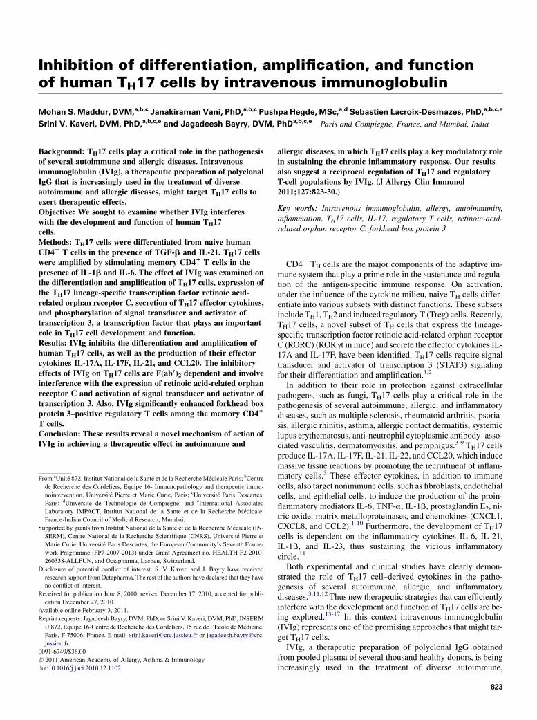
IVIg was added to the T-cell culture 12 hours after initiation ofstimulation to investigate the effect of IVIg on the differentiationof TH17 cells. Compared with cells differentiated in the presenceof TGF-b and IL-21 alone, IVIg significantly reduced thepercentage of IL-17A1, RORC1, and IL-17A1/IFN-g1 cells(Fig 1). Equimolar concentrations of HSA, used as a protein con-trol for IVIg, did not inhibit the differentiation of TH17 cells (Fig1). We excluded the possibility that the reduction of IL-17–pro-ducing cell numbers by IVIg is not due to apoptosis or the toxiceffects of IVIg on cells (data not shown). Furthermore, IVIg didnot alter the expression of Foxp3 (Treg cells) among the naiveTH cells cultured under TH17-polarizing conditions (Fig 1, B).A prominent decrease in IFN-g–producing cell numbers was
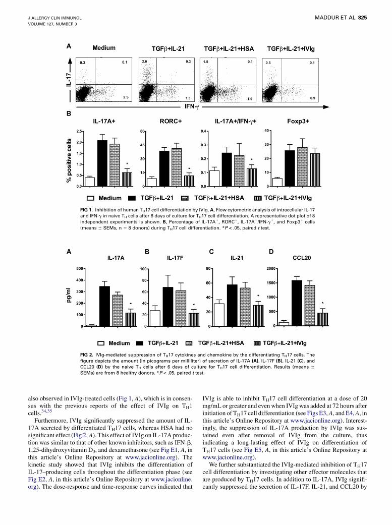
FIG 1. Inhibition of human TH17 cell differentiation by IVIg. A, Flow cytometric analysis of intracellular IL-17
and IFN-g in naive TH cells after 6 days of culture for TH17 cell differentiation. A representative dot plot of 8
independent experiments is shown. B, Percentage of IL-17A1, RORC1, IL-17A1/IFN-g1, and Foxp31 cells
(means 6 SEMs, n 5 8 donors) during TH17 cell differentiation. *P < .05, paired t test.
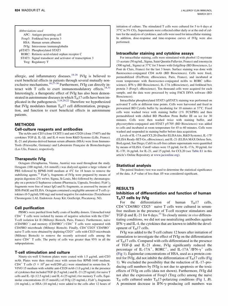
FIG 2. IVIg-mediated suppression of TH17 cytokines and chemokine by the differentiating TH17 cells. The
figure depicts the amount (in picograms per milliliter) of secretion of IL-17A (A), IL-17F (B), IL-21 (C), and
CCL20 (D) by the naive TH cells after 6 days of culture for TH17 cell differentiation. Results (means 6SEMs) are from 8 healthy donors. *P < .05, paired t test.
J ALLERGY CLIN IMMUNOL
VOLUME 127, NUMBER 3
MADDUR ET AL 825
also observed in IVIg-treated cells (Fig 1, A), which is in consen-sus with the previous reports of the effect of IVIg on TH1cells.34,35
Furthermore, IVIg significantly suppressed the amount of IL-17A secreted by differentiated TH17 cells, whereas HSA had nosignificant effect (Fig 2,A). This effect of IVIg on IL-17A produc-tion was similar to that of other known inhibitors, such as IFN-b,1,25-dihydroxyvitamin D3, and dexamethasone (see Fig E1, A, inthis article’s Online Repository at www.jacionline.org). Thekinetic study showed that IVIg inhibits the differentiation ofIL-17–producing cells throughout the differentiation phase (seeFig E2, A, in this article’s Online Repository at www.jacionline.org). The dose-response and time-response curves indicated that
IVIg is able to inhibit TH17 cell differentiation at a dose of 20mg/mL or greater and evenwhen IVIg was added at 72 hours afterinitiation of TH17 cell differentiation (see Figs E3,A, and E4,A, inthis article’s Online Repository at www.jacionline.org). Interest-ingly, the suppression of IL-17A production by IVIg was sus-tained even after removal of IVIg from the culture, thusindicating a long-lasting effect of IVIg on differentiation ofTH17 cells (see Fig E5, A, in this article’s Online Repository atwww.jacionline.org).
We further substantiated the IVIg-mediated inhibition of TH17cell differentiation by investigating other effector molecules thatare produced by TH17 cells. In addition to IL-17A, IVIg signifi-cantly suppressed the secretion of IL-17F, IL-21, and CCL20 by
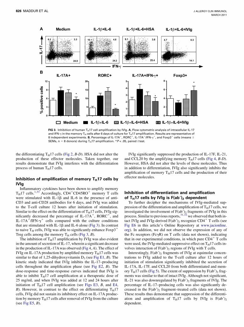
FIG 3. Inhibition of human TH17 cell amplification by IVIg. A, Flow cytometric analysis of intracellular IL-17
and IFN-g in the memory TH cells after 6 days of culture for TH17 amplification. Results are representative of
8 independent experiments. B, Percentage of IL-17A1, RORC1, IL-17A1/IFN-g1, and Foxp31 cells (means 6SEMs, n 5 8 donors) during TH17 amplification. *P < .05, paired t test.
J ALLERGY CLIN IMMUNOL
MARCH 2011
826 MADDUR ET AL
the differentiating TH17 cells (Fig 2, B-D). HSA did not alter theproduction of these effector molecules. Taken together, ourresults demonstrate that IVIg interferes with the differentiationprocess of human TH17 cells.
Inhibition of amplification of memory TH17 cells by
IVIgInflammatory cytokines have been shown to amplify memory
TH17 cells.1,33 Accordingly, CD41CD45RO1 memory T cellswere stimulated with IL-1b and IL-6 in the presence of anti-CD3 and anti-CD28 antibodies for 6 days, and IVIg was addedto the T-cell culture 12 hours after initiation of stimulation.Similar to the effect on the differentiation of TH17 cells, IVIg sig-nificantly decreased the percentage of IL-17A1, RORC1, andIL-17A1/IFN-g1 cells compared with the culture conditionsthat are stimulated with IL-1b and IL-6 alone (Fig 3). In contrastto naive TH cells, IVIg was able to significantly enhance Foxp31
Treg cells among the memory TH cells (Fig 3, B).The inhibition of TH17 amplification by IVIg was also evident
in the amount of secretion of IL-17, wherein a significant decreasein the production of IL-17Awas observed (Fig 4, A). The effect ofIVIg on IL-17A production by amplified memory TH17 cells wassimilar to that of 1,25-dihydroxyvitamin D3 (see Fig E1, B). Thekinetic study indicated that IVIg inhibits the IL-17–producingcells throughout the amplification phase (see Fig E2, B). Thedose-response and time-response curves indicated that IVIg isable to inhibit TH17 cell amplification at a therapeutic dose of25 mg/mL and when IVIg was added at 12 and 24 hours afterinitiation of TH17 cell amplification (see Figs E3, B, and E4,B). However, in contrast to the effect on differentiating TH17cells, IVIg did not sustain its inhibitory effect on IL-17A produc-tion by memory TH17 cells after removal of IVIg from the culture(see Fig E5, B).
IVIg significantly suppressed the production of IL-17F, IL-21,and CCL20 by the amplifying memory TH17 cells (Fig 4, B-D).However, HSA did not alter the levels of these molecules. Thusin addition to differentiation, IVIg also significantly inhibits theamplification of memory TH17 cells and the production of theireffector molecules.
Inhibition of differentiation and amplification
of TH17 cells by IVIg is F(ab9)2 dependentTo further decipher the mechanisms of IVIg-mediated sup-
pression of the differentiation and amplification of TH17 cells, weinvestigated the involvement of F(ab9)2 fragments of IVIg in thisprocess. Similar to previous reports,31,32 we observed that both in-tact IVIg and IVIg-derived F(ab9)2 recognize CD4
1 T cells (seeFig E6 in this article’s Online Repository at www.jacionline.org). In addition, we did not observe the expression of any ofthe Fc receptors (FcgR) on T cells (data not shown), indicatingthat in our experimental conditions, in which pure CD41 T cellswere used, the IVIg-mediated suppressive effect on TH17 cells in-volves interaction of F(ab9)2 regions of IVIg with T cells.
Interestingly, F(ab9)2 fragments of IVIg at equimolar concen-trations to IVIg added to the T-cell culture after 12 hours ofinitiation of stimulation significantly inhibited the secretion ofIL-17A, IL-17F, and CCL20 from both differentiated and mem-ory TH17 cells (Fig 5). The extent of suppression by F(ab9)2 frag-ments was similar to that of intact IVIg. Although not significant,IL-21 was also downregulated by F(ab9)2 fragments of IVIg. Thepercentage of IL-17–producing cells was also significantly de-creased in the F(ab9)2 fragment–treated cells (data not shown).These results thus demonstrate that suppression of the differenti-ation and amplification of TH17 cells by IVIg is F(ab9)2dependent.

FIG 4. Suppression of TH17 cytokines and chemokine in the amplifying memory TH17 cells by IVIg. The fig-
ure shows the amount (in picograms per milliliter) of secretion of IL-17A (A), IL-17F (B), IL-21 (C), and CCL20
(D) by the memory TH cells after 6 days of culture for TH17 amplification. Results (means6 SEMs) are from 8
healthy donors. *P < .05, paired t test.
FIG 5. IVIg-mediated suppression of differentiation and amplification of TH17 cells is F(ab9)2 dependent. Theamount (in picograms per milliliter) of secretion of IL-17A, IL-17F, IL-21, and CCL20 by the naive CD41 T cells
cultured for TH17 cell differentiation (A) and the memory CD41 T cells cultured for TH17 amplification (B) is
depicted. Results (means 6 SEMs) are from 5 donors. *P < .05, paired t test.
J ALLERGY CLIN IMMUNOL
VOLUME 127, NUMBER 3
MADDUR ET AL 827
IVIg interferes with cytokine signaling to mediate
the suppression of differentiation and amplification
of TH17 cellsSTAT3 signaling plays a crucial role early in the development
of TH17 cells.11 After binding of IL-21 and IL-6 to their respective
receptors on cells, STAT3 undergoes activation by phosphoryla-tion (at Y705) and facilitates the expression of RORC andthe inhibition of Foxp3. Therefore we explored the probablemodulation of sustained STAT3 phosphorylation (pSTAT3) by
IVIg as a mechanism of inhibition of TH17 cell developmentand function.A significant level of pSTAT3 was observed in both naive and
memory TH cells under TH17 culture conditions after 18 hoursof cytokine stimulation (Fig 6). Interestingly, IVIg reducedpSTAT3 levels in both populations of TH cells (Fig 6). Duringthe differentiation phase of TH17 cells, IVIg inhibited pSTAT3from the early time points, whereas in memory TH17 cells theinhibition of pSTAT3 was evident from 24 hours of culture.
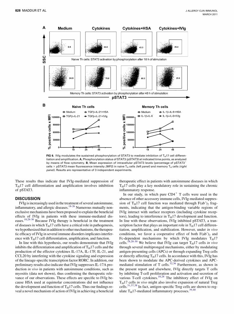
FIG 6. IVIg modulates the sustained phosphorylation of STAT3 to mediate inhibition of TH17 cell differen-
tiation and amplification.A, Phosphorylation status of STAT3 (pSTAT3) at indicated time points, as analyzed
by means of flow cytometry. B, Mean expression of intracellular pSTAT3 levels (percentage of pSTAT31
cells 3 pSTAT3 mean fluorescence intensity [MFI]) in naive TH cells (left panel) and memory TH cells (rightpanel). Results are representative of 3 independent experiments.
J ALLERGY CLIN IMMUNOL
MARCH 2011
828 MADDUR ET AL
These results thus indicate that IVIg-mediated suppression ofTH17 cell differentiation and amplification involves inhibitionof pSTAT3.
DISCUSSIONIVIg is increasingly used in the treatment of several autoimmune,
inflammatory, and allergic diseases.18-26 Numerous mutually non-exclusivemechanisms have been proposed to explain the beneficialeffects of IVIg in patients with these immune-mediated dis-eases.18,28-30 Because IVIg therapy is beneficial in the treatmentof diseases in which TH17 cells have a critical role in pathogenesis,wehypothesized that in addition toothermechanisms, the therapeu-tic efficacy of IVIg in several immune disorders implicates interfer-ence with TH17 cell differentiation, amplification, and function.
In line with this hypothesis, our results demonstrate that IVIginhibits the differentiation and amplification of TH17 cells and theproduction of the effector cytokines IL-17A, IL-17F, IL-21, andCCL20 by interfering with the cytokine signaling and expressionof the lineage-specific transcription factor RORC. In addition, ourpreliminary results also indicate that IVIg suppresses IL-17A pro-duction in vivo in patients with autoimmune conditions, such asmyositis (data not shown), thus confirming the therapeutic rele-vance of our observations. These effects are specific to IVIg be-cause HSA used at equimolar concentrations did not influencethe development and function of TH17 cells. Thus our findings re-veal a novel mechanism of action of IVIg in achieving a beneficial
therapeutic effect in patients with autoimmune diseases in whichTH17 cells play a key modulatory role in sustaining the chronicinflammatory response.In our study, in which pure CD41 T cells were used in the
absence of other accessory immune cells, IVIg-mediated suppres-sion of TH17 cell function was mediated through F(ab9)2 frag-ments, indicating that the antigen-binding variable regions ofIVIg interact with surface receptors (including cytokine recep-tors), leading to interference in TH17 development and function.In line with these observations, IVIg inhibited pSTAT3, a tran-scription factor that plays an important role in TH17 cell differen-tiation, amplification, and stabilization. However, under in vivoconditions, we favor a cooperative effect of both F(ab9)2 andFc-dependent mechanisms by which IVIg modulates TH17cells.18,28-30 We believe that IVIg can target TH17 cells in vivothrough several multipronged mechanisms, either by modulatingantigen-presenting cells (APCs) or through expanding Treg cellsor directly affecting TH17 cells. In accordance with this, IVIg hasbeen shown to modulate the APC-derived cytokines and APC-mediated stimulation of T cells.32,36 Furthermore, as shown inthe present report and elsewhere, IVIg directly targets T cellsby inhibiting T-cell proliferation and activation and secretion ofvarious T-cell cytokines.18,28 The inhibitory effect of IVIg onTH17 cells in vivo might also involve expansion of natural Tregcells.31,37,38 In fact, antigen-specific Treg cells are shown to reg-ulate TH17-mediated inflammatory processes.39,40
J ALLERGY CLIN IMMUNOL
VOLUME 127, NUMBER 3
MADDUR ET AL 829
Interestingly, in our study IVIg-mediated inhibition of TH17cell differentiation from naive TH cells was not associatedwith the enhancement of Foxp31 Treg cells. Moreover, the na-ive TH cells were depleted of CD251 cells to remove the re-cently activated TH cells and the naive Treg cells. This findingis in consensus with our previous observation, wherein IVIgdid not induce de novo generation of Treg cells in mice.31 How-ever, among the memory cells, suppression of TH17 amplifica-tion and function by IVIg was associated with a significantincrease in Foxp31 Treg cell numbers. These results thusconfirm the previous reports that IVIg expands the Treg cellpopulation both in vitro and in vivo.31,37,41 Together, our resultssuggest a reciprocal regulation of the TH17 and Treg cell popu-lation by IVIg.We observed that in addition to inhibition of TH17 cell dif-
ferentiation and amplification, IVIg also suppressed IL-171/IFN-g1 and IFN-g1 cells. This is in contrast to some of themolecules explored for TH17 inhibition, such as halofuginoneand pioglitazone, which selectively inhibit TH17 cells withoutaffecting TH1 development.14,15 However, 1,25-dihydroxyvita-min D3 and dexamethasone inhibit IFN-g in addition toIL-17.17 Despite the prominent role of TH17 cells in severalinflammatory and autoimmune diseases, the importance ofIFN-g–producing TH1 cells is not clearly ruled out in these dis-eases.1,2 Moreover, memory TH17 cells can express IFN-g afteractivation, as evidenced by the high percentage of IL-171IFN-g1 T cells in the memory CD41 T-cell pool, thus suggestingthe plasticity of TH17 cells.2 Given that both TH1 and TH17cells mediate diverse autoimmune processes, therapies thathave broad cellular targets, as in the case of IVIg, enablemore efficient control of these pathologies.
Key messages
d IVIg inhibits TH17 cell differentiation, propagation, andproduction of the effector cytokines IL-17A, IL-17F,IL-21, and CCL20.
d In addition to other mechanisms, the therapeutic efficacyof IVIg in patients with autoimmune and allergic diseasesmight implicate interference with TH17 cell developmentand function.
REFERENCES
1. Bettelli E, Korn T, Oukka M, Kuchroo VK. Induction and effector functions of
T(H)17 cells. Nature 2008;453:1051-7.
2. Zhou L, Chong MM, Littman DR. Plasticity of CD41 T cell lineage differentia-
tion. Immunity 2009;30:646-55.
3. Miossec P, Korn T, Kuchroo VK. Interleukin-17 and type 17 helper T cells. N Engl
J Med 2009;361:888-98.
4. Louten J, Boniface K, de Waal Malefyt R. Development and function of TH17
cells in health and disease. J Allergy Clin Immunol 2009;123:1004-11.
5. Annunziato F, Cosmi L, Liotta F, Maggi E, Romagnani S. Type 17 T helper cells-
origins, features and possible roles in rheumatic disease. Nat Rev Rheumatol 2009;
5:325-31.
6. Hashimoto T, Akiyama K, Kobayashi N, Mori A. Comparison of IL-17 production
by helper T cells among atopic and nonatopic asthmatics and control subjects. Int
Arch Allergy Immunol 2005;137(suppl 1):51-4.
7. van Beelen AJ, Teunissen MB, Kapsenberg ML, de Jong EC. Interleukin-17 in
inflammatory skin disorders. Curr Opin Allergy Clin Immunol 2007;7:374-81.
8. Larsen JM, Bonefeld CM, Poulsen SS, Geisler C, Skov L. IL-23 and T(H)17-
mediated inflammation in human allergic contact dermatitis. J Allergy Clin
Immunol 2009;123:486-92.
9. Ciprandi G, Filaci G, Battaglia F, Fenoglio D. Peripheral Th-17 cells in allergic
rhinitis: New evidence. Int Immunopharmacol 2010;10:226-9.
10. Dong C. TH17 cells in development: an updated view of their molecular identity
and genetic programming. Nat Rev Immunol 2008;8:337-48.
11. Korn T, Bettelli E, Oukka M, Kuchroo VK. IL-17 and Th17 Cells. Annu Rev
Immunol 2009;27:485-517.
12. Wang YH, Liu YJ. The IL-17 cytokine family and their role in allergic inflamma-
tion. Curr Opin Immunol 2008;20:697-702.
13. Zhang X, Jin J, Peng X, Ramgolam VS, Markovic-Plese S. Simvastatin inhibits
IL-17 secretion by targeting multiple IL-17-regulatory cytokines and by inhibiting
the expression of IL-17 transcription factor RORC in CD41 lymphocytes.
J Immunol 2008;180:6988-96.
14. Sundrud MS, Koralov SB, Feuerer M, Calado DP, Kozhaya AE, Rhule-Smith A,
et al. Halofuginone inhibits TH17 cell differentiation by activating the amino
acid starvation response. Science 2009;324:1334-8.
15. Klotz L, Burgdorf S, Dani I, Saijo K, Flossdorf J, Hucke S, et al. The nuclear re-
ceptor PPAR gamma selectively inhibits Th17 differentiation in a T cell-intrinsic
fashion and suppresses CNS autoimmunity. J Exp Med 2009;206:2079-89.
16. Ramgolam VS, Sha Y, Jin J, Zhang X, Markovic-Plese S. IFN-b inhibits human
Th17 cell differentiation. J Immunol 2009;183:5418-27.
17. Colin EM, Asmawidjaja PS, van Hamburg JP, Mus AM, van Driel M, Hazes JM,
et al. 1,25-Dihydroxyvitamin D3 modulates Th17 polarization and interleukin-22
expression by memory T cells from patients with early rheumatoid arthritis. Arthri-
tis Rheum 2010;62:132-42.
18. Kazatchkine MD, Kaveri SV. Immunomodulation of autoimmune and inflamma-
tory diseases with intravenous immune globulin. N Engl J Med 2001;345:747-55.
19. Orange JS, Hossny EM, Weiler CR, Ballow M, Berger M, Bonilla FA, et al. Use of
intravenous immunoglobulin in human disease: a review of evidence by members
of the Primary Immunodeficiency Committee of the American Academy of
Allergy, Asthma and Immunology. J Allergy Clin Immunol 2006;117(suppl):
S525-53.
20. Gold R, Stangel M, Dalakas MC. Drug Insight: the use of intravenous immuno-
globulin in neurology—therapeutic considerations and practical issues. Nat Clin
Pract Neurol 2007;3:36-44.
21. Mazer BD, Giclas PC, Gelfand EW. Immunomodulatory effects of intravenous im-
munoglobulin in severe steroid-dependent asthma. Clin Immunol Immunopathol
1989;53(suppl):S156-63.
22. Mazer BD, Gelfand EW. An open-label study of high-dose intravenous immuno-
globulin in severe childhood asthma. J Allergy Clin Immunol 1991;87:976-83.
23. Leung DY. The immunoregulatory effects of IVIG in Kawasaki disease and other
autoimmune diseases. Clin Rev Allergy 1992;10:93-104.
24. Gelfand EW, Landwehr LP, Esterl B, Mazer B. Intravenous immune globulin: an
alternative therapy in steroid-dependent allergic diseases. Clin Exp Immunol
1996;104(suppl 1):61-6.
25. Rabinovitch N, Gelfand EW, Leung DY. The role of immunoglobulin therapy in
allergic diseases. Allergy 1999;54:662-8.
26. Prins C, Gelfand EW, French LE. Intravenous immunoglobulin: properties, mode
of action and practical use in dermatology. Acta Derm Venereol 2007;87:206-18.
27. Lemieux R, Bazin R, Neron S. Therapeutic intravenous immunoglobulins. Mol
Immunol 2005;42:839-48.
28. Tha-In T, Bayry J, Metselaar HJ, Kaveri SV, Kwekkeboom J. Modulation of the
cellular immune system by intravenous immunoglobulin. Trends Immunol 2008;
29:608-15.
29. Nimmerjahn F, Ravetch JV. Anti-inflammatory actions of intravenous immuno-
globulin. Annu Rev Immunol 2008;26:513-33.
30. Seite JF, Shoenfeld Y, Youinou P, Hillion S. What is the contents of the magic draft
IVIg? Autoimmun Rev 2008;7:435-9.
31. Ephrem A, Chamat S, Miquel C, Fisson S, Mouthon L, Caligiuri G, et al. Expan-
sion of CD41CD251 regulatory T cells by intravenous immunoglobulin: a critical
factor in controlling experimental autoimmune encephalomyelitis. Blood 2008;
111:715-22.
32. Bayry J, Lacroix-Desmazes S, Carbonneil C, Misra N, Donkova V, Pashov A, et al.
Inhibition of maturation and function of dendritic cells by intravenous immuno-
globulin. Blood 2003;101:758-65.
33. Yang L, Anderson DE, Baecher-Allan C, Hastings WD, Bettelli E, Oukka M, et al.
IL-21 and TGF-b are required for differentiation of human T(H)17 cells. Nature
2008;454:350-2.
34. Andersson J, Skansen-Saphir U, Sparrelid E, Andersson U. Intravenous immune
globulin affects cytokine production in T lymphocytes and monocytes/macro-
phages. Clin Exp Immunol 1996;104(suppl 1):10-20.
35. Tha-In T, Metselaar HJ, Tilanus HW, Boor PP, Mancham S, Kuipers EJ, et al.
Superior immunomodulatory effects of intravenous immunoglobulins on human
T-cells and dendritic cells: comparison to calcineurin inhibitors. Transplantation
2006;81:1725-34.

J ALLERGY CLIN IMMUNOL
MARCH 2011
830 MADDUR ET AL
36. Aubin E, Lemieux R, Bazin R. Indirect inhibition of in vivo and in vitro T-cell
responses by intravenous immunoglobulins due to impaired antigen presentation.
Blood 2010;115:1727-34.
37. Kessel A, Ammuri H, Peri R, Pavlotzky ER, Blank M, Shoenfeld Y, et al. Intrave-
nous immunoglobulin therapy affects T regulatory cells by increasing their
suppressive function. J Immunol 2007;179:5571-5.
38. De Groot AS, Moise L, McMurry JA, Wambre E, Van Overtvelt L, Moingeon P,
et al. Activation of natural regulatory T cells by IgG Fc-derived peptide ‘‘Tregi-
topes.’’ Blood 2008;112:3303-11.
39. Fletcher JM, Lonergan R, Costelloe L, Kinsella K, Moran B, O’Farrelly C, et al.
CD391Foxp31 regulatory T Cells suppress pathogenic Th17 cells and are
impaired in multiple sclerosis. J Immunol 2009;183:7602-10.
40. Crome SQ, Clive B, Wang AY, Kang CY, Chow V, Yu J, et al. Inflammatory effects
of ex vivo human Th17 cells are suppressed by regulatory T cells. J Immunol 2010;
185:3199-208.
41. Tha-In T, Metselaar HJ, Bushell AR, Kwekkeboom J, Wood KJ. Intravenous im-
munoglobulins promote skin allograft acceptance by triggering functional activa-
tion of CD41Foxp31 T cells. Transplantation 2010;89:1446-55.
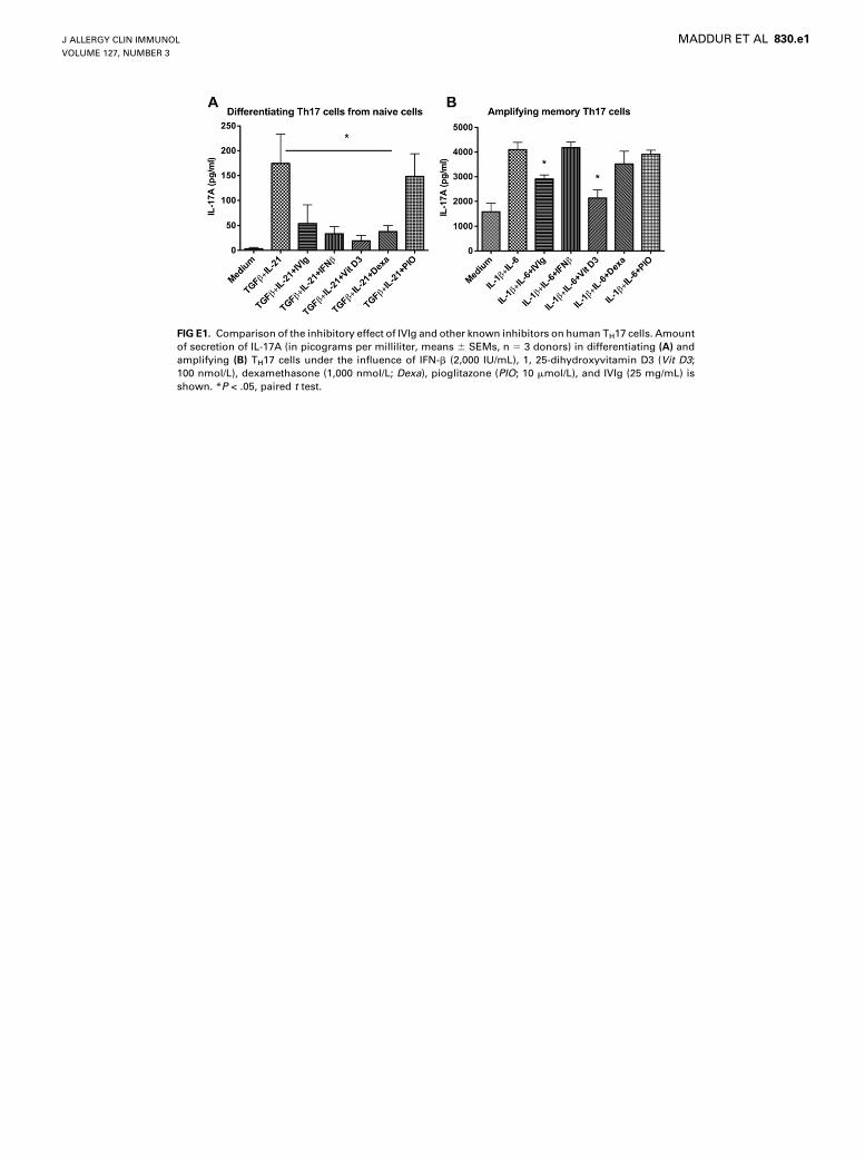
FIG E1. Comparison of the inhibitory effect of IVIg and other known inhibitors on human TH17 cells. Amount
of secretion of IL-17A (in picograms per milliliter, means 6 SEMs, n 5 3 donors) in differentiating (A) and
amplifying (B) TH17 cells under the influence of IFN-b (2,000 IU/mL), 1, 25-dihydroxyvitamin D3 (Vit D3;100 nmol/L), dexamethasone (1,000 nmol/L; Dexa), pioglitazone (PIO; 10 mmol/L), and IVIg (25 mg/mL) is
shown. *P < .05, paired t test.
J ALLERGY CLIN IMMUNOL
VOLUME 127, NUMBER 3
MADDUR ET AL 830.e1
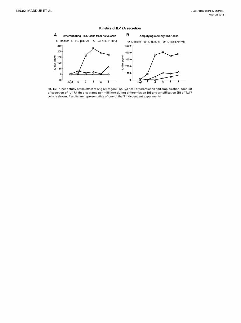
FIG E2. Kinetic study of the effect of IVIg (25 mg/mL) on TH17 cell differentiation and amplification. Amount
of secretion of IL-17A (in picograms per milliliter) during differentiation (A) and amplification (B) of TH17
cells is shown. Results are representative of one of the 3 independent experiments.
J ALLERGY CLIN IMMUNOL
MARCH 2011
830.e2 MADDUR ET AL
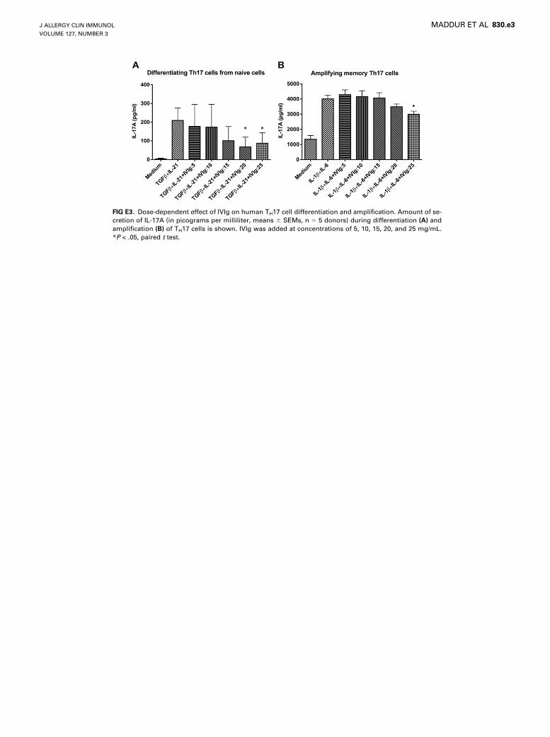
FIG E3. Dose-dependent effect of IVIg on human TH17 cell differentiation and amplification. Amount of se-
cretion of IL-17A (in picograms per milliliter, means 6 SEMs, n 5 5 donors) during differentiation (A) and
amplification (B) of TH17 cells is shown. IVIg was added at concentrations of 5, 10, 15, 20, and 25 mg/mL.
*P < .05, paired t test.
J ALLERGY CLIN IMMUNOL
VOLUME 127, NUMBER 3
MADDUR ET AL 830.e3
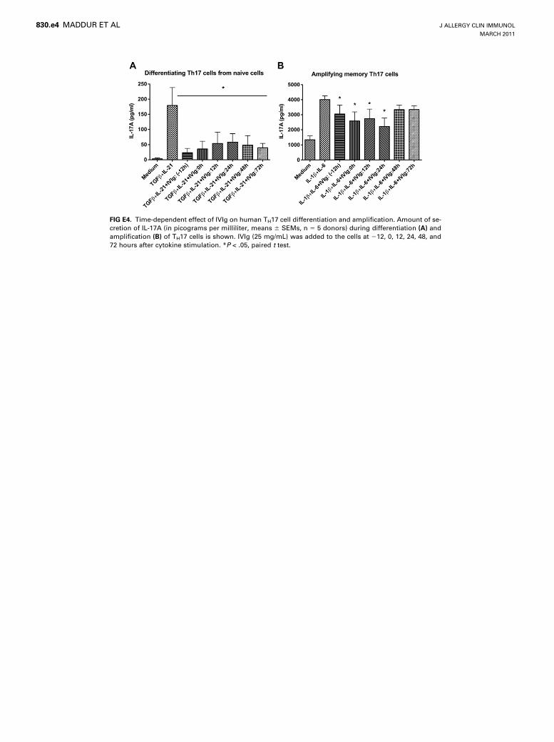
FIG E4. Time-dependent effect of IVIg on human TH17 cell differentiation and amplification. Amount of se-
cretion of IL-17A (in picograms per milliliter, means 6 SEMs, n 5 5 donors) during differentiation (A) and
amplification (B) of TH17 cells is shown. IVIg (25 mg/mL) was added to the cells at 212, 0, 12, 24, 48, and
72 hours after cytokine stimulation. *P < .05, paired t test.
J ALLERGY CLIN IMMUNOL
MARCH 2011
830.e4 MADDUR ET AL
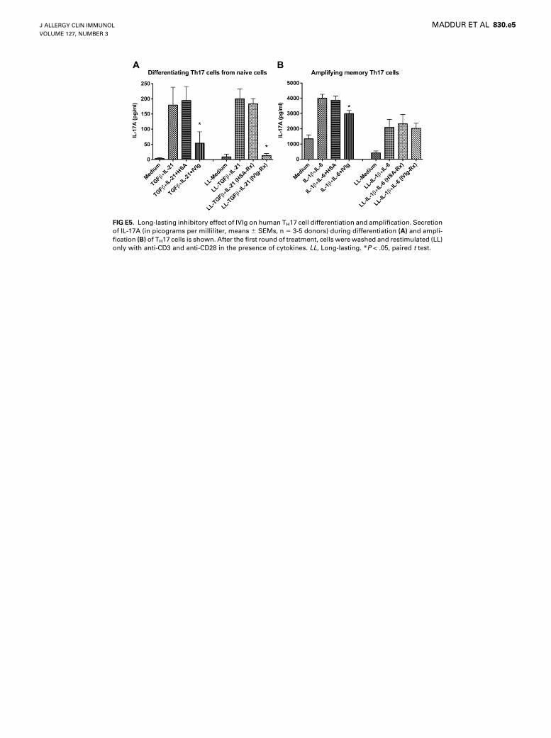
FIG E5. Long-lasting inhibitory effect of IVIg on human TH17 cell differentiation and amplification. Secretion
of IL-17A (in picograms per milliliter, means 6 SEMs, n 5 3-5 donors) during differentiation (A) and ampli-
fication (B) of TH17 cells is shown. After the first round of treatment, cells were washed and restimulated (LL)
only with anti-CD3 and anti-CD28 in the presence of cytokines. LL, Long-lasting. *P < .05, paired t test.
J ALLERGY CLIN IMMUNOL
VOLUME 127, NUMBER 3
MADDUR ET AL 830.e5
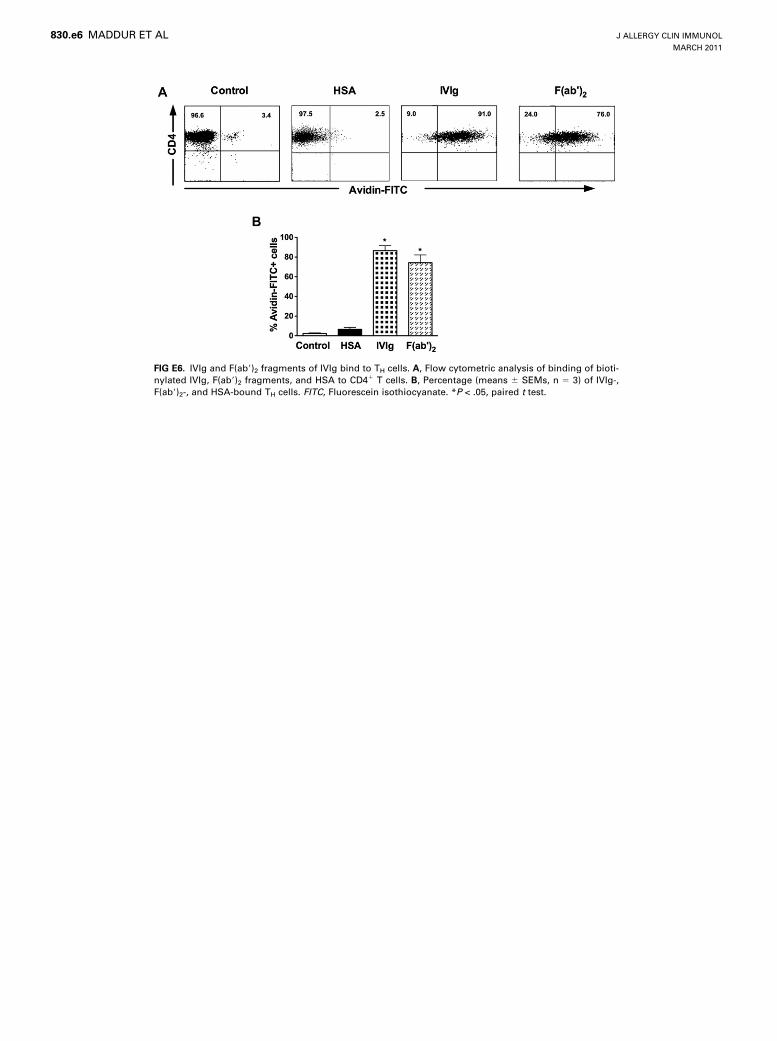
FIG E6. IVIg and F(ab9)2 fragments of IVIg bind to TH cells. A, Flow cytometric analysis of binding of bioti-
nylated IVIg, F(ab9)2 fragments, and HSA to CD41 T cells. B, Percentage (means 6 SEMs, n 5 3) of IVIg-,
F(ab9)2-, and HSA-bound TH cells. FITC, Fluorescein isothiocyanate. *P < .05, paired t test.
J ALLERGY CLIN IMMUNOL
MARCH 2011
830.e6 MADDUR ET AL
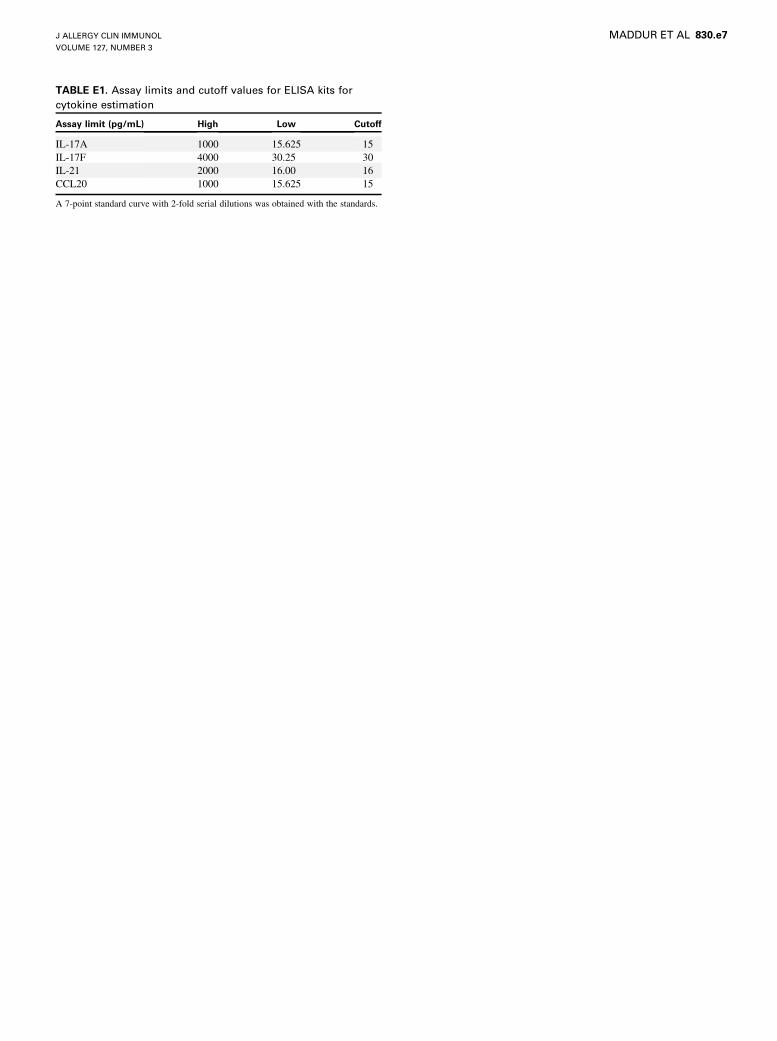
TABLE E1. Assay limits and cutoff values for ELISA kits for
cytokine estimation
Assay limit (pg/mL) High Low Cutoff
IL-17A 1000 15.625 15
IL-17F 4000 30.25 30
IL-21 2000 16.00 16
CCL20 1000 15.625 15
A 7-point standard curve with 2-fold serial dilutions was obtained with the standards.
J ALLERGY CLIN IMMUNOL
VOLUME 127, NUMBER 3
MADDUR ET AL 830.e7