inhibition of bet recruitment to chromatin as an effective
TRANSCRIPT
LETTERdoi:10.1038/nature10509
Inhibition of BET recruitment to chromatin as aneffective treatment for MLL-fusion leukaemiaMark A. Dawson1,2*, Rab K. Prinjha3*, Antje Dittmann4*, George Giotopoulos1, Marcus Bantscheff4, Wai-In Chan1,Samuel C. Robson2, Chun-wa Chung5, Carsten Hopf4, Mikhail M. Savitski4, Carola Huthmacher4, Emma Gudgin1, Dave Lugo3,Soren Beinke3, Trevor D. Chapman3, Emma J. Roberts3, Peter E. Soden3, Kurt R. Auger6, Olivier Mirguet7, Konstanze Doehner8,Ruud Delwel9, Alan K. Burnett10, Phillip Jeffrey3, Gerard Drewes4, Kevin Lee3, Brian J. P. Huntly1* & Tony Kouzarides2*
Recurrent chromosomal translocations involving the mixed lineageleukaemia (MLL) gene initiate aggressive forms of leukaemia, whichare often refractory to conventional therapies1. Many MLL-fusionpartners are members of the super elongation complex (SEC), acritical regulator of transcriptional elongation, suggesting thataberrant control of this process has an important role in leukaemiainduction2,3. Here we use a global proteomic strategy to demon-strate that MLL fusions, as part of SEC2,3 and the polymerase-associated factor complex (PAFc)4,5, are associated with the BETfamily of acetyl-lysine recognizing, chromatin ‘adaptor’ proteins.These data provided the basis for therapeutic intervention in MLL-fusion leukaemia, via the displacement of the BET family of proteinsfrom chromatin. We show that a novel small molecule inhibitor ofthe BET family, GSK1210151A (I-BET151), has profound efficacyagainst human and murine MLL-fusion leukaemic cell lines,through the induction of early cell cycle arrest and apoptosis.I-BET151 treatment in two human leukaemia cell lines with differ-ent MLL fusions alters the expression of a common set of geneswhose function may account for these phenotypic changes. Themode of action of I-BET151 is, at least in part, due to the inhibitionof transcription at key genes (BCL2, C-MYC and CDK6) throughthe displacement of BRD3/4, PAFc and SEC components fromchromatin. In vivo studies indicate that I-BET151 has significanttherapeutic value, providing survival benefit in two distinct mousemodels of murine MLL–AF9 and human MLL–AF4 leukaemia.Finally, the efficacy of I-BET151 against human leukaemia stemcells is demonstrated, providing further evidence of its potent thera-peutic potential. These findings establish the displacement of BETproteins from chromatin as a promising epigenetic therapy forthese aggressive leukaemias.
Dysregulation of chromatin modifiers is a recurrent and sentinelevent in oncogenesis6. Therapeutic strategies that selectively alter therecruitment and/or catalytic activity of these enzymes at chromatintherefore hold great promise as targeted therapies6. In this regard thebromodomain and extra terminal (BET) family of proteins (BRD2,BRD3, BRD4 and BRDT) provide an ideal ‘druggable’ target, becausethey share a common highly conserved tandem bromodomain at theiramino terminus. Selective bromodomain inhibitors that disrupt thebinding of BET proteins to histones have recently been described7,8;however, their true therapeutic scope remains untested.
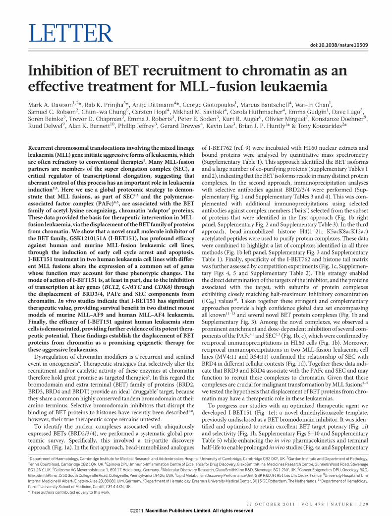
To identify the nuclear complexes associated with ubiquitouslyexpressed BETs (BRD2/3/4), we performed a systematic global pro-teomic survey. Specifically, this involved a tri-partite discoveryapproach (Fig. 1a). In the first approach, bead-immobilized analogues
of I-BET762 (ref. 9) were incubated with HL60 nuclear extracts andbound proteins were analysed by quantitative mass spectrometry(Supplementary Table 1). This approach identified the BET isoformsand a large number of co-purifying proteins (Supplementary Tables 1and 2), indicating that the BET isoforms reside in many distinct proteincomplexes. In the second approach, immunoprecipitation analyseswith selective antibodies against BRD2/3/4 were performed (Sup-plementary Fig. 1 and Supplementary Tables 3 and 4). This was com-plemented with additional immunoprecipitations using selectedantibodies against complex members (‘baits’) selected from the subsetof proteins that were identified in the first approach (Fig. 1b rightpanel, Supplementary Fig. 2 and Supplementary Table 3). In the thirdapproach, bead-immobilized histone H4(1–21; K5acK8acK12ac)acetylated peptides were used to purify protein complexes. These datawere combined to highlight a list of complexes identified in all threemethods (Fig. 1b left panel, Supplementary Fig. 3 and SupplementaryTable 1). Finally, specificity of the I-BET762 and histone tail matrixwas further assessed by competition experiments (Fig. 1c, Supplemen-tary Figs 4, 5 and Supplementary Table 2). This strategy enabledthe direct determination of the targets of the inhibitor, and the proteinsassociated with the target, with subunits of protein complexesexhibiting closely matching half-maximum inhibitory concentration(IC50) values10. Taken together these stringent and complementaryapproaches provide a high confidence global data set encompassingall known11–13 and several novel BET protein complexes (Fig. 1b andSupplementary Fig. 3). Among the novel complexes, we observed aprominent enrichment and dose-dependent inhibition of several com-ponents of the PAFc4,5 and SEC2,3 (Fig. 1b, c), which were confirmed byreciprocal immunoprecipitations in HL60 cells (Fig. 1b). Moreover,reciprocal immunoprecipitations in two MLL-fusion leukaemia celllines (MV4;11 and RS4;11) confirmed the relationship of SEC withBRD4 in different cellular contexts (Fig. 1d). Together these data indi-cate that BRD3 and BRD4 associate with the PAFc and SEC and mayfunction to recruit these complexes to chromatin. Given that thesecomplexes are crucial for malignant transformation by MLL fusions2–5
we tested the hypothesis that displacement of BET proteins from chro-matin may have a therapeutic role in these leukaemias.
To progress our studies with an optimized therapeutic agent wedeveloped I-BET151 (Fig. 1e); a novel dimethylisoxazole template,previously undisclosed as a BET bromodomain inhibitor. It was iden-tified and optimized to retain excellent BET target potency (Fig. 1i)and selectivity (Fig. 1h, Supplementary Figs 5–10 and SupplementaryTable 5) while enhancing the in vivo pharmacokinetics and terminalhalf-life to enable prolonged in vivo studies (Fig. 4a and Supplementary
*These authors contributed equally to this work.
1Department of Haematology, Cambridge Institute for Medical Research and Addenbrookes Hospital, University of Cambridge, Cambridge CB2 0XY, UK. 2Gurdon Institute and Department of Pathology,Tennis Court Road, Cambridge CB2 1QN, UK. 3Epinova DPU, Immuno-Inflammation Centre of Excellence for Drug Discovery, GlaxoSmithKline, Medicines Research Centre, Gunnels Wood Road, StevenageSG1 2NY, UK. 4Cellzome AG Meyerhofstrasse 1, 69117 Heidelberg, Germany. 5Molecular Discovery Research, GlaxoSmithKline R&D, Stevenage SG1 2NY, UK. 6Cancer Epigenetics DPU, Oncology R&D,GlaxoSmithKline, 1250South Collegeville Road,Collegeville, Pennsylvania 19426,USA. 7Lipid MetabolismDiscovery Performance Unit, GSK R&D, 91951Les Ulis Cedex, France. 8University Hospital ofUlmInternal Medicine III Albert- Einstein-Allee 23, 89081 Ulm, Germany. 9Department of Hematology, Erasmus University Medical Center, 3015 GE Rotterdam, The Netherlands. 10Department of Hematology,Cardiff University School of Medicine, Cardiff, CF14 4XN, UK.
2 7 O C T O B E R 2 0 1 1 | V O L 4 7 8 | N A T U R E | 5 2 9
Macmillan Publishers Limited. All rights reserved©2011
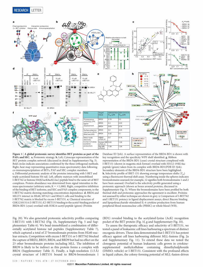
Fig. 20). We also generated proteomic selectivity profiles comparingI-BET151 with I-BET762 (Fig. 1h, Supplementary Fig. 5 and Sup-plementary Table 6). We bead-immobilized a combination of differ-entially acetylated histone tail peptides (Supplementary Table 7),which captured a total of 27 bromodomain proteins from HL60 nuc-lear extracts. Competition with excess I-BET151 or I-BET762 blockedthe capture of BRD2, BRD3, BRD4, and BRD9 but had no effect on the23 other bromodomain proteins including MLL. The inhibition ofBRD9 is likely to be indirect as this protein forms a complex withBRD4 (Supplementary Table 3). Finally, a high-resolution (1.5 A)crystal structure of I-BET151 bound to BRD4-bromodomain 1
(BD1) revealed binding to the acetylated-lysine (AcK) recognitionpocket of the BET protein (Fig. 1f, g and Supplementary Fig. 10).
To assess the therapeutic efficacy and selectivity of I-BET151, wetested a panel of leukaemic cell lines harbouring a spectrum of distinctoncogenic drivers. These data demonstrated that I-BET151 has potentefficacy against cell lines harbouring different MLL-fusions (Fig. 2aand Supplementary Fig. 11). To extend these data we tested theclonogenic potential of human leukaemic cells grown in cytokine-supplemented methylcellulose containing dimethylsulphoxide(DMSO; vehicle) or I-BET151. Consistent with the profound effectsin liquid culture, the colony-forming potential of MLL-fusion-driven
Proteinsquantified
IP with antibody for:BRD2 BRD3 BRD4 CDK9 MLLT1 PAF1
BE
T
BRD2BRD3BRD4
PA
F
CDC73CTR9LEO1PAF1RTF1
WDR61
PT
EF
b-S
EC
AFF1AFF2AFF4
CCNT1CDK9
ELLMLLT1
3.3 0.19
Relative abundance vs bait(log scale)
N
NH
N
O
N
O
O
N
a b c
e
f g
h
i
Chemoproteomics Interaction proteomics
Immunoprecipitation Histone tails = BET inhibitor
Proteinsbinding to
histone marks
Proteinsbinding to
BET inhibitor
BET
complexes
IP
IB: MLLT1 IB: BRD4
MV
41
1 n
ucle
ar
extr
act
ML
LT1
BR
D4
IgG
Co
ntr
ol
IP
10 μM
I-BET151
+ –
Matrix
MV
41
1 n
ucle
ar
extr
act
ML
LT1
BR
D4
IgG
Co
ntr
ol
10 μM
I-BET151
+ –
Matrix
HL
60
nu
cle
ar
extr
act
RS
41
1 n
ucle
ar
extr
act
10 μM
I-BET151
+ –
Matrix
10 μM
I-BET151
+ –
Matrix
IB: BRD4
IB: MLLT1
Assay Target/
tissue
GSK525762A
IC50 (μM)
GSK1210151A
IC50 (μM)
Fluorescence
anisotropy (FP)
ligand
displacement
BRD2 0.79 0.5
BRD3 0.4 0.25
BRD4 0.63 0.79
Surface plasmon
resonance affinity
(BiaCore) (KD)
BRD3 0.2 0.02
BRD4 0.32 0.1
Meso-scale
discovery
human IL6 assay
(cytokine
production)
PBMC LPS/IL-6
0.2 0.16
Human WB LPS/IL-6
0.63 1.26
Rat WB LPS/IL6
1.58 1.26
d
Resid
ual b
ind
ing
1010.10.010
0.5
1.0 Kdapp(nM)
BRD2BRD3 32BRD4
BRD4BRD2
BRD3
4.0 5.0 6.0 7.0 8.04.0
5.0
6.0
7.0
8.0
Log abundance I-BET matrix
log
ab
un
dan
ce H
4ac3
matr
ix
0.01 0.1 1 100
0.5
1.0
Resid
ual b
ind
ing
CDC73LEO1PAF1WDR61
Ab
un
dan
ce
H4
ac3
matr
ix
MLLT1
ELL
AFF1
AFF4
500K
1,000K
0 100K 200K 300K0
Abundance I-BET matrix
1010.10.010
0.5
1.0
Resid
ual b
ind
ing AFF2 54
AFF4CCNT1CDK9ELLMLLT1
SECcomplex
I-
BET concentration (μM)
BRD4
PAF
PTEFb-SEC
Heatshock
COMPASS
Splicosome assoc
RLC
BRD3BRD2
JMJD6
NOLC 64696560
39
37
9062515089
250150
100
75
50
37
252015
Figure 1 | A global proteomic survey identifies BET proteins as part of thePAFc and SEC. a, Proteomic strategy. b, Left, Cytoscape representation of theBET protein complex network (discussed in detail in Supplementary Fig. 3).Bold circles indicate associations confirmed by the three orthogonal methods.Right, heat map representing quantitative-mass spectrometry data followingco-immunoprecipitation of BETs, PAF and SEC complex members.c, Differential proteomic analysis of the proteins interacting with I-BET andtriple acetylated histone H4 tail. Left, affinity matrices with immobilizedI-BET762 or histone H4(K5acK8acK12ac) peptide bind to the same set of BETcomplexes. Protein abundance was determined from signal intensities in themass spectrometer (arbitrary units, K 5 31,000). Right, competitive inhibitionof the binding of BET isoforms, and SEC and PAF complex components, to theI-BET762 matrix showing matching concentration dependence. d, BRD4 andMLLT1 interact in HL60, MV4;11 and RS4;11 cells and binding to theI-BET762 matrix is blocked by excess I-BET151. e, Chemical structure ofGSK1210151A (I-BET151). f, I-BET151 binding to the acetyl-binding pocket ofBRD4-BD1 (cyan) overlaid with H3K14-acetyl peptide (green) (Protein
Database ID 3jvk). A surface representation of the BRD4-BD1 is shown withkey recognition and the specificity WPF shelf identified. g, Ribbonrepresentation of the BRD4-BD1 (cyan) crystal structure complexed withI-BET151 (shown in magenta stick format) overlaid with H3(12-19)K14acpeptide (green) taken from its complex with BRD4-BD1(PDB ID 3jvk).Secondary elements of the BRD4-BD1 structure have been highlighted.h, Selectivity profile of IBET-151 showing average temperature shifts (Tm)using a fluorescent thermal shift assay. Numbering inside the spheres indicatesbromodomains assessed; for example, 12 signifies both bromodomains 1 and 2have been assessed. Overlaid is the selectivity profile generated using aproteomic approach (shown as boxes around proteins, discussed inSupplementary Fig. 5). Where the bromodomains have been profiled by boththermal shift and proteomic approaches the agreement is excellent. Proteinsnot assessed by either technique are shown in grey. i, Comparison of I-BET762and I-BET151 potency in ligand displacement assays, direct Biacore bindingand lipopolysaccharide-stimulated IL-6 cytokine production from humanperipheral blood mononuclear cells (PBMC) or whole blood (WB).
RESEARCH LETTER
5 3 0 | N A T U R E | V O L 4 7 8 | 2 7 O C T O B E R 2 0 1 1
Macmillan Publishers Limited. All rights reserved©2011
leukaemias (MOLM13) was completely ablated by I-BET151, whereasleukaemias driven by tyrosine kinase activation (K562) were un-affected (Fig. 2b). In addition to the data with human leukaemiccell lines, we also confirmed the potent efficacy of I-BET151 in bothliquid culture and clonogenic assays using primary murine progenitors
retrovirally transformed with either MLL-ENL or MLL–AF9 (Fig. 2c).To investigate the mechanism of action for I-BET151, we performed
fluorescence-activated cell sorting (FACS) analysis to assess apoptosisand cell cycle progression after I-BET151 treatment. Figure 2d–e andSupplementary Fig. 12 show a marked induction of apoptosis and a
Cell line Oncogenic
driver
I-BET151
IC50
MV4;11 MLL-AF4 26 nM
RS4;11 MLL-AF4
MOLM13 MLL-AF9 120 nM
NOMO1 MLL-AF9 15 nM
HEL JAK2V617F 1 μM
K562 BCR-ABL >100 μM
MEG01 BCR-ABL 25 μM
HL60 N-RAS
192 nM
890 nM
a
bDMSO I-BET151
MOLM13
K562
I-BET151DMSO
Nu
mb
er
of
co
lon
ies
Nu
mb
er
of
co
lon
ies
MOLM13
750500250
125250375500
I-BET151DMSO
K5621,000MLL-ENL
MSCV MLL-ENLMSCV MLL-AF9
c
Nu
mb
er
of
co
lon
ies
Nu
mb
er
of
co
lon
ies
MLL-AF9
Liquid culture
Methylcellulose
IC50 assay
Colony assay
IC50 =579 nM
MLL-AF9
Log[I-BET] (M)
Pro
lifera
tio
n (%
)
100
75
50
25
–10–7.5–5.0–2.5
IC50 =418 nM
MLL-ENL
Log[I-BET] (M)
Pro
lifera
tio
n (%
)
100
75
50
25
–10–7.5–5.0–2.5
5001,0001,500
500
1,500
2,500
I-BET151DMSO
I-BET151DMSO
DMSO I-BET151
MOLM137-A
AD
Annexin V
DMSO I-BET151K562d104
103
102
101
100
104
103
102
101
100
104103102101100104103102101100
104
103
102
101
100
104
103
102
101
100
104103102101100104103102101100
70.4%1.72%
9.56%
50.2%16.1%
23.2%
47.4%15.5%
23.1%
51.3%16.1%
25.4%
DMSO I-BET151K562
MOLM13
e300
200
100
00 20K 40K 60K
200
150
100
50
00 20K 40K 60K
300
200
100
00 20K 40K 60K
600
400
200
00 20K 40K 60K
Figure 2 | I-BET151 selectively and potently inhibits MLL-fusion leukaemiccell lines in vitro. a, Human leukaemia cell lines tested using I-BET151.b, Clonogenic assays performed in the presence of DMSO or I-BET151.c, Haematopoietic progenitors were isolated from mouse bone marrow andretrovirally transformed with MLL–ENL or MLL–AF9. These cells were used inboth proliferation and clonogenic assays d, Apoptosis was assessed by FACS
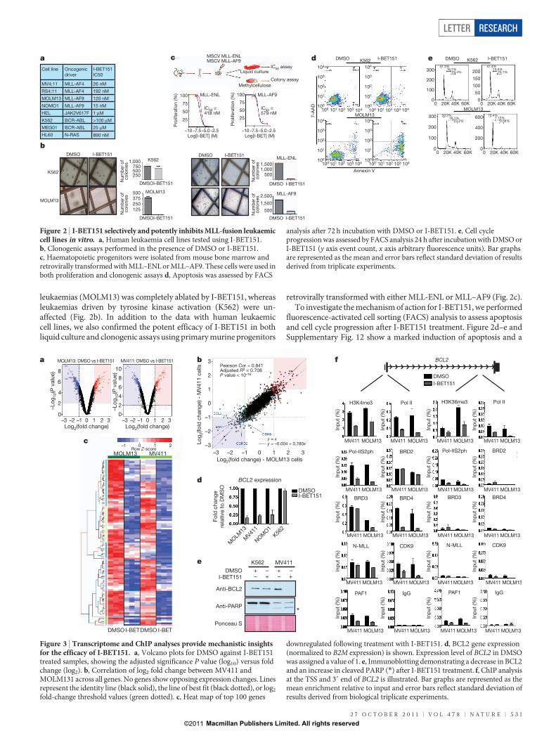
analysis after 72 h incubation with DMSO or I-BET151. e, Cell cycleprogression was assessed by FACS analysis 24 h after incubation with DMSO orI-BET151 (y axis event count, x axis arbitrary fluorescence units). Bar graphsare represented as the mean and error bars reflect standard deviation of resultsderived from triplicate experiments.
MOLM13 MV411
DMSO DMSOI-BET I-BET
a b
c
d
K562 MV411
DMSOI-BET151
+ ++ +– –
– –
Anti-BCL2
Anti-PARP
Ponceau S
e
*
MOLM
13
MV41
1
NOM
O1
K562
BCL2 expression
Fo
ld c
hang
e
rela
tive t
o D
MS
O
DMSOI-BET151
–Lo
g1
0(P
valu
e)
Lo
g2(fo
ld c
hang
e) -
MV
411 c
ells
–Lo
g1
0(P
valu
e)8
6
4
2
0
10
8
6
4
2
0
3
2
1
0
–1
–2
–3
3210–1–2–3
Log2(fold change) - MOLM13 cells
Pearson Cor = 0.841Adjusted R2 = 0.708P value < 10–16
y = xy = –0.004 + 0.780x–1 0 1 2
Row Z-score
BCL2f
MV411 MOLM13
Inp
ut
(%)
PAF1
MV411 MOLM13
Inp
ut
(%)
IgG
MV411 MOLM13
Inp
ut
(%)
PAF1
MV411 MOLM13
Inp
ut
(%)
IgG
Inp
ut
(%)
MV411 MOLM13
N-MLL
Inp
ut
(%)
MV411 MOLM13
CDK9
Inp
ut
(%)
MV411MOLM13
N-MLL
Inp
ut
(%)
MV411 MOLM13
CDK9
Inp
ut
(%)
MV411 MOLM13
BRD3
Inp
ut
(%)
MV411 MOLM13
BRD4
Inp
ut
(%)
MV411 MOLM13
BRD3
Inp
ut
(%)
MV411 MOLM13
BRD4
Pol-IIS2ph
MV411 MOLM13
Inp
ut
(%)
BRD2
MV411 MOLM13
Inp
ut
(%)
MV411 MOLM13
Inp
ut
(%)
Pol-IIS2ph
MV411 MOLM13
Inp
ut
(%)
BRD2
H3K4me3
MV411 MOLM13
Inp
ut
(%)
Pol II
MV411 MOLM13
Inp
ut
(%)
MV411 MOLM13
Inp
ut
(%)
H3K36me3
MV411 MOLM13
Pol II
Inp
ut
(%)
DMSO
I-BET151
MOLM13: DMSO vs I-BET151 MV411: DMSO vs I-BET151
–3 –2 –1 0 1 2 3 –3 –2 –1 0 1 2 3Log2(fold change) Log2(fold change)
Figure 3 | Transcriptome and ChIP analyses provide mechanistic insightsfor the efficacy of I-BET151. a, Volcano plots for DMSO against I-BET151treated samples, showing the adjusted significance P value (log10) versus foldchange (log2). b, Correlation of log2 fold change between MV411 andMOLM131 across all genes. No genes show opposing expression changes. Linesrepresent the identity line (black solid), the line of best fit (black dotted), or log2
fold-change threshold values (green dotted). c, Heat map of top 100 genes
downregulated following treatment with I-BET151. d, BCL2 gene expression(normalized to B2M expression) is shown. Expression level of BCL2 in DMSOwas assigned a value of 1. e, Immunoblotting demonstrating a decrease in BCL2and an increase in cleaved PARP (*) after I-BET151 treatment. f, ChIP analysisat the TSS and 39 end of BCL2 is illustrated. Bar graphs are represented as themean enrichment relative to input and error bars reflect standard deviation ofresults derived from biological triplicate experiments.
LETTER RESEARCH
2 7 O C T O B E R 2 0 1 1 | V O L 4 7 8 | N A T U R E | 5 3 1
Macmillan Publishers Limited. All rights reserved©2011

prominent G0/G1 arrest in two MLL-fusion cell lines driven by distinctMLL fusions (MOLM13 and MV4;11 containing MLL–AF9 andMLL–AF4, respectively). In contrast, the cell cycle characteristicsand apoptotic rate of K562 cells were largely unaffected at this time.These data indicate that I-BET151 alters the transcriptional pro-grammes regulating apoptosis and cell-cycle progression in MLL-fusion leukaemias.
To identify the precise transcriptional pathways controlled byI-BET151, global gene-expression analysis was performed inMOLM13 and MV4;11 cells after treatment with I-BET151 orDMSO for 6 h. This strategy allowed us to identify early I-BET151-responsive genes, before any discernable phenotypic alteration in cellcycle or apoptosis (Supplementary Fig. 12). As demonstrated previ-ously7, we observed differential expression of a selective subset of genes(Fig. 3a), rather than global transcriptional dysregulation. Remarkably,the transcriptional programmes altered in the two MLL-fusion celllines were highly correlated (Fig. 3b) and gene set enrichment analysisdocumented significant overlap with published MLL fusion signaturesincluding MLL-fusion leukaemia stem cells (LSC)14,15 (SupplementaryFig. 13). These data are consistent with the notion that MLL fusionsaberrantly co-opt the SEC and PAFc to regulate similar transcriptional
programmes. Notably, the top 100 genes concomitantly decreased inboth MOLM13 and MV4;11 (Fig. 3c) contained several previouslyreported direct MLL targets, such as BCL2, CDK6 and MYC, the down-regulation of which was consistent with the phenotypic consequencesof I-BET151 treatment.
BCL2 is a key antiapoptotic gene implicated in the pathogenesis ofMLL-fusion leukaemias16,17. Consistent with these data, I-BET151reduced the expression of BCL2 in a third MLL-fusion cell line(NOMO1) but not in the unresponsive K562 cells (Fig. 3d), and induc-tion of apoptosis coincided with a marked reduction in BCL2 proteinexpression (Fig. 3e). Moreover, overexpression of BCL2 in the pres-ence of I-BET151 rescued the apoptotic phenotype (SupplementaryFig. 14). Chromatin immunoprecipitation (ChIP) analyses at the BCL2locus showed that 6 h of I-BET151 treatment selectively decreased therecruitment of BRD3/4 and impaired recruitment of CDK9 and PAF1(part of SEC and PAFc, respectively) to the transcriptional start site(TSS). This correlated with reduced phosphorylation of RNA poly-merase II (Pol II) on serine 2 of its carboxy-terminal domain (Pol-IIS2ph) (Fig. 3f). A similar pattern was observed at two other MLLtarget genes (MYC and CDK6), but not at housekeeping genes (B2M)whose expression was unaltered by I-BET151 (Supplementary Fig. 15).
ControlI-BET151
10
1
0.1
0.01
0.001
100
80
60
40
20
00 10 20 30 40 50
Dis
ease-f
ree s
urv
ival % Control
I-BET151
e
P = 0.0002
100
75
50
25
00 4 8 12 16 20
Brd3/4
ONAc
K
Ac
K
PAFc
MLL AF4 / AF9
BCL2CDK6
C-MYC
Ac
K
Ac
K
SEC
BCL2CDK6
C-MYC
OFF
I-BET I-BET
I-BET
Brd3/4
PAFc
MLL AF4 / AF9
SEC
Log2(fold change)
With
I-BET151
With
I-BET151
No
change
PT1 (MLL-AF9)
PT2 (MLL-AF17)
PT3 (MLL-AF6)
PT4 (MLL-AF10)
PT5 (MLL-ENL)
PT6 (MLL-AF9)
PT11 (MLL-AF9)
PT8 (MLL-AF6)
PT9 (MLL-AF10)
PT10 (MLL-ENL)
PT7 (MLL-AF17)
BC
L2
CD
K6
MY
C
CC
ND
2
B2M
n = 8
DA
A-7
Annexin V
DMSO I-BET151
Days
Overa
ll su
rviv
al (%
)
Nu
mb
er
of
co
lon
ies
Control I-BET151
7-A
AD
Human HLA
Control I-BET151a b c d
ControlI-BET151f ih j
k l m nHuman
(MLL-AF6)
g
Control I-BET151
I-BET151I-BET762JQ1In vitro IC50of MV411
30 mg kg–1
single IPdose
Time (h)
Blo
od
co
ncen
tratio
n (μM
)
0 3 6 9 12 15 18 21 24 27 30 33 36 Days
P = 0.017
120
90
60
30
0
2.5
2.0
1.5
1.0
0.5
0.0
0.5
0.4
0.3
0.2
0.1
0.0
I-BET151 Control I-BET151 Control I-BET151 Control
300
250
150
100
50
0
DMSO I-BET151
Human LSC
(CD34+/CD38–)
–2 –1 0 1 2
Wh
ite b
loo
d c
ells
(10
3 p
er μl
)
Liv
er
weig
ht
(g)
Sp
leen
weig
ht
(g)
104
103
102
101
100
104
103
102
101
100
104103102101100 104103102101100
104
103
102
101
100
104103102101
104
103
102
101
100
104103102101100
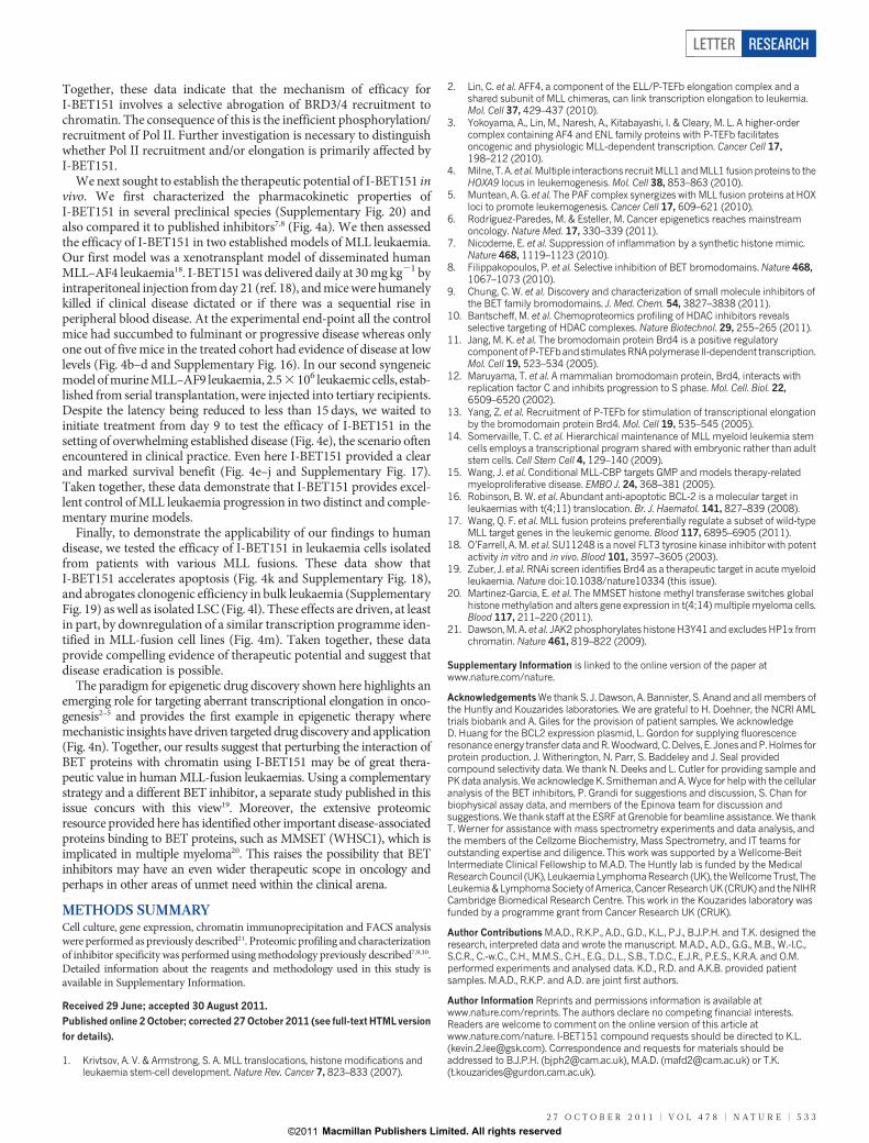
Figure 4 | I-BET151 is efficacious in in vivo murine models and primarypatient samples of MLL-fusion leukaemia. a, Murine pharmacokineticstudies (mean 6 s.d., n 5 4 per compound) comparing the blood concentrationof I-BET151 with I-BET762 and JQ1. b, Kaplan–Meier curve of control andtreated NOD-SCID mice transplanted with 1 3 107 MV4;11 cells. Greenarrowhead, treatment commencement on day 21. c, Haematoxylin and eosin-stained histological sections of the renal parenchyma of control and treatedmice. Black arrows highlight leukaemic infiltration. d, Representative FACSanalysis from the peripheral blood of control or I-BET151-treated mice.e, Kaplan–Meier curve of control and treated C57BL/6 mice transplanted with2.5 3 106 syngeneic MLL–AF9 leukaemic cells. Green arrowhead, treatmentcommencement on day 9. f, Photomicrograph of the spleen size from 5/8control and 1/12 I-BET151-treated mice that died on day 12. g, Haematoxylin
and eosin-stained histological sections of the liver parenchyma from controland I-BET151-treated mice demonstrating reduced disease burden in thetreated animal. h–j, Peripheral blood white cell count (h), liver weight (i) andspleen weights (j) from all the control and treated mice at the time of necropsy.k, Representative FACS analysis assessing apoptosis from a patient with MLL–AF6 leukaemia. l, Clonogenic assays with human MLL-fusion LSC isolated byFACS sorting (CD341/CD382) and plated in the presence of DMSO orI-BET151. m, Gene expression changes in human MLL-fusion leukaemia cellsfollowing treatment with I-BET151 or DMSO. The log2 fold change in theexpression level for all genes (expression level with I-BET151 treatment/expression level with DMSO) is represented. n, Schematic model proposing themode of action for I-BET151 in MLL-fusion leukaemia.
RESEARCH LETTER
5 3 2 | N A T U R E | V O L 4 7 8 | 2 7 O C T O B E R 2 0 1 1
Macmillan Publishers Limited. All rights reserved©2011
Together, these data indicate that the mechanism of efficacy forI-BET151 involves a selective abrogation of BRD3/4 recruitment tochromatin. The consequence of this is the inefficient phosphorylation/recruitment of Pol II. Further investigation is necessary to distinguishwhether Pol II recruitment and/or elongation is primarily affected byI-BET151.
We next sought to establish the therapeutic potential of I-BET151 invivo. We first characterized the pharmacokinetic properties ofI-BET151 in several preclinical species (Supplementary Fig. 20) andalso compared it to published inhibitors7,8 (Fig. 4a). We then assessedthe efficacy of I-BET151 in two established models of MLL leukaemia.Our first model was a xenotransplant model of disseminated humanMLL–AF4 leukaemia18. I-BET151 was delivered daily at 30 mg kg21 byintraperitoneal injection from day 21 (ref. 18), and mice were humanelykilled if clinical disease dictated or if there was a sequential rise inperipheral blood disease. At the experimental end-point all the controlmice had succumbed to fulminant or progressive disease whereas onlyone out of five mice in the treated cohort had evidence of disease at lowlevels (Fig. 4b–d and Supplementary Fig. 16). In our second syngeneicmodel of murine MLL–AF9 leukaemia, 2.5 3 106 leukaemic cells, estab-lished from serial transplantation, were injected into tertiary recipients.Despite the latency being reduced to less than 15 days, we waited toinitiate treatment from day 9 to test the efficacy of I-BET151 in thesetting of overwhelming established disease (Fig. 4e), the scenario oftenencountered in clinical practice. Even here I-BET151 provided a clearand marked survival benefit (Fig. 4e–j and Supplementary Fig. 17).Taken together, these data demonstrate that I-BET151 provides excel-lent control of MLL leukaemia progression in two distinct and comple-mentary murine models.
Finally, to demonstrate the applicability of our findings to humandisease, we tested the efficacy of I-BET151 in leukaemia cells isolatedfrom patients with various MLL fusions. These data show thatI-BET151 accelerates apoptosis (Fig. 4k and Supplementary Fig. 18),and abrogates clonogenic efficiency in bulk leukaemia (SupplementaryFig. 19) as well as isolated LSC (Fig. 4l). These effects are driven, at leastin part, by downregulation of a similar transcription programme iden-tified in MLL-fusion cell lines (Fig. 4m). Taken together, these dataprovide compelling evidence of therapeutic potential and suggest thatdisease eradication is possible.
The paradigm for epigenetic drug discovery shown here highlights anemerging role for targeting aberrant transcriptional elongation in onco-genesis2–5 and provides the first example in epigenetic therapy wheremechanistic insights have driven targeted drug discovery and application(Fig. 4n). Together, our results suggest that perturbing the interaction ofBET proteins with chromatin using I-BET151 may be of great thera-peutic value in human MLL-fusion leukaemias. Using a complementarystrategy and a different BET inhibitor, a separate study published in thisissue concurs with this view19. Moreover, the extensive proteomicresource provided here has identified other important disease-associatedproteins binding to BET proteins, such as MMSET (WHSC1), which isimplicated in multiple myeloma20. This raises the possibility that BETinhibitors may have an even wider therapeutic scope in oncology andperhaps in other areas of unmet need within the clinical arena.
METHODS SUMMARYCell culture, gene expression, chromatin immunoprecipitation and FACS analysiswere performed as previously described21. Proteomic profiling and characterizationof inhibitor specificity was performed using methodology previously described7,9,10.Detailed information about the reagents and methodology used in this study isavailable in Supplementary Information.
Received 29 June; accepted 30 August 2011.
Published online 2 October; corrected 27 October 2011 (see full-text HTML version
for details).
1. Krivtsov, A. V. & Armstrong, S. A. MLL translocations, histone modifications andleukaemia stem-cell development. Nature Rev. Cancer 7, 823–833 (2007).
2. Lin, C. et al. AFF4, a component of the ELL/P-TEFb elongation complex and ashared subunit of MLL chimeras, can link transcription elongation to leukemia.Mol. Cell 37, 429–437 (2010).
3. Yokoyama, A., Lin, M., Naresh, A., Kitabayashi, I. & Cleary, M. L. A higher-ordercomplex containing AF4 and ENL family proteins with P-TEFb facilitatesoncogenic and physiologic MLL-dependent transcription. Cancer Cell 17,198–212 (2010).
4. Milne, T. A. et al. Multiple interactions recruit MLL1 andMLL1 fusion proteins to theHOXA9 locus in leukemogenesis. Mol. Cell 38, 853–863 (2010).
5. Muntean, A. G. et al. The PAF complex synergizes with MLL fusion proteins at HOXloci to promote leukemogenesis. Cancer Cell 17, 609–621 (2010).
6. Rodrıguez-Paredes, M. & Esteller, M. Cancer epigenetics reaches mainstreamoncology. Nature Med. 17, 330–339 (2011).
7. Nicodeme, E. et al. Suppression of inflammation by a synthetic histone mimic.Nature 468, 1119–1123 (2010).
8. Filippakopoulos, P. et al. Selective inhibition of BET bromodomains. Nature 468,1067–1073 (2010).
9. Chung, C. W. et al. Discovery and characterization of small molecule inhibitors ofthe BET family bromodomains. J. Med. Chem. 54, 3827–3838 (2011).
10. Bantscheff, M. et al. Chemoproteomics profiling of HDAC inhibitors revealsselective targeting of HDAC complexes. Nature Biotechnol. 29, 255–265 (2011).
11. Jang, M. K. et al. The bromodomain protein Brd4 is a positive regulatorycomponent ofP-TEFbandstimulatesRNApolymerase II-dependent transcription.Mol. Cell 19, 523–534 (2005).
12. Maruyama, T. et al. A mammalian bromodomain protein, Brd4, interacts withreplication factor C and inhibits progression to S phase. Mol. Cell. Biol. 22,6509–6520 (2002).
13. Yang, Z. et al. Recruitment of P-TEFb for stimulation of transcriptional elongationby the bromodomain protein Brd4. Mol. Cell 19, 535–545 (2005).
14. Somervaille, T. C. et al. Hierarchical maintenance of MLL myeloid leukemia stemcells employs a transcriptional program shared with embryonic rather than adultstem cells. Cell Stem Cell 4, 129–140 (2009).
15. Wang, J. et al. Conditional MLL-CBP targets GMP and models therapy-relatedmyeloproliferative disease. EMBO J. 24, 368–381 (2005).
16. Robinson, B. W. et al. Abundant anti-apoptotic BCL-2 is a molecular target inleukaemias with t(4;11) translocation. Br. J. Haematol. 141, 827–839 (2008).
17. Wang, Q. F. et al. MLL fusion proteins preferentially regulate a subset of wild-typeMLL target genes in the leukemic genome. Blood 117, 6895–6905 (2011).
18. O’Farrell, A. M. et al. SU11248 is a novel FLT3 tyrosine kinase inhibitor with potentactivity in vitro and in vivo. Blood 101, 3597–3605 (2003).
19. Zuber, J. et al. RNAi screen identifies Brd4 as a therapeutic target in acute myeloidleukaemia. Nature doi:10.1038/nature10334 (this issue).
20. Martinez-Garcia, E. et al. The MMSET histone methyl transferase switches globalhistone methylation and alters gene expression in t(4;14) multiple myeloma cells.Blood 117, 211–220 (2011).
21. Dawson, M. A. et al. JAK2 phosphorylates histone H3Y41 and excludes HP1a fromchromatin. Nature 461, 819–822 (2009).
Supplementary Information is linked to the online version of the paper atwww.nature.com/nature.
Acknowledgements We thank S. J. Dawson, A. Bannister, S. Anand and all members ofthe Huntly and Kouzarides laboratories. We are grateful to H. Doehner, the NCRI AMLtrials biobank and A. Giles for the provision of patient samples. We acknowledgeD. Huang for the BCL2 expression plasmid, L. Gordon for supplying fluorescenceresonance energy transfer data and R. Woodward, C. Delves, E. Jones and P. Holmes forprotein production. J. Witherington, N. Parr, S. Baddeley and J. Seal providedcompound selectivity data. We thank N. Deeks and L. Cutler for providing sample andPK data analysis. We acknowledge K. Smitheman and A. Wyce for help with the cellularanalysis of the BET inhibitors, P. Grandi for suggestions and discussion, S. Chan forbiophysical assay data, and members of the Epinova team for discussion andsuggestions. We thank staff at the ESRF at Grenoble for beamline assistance. We thankT. Werner for assistance with mass spectrometry experiments and data analysis, andthe members of the Cellzome Biochemistry, Mass Spectrometry, and IT teams foroutstanding expertise and diligence. This work was supported by a Wellcome-BeitIntermediate Clinical Fellowship to M.A.D. The Huntly lab is funded by the MedicalResearchCouncil (UK), Leukaemia Lymphoma Research (UK), the Wellcome Trust, TheLeukemia & Lymphoma Society of America, Cancer Research UK (CRUK) and the NIHRCambridge Biomedical Research Centre. This work in the Kouzarides laboratory wasfunded by a programme grant from Cancer Research UK (CRUK).
Author Contributions M.A.D., R.K.P., A.D., G.D., K.L., P.J., B.J.P.H. and T.K. designed theresearch, interpreted data and wrote the manuscript. M.A.D., A.D., G.G., M.B., W.-I.C.,S.C.R., C.-w.C., C.H., M.M.S., C.H., E.G., D.L., S.B., T.D.C., E.J.R., P.E.S., K.R.A. and O.M.performed experiments and analysed data. K.D., R.D. and A.K.B. provided patientsamples. M.A.D., R.K.P. and A.D. are joint first authors.
Author Information Reprints and permissions information is available atwww.nature.com/reprints. The authors declare no competing financial interests.Readers are welcome to comment on the online version of this article atwww.nature.com/nature. I-BET151 compound requests should be directed to K.L.([email protected]). Correspondence and requests for materials should beaddressed to B.J.P.H. ([email protected]), M.A.D. ([email protected]) or T.K.([email protected]).
LETTER RESEARCH
2 7 O C T O B E R 2 0 1 1 | V O L 4 7 8 | N A T U R E | 5 3 3
Macmillan Publishers Limited. All rights reserved©2011