influenzaavirusneuraminidaseproteinenhancescell ... survivalthroughinteractionwithcarcinoembryonic...
TRANSCRIPT
Influenza A Virus Neuraminidase Protein Enhances CellSurvival through Interaction with CarcinoembryonicAntigen-related Cell Adhesion Molecule 6 (CEACAM6) Protein*□S
Received for publication, November 28, 2011, and in revised form, February 22, 2012 Published, JBC Papers in Press, March 6, 2012, DOI 10.1074/jbc.M111.328070
Pratibha Gaur‡, Priya Ranjan§, Shipra Sharma‡, Jenish R. Patel§, J. Bradford Bowzard§, Shah K. Rahman‡,Rashmi Kumari‡, Shivaprakash Gangappa§, Jacqueline M. Katz§, Nancy J. Cox§, Renu B. Lal§,Suryaprakash Sambhara§, and Sunil K. Lal‡1
From the ‡Virology Group, International Centre for Genetic Engineering and Biotechnology, Aruna Asaf Ali Road, New Delhi110067, India and the §Influenza Division, National Center for Immunization and Respiratory Diseases, Centers for Disease Controland Prevention, Atlanta, Georgia 30033
Background: The NA protein is required for the release of progeny virions.Results: The NA/C6 interaction leads to increased tyrosyl phosphorylation of Src, FAK, Akt, GSK3�, and Bcl-2, which affectscell survival.Conclusion: A novel role exists for NA in enhancing host cell survival.Significance: NA not only aids in the release of progeny virions, but also cell survival during viral replication.
The influenza virus neuraminidase (NA) protein primarilyaids in the release of progeny virions from infected cells. Here,we demonstrate a novel role for NA in enhancing host cell sur-vival by activating theSrc/Akt signalingaxis viaan interactionwithcarcinoembryonic antigen-related cell adhesion molecule 6/clus-ter of differentiation 66c (C6). NA/C6 interaction leads toincreased tyrosyl phosphorylation of Src, FAK, Akt, GSK3�, andBcl-2,which affects cell survival, proliferation,migration, differen-tiation, andapoptosis. siRNA-mediated suppressionofC6resultedinadown-regulationofactivatedSrc,FAK,andAkt, increasedapo-ptosis, and reduced expression of viral proteins and viral titers ininfluenza virus-infected human lung adenocarcinoma epithelialand normal human bronchial epithelial cells. These findings indi-cate that influenza NA not only aids in the release of progeny viri-ons, but also cell survival during viral replication.
Influenza viruses are human respiratory pathogens thatcause both seasonal epidemics and periodic, but unpredictable,pandemics (1). The influenza type A virus genome consists ofeight single-stranded negative-sense RNA segments, whichencode up to 11 viral proteins. Annual epidemics occur fre-quently due to antigenic shift, the process by which the majorviral surface glycoproteins, hemagglutinin (HA) and neuramin-idase (NA),2 acquire point mutations allowing the virus to par-tially overcome preexisting immunity. The less frequent, but
potentially more deadly pandemics occur when a virus with anovelHAand one ormore other genes emerges to cause diseaseand spreads efficiently among humans with little or no preex-isting immunity to the novel strain (2).In addition to their roles in entry, genome mRNA synthesis,
assembly, and budding, many influenza viral proteins also havesecondary functions that alter the cellular environment to permitviral replication. For example, thenonstructural protein1 (NS1)ofinfluenza virus has been reported to activate the phosphatidyli-nositol 3-kinase (PI3K)/Akt pathway, which suppresses apoptosis(3–5), whereas overexpression of the nucleoprotein (NP) andmatrix 1 (M1) protein was shown to activate NF-�B (6). Likewise,binding of recombinant HA to cell receptors induces proteinkinase C signaling, and the overexpression of HA activates NF-�B(7, 8). The NA protein is critical for virus assembly, budding, andrelease fromthe infectedcell (9) andactivatesTGF-� tocontributeto apoptosis (10). NA activity has also been associated with theability to induce type I interferon in a strain-dependent manner(11), and treatment of cells with purifiedNAwas shown to stimu-late the expression of TNF-� (12).In the present study, we show that influenza NA interacts
with human carcinoembryonic antigen-related cell adhesionmolecule 6/cluster of differentiation 66c (CEACAM6/CD66c,orC6), a glycosylphosphatidylinositol-linked Ig-likemembraneprotein. Overexpression of C6 causes resistance to anoikis (13),a form of cell death, and activated c-Src signaling in a caveolin-1-dependent manner (14). Inhibition of C6 expression usingsiRNA led to a loss of activation of Src, FAK, and Akt, increasedcell death, and a significant loss of expression of viral proteins.These findings propose a novel role for the NA protein in sus-taining cell survival for enhanced viral replication.
EXPERIMENTAL PROCEDURES
Plasmid Constructs, Antibodies, siRNA, Virus Strains, andMammalian Cell Lines—For mammalian expression, NA wascloned into pXJ41neo (Dr. Yee-Joo Tan, National University ofSingapore). The full-length human C6 gene cloned into the
* This work was supported by internal funds from the International Centre forGenetic Engineering and Biotechnology (ICGEB), New Delhi, a research grantfrom the Department of Biotechnology, India (to S. K. L.), and a training andresearch grant from the Centers for Disease Control (CDC), Atlanta (to S. K. L.).
□S This article contains supplemental Figs. S1–S4.1 To whom correspondence should be addressed. Tel.: 91-9818522900; Fax:
91-11-26742316; E-mail: [email protected] The abbreviations used are: NA, neuraminidase; C6, CEACAM6/CD66c;
CD66c, cluster of differentiation 66c; CEACAM6, carcinoembryonic anti-gen-related cell adhesion molecule 6; m.o.i., multiplicity of infection; NHBE,normal human bronchial epithelial; NP, nucleoprotein; NS1, nonstructuralprotein 1; TPCK, L-1-tosylamido-2-phenylethyl chloromethyl ketone.
THE JOURNAL OF BIOLOGICAL CHEMISTRY VOL. 287, NO. 18, pp. 15109 –15117, April 27, 2012Published in the U.S.A.
APRIL 27, 2012 • VOLUME 287 • NUMBER 18 JOURNAL OF BIOLOGICAL CHEMISTRY 15109
by guest on July 7, 2018http://w
ww
.jbc.org/D
ownloaded from

pRc/CMV plasmid was a kind gift from Wolfgang Zimmer-mann (Tumor Immunology Laboratory Life Center, UniversityClinic-Grosshadern Muenchen, Germany). Anti-NA antibodywas purchased from Meridian Life Sciences (Saco, ME), andanti-C6 and anti-HA tag antibodies were purchased from SantaCruz Biotechnology (Santa Cruz, CA). Antibodies against FAK,P-Y925-FAK, Akt, P-Akt, P-GSK3�, P-Bcl-2, Bcl-2, and P-Srcwere obtained from Cell Signaling Technology (Danvers, MA).Anti-�-actin antibody was purchased from Sigma-Aldrich. Apool of siRNAs specific for C6, Akt1 (L-003000), Akt2(L-003001), andAkt3 (L-003002)was purchased fromDharma-con (Lafayette, CO), and the sequences are listed in Table 1. Forvirus infection experiments, the A/Puerto Rico/8/34 (PR8)influenza virus strain was used at a multiplicity of infection(m.o.i.) of 1 unless specified otherwise. An 3-(4,5-dimethylthi-azol-2-yl)-2,5-diphenyltetrazolium bromide (MTT) assay kitfrom Biotium (Hayward, CA) and an Annexin-V kit (BD Bio-sciences) were used for measuring cell growth and apoptosis,respectively.Other influenza virus strains used in the co-immu-noprecipitation experiments are listed in Fig. 1F. Humanembryonic kidney 293 (HEK-293) and human lung adenocarci-noma epithelial (A549) cell lines were purchased from ATCC.Normal human bronchial epithelial (NHBE) cells (Lonza, Swit-zerland) were maintained as specified by the supplier. Akt acti-vator YS-49 was purchased from Sigma. Akt inhibitors GSK690693 was purchased from Tocris Bioscience, MN. Akt inhib-itors Triciribine and 124005 were purchased from EMDMillipore.Yeast Two-hybrid Screening—Hybrid Hunter (Invitrogen)
yeast two-hybrid system was used for screening. The A/Chick-en/Hatay/2004 NA gene (H5N1) cloned in the pHybLex/Zeovector was used as a bait to screen a human lung cDNA librarycloned in pYESTrp2 vector. The L40 strain of Saccharomycescerevisiae was used as host for co-transformation of bait andlibrary plasmids. Colonies that grewonT�H�/L�Zeo� (stand-ard dextrose plates with zeocine but lacking leucine tryptophanand histidine) medium supplemented with 10 mM aminotria-zole were considered as initial positives.�-Galactosidase assayswere performed as described in the Invitrogen manual (15, 16)as an additional evidence for determining the strength ofinteraction.In Vitro Translation and Western Blot Analysis—In vitro
transcription/translation was performed using the TNT T7 kitfrom Promega according to the manufacturer’s recommenda-tions. For Western blotting, cells were treated with lysis buffer(20mMHEPES, pH 7.5, 150mMNaCl, 1mMEDTA, 0.1%TritonX-100) supplemented with Complete protease-inhibitor mix-ture (Roche Diagnostics), and the lysates were subjected toSDS-PAGE. Proteins were transferred to nitrocellulose andprobed with the indicated antibodies.Co-immunoprecipitation—Cells were harvested in radioim-
muneprecipitation assay buffer (Sigma-Aldrich), and cell
lysates were incubated with primary antibody overnight fol-lowed by incubation with protein ADynabeads (Invitrogen) for2 h. The beads were washed three times with PBS, suspended inLaemmli buffer (62.5 mM Tris-HCl, pH 6.8, 25% glycerol, 2%SDS, 0.01% bromphenol blue), boiled for 10 min, and spundown. Supernatants were collected and analyzed by Westernblotting.Immunofluorescence Microscopy—A549 cells infected with
PR8 influenza virus at a m.o.i. of 1 were fixed at different timepoints in PBS with 2% paraformaldehyde for 30 min, permea-bilized with 0.5% Triton X-100 for 5 min at room temperature,and blockedwith PBS containing 2%BSA. Immunostainingwasperformed usingmouse anti-C6 and rabbit anti-NA antibodies.Unbound antibodies were washed away with PBS, and cellswere incubated with goat anti-rabbit Alexa Fluor 488 antibod-ies and goat anti-mouse Alexa Fluor 594 conjugated antibodies.Nuclei were stained with DAPI. Images were captured using aLeica DM6000 CFS Confocal Microscope.Quantification ofC6andNPmRNAbyReal-timeRT-PCR—Total
RNA from cells was extracted using the RNeasy Mini Kit (Qia-gen) and treatedwithDNase I (Invitrogen). Two�g of RNAwasreverse-transcribed using the ThermoScript RT (Invitrogen) ina volume of 20 �l. Resulting cDNA was diluted 1:10 and 2.5 �lwas used in a SYBR Green (SA Biosciences, Frederick, MD)-based real-time PCR in a volume of 25 �l using a Mx3000 real-time PCR instrument (Stratagene). Primers used for real-timeRT-PCR were: C6 forward, GTTGTTGCTGGAGATGGAGG;C6 reverse, ACGTCACCCAGAATGACACA. Primer setsspecific for an internal control �-actin, ACCAACTGGGAC-GACATGGAGAAA (forward) and TAGCACAGCCTGGAT-AGCAACGTA (reverse) and NP gene, CTCGTCGCTT-ATGACAAAGAAG (forward) and AGATCATCATGTGA-GTCAGAC (reverse) were used for real-time PCR (��Ctmethod) and -fold changes were calculated by comparing withthe uninfected samples. These experiments were performed atleast three times with similar results.Virus Infection, C6, and Akt Silencing—Scrambled and tran-
script-specific siRNAs specific for human C6 (Table 1) andAkt1, Akt2, and Akt3 were transfected into A549 and NHBEcells usingDharmafect1 siRNA transfection reagent. Cells wereplated at a density of 106/well in a 6-well plate and transfectedwith a 50 nM concentration of each of the indicated siRNAs 24 hprior to infection with A/PR/8/34. One day later, cells wereharvested, and lysates were analyzed for NS1, NP, FAK, P-FAK,Akt, P-Akt, P-GSK3�, P-Bcl-2, and Bcl-2, P-Src, and �-actinusing Western blotting.PlaqueAssay—Madin-Darby canine kidney cellswere seeded
in 6-well plates (�106 cells/well), and the plates were incubatedat 37 °C overnight. Cell monolayers in all 6-well plates werewashed twice with DMEM, and the supernatant-containingvirus was added in a volume of 200 �l at different dilutions.Each dilution was plated in duplicate. Plates were incubatedwith virus for 1 h, followed by washing with DMEM with 0.3%BSA. The cells were overlaid with 1.6% agarose (Invitrogen), inL15medium (2� L15, 1 MHEPES, 200mM glutamine, 50�g/mlgentamycin, NaHCO3, and penicillin-streptomycin) with 1�g/ml TPCK-treated trypsin (Sigma-Aldrich). We placed theplates in an incubator for 2–3 days, carefully removed the aga-
TABLE 1C6-specific siRNA sequences
GAUCACAGUCUCUGGAAGUGAACAUGGCUAAAUACAAUGAGGGUAACUUAACAGAGUCUACAUACUCCAACUGAAA
Influenza A NA Enhances Cell Survival via CEACAM6
15110 JOURNAL OF BIOLOGICAL CHEMISTRY VOLUME 287 • NUMBER 18 • APRIL 27, 2012
by guest on July 7, 2018http://w
ww
.jbc.org/D
ownloaded from

rose, and fixed the cells with 70% ethanol for 5–10 min. Later,we stained them with crystal violet for 30 min.MTT Assay—A549 and NHBE cells were seeded at 10,000
cells/well in a 96-well dish. After adherence, they were treatedwith either scrambled or C6-specific siRNAs and infected withinfluenza virus 24 h later. Ten �l of MTT solution/well wasadded and incubated at 37 °C for 4 h to allow for the formationof formazan. Themediumwas removed, and 200�l DMSOwasadded to each well to dissolve the formazan. Absorbance wasmeasured on an ELISA plate reader with a test wavelength of570 nm and a reference wavelength of 630 nm to obtain samplesignal (A570–A630). DMSO was used as a reference.Flow Cytometric Analysis for Annexin-V Cell Death
Assay—A549 cells were transfected with scrambled andC6-specific siRNA and infected with influenza virus 24 h later,as described above. Cells were harvested using 0.05% trypsin/EDTA (Invitrogen) and washed twice with cold 1� PBS (Invit-rogen). 200,000 cells were added per well in a 96-well round-bottom plate (Corning, Corning, NY) and stained with 5 �l ofAnnexin-V FITC (BD Biosciences) in 100 �l of 1� Annexin-Vbinding buffer for 15 min at room temperature. Samples werediluted 1:5 and analyzed using a FACS Calibur flow cytometer(BD Biosciences) and Flowjo software (Treestar, Ashland, OR).
RESULTS
Conserved Interaction of NA with C6 among Different Sub-types of Influenza Virus—To identify new cellular binding part-ners for the influenza virus NA protein, a mammalian lungcDNA library was screened using the NA gene from a highlypathogenic avian influenza virus as a bait, A/Chicken/Hatay/2004, in a yeast two-hybrid assay (supplemental Fig. S1A).Potential positive colonies were confirmed using �-galactosid-ase assays (supplemental Fig. S1B), and the inserts in these plas-mids were sequenced. BLAST analysis identified the insert asthemammalian C6 cDNA (supplemental Fig. S1C). To confirmthe NA/C6 interaction, both proteins were expressed using anin vitro coupled transcription-translation rabbit reticulocytesystem in the presence of [35S]Met (Fig. 1A). Immunoprecipi-tation of these reactions showed that NA co-precipitated withC6 and vice versa (Fig. 1A, lane 2). Furthermore, immunofluo-rescence analysis, following co-transfection of HEK-293 cells,which do not express endogenous C6, with NA and C6 expres-sion plasmids revealed a co-localization of both proteins to theplasma membrane (Fig. 1B). Thus, NA co-localizes and inter-acts with C6.Todeterminewhether such an interaction also occurs during
viral infection, A549 cells were infected at an m.o.i. of 1 witha laboratory-adapted influenza virus, A/Puerto Rico/8/34(H1N1; PR8). After 24 h, lysateswere harvested, andNAandC6proteins were immunoprecipitated using specific antibodies.The NA of PR8 was found to co-precipitate with C6; and con-versely, C6 was co-precipitated with NA (Fig. 1C, panels 1 and2, respectively). Immunoprecipitation of each antigen alone(Fig. 1C, panels 3 and 4) and Western blotting of total celllysates (Fig. 1C, panels 6 and 7) also shown the expression ofthese proteins. In similar experiments, A549 and NHBE cellswere infected with other influenza virus subtypes (Fig. 1,D andE), including a reassortant H3N2 (A/X-31), a neurotropic
H1N1 (A/WSN/1933), and a recently emerged pandemicH1N1 (A/California/08/2009). The NA/C6 interaction wasconserved among all virus strains (Fig. 1F). Furthermore, co-lo-calization of NA and C6 in A549 cells infected with PR8 byimmunofluorescence indicates that this interaction is alsolikely to occur within infected cells (Fig. 2).Influenza Infection Increases C6 mRNA and Protein Expres-
sion, and Knocking Down C6 Down-regulates Viral ProteinExpression—To test whether influenza virus infection alters C6expression levels, we infected A549 cells with PR8 (m.o.i. � 1)and extracted RNA and proteins at various times after infec-tion. Quantitative real-time PCR analyses indicated a low con-stitutive level of C6 mRNA expression in A549 cells which wasincreased after viral infection (Fig. 3Aa). These mRNA levelscorresponded to comparable increase in C6 protein expressionas indicated byWestern blot analyses (Fig. 3Ab).Wenext deter-mined the effect of different doses of virus on the enhancementof C6 expression. A549 cells were infected with PR8 at threedifferent m.o.i.s (0.1, 0.5, and 5) for 24 h, and C6 mRNA (Fig.3Ba) andprotein (Fig. 3Bb) levelswere quantified.Here,NPwasused as a control for mRNA and protein expression (Fig. 3, A, cand d; and B, c and d). C6 expression was induced in virus-infected A549 cells in a PR8 dose-dependent manner.To determine whether the increase in C6 protein affects viral
replication, C6 expression in A549 cells was abrogated usingsiRNA prior to infection with PR8. In addition to knockingdown C6 mRNA and protein levels, the siRNA (Table 1) alsosubstantially down-regulated the mRNA and protein levels ofNS1 and NP (Fig. 4). In contrast, a negative control scrambledsiRNA did not affect the levels of C6 or either viral protein.Furthermore, there was significant reduction in viral titer (�1log) in cells treated with C6 siRNA and subsequently infectedwith PR8 in contrast to those treated with scrambled siRNAand infected with PR8 virus (supplemental Fig. S2B). Alto-gether, these results clearly suggest that the decrease in C6observed upon viral infection is necessary for an efficient pro-duction of viral proteins and responsible for viral replication.NA Activates Akt Cell Survival Pathway through Its Interac-
tionwith C6—Given the role of C6 in cell survival pathways (14,17, 18), we hypothesized that the interaction of NA with C6could lead to the activation ofAkt-mediated cell survival (5). Toaddress this, we transfected HEK-293 cells with influenza NAand human C6 expression vectors or with C6 and vector-onlycontrol plasmids. Cell lysates prepared 48 h after transfectionwere subject toWestern blot analyses using antibodies specificfor phospho-Src (Tyr-416), phospho-FAK (Tyr-925), phospho-ERK (Tyr-204), and phospho-Akt (Thr-308). As shown in Fig.5A, phosphorylation of Src, FAK, andAktwas stimulated inNAand C6 co-transfected cells relative to the controls. However,phosphorylation of ERK was unaffected. In A549 cells, expres-sion of NA alone was sufficient to induce phosphorylation ofSrc, FAK, and Akt compared with the vector-only control (Fig.5B). The densitometry graphs compare the relative intensity ofthe bands (Fig. 5, A and B, respectively). These results suggestthat the NA/C6 interaction activates the cell survival signalingpathway through Akt.To ascertain further the importance of C6 in the activation of
the Akt cell survival pathway, we used siRNA to inhibit C6
Influenza A NA Enhances Cell Survival via CEACAM6
APRIL 27, 2012 • VOLUME 287 • NUMBER 18 JOURNAL OF BIOLOGICAL CHEMISTRY 15111
by guest on July 7, 2018http://w
ww
.jbc.org/D
ownloaded from

expression inA549 andNHBE cells. As expected, infectionwithPR8 virus resulted in enhanced phosphorylation of Src, FAK,and Akt (Fig. 5, C and D; densitometric analysis shown in Fig.5C). In contrast, infection of cells after C6-specific siRNA treat-ment resulted in a substantial suppression in phosphorylationof Src, FAK, Akt, and Bcl-2 molecules in both A549 (Fig. 5C)and NHBE cells (Fig. 5D). Total levels of each of these proteinswere unaffected by virus infection. These data demonstrate acritical role for C6 in virus-induced activation of Akt.
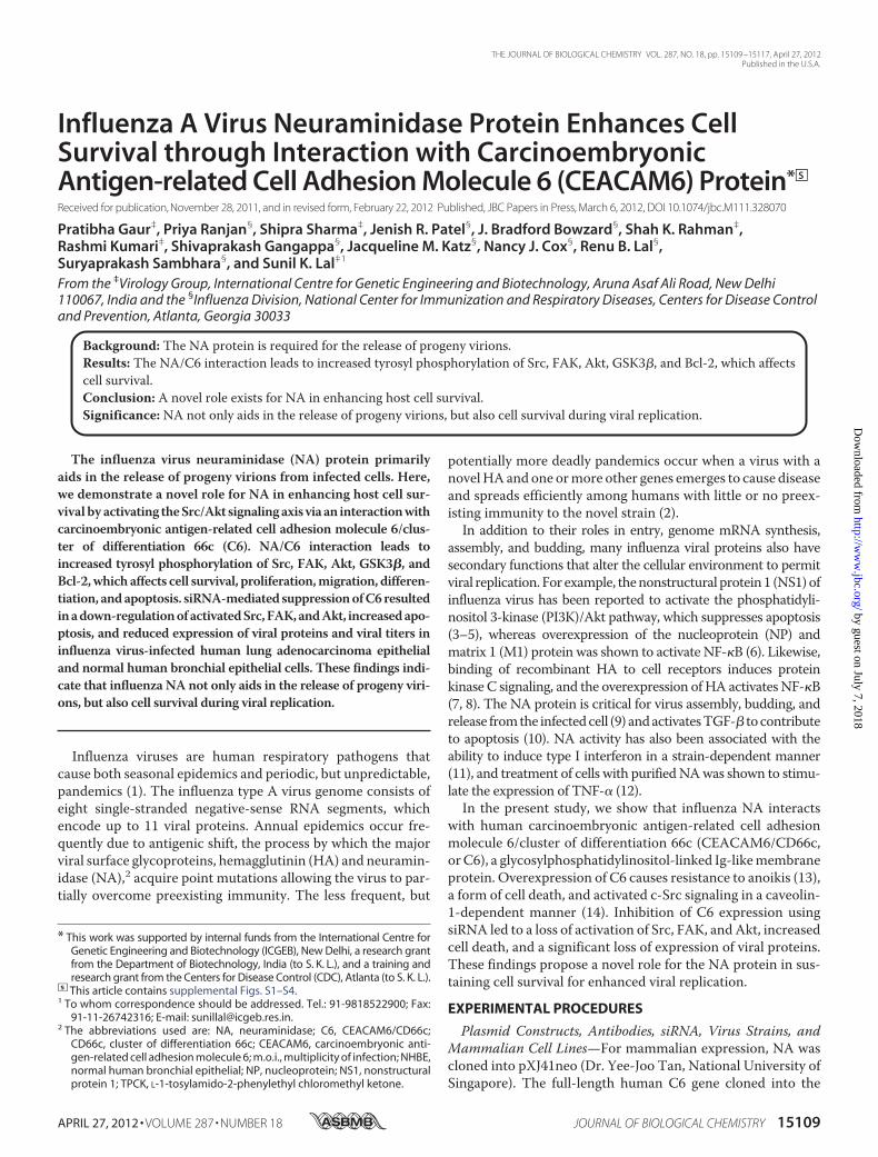
We next studied the biological effect of the NA-C6 complexon cell survival using an MTT assay. A549 and NHBE cellstransfected with scrambled siRNA and C6-specific siRNAwereinfected with PR8 virus and then subjected toMTT assay. Sub-stantial cell death was observed in C6 siRNA-treated cells com-pared with the scrambled controls (�67 and�50% relative via-bility, respectively, Fig. 6, A and B). Staining for Annexin-V, amarker for the early stages of cell death, also indicated thatviability was decreased upon inhibition of C6 in A549 cells (Fig.
FIGURE 1. Influenza A virus NA interacts with host protein C6. A, in vitro co-immunoprecipitation of NA and C6. HA-tagged NA and untagged C6 weretranslated in vitro in the presence of [35S]methionine and immunoprecipitated with �-HA or �-C6 antibodies, respectively. Complexes were subjected to 12%SDS-PAGE followed by autoradiography. NA co-immunoprecipitated with C6 pulled down by C6-specific antibody (panel 2, lane 2). Similarly, C6 co-immuno-precipitated with NA protein pulled down by �-HA antibody (panel 1, lane 2). B, NA co-localization with C6 in HEK-293 cells. NA and C6 expression plasmids weretransfected into HEK-293 cells. 48 h after transfection, cells were fixed, and nuclei were stained with DAPI (blue). C6 was labeled with goat anti-mouse AlexaFluor 594 (red), and NA was labeled with goat anti-rabbit Alexa Fluor 488 (green) antibody. The rightmost panel shows that 48 h after transfection NA and C6co-localize on the cell membrane. C, co-immunoprecipitation of NA with C6 in PR8 virus-infected cells. C6 is endogenously present in A549 cells. A549 cells wereinfected with A/PR8/34 influenza viruses at a m.o.i. of 1 and harvested at 24 h after infection. Co-immunoprecipitation and Western blot analysis were doneusing NA- and C6-specific antibodies. Lane 1 shows uninfected A549 cells, and lane 2 shows interaction between NA and C6 in PR8-infected cells. D, co-immunoprecipitations of NA and C6 in different subtypes of influenza virus. A549 cells were infected with different subtypes of influenza virus at a m.o.i. of 1and harvested at 24 h after infection. Co-immunoprecipitation and Western blot analysis were conducted using NA- and C6-specific antibodies. Lane 1 showsuninfected A549 cells, lane 2-4 show interactions between C6 and NA from A/X-31H3N2, A/WSN/1933, H1N1, and A/California/08/2009 H1N1 subtypes,respectively. E, co-immunoprecipitation of NA and C6 in primary cells infected with different subtypes of influenza viruses. NHBE cells were transfected with C6expression vectors or control vectors. 24 h after transfection, cells were infected with different subtypes of influenza viruses at a m.o.i. of 1 and harvested after24 h. Co-immunoprecipitation and Western blot analysis were conducted using NA- and C6-specific antibodies. Lane 1 shows uninfected NHBE cells, lane 2-4show interactions between C6 and NA from A/X-31(H3N2), A/WSN/1933 (H1N1), and A/California/08/2009 (pH1N1) viruses, respectively. F, various influenzavirus strains used for this study.
FIGURE 2. NA and C6 co-localize on the membrane of mammalian cells (A549) infected with PR8 influenza A virus. A549 cells were infected with PR8influenza virus at a m.o.i. of 1 and fixed at 16 h after infection. Nuclei were stained with DAPI (blue). NA was labeled with goat anti-rabbit Alexa Fluor 488 (green)antibody, and C6 was labeled with goat anti-mouse Alexa Fluor 594 (red) antibody. Protein localization was visualized by confocal microscopy.
Influenza A NA Enhances Cell Survival via CEACAM6
15112 JOURNAL OF BIOLOGICAL CHEMISTRY VOLUME 287 • NUMBER 18 • APRIL 27, 2012
by guest on July 7, 2018http://w
ww
.jbc.org/D
ownloaded from

6C). We have also checked the C6 siRNA effect on cell growthand viability in uninfected samples (supplemental Fig. S2A).There was a significant decrease in cell viability with the knock-down of C6 and virus-infected samples in contrast to theircontrol.Data shown above clearly indicateNAoverexpression and its
interaction with C6-increased Akt phosphorylation. To inves-
tigate the role of Akt phosphorylation in influenza virus infec-tion, A549 and NHBE cells were treated with PI3K inhibitors(LY294002 and wortmanin), Akt activator, YS-49 or Akt inhib-itors, Triciribine, 124005, or GSK690693 followed by infectionwith PR8 virus at a m.o.i. of 1. The samples were analyzed byWestern blotting to check the phosphorylation of Akt (Fig.7, Aa andBa). It was clearly evident that phosphorylation levels
FIGURE 3. Influenza infection elevates C6 mRNA and protein expression levels in a time- and dose-dependent manner. A549 cells were infected with PR8influenza virus at a m.o.i. of 1 and harvested 24 h after infection. A, real-time PCR or Western blot analysis of C6 (a and b) and NP (c and d). Expression increaseswith time of PR8 infection (4, 6, and 16 h at m.o.i. of 0.1and 1.0). B, real-time PCR analysis (n � 2) of C6 (a) and NP (c) expression increases with increasing doseof PR8 infection (m.o.i. of 0.1, 0.5, and 5.0); the same samples were checked for protein expression of C6 (b) and NP (d).
FIGURE 4. C6 siRNA-treated influenza A virus-infected cells down-regulate NP and NS1. A, A549 cells were treated with siRNA against C6 and scrambledsiRNA for 48 h and then infected with PR8 virus at an m.o.i. of 1. Cells were harvested at 24 h after infection, and equal amounts of protein from control andtreated cell extracts were subjected to Western blot analysis. First lane shows control levels of C6, NP, and NS1. Second lane shows minimal effect of scrambledsiRNA on the same protein levels. Third lane in top panel shows down-regulated levels of C6. Third lane in second panel from top shows down-regulation of NPafter using siRNA against C6. Third lane in second panel from bottom shows down-regulation of NS1 after using siRNA against C6. Bottom panel shows �-actinloading control. B, NHBE cells were treated with siRNA against C6 and control siRNA for 24 h and then infected with PR8 virus at m.o.i. of 1. Cells were harvestedat 24 h after infection and analyzed for mRNA expression of C6, NP, and NS1 by quantitative real-time PCR.
Influenza A NA Enhances Cell Survival via CEACAM6
APRIL 27, 2012 • VOLUME 287 • NUMBER 18 JOURNAL OF BIOLOGICAL CHEMISTRY 15113
by guest on July 7, 2018http://w
ww
.jbc.org/D
ownloaded from

of Akt were reduced in cells treated with PI3K inhibitors andinfected with PR8 virus (Fig. 7). PR8 infection itself activatedphosphorylation of Akt in A549 and NHBE cells.We have also assessed the viability of NHBE cells in the pres-
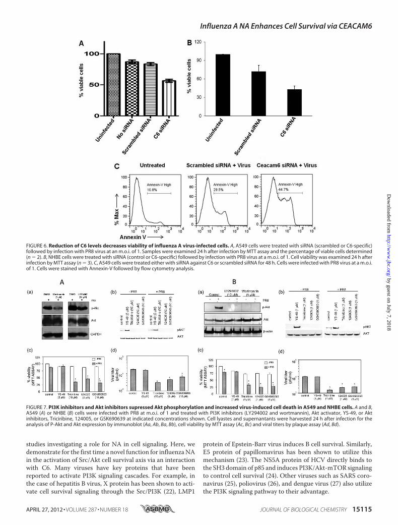
ence of PI3K inhibitors (supplemental Fig. S4) and our findingsindicate a decrease in cell viability with LY294002 and wort-mannin. PI3K inhibitors act upstream of Akt phosphorylation.Therefore, we investigated further the role of Akt phosphory-lation utilizing Akt-specific inhibitors. As a control, we usedAkt activator YS-49. YS-49 not only increased Akt phosphory-lation (Fig. 7, Ab and Bb) but also inhibited virus-induced celldeath (Fig. 7, A and B) in both A549 and NHBE cells. In con-trast, Akt inhibitors Triciribine, 124005, andGSK690693 inhib-ited virus-induced Akt phosphorylation (Fig. 7, Ab and Bb) inA549 and NHBE cells. Akt inhibitors decreased cell viability(Fig. 7, Ac and Bc) and virus replication (Fig. 7, Ad and Bd) inPR8-infected A549 and NHBE cells. Apart from pharmacolog-ical inhibitors we also evaluated the role of Akt in survival path-way by siRNA knockdown studies. siRNA pools against Akt1,
Akt2, and Akt3 significantly reduced the Akt expression ininfected or uninfected NHBE cells (Fig. 8, Aa and Ba). PR8infection resulted in significant decrease in cell viability (Fig. 8,Ab and Bb) and viral titers in both cell lines (Fig. 8, Ac and Bc).However, despite the change in cell viability and viral titers as aresult of Akt phosphorylation, we did not see significant changein plaque size (data not shown). These results suggest a role forNA-C6 in augmenting cell survival following infection withinfluenza virus via the Src/Akt axis.
DISCUSSION
To date, the primary role of influenza NA protein has beenbelieved to be in the release of progeny viruses from infectedcells and the subsequent spread of the virus to the surroundinguninfected cells. NA is also critical for the initiation of infectionand resultant influenza pathogenesis (19, 20). During influenzainfection, it triggers a host of intracellular signaling. Thesepathways carry out various cellular functions which usually arehijacked by the infecting virus (21). There have been limited
FIGURE 5. NA expression in mammalian cells leads to up-regulation of FAK and Akt phosphorylation. HEK-293 and A549 cells were transfected with theplasmid expressing H5N1 NA or empty plasmid (pcDNA 3.1 myc/his) as control and harvested at 48 h after transfection. Equal amounts of protein from test andcontrol sets were subjected to Western blot analysis. A, NA and C6 expression led to significant up-regulation of FAK and Src phosphorylation at 48 h as shownin second lane of top two panels. The pathway is Akt-dependent but independent of ERK activation as shown in second lane of bottom two panels in HEK-293 cells.Densitometry analysis (n � 2) of phosphorylation levels of Akt, FAK, and Src molecules in HEK-293 cells transfected with vector only and NA�C6 expressionconstructs are shown. Results were analyzed using paired t tests, and differences were considered significant at p � 0.005, p � 0.05, and p � 0.01 for P-FAK,P-Src, and P-Akt, respectively. Error bars, S.E. B, NA expression led to significant up-regulation of FAK and Akt phosphorylation at 48 h shown in second lane oftop two panels. P-Src and P-GSK3� expression at 48 h after transfection is shown in middle lane of bottom two panels. Bottom panel shows �-actin control in A549cells where endogenous levels of C6 were checked by immunofluorescence staining. Densitometry analysis of phosphorylation levels of Akt, FAK, Src mole-cules in A549 cells transfected with vector only and NA expression constructs is shown. Results were analyzed as in A with values of p � 0.04, p � 0.01, p � 0.008,and p � 0.02 for P-Akt, P-FAK, P-Src, and P-GSK3�, respectively. C, influenza infection led to significant up-regulation of P-Akt, P-Src, and P-FAK at 24 h, shownin second lane of top three panels in A549 cells. Total Akt and FAK antibody was used as a control (a). In contrast, knock down of C6 shows down-regulation ofP-Akt, P-Src, and P-FAK at 24 h (b). Densitometry analysis of phosphorylation levels of Akt, FAK, Src molecules in infected with PR8 virus in A549 cells is shown.Results were analyzed as in A with values of p � 0.009, p � 0.001, and p � 0.005 for P-Akt, P-FAK, and P-Src, respectively. D, NHBE cells were transfected withcontrol siRNA or C6 siRNA for 24 h. Cells were infected with PR8 (1 m.o.i.) for 24 h and harvested for the expression analysis of C6, P-Akt, P-Src, and P-Bcl-2 byWestern blotting. Total Akt and Bcl-2 Western blotting was used as a control.
Influenza A NA Enhances Cell Survival via CEACAM6
15114 JOURNAL OF BIOLOGICAL CHEMISTRY VOLUME 287 • NUMBER 18 • APRIL 27, 2012
by guest on July 7, 2018http://w
ww
.jbc.org/D
ownloaded from
studies investigating a role for NA in cell signaling. Here, wedemonstrate for the first time a novel function for influenzaNAin the activation of Src/Akt cell survival axis via an interactionwith C6. Many viruses have key proteins that have beenreported to activate PI3K signaling cascades. For example, inthe case of hepatitis B virus, X protein has been shown to acti-vate cell survival signaling through the Src/PI3K (22), LMP1
protein of Epstein-Barr virus induces B cell survival. Similarly,E5 protein of papillomavirus has been shown to utilize thismechanism (23). The NS5A protein of HCV directly binds tothe SH3 domain of p85 and induces PI3K/Akt-mTOR signalingto control cell survival (24). Other viruses such as SARS coro-navirus (25), poliovirus (26), and dengue virus (27) also utilizethe PI3K signaling pathway to their advantage.
FIGURE 6. Reduction of C6 levels decreases viability of influenza A virus-infected cells. A, A549 cells were treated with siRNA (scrambled or C6-specific)followed by infection with PR8 virus at an m.o.i. of 1. Samples were examined 24 h after infection by MTT assay and the percentage of viable cells determined(n � 2). B, NHBE cells were treated with siRNA (control or C6-specific) followed by infection with PR8 virus at a m.o.i. of 1. Cell viability was examined 24 h afterinfection by MTT assay (n � 3). C, A549 cells were treated either with siRNA against C6 or scrambled siRNA for 48 h. Cells were infected with PR8 virus at a m.o.i.of 1. Cells were stained with Annexin-V followed by flow cytometry analysis.
FIGURE 7. PI3K inhibitors and Akt inhibitors supressed Akt phosphorylation and increased virus-induced cell death in A549 and NHBE cells. A and B,A549 (A) or NHBE (B) cells were infected with PR8 at m.o.i. of 1 and treated with PI3K inhibitors (LY294002 and wortmannin), Akt activator, YS-49, or Aktinhibitors, Triciribine, 124005, or GSK690639 at indicated concentrations shown. Cell lyastes and supernantants were harvested 24 h after infection for theanalysis of P-Akt and Akt expression by immunoblot (Aa, Ab, Ba, Bb), cell viability by MTT assay (Ac, Bc) and viral titers by plaque assay (Ad, Bd).
Influenza A NA Enhances Cell Survival via CEACAM6
APRIL 27, 2012 • VOLUME 287 • NUMBER 18 JOURNAL OF BIOLOGICAL CHEMISTRY 15115
by guest on July 7, 2018http://w
ww
.jbc.org/D
ownloaded from

C6 is a member of a family of membrane-associated glyco-proteins that contain SLex moieties, which are involved in thebinding of influenza virions to the host cell (28). CEACAMfamily members are localized to lipid rafts at the cell surface byeither transmembrane domains or glycosylphosphatidylinosi-tol anchors (29). These plasmamembranemicrodomains accu-mulate numerous signal transduction components and havebeen implicated in transmembrane signaling via Src familykinases (30). Although CEACAM family members have beenshown to activate tyrosine kinase activity and play a role ininhibition of apoptosis (31, 32), the ligands that initiate thesesignaling pathways have not been fully elucidated.The NA/C6 interaction described here was confirmed in a
cell-free translation systemand in influenza virus infectedA549andNHBE cells, where the proteinswere found to co-localize inthe plasma membrane. The interaction was well conservedacross different subtypes of influenza viruses, including the2009 pandemic H1N1 strain, despite significant differences inthe amino acid sequences of NA.It has previously been shown that cell adhesion-mediated
FAK-Src signaling has a role in the regulation of human intes-tinal epithelial cell survival, with inhibition of FAK leading toincreased anoikis (33). The NA/C6 interaction described hereappears to activate this cell survival pathway by enhancing thephosphorylation levels of Src, FAK, Akt, and Bcl-2. The criticalnature of C6 in these processes during virus infection was fur-ther established by siRNA-mediated knockdown.We have alsoprovided evidence that indicates the involvement of Akt signal-ing PI3K inhibitors reduced the phosphorylation of Akt. Involve-ment of Akt phosphorylation in cell survival and virus replicationwas further confirmed using inhibitors that act directly on Akt
which decreased cell viability and viral titer in infected cells. Addi-tional evidence in support ofAkt involvent comes fromthe studieswhere we knocked down all the isoforms of Akt and observedreducedcell viability andviral titer in virus-infectedcells.Thephe-nomenon isnot cell-dependent aswehaveobtained similar resultswithHEK-293,A549, andNHBEcells. In the context of viral infec-tion, silencing C6 led to a reduction in the expression of NP andNS1proteins aswell as vRNA levels ofNP (Fig. S3). Influenza virusinfection has previously been shown to result in the up-regulationof expression of integrins and other neutrophil adhesion mole-cules (28, 34). Influenza NA has been shown to activate TGF-�(10), andCEACAM6is amajor target forSmad3-mediatedTGF-�signaling (35). These observations have led us to investigate theeffect of influenza virus on C6 expression levels. In this study weshowed for the first time that influenzaAvirus-infectedA549 cellsshow elevatedC6mRNAand protein expression. Taken together,these findings demonstrate that influenza NA protein may haveadditional roles in promoting cell survival during viral infection tofacilitate viral replication.
Acknowledgments—We thank Dr. Yee-Joo Tan (National Universityof Singapore) and Dr. Wolfgang Zimmermann (Tumor ImmunologyLaboratory, Muenchen, Germany) for providing pJX41-NA and pRc/CMV-C6 plasmids, respectively, and Dr. Dinh Duy Khang (Instituteof Biotechnology, Hanoi, Vietnam) for providing the influenza virus(Hatay isolate) cDNAandAdarshMayank for help with figures in themanuscript.
REFERENCES1. Taubenberger, J. K., andMorens, D.M. (2008) The pathology of influenza
virus infections. Annu. Rev. Pathol. 3, 499–522
FIGURE 8. siRNA-mediated Akt knockdown impaired cell viability and influenza virus replication in A549 and NHBE cells. A549 cells (A) or (B) NHBE cellswere transfected with control siRNA or siRNA specific for Akt1, Akt2, and Akt3 for 24 h and infected with PR8 (1 m.o.i.) for 24 h. Cell lysates and cell supernatantswere harvested for Akt expression by immunoblot (Aa, Ba), cell viability by MTT (Ab, Bb), and viral titers by plaque assay (Ac, Bc).
Influenza A NA Enhances Cell Survival via CEACAM6
15116 JOURNAL OF BIOLOGICAL CHEMISTRY VOLUME 287 • NUMBER 18 • APRIL 27, 2012
by guest on July 7, 2018http://w
ww
.jbc.org/D
ownloaded from

2. Bouvier, N. M., and Palese, P. (2008) The biology of influenza viruses.Vaccine 26, D49–53
3. Ehrhardt, C., Wolff, T., Pleschka, S., Planz, O., Beermann, W., Bode, J. G.,Schmolke, M., and Ludwig, S. (2007) Influenza A virus NS1 protein acti-vates the PI3K/Akt pathway tomediate antiapoptotic signaling responses.J. Virol. 81, 3058–3067
4. Hale, B. G., and Randall, R. E. (2007) PI3K signalling during influenza Avirus infections. Biochem. Soc. Trans. 35, 186–187
5. Shin, Y. K., Liu, Q., Tikoo, S. K., Babiuk, L. A., and Zhou, Y. (2007) Effect ofthe phosphatidylinositol 3-kinase/Akt pathway on influenza A virus prop-agation. J. Gen. Virol. 88, 942–950
6. Flory, E., Kunz, M., Scheller, C., Jassoy, C., Stauber, R., Rapp, U. R., andLudwig, S. (2000) Influenza virus-inducedNF-�B-dependent gene expres-sion is mediated by overexpression of viral proteins and involves oxidativeradicals and activation of I�B kinase. J. Biol. Chem. 275, 8307–8314
7. Arora, D. J., andGasse, N. (1998) Influenza virus hemagglutinin stimulatesthe protein kinase C activity of human polymorphonuclear leucocytes.Arch. Virol. 143, 2029–2037
8. Pahl, H. L., and Baeuerle, P. A. (1995) Expression of influenza virus he-magglutinin activates transcription factor NF-�B. J. Virol. 69, 1480–1484
9. Chazal, N., and Gerlier, D. (2003) Virus entry, assembly, budding, andmembrane rafts.Microbiol. Mol. Biol. Rev. 67, 226–237, table of contents
10. Schultz-Cherry, S., and Hinshaw, V. S. (1996) Influenza virus neuramini-dase activates latent transforming growth factor-�. J. Virol. 70,8624–8629
11. Miller, J. L., and Anders, E. M. (2003) Virus-cell interactions in the induc-tion of type 1 interferon by influenza virus in mouse spleen cells. J. Gen.Virol. 84, 193–202
12. Houde, M., and Arora, D. J. (1990) Stimulation of tumor necrosis factorsecretion by purified influenza virus neuraminidase. Cell. Immunol. 129,104–111
13. Duxbury, M. S., Ito, H., Zinner, M. J., Ashley, S. W., and Whang, E. E.(2004) CEACAM6 gene silencing impairs anoikis resistance and in vivometastatic ability of pancreatic adenocarcinoma cells. Oncogene 23,465–473
14. Duxbury,M. S., Ito, H., Ashley, S.W., andWhang, E. E. (2004) CEACAM6cross-linking induces caveolin-1-dependent, Src-mediated focal adhesionkinase phosphorylation in BxPC3 pancreatic adenocarcinoma cells. J. Biol.Chem. 279, 23176–23182
15. Kumar, P., Gunalan, V., Liu, B., Chow, V. T., Druce, J., Birch, C., Catton,M., Fielding, B. C., Tan, Y. J., and Lal, S. K. (2007) The nonstructuralprotein 8 (nsp8) of the SARS coronavirus interacts with its ORF6 acces-sory protein. Virology 366, 293–303
16. Ratra, R., Kar-Roy, A., and Lal, S. K. (2009) ORF3 protein of hepatitis Evirus interacts with the � chain of fibrinogen resulting in decreased fibrin-ogen secretion from HuH-7 cells. J. Gen. Virol. 90, 1359–1370
17. Duxbury,M. S., Ito, H., Benoit, E.,Waseem, T., Ashley, S.W., andWhang,E. E. (2004) A novel role for carcinoembryonic antigen-related cell adhe-sion molecule 6 as a determinant of gemcitabine chemoresistance in pan-creatic adenocarcinoma cells. Cancer Res. 64, 3987–3993
18. Duxbury,M. S., Ito, H., Benoit, E., Zinner,M. J., Ashley, S.W., andWhang,E. E. (2004) Overexpression of CEACAM6 promotes insulin-like growthfactor I-induced pancreatic adenocarcinoma cellular invasiveness. Onco-gene 23, 5834–5842
19. Bhatia, A., andKast, R. E. (2007)How influenza’s neuraminidase promotesvirulence and creates localized lung mucosa immunodeficiency. Cell Mol.Biol. Lett. 12, 111–119
20. Matrosovich, M. N., Matrosovich, T. Y., Gray, T., Roberts, N. A., andKlenk, H. D. (2004) Neuraminidase is important for the initiation of influ-enza virus infection in human airway epithelium. J. Virol. 78,12665–12667
21. Gaur, P., Munjhal, A., and Lal, S. K. (2011) Influenza virus and cell signal-ing pathways.Med. Sci. Monit. 17, RA148–154
22. Shih,W. L., Kuo,M. L., Chuang, S. E., Cheng, A. L., andDoong, S. L. (2003)Hepatitis B virus X protein activates a survival signaling by linking SRC tophosphatidylinositol 3-kinase. J. Biol. Chem. 278, 31807–31813
23. Cooray, S. (2004) The pivotal role of phosphatidylinositol 3-kinase/Aktsignal transduction in virus survival. J. Gen. Virol. 85, 1065–1076
24. Mannová, P., and Beretta, L. (2005) Activation of the N-Ras-PI3K-Akt-mTOR pathway by hepatitis C virus: control of cell survival and viralreplication. J. Virol. 79, 8742–8749
25. Mizutani, T., Fukushi, S., Saijo, M., Kurane, I., and Morikawa, S. (2004)Importance of Akt signaling pathway for apoptosis in SARS-CoV-infectedVero E6 cells. Virology 327, 169–174
26. Autret, A., Martin-Latil, S., Brisac, C., Mousson, L., Colbère-Garapin, F.,and Blondel, B. (2008) Early phosphatidylinositol 3-kinase/Akt pathwayactivation limits poliovirus-induced JNK-mediated cell death. J. Virol. 82,3796–3802
27. Lee, C. J., Liao, C. L., and Lin, Y. L. (2005) Flavivirus activates phosphati-dylinositol 3-kinase signaling to block caspase-dependent apoptotic celldeath at the early stage of virus infection. J. Virol. 79, 8388–8399
28. Hartshorn, K. L., and White, M. R. (1999) Influenza A virus up-regulatesneutrophil adhesionmolecules and adhesion to biological surfaces. J. Leu-koc. Biol. 65, 614–622
29. Kuespert, K., Pils, S., and Hauck, C. R. (2006) CEACAMs: their role inphysiology and pathophysiology. Curr. Opin. Cell Biol. 18, 565–571
30. Kasahara, K.,Watanabe, K., Kozutsumi, Y., Oohira, A., Yamamoto, T., andSanai, Y. (2002) Association of GPI-anchored protein TAG-1 with src-family kinase Lyn in lipid rafts of cerebellar granule cells.Neurochem. Res.27, 823–829
31. Kirshner, J., Chen, C. J., Liu, P., Huang, J., and Shively, J. E. (2003)CEACAM1–4S, a cell-cell adhesion molecule, mediates apoptosis andreverts mammary carcinoma cells to a normal morphogenic phenotype ina 3D culture. Proc. Natl. Acad. Sci. U.S.A. 100, 521–526
32. Najjar, S. M. (2002) Regulation of insulin action by CEACAM1. TrendsEndocrinol. Metab. 13, 240–245
33. Bouchard, V., Demers, M. J., Thibodeau, S., Laquerre, V., Fujita, N., Tsu-ruo, T., Beaulieu, J. F., Gauthier, R., Vézina, A., Villeneuve, L., andVachon,P. H. (2007) Fak/Src signaling in human intestinal epithelial cell survivaland anoikis: differentiation state-specific uncoupling with the PI3-K/Akt-1 and MEK/Erk pathways. J. Cell Physiol. 212, 717–728
34. Ducker, T. P., and Skubitz, K. M. (1992) Subcellular localization of CD66,CD67, and NCA in human neutrophils. J. Leukoc. Biol. 52, 11–16
35. Han, S. U., Kwak, T. H., Her, K. H., Cho, Y. H., Choi, C., Lee, H. J., Hong, S.,Park, Y. S., Kim, Y. S., Kim, T. A., Kim, S. J. (2008) CEACAM5 andCEACAM6 are major target genes for Smad3-mediated TGF-� signaling.Oncogene 27, 675–683
Influenza A NA Enhances Cell Survival via CEACAM6
APRIL 27, 2012 • VOLUME 287 • NUMBER 18 JOURNAL OF BIOLOGICAL CHEMISTRY 15117
by guest on July 7, 2018http://w
ww
.jbc.org/D
ownloaded from

Cox, Renu B. Lal, Suryaprakash Sambhara and Sunil K. LalK. Rahman, Rashmi Kumari, Shivaprakash Gangappa, Jacqueline M. Katz, Nancy J.
Pratibha Gaur, Priya Ranjan, Shipra Sharma, Jenish R. Patel, J. Bradford Bowzard, Shah(CEACAM6) Protein
Interaction with Carcinoembryonic Antigen-related Cell Adhesion Molecule 6 Influenza A Virus Neuraminidase Protein Enhances Cell Survival through
doi: 10.1074/jbc.M111.328070 originally published online March 6, 20122012, 287:15109-15117.J. Biol. Chem.
10.1074/jbc.M111.328070Access the most updated version of this article at doi:
Alerts:
When a correction for this article is posted•
When this article is cited•
to choose from all of JBC's e-mail alertsClick here
Supplemental material:
http://www.jbc.org/content/suppl/2012/03/06/M111.328070.DC1
http://www.jbc.org/content/287/18/15109.full.html#ref-list-1
This article cites 35 references, 15 of which can be accessed free at
by guest on July 7, 2018http://w
ww
.jbc.org/D
ownloaded from