influenza a virus-host interactions and their control by
TRANSCRIPT
Influenza A Virus-Host Interactionsand Their Control by ViralNon-Structural Protein NS1
Maria Anastasina
Institute for Molecular Medicine Finlandand
Division of MicrobiologyDepartment of Biosciences
Faculty of Biological and Environmental Sciencesand
Doctoral Program in BiomedicineUniversity of Helsinki
Academic Dissertation
To be presented for public examination with the permission of theFaculty of Biological and Environmental Sciences of the University of
Helsinki in the lecture hall 2402 of Biocenter 3, Viikinkaari 1 onFriday, 17th of April, 2015, at 12 o’clock noon
Helsinki 2015
Supervisors Docent Denis Kainov, Ph. D.Institute for Molecular Medicine Finland,University of Helsinki, Finland
Professor Sarah Butcher, Ph. D.Research DirectorStructural Biology and Biophysics Program,Institute of Biotechnology,University of Helsinki, Finland
Reviewers Docent Thedi Ziegler, Ph. D.Research Center for Child PsychiatryInstitute of Clinical MedicineUniversity of Turku, Finland
Docent Maija Vihinen-Ranta, Ph. D.Nanoscience Center,Department of Biological and Environmental Science,University of Jyvaskyla, Finland
Opponent Professor Stephan Ludwig, Ph. D.Institute of Molecular VirologyCentre for Molecular Biology of InflammationUniversity of Munster, Germany
Custos Professor Dennis Bamford, Ph. D.Department of Biosciences andInstitute of BiotechnologyUniversity of Helsinki, Finland
Thesis Committee Professor Kalle Saksela, Ph. D.Department of Virology, Haartman InstituteUniversity of Helsinki, Finland
Professor Dennis Bamford, Ph. D.Department of Biosciences andInstitute of BiotechnologyUniversity of Helsinki, Finland
c○Maria Anastasina 2015
ISBN 978-951-51-0960-6 (paperback)ISBN 978-951-51-0961-3 (PDF)
Hansaprint Oy, Helsinki 2015

To my family
Contents
List of original publications i
Summary iii
Abbreviations v
1 Review of the Literature 1
1.1 Introduction . . . . . . . . . . . . . . . . . . . . . . . . . . . . . . . . . 1
1.2 Influenza A virus: an overview . . . . . . . . . . . . . . . . . . . . . . . 4
1.3 Influenza A virus organization and replication cycle . . . . . . . . . . . 7
1.4 Host factors involved in influenza A virus replication cycle . . . . . . . 13
1.5 Innate immune responses to influenza A infection . . . . . . . . . . . . 16
1.5.1 Virus recognition by innate immunity . . . . . . . . . . . . . . . 17
1.5.2 Antiviral responses by interferons and interferon-stimulated genes 23
1.5.3 Pro-inflammatory responses . . . . . . . . . . . . . . . . . . . . 27
1.6 Apoptosis . . . . . . . . . . . . . . . . . . . . . . . . . . . . . . . . . . 28
1.7 Viral counteraction to innate responses. NS1. . . . . . . . . . . . . . . . 29
1.7.1 NS1 synthesis and localization . . . . . . . . . . . . . . . . . . . 30
1.7.2 Post-translational modifications of NS1 . . . . . . . . . . . . . . 32
1.7.3 Structure of NS1 . . . . . . . . . . . . . . . . . . . . . . . . . . 34
1.7.4 Inhibition of interferon signaling at pre-transcriptional level . . 36
1.7.5 Inhibition of interferon signaling at post-transcriptional level . . 39
1.7.6 Direct inhibition of interferon-stimulated gene products . . . . . 42
1.7.7 Other pro-viral functions of NS1 . . . . . . . . . . . . . . . . . . 43
1.7.8 NS1 diversity . . . . . . . . . . . . . . . . . . . . . . . . . . . . 47
2 Objectives of the Present Study 51
3 Materials and Methods 52
4 Results and Discussion 66
4.1 Conserved residues within NS1 bind dsDNA and control transcription
of cellular genes (Study I) . . . . . . . . . . . . . . . . . . . . . . . . . 66
4.2 Regulation of general protein synthesis by NS1 (Studies II, IV) . . . . . 72
4.3 C-terminus of NS1 contributes to modulation of host antiviral responses
(Study III) . . . . . . . . . . . . . . . . . . . . . . . . . . . . . . . . . . 78
4.4 NS1 as a tool to improve cell-free protein synthesis system (Study IV) . 81
5 Conclusions and Future Perspectives 84
Acknowledgements 89
References 94

List of Original Publications
This thesis is based on the following publications and manuscripts which are referred to
in the text by their roman numerals. In addition, unpublished data will be presented.
I Anastasina M*, Le May N*, Butcher SJ, Egly JM, Kainov DE. Influenza A
non-structural protein NS1 binds DNA to control host antiviral gene expression.
Manuscript. (* = equal contribution).
II Kainov DE, Muller KH, Theisen LL, Anastasina M, Kaloinen M, Muller CP.
Differential effects of NS1 proteins of human pandemic H1N1/2009, avian highly
pathogenic H5N1, and low pathogenic H5N2 influenza A viruses on cellular pre-
mRNA polyadenylation and mRNA translation. Journal of Biological Chemistry.
2011 Mar 4;286(9).
III Anastasina M, Schepens B, Saksela K, Saelens X, Kainov D. The length of
C-terminus of influenza NS1 is essential for virus-host interplay. Manuscript.
IV Anastasina M, Terenin I, Butcher S, Kainov D. A technique to increase pro-
tein yield in a rabbit reticulocyte lysate translation system. Biotechniques. 2014.
56(1):36-9.
i
Candidate’s independent contribution to this work:
I MA participated in design and performed experiments on DNA binding by NS1,
generated recombinant viruses, did transcriptional and cytokine profiling of in-
fected cells, did protein localization and ChIP experiments and interpreted the
obtained data. NLM did in vitro transcription experiments and studied immune
responses activation in transfected cells. MA, SB and DK wrote the manuscript
II MA did experiments addressing mRNA concentration effects on in vitro transla-
tion, effects of NS1 on mRNA stability and loss- and gain-of-function experiments
with NS1 mutants. MA interpreted the obtained results and contributed to writing
of the manuscript.
III MA generated recombinant viruses, designed and performed gene expression anal-
ysis, did cytokine and phosphoprotein profiling in infected cells and interpreted
the data. MA and DK wrote the manuscript.
IV MA designed and performed experiments on cellular mRNA translation and polysome
formation in rabbit lysate and interpreted the data. MA, SB, and DK wrote the
manuscript.
ii

Summary
Viruses infect all domains of life. They establish complex interactions with their host
cells to subvert and hijack multiple cellular processes and warrant their own replication.
Understanding virus-host interactions is critical to control spread of pathogenic viruses,
develop vaccines and search for antivirals. Besides that, understanding virus-host in-
teractions allows deciphering complex cellular processes and provides useful tools for
biotechnology.
My research is dedicated to influenza A virus, an important pathogen that infects
humans worldwide, represents a constant health care threat and elicits continuous ef-
forts to control the human spread of the disease. Influenza A expresses a non-structural
protein NS1 that is a key regulator of viral interactions with the host cell and an impor-
tant virulence factor. Versatile functions of NS1 modulate multiple cellular functions
to secure viral replication.
This work addresses several aspects of NS1-mediated modulation of core cellular
processes. We discovered that NS1 binds to dsDNA and inhibits transcription of cellular
genes, thus limiting antiviral responses. We found that NS1 secures general protein
synthesis and mapped several residues within NS1 that are essential for this function.
Further, we showed that the length of C-terminal “tail” of NS1 is essential for control
of cellular antiviral responses and virus pathogenicity. The presented results increase
the understanding of influenza A virus-host interactions and can be further utilized
in the search for antivirals and vaccine development. In addition, this work provides
a biotechnological application of influenza A NS1 protein for improvement of cell-free
translation system.
iii

Abbreviations
5’meG 5’ methylated guanine
aa amino acid
AP-1 activator protein 1
CARD caspase recruitment domain
ChIP chromatin immunoprecipitation
CPSF cleavage and polyadenylation specific factor
cRNP complementary ribonucleoprotein
CTD C-terminal domain
CTL cytotoxic T lymphocyte
DC dendritic cell
ED effector domain
eIF2𝛼 translation initiation factor 2𝛼
eIF4GI initiation factor 4GI
EMCV encephalomyocarditis virus
GCN2 general control non-derepressible kinase 2
HA hemagglutinin
HRI heme-regulated inhibitor
hStaufen human homolog of Drosophila melanogaster Staufen protein
IFITM3 interferon-inducible transmembrane protein 3
IFN interferon
IKK inhibitor of nuclear factor kappa-B kinase
IL interleukin
IRES internal ribosome entry site
IRF interferon regulatory factor
ISG interferon-stimulated gene
ISG15 ubiquitin-like protein ISG15
JAK Janus kinases
JNK c-Jun N-terminal kinase
Kd dissociation constant
M1 matrix protein
M2 M2 proton channel
MAPK mitogen-activated protein kinase
MAVS mitochondrial antiviral-signaling protein
MDA5 melanoma differentiation-associated protein 5
iv

MxA myxovirus resistance gene product
NA neuraminidase
NEP nuclear export protein
NES nuclear export sequence
NFkB nuclear factor kappa-light-chain-enhancer of activated B cells
NLR NOD-like receptor
NLRP3 LRR- and pyrin domain-containing protein 3
NLS nuclear localization sequence
NP nucleoprotein
NS1 non-structural protein
OAS 2’-5’-oligoadenylate syntethase
PA polymerase acidic protein
PABP poly(A)-binding protein
PAMP pathogen-associated molecular pattern
PB1 polymerase basic protein 1
PB2 polymerase basic protein 2
PDZ postsynaptic density protein 95, Drosophila disc large tumor suppressor, and zonulaoccludens 1 protein
PERK PKR-like endoplasmic reticulum kinase
PI3K phosphoinositide-3-kinase
PKR protein kinase R
poly(A) polyadenine stretch
poly(I:C) polyinosinic:polycytidylic acid
PRR pattern recognition receptor
RBD RNA-binding domain
RdRp RNA-dependent RNA polymerase
RIG-I retinoic acid inducible gene I
RLR RIG-I-like receptor
RNAse L ribonuclease L
RRL rabbit reticulocyte lysate
SH3 Src-homology 3 domain
ssRNA single-stranded RNA
STAT signal transducer and activator of transcription
SUMO small ubiquitin-like modifier protein
TLR Toll-like receptor
UTR untranslated region
vRNA viral RNA
vRNP viral ribonucleoprotein
v
1 Review of the Literature
1.1 Introduction
Viruses are seemingly simple in comparison to organisms that they infect and consist
of just a genome, structural proteins and sometimes a lipid bilayer envelope. Because of
this simplicity they have very limited capacity to encode factors essential for their own
replication. Viruses are obligate parasites of cells and have evolved multiple strategies
to interact with the cell at each step of viral life cycle. For this, specific viral proteins
interact with the numerous host factors and subvert cellular processes to fulfill the
needs of virus replication.
The initial interaction event between the virus and a susceptible cell occurs when
viral receptor-binding proteins recognize cellular proteins, carbohydrates or lipids ex-
posed on the cell surface (Grove and Marsh, 2011). Viruses enter the cell via non-
endocytic or endocytic routes, which often require interaction with cellular factors,
such as clathrin-coated vesicle components (Dimitrov, 2004; Yamauchi and Helenius,
2013). Interaction with the cellular cytoskeleton is widely used by viruses to accelerate
and direct their antero- and retrograde transport through the crowded intra-cellular
environment (Ploubidou and Way, 2001). Uncoating and release of viral genomes is
often triggered by interaction with specific cellular proteins (Suomalainen and Greber,
2013; Haywood, 2010). Furthermore, viruses that replicate their genomes in the nu-
cleus translocate viral components there in an active way which requires binding to
the proteins of nuclear pore complex (Kobiler et al., 2012).
Independently on whether they encode their own RNA polymerase or use the cel-
lular enzyme, complex interactions with the cellular transcription machinery ensure
effective synthesis of viral mRNA(s). For example, poliovirus and Rift Valley fever
virus, which use their own enzymes for RNA synthesis both shut down host transcrip-
tion, which is not essential for viral replication (Le May et al., 2004; Kundu et al.,
2005). In contrast, the viruses that require host transcription machinery, for example
1

herpes simplex virus, set up regulatory interactions with cellular transcription factors
to support their own DNA synthesis (Wysocka and Herr, 2003).
Furthermore, being restricted in their encoding capacity viruses lack their own
functional translation machinery and fully rely on host protein synthesis (Walsh and
Mohr, 2011). Even Pandoravirus salinus with the biggest known viral genomes of 2.77
mega base pairs encodes only few required translation factors remaining dependent on
protein synthesis of its Acantamoeba host (Philippe et al., 2013). Viruses target the
cellular translational machinery to secure preferential translation of viral transcripts or
to shut off host translation when it is not required. For instance, vesicular stomatitis
virus utilizes specific 3’ structures on its mRNA and also interacts with the ribosome to
secure preferential synthesis of viral proteins (Whitlow et al., 2006; Lee et al., 2013a).
Viruses further utilize cellular processes for transport of their components or assembled
virions and for escape from the cell (Bartenschlager et al., 2011; Lyles, 2013). Thus,
human immunodeficiency virus type 1 usurps cellular endosomal sorting complexes
required for transport pathway to facilitate its effective budding (Morita et al., 2011),
whereas adenoviruses induce autophagy to enable cell lysis and viral exit (Jiang et al.,
2011).
The cells of plants, many invertebrates and vertebrates respond to infection with
robust induction of innate immune responses and no successful virus replication would
be possible without control over these responses. For this, viruses have evolved a mul-
titude of approaches to counteract their recognition by cellular detectors (Zinzula and
Tramontano, 2013), subvert signal transduction, prevent activation of antiviral genes
(Short, 2009), and, when necessary, limit apoptosis (Galluzzi et al., 2008).
The number of viral strategies to interact with the host cell is overwhelming. Al-
though research helped to deduce the basic strategies of viral life cycle from the nature
of viral genomes already in 1971 (Baltimore, 1971), the exact mechanisms of viral
replication are so diverse and complicated that we are still striving to understand
them. Studies of virus-host interactions are largely driven by attempts to improve the
2

surveillance of pathogenic viruses, search for antivirals and development of vaccines
(Webby and Webster, 2003; Schwegmann and Brombacher, 2008). However, viruses
have also proven many times to be valuable tools for understanding cell functions,
from the initial discovery of DNA as genetic material (Hershey and Chase, 1952), up
to recent advances in tackling complex processes such as endocytosis (Pelkmans and
Helenius, 2003). Finally, understanding virus-host interactions brings novel applicable
tools widely used in biotechnology, such as viral vectors for gene transfer (Vannucci
et al., 2013) or baculovirus systems for heterologous protein production (van Oers,
2011). Although a great deal of mechanisms have been already discovered, the amount
of information that we get now is growing exponentially and the majority of discoveries
is perhaps still ahead.
1.2 Influenza A virus: an overview
This work is dedicated to influenza A virus, a member of the Orthomyxoviridae fam-
ily. Influenza A viruses are commonly classified based on their surface antigens—
hemagglutinin (HA) and neuraminidase (NA). HA subtypes 1–16 and all NA subtypes
1–9 are found in wild birds which, apparently, represent the main natural reservoir for
influenza A virus (Stallknecht and Brown, 2007). In addition, H17N10 and H18N11
viruses were recently found in bats that seem to represent a sylvatic mammalian reser-
voir for influenza A viruses (Tong et al., 2012; Tong et al., 2013). Certain viral subtypes
can also infect domesticated birds and multiple species of mammals, including humans.
Whereas influenza A virus is usually asymptomatic in its natural hosts, it can cause
mild to severe intestinal infections in poultry and asymptomatic to severe respiratory
infections in mammals (Webster et al., 1992).
Although the first human influenza A virus was isolated in 1933 (Smith et al.,
1933) and one of the first well-publicized influenza A pandemics occurred in 1918
(Taubenberger et al., 1997), numerous records indicate that humankind has been facing
influenza epidemics and probably also pandemics for at least several centuries (Potter,
3

2001). There is molecular evidence that influenza A HA subtypes 1, 2, 3, 5, 7, 9 and 10
can infect humans, but currently only influenza A of H1 and H3 subtypes are circulating
in humans and are causing annual epidemics.
Circulating strains of influenza A cause seasonal infections in humans. In most
countries these infections result in annual epidemics which, according to World Health
Organization (WHO), affect up to 10 % of the population worldwide and result in
up to 500, 000 deaths. In addition to these annual epidemics, global pandemics can
occur when humans are infected with viruses capable of human-to-human spread to
which they are immunologically naıve. Although influenza A pandemics are relatively
rare events, humankind has faced three major pandemics in the twentieth century and
already one in the twenty-first (Lagace-Wiens et al., 2010; Fineberg, 2014). Whereas
the mortality of seasonal influenza is modest, the mortality of pandemic influenza is
unpredictable and can vary: for example, during the H1N1 pandemic in 2009 it was
below 0.5 %, but during the H5N1 outbreak in 1997 it reached 60 % (Forrest and
Webster, 2010; Noah and Noah, 2013). In addition to annual influenza-related deaths,
the virus imposes an enormous economic burden on multiple sectors of societies (Szucs,
1999; Noah and Noah, 2013).
Influenza A virus genome is composed of eight single-stranded RNA (ssRNA)
molecules of negative polarity (Palese, 1977). It is replicated with the viral RNA-
dependent RNA polymerase (RdRp) which is error-prone and produces between 1.5
and 7.5 × 10⁻⁵ misincorporations per nucleotide. Because RdRp also lacks proofread-
ing activity these misincorporations cannot be repaired and on average one mutation
appears in the viral genome after each cycle of RNA replication (Parvin et al., 1986;
Drake, 1993). The gradual evolution of influenza A viruses due to frequent mutations
in viral proteins is referred to as antigenic drift. In addition, the viral genomic segments
can reassort during the co-infection of the same cell with two or more influenza A viruses
and give rise to progeny virions that contain segments derived from both “parental”
viruses (McGeoch et al., 1976; Desselberger et al., 1978). If the “parental” viruses be-
4

long to different subtypes, the reassortant progeny virion(s) may harbor major changes
that are referred to as antigenic shift. Antigenic drift, antigenic shift and inter-species
transmission are the key drivers of viral evolution (Forrest and Webster, 2010). Anti-
genic drift limits efficacy of vaccines and antivirals and antigenic shift imposes constant
risk for new pandemics. Thus, improvements of options to control influenza are needed.
These efforts require careful virus surveillance, vaccine development and search for an-
tivirals with novel mechanisms of action. This is impossible without a comprehensive
understanding of influenza A virus-host interactions.
1.3 Influenza A virus organization and replication cycle
Influenza A virions are pleiomorphic, i.e. their shapes are not uniform and can be spher-
ical, kidney- or rod-shaped with an average size of 100–150 nm (Fujiyoshi et al., 1994).
The outer shell of the virion is composed of the host-derived lipid bilayer in which
viral HA, NA and M2 proton channel (M2) are incorporated. This shell is underlined
with the viral matrix protein (M1) (Harris et al., 2006). Each virion encompasses eight
genomic RNA segments packed in viral ribonucleoproteins (vRNPs)—supercoiled ring-
like structures in which paired 5’ and 3’ ends of the viral RNA are associated with
heterotrimetic viral polymerase complex and the rest of the RNA is densely covered
with viral nucleoprotein (NP) (Arranz et al., 2012). In the virion the vRNPs are asso-
ciated with the M1 protein (Rees and Dimmock, 1982; Ye et al., 1999). Eight genes of
all influenza A viruses encode 10 essential viral proteins: HA, NA, M1, M2, NP, poly-
merase basic protein 1 (PB1), polymerase basic protein 2 (PB2), polymerase acidic
protein (PA), non-structural protein (NS1), and nuclear export protein (NEP) (Lamb,
1983). In addition, some influenza A strains may encode accessory proteins PB1-F2,
PB1-N40, PA-X, PA-155 and PA-182. Whereas HA, NA, M1, M2, NP, PB1, PB2, PA
and NEP are structural components of viral particle, NS1, PB1-F2, PB1-N40, PA-X,
PA-155 and PA-182 are considered to be non-structural and are involved in regulation
of virus-host interactions (Chen et al., 2001; Hale et al., 2008c; Wise et al., 2009; Jagger
5

et al., 2012; Muramoto et al., 2013).
Influenza A replication cycle begins when the viral HA binds to the specific virus
receptor on cell surface. The key, but possibly not the only, receptors for influenza A
virus are sialic acids linked to cellular surface glycoproteins or glycolipids (Martın et al.,
1998; Skehel and Wiley, 2000; Stray et al., 2000). HA molecules of avian influenza A
viruses recognize 𝛼-2,3-linked sialic acids and HAs of human influenza A viruses recog-
nize 𝛼-2,6-linked sialic acids (Connor et al., 1994; van Riel et al., 2010). HA recognition
of specific sialic acids to a large extent determines viral host specificity. After receptor
binding the viruses are endocytosed via clathrin-dependent or clathrin- and caveolin-
independent routes and are transferred towards the perinuclear space in endosomes
(Dourmashkin and Tyrrell, 1974; Matlin et al., 1981; Sieczkarski and Whittaker, 2002;
Lakadamyali et al., 2003). Acidification of late endosomes in perinuclear space triggers
two essential events that allow virus uncoating and delivery of vRNPs to the cyto-
plasm. First, low pH mediates the conformational change in the HA and enables fusion
of viral and endosomal membranes (Carr and Kim, 1993). Second, acidification of the
virus interior leads to dissociation of vRNPs from M1 which is a prerequisite for their
import into the nucleus, a site of viral RNA transcription and replication (Bui et al.,
1996; Stauffer et al., 2014).
Cytoplasmic transport of the vRNPs towards the nucleus is passive: it does not
require cellular cytoskeleton components and appears to rely on diffusion (Martin and
Helenius, 1991; Babcock et al., 2004). The nuclear import of vRNPs, in contrast, occurs
in an active way (Kemler et al., 1994). For this, importin 𝛼 isoforms 1 and 5 likely
recognize the surface-exposed nuclear localization sequences (NLSs) on viral NPs and,
together with the importin 𝛽, mediate traverse of vRNPs through the nuclear pore
complex and its delivery to the nucleus (Martin and Helenius, 1991; O’Neill et al.,
1995; Cros et al., 2005; Moeller et al., 2012; ying Chou et al., 2013).
In the nucleus the viral RNAs (vRNAs) are transcribed in cis by the viral poly-
merase associated with the vRNP (Moeller et al., 2012). Synthesis of viral mRNA is
6

initiated using 10–13 nucleotide long primers with 5’ methylated guanine (5’meG) cap
(Beaton and Krug, 1981; Plotch et al., 1981). These primers are stolen from cellular
mRNAs during “cap-snatching” when 5’meG cap structures on host mRNAs are rec-
ognized and bound by PB2 subunit of viral polymerase (Guilligay et al., 2008) and
further endonucleolytically cleaved by viral PB1 and PA (Li et al., 2001a; Dias et al.,
2009; Yuan et al., 2009). The “snatched” 5’meG-caps also provide 3’-OH ends for viral
mRNA chain elongation by PB1 (Poch et al., 1989). The synthesis of viral mRNA ends
after reiterative copying of short uridine stretches located at the 5’ end of vRNA and
the resulting viral mRNA contains 150–200 adenine bases at its 3’ end (Plotch and
Krug, 1977; Robertson et al., 1981; Poon et al., 1999). Cap-containing polyadenylated
viral mRNAs are structurally indistinguishable from host transcripts. They are ex-
ported from the nucleus via the cellular mRNA export route and are translated in the
cytoplasm (Chen and Krug, 2000; Read and Digard, 2010). Many of the synthesized
viral proteins shuttle back to the nucleus to facilitate production of new vRNPs and
their export to the cytoplasm (Greenspan et al., 1988; Neumann et al., 1997; Huet
et al., 2010; Wang et al., 2013).
Replication of influenza A viral genomes occurs through two steps. First, com-
plementary ribonucleoproteins (cRNPs) containing positive single-stranded cRNA are
produced. Next, these cRNPs serve as templates for production of progeny vRNP (El-
ton et al., 2005). In contrast to mRNA, the synthesis of both cRNA and vRNA is
carried out in trans by the free viral polymerase available in the nucleus after synthesis
and import of new viral proteins (Jorba et al., 2009; Moeller et al., 2012). Moreover,
initiation of both cRNA and vRNA synthesis does not require cell- or virus-derived
primers and occurs de novo resulting in the presence of triphosphates at their 5’ ends
(Hay et al., 1982; Zhang et al., 2010). The progeny vRNPs are assembled in the nucleus
and contain copies of parental vRNA, a single viral polymerase and multiple copies of
NP. They can be transcribed and later exported from the nucleus for virion assembly
(Resa-Infante et al., 2011).
7

The ultimate virion assembly and budding occur at the cellular plasma membrane
and require transport of essential components of progeny virions to the site of assembly.
The vRNPs in a complex with M1 and NEP are exported from the nucleus via cellular
Crm1/exportin-1 pathway and then are transported to the site of budding via cellular
microtubules (Akarsu et al., 2003; Momose et al., 2007; Kawaguchi et al., 2012). The
transport of vRNPs along microtubule tracks is characterized by an intermittent mo-
tion typical for microtubule-mediated cargo, although no specific motor complex has
yet been identified (Amorim et al., 2011; Momose et al., 2011; Avilov et al., 2012). Viral
envelope proteins (HA, NA and M2) obtain specific sorting signals for their targeting
to the budding site (Hughey et al., 1992; Kundu et al., 1996; Tall et al., 2003) and are
transported there through the Golgi network (Daniels-Holgate and Edwardson, 1989).
The virion assembly is localized to specific cholesterol- and sphingolipid-enriched re-
gions of plasma membrane referred to as lipid rafts (Scheiffele et al., 1999). The budding
requires HA for initiation of cellular membrane curvature, coordinated interaction of
M1 and vRNPs for packaging of viral genomes and M2 for bud scission (Nayak et al.,
2009; Rossman and Lamb, 2011). After virion assembly and bud formation NA cleaves
the sialic acids off the cellular surface releasing new virions that can initiate another
infection cycle (Barman et al., 2004).
1.4 Host factors involved in influenza A virus replication cycle
Because of the limited capacity of influenza A virus to encode its own proteins, its
effective replication relies on cellular factors. A large number of such factors has been
recently identified using yeast two-hybrid assay, genome-wide RNAi screening, and
proteomic approaches (Mayer et al., 2007; Hao et al., 2008; Brass et al., 2009; Shapira
et al., 2009; Karlas et al., 2010; Konig et al., 2010; Shaw, 2011; Song et al., 2011).
At least 128 of them were identified in two or more screens simultaneously. Functional
clustering of these factors revealed their involvement in essentially all stages of viral
replication (Watanabe et al., 2010). A scheme summarizing host factors involved in
8

different steps of the influenza A replication cycle is presented on Figure 1.
The clathrin-mediated endocytosis of influenza A requires cellular clathrin and
epsin-1 (Chen and Zhuang, 2008) and the efficient endosomal transport depends on
cellular GTPases Rab5 and Rab7 (Sieczkarski and Whittaker, 2003). Fusion of vi-
ral and endosomal membranes is dependent on vacuolar proton-ATPase that acidifies
endosomal interior and several subunits of this macromolecular complex have been
identified as host factors required for influenza A replication (Watanabe et al., 2010).
Nuclear import of vRNPs occurs in an active way and requires the interaction of NP
with importin 𝛼1 or importin 𝛼5 (Cros et al., 2005). Although viral RdRp is sufficient to
transcribe influenza A RNAs and does not require additional factors, it interacts with
cellular DNA-dependent RNA polymerase II presumably to facilitate cap-snatching
(Engelhardt et al., 2005). Furthermore, influenza A utilizes cellular splicing machinery
to process its mRNAs derived from segments 7 and 8 (Dubois et al., 2014) and cellular
nuclear export machinery to deliver its transcripts to the cytoplasm (York and Fodor,
2013). As the virus encodes none of the translation machinery components, its protein
synthesis completely depends on the host translation machinery and the efficacy of
viral protein production is secured via the tight interaction between viral NS1 and
cellular translation factors (de la Luna et al., 1995; Aragon et al., 2000; Burgui et al.,
2003).
Effective synthesis of vRNPs requires interaction of RdRp with cellular minichro-
mosome maintainance complex (Kawaguchi and Nagata, 2007) and serine/threonine
phosphatase 6 (York et al., 2014). The export of vRNPs is dependent on the interac-
tion of M1-vRNP with the cellular nuclear export receptor Crm1, which presumably is
bridged by viral NEP (Brunotte et al., 2014) and their further transport to the bud-
ding site requires interaction of viral NP with cellular Rab11 GTPase (Eisfeld et al.,
2011). Finally, assembly of the virion at the budding site and bud formation requires
functional actin microfilaments and cellular energy sources (Nayak et al., 2004). The
above mentioned interactions give just a few examples of a complex interactome that
9

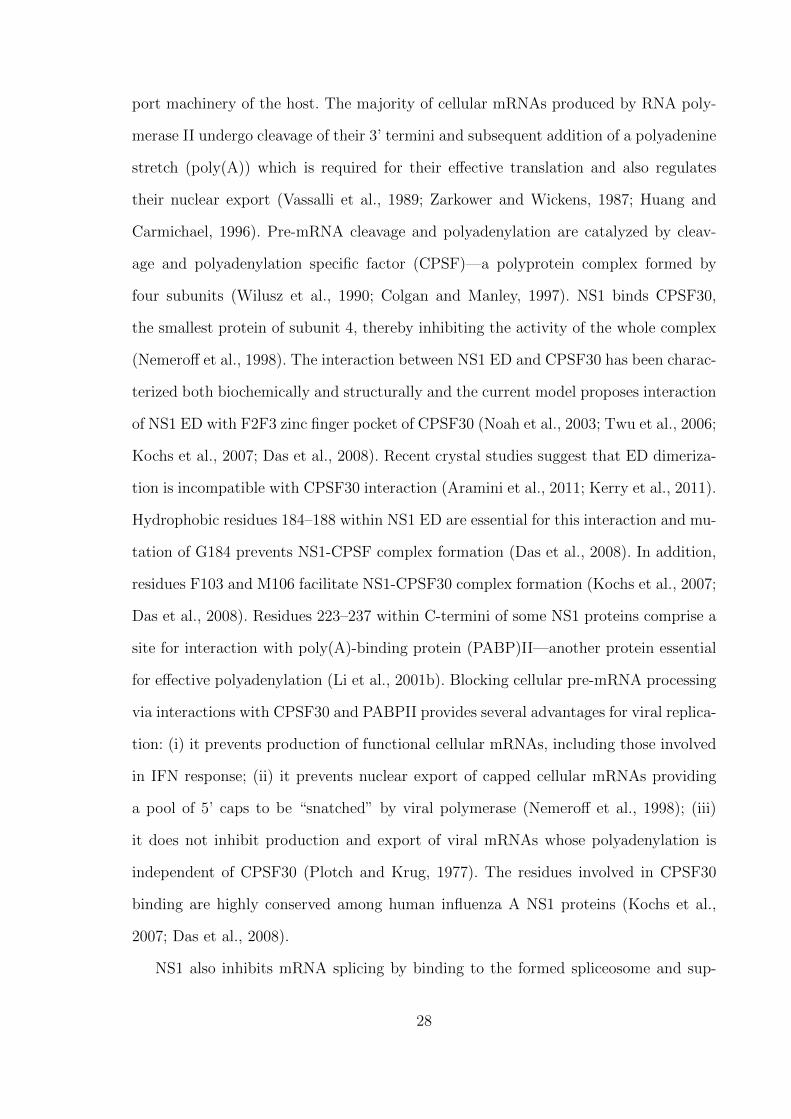
Figure 1. Influenza A virus replication cycle and cellular factors involved in it. The abbreviated hostfactors are: 𝛼-2,6-SA—𝛼-2,6-linked sialic acids; EGFR—epidermal growth factor receptor; COPI—coatomer 1 vesicular transport complex; vATPase, vacuolar H+-ATPase; Rab 5/7/8/10/11—small GTPases; Mcl-1—induced myeloid leukemia cell differentiation protein Mcl-1; NPC—nuclearpore complex; CRM1—exportin-1; HRB — HIV rev-binding protein; HSP40/70/90—heat shockprotein 40, 70 or 90 kDa; CK2—casein kinase 2; Rab 8/11—small GTPases; eIF4GI—eukaryoticinitiation factor 4 gamma 1; PABPI/II—polyadenylate-binding protein cytoplasmic isoforms I andII; GRSF1—G-rich sequence factor 1; TCP1, T-complex protein 1; NXF1—nuclear mRNA exportfactor 1; P15—mRNA export factor; Rae1—mRNA export factor 1; E1B-AP5—heterogeneousnuclear ribonucleoprotein U-like protein 1; MCM—minichromosome maintenance complex IREF-1; Tat-SF1—Tat-specific factor 1; UAP56—helicase UAP56; RNR—ribonucleotide reductase;ACC—acetyl-CoA carboxylase; FAS—fatty acid synthase; COX-2—cyclooxygenase 2; HMGCR—3-hydroxy-3-methylglutaryl-coenzyme A reductase; Raf/MEK/ERK—Ras/Raf/mitogen-activatedprotein kinase/extracellular signal-regulated kinase pathway; PI3K/Akt, phosphatidylinositol-3-kinase/RAC-alpha serine/threonine-protein kinase pathway; DHODH—dihydroorotate dehydro-genase.
the virus establishes during infection: accession of host-pathogen interaction database
(Kumar and Nanduri, 2010) yielded published associations of viral proteins with over
400 host factors (www.agbase.msstate.edu, accessed on 10.12.2014).
Influenza A replication triggers innate immune responses and therefore its effec-
tiveness is not limited to recruiting essential host factors but expands far beyond that.
10

Establishment of tight control over innate immune responses to infection is absolutely
critical for successful viral replication in an immune-competent system.
1.5 Innate immune responses to influenza A infection
In mammals influenza A is transmitted mainly through aerosols and droplets and en-
ters the host through the respiratory tract (Brankston et al., 2007). The first line of
antiviral defense in the respiratory tract is represented by the airway mucus. It consists
mainly of glycoproteins, antimicrobial and antiviral substances and is an essential bar-
rier for virus infection (Thornton et al., 2008; Nicholas et al., 2006). The viruses that
penetrate the airway mucus barrier initiate infections of the respiratory tract epithelial
cells and can also spread to immune cells of the respiratory tract, mainly macrophages
and dendritic cells (DCs) (Perrone et al., 2008; Bender et al., 1998). Infection of suscep-
tible cells with influenza A is rapidly detected by innate immune sensors that trigger
induction of antiviral gene expression and activation of pro-inflammatory responses.
Antiviral gene products are essential for restriction of viral replication and reduction
of virus burden, whereas pro-inflammatory responses are required for establishment
of inflammation (Iwasaki and Pillai, 2014). Furthermore, the onset of innate immune
responses is required for informing the adaptive immunity that regulates clearance of
the infection site and generation of immune memory (Iwasaki and Medzhitov, 2010).
Although innate responses involve a complex network of events that are hard to
tackle, in recent years substantial progress has been made towards the understanding
of critical processes that regulate virus detection, antiviral signaling and activation of
immune-related genes.
1.5.1 Virus recognition by innate immunity
Eukaryotic cells evolved a way to distinguish between “self” and “non-self” via ex-
pression of specific detection molecules called pattern recognition receptors (PRRs)
(Janeway and Medzhitov, 2002). These PRRs recognize specific molecular signatures
11

produced by invading microorganisms that are called pathogen-associated molecular
patterns (PAMPs) and initiate downstream signaling events to activate innate immune
responses (Janeway, 1989). The current paradigm of innate immunity to influenza A
virus assumes that during replication the virus produces three types of PAMPs. These
PAMPs are single- and double-stranded viral RNA and 5’ triphosphates generated dur-
ing viral genome synthesis by RdRp (Lund et al., 2004; Guillot et al., 2005; Hornung
et al., 2006; Kato et al., 2006). They are recognized in the endosome or in the cyto-
plasm by three major classes of cellular PRRs: NOD-like receptors (NLRs), Toll-like
receptors (TLRs), and RIG-I-like receptors (RLRs) (Iwasaki and Pillai, 2014).
Viral recognition in the endosome relies on three different TLR class members:
TLR3 which recognizes dsRNA, and TLR7 and TLR8 which recognize ssRNA (Fig. 2)
(Iwasaki and Pillai, 2014). TLR3 is constitutively expressed in pulmonary and airway
epithelial cells and in DCs (Guillot et al., 2005; Schulz et al., 2005; Ioannidis et al.,
2013). It has been initially shown to recognize dsRNA and induce interferon (IFN)
production in response to it (Alexopoulou et al., 2001; Guillot et al., 2005). TLR3
signaling is activated in response to replicating influenza A virus (Guillot et al., 2005).
TLR7 is expressed by airway epithelial cells and DCs and plasmocytoid DCs (Ioannidis
et al., 2013; Lund et al., 2004). TLR7 can be activated by ssRNA and is proposed to
recognize genomic vRNA of influenza A virus in the endosome (Diebold et al., 2004).
Unlike other TLRs, TLR8 has been so far only found in macrophages and monocytes
where it is activated in response to ssRNA and 5’ triphosphates (Ablasser et al., 2009).
TLR8 signaling is activated upon influenza A infection and leads to production of
interleukin (IL)-12, however its distinct role in regulation of innate immunity is yet to
be determined (Lee et al., 2013b).
The listed TLRs are expressed in endosomal compartments of the cells, except
TLR3, which was also found in the outer membrane (Diebold et al., 2004; Schulz et al.,
2005; Ablasser et al., 2009). The proposed mechanisms for their activation in response
to influenza A are, however, unclear for several reasons: (i) influenza A does not produce
12
detectable amounts of dsRNA during its replication due to activity of cellular RNA
helicase UAP56 (Wisskirchen et al., 2011); (ii) paired 5’ and 3’ ends of vRNA are
bound to viral RdRp, which can hinder 5’ triphosphates from their recognition by
TLRs (Arranz et al., 2012); (iii) TLR3 and TLR8 are dispensable for influenza A
recognition and only TLR7 seems to be critical for it (Lund et al., 2004); and (iv) it is
not clear whether TLR7 recognizes any specific structures or sequences within RNA,
as it was shown to become readily activated in response to both “self” and “non-self”
RNA (Diebold et al., 2004).
TLR7 and TLR8 interact with their common adapter MyD88 (Figure 2) (Medzhitov
et al., 1998). MyD88 recruits IRAK family kinases which mediate phosphorylation and
nuclear translocation of interferon regulatory factor (IRF)3 and IRF7—transcription
factors that induce IFN gene expression (Burns et al., 2003; Honda et al., 2005). In addi-
tion, MyD88 activates mitogen-activated protein kinases (MAPKs) signaling and tran-
scription factor activator protein 1 (AP-1), that controls expression of pro-inflammatory
genes (Kawai and Akira, 2007). TLR3 induces its signaling via interaction with TIR-
domain-containing adapter-inducing interferon-𝛽 (TRIF) and its downstream pathways
bifurcate (Guillot et al., 2005; Kumar et al., 2009). One of those induces type I IFN
production via TBK1 and inhibitor of nuclear factor kappa-B kinase (IKK) kinases and
IRF3 and IRF7. Another one stimulates production of pro-inflammatory cytokines via
IKK and nuclear factor kappa-light-chain-enhancer of activated B cells (NFkB) or via
MAPK signaling and transcription factor AP-1 (Guillot et al., 2005; Vercammen et al.,
2008).
Viral recognition in the cytoplasm relies on RLRs and NLRs. RLRs is a group of
helicases named after its representative retinoic acid inducible gene I (RIG-I). RLRs are
constitutively present in low amounts in multiple cell types, but their most prominent
location is airway epithelium (Bogefors et al., 2011) where they play an essential role
in detection of airborne pathogens. The RLR group consists of three proteins: RIG-I,
melanoma differentiation-associated protein 5 (MDA5) and laboratory of genetics and
13

physiology-2 (LGP-2) (Kang et al., 2004; Yoneyama et al., 2004; Yoneyama et al., 2005).
They are structurally similar and contain RNA-binding C-terminal domain (CTD) and
a DExD/H box helicase domain (Cui et al., 2008; Takahasi et al., 2009). RIG-I and
MDA5 also contain two consecutive N-terminal caspase recruitment domains (CARDs)
that mediate signaling (Yoneyama et al., 2004; Kang et al., 2004). All RLRs recognize
dsRNA, and RIG-I can also recognize ssRNA with 5’ triphosphates (Cui et al., 2008).
In the cytoplasm RIG-I is normally present in an inactive autorepressed state in
which its CARDs are sequestered by the helical domain, preventing non-specific induc-
tion of RIG-I downstream signaling (Figure 2) (Kowalinski et al., 2011). Upon sensing
its ligands by CTD, RIG-I undergoes conformational rearrangement which liberates
its CARDs for downstream signaling (Kowalinski et al., 2011). Activation of RIG-I
is dependent on its ubiquitination by E3 ubiquitin ligases TRIM25 and Riplet or on
binding to free polyubiquitin chains generated by TRIM25. Both TRIM25 and Riplet
are required for RIG-I signaling in vitro and in vivo (Gack et al., 2007; Oshiumi et
al., 2010; Zeng et al., 2010). The modified RIG-I oligomerizes (Patel et al., 2013) and
undergoes additional conformational rearrangements that enable interaction with its
adapter mitochondrial antiviral-signaling protein (MAVS) (Kawai et al., 2005; Seth
et al., 2005). For this, RIG-I is targeted to mitochondria in a “translocon” complex
containing TRIM25 and mitochondrial targeting chaperone 14-3-3𝜖 (Liu et al., 2012).
Upon binding RIG-I, MAVS oligomerizes and forms a scaffold for a multi-kinase sig-
naling complex which includes c-Jun N-terminal kinase (JNK), TANK-binding kinase
1 and IKK𝜖 complex, and IKK𝛼/𝛽/𝛾 complex (McWhirter et al., 2005). These kinases
eventually activate transcription factors IRF3, AP-1 and NFkB which regulate type I
IFN genes (McWhirter et al., 2005).
The only NLR that detects influenza A is LRR- and pyrin domain-containing pro-
tein 3 (NLRP3) found in lung and bronchial epithelial cells, monocytes, macrophages
and DCs (Guarda et al., 2011; Kim et al., 2014). It is constitutively present in an
inactive form in the cell cytoplasm. During influenza A infection NLRP3 is activated
14

Figure 2. Viral PAMPs detection by endosomal and cytoplasmic PRRs. TLRs 3, 7 and 8 recognizedsRNA in the endosome and induce downstream signaling via the adapter proteins MyD88 orTRIF. The signal transduction is mediated by protein kinases MAPK, IRAK4,1, TBK1/IKK andIKK𝛼/𝛽. RIG-I recognizes dsRNA and 5’triphosphorylated-ssRNA and undergoes conformationalchanges followed by its uniquitination by TRIM25 and Riplet. The modified RIG-I oligomerizesand translocates to mitochondrion where it triggers oligomerization of the adapter protein MAVS.MAVS facilitates signal transduction by protein kinases JNK, IKK𝜖, TBK1 and IKK𝛼/𝛽/𝛾. TLRand RIG-I signaling activates transcription factors AP-1, NF𝜅B, IRF3 and IRF7 which translocateto the nucleus and transcriptionally induce expression of interferons, interferon-stimulated genesand pro-inflammatory cytokines, thus activating the innate immune responses to influenza Ainfection.
by sensing viral ssRNA or proton flux mediated by viral M2 in trans Golgi network
(Thomas et al., 2009; Allen et al., 2009; Ichinohe et al., 2010). Virus-mediated activation
and oligomerization of NLRP3 leads to formation of the inflammasome—a multipro-
15

tein complex that includes NLRP3, apoptosis-associated speck-like protein containing
a CARD domain and pro-caspase 1 (Tschopp and Schroder, 2010). The inflammasome
is required for proteolytic self-activation of pro-caspase 1, which afterwards cleaves
IL1𝛽 and IL18 precursors, resulting in production of IL1𝛽 and IL18.
Whereas RIG-I activation results in induction of antiviral responses by IFNs and
interferon-stimulated genes (ISGs), signaling by NLRP3 and TLRs can also induce
pro-inflammatory responses (see section 1.5.3. for more details) (Le Goffic et al., 2007;
Allen et al., 2009; Kawai and Akira, 2007).
1.5.2 Antiviral responses by interferons and interferon-stimulated genes
Following detection of viral PAMPs and establishment of PRR signaling, the infected
cells produce and secrete small regulatory proteins known as interferons (Fensterl and
Sen, 2009). Interferons are subdivided into three types (I–III) based on their respective
receptors (Branca and Baglioni, 1981; Sheppard et al., 2003). Type I and III IFN signal-
ing activates ISGs and antiviral responses in autocrine and paracrine manner (Kotenko
et al., 2003; Garcıa-Sastre and Biron, 2006), reinforces PRR production (Pothlichet et
al., 2013), and regulates adaptive responses via enhancement of antigen presentation
to CD4+ and CD8+ T-cells (Zietara et al., 2009).
Both type I and III IFNs are secreted by nearly all cell types, although the majority
of them is secreted by DCs (Siegal et al., 1999; Odendall et al., 2014). Type I IFNs
include IFN𝛼 and IFN𝛽 and utilize dimeric receptor IFNAR1/IFNAR2 on cell surface
(Mogensen et al., 1999). Type III IFNs are IFN𝜆1, IFN𝜆2, IFN𝜆3 (also called IL29,
IL28A and IL28B, respectively), and IFN𝜆4. They bind to their heterodimeric receptor
IL10R2/IFNLR1 (Kotenko et al., 2003; Sheppard et al., 2003). Both type I and III IFN
receptors activate signaling through the Janus kinases (JAKs) / signal transducer and
activator of transcription (STAT) pathway (Figure 3). Binding of IFN to its receptor
triggers a series of phosphorylation events in which receptor-bound JAKs phosphory-
late themselves, the IFN receptor and receptor-associated proteins STAT1 and STAT2
16

(van Boxel-Dezaire et al., 2006). Phosphorylation of STAT1 and STAT2 triggers their
heterodimerization and formation of regulatory complex with IRF9 (Fu et al., 1990).
This complex is translocated to the nucleus where it transcriptionally activates ISGs
(Levy et al., 1988). A schematic illustration of IFN signaling is presented in Figure 3.
Figure 3. A schematic representation of type I and type III IFN responses. Interaction of typeI and type III interferons with their corresponding receptors results in autophosphorylation ofreceptor-associated JAK kinases JAK1 and TYK2, which phosphorylate STAT1 and STAT2 pro-teins. Phosphorylated STAT1 and STAT2 form heterodimers that interact with regulatory proteinIRF9. The STAT1-STAT2-IRF9 complex translocates to the nucleus where it transcriptionallyactivates interferon-stimulated genes. Interferon-stimulated gene products inhibit viral entry anduncoating, suppress protein synthesis in infected cell, regulate degradation of vRNAs and controlinterferon signaling via feedback loops.
ISGs are a diverse group of genes that control multiple cellular processes: they
enhance virus sensing by PRRs, target pathways essential for viral replication, upregu-
late cytokine and chemokine production, control IFN response via positive and negative
feedback loops. The best-described ISGs with antiviral action include myxovirus resis-
tance gene product (MxA), interferon-inducible transmembrane protein 3 (IFITM3),
cholesterol 25-hydroxylase, ubiquitin-like protein ISG15, protein kinase R (PKR), and
2’-5’-oligoadenylate syntethase (OAS) (Sadler and Williams, 2008).
IFITM3 is localized in late endosomes and exerts its antiviral activity during virus
entry. It alters properties of the endosomal membrane and prevents viral fusion (Desai
17

et al., 2014). Indeed, in cells overexpressing IFITM3 virus-like particles bearing 𝛽-
lactamase do not fuse in late endosomes and no cytoplasmic 𝛽-lactamase activity can
be detected (Desai et al., 2014).
Human MxA gene product is a dynamin-like guanosine triphosphatase (Nakayama
et al., 1992). It is localized in the cytoplasm of infected cells where it self-assembles
into ring-like ordered structures (Gao et al., 2010). Although the detailed mechanism
of its antiviral activity is yet do be established, MxA oligomers presumably recognize
and bind vRNPs in the cytoplasm, preventing their nuclear import and thus attenuate
influenza A replication (Haller et al., 2010).
PKR and OAS act at later stages of viral replication cycle. PKR is a multifunctional
serine/threonine kinase that has a critical role in host antiviral responses (Garcıa et
al., 2006). It is constitutively present in the cytoplasm at low abundance, and its
expression is trancsriptionally induced by type I and III IFNs (Meurs et al., 1990). PKR
is activated via its interaction with dsRNA or protein activator of the interferon-induced
protein kinase (Li et al., 2006b). It phosphorylates and inactivates translation initiation
factor 2𝛼 (eIF2𝛼) (Levin and London, 1978), which is indispensable for initiation of cap-
and often of internal ribosome entry site (IRES)-driven translation. Thus, activation
of PKR in response to virus infection shuts down protein synthesis (Kimball, 1999).
In addition to controlling translation in infected cells, PKR can also detect dsRNA,
mediate IFN𝛾-induced NFkB activation (Deb et al., 2001), modulate STAT1 signaling
(Wong et al., 1997), induce JNK and MAPK signaling in response to viral infection
(Chu et al., 1999), induce apoptosis and autophagy following the shut down of protein
synthesis (Gil and Esteban, 2000; Talloczy et al., 2002). PKR mediates death of infected
cells unless the virus counteracts its action (Takizawa et al., 1996; Hatada et al., 1999).
As no cytoplasmic dsRNA is detectable during influenza A replication (Wisskirchen et
al., 2011), PKR activation during infection is likely mediated by TLR signaling (Jiang
et al., 2003) or by binding to its protein activator (Garcıa et al., 2006).
OAS and ribonuclease L (RNAse L) act together in the antiviral RNA decay path-
18

way. Both enzymes are upregulated by IFN, but they are also present constitutively in
cell cytoplasm (Sadler and Williams, 2008). Like PKR, OAS can be activated by bind-
ing to dsRNA (Castelli et al., 1998). Upon its activation, OAS synthesizes 2’-5’-linked
adenosine triphosphate oligomers, which, in turn, act as inducers of latent RNAse
L (Rebouillat and Hovanessian, 1999). Activated RNAse L catalyzes endonucleolytic
degradation of cellular and viral ssRNAs and mRNAs thus contributing to host an-
tiviral responses (Dyer and Rosenberg, 2006). In addition to viral RNA elimination,
RNAse L reinforces virus detection by TLRs and RIG-I, supports IFN response, and
regulates apoptosis in infected cells (Liang et al., 2006). Replication of influenza A
virus that is unable to inhibit OAS/RNAse L pathway is attenuated (Min and Krug,
2006).
Some ISGs are also implicated in interferon response control via negative feedback
loops (Schneider et al., 2014). For example, SOCS proteins inhibit JAK/STAT signaling
(Hong and Carmichael, 2013) and USP18 binds to IFNAR2 and inhibits it (Ritchie et
al., 2004).
1.5.3 Pro-inflammatory responses
Pro-inflammatory responses induced by TLR and NLRP3 signaling activate production
of chemokines including IL1-𝛽, IL6, IL8, IL18, RANTES, MCP-1, MCP-3 and MIP-3𝛼
(Julkunen et al., 2000; Le Goffic et al., 2007). Unlike type I IFNs, pro-inflammatory
cytokines may not induce direct antiviral resistance, but are required for antigen presen-
tation, establishment of inflammation, recruitment of leukocytes to the site of infection
and proliferation of CD8+ cytotoxic T lymphocytes (CTLs) (Van Der Sluijs et al.,
2005; Schulz et al., 2005; Le Goffic et al., 2006). Consequently, they do not inhibit viral
replication, but are essential for the host resistance to infection (Pang et al., 2013). In
addition, the correct onset of pro-inflammatory responses regulates adaptive immunity
to influenza A infection (Trinchieri, 2003; Ichinohe et al., 2009). However, robust in-
duction of pro-inflammatory response to some influenza A subtypes, e.g. H5N1, can be
19

detrimental to the host and lead to severe immunopathology, thereby increasing viral
pathogenicity (La Gruta et al., 2007).
1.6 Apoptosis
In addition to activating antiviral and pro-inflammatory responses, influenza A was
shown to induce apoptosis in infected cells (Fesq et al., 1994; Hinshaw et al., 1994;
Mori et al., 1995; Brydon et al., 2005). The central role in apoptotic response belongs
to cysteinyl proteases (caspases) which proteolytically inactivate numerous cellular
proteins leading to cell death (Cohen, 1997; Thornberry and Lazebnik, 1998)
A variety of cellular signaling pathways, including MAPK, NFkB, PKR and phospho-
inisitide-3-kinase/Akt, are implicated in apoptosis regulation during infection (Gil and
Esteban, 2000; Xing et al., 2010; Lu et al., 2010). Activation of apoptosis can play a
role in host defense via facilitating premature cell death and also via triggering pro-
duction of pro-inflammatory cytokines (Julkunen et al., 2000). It also contributes to
viral clearance in cell culture and in vivo being essential for CD8+-mediated killing of
infected cells (Ishikawa et al., 2005; Brincks et al., 2008). On the other hand, onset of
apoptic responses can be beneficial for the virus and influenza A propagation is atten-
uated in the presence of caspase inhibitors due to retention of vRNPs in the nucleus
(Wurzer et al., 2003). Due to this twofold role of apoptosis in influenza A infection, it
is tightly controlled by both cellular and viral factors.
1.7 Viral counteraction to innate responses. NS1.
Successful counteraction to antiviral responses is critical for influenza A replication
and several viral proteins support overcoming of intrinsic barriers and replication in
the context of immune response. For instance, NA cleaves sialic acids in host mucus to
facilitate viral penetration (Cohen et al., 2013), PB1-F2 inactivates RIG-I/MAVS and
NFkB signaling (Varga et al., 2011; Dudek et al., 2011; Reis and McCauley, 2013), and
20
NP mediates resistance to MxA (Dittmann et al., 2008). Both PB1-F2 and M2 can
contribute to regulation of apoptosis during infection (Herold et al., 2012).
However, the critical role in inhibition of antiviral responses and regulation of virus-
host interactions is assigned to NS1 (Garcia-Sastre et al., 1998). NS1 is a non-structural
protein, but it is expressed in high quantities in infected cells (Ritchey et al., 1976).
Initial studies indicated that NS1 is essential for viral replication (Koennecke et al.,
1981) and further investigations proved that it is a versatile viral protein which is a
key regulator of influenza A virus-host interactions (Ayllon and Garcia-Sastre, 2015).
Indeed, viruses lacking functional NS1 are severely attenuated, especially in immune-
competent systems, and can only replicate in the absence of STAT1 or PKR (Garcia-
Sastre et al., 1998; Egorov et al., 1998; Donelan et al., 2003; Falcon et al., 2004). Due
to its critical role in viral replication NS1 has been extensively studied and its roles
in regulation of virus-host interactions stretch beyond regulation of IFN responses
and include regulation of vRNA synthesis, enhancement of viral protein production,
regulation of virion assembly, modulation of cellular signaling, apoptosis inhibition,
contribution to host range definition and pathogenesis.
1.7.1 NS1 synthesis and localization
The mRNA of NS1 is generated by colinear transcription of the 8th genomic segment
(NS). About 10 % of NS transcripts are spliced and generate the mRNA of another
viral protein, NEP (Lamb et al., 1980), which shares the first 10 amino acids with NS1
(Inglis et al., 1979; Lamb and Choppin, 1979; Lamb et al., 1980). As NS1 is not found
in virions, it appears in infected cells only after viral transcripts have been generated
and translated. Although intracellular localization of NS1 may vary depending on its
abundance, the virus strain, cell type and polarity, and time post infection, the majority
NS1 is localized in the cellular nucleus, but a fraction of it is also present in the
cytoplasm (Greenspan et al., 1988; Li et al., 1998; Melen et al., 2007; Newby et al.,
2007; Melen et al., 2012).
21

Generally, proteins of under 30 kDa smoothly diffuse through the nuclear pore com-
plex channel and do not require specific NLSs, whereas proteins of 40–60 kDa usually
are delivered to the nucleus via active import (Macara, 2001; Wang and Brattain, 2007;
Ma et al., 2012). NS1 is a relatively small protein and its molecular mass is only 26 kDa
(Ward et al., 1994), however, its nuclear import occurs in an active way. Depending
on the virus subtype, NS1 can contain one or two NLSs which mediate interaction
with cellular importin 𝛼 (Melen et al., 2007), thus securing rapid nuclear import of
NS1 (Privalsky and Penhoet, 1981). The monopartite NLS1 is located close to the
protein N-terminus, involves amino acids (aas) R35, R38 and K41, and is conserved
across most influenza A isolates. The bipartite C-terminal NLS2 is present in a subset
of viral strains expressing extended 237 aa NS1. It is located around aa 219–237 and
also serves as a nucleolar localization sequence (Melen et al., 2007; Melen et al., 2012).
Cytoplasmic localization of NS1 seems to depend on nuclear export sequence (NES)
which lies within residues 138–147 (Li et al., 1998). This NES, however, is masked by
adjacent residues 148–161 and its activation requires “unmasking” which presumably
occurs via the conformational change upon interaction of NS1 with and unidentified
protein partner(s).
1.7.2 Post-translational modifications of NS1
Initial studies indicated that a large portion of NS1 is phosphorylated during infection
and this phosphorylation occurs in the nucleus (Privalsky and Penhoet, 1981). Four
residues within NS1 can be phosphorylated—S42, S48, T197 and T215—although their
phosphorylation may be virus subtype specific (Petri et al., 1982). Phosphorylation of
NS1 at S48 by protein kinase A, at T197 by an unidentified kinase and at T215 by
cyclin-dependent kinase 5 and extracellular signal-regulated kinase 2 do not seem to
affect viral replication and roles of these modifications need further elucidation (Hale
et al., 2009; Hutchinson et al., 2012; Hsiang et al., 2012). Phosphorylation at S42
by protein kinase C alpha is proposed to attenuate viral replication presumably via
22

impairing the nucleic acid binding function of NS1 (Hsiang et al., 2012).
NS1 can be also modified by linkage of small ubiquitin-like modifier protein (SUMO)—
a small regulatory protein that affects activity, stability, localization and interactions
of its targets (Pal et al., 2010; Johnson, 2004). NS1 extensively interacts with the
cellular SUMOylation system in the nucleus and can be modified with three SUMO
isoforms—SUMO1 and SUMO2/3 (Pal et al., 2011; Santos et al., 2013). This modifica-
tion is subtype-specific: NS1 from some, but not all H5N1, H9N2 and H1N1 influenza
A viruses can be SUMOylated (Xu et al., 2011). SUMOylation sites are lysines 219
and 221 in NS1 C-terminus (Xu et al., 2011). So far only two studies have addressed
the functional role of this modification. They indicate that it regulates NS1 stability,
abundance of NS1 dimers and trimers and may facilitate immunomodulatory functions
of NS1 (Xu et al., 2011; Santos et al., 2013).
Another modification of NS1 that occurs in infected cells is conjugation of a small
ubiquitin-like protein ISG15, that is produced in response to various stress stimuli in-
cluding influenza A infection (Pitha-Rowe and Pitha, 2007; Sadler and Williams, 2008;
Hsiang et al., 2009). ISG15 conjugation to NS1 by its IFN-induced ligases Ube1L,
UbcH8 and Herc5 occurs primarily at lysines 41, 126, 217 and 219 (Zhao et al., 2010;
Tang et al., 2010). It inhibits NS1 dimerization, interaction with PKR and with im-
portin 𝛼, alleviates NS1-mediated inhibition of cytokine production by the infected
cell and attenuates viral growth kinetics (Zhao et al., 2010; Tang et al., 2010). Like
other modifications of NS1, ISGylation is strain-specific: avian NS1s differ from human
in their ISGylation profiles (Tang et al., 2010). Experiments with recombinant human
H3N2 influenza A virus showed that mutation K41R in NS1 renders ISGylation inef-
ficient without compromising NS1 functions and hence acquisition of such mutations
may be beneficial for the virus (Zhao et al., 2010).
23

1.7.3 Structure of NS1
NS1 is a relatively small protein of 219–237 aa depending on the virus strain (Hale et al.,
2008c). It is subdivided in four regions: the N-terminal RNA-binding domain (RBD),
the inter-domain linker, the effector domain (ED) and a disordered C-terminal “tail”,
for which no crystal structure is available (Figure 4A) (Hale, 2014). Several structural
studies have provided detailed information on the organization of NS1 domains, the
full-length protein and on the structural polymorphisms of NS1s from different viral
sybtypes (Chien et al., 1997; Liu et al., 1997; Wang et al., 1999; Bornholdt and Prasad,
2006; Yin et al., 2007b; Hale et al., 2008a; Cheng et al., 2009; Xia et al., 2009; Kerry
et al., 2011; Carrillo et al., 2014). As implied by their names, the RBD of NS1 interacts
with the RNA, whereas ED accommodates the majority of interaction sites with NS1
cellular partners (Hale et al., 2008c).
Figure 4. Crystal structure of H5N1 NS1 R38A, K41A mutant protein (Bornholdt and Prasad,2008). (A) NS1 monomer. The three 𝛼-helices of RBD are marked as 1, 2 and 3 (B) NS1 dimer.Groove-forming 𝛼-helices of distinct RBD’s are marked as 2 and 2’. Residues 38 and 41 are shownin red. (C,D) Oligomerized NS1. The dimers are marked with distinct colors, residues 38 and 41shown in red can be seen inside the transparent helix.
24

RBD comprises the first 73 N-terminal aa of NS1 (Qian et al., 1995a; Yin et al.,
2007b). About 80 % of its residues are organized into three positively charged 𝛼-helices
(Qian et al., 1995a; Liu et al., 1997). RBD itself forms highly stable dimers in which
anti-parallel 𝛼-helices 2 and 2’ of corresponding monomers form the groove in which
RNA can be accommodated (Figure 4B) (Chien et al., 1997; Wang et al., 1999). As
a dimer NS1 can bind ss- and, with higher affinity, dsRNA in a sequence-unspecific
manner (Hatada and Fukuda, 1992; Qian et al., 1995b; Chien et al., 1997). Residue
R38 is critical and residue K41 is important for both RBD dimerization and NS1 ability
to interact with RNA (Hatada and Fukuda, 1992; Wang et al., 1999).
Inter-domain linker in most cases is comprised of residues 74-84, but its length may
vary between viral subtypes, contributing to NS1 structural polymorphism (Bornholdt
and Prasad, 2006; Carrillo et al., 2014; Kerry et al., 2011).
ED in most viral subtypes encompasses residues 88–202 (Hale, 2014). It comprises
seven 𝛽-strands and three 𝛼-helices and can also homodimerize (Bornholdt and Prasad,
2006; Hale et al., 2008a; Xia et al., 2009). The dimerization occurs primarily via helix-
helix interface, in which strictly conserved residue T187 plays a critical role (Hale et al.,
2008a; Kerry et al., 2011). Unlike stable RBD dimerization, the interactions between
ED monomers are likely to be transient (Kerry et al., 2011; Hale, 2014).
Dimerization is important for NS1 function and NS1 monomers have not been ob-
served in vitro or in vivo (Hale, 2014). The crystal structure of NS1 dimer suggests
that its formation relies on the stable interactions between RBDs of NS1 monomers
whereas EDs are not directly involved in dimerization and are probably free for inter-
actions with cellular proteins (Figure 4B) (Bornholdt and Prasad, 2008). Interestingly,
the full-length protein not only can dimerize, but also may form hollow helices (Born-
holdt and Prasad, 2008). The formation of such oligomers is mediated by inter-NS1
interactions of both RBD and ED (Figure 4C, D) (Bornholdt and Prasad, 2008; Car-
rillo et al., 2014). In addition, full-length NS1 retains conformational plasticity with
three possible orientations of ED to RBD. Preference for certain states is dependent
25

on NS1 inter-domain linker length, residue 71 and a mechanical hinge, and determines
strain-specific variations in NS1 structure and function (Carrillo et al., 2014).
1.7.4 Inhibition of interferon signaling at pre-transcriptional level
Inhibition of the interferon response is a function of NS1. The mechanisms behind
this function have been extensively studied over the past two decades and according to
the current paradigm NS1 subverts development of immune responses by counteracting
PRR signaling, co- and post-transcriptional inhibiting of host gene expression and post-
translationally inactivating interferon-stimulated gene products (Ayllon and Garcia-
Sastre, 2014). For this, the multi-functional NS1 targets numerous factors, which are
discussed below. The mapped interactions of NS1 are schematically shown in Figure 5.
Figure 5. Schematic representation of NS1 and its described interactions. NS1 length is 219–237amino acids, depending on the viral subtype. Residues 1–73 comprise an RNA-binding domain(RBD), residues 85-202 comprise an effector domain (ED) and residues 202–219/230/237 com-prise a C-terminal “tail”. NS1 contains two nuclear localization signals (NLS1 and NLS2) and anuclear export signal (NES). NS1 is thought to interact with RNA, RIG-I, TRIM25 and Ripletto alleviate RIG-I signaling, with PABPII and CPSF30 to inhibit cellular mRNA processing, withPKR, eIF4GI and PABPI to facilitate viral protein synthesis, with nuclear pore complex compo-nents NXFI, p15, Rae1 and E1B-AP5 to inhibit nuclear export of cellular mRNAs, with p85𝛽,PDZ-domain containing proteins, Crk and Crk-like proteins to regulate cellular signaling.
NS1 inhibits interferon signaling at the pre-transcriptional level by preventing ac-
tivation and nuclear translocation of IRF3, AP-1, NFkB mainly via alleviating RIG-I
signaling (Talon et al., 2000; Ludwig et al., 2002; Wang et al., 2000; Geiss et al., 2002;
Munir et al., 2012). Multiple studies indicate that NS1 employs both its RBD and ED
26

to subvert RIG-I signaling at multiple steps (Wang et al., 2000; Ludwig et al., 2002;
Haye et al., 2009; Tisoncik et al., 2011). NS1 binds RIG-I, although direct inhibitory
effects of this interaction have not been reported yet (Opitz et al., 2007; Mibayashi
et al., 2007). It also established inhibitory interactions with two indispensable RIG-I
regulators: TRIM25 and Riplet (Gack et al., 2009; Rajsbaum et al., 2012). Interaction
with TRIM25 requires E96, E97 residues within NS1 ED and RNA-binding residues
R38, K41, although it is not clear whether the latter are involved in interaction with
TRIM-25 (direct or RNA-mediated) or just support suitable NS1 conformation (Gack
et al., 2009). Interaction with Riplet requires R38, K41 although again their exact roles
in this interaction still need clarification (Rajsbaum et al., 2012). The involvement of
R38 and K41 residues in regulation of RIG-I has raised discussions of another possi-
ble mechanism of RIG-I inhibition by NS1 in which NS1 sequesters dsRNA, a known
RIG-I inducer, thereby preventing activation of the RIG-I signaling axis. The role of
NS1 RNA-binding in pre-transcriptional control of immune responses, however, needs
to be elucidated, because (i) influenza A does not seem to generate dsRNA during its
replication (Wisskirchen et al., 2011) and (ii) the affinity of NS1 for dsRNA is much
lower than that of RIG-I (Chien et al., 2004; Yin et al., 2007a; Vela et al., 2012).
In addition to inhibition of RIG-I signaling, NS1 has evolved several other ways
to effectively inhibit interferon induction at the transcriptional level. It subverts both
canonical and non-canonical NFkB pathways (Ruckle et al., 2012) and prevents nuclear
translocation of NFkB via direct inhibition of alpha and beta subunits of IKK (Gao
et al., 2012). NS1 impairs c-Jun and JNK signaling, preventing AP-1-regulated gene
expression (Ludwig et al., 2002). It also alleviates IFN response by inducing suppressor
of cytokine signaling-3, a negative regulator of JAK-STAT signaling (Pauli et al., 2008).
1.7.5 Inhibition of interferon signaling at post-transcriptional level
NS1 acts beyond pre-transcriptional level and controls development of antiviral re-
sponses also post-transcriptionally by targeting pre-mRNA processing and nuclear ex-
27
port machinery of the host. The majority of cellular mRNAs produced by RNA poly-
merase II undergo cleavage of their 3’ termini and subsequent addition of a polyadenine
stretch (poly(A)) which is required for their effective translation and also regulates
their nuclear export (Vassalli et al., 1989; Zarkower and Wickens, 1987; Huang and
Carmichael, 1996). Pre-mRNA cleavage and polyadenylation are catalyzed by cleav-
age and polyadenylation specific factor (CPSF)—a polyprotein complex formed by
four subunits (Wilusz et al., 1990; Colgan and Manley, 1997). NS1 binds CPSF30,
the smallest protein of subunit 4, thereby inhibiting the activity of the whole complex
(Nemeroff et al., 1998). The interaction between NS1 ED and CPSF30 has been charac-
terized both biochemically and structurally and the current model proposes interaction
of NS1 ED with F2F3 zinc finger pocket of CPSF30 (Noah et al., 2003; Twu et al., 2006;
Kochs et al., 2007; Das et al., 2008). Recent crystal studies suggest that ED dimeriza-
tion is incompatible with CPSF30 interaction (Aramini et al., 2011; Kerry et al., 2011).
Hydrophobic residues 184–188 within NS1 ED are essential for this interaction and mu-
tation of G184 prevents NS1-CPSF complex formation (Das et al., 2008). In addition,
residues F103 and M106 facilitate NS1-CPSF30 complex formation (Kochs et al., 2007;
Das et al., 2008). Residues 223–237 within C-termini of some NS1 proteins comprise a
site for interaction with poly(A)-binding protein (PABP)II—another protein essential
for effective polyadenylation (Li et al., 2001b). Blocking cellular pre-mRNA processing
via interactions with CPSF30 and PABPII provides several advantages for viral replica-
tion: (i) it prevents production of functional cellular mRNAs, including those involved
in IFN response; (ii) it prevents nuclear export of capped cellular mRNAs providing
a pool of 5’ caps to be “snatched” by viral polymerase (Nemeroff et al., 1998); (iii)
it does not inhibit production and export of viral mRNAs whose polyadenylation is
independent of CPSF30 (Plotch and Krug, 1977). The residues involved in CPSF30
binding are highly conserved among human influenza A NS1 proteins (Kochs et al.,
2007; Das et al., 2008).
NS1 also inhibits mRNA splicing by binding to the formed spliceosome and sup-
28
pressing its catalytic activity (Lu et al., 1994; Qiu et al., 1995). Interestingly, this effect
is specific to host mRNAs. Although viral mRNAs recruit cellular spliceosome for their
post-transcriptional processing, NS1 does not affect splicing of its own mRNA (Robb
et al., 2010) and has little, if any, effect on M mRNA splicing (Salvatore et al., 2002;
Robb and Fodor, 2012). The possible reason for such selectivity could be recognition
of specific motifs within viral mRNAs by NS1, which, however, is questionable, since
no sequence specificity is so far known for NS1 RNA binding. Another possibility is
recruitment of different spliceosomal factors to viral transcripts by viral polymerase
(Fournier et al., 2014).
In addition to its direct effects on mRNA synthesis and processing, NS1 also in-
hibits nuclear export of cellular mRNAs. It specifically binds to nuclear pore complex
components NXF1, p15, Rae1 and E1B-AP5, thus contributing to retention of cellular
mRNAs in the nucleus (Satterly et al., 2007). Importantly, inhibition of nuclear pore
complex by NS1 does not attenuate viral replication, as export of viral RNAs relies on
the alternative Crm1-mediated pathway (Neumann et al., 2000).
The combination of NS1 effects on host mRNAs production, processing and ex-
port contributes to host protein synthesis shut-off which is commonly observed during
influenza A infection (Beloso et al., 1992).
1.7.6 Direct inhibition of interferon-stimulated gene products
In addition to the control at pre-transcriptional, transcriptional and post-transcriptional
level, NS1 antagonizes IFN responses by directly targeting PKR and OAS.
Influenza A mRNAs are structurally indistinguishable from cellular mRNA and
hence production of viral proteins requires functional cap-dependent translation. For
this, the virus prevents activation of the negative translational regulator PKR (Katze
et al., 1986; Katze et al., 1988). Inhibition of PKR is to a large extent a function of NS1,
as viruses lacking functional NS1 can only replicate in the absence of PKR (Bergmann
et al., 2000). NS1 was proposed to prevent activation of PKR by binding to dsRNA and
29

thus sequestering it away from PKR (Lu et al., 1995). Such regulation, however, seems
controversial for several reasons: (i) while PKR senses dsRNA in the cytoplasm, the site
of influenza A RNA transcription and replication is the nucleus, and thus the presence
of virus replication intermediates in the cellular cytoplasm seems unlikely (Jackson et
al., 1982), (ii) viral genomes are exported to the cytoplasm as vRNPs, and thus base-
paired regions of vRNA are likely to be inaccessible to PKR (Coloma et al., 2009), (iii)
the affinity of NS1 for dsRNA is much lower than that of PKR (Chien et al., 2004;
Husain et al., 2012), and (iv) dsRNA binding function of NS1 is not required for PKR
inhibition (Li et al., 2006a). NS1 has been shown to form a complex with PKR which
appears to be inhibitory for PKR activation (Tan and Katze, 1998; Li et al., 2006a).
It has been shown that residues 123–127 within NS1 are required for interaction with
PKR and its inhibition (Min et al., 2007).
Inhibition of OAS/RNAse L pathway is also a function of NS1. So far only one
study has described the putative mechanism of NS1 control over the OAS/RNAse L
pathway which presumes that dsRNA binding by NS1 is required for sequestration
of dsRNA away from OAS (Min and Krug, 2006). This observation is supported by
low affinity of OAS to dsRNA (Hartmann et al., 2003), however, the abundancy of
influenza-generated dsRNA in cell cytoplasm still remains an open question.
1.7.7 Other pro-viral functions of NS1
In addition to its critical role in control of innate immune responses, NS1 targets a
number of other cellular factors to facilitate virus replication. These additional pro-viral
functions of NS1 include regulation of production of viral RNA and protein synthesis,
control of apoptosis and modulation of cell signaling.
Influenza A virus RNA production occurs in two phases: early, when NS1 and NP
vRNAs are preferentially synthesized, and late, when all eight segments are produced
in equimolar quantities (Skehel, 1973; Shapiro et al., 1987). This temporal regulation
of vRNAs production has been shown to require functional NS1 (Falcon et al., 2004)
30

and is linked to residues 123 and 124 within its ED (Min et al., 2007). Although these
residues overlap with the PKR binding site on NS1, NS1 regulates vRNA independently
of its interaction with PKR, which has been proven using PKR-deficient mice (Min
et al., 2007). NS1 through an as yet unknown mechanism also specifically regulates
production of HA vRNA (Maamary et al., 2012). The regulation of vRNA production
by NS1 is likely linked to its interaction with vRNPs which disrupts inhibitory binding
of cellular helicase DDX21 to PB1 (Marion et al., 1997; Chen et al., 2014).
NS1 has been also discussed as a factor that regulates protein synthesis in infected
cells. As viral mRNAs contain 5’meG “cap” and 3’ poly(A), they are structurally in-
distinguishable from cellular mRNAs and require the same subset of translation factors
(Poch et al., 1989; Poon et al., 1999). The bottleneck of the complex translation pro-
cess is its initiation which requires coordinated assembly of translation factors and
43S pre-initiation complex on mRNA followed by start codon scanning and ribosome
assembly (Pestova et al., 2001). In addition to the prevention of eIF2𝛼 inhibition by
PKR described in section 1.7.6, NS1 contributes to translational control by direct in-
teractions with translation initiation factors. Thus, fragment 1–81 has been shown to
bind the translation initiation factor 4GI (eIF4GI) via protein-protein interactions and
is supposed to facilitate recruitment of this factor onto viral mRNAs (Aragon et al.,
2000). In addition, it interacts with PABPI, another factor essential for translation
initiation (Burgui et al., 2003). The proposed mechanism of translational regulation by
NS1 suggests preferential recruitment of translation initiation factors on viral mRNAs
due to specific recognition of their 5’ untranslated region (UTR) by NS1 (de la Luna
et al., 1995). Such a mechanism, however, is in controversy with the very low affinity
of NS1 to ssRNA and with its proposed nonsequence-specific binding to RNA (Hatada
and Fukuda, 1992) and requires further description of NS1 interactions with translation
factors and/or search for possible specificity of NS1 towards viral mRNAs.
Influenza A infection induces prolonged activation of PI3K and its downstream effec-
tor Akt several hours post infection due to NS1 activity (Ehrhardt et al., 2006; Ehrhardt
31
and Ludwig, 2009). Structural information suggests that NS1 ED forms heterotrimeric
complex with regulatory (p85) and catalytic (p110) subunits of PI3K. Interaction with
NS1 alters positions of p85 and p110 in respect to each other and results in PI3K/Akt
signaling activation (Hale et al., 2008b; Hale et al., 2010a). The interaction between
NS1 and p85 is dependent on the 164PxxP1167 Src-homology 3 domain (SH3) motif
and a conservative tyrosine 89 within NS1 ED (Hale et al., 2006; Shin et al., 2007a;
Shin et al., 2007b). As PI3K/Akt signaling inhibits apoptosis, its activation by NS1
was initially regarded as a mechanism to prevent premature cell death and secure viral
replication (Ehrhardt et al., 2007; Zhirnov and Klenk, 2007). However, p85-binding de-
ficient NS1 also exibits anti-apoptotic activity indicating possible redundancy in NS1
approaches to control apoptosis (Jackson et al., 2010a).
In addition to the PI3K/Akt branch, NS1 also controls the JNK branch of apoptosis.
This control is realized through binding of SH3 domains present in some NS1 subtypes
to cellular adapter proteins Crk and Crk-like (Heikkinen et al., 2008; Hrincius et al.,
2010).
Another way by which NS1 protects infected cells from apoptosis could be its ability
to bind to signaling proteins that contain a domain called PDZ (postsynaptic density
protein 95, Drosophila disc large tumor suppressor, and zonula occludens 1 protein)
(Liu et al., 2010). The C-terminus of 230 aa long avian NS1s can contain a PDZ-binding
motif ESEV/EPEV, which mediates interaction with PDZ-domain containing proteins
(Golebiewski et al., 2011). In addition to apoptosis regulation, PDZ binding may pro-
vide control over formation of cellular junctions and establishment of cell polarity,
although these possibilities, as well as the overall effect of PDZ-binding on influenza A
virulence are yet to be investigated (Jackson et al., 2010b; Zielecki et al., 2010; Javier
and Rice, 2011).
32
1.7.8 NS1 diversity
NS1 contributes to influenza A variability by displaying a substantial number of se-
quence, length, structure, modification and functional polymorphisms. Initial studies of
sequence similarity revealed two groups of NS1 proteins designated as alleles A and B
(Scholtissek and von Hoyningen-Huene, 1980). Allele A NS1s have been found in avian,
as well as in mammalian strains, whereas allele B appears to be exclusively avian indi-
cating the contribution of NS1 to host-specificity (Treanor et al., 1989; Ludwig et al.,
1991). The percentage of sequence identity is over 90 % within alleles, but between
alleles it is below 70 % (Scholtissek and von Hoyningen-Huene, 1980).
Structural polymorphisms of NS1 are mainly dependent on the length of inter-
domain linker. A recent study has shown that the linker length and residue 71 determine
the preference of RBD orientation towards ED and provide the structural basis for
strain-dependent NS1 functions (Carrillo et al., 2014). Interestingly, whereas the inter-
domain linker is 12 amino acid long in the majority of NS1s, its length in highly
pathogenic avian influenza A viruses is only 7 amino acids, indicating that although
there are no known interaction motifs within the linker, it still can determine virulence
(Long et al., 2008).
Although functional NS1 is required for replication in immune-competent systems
(Garcia-Sastre et al., 1998; Donelan et al., 2003), NS1s from different subtypes may
vary in their ability to modulate certain cellular responses. For example, although
different NS1s are able to bind components of the RIG-I pathway, the extent to which
they inhibit RIG-I signaling varies and some H1N1, H2N2 and human H3N2 viruses
fail to pre-transcriptionally block IFN induction (Kochs et al., 2007; Haye et al., 2009;
Munir et al., 2011; Munir et al., 2012). These viruses compensate for their inefficiency
in pre-transcriptional control with strong CPSF30 binding, whereas the mouse-adapted
PR8 strain as well as the pandemic H1N1/2009 (pH1N1/2009) acquired mutations that
prevent interactions with CPSF30 (Kochs et al., 2007; Hale et al., 2010c). Human and
avian NS1 proteins also markedly differ in their ability to modulate signaling pathways:
33

instead of functional PDZ-binding motifs ESEV/EPEV found in avian strains human
NS1s contain RSKV/RSEV motifs that can not interact with PDZ-domain containing
proteins (Liu et al., 2010). Furthermore, interaction with CRK and CRK-like protein
adapters is also a characteristic of avian strains and the 1918 pandemic influenza A
virus which contain SH3-binding motif (Heikkinen et al., 2008).
Naturally occurring NS1 proteins vary in length due to C-terminal truncations
and extensions that arise via appearance or loss of nonsense codons (Parvin et al.,
1983). Although selective pressure maintains avian and avian-originated human NS1
at 230 aa and human pH1N1/2009 at 219 aa, the appearance of truncated isolates
has been observed (Suarez and Perdue, 1998; Lakspere et al., 2014). The attempts
to address the effects of NS1 C-terminal truncations on viral pathogenicity brought
controversial results and the significance of these truncations is still unclear (Hale et
al., 2010b; Tu et al., 2011). The relatively short NS1 C-terminus can accommodate,
lose or acquire a number of binding motifs and modification sites that can mediate
virus-host interactions, including PDZ- and Crk/CrkL-binding sites (Heikkinen et al.,
2008; Liu et al., 2010), histone mimic motif (Marazzi et al., 2012), PABPII (Li et al.,
2001b), nuclear and nucleolar localization sites (Melen et al., 2012), phosphorylation
(Hsiang et al., 2012) and ISGylation sites (Hsiang et al., 2009). Therefore addressing
the role of NS1 C-terminal polymorphisms could be beneficial for understanding viral
evolution and pathogenicity.
34

2 Objectives of the Present Study
The objective of the present study was to provide novel insights into modulation of
influenza A virus-host cell interactions by NS1 and to utilize that knowledge further.
The specific aims were:
1. To study the functional role of the NS1 nucleic acid binding in transcriptional
control of cellular genes
2. To understand how NS1 regulates protein synthesis
3. To determine the contribution of the NS1 C-terminal “tail” to viral replication
and pathogenicity
4. To utilize understanding of translational modulation by NS1 for improvement of
a biotechnologically important cell-free protein synthesis system
35
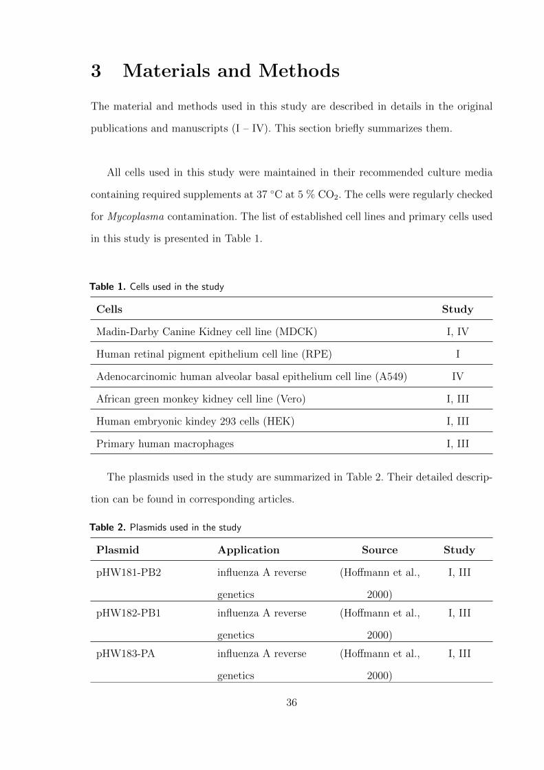
3 Materials and Methods
The material and methods used in this study are described in details in the original
publications and manuscripts (I – IV). This section briefly summarizes them.
All cells used in this study were maintained in their recommended culture media
containing required supplements at 37 ∘C at 5 % CO2. The cells were regularly checked
for Mycoplasma contamination. The list of established cell lines and primary cells used
in this study is presented in Table 1.
Table 1. Cells used in the study
Cells Study
Madin-Darby Canine Kidney cell line (MDCK) I, IV
Human retinal pigment epithelium cell line (RPE) I
Adenocarcinomic human alveolar basal epithelium cell line (A549) IV
African green monkey kidney cell line (Vero) I, III
Human embryonic kindey 293 cells (HEK) I, III
Primary human macrophages I, III
The plasmids used in the study are summarized in Table 2. Their detailed descrip-
tion can be found in corresponding articles.
Table 2. Plasmids used in the study
Plasmid Application Source Study
pHW181-PB2 influenza A reverse
genetics
(Hoffmann et al.,
2000)
I, III
pHW182-PB1 influenza A reverse
genetics
(Hoffmann et al.,
2000)
I, III
pHW183-PA influenza A reverse
genetics
(Hoffmann et al.,
2000)
I, III
36

Table 2. Plasmids used in the study
Plasmid Application Source Study
pHW184-HA influenza A reverse
genetics
(Hoffmann et al.,
2000)
I, III
pHW185-NA influenza A reverse
genetics
(Hoffmann et al.,
2000)
I, III
pHW186-NP influenza A reverse
genetics
(Hoffmann et al.,
2000)
I, III
pHW187-M influenza A reverse
genetics
(Hoffmann et al.,
2000)
I, III
pHW188-NS influenza A reverse
genetics
(Hoffmann et al.,
2000)
I, III
pHW188-NS1-RK/AA influenza A reverse
genetics
in-house I, III
pHW188-NS1-202 influenza A reverse
genetics
in-house III
pHW188-NS1-220 influenza A reverse
genetics
in-house III
pCMV-tag4A transfection
experiments
Agilent
Technologies
I
pCMV-tag4A-NS1-WT NS1 expression in cells in-house I
pCMV-tag4A-NS1-
RK/AA
NS1 expression in cells in-house I
pNic28-H5N1-NS1 NS1 protein production Dr. Denis Kainov II
pNic28-H5N1-NS1-
RK/AA
NS1 protein production Dr. Denis Kainov II, IV
pNic28-H5N1-NS1-
RK/AA-25,26
NS1 protein production in-house II
pNic28-H5N1-NS1-
RK/AA-48
NS1 protein production in-house II
37

Table 2. Plasmids used in the study
Plasmid Application Source Study
pNic28-H5N1-NS1-
RK/AA-67
NS1 protein production in-house II
pNic28-H5N1-NS1-
RK/AA-25,26,48,67
NS1 protein production in-house II
pNic28-H5N1-RBD NS1 protein production in-house II
pNic28-H5N2-NS1 NS1 protein production Dr. Denis Kainov II
pNic28-H5N2-NS1-
RK/AA
NS1 protein production Dr. Denis Kainov II
pNic28-H5N2-NS1-
delC
NS1 protein production Dr. Denis Kainov II
pET151-H1N1-NS1-
RK/AA
NS1 protein production Dr. Denis Kainov II
pET151-H1N1-NS1-
RK/AA-25,26
NS1 protein production MA II
pET151-H1N1-NS1-
RK/AA-48
NS1 protein production in-house II
pET151-H1N1-NS1-
RK/AA-67
NS1 protein production in-house II
pET151-H1N1-NS1-
RK/AA-25,26,48,67
NS1 protein production in-house II
Luciferase control DNA mRNA synthesis Promega II, IV
pGL-HCV-poly(A) mRNA synthesis Dr. Ilya Terenin IV
pGL-HCV-poly(A) mRNA synthesis Dr. Ilya Terenin IV
pGL-globin-SV40 mRNA synthesis Dr. Ilya Terenin IV
pGL-globin-poly(A) mRNA synthesis Dr. Ilya Terenin IV
pGL-EMCV-poly(A) mRNA synthesis Dr. Ilya Terenin IV
The viruses used in this study were generated using a reverse genetics system for
38
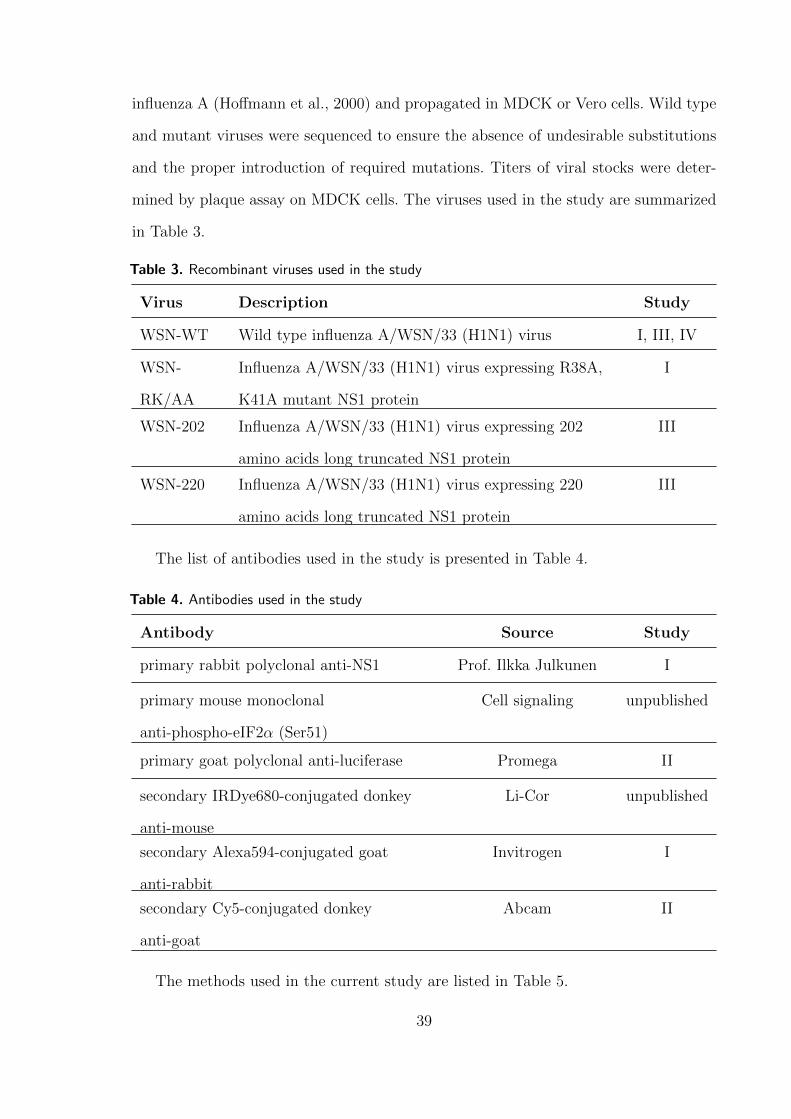
influenza A (Hoffmann et al., 2000) and propagated in MDCK or Vero cells. Wild type
and mutant viruses were sequenced to ensure the absence of undesirable substitutions
and the proper introduction of required mutations. Titers of viral stocks were deter-
mined by plaque assay on MDCK cells. The viruses used in the study are summarized
in Table 3.
Table 3. Recombinant viruses used in the study
Virus Description Study
WSN-WT Wild type influenza A/WSN/33 (H1N1) virus I, III, IV
WSN-
RK/AA
Influenza A/WSN/33 (H1N1) virus expressing R38A,
K41A mutant NS1 protein
I
WSN-202 Influenza A/WSN/33 (H1N1) virus expressing 202
amino acids long truncated NS1 protein
III
WSN-220 Influenza A/WSN/33 (H1N1) virus expressing 220
amino acids long truncated NS1 protein
III
The list of antibodies used in the study is presented in Table 4.
Table 4. Antibodies used in the study
Antibody Source Study
primary rabbit polyclonal anti-NS1 Prof. Ilkka Julkunen I
primary mouse monoclonal
anti-phospho-eIF2𝛼 (Ser51)
Cell signaling unpublished
primary goat polyclonal anti-luciferase Promega II
secondary IRDye680-conjugated donkey
anti-mouse
Li-Cor unpublished
secondary Alexa594-conjugated goat
anti-rabbit
Invitrogen I
secondary Cy5-conjugated donkey
anti-goat
Abcam II
The methods used in the current study are listed in Table 5.
39

Table 5. Methods used in the study
Method Study
Mammalian cell culture I, III, IV
Isolation of human peripheral blood mononuclear cells I, III
DNA-mediated transformation of mammalian cells I, III
Plasmid-based reverse genetics system for Influenza A I, III
Virus culture and purification I, III
Plaque assay I, III
Influenza A infections I, III, IV
RNA isolation I, III, IV
Reverse transcription I, III
Polymerase chain reaction I, III
One step reverse transcription and polymerase chain reaction I, III
Quantitative polymerase chain reaction I, II
Nuclear protein isolation I
Cytokine profiling I, III
Phosphoprotein profiling III
Enzyme-linked immunosorbent assay I
Immunofluorescence I
Gene expression profiling I, III
Chromatin immunoprecipitation (ChIP) I
Chomatin immunoprecipitation followed by next generation
sequencing (ChIPseq)
I
Recombinant protein production and purification II, IV
Electrophoretic mobility shift assay I, II
In vitro transcription I, II, IV
Sodium dodecyl sulphate polyacrylamide gel electrophoresis I, II, III
40

Table 5. Methods used in the study
Method Study
Molecular cloning I, III, II
Agarose gel electrophoresis I, IV
Cell free protein synthesis II, IV
Luciferase reporter assay II, IV
Microscale thermophoresis I
Density centrifugation in sucrose gradient IV
Determination of influenza A morbidity and mortality in vivo III
41

4 Results and Discussion
4.1 Conserved residues within NS1 bind dsDNA and control
transcription of cellular genes (Study I)
The nucleic acid binding function of NS1 is extremely important for influenza A repli-
cation and residues R38 and K41 are conserved across viral subtypes (Hatada and
Fukuda, 1992; Zohari et al., 2008). The mutations R38A and K41A were shown to pre-
vent dsRNA binding by NS1, and result in increased IFN production by infected cells
and severely attenuated viral phenotype (Donelan et al., 2003). Although the proposed
role of nucleic acid binding by NS1 is sequestration of viral replication intermediates
away from cellular PRRs, it is controversial as stated in Section 1.7.4. We addressed the
role of nucleic acid binding function of NS1 using the recombinant influenza A/WSN/33
(WSN) viruses expressing wild type NS1 or its R38A, K41A (RK/AA) mutant which
can not bind nucleic acids. In agreement with previous studies (Min and Krug, 2006),
RK/AA WSN virus exhibited severely attenuated phenotype in cell culture.
Gene expression profiling of human macrophages infected with RK/AA viruses re-
sulted in strong up-regulation of a large set of cellular genes (I, Fig. 1A and B). Gene
set enrichment analysis (Subramanian et al., 2005) indicated that these genes were in-
volved in the regulation of innate and adaptive immune responses and belonged to TLR
and Jak/STAT signaling pathways, cytokine-cytokine receptor interaction and natural
killer cell mediated cytotoxicity pathways. Accordingly, RK/AA infection resulted in
increased production of both antiviral and pro-inflammatory cytokines (I, Fig. 1C).
Importantly, the effect of RK/AA mutations on cellular gene expression and cytokine
production was not specific to immune cells and was also observed in a retinal pigment
epithelium (RPE) cell line (I, suppl. Fig. 1A and B).
Wild type NS1 is abundant in the insoluble fraction of cell lysate (I, Fig. 2A), where
it associates with cellular chromatin, presumably via protein-DNA, protein-protein or
both interactions (I, Fig. 2B). Although the direct binding of NS1 to dsDNA has
42

not been shown previously, the structure of NS1 RBD and full-length dimer does not
exclude its interaction with dsDNA (Bornholdt and Prasad, 2008; Cheng et al., 2009).
Therefore, we tested this possibility in an electophoretic mobility shift assay using
purified recombinant wild type and RK/AA NS1 proteins and synthetic DNA. Indeed,
purified wild-type NS1 did interact with 190 bp long linear dsDNA and the dissociation
constant (Kd) was 11.1 ± 0.7 𝜇M, indicative of a probably nonspecific binding. The
residues R38 and K41 were absolutely required for dsDNA interaction (I, Fig. 3B, C).
The ability of NS1 to associate with cellular chromatin and to bind dsDNA prompted
us to test its effects on transcription. To dissect effects of NS1-dsDNA binding and
upstream events we addressed this question in a cell-free run-off transcription assay
using purified wild type and RK/AA NS1 proteins. Binding of wt NS1 to DNA had
inhibitory effects on RNA synthesis by RNA polymerase II and time of addition exper-
iment suggested that NS1 inhibited transcription at its initiation step (I, Fig. 3D, E).
Interestingly, NS1 prevented RNA synthesis after preincubation with dsDNA or with
transcription factors and RNA polymerase II (I, Fig. 3F). This finding indicates that
NS1 binds dsDNA and inhibits transcription initiation by preventing pre-initiation
complex and RNA polymerase II loading on dsDNA. Alternatively, dsDNA binding
could coordinate inhibitory protein-protein interactions between NS1 and components
of transcription initiation machinery.
Is the dsDNA binding by NS1 that we observed in vitro relevant to infection? To
answer this question we addressed NS1 interaction with dsDNA in infected cells. Chro-
matin immunoprecipitation (ChIP) experiment in infected cells revealed the presence
of NS1 on the promoter and exon regions of the IFNB1 gene which is strongly in-
duced upon influenza A infection. Interaction of NS1 with IFNB1 DNA was R38-,
K41-dependent, suggesting direct dsDNA binding as was observed in an in vitro assay
(I, Fig. 4A). The pattern of NS1 association with IFNB1 promoter and exon regions
was opposite to that of RNA polymerase II, which may indicate possible inhibition of
IFNB1 gene expression due to NS1 DNA-binding.
43

Functional versatility of NS1 complicates studying the role of its dsDNA binding in
the context of viral infection, because in addition to nucleic acid binding residues R38
and K41 are involved in inhibitory interactions with TRIM25, RIPLET and RIG-I,
which also prevent transcriptional activation of IFNB1 (Gack et al., 2009; Rajsbaum
et al., 2012). To separate NS1 transcriptional effects from its role in virus-induced RIG-
I signaling, we addressed the function of residues R38 and K41 in cells overexpressing
NS1, taking advantage of the absence of RIG-I activation in such a set up. In this system
the wild type NS1 was detected on promoter and exon regions of housekeeping gene
EML4, indicating its ability to interact with cellular dsDNA (I, Fig. 4C). As expected,
the RK/AA mutant NS1 was not able to do so. To address direct effects of NS1 dsDNA
binding on transcriptional regulation of cellular genes we exposed cells expressing wild
type or RK/AA mutant NS1 to polyinosinic:polycytidylic acid (poly(I:C)). External
poly(I:C) is detected by cell surface TLR3 and thus IFN responses are induced without
activation of dsRNA signaling (Karpala et al., 2005). As expected, the wild type NS1
capable of dsDNA binding strongly inhibited poly(I:C)-induced activation of IFNB1,
IFNA1 and IFNA16, supporting our hypothesis of its role in direct transcriptional
control of cellular immune-related genes (I, fig. 4D).
In summary, these results provide a novel insight into IFN response control by
NS1, suggesting direct binding of NS1 to promoter and exon regions of DNA and
prevention of effective transcription of cellular genes. The Kd of NS1-dsDNA binding
was 11.1 ± 0.7 𝜇M and is comparable to the Kd of NS1-dsRNA binding, which is also
in micromolar range (Yin et al., 2007a). Provided this, and also given that the NS1
is predominantly localized in cellular nucleus, where its concentration is very high (Li
et al., 2010; Marazzi et al., 2012), we can propose that the dsDNA-binding of NS1 is as
important as its well-described dsRNA-binding. Further studies in RIG-I-, PKR- and
OAS-deficient systems would be helpful for dissecting these two essential functions of
NS1.
From our studies we can not determine, whether NS1 interacts with dsDNA as a
44

dimer or as an oligomer. Interestingly, the tunnel diameter inside the NS1 oligomer
was reported to be 20 Angstrom (A) (Bornholdt and Prasad, 2008), and, if no con-
formational changes occur during binding, it is more likely to accommodate a 20 A
B-form DNA double helix, rather than 26 A RNA double helix. This question could
be addressed in detail with further biophysical and structural studies of NS1-dsDNA
complex.
With regards sequence specificity of NS1 dsDNA binding, our results suggest the
lack of it, however, one can speculate that during infection transcriptionally active
chromatin, i.e. IFN genes, is more accessible to NS1. This question, however, needs
to be addressed in detail using genome-wide studies, for example, ChIP combined
with sequencing. Furthermore, our in vitro results suggest that NS1 is able to bind
dsDNA per se, however it would be interesting to investigate whether NS1 interacts
with possible cellular factors to facilitate its targeting to dsDNA in a crowded nucleus
environment.
When this study was initiated, NS1 had been thought to control activation of an-
tiviral genes at pre-transcriptional level (see section 1.7.5). However, later it was shown
that NS1 also inhibits cellular transcription by targeting histone H3-interacting tran-
scription elongation complex PAF1 (Marazzi et al., 2012). Such an inhibition, however,
required histone-mimic sequence 226ARSK229 in NS1 C-terminus and was specific to
viruses of H3N2 subtype (Marazzi et al., 2012). The mechanism of transcriptional con-
trol by NS1 discovered in our study, in contrast, seems to be general across different
influenza A subtypes and involves conserved residues R38, K41. The importance of NS1
dsDNA binding for viral replication and conservation of R38 and K41 suggest that this
function of NS1 could be an interesting target for development of antivirals. Indeed, a
recent study utilized SELEX approach to select ssDNA aptamers that are bound by
NS1 with high affinity and suppress its anti-interferon functions (Woo et al., 2013).
45

4.2 Regulation of general protein synthesis by NS1 (Studies
II, IV)
Another essential function of NS1 is to support effective viral protein synthesis in the
context of infection and thus secure viral replication. Influenza A infection activates
cellular PKR which phosphorylates and inactivates translation initiation factor eIF2𝛼,
thus inhibiting protein synthesis and inducing death of infected cells (Levin and Lon-
don, 1978). NS1 regulates general protein synthesis by preventing PKR activation and
its residues 123–127 are thought to be essential for this (Lu et al., 1995; Min et al.,
2007). In addition, NS1 interacts with translation initiation factors eIF4GI and PABPI
and few studies proposed that it recruits these factors to viral mRNAs thus specifi-
cally enhancing viral protein synthesis (de la Luna et al., 1995; Aragon et al., 2000;
Burgui et al., 2003). Such recruitment, however, would require specific binding of NS1
to distinct sequences on viral mRNAs.
We addressed regulation of protein synthesis by NS1. To separate the effects of
NS1 on protein synthesis from its other functions we used a rabbit reticulocyte lysate
(RRL) cell-free translation system.
Avian high pathogenic H5N1 NS1 enhanced translation of reporter mRNA and
protein production by RRL over six fold (II, Fig. 2A and C). Importantly, the RNA-
binding function of NS1 was not required for translation regulation in RRL and the
RK/AA mutant protein was as efficient enhancer of translation as the wild type NS1
(II, Fig. 2A). This observation allowed us to use the RK/AA NS1 proteins in fur-
ther experiments as they show remarkably better solubility compared to the wild type
proteins (Bornholdt and Prasad, 2008).
NS1 effects on translation in RRL were subtype specific: avian H5N1 and H5N2
NS1s were efficient translational enhancers, whereas pH1N1/2009 NS1 did not affect
protein synthesis (II, Fig. 2B and C).
Translational enhancement by NS1 was not limited to viral mRNAs. In RRL pro-
46

grammed with RNAs extracted from infected cells at different time points post infection
NS1 promoted production of both viral and cellular proteins (IV, Fig. 1A). This finding
was supported by our observation that RNA-binding was not required for NS1 transla-
tional control in RRL and hence in this system the protein synthesis rate does not seem
to depend on virus-specific sequences on RNA with which NS1 could interact, meaning
that one should observe enhancement of both cellular and viral mRNA translation.
In addition to its interactions with translation factors, NS1 has been shown to
associate with human homolog of Drosophila melanogaster Staufen protein (hStaufen)
on polysomes in infected cells (Falcon et al., 1999). As the hStaufen has been implicated
in regulation of mRNA stability, we addressed if translational enhancement in RRL in
the presence of NS1 was a result of increased mRNA stability. This appeared not to
be the case and NS1 did not affect the mRNA degradation rate (II, Fig. 2E and F).
Instead, NS1 enhanced association of mRNA with ribosomes and polysomes in RRL
reactions which suggested its possible role in translation initiation (IV, Fig. 1B).
We utilized differential effects of H5N1, H5N2 and pH1N1/2009 NS1 proteins on
RRL protein synthesis to map NS1 residues essential for translation enhancement. The
NS1 protein of pH1N1/2009 is 11 aa shorter than H5N1 and H5N2 NS1s due to a
C-terminal truncation. However, neither 11 C-terminal residues, nor the whole ED was
essential for translation modulation by NS1. RBD alone was sufficient for this function
(II, Fig. 2G and H). Importantly, the RBD of NS1 can be involved in protein-protein
interactions and therefore we searched to potential determinants of those. The RBD of
pH1N1/2009 NS1 contained several unique amino acids compared to H5N1 and H5N2:
N25, G26, N48 and W67 (II, Fig. 1A). Introduction of these residues singularly or all
at once into H5N1 NS1 resulted in loss of its function in RRL protein synthesis. In a
reciprocal experiment introduction of Q25/N26 and R67 from H5N1 in pH1N1/2009
NS1 resulted in its gain of function confirming that these residues are essential for
translational control in RRL (II, Fig. 2I). In the 3D structure of NS1 the side chains
of these amino acids are exposed to solvent, indicating their possible involvement in
47
interaction(s) between NS1 and its protein partner(s) (II, Fig. 4B). Thus, our results
clearly indicate that in RRL NS1 non-specifically enhances production of both viral
and cellular proteins and critical residues for this function are Q25, E26, S48 and R67
with NS1 RBD.
In the system that we used, the stimulation of protein synthesis in RRL was not
specific to viral mRNAs and also did not require NS1 RNA-binding function. Therefore,
this approach can not address the ability of NS1 to target translation factors to viral
mRNAs and our data neither contradict, nor support previous studies.
Translational enhancement by NS1 in RRL occurred due to prevention of eIF2𝛼
phosphorylation (Fig. 6). There are four kinases that are known to phosphorylate
eIF2𝛼: PKR, PKR-like endoplasmic reticulum kinase (PERK), general control non-
derepressible kinase 2 (GCN2) and heme-regulated inhibitor (HRI) (Donnelly et al.,
2013). Of these, only PKR has an assigned role in influenza infection. NS1 is proposed
to prevent activation of the PKR in infected cells by two mechanisms: (i) by binding
to dsRNA with its R38 and K41 and sequestering it away from PKR (Lu et al., 1995)
or (ii) by direct interaction with PKR via its residues 123–127 (Min et al., 2007) and
the latter activity is proposed to be critical for PKR inactivation. It is possible that
endogenous PKR is activated in RRL and NS1 prevents this activation thus securing
protein synthesis. However, three observations are not in line with this mechanism: (i)
RNA-binding function of NS1 was not required for its function in RRL. Therefore, NS1
could not prevent eIF2𝛼 phosphorylation by sequestering possible dsRNA substrates of
PKR; (ii) in our experiments NS1 RBD alone was sufficient to prevent eIF2𝛼 phospho-
rylation in RRL via its residues Q25, E26, S48 and R67, whereas previously published
data suggests that PKR inhibition by NS1 requires aas 123–127 within its ED; and
(iii) a previous study by Min et al. reported that RBD of A/Udorn/72 H3N2 which is
highly similar to H5N1 and H5N2 NS1 RBDs and contains residues Q25, E26, S48 and
K67 did not inhibit PKR (Min et al., 2007). Therefore, it is possible that the protein
target of NS1 in RRL is not PKR. The other kinases possibly inhibited by NS1 are
48

HRI, GCN2 and PERK. Although their role in influenza A infection are not known, all
of them are induced in response to various stress stimuli (Donnelly et al., 2013). The
possible inhibition of one of these kinases by NS1 could represent an additional ap-
proach by which influenza A secures protein synthesis during cellular stress. However,
the exact target is yet to be identified.
Figure 6. NS1 inhibits eIF2𝛼 phosphorylation in RRL. RRL reactions were supplied with H5N1RK/AA NS1 or control buffer and incubated for the time indicated. Equal amounts of reactionmixes were loaded on the SDS-polyacrylamide gel and phosphorylation status of eIF2𝛼 was assayedusing immunoblot.
Because NS1 proteins used in our work were deficient of RNA binding, our results
do not address possible targeting of NS1 on specific sequences on mRNA. However,
in a recent study Marc et al. have identified virus-specific motifs on RNAs to which
NS1 bound with nanomolar Kd (Marc et al., 2013). Biologically, such binding could be
related to several essential processes in virus life cycle, including enhanced translation
of viral mRNAs, however, it is still yet to be confirmed in infected cells. Altogether, our
results do not contradict possible targeting of NS1 to viral mRNAs, but demonstrate
a strategy of general protein synthesis stabilisation during infection employed by the
virus.
4.3 C-terminus of NS1 contributes to modulation of host an-
tiviral responses (Study III)
Although NS1 is a well structured protein, its C-terminus is likely to be unstructured
(Hale et al., 2008c). Nevertheless, it can play an essential regulatory role in infection,
as it accommodates numerous interaction motifs and modification sites that can mod-
ulate virus-host interactions, ofter in strain-specific manner (Li et al., 2001a; Hsiang
49
et al., 2009; Liu et al., 2010; Hsiang et al., 2012; Marazzi et al., 2012; Melen et al.,
2012). Naturally occurring truncations and extensions of NS1 C-terminus are have been
described in influenza A viruses of different genetic backgrounds (Suarez and Perdue,
1998). The NS1 protein of H1N1 viruses isolated in 2009–2014 is typically 219 aa long,
and H1N1 viruses encoding NS1 of 202 and 230 aa have been recently reported in
Finnish patients (Lakspere et al., 2014). However, the relevance of NS1 C-terminal
truncations and extensions to viral replication and virulence is unclear.
We aimed to address the functional role of NS1 C-terminus length using influenza A
viruses of well-characterized A/WSN/33 background that express NS1 proteins of 202,
220 and 230 amino acids (III, Fig. 1B). All viruses were able to propagate in immune-
competent MDCK cells, but the virus with the shortest NS1 (202 aa) displayed delayed
growth kinetics (III, suppl. Fig. 1A).
We addressed the effects of NS1 C-terminus length in human macrophages. Se-
quential truncations of NS1 C-terminus increased transcriptional activation of cellular
genes and the virus expressing the shortest NS1 was least successful in control of host
gene expression (III, Fig. 2A). The majority of genes differentially regulated by NS1
C-terminus length, e.g. MX1, ISG15, OASL, CCL8, IL6, and IL8, was involved in the
regulation of innate and adaptive immune responses (III, Fig. 2B). This effect was to
some extent reflected on the level of cytokine secretion and amounts of IL-6 and IL-8
secreted by macrophages were increased upon NS1 truncations (III, Fig. 2C and D).
Differential regulation of immune-related gene expression and cytokine production
prompted us to address the role of NS1 C-terminus in cellular signaling pathways. We
observed that C-terminal truncations of NS1 upregulated phosphorylation of several
phosphoproteins involved in control of immune responses. The shorter the NS1 C-
terminus was, the stronger was observed phosphorylation of STAT3, JNK, HSP60 and
AMPK𝛼1 (III, Fig. 3).
We used the viruses that express 230 and 202 aa long NS1 to address the effect
of NS1 C-terminus in vivo. NS1 C-terminus deletion reduced influenza A virulence
50

and pathogenicity in mice (III, Fig. 4A and B), suggesting the regulatory role of
NS1 C-terminus in infection. Thus, our results indicate that the length of NS1 C-
terminus is essential for interactions with the host cell, control of antiviral responses
and modulation of viral pathogenicity.
The C-terminus of A/WSN/33 NS1 contains human-like PDZ-binding motif for
which no interactions have yet been identified and a phosphorylation site T215 for
which the effect is also unclear (Jackson et al., 2010b; Hsiang et al., 2012). Nevertheless,
our data indicate the functional role of NS1 C-terminus length in the regulation of the
host immune responses and in viral pathogenicity and virulence. Sporadic truncations
and extensions of NS1 are quite seldom events, but they could provide opportunities
to lose, acquire or activate functional motifs within NS1 C-terminus. For example, de-
spite that in initial experiments appearance of functional Crk/CrkL-binding motif in
H1N1 NS1 C-terminus did not bring on notable effects (Hale et al., 2010b), it seems
that a combination of Crk/CrkL and NS1 C-terminus extension could promote viral
replication and increase virulence (Dr. Eike Hrincius, personal communication). Fur-
ther modeling of C-terminal polymorphisms on pH1N1/2009 genetic background could
provide deeper insights in virus-host interactions and influenza A adaptation to host.
The H1N1 influenza A viruses isolated from 2009 demonstrate the highest substi-
tution rate in NS1 compared to other known viral subtypes (Xu et al., 2011). A recent
study conducted in UK indeed reported that currently circulating pH1N1/2009 virus
rapidly adapted to human, and acquired mutations in multiple viral genes, includ-
ing NS1, that increase viral replication and limit host cytokine production (Elderfield
et al., 2014). Several isolates of pH1N1/2009 influenza A have acquired a functional
Crk/CrkL-binding domain or an extended C-terminus (III, Fig. 1A). Although such
viruses may not be of immediate concern, monitoring of changes within NS1 and its
C-terminus is worth undertaking, as while amino acid variations in NS1 C-terminus
are not detrimental, they provide the opportunity for influenza A viruses to probe
additional ways of virus-host interactions under human selection pressure.
51
4.4 NS1 as a tool to improve cell-free protein synthesis system
(Study IV)
The effects of NS1 on translation in RRL observed in Study II prompted us to utilize
NS1 for improvement of RRL cell-free translation system. RRL is widely used for
translation of specific templates, membrane protein studies, to study post-translational
events in folding and protein modification, and high-throughput screening for protein-
protein interactions (Fuller and Cuthbert, 2000; Douthwaite, 2012; Fixsen and Howard,
2010; Wang et al., 2011). However, low protein yield is a considerable limitation of RRL,
which should be overcome to improve its current applications (Carlson et al., 2012).
As viruses have evolved multiple ways to facilitate protein synthesis, a possible way to
improve cell-free protein synthesis is addition of viral factors which regulate translation.
These include viral proteins and specific regulatory structures on mRNA such as UTRs
or IRESs.
The NS1-mediated enhancement of RRL translation occurred through inhibition
of eIF2𝛼 phosphorylation (Fig. 6) and stabilization of translation initiation and was
not specific to viral mRNAs (IV, fig. 1). We reasoned that RRL translation could be
reinforced using mRNAs with specific 5’ and 3’ regulatory structures which together
with NS1 would exert a cumulative effect. For this several reporter mRNAs with 5’meG
or IRES and with different 3’ UTRs were tested in RRL at their optimal concentration
(II, Fig. 2D; IV, Fig. 2A). We observed the most robust enhancement of translation
when mRNA containing 5’ encephalomyocarditis virus (EMCV) IRES and 3’ poly(A)
sequence was used together with NS1. The protein production by RRL in this case was
increased over 11 fold (IV, Fig. 2B). The EMCV IRES is known to initiate translation
with limited initiation factor requirement and without ribosome scanning for AUG
codon (Pestova et al., 2001). However, it is still dependent on non-phosphorylated
eIF2𝛼 (Terenin et al., 2008), which in our system is controlled by NS1.
In summary, our strategy to use viral enhancers of translation provides an easy and
52

efficient way to improve cell-free translation in RRL and enhance its protein yield. This
strategy appears to be more effective compared to traditional usage of small molecular
inhibitors of eIF2𝛼 kinases (Jammi et al., 2003). It also provides a proof-of-concept for
generally utilizing viral proteins to modify cell-free translation systems from different
origins.
53

5 Conclusions and Future Perspectives
NS1 is an extremely versatile protein that controls influenza A virus-host interactions
at multiple levels. It has been extensively studied over the past few decades, but despite
a great deal of information obtained, the understanding of multiple functions of NS1 is
still far from being complete. For example, nucleic acid binding by R38 and K41 of NS1
has been well-described, but its role in the viral replication cycle still needs elucidation.
NS1 interactions with cellular factors of translation might facilitate selective synthesis
of viral proteins, but the exact mechanism needs to be described. Furthermore, a C-
terminal disordered “tail” of NS1 is a key contributor to NS1 diversity and length
polymorphisms, but its role in viral replication is far from being understood.
In this work I set specific questions aiming to provide novel insights into modulation
of influenza A virus-host interactions by NS1: What is the role of NS1 nucleic acid
binding and residues R38 and K41 in infection? Does NS1 specifically or non-specifically
enhance translation? What is the contribution of NS1 C-terminus in modulation of
virus-host interactions?
We approached of nucleic acid binding by NS1 in our study I and demonstrated that
NS1 can directly bind dsDNA and inhibit cellular gene expression at transcriptional
level. This does not exclude the RNA-binding—a well-characterized function of NS1—
but seems to be as important and provides additional level of NS1 control over host
immune responses. As it often occurs in science, our observation not only uncovers a
previously undescribed function of NS1, but also brings up several questions for further
elucidation.
How does NS1 inhibit transcription in the cell, without leading to its premature
death? Does it target specific cellular gene subsets? Are there any cellular factors hi-
jacked by the virus to secure this targeting? Addressing these questions in genome-wide
distribution of NS1 on cellular dsDNA using a high-throughput method such as ChIP-
seq, and identification of NS1 interacting partners using immunoprecipitation coupled
54
with mass-spectrometry analysis could contribute to understanding the mechanistic
details of transcriptional control by NS1. As the NS1 residues involved in this function
are conserved, one would expect that the transcriptional control by dsDNA binding is a
common function of NS1 proteins. Is it so? This can be addressed in question with sim-
ilar studies using other influenza A viruses, such as A/Udorn/72 (H3N2), A/PR8/34
(H1N1) and A/California/07/2009 (pH1N1/2009) for which reverse genetics systems
are available. Furthermore, as the NS1 nucleic acid binding function(s) appear to be
critical for influenza A replication in immune competent systems and residues R38 and
K41 are conserved, targeting this function with an inhibitor molecules may provide a
promising antiviral, to which resistance is less likely to develop.
In our study II we addressed the translation modulation by NS1 in a cell-free
translation system. We observed that NS1 non-specifically enhanced translation of
viral and cellular mRNAs. The mapped residues within NS1 that secure this function
indicate that it occurs via protein-protein interactions and RNA binding is not involved.
Thus, our results did not disprove or support previous suggestions that NS1 specifically
targets viral mRNAs and recruits translation factors therein. We rather added a new
level of translational control by NS1. Although our studies of translational control by
NS1 might be limited by the system used, our results provoke further investigation
of cellular responses to influenza A infection. Are there translational regulators, other
than PKR, induced by viral infection and the linked cellular stress? Answering this
question by specific studies of activation of eIF2𝛼 kinases upon influenza A infection
and probing possible protein-protein interactions between NS1 and these kinases can
bring new insights in understanding of complex control over cellular protein synthesis
during infection.
Interestingly, our observation that NS1 prevents eIF2𝛼 phosphorylation in RRL al-
lowed us to develop an approach to utilize NS1 to improve biotechnologically important
cell-free translation system and to increase the protein yield. Our study IV has laid
a proof-of-principle for using viral translational enhancers in biotechnological systems.
55
As Influenza A infects mammals, our method improved mammalian cell-free transla-
tion system based on RRL. Viruses infect all forms of life and are obligate parasites
of host translation, hence further studies may identify viral enhancers for any given
cell-free translation system.
Finally, in our study III we showed that the length of NS1 C-terminal tail con-
tributes to control over cellular antiviral responses and regulates virus pathogenicity.
These findings suggests that further monitoring of NS1 length polymorphisms should
be undertaken. As human infections with influenza A of pH1N1/2009 origin are still a
cause of concern, it would be important to address the role of NS1 C-terminal trun-
cations and extensions using reverse genetics system for pH1N1/2009. Furthermore,
influenza A strains lacking NS1 protein are considered as potential vaccine candidates
(Mossler et al., 2013), however, in my experience propagation of such viruses in cell
culture is extremely challenging. As viruses expressing C-terminally truncated NS1 in-
duce stronger antiviral responses and their virulence is limited, such viruses could be
investigated further as potential candidates for vaccine development.
Altogether, this work added three new functions of different NS1 determinants in
regulation of several aspects of virus host interactions. Although it would be interesting
to study each of these functions in more detail, it reminds us of a bigger question to
address: How does such a small protein like NS1 retain its functional versatility? Such a
versatility is apparently controlled at the level of protein structure, modifications, and
temporal and spatial regulation. Unfortunately, multiple studies addressingJAKAKA
these issues, were performed using different viral strains and experimental approaches
and, although they provided a huge amount of information, the detailed control of NS1
multifunctionality in a cell during infection still remains largely elusive. Undoubtedly,
further studies of influenza A virus-host interactions and their modulation by NS1 will
bring deeper understanding of multiple functions of this extremely interesting protein
and hopefully will contribute to improvement of current means to control influenza A.
56

Acknowledgements
I have been working on this dissertation for five years at the Institute for Molecular
Medicine Finland, FIMM. This has been an exciting time but also a long way which
could not be completed without assistance, support and friendship of people that I was
lucky to have around.
I thank my supervisor Dr. Denis Kainov for providing me with never-ending op-
portunities to develop, helping to find ways out of what could seem to be a dead end
and for being an example of real enthusiasm and determination. I clearly learned a lot
while working with you, Denis.
I am grateful to my co-supervisor Prof. Sarah Butcher for her never-failing support,
wise advices on every issue, and hours of inspiring discussions. Sarah, your door was
always open for me and your energizing laughter and “It’s gonna be fine!” helped me
keep on going when I could not believe that this dissertation would ever be completed.
Thank you.
Special thanks to my thesis advisory board Prof. Kalle Saksela and Prof. Dennis
Bamford for their guidance and advices during all five years.
I am grateful to Docent Maija Vihinen-Ranta and to Docent Thedi Ziegler who
kindly agreed to serve as pre-examiners of my dissertation and provided useful com-
ments that helped me improve this manuscript. I also thank Maija and Thedi for
doing a great job in a very short time. I am honored to have Prof. Stephan Ludwig as
an examiner of my dissertation and thank him for accepting the invitation to be my
opponent.
I would like to thank my colleagues and collaborators that brought their expertise
and best efforts to facilitate my work: Prof. Jean-Marc Egly, Dr. Nicolas Le May, Prof.
Xavier Saelens, Dr. Bert Schepens, Docent Tuula Nyman, Dr. Tiina Ohman, Docent
Sampsa Matikainen, Sandra Soderholm, Dr. Ilya Terenin, Prof. Ilkka Julkunen, Janne
Tynell, Dr. Maxim Bespalov, Laura Turunen, Lin Feng and Dr. Jakob Stenman. I would
57

like to express special gratitude to Prof. Jean-Marc Egly and Dr. Nicolas Le May for
an exciting opportunity to work with them during my lab visit to IGBMC which was
an invaluable experience during my PhD studies.
I thank former members of Denis Kainov group Andrey, Laura, Johanna, Oxana,
Minttu, Petri, Triin, Al, Maarten and Jens. Thank you for being excellent colleagues,
great office neighbors and fun company outside the lab. Special thanks Andrey and Al
for their friendship and for deep discussions about everything. Laura, you mentioned
recently that there would be no words to thank you, but seriosno, I am very grateful to
you for your rock-solid support and friendship over these almost five years that I know
you. Thank you for your help in the lab (and for donating macrophages), for advising
on all kinds of matters (good that you know everything), for proofreading whatever I
have ever written (even this thesis, at least twice), and for a great company on any
occasion.
I thank current and former members of Sarah’s lab with whom I had a chance
to interact: Lassi, Pasi, Ari, Shabih, Ausra, James, Jani, Veli-Pekka, Sergei, Noha and
Benita. Thank you guys for welcoming me in your lab, you’ve always been very friendly
and helpful.
I thank people from Sergey’s and Emmy’s groups at FIMM with whom I have been
working side-by-side for these years. Dasha, Pauliina, Manu, Yuexi, Sharif, Sergey,
Katja, Rita, Ashwini, Dat, Jenni, Annabrita, Emmy for a great working environment
and friendly atmosphere. I also thank JP, the most patient bioinformatician, for help-
ing with data analysis. I thank FIMM personnel for stimulating atmosphere during
seminars and retreats and for many bright moments. I thank FIMM director Prof. Olli
Kallioniemi and administrative manager Reetta Niemela also for the back-up during
some of the not-so-bright moments. Gretchen, thank you for your excellent work to
manage the information flow and make whichever tricky organizational issues become
crystal clear. I am grateful to Helsinki Biomedical Graduate School (now Doctoral
Program in Biomedicine).
58

I thank my friends Zaza, Steffi, Masha, Nitai, Alla, Ira, Asja, Shvilya and Kristiina.
Despite some of you live miles away you all have stayed close by and I appreciate
sharing happy moments with you and knowing that at darker days you are also there
for me. I want to express my special gratitude to my friends Dima M. and Karina.
Guys, you’ve always been around, you’ve seen all my ups and downs and I thank you
for your support, wisdom and occasional magic kicks. You helped me enjoy the happy
moments and overcome incidental asperities.
I thank Dima whose path happened to join mine at this exciting time of thesis
finalization. Thank you for the patience, understanding, courage and for staying calm
even during my “now-panic-and-freak-out” moments. And a special thank you for your
extraordinary ability to listen.
This work would have been be absolutely impossible without my family, which is my
true fortress at all times. I thank my grandmas, my siblings and my parents for their
never ending support. I am extremely grateful to my parents for their unconditional
love, care and the whole world of opportunities that they brought to me (and also for
the good genes). I thank mom for her patience, wisdom, understanding and for that
unbelievable source of energy which she generously shares with her children.
My research has been financially supported by the Finnish Committee for International
Mobility (CIMO), Helsinki Graduate Program in Biomedicine, University of Helsinki,
European Molecular Biology Organization (EMBO) and Finnish Cultural Foundation,
to which I am very grateful.
Helsinki, March 2015
59

References
Ablasser A, Poeck H, Anz D, Berger M, Schlee M, Kim S, Bourquin C, Goutagny N,Jiang Z, Fitzgerald KA, Rothenfusser S, Endres S, Hartmann G, and Hornung V.“Selection of molecular structure and delivery of RNA oligonucleotides to activateTLR7 versus TLR8 and to induce high amounts of IL-12p70 in primary humanmonocytes.” In: Journal of Immunology 182 (2009), pp. 6824–6833.
Akarsu H, Burmeister WP, Petosa C, Petit I, Muller CW, Ruigrok RWH, and BaudinF. “Crystal structure of the M1 protein-binding domain of the influenza A virusnuclear export protein (NEP/NS2)”. In: EMBO Journal 22 (2003), pp. 4646–4655.
Alexopoulou L, Holt AC, Medzhitov R, and Flavell RA. “Recognition of double-stranded RNA and activation of NF-kappaB by Toll-like receptor 3.” In: Nature413 (2001), pp. 732–738.
Allen IC, Scull MA, Moore CB, Holl EK, McElvania-TeKippe E, Taxman DJ, GuthrieEH, Pickles RJ, and Ting JPY. “The NLRP3 Inflammasome Mediates In VivoInnate Immunity to Influenza A Virus through Recognition of Viral RNA”. In:Immunity 30 (2009), pp. 556–565.
Amorim MJ, Bruce EA, Read EKC, Foeglein A, Mahen R, Stuart AD, and DigardP. “A Rab11- and microtubule-dependent mechanism for cytoplasmic transport ofinfluenza A virus viral RNA.” In: Journal of Virology 85 (2011), pp. 4143–4156.
Aragon T, de la Luna S, Novoa I, Carrasco L, Ortin J, and Nieto A. “Eukaryotic trans-lation initiation factor 4GI is a cellular target for NS1 protein, a translational acti-vator of influenza virus”. In: Molecular and Cellular Biology 20.17 (2000), pp. 6259–6268.
Aramini JM, Ma LC, Zhou L, Schauder CM, Hamilton K, Amer BR, Mack TR, LeeHW, Ciccosanti CT, Zhao L, Xiao R, Krug RM, and Montelione GT. “Dimer inter-face of the effector domain of non-structural protein 1 from influenza A virus: Aninterface with multiple functions”. In: Journal of Biological Chemistry 286 (2011),pp. 26050–26060.
Arranz R, Coloma R, Chichon FJ, Conesa JJ, Carrascosa JL, Valpuesta JM, Ortın J,and Martın-Benito J. “The structure of native influenza virion ribonucleoproteins.”In: Science 338 (2012), pp. 1634–7.
Avilov SV, Moisy D, Naffakh N, and Cusack S. “Influenza A virus progeny vRNPtrafficking in live infected cells studied with the virus-encoded fluorescently taggedPB2 protein”. In: Vaccine 30 (2012), pp. 7411–7417.
Ayllon J and Garcia-Sastre A. “The NS1 Protein: A Multitasking Virulence Factor”.In: Current Topics in Microbiology and Immunology (2014).
Ayllon J and Garcia-Sastre A. “The NS1 Protein: A Multitasking Virulence Factor.”In: Current Topics in Microbiology and Immunology 386 (2015), pp. 73–107.
Babcock HP, Chen C, and Zhuang X. “Using single-particle tracking to study nucleartrafficking of viral genes.” In: Biophysical journal 87 (2004), pp. 2749–2758.
Baltimore D. “Expression of animal virus genomes.” In: Bacteriological Reviews 35(1971), pp. 235–241.
Barman S, Adhikary L, Chakrabarti AK, Bernas C, Kawaoka Y, and Nayak DP. “Roleof transmembrane domain and cytoplasmic tail amino acid sequences of influenza a
60

virus neuraminidase in raft association and virus budding.” In: Journal of Virology78.10 (2004), pp. 5258–5269.
Bartenschlager R, Penin F, Lohmann V, and Andre P. “Assembly of infectious hepatitisC virus particles”. In: Trends in Microbiology 19 (2011), pp. 95–103.
Beaton AR and Krug RM. “Selected host cell capped RNA fragments prime influenzaviral RNA transcription in vivo.” In: Nucleic Acids Research 9 (1981), pp. 4423–4436.
Beloso A, Martinez C, Valcarcel J, Santaren JF, and Ortin J. “Degradation of cel-lular mRNA during influenza virus infection: Its possible role in protein synthesisshutoff”. In: Journal of General Virology 73 (1992), pp. 575–581.
Bender A, Albert M, Reddy A, Feldman M, Sauter B, Kaplan G, Hellman W, andBhardwaj N. “The Distinctive Features of Influenza Virus Infection of DendriticCells”. In: Immunobiology 198.5 (1998), pp. 552–567.
Bergmann M, Garcia-Sastre A, Carnero E, Pehamberger H, Wolff K, Palese P, andMuster T. “Influenza virus NS1 protein counteracts PKR-mediated inhibition ofreplication.” In: Journal of Virology 74 (2000), pp. 6203–6206.
Bogefors J, Kvarnhammar AM, Latif L, Petterson T, Uddman R, and Cardell LO.“Retinoic acid-inducible gene 1-like receptors in the upper respiratory tract.” In:American Journal of Rhinology & Allergy 25.6 (2011), e262–7.
Bornholdt ZA and Prasad BVV. “X-ray structure of influenza virus NS1 effector do-main.” In: Nature Structural & Molecular Biology 13 (2006), pp. 559–560.
Bornholdt ZA and Prasad BVV. “X-ray structure of NS1 from a highly pathogenicH5N1 influenza virus.” In: Nature 456 (2008), pp. 985–988.
Branca AA and Baglioni C. “Evidence that types I and II interferons have differentreceptors.” In: Nature 294 (1981), pp. 768–770.
Brankston G, Gitterman L, Hirji Z, Lemieux C, and Gardam M. “Transmission ofinfluenza A in human beings”. In: Lancet Infectious Diseases 7 (2007), pp. 257–265.
Brass AL, Huang IC, Benita Y, John SP, Krishnan MN, Feeley EM, Ryan BJ, WeyerJL, van der Weyden L, Fikrig E, Adams DJ, Xavier RJ, Farzan M, and Elledge SJ.“The IFITM proteins mediate cellular resistance to influenza A H1N1 virus, WestNile virus, and dengue virus”. In: Cell 139 (2009), pp. 1243–1254.
Brincks EL, Kucaba TA, Legge KL, and Griffith TS. “Influenza-induced expressionof functional tumor necrosis factor-related apoptosis-inducing ligand on human pe-ripheral blood mononuclear cells”. In: Human Immunology 69 (2008), pp. 634–646.
Brunotte L, Flies J, Bolte H, Reuther P, Vreede F, and Schwemmle M. “The nuclear ex-port protein of H5N1 influenza A viruses recruits M1 to the viral ribonucleoproteinto mediate nuclear export.” In: Journal of Biological Chemistry (2014).
Brydon EWA, Morris SJ, and Sweet C. “Role of apoptosis and cytokines in influenzavirus morbidity”. In: FEMS Microbiology Reviews 29 (2005), pp. 837–850.
Bui M, Whittaker G, and Helenius A. “Effect of M1 protein and low pH on nucleartransport of influenza virus ribonucleoproteins.” In: Journal of Virology 70 (1996),pp. 8391–8401.
Burgui I, Aragon T, Ortin J, and Nieto A. “PABP1 and eIF4GI associate with influenzavirus NS1 protein in viral mRNA translation initiation complexes”. In: Journal ofGeneral Virology 84.Pt 12 (2003), pp. 3263–3274.
61

Burns K, Janssens S, Brissoni B, Olivos N, Beyaert R, and Tschopp J. “Inhibitionof Interleukin 1 Receptor/Toll-like Receptor Signaling through the AlternativelySpliced, Short Form of MyD88 Is Due to Its Failure to Recruit IRAK-4”. In: TheJournal of Experimental Medicine 197.2 (2003), pp. 263–268.
Carlson ED, Gan R, Hodgman CE, and Jewett MC. “Cell-free protein synthesis: ap-plications come of age”. In: Biotechnology Advances 30.5 (2012), pp. 1185–1194.
Carr CM and Kim PS. “A spring-loaded mechanism for the conformational change ofinfluenza hemagglutinin”. In: Cell 73 (1993), pp. 823–832.
Carrillo B, Choi JM, Bornholdt ZA, Sankaran B, Rice AP, and Prasad BV. “Theinfluenza A virus protein NS1 displays structural polymorphism”. In: Journal ofVirology 88.8 (2014), pp. 4113–4122.
Castelli J, Wood KA, and Youle RJ. “The 2-5A system in viral infection and apoptosis”.In: Biomedicine and Pharmacotherapy 52 (1998), pp. 386–390.
Chen C and Zhuang X. “Epsin 1 is a cargo-specific adaptor for the clathrin-mediatedendocytosis of the influenza virus.” In: Proceedings of the National Academy ofSciences of the USA 105 (2008), pp. 11790–11795.
Chen G, Liu CH, Zhou L, and Krug RM. “Cellular DDX21 RNA Helicase InhibitsInfluenza A Virus Replication but Is Counteracted by the Viral NS1 Protein”. In:Cell Host and Microbe 15.4 (2014), pp. 484–493.
Chen W, Calvo PA, Malide D, Gibbs J, Schubert U, Bacik I, Basta S, O’Neill R,Schickli J, Palese P, Henklein P, Bennink JR, and Yewdell JW. “A novel influenza Avirus mitochondrial protein that induces cell death.” In: Nature Medicine 7 (2001),pp. 1306–1312.
Chen Z and Krug RM. “Selective nuclear export of viral mRNAs in influenza-virus-infected cells”. In: Trends in Microbiology 8 (2000), pp. 376–383.
Cheng A, Wong SM, and Yuan YA. “Structural basis for dsRNA recognition by NS1protein of influenza A virus.” In: Cell Research 19.2 (2009), pp. 187–95.
Chien CY, Tejero R, Huang YP, Zimmerman DE, Rios CB, Krug RM, and MontelioneGT. “A novel RNA-binding motif in influenza A virus non-structural protein 1”.In: Nature Structural Biology 4 (1997), pp. 891–895.
Chien CY, Xu Y, Xiao R, Aramini JM, Sahasrabudhe PV, Krug RM, and MontelioneGT. “Biophysical characterization of the complex between double-stranded RNAand the N-terminal domain of the NS1 protein from influenza A virus: evidence fora novel RNA-binding mode”. In: Biochemistry 43.7 (2004), pp. 1950–1962.
Chu WM, Ostertag D, Li ZW, Chang L, Chen Y, Hu Y, Williams B, Perrault J, andKarin M. “JNK2 and IKKbeta are required for activating the innate response toviral infection.” In: Immunity 11 (1999), pp. 721–731.
Cohen GM. “Caspases: the executioners of apoptosis.” In: The Biochemical Journal326 ( Pt 1 (1997), pp. 1–16.
Cohen M, Zhang XQ, Senaati HP, Chen HW, Varki NM, Schooley RT, and Gagneux P.“Influenza A penetrates host mucus by cleaving sialic acids with neuraminidase.”In: Virology Journal 10 (2013), p. 321.
Colgan DF and Manley JL. “Mechanism and regulation of mRNA polyadenylation.”In: Genes & Development 11.21 (1997), pp. 2755–2766.
62

Coloma R, Valpuesta JM, Arranz R, Carrascosa JL, Ortın J, and Martın-Benito J.“The structure of a biologically active influenza virus ribonucleoprotein complex”.In: PLoS Pathogens 5 (2009).
Connor RJ, Kawaoka Y, Webster RG, and Paulson JC. “Receptor specificity in human,avian, and equine H2 and H3 influenza virus isolates.” In: Virology 205 (1994),pp. 17–23.
Cros JF, Garcia-Sastre A, and Palese P. “An unconventional NLS is critical for thenuclear import of the influenza A virus nucleoprotein and ribonucleoprotein.” In:Traffic 6.3 (2005), pp. 205–213.
Cui S, Eisenacher K, Kirchhofer A, Brzozka K, Lammens A, Lammens K, Fujita T,Conzelmann KK, Krug A, and Hopfner KP. “The C-Terminal Regulatory DomainIs the RNA 5’-Triphosphate Sensor of RIG-I”. In: Molecular Cell 29 (2008), pp. 169–179.
Daniels-Holgate PU and Edwardson JM. “Transport of influenza virus envelope pro-teins from the Golgi complex to the apical plasma membrane in MDCK cells: pH-controlled interaction with a cycling receptor is not involved.” In: FEBS Letters249 (1989), pp. 407–410.
Das K, Ma LC, Xiao R, Radvansky B, Aramini J, Zhao L, Marklund J, Kuo RL, TwuKY, Arnold E, Krug RM, and Montelione GT. “Structural basis for suppressionof a host antiviral response by influenza A virus.” In: Proceedings of the NationalAcademy of Sciences of the USA 105.35 (2008), pp. 13093–8.
De la Luna S, Fortes P, Beloso A, and Ortin J. “Influenza virus NS1 protein enhancesthe rate of translation initiation of viral mRNAs”. In: Journal of Virology 69.4(1995), pp. 2427–2433.
Deb A, Haque SJ, Mogensen T, Silverman RH, and Williams BR. “RNA-dependentprotein kinase PKR is required for activation of NF-kappa B by IFN-gamma in aSTAT1-independent pathway.” In: Journal of Immunology 166 (2001), pp. 6170–6180.
Desai TM, Marin M, Chin CR, Savidis G, Brass AL, and Melikyan GB. “IFITM3 Re-stricts Influenza A Virus Entry by Blocking the Formation of Fusion Pores followingVirus-Endosome Hemifusion”. In: PLoS Pathogens 10 (2014).
Desselberger U, Nakajima K, Alfino P, Pedersen FS, Haseltine WA, Hannoun C, andPalese P. “Biochemical evidence that ”new” influenza virus strains in nature mayarise by recombination (reassortment).” In: Proceedings of the National Academyof Sciences of the USA 75 (1978), pp. 3341–3345.
Dias A, Bouvier D, Crepin T, McCarthy AA, Hart DJ, Baudin F, Cusack S, andRuigrok RWH. “The cap-snatching endonuclease of influenza virus polymerase re-sides in the PA subunit.” In: Nature 458 (2009), pp. 914–918.
Diebold SS, Kaisho T, Hemmi H, Akira S, and Reis e Sousa C. “Innate antiviralresponses by means of TLR7-mediated recognition of single-stranded RNA.” In:Science 303 (2004), pp. 1529–1531.
Dimitrov DS. “Virus entry: molecular mechanisms and biomedical applications.” In:Nature Reviews. Microbiology 2 (2004), pp. 109–122.
Dittmann J, Stertz S, Grimm D, Steel J, Garcıa-Sastre A, Haller O, and Kochs G.“Influenza A virus strains differ in sensitivity to the antiviral action of Mx-GTPase.”In: Journal of Virology 82 (2008), pp. 3624–3631.
63
Donelan NR, Basler CF, and Garcia-Sastre A. “A recombinant influenza A virus ex-pressing an RNA-binding-defective NS1 protein induces high levels of beta inter-feron and is attenuated in mice”. In: Journal of Virology 77.24 (2003), pp. 13257–13266.
Donnelly N, Gorman AM, Gupta S, and Samali A. “The eIF2alpha kinases: theirstructures and functions”. In: Cellular and Molecular Life Sciences 70.19 (2013),pp. 3493–3511.
Dourmashkin RR and Tyrrell DA. “Electron microscopic observations on the entryof influenza virus into susceptible cells.” In: The Journal of General Virology 24.1(1974), pp. 129–141.
Douthwaite JA. “Eukaryotic ribosome display selection using rabbit reticulocyte lysate”.In: Methods in Molecular Biology 805 (2012), pp. 45–57.
Drake JW. “Rates of spontaneous mutation among RNA viruses.” In: Proceedings ofthe National Academy of Sciences of the USA 90 (1993), pp. 4171–4175.
Dubois J, Terrier O, and Rosa-Calatrava M. “Influenza viruses and mRNA splicing:Doing more with less”. In: mBio 5 (2014).
Dudek SE, Wixler L, Nordhoff C, Nordmann A, Anhlan D, Wixler V, and LudwigS. “The influenza virus PB1-F2 protein has interferon antagonistic activity”. In:Biological Chemistry 392 (2011), pp. 1135–1144.
Dyer KD and Rosenberg HF. “The RNase a superfamily: Generation of diversity andinnate host defense”. In: Molecular Diversity 10 (2006), pp. 585–597.
Egorov A, Brandt S, Sereinig S, Romanova J, Ferko B, Katinger D, Grassauer A,Alexandrova G, Katinger H, and Muster T. “Transfectant influenza A viruses withlong deletions in the NS1 protein grow efficiently in Vero cells.” In: Journal ofVirology 72 (1998), pp. 6437–6441.
Ehrhardt C and Ludwig S. “A new player in a deadly game: Influenza viruses and thePI3K/Akt signalling pathway”. In: Cellular Microbiology 11 (2009), pp. 863–871.
Ehrhardt C, Marjuki H, Wolff T, Nurnberg B, Planz O, Pleschka S, and Ludwig S.“Bivalent role of the phosphatidylinositol-3-kinase (PI3K) during influenza virusinfection and host cell defence.” In: Cellular Microbiology 8.8 (2006), pp. 1336–1348.
Ehrhardt C, Wolff T, Pleschka S, Planz O, Beermann W, Bode JG, Schmolke M,and Ludwig S. “Influenza A virus NS1 protein activates the PI3K/Akt pathwayto mediate antiapoptotic signaling responses.” In: Journal of Virology 81 (2007),pp. 3058–3067.
Eisfeld AJ, Kawakami E, Watanabe T, Neumann G, and Kawaoka Y. “RAB11A isessential for transport of the influenza virus genome to the plasma membrane.” In:Journal of Virology 85 (2011), pp. 6117–6126.
Elderfield RA, Watson SJ, Godlee A, Adamson WE, Thompson CI, Dunning J, Fernandez-Alonso M, Blumenkrantz D, Hussell T, Zambon M, Openshaw P, Kellam P, andBarclay WS. “Accumulation of human-adapting mutations during circulation ofA(H1N1)pdm09 influenza virus in humans in the United Kingdom.” In: Journal ofVirology 88.22 (2014), pp. 13269–13283.
Elton D, Digard P, Tiley L, and Ortin J. “Structure and function of the influenza virusRNP”. In: Current Topics in Influenza Virology (2005), pp. 1–92.
64

Engelhardt OGO, Smith M, and Fodor E. “Association of the Influenza A Virus RNA-Dependent RNA Polymerase with Cellular RNA Polymerase II”. In: Journal ofVirology 79 (2005), pp. 5812–8.
Falcon AM, Fortes P, Marion RM, Beloso A, and Ortın J. “Interaction of influenzavirus NS1 protein and the human homologue of Staufen in vivo and in vitro.” In:Nucleic Acids Research 27 (1999), pp. 2241–2247.
Falcon AM, Marion RM, Zurcher T, Gomez P, Portela A, Nieto A, and Ortın J. “De-fective RNA replication and late gene expression in temperature-sensitive influenzaviruses expressing deleted forms of the NS1 protein.” In: Journal of Virology 78(2004), pp. 3880–3888.
Fensterl V and Sen GC. “Interferons and viral infections”. In: BioFactors 35 (2009),pp. 14–20.
Fesq H, Bacher M, Nain M, and Gemsa D. “Programmed cell death (apoptosis) inhuman monocytes infected by influenza A virus.” In: Immunobiology 190 (1994),pp. 175–182.
Fineberg HV. “Pandemic Preparedness and Response — Lessons from the H1N1 In-fluenza of 2009”. In: New England Journal of Medicine 370.14 (2014), pp. 1335–1342.
Fixsen SM and Howard MT. “Processive selenocysteine incorporation during synthe-sis of eukaryotic selenoproteins”. In: Journal of Molecular Biology 399.3 (2010),pp. 385–396.
Forrest HL and Webster RG. “Perspectives on influenza evolution and the role ofresearch.” In: Animal Health Research Reviews 11 (2010), pp. 3–18.
Fournier G, Chiang C, Munier S, Tomoiu A, Demeret C, Vidalain PO, Jacob Y, andNaffakh N. “Recruitment of RED-SMU1 Complex by Influenza A Virus RNA Poly-merase to Control Ciral mRNA Splicing”. In: PLoS Pathogens 10 (2014).
Fu XY, Kessler DS, Veals SA, Levy DE, and Darnell JE. “ISGF3, the transcriptionalactivator induced by interferon alpha, consists of multiple interacting polypeptidechains.” In: Proceedings of the National Academy of Sciences of the USA 87 (1990),pp. 8555–8559.
Fujiyoshi Y, Kume NP, Sakata K, and Sato SB. “Fine structure of influenza A virusobserved by electron cryo-microscopy.” In: The EMBO journal 13 (1994), pp. 318–326.
Fuller W and Cuthbert AW. “Post-translational disruption of the delta F508 cysticfibrosis transmembrane conductance regulator (CFTR)-molecular chaperone com-plex with geldanamycin stabilizes delta F508 CFTR in the rabbit reticulocytelysate”. In: Journal of Biological Chemistry 275.48 (2000), pp. 37462–37468.
Gack MU, Shin YC, Joo CH, Urano T, Liang C, Sun L, Takeuchi O, Akira S, Chen Z,Inoue S, and Jung JU. “TRIM25 RING-finger E3 ubiquitin ligase is essential forRIG-I-mediated antiviral activity.” In: Nature 446 (2007), pp. 916–920.
Gack MU, Albrecht RA, Urano T, Inn KS, Huang IC, Carnero E, Farzan M, Inoue S,Jung JU, and Garcia-Sastre A. “Influenza A virus NS1 targets the ubiquitin ligaseTRIM25 to evade recognition by the host viral RNA sensor RIG-I.” In: Cell Hostand Microbe 5.5 (2009), pp. 439–449.
Galluzzi L, Brenner C, Morselli E, Touat Z, and Kroemer G. “Viral control of mito-chondrial apoptosis”. In: PLoS Pathogens 4 (2008).
65
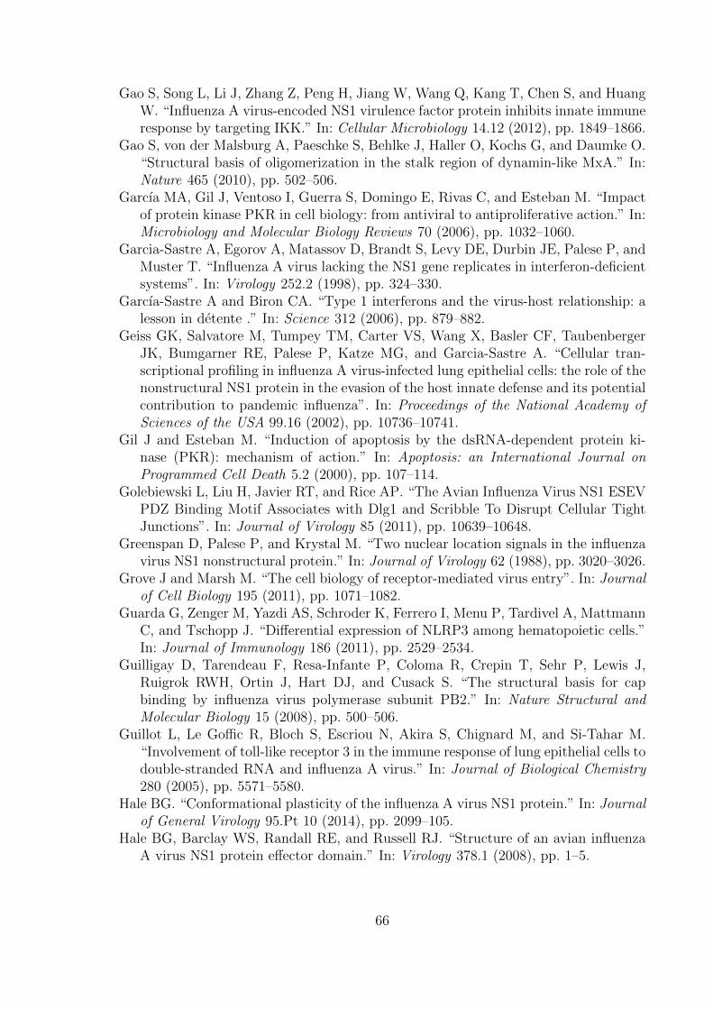
Gao S, Song L, Li J, Zhang Z, Peng H, Jiang W, Wang Q, Kang T, Chen S, and HuangW. “Influenza A virus-encoded NS1 virulence factor protein inhibits innate immuneresponse by targeting IKK.” In: Cellular Microbiology 14.12 (2012), pp. 1849–1866.
Gao S, von der Malsburg A, Paeschke S, Behlke J, Haller O, Kochs G, and Daumke O.“Structural basis of oligomerization in the stalk region of dynamin-like MxA.” In:Nature 465 (2010), pp. 502–506.
Garcıa MA, Gil J, Ventoso I, Guerra S, Domingo E, Rivas C, and Esteban M. “Impactof protein kinase PKR in cell biology: from antiviral to antiproliferative action.” In:Microbiology and Molecular Biology Reviews 70 (2006), pp. 1032–1060.
Garcia-Sastre A, Egorov A, Matassov D, Brandt S, Levy DE, Durbin JE, Palese P, andMuster T. “Influenza A virus lacking the NS1 gene replicates in interferon-deficientsystems”. In: Virology 252.2 (1998), pp. 324–330.
Garcıa-Sastre A and Biron CA. “Type 1 interferons and the virus-host relationship: alesson in detente .” In: Science 312 (2006), pp. 879–882.
Geiss GK, Salvatore M, Tumpey TM, Carter VS, Wang X, Basler CF, TaubenbergerJK, Bumgarner RE, Palese P, Katze MG, and Garcia-Sastre A. “Cellular tran-scriptional profiling in influenza A virus-infected lung epithelial cells: the role of thenonstructural NS1 protein in the evasion of the host innate defense and its potentialcontribution to pandemic influenza”. In: Proceedings of the National Academy ofSciences of the USA 99.16 (2002), pp. 10736–10741.
Gil J and Esteban M. “Induction of apoptosis by the dsRNA-dependent protein ki-nase (PKR): mechanism of action.” In: Apoptosis: an International Journal onProgrammed Cell Death 5.2 (2000), pp. 107–114.
Golebiewski L, Liu H, Javier RT, and Rice AP. “The Avian Influenza Virus NS1 ESEVPDZ Binding Motif Associates with Dlg1 and Scribble To Disrupt Cellular TightJunctions”. In: Journal of Virology 85 (2011), pp. 10639–10648.
Greenspan D, Palese P, and Krystal M. “Two nuclear location signals in the influenzavirus NS1 nonstructural protein.” In: Journal of Virology 62 (1988), pp. 3020–3026.
Grove J and Marsh M. “The cell biology of receptor-mediated virus entry”. In: Journalof Cell Biology 195 (2011), pp. 1071–1082.
Guarda G, Zenger M, Yazdi AS, Schroder K, Ferrero I, Menu P, Tardivel A, MattmannC, and Tschopp J. “Differential expression of NLRP3 among hematopoietic cells.”In: Journal of Immunology 186 (2011), pp. 2529–2534.
Guilligay D, Tarendeau F, Resa-Infante P, Coloma R, Crepin T, Sehr P, Lewis J,Ruigrok RWH, Ortin J, Hart DJ, and Cusack S. “The structural basis for capbinding by influenza virus polymerase subunit PB2.” In: Nature Structural andMolecular Biology 15 (2008), pp. 500–506.
Guillot L, Le Goffic R, Bloch S, Escriou N, Akira S, Chignard M, and Si-Tahar M.“Involvement of toll-like receptor 3 in the immune response of lung epithelial cells todouble-stranded RNA and influenza A virus.” In: Journal of Biological Chemistry280 (2005), pp. 5571–5580.
Hale BG. “Conformational plasticity of the influenza A virus NS1 protein.” In: Journalof General Virology 95.Pt 10 (2014), pp. 2099–105.
Hale BG, Barclay WS, Randall RE, and Russell RJ. “Structure of an avian influenzaA virus NS1 protein effector domain.” In: Virology 378.1 (2008), pp. 1–5.
66
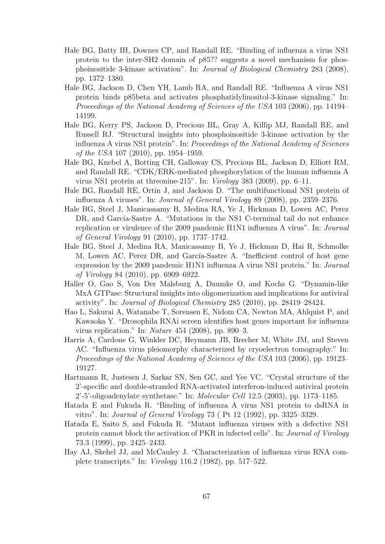
Hale BG, Batty IH, Downes CP, and Randall RE. “Binding of influenza a virus NS1protein to the inter-SH2 domain of p85?? suggests a novel mechanism for phos-phoinositide 3-kinase activation”. In: Journal of Biological Chemistry 283 (2008),pp. 1372–1380.
Hale BG, Jackson D, Chen YH, Lamb RA, and Randall RE. “Influenza A virus NS1protein binds p85beta and activates phosphatidylinositol-3-kinase signaling.” In:Proceedings of the National Academy of Sciences of the USA 103 (2006), pp. 14194–14199.
Hale BG, Kerry PS, Jackson D, Precious BL, Gray A, Killip MJ, Randall RE, andRussell RJ. “Structural insights into phosphoinositide 3-kinase activation by theinfluenza A virus NS1 protein”. In: Proceedings of the National Academy of Sciencesof the USA 107 (2010), pp. 1954–1959.
Hale BG, Knebel A, Botting CH, Galloway CS, Precious BL, Jackson D, Elliott RM,and Randall RE. “CDK/ERK-mediated phosphorylation of the human influenza Avirus NS1 protein at threonine-215”. In: Virology 383 (2009), pp. 6–11.
Hale BG, Randall RE, Ortin J, and Jackson D. “The multifunctional NS1 protein ofinfluenza A viruses”. In: Journal of General Virology 89 (2008), pp. 2359–2376.
Hale BG, Steel J, Manicassamy B, Medina RA, Ye J, Hickman D, Lowen AC, PerezDR, and Garcıa-Sastre A. “Mutations in the NS1 C-terminal tail do not enhancereplication or virulence of the 2009 pandemic H1N1 influenza A virus”. In: Journalof General Virology 91 (2010), pp. 1737–1742.
Hale BG, Steel J, Medina RA, Manicassamy B, Ye J, Hickman D, Hai R, SchmolkeM, Lowen AC, Perez DR, and Garcıa-Sastre A. “Inefficient control of host geneexpression by the 2009 pandemic H1N1 influenza A virus NS1 protein.” In: Journalof Virology 84 (2010), pp. 6909–6922.
Haller O, Gao S, Von Der Malsburg A, Daumke O, and Kochs G. “Dynamin-likeMxA GTPase: Structural insights into oligomerization and implications for antiviralactivity”. In: Journal of Biological Chemistry 285 (2010), pp. 28419–28424.
Hao L, Sakurai A, Watanabe T, Sorensen E, Nidom CA, Newton MA, Ahlquist P, andKawaoka Y. “Drosophila RNAi screen identifies host genes important for influenzavirus replication.” In: Nature 454 (2008), pp. 890–3.
Harris A, Cardone G, Winkler DC, Heymann JB, Brecher M, White JM, and StevenAC. “Influenza virus pleiomorphy characterized by cryoelectron tomography.” In:Proceedings of the National Academy of Sciences of the USA 103 (2006), pp. 19123–19127.
Hartmann R, Justesen J, Sarkar SN, Sen GC, and Yee VC. “Crystal structure of the2’-specific and double-stranded RNA-activated interferon-induced antiviral protein2’-5’-oligoadenylate synthetase.” In: Molecular Cell 12.5 (2003), pp. 1173–1185.
Hatada E and Fukuda R. “Binding of influenza A virus NS1 protein to dsRNA invitro”. In: Journal of General Virology 73 ( Pt 12 (1992), pp. 3325–3329.
Hatada E, Saito S, and Fukuda R. “Mutant influenza viruses with a defective NS1protein cannot block the activation of PKR in infected cells”. In: Journal of Virology73.3 (1999), pp. 2425–2433.
Hay AJ, Skehel JJ, and McCauley J. “Characterization of influenza virus RNA com-plete transcripts.” In: Virology 116.2 (1982), pp. 517–522.
67

Haye K, Burmakina S, Moran T, Garcıa-Sastre A, and Fernandez-Sesma A. “TheNS1 protein of a human influenza virus inhibits type I interferon production andthe induction of antiviral responses in primary human dendritic and respiratoryepithelial cells.” In: Journal of Virology 83 (2009), pp. 6849–6862.
Haywood AM. “Membrane uncoating of intact enveloped viruses.” In: Journal of Vi-rology 84 (2010), pp. 10946–10955.
Heikkinen LS, Kazlauskas A, Melen K, Wagner R, Ziegler T, Julkunen I, and SakselaK. “Avian and 1918 Spanish influenza a virus NS1 proteins bind to Crk/CrkLSrc homology 3 domains to activate host cell signaling”. In: Journal of BiologicalChemistry 283.9 (2008), pp. 5719–5727.
Herold S, Ludwig S, Pleschka S, and Wolff T. “Apoptosis signaling in influenza viruspropagation, innate host defense, and lung injury”. In: Journal of Leukocyte Biology92 (2012), pp. 75–82.
Hershey A and Chase M. “Independent functions of viral protein and nucleic acid ingrowth of bacteriophage”. In: The Journal of General Physiology 36 (1952), pp. 39–56.
Hinshaw VS, Olsen CW, Dybdahl-sissoko N, and Evans D. “Apoptosis: a mechanismof cell killing by influenza A and B viruses.” In: Journal of Virology 68 (1994),pp. 3667–73.
Hoffmann E, Neumann G, Kawaoka Y, Hobom G, and Webster RG. “A DNA transfec-tion system for generation of influenza A virus from eight plasmids”. In: Proceedingsof the National Academy of Sciences of the USA 97.11 (2000), pp. 6108–6113.
Honda K, Yanai H, Negishi H, Asagiri M, Sato M, Mizutani T, Shimada N, Ohba Y,Takaoka A, Yoshida N, and Taniguchi T. “IRF-7 is the master regulator of type-Iinterferon-dependent immune responses”. In: Nature 434.7034 (2005), pp. 772–777.
Hong XX and Carmichael GG. “Innate immunity in pluripotent human cells: Attenu-ated response to interferon-beta”. In: Journal of Biological Chemistry 288 (2013),pp. 16196–16205.
Hornung V, Ellegast J, Kim S, Brzozka K, Jung A, Kato H, Poeck H, Akira S, Conzel-mann KK, Schlee M, Endres S, and Hartmann G. “5’-Triphosphate RNA is theligand for RIG-I.” In: Science 314 (2006), pp. 994–997.
Hrincius ER, Wixler V, Wolff T, Wagner R, Ludwig S, and Ehrhardt C. “CRK adap-tor protein expression is required for efficient replication of avian influenza Aviruses and controls JNK-mediated apoptotic responses”. In: Cellular Microbiol-ogy 12 (2010), pp. 831–843.
Hsiang TY, Zhou L, and Krug RM. “Roles of the phosphorylation of specific serinesand threonines in the NS1 protein of human influenza A viruses”. In: Journal ofVirology 86.19 (2012), pp. 10370–10376.
Hsiang TY, Zhao C, and Krug RM. “Interferon-induced ISG15 conjugation inhibitsinfluenza A virus gene expression and replication in human cells.” In: Journal ofVirology 83 (2009), pp. 5971–5977.
Huang Y and Carmichael GG. “Role of polyadenylation in nucleocytoplasmic transportof mRNA.” In: Molecular and Cellular Biology 16 (1996), pp. 1534–1542.
Huet S, Avilov S, Ferbitz L, Daigle N, Cusack S, and Ellenberg J. “Nuclear importand assembly of the influenza A virus RNA polymerase studied in live cells by
68

Fluorescence Cross Correlation Spectroscopy”. In: Journal of Virology 84 (2010),pp. 1254–1264.
Hughey PG, Compans RW, Zebedee SL, and Lamb RA. “Expression of the influenzaA virus M2 protein is restricted to apical surfaces of polarized epithelial cells.” In:Journal of Virology 66.9 (1992), pp. 5542–5552.
Husain B, Mukerji I, and Cole JL. “Analysis of high-affinity binding of protein kinaseR to double-stranded RNA.” In: Biochemistry 51.44 (2012), pp. 8764–8770.
Hutchinson EC, Denham EM, Thomas B, Trudgian DC, Hester SS, Ridlova G, YorkA, Turrell L, and Fodor E. “Mapping the Phosphoproteome of Influenza A and BViruses by Mass Spectrometry”. In: PLoS Pathogens 8 (2012).
Ichinohe T, Lee HK, Ogura Y, Flavell R, and Iwasaki A. “Inflammasome recognitionof influenza virus is essential for adaptive immune responses.” In: The Journal ofExperimental Medicine 206 (2009), pp. 79–87.
Ichinohe T, Pang IK, and Iwasaki A. “Influenza virus activates inflammasomes via itsintracellular M2 ion channel.” In: Nature Immunology 11 (2010), pp. 404–410.
Inglis SC, Barrett T, Brown CM, and Almond JW. “The smallest genome RNA segmentof influenza virus contains two genes that may overlap.” In: Proceedings of theNational Academy of Sciences of the USA 76 (1979), pp. 3790–3794.
Ioannidis I, Ye F, McNally B, Willette M, and Flano E. “Toll-like receptor expressionand induction of type I and type III interferons in primary airway epithelial cells.”In: Journal of Virology 87 (2013), pp. 3261–70.
Ishikawa E, Nakazawa M, Yoshinari M, and Minami M. “Role of tumor necrosis factor-related apoptosis-inducing ligand in immune response to influenza virus infectionin mice”. In: Journal of Virology 79 (2005), pp. 7658–7663.
Iwasaki A and Medzhitov R. “Regulation of adaptive immunity by the innate immunesystem.” In: Science 327 (2010), pp. 291–295.
Iwasaki A and Pillai PS. “Innate immunity to influenza virus infection”. In: NatureReviews Microbiology Immunol 14.5 (2014), pp. 315–328.
Jackson DA, Caton AJ, McCready SJ, and Cook PR. “Influenza virus RNA is synthe-sized at fixed sites in the nucleus.” In: Nature 296 (1982), pp. 366–368.
Jackson D, Killip MJ, Galloway CS, Russell RJ, and Randall RE. “Loss of functionof the influenza A virus NS1 protein promotes apoptosis but this is not due to afailure to activate phosphatidylinositol 3-kinase (PI3K)”. In: Virology 396 (2010),pp. 94–105.
Jackson RJ, Hellen CU, and Pestova TV. “The mechanism of eukaryotic translation ini-tiation and principles of its regulation”. In: Nature Reviews Molecular and CellularBiology 11.2 (2010), pp. 113–127.
Jagger BW, Wise HM, Kash JC, Walters KA, Wills NM, Xiao YL, Dunfee RL, Schwartz-man LM, Ozinsky A, Bell GL, Dalton RM, Lo A, Efstathiou S, Atkins JF, FirthAE, Taubenberger JK, and Digard P. “An Overlapping Protein-Coding Region inInfluenza A Virus Segment 3 Modulates the Host Response”. In: Science 337 (2012),pp. 199–204.
Jammi NV, Whitby LR, and Beal PA. “Small molecule inhibitors of the RNA-dependentprotein kinase”. In: Biochemical and Biophysical Research Communications 308.1(2003), pp. 50–57.
69

Janeway CA. “Approaching the asymptote? Evolution and revolution in immunology”.In: Cold Spring Harbor Symposia on Quantitative Biology. Vol. 54. 1989, pp. 1–13.
Janeway CA and Medzhitov R. “Innate immune recognition.” In: Annual Review ofImmunology 20 (2002), pp. 197–216.
Javier RT and Rice AP. “Emerging Theme: Cellular PDZ Proteins as Common Targetsof Pathogenic Viruses”. In: Journal of Virology 85 (2011), pp. 11544–11556.
Jiang H, White EJ, Rıos-Vicil CI, Xu J, Gomez-Manzano C, and Fueyo J. “Humanadenovirus type 5 induces cell lysis through autophagy and autophagy-triggeredcaspase activity.” In: Journal of Virology 85 (2011), pp. 4720–4729.
Jiang Z, Zamanian-Daryoush M, Nie H, Silva AM, Williams BRG, and Li X. “Poly(I-C)-induced Toll-like receptor 3 (TLR3)-mediated activation of NFkappa B andMAP kinase is through an interleukin-1 receptor-associated kinase (IRAK)-independentpathway employing the signaling components TLR3-TRAF6-TAK1-TAB2-PKR .”In: Journal of Biological Chemistry 278.19 (2003), pp. 16713–16719.
Johnson ES. “Protein modification by SUMO.” In: Annual Review of Biochemistry 73(2004), pp. 355–382.
Jorba N, Coloma R, and Ortın J. “Genetic trans-complementation establishes a newmodel for influenza virus RNA transcription and replication”. In: PLoS Pathogens5 (2009).
Julkunen I, Melen K, Nyqvist M, Pirhonen J, Sareneva T, and Matikainen S. “Inflam-matory responses in influenza A virus infection.” In: Vaccine 19 Suppl 1 (2000),S32–S37.
Kang DC, Gopalkrishnan RV, Lin L, Randolph A, Valerie K, Pestka S, and FisherPB. “Expression analysis and genomic characterization of human melanoma differ-entiation associated gene-5, mda-5: a novel type I interferon-responsive apoptosis-inducing gene.” In: Oncogene 23 (2004), pp. 1789–1800.
Karlas A, Machuy N, Shin Y, Pleissner KP, Artarini A, Heuer D, Becker D, KhalilH, Ogilvie LA, Hess S, Maurer AP, Muller E, Wolff T, Rudel T, and Meyer TF.“Genome-wide RNAi screen identifies human host factors crucial for influenza virusreplication.” In: Nature 463 (2010), pp. 818–822.
Karpala AJ, Doran TJ, and Bean AGD. “Immune responses to dsRNA: Implications forgene silencing technologies”. In: Immunology and Cell Biology 83 (2005), pp. 211–216.
Kato H, Takeuchi O, Sato S, Yoneyama M, Yamamoto M, Matsui K, Uematsu S, JungA, Kawai T, Ishii KJ, Yamaguchi O, Otsu K, Tsujimura T, Koh CS, Reis e Sousa C,Matsuura Y, Fujita T, and Akira S. “Differential roles of MDA5 and RIG-I helicasesin the recognition of RNA viruses.” In: Nature 441 (2006), pp. 101–105.
Katze MG, Detjen BM, Safer B, and Krug RM. “Translational control by influenzavirus: suppression of the kinase that phosphorylates the alpha subunit of initiationfactor eIF-2 and selective translation of influenza viral mRNAs”. In: Molecular andCellular Biology 6.5 (1986), pp. 1741–1750.
Katze MG, Tomita J, Black T, Krug RM, Safer B, and Hovanessian A. “Influenza virusregulates protein synthesis during infection by repressing autophosphorylation andactivity of the cellular 68,000-Mr protein kinase.” In: Journal of Virology 62.10(1988), pp. 3710–3717.
70
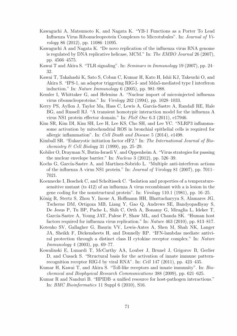
Kawaguchi A, Matsumoto K, and Nagata K. “YB-1 Functions as a Porter To LeadInfluenza Virus Ribonucleoprotein Complexes to Microtubules”. In: Journal of Vi-rology 86 (2012), pp. 11086–11095.
Kawaguchi A and Nagata K. “De novo replication of the influenza virus RNA genomeis regulated by DNA replicative helicase, MCM.” In: The EMBO Journal 26 (2007),pp. 4566–4575.
Kawai T and Akira S. “TLR signaling”. In: Seminars in Immunology 19 (2007), pp. 24–32.
Kawai T, Takahashi K, Sato S, Coban C, Kumar H, Kato H, Ishii KJ, Takeuchi O, andAkira S. “IPS-1, an adaptor triggering RIG-I- and Mda5-mediated type I interferoninduction.” In: Nature Immunology 6 (2005), pp. 981–988.
Kemler I, Whittaker G, and Helenius A. “Nuclear import of microinjected influenzavirus ribonucleoproteins.” In: Virology 202 (1994), pp. 1028–1033.
Kerry PS, Ayllon J, Taylor Ma, Hass C, Lewis A, Garcıa-Sastre A, Randall RE, HaleBG, and Russell RJ. “A transient homotypic interaction model for the influenza Avirus NS1 protein effector domain.” In: PloS One 6.3 (2011), e17946.
Kim SR, Kim DI, Kim SH, Lee H, Lee KS, Cho SH, and Lee YC. “NLRP3 inflamma-some activation by mitochondrial ROS in bronchial epithelial cells is required forallergic inflammation”. In: Cell Death and Disease 5 (2014), e1498.
Kimball SR. “Eukaryotic initiation factor eIF2.” In: The International Journal of Bio-chemistry & Cell Biology 31 (1999), pp. 25–29.
Kobiler O, Drayman N, Butin-Israeli V, and Oppenheim A. “Virus strategies for passingthe nuclear envelope barrier.” In: Nucleus 3 (2012), pp. 526–39.
Kochs G, Garcıa-Sastre A, and Martınez-Sobrido L. “Multiple anti-interferon actionsof the influenza A virus NS1 protein.” In: Journal of Virology 81 (2007), pp. 7011–7021.
Koennecke I, Boschek C, and Scholtissek C. “Isolation and properties of a temperature-sensitive mutant (ts 412) of an influenza A virus recombinant with a is lesion in thegene coding for the nonstructural protein”. In: Virology 110.1 (1981), pp. 16–25.
Konig R, Stertz S, Zhou Y, Inoue A, Hoffmann HH, Bhattacharyya S, Alamares JG,Tscherne DM, Ortigoza MB, Liang Y, Gao Q, Andrews SE, Bandyopadhyay S,De Jesus P, Tu BP, Pache L, Shih C, Orth A, Bonamy G, Miraglia L, Ideker T,Garcıa-Sastre A, Young JAT, Palese P, Shaw ML, and Chanda SK. “Human hostfactors required for influenza virus replication.” In: Nature 463 (2010), pp. 813–817.
Kotenko SV, Gallagher G, Baurin VV, Lewis-Antes A, Shen M, Shah NK, LangerJA, Sheikh F, Dickensheets H, and Donnelly RP. “IFN-lambdas mediate antivi-ral protection through a distinct class II cytokine receptor complex.” In: NatureImmunology 4 (2003), pp. 69–77.
Kowalinski E, Lunardi T, McCarthy AA, Louber J, Brunel J, Grigorov B, GerlierD, and Cusack S. “Structural basis for the activation of innate immune pattern-recognition receptor RIG-I by viral RNA”. In: Cell 147 (2011), pp. 423–435.
Kumar H, Kawai T, and Akira S. “Toll-like receptors and innate immunity”. In: Bio-chemical and Biophysical Research Communications 388 (2009), pp. 621–625.
Kumar R and Nanduri B. “HPIDB–a unified resource for host-pathogen interactions.”In: BMC Bioinformatics 11 Suppl 6 (2010), S16.
71

Kundu A, Avalos RT, Sanderson CM, and Nayak DP. “Transmembrane domain ofinfluenza virus neuraminidase, a type II protein, possesses an apical sorting signalin polarized MDCK cells.” In: Journal of Virology 70 (1996), pp. 6508–6515.
Kundu P, Raychaudhuri S, Tsai W, and Dasgupta A. “Shutoff of RNA polymerase IItranscription by poliovirus involves 3C protease-mediated cleavage of the TATA-binding protein at an alternative site: incomplete shutoff of transcription interfereswith efficient viral replication.” In: Journal of Virology 79.15 (2005), pp. 9702–9713.
La Gruta NL, Kedzierska K, Stambas J, and Doherty PC. “A question of self-preservation:immunopathology in influenza virus infection”. In: Immunology & Cell Biology 85.2(2007), pp. 85–92.
Lagace-Wiens PRS, Rubinstein E, and Gumel A. “Influenza epidemiology–past, present,and future.” In: Critical care medicine 38 (2010), e1–e9.
Lakadamyali M, Rust MJ, Babcock HP, and Zhuang X. “Visualizing infection of in-dividual influenza viruses.” In: Proceedings of the National Academy of Sciences ofthe USA 100 (2003), pp. 9280–9285.
Lakspere T, Tynell J, Kaloinen M, Vanlede M, Parsons A, Ikonen N, Kallio-Kokko H,Kantele A, Mattila P, Almusa H, Julkunen I, Kainov D, and Kakkola L. “Full-Genome Sequences of Influenza A(H1N1)pdm09 Viruses Isolated from Finnish Pa-tients from 2009 to 2013”. In: Genome Announcements 2.1 (2014).
Lamb RA. “The Influenza Virus RNA Segments and Their Encoded Proteins”. In:Genetics of Influenza Viruses SE - 2. Ed. by Palese P and Kingsbury D. SpringerVienna, 1983, pp. 21–69.
Lamb RA and Choppin PW. “Segment 8 of the influenza virus genome is unique incoding for two polypeptides.” In: Proceedings of the National Academy of Sciencesof the USA 76 (1979), pp. 4908–4912.
Lamb RA, Choppin PW, Chanock RM, and Lai CJ. “Mapping of the two overlappinggenes for polypeptides NS1 and NS2 on RNA segment 8 of influenza virus genome.”In: Proceedings of the National Academy of Sciences of the USA 77 (1980), pp. 1857–1861.
Le Goffic R, Balloy V, Lagranderie M, Alexopoulou L, Escriou N, Flavell R, ChignardM, and Si-Tahar M. “Detrimental contribution of the Toll-like receptor (TLR)3to influenza A virus-induced acute pneumonia”. In: PLoS Pathogens 2 (2006),pp. 0526–0535.
Le Goffic R, Pothlichet J, Vitour D, Fujita T, Meurs E, Chignard M, and Si-TaharM. “Cutting Edge: Influenza A virus activates TLR3-dependent inflammatory andRIG-I-dependent antiviral responses in human lung epithelial cells.” In: Journal ofImmunology 178 (2007), pp. 3368–3372.
Le May N, Dubaele S, De Santis LP, Billecocq A, Bouloy M, and Egly JM. “TFIIHTranscription Factor, a Target for the Rift Valley Hemorrhagic Fever Virus”. In:Cell 116 (2004), pp. 541–550.
Lee ASy, Burdeinick-Kerr R, and Whelan SPJ. “A ribosome-specialized translationinitiation pathway is required for cap-dependent translation of vesicular stomatitisvirus mRNAs.” In: Proceedings of the National Academy of Sciences of the USA110 (2013), pp. 324–9.
Lee N, Wong CK, Hui DSC, Lee SKW, Wong RYK, Ngai KLK, Chan MCW, Chu YJ,Ho AWY, Lui GCY, Wong BCK, Wong SH, Yip SP, and Chan PKS. “Role of human
72

Toll-like receptors in naturally occurring influenza A infections”. In: Influenza andother Respiratory Viruses 7 (2013), pp. 666–675.
Levin D and London IM. “Regulation of protein synthesis: activation by double-stranded RNA of a protein kinase that phosphorylates eukaryotic initiation factor2”. In: Proceedings of the National Academy of Sciences of the USA 75.3 (1978),pp. 1121–1125.
Levy DE, Kessler DS, Pine R, Reich N, and Darnell JE. “Interferon-induced nuclearfactors that bind a shared promoter element correlate with positive and negativetranscriptional control.” In: Genes & Development 2 (1988), pp. 383–393.
Li ML, Rao P, and Krug RM. “The active sites of the influenza cap-dependent en-donuclease are on different polymerase subunits”. In: EMBO Journal 20 (2001),pp. 2078–2086.
Li S, Min JY, Krug RM, and Sen GC. “Binding of the influenza A virus NS1 protein toPKR mediates the inhibition of its activation by either PACT or double-strandedRNA”. In: Virology 349.1 (2006), pp. 13–21.
Li S, Peters GA, Ding K, Zhang X, Qin J, and Sen GC. “Molecular basis for PKR ac-tivation by PACT or dsRNA.” In: Proceedings of the National Academy of Sciencesof the USA 103 (2006), pp. 10005–10010.
Li Y, Chen ZY, Wang W, Baker CC, and Krug RM. “The 3’-end-processing factorCPSF is required for the splicing of single-intron pre-mRNAs in vivo.” In: RNA 7.6(2001), pp. 920–931.
Li Y, Yamakita Y, and Krug RM. “Regulation of a nuclear export signal by an adjacentinhibitory sequence: the effector domain of the influenza virus NS1 protein.” In:Proceedings of the National Academy of Sciences of the USA 95 (1998), pp. 4864–4869.
Li Y, Lu X, Li J, Berube N, Giest KL, Liu Q, Anderson DH, and Zhou Y. “Geneticallyengineered, biarsenically labeled influenza virus allows visualization of viral NS1protein in living cells.” In: Journal of Virology 84 (2010), pp. 7204–7213.
Liang SL, Quirk D, and Zhou A. “RNase L: its biological roles and regulation.” In:IUBMB Life 58 (2006), pp. 508–514.
Liu HM, Loo YM, Horner SM, Zornetzer GA, Katze MG, and Gale M. “The mito-chondrial targeting chaperone 14-3-3𝜖 regulates a RIG-I translocon that mediatesmembrane association and innate antiviral immunity”. In: Cell Host and Microbe11 (2012), pp. 528–537.
Liu H, Golebiewski L, Dow EC, Krug RM, Javier RT, and Rice AP. “The ESEV PDZBinding-Motif of the Avian Influenza A Virus NS1 Protein Protects Infected Cellsfrom Apoptosis through Directly Targeting Scribble.” In: Journal of Virology 84(2010), pp. 11164–11174.
Liu J, Lynch PA, Chien CY, Montelione GT, Krug RM, and Berman HM. “Crystalstructure of the unique RNA-binding domain of the influenza virus NS1 protein.”In: Nature Structural Biology 4 (1997), pp. 896–899.
Long JX, Peng DX, Liu YL, Wu YT, and Liu XF. “Virulence of H5N1 avian influenzavirus enhanced by a 15-nucleotide deletion in the viral nonstructural gene”. In:Virus Genes 36 (2008), pp. 471–478.
73

Lu X, Masic A, Li Y, Shin Y, Liu Q, and Zhou Y. “The PI3K/Akt pathway inhibitsinfluenza A virus-induced Bax-mediated apoptosis by negatively regulating the JNKpathway via ASK1”. In: Journal of General Virology 91 (2010), pp. 1439–1449.
Lu Y, Qian XY, and Krug RM. “The influenza virus NS1 protein: a novel inhibitor ofpre-mRNA splicing.” In: Genes & Development 8 (1994), pp. 1817–1828.
Lu Y, Wambach M, Katze MG, and Krug RM. “Binding of the influenza virus NS1protein to double-stranded RNA inhibits the activation of the protein kinase thatphosphorylates the elF-2 translation initiation factor”. In: Virology 214.1 (1995),pp. 222–228.
Ludwig S, Schultz U, Mandler J, Fitch WM, and Scholtissek C. “Phylogenetic rela-tionship of the nonstructural (NS) genes of influenza A viruses.” In: Virology 183(1991), pp. 566–577.
Ludwig S, Wang X, Ehrhardt C, Zheng H, Donelan N, Planz O, Pleschka S, Garcia-Sastre A, Heins G, and Wolff T. “The influenza A virus NS1 protein inhibits ac-tivation of Jun N-terminal kinase and AP-1 transcription factors”. In: Journal ofVirology 76.21 (2002), pp. 11166–11171.
Lund JM, Alexopoulou L, Sato A, Karow M, Adams NC, Gale NW, Iwasaki A, andFlavell RA. “Recognition of single-stranded RNA viruses by Toll-like receptor 7.” In:Proceedings of the National Academy of Sciences of the USA 101 (2004), pp. 5598–5603.
Lyles DS. “Assembly and Budding of Negative-Strand RNA Viruses”. In: Advances inVirus Research 85 (2013), pp. 57–90.
Ma J, Goryaynov A, Sarma A, and Yang W. “Self-regulated viscous channel in thenuclear pore complex”. In: Proceedings of the National Academy of Sciences of theUSA 109 (2012), pp. 7326–7331.
Maamary J, Pica N, Belicha-Villanueva A, Chou Yy, Krammer F, Gao Q, Garcia-Sastre A, and Palese P. “Attenuated Influenza Virus Construct with EnhancedHemagglutinin Protein Expression”. In: Journal of Virology 86 (2012), pp. 5782–5790.
Macara IG. “Transport into and out of the nucleus.” In: Microbiology and MolecularBiology Reviews 65 (2001), 570–594, table of contents.
Marazzi I, Ho JS, Kim J, Manicassamy B, Dewell S, Albrecht RA, Seibert CW, SchaeferU, Jeffrey KL, Prinjha RK, Lee K, Garcia-Sastre A, Roeder RG, and TarakhovskyA. “Suppression of the antiviral response by an influenza histone mimic”. In: Nature483.7390 (2012), pp. 428–433.
Marc D, Barbachou S, and Soubieux D. “The RNA-binding domain of influenzavirusnon-structural protein-1 cooperatively binds to virus-specific RNA sequences in astructure-dependent manner.” In: Nucleic acids research 41.1 (2013), pp. 434–449.
Marion RM, Zurcher T, De La Luna S, and Ortın J. “Influenza virus NS1 protein inter-acts with viral transcription-replication complexes in vivo”. In: Journal of GeneralVirology 78 (1997), pp. 2447–2451.
Martın J, Wharton SA, Lin YP, Takemoto DK, Skehel JJ, Wiley DC, and SteinhauerDA. “Studies of the binding properties of influenza hemagglutinin receptor-sitemutants.” In: Virology 241 (1998), pp. 101–111.
Martin K and Helenius A. “Transport of incoming influenza virus nucleocapsids intothe nucleus.” In: Journal of Virology 65 (1991), pp. 232–244.
74
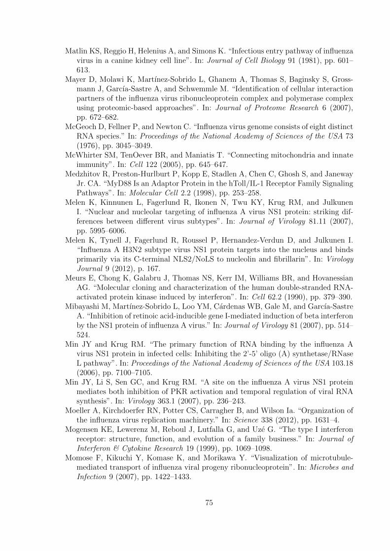
Matlin KS, Reggio H, Helenius A, and Simons K. “Infectious entry pathway of influenzavirus in a canine kidney cell line”. In: Journal of Cell Biology 91 (1981), pp. 601–613.
Mayer D, Molawi K, Martınez-Sobrido L, Ghanem A, Thomas S, Baginsky S, Gross-mann J, Garcıa-Sastre A, and Schwemmle M. “Identification of cellular interactionpartners of the influenza virus ribonucleoprotein complex and polymerase complexusing proteomic-based approaches”. In: Journal of Proteome Research 6 (2007),pp. 672–682.
McGeoch D, Fellner P, and Newton C. “Influenza virus genome consists of eight distinctRNA species.” In: Proceedings of the National Academy of Sciences of the USA 73(1976), pp. 3045–3049.
McWhirter SM, TenOever BR, and Maniatis T. “Connecting mitochondria and innateimmunity”. In: Cell 122 (2005), pp. 645–647.
Medzhitov R, Preston-Hurlburt P, Kopp E, Stadlen A, Chen C, Ghosh S, and JanewayJr. CA. “MyD88 Is an Adaptor Protein in the hToll/IL-1 Receptor Family SignalingPathways”. In: Molecular Cell 2.2 (1998), pp. 253–258.
Melen K, Kinnunen L, Fagerlund R, Ikonen N, Twu KY, Krug RM, and JulkunenI. “Nuclear and nucleolar targeting of influenza A virus NS1 protein: striking dif-ferences between different virus subtypes”. In: Journal of Virology 81.11 (2007),pp. 5995–6006.
Melen K, Tynell J, Fagerlund R, Roussel P, Hernandez-Verdun D, and Julkunen I.“Influenza A H3N2 subtype virus NS1 protein targets into the nucleus and bindsprimarily via its C-terminal NLS2/NoLS to nucleolin and fibrillarin”. In: VirologyJournal 9 (2012), p. 167.
Meurs E, Chong K, Galabru J, Thomas NS, Kerr IM, Williams BR, and HovanessianAG. “Molecular cloning and characterization of the human double-stranded RNA-activated protein kinase induced by interferon”. In: Cell 62.2 (1990), pp. 379–390.
Mibayashi M, Martınez-Sobrido L, Loo YM, Cardenas WB, Gale M, and Garcıa-SastreA. “Inhibition of retinoic acid-inducible gene I-mediated induction of beta interferonby the NS1 protein of influenza A virus.” In: Journal of Virology 81 (2007), pp. 514–524.
Min JY and Krug RM. “The primary function of RNA binding by the influenza Avirus NS1 protein in infected cells: Inhibiting the 2’-5’ oligo (A) synthetase/RNaseL pathway”. In: Proceedings of the National Academy of Sciences of the USA 103.18(2006), pp. 7100–7105.
Min JY, Li S, Sen GC, and Krug RM. “A site on the influenza A virus NS1 proteinmediates both inhibition of PKR activation and temporal regulation of viral RNAsynthesis”. In: Virology 363.1 (2007), pp. 236–243.
Moeller A, Kirchdoerfer RN, Potter CS, Carragher B, and Wilson Ia. “Organization ofthe influenza virus replication machinery.” In: Science 338 (2012), pp. 1631–4.
Mogensen KE, Lewerenz M, Reboul J, Lutfalla G, and Uze G. “The type I interferonreceptor: structure, function, and evolution of a family business.” In: Journal ofInterferon & Cytokine Research 19 (1999), pp. 1069–1098.
Momose F, Kikuchi Y, Komase K, and Morikawa Y. “Visualization of microtubule-mediated transport of influenza viral progeny ribonucleoprotein”. In: Microbes andInfection 9 (2007), pp. 1422–1433.
75

Momose F, Sekimoto T, Ohkura T, Jo S, Kawaguchi A, Nagata K, and Morikawa Y.“Apical transport of influenza A virus ribonucleoprotein requires Rab11-positiverecycling endosome”. In: PLoS ONE 6 (2011).
Mori I, Komatsu T, Takeuchi K, Nakakuki K, Sudo M, and Kimura Y. “In vivo induc-tion of apoptosis by influenza virus.” In: Journal of General Virology 76 ( Pt 11(1995), pp. 2869–2873.
Morita E, Sandrin V, McCullough J, Katsuyama A, Baci Hamilton I, and SundquistWI. “ESCRT-III protein requirements for HIV-1 budding”. In: Cell Host and Mi-crobe 9 (2011), pp. 235–242.
Mossler C, Groiss F, Wolzt M, Wolschek M, Seipelt J, and Muster T. “Phase I/II trialof a replication-deficient trivalent influenza virus vaccine lacking NS1”. In: Vaccine31.52 (2013), pp. 6194–6200.
Munir M, Zohari S, Belak S, and Berg M. “Double-Stranded RNA-Induced Acti-vation of Activating Protein-1 Promoter Is Differentially Regulated by the Non-structural Protein 1 of Avian Influenza A Viruses”. In: Viral Immunology (2012),p. 120112065140000.
Munir M, Zohari S, and Berg M. “Non-structural protein 1 of avian influenza Aviruses differentially inhibit NF-kappaB promoter activation.” In: Virology Jour-nal 8 (2011), p. 383.
Muramoto Y, Noda T, Kawakami E, Akkina R, and Kawaoka Y. “Identification of novelinfluenza A virus proteins translated from PA mRNA.” In: Journal of Virology 87(2013), pp. 2455–62.
Nakayama M, Nagata K, and Ishihama A. “Enzymatic properties of the mouse Mx1protein-associated GTPase.” In: Virus Research 22 (1992), pp. 227–234.
Nayak DP, Balogun RA, Yamada H, Zhou ZH, and Barman S. Influenza virus mor-phogenesis and budding. 2009.
Nayak DP, Hui EKW, and Barman S. “Assembly and budding of influenza virus”. In:Virus Research 106 (2004), pp. 147–165.
Nemeroff ME, Barabino SM, Li Y, Keller W, and Krug RM. “Influenza virus NS1protein interacts with the cellular 30 kDa subunit of CPSF and inhibits 3’endformation of cellular pre-mRNAs.” In: Molecular Cell 1 (1998), pp. 991–1000.
Neumann G, Castrucci MR, and Kawaoka Y. “Nuclear import and export of influenzavirus nucleoprotein.” In: Journal of Virology 71 (1997), pp. 9690–9700.
Neumann G, Hughes MT, and Kawaoka Y. “Influenza A virus NS2 protein medi-ates vRNP nuclear export through NES-independent interaction with hCRM1”. In:EMBO Journal 19 (2000), pp. 6751–6758.
Newby CM, Sabin L, and Pekosz A. “The RNA binding domain of influenza A virus NS1protein affects secretion of tumor necrosis factor alpha, interleukin-6, and interferonin primary murine tracheal epithelial cells”. In: Journal of Virology 81.17 (2007),pp. 9469–9480.
Nicholas B, Skipp P, Mould R, Rennard S, Davies DE, O’Connor CD, and DjukanovicR. “Shotgun proteomic analysis of human-induced sputum”. In: Proteomics 6 (2006),pp. 4390–4401.
Noah D, Twu K, and Krug R. “Cellular antiviral responses against influenza A virus arecountered at the posttranscriptional level by the viral NS1A protein via its binding
76

to a cellular protein required for the 3’ end processing of cellular pre-mRNAS.” In:Virology 307.2 (2003), pp. 386–395.
Noah DL and Noah JW. “Adapting global influenza management strategies to addressemerging viruses.” In: American journal of physiology. Lung cellular and molecularphysiology 305 (2013), pp. L108–17.
Odendall C, Dixit E, Stavru F, Bierne H, Franz KM, Durbin AF, Boulant S, GehrkeL, Cossart P, and Kagan JC. “Diverse intracellular pathogens activate type IIIinterferon expression from peroxisomes.” In: Nature Immunology 15 (2014), pp. 717–728.
O’Neill RE, Jaskunas R, Blobel G, Palese P, and Moroianu J. “Nuclear import ofinfluenza virus RNA can be mediated by viral nucleoprotein and transport fac-tors required for protein import”. In: Journal of Biological Chemistry 270 (1995),pp. 22701–22704.
Opitz B, Rejaibi A, Dauber B, Eckhard J, Vinzing M, Schmeck B, Hippenstiel S,Suttorp N, and Wolff T. “IFNbeta induction by influenza A virus is mediated byRIG-I which is regulated by the viral NS1 protein.” In: Cellular Microbiology 9.4(2007), pp. 930–938.
Oshiumi H, Miyashita M, Inoue N, Okabe M, Matsumoto M, and Seya T. “The ubiqui-tin ligase riplet is essential for RIG-I-dependent innate immune responses to RNAvirus infection”. In: Cell Host and Microbe 8 (2010), pp. 496–509.
Pal S, Rosas JM, and Rosas-Acosta G. “Identification of the non-structural influenzaA viral protein NS1A as a bona fide target of the Small Ubiquitin-like MOdifierby the use of dicistronic expression constructs”. In: Journal of Virological Methods163 (2010), pp. 498–504.
Pal S, Santos A, Rosas JM, Ortiz-Guzman J, and Rosas-Acosta G. “Influenza A virusinteracts extensively with the cellular SUMOylation system during infection”. In:Virus Research 158 (2011), pp. 12–27.
Palese P. “The genes of influenza virus”. In: Cell 10.1 (1977), pp. 1–10.Pang IK, Ichinohe T, and Iwasaki A. “IL-1R signaling in dendritic cells replaces
pattern-recognition receptors in promoting CD8 T cell responses to influenza Avirus.” In: Nature Immunology 14 (2013), pp. 246–53.
Parvin JD, Moscona A, Pan WT, Leider JM, and Palese P. “Measurement of themutation rates of animal viruses: influenza A virus and poliovirus type 1.” In:Journal of Virology 59 (1986), pp. 377–383.
Parvin JD, Young JF, and Palese P. “Nonsense mutations affecting the lengths of theNS1 nonstructural proteins of influenza A virus isolates”. In: Virology 128.2 (1983),pp. 512–517.
Patel JR, Jain A, Chou YY, Baum A, Ha T, and Garcıa-Sastre A. “ATPase-drivenoligomerization of RIG-I on RNA allows optimal activation of type-I interferon.”In: EMBO Reports 14 (2013), pp. 1–8.
Pauli EK, Schmolke M, Wolff T, Viemann D, Roth J, Bode JG, and Ludwig S. “In-fluenza A virus inhibits type I IFN signaling via NF-kappaB-dependent inductionof SOCS-3 expression.” In: PLoS Pathogens 4.11 (2008), e1000196.
Pelkmans L and Helenius A. “Insider information: What viruses tell us about endocy-tosis”. In: Current Opinion in Cell Biology 15 (2003), pp. 414–422.
77

Perrone LA, Plowden JK, Garcıa-Sastre A, Katz JM, and Tumpey TM. “H5N1 and1918 Pandemic Influenza Virus Infection Results in Early and Excessive Infiltrationof Macrophages and Neutrophils in the Lungs of Mice”. In: PLoS Pathogens 4.8(2008), e1000115.
Pestova TV, Kolupaeva VG, Lomakin IB, Pilipenko EV, Shatsky IN, Agol VI, andHellen CU. “Molecular mechanisms of translation initiation in eukaryotes.” In: Pro-ceedings of the National Academy of Sciences of the USA 98 (2001), pp. 7029–7036.
Petri T, Patterson S, and Dimmock NJ. “Polymorphism of the NS1 proteins of type Ainfluenza virus.” In: Journal of General Virology 61 (Pt 2) (1982), pp. 217–231.
Philippe N, Legendre M, Doutre G, Coute Y, Poirot O, Lescot M, Arslan D, SeltzerV, Bertaux L, Bruley C, Garin J, Claverie JM, and Abergel C. “Pandoraviruses:amoeba viruses with genomes up to 2.5 Mb reaching that of parasitic eukaryotes.”In: Science 341 (2013), pp. 281–6.
Pitha-Rowe IF and Pitha PM. “Viral defense, carcinogenesis and ISG15: Novel rolesfor an old ISG”. In: Cytokine and Growth Factor Reviews 18 (2007), pp. 409–417.
Plotch SJ, Bouloy M, Ulmanen I, and Krug RM. “A unique cap(m7GpppXm)-dependentinfluenza virion endonuclease cleaves capped RNAs to generate the primers that ini-tiate viral RNA transcription.” In: Cell 23 (1981), pp. 847–858.
Plotch SJ and Krug RM. “Influenza virion transcriptase: synthesis in vitro of large,polyadenylic acid-containing complementary RNA.” In: Journal of Virology 21(1977), pp. 24–34.
Ploubidou A and Way M. “Viral transport and the cytoskeleton”. In: Current Opinionin Cell Biology 13 (2001), pp. 97–105.
Poch O, Sauvaget I, Delarue M, and Tordo N. “Identification of four conserved motifsamong the RNA-dependent polymerase encoding elements.” In: The EMBO Journal8 (1989), pp. 3867–3874.
Poon LL, Pritlove DC, Fodor E, and Brownlee GG. “Direct evidence that the poly(A)tail of influenza A virus mRNA is synthesized by reiterative copying of a U trackin the virion RNA template”. In: Journal of Virology 73.4 (1999), pp. 3473–3476.
Pothlichet J, Meunier I, Davis BK, Ting JPY, Skamene E, von Messling V, and VidalSM. “Type I IFN triggers RIG-I/TLR3/NLRP3-dependent inflammasome activa-tion in influenza A virus infected cells.” In: PLoS Pathogens 9 (2013), e1003256.
Potter CW. “A history of influenza”. In: Journal of Applied Microbiology. Vol. 91. 2001,pp. 572–579.
Privalsky ML and Penhoet EE. “The structure and synthesis of influenza virus phos-phoproteins”. In: Journal of Biological Chemistry 256 (1981), pp. 5368–5376.
Qian XY, Chien CY, Lu Y, Montelione GT, and Krug RM. “An amino-terminalpolypeptide fragment of the influenza virus NS1 protein possesses specific RNA-binding activity and largely helical backbone structure.” In: RNA 1 (1995), pp. 948–956.
Qian XY, Chien CY, Lu Y, Montelione GT, and Krug RM. “An amino-terminalpolypeptide fragment of the influenza virus NS1 protein possesses specific RNA-binding activity and largely helical backbone structure”. In: RNA 1.9 (1995), pp. 948–956.
78

Qiu Y, Nemeroff M, and Krug RM. “The influenza virus NS1 protein binds to a specificregion in human U6 snRNA and inhibits U6-U2 and U6-U4 snRNA interactionsduring splicing.” In: RNA 1 (1995), pp. 304–316.
Rajsbaum R, Albrecht RA, Wang MK, Maharaj NP, Versteeg GA, Nistal-Villan E,Garcia-Sastre A, and Gack MU. “Species-specific inhibition of RIG-I ubiquitinationand IFN induction by the influenza A virus NS1 protein.” In: PLoS Pathogens 8.11(2012), e1003059.
Read EKC and Digard P. “Individual influenza A virus mRNAs show differential de-pendence on cellular NXF1/TAP for their nuclear export”. In: Journal of GeneralVirology 91 (2010), pp. 1290–1301.
Rebouillat D and Hovanessian AG. “The human 2’,5’-oligoadenylate synthetase fam-ily: interferon-induced proteins with unique enzymatic properties.” In: Journal ofInterferon & Cytokine Research 19 (1999), pp. 295–308.
Rees PJ and Dimmock NJ. “Kinetics of synthesis of influenza virus ribonucleoproteinstructures”. In: Journal of General Virology 59 (1982), pp. 403–408.
Reis AL and McCauley JW. “The Influenza Virus Protein PB1-F2 Interacts with IKK𝛽and Modulates NF-𝜅B Signalling”. In: PLoS ONE 8 (2013).
Resa-Infante P, Jorba N, Coloma R, and Ortin J. “The influenza virus RNA synthe-sis machine: Advances in its structure and function”. In: RNA Biology 8 (2011),pp. 207–215.
Ritchey MB, Palese P, and Schulman JL. “Mapping of the influenza virus genome. III.Identification of genes coding for nucleoprotein, membrane protein, and nonstruc-tural protein.” In: Journal of Virology 20 (1976), pp. 307–313.
Ritchie KJ, Hahn CS, Kim KI, Yan M, Rosario D, Li L, de la Torre JC, and Zhang DE.“Role of ISG15 protease UBP43 (USP18) in innate immunity to viral infection.”In: Nature Medicine 10 (2004), pp. 1374–1378.
Robb NC and Fodor E. “The accumulation of influenza A virus segment 7 splicedmRNAs is regulated by the NS1 protein”. In: Journal of General Virology 93 (2012),pp. 113–118.
Robb NC, Jackson D, Vreede FT, and Fodor E. “Splicing of influenza A virus NS1mRNA is independent of the viral NS1 protein”. In: Journal of General Virology91 (2010), pp. 2331–2340.
Robertson JS, Schubert M, and Lazzarini RA. “Polyadenylation sites for influenza virusmRNA.” In: Journal of Virology 38 (1981), pp. 157–163.
Rossman JS and Lamb RA. “Influenza virus assembly and budding”. In: Virology 411(2011), pp. 229–236.
Ruckle A, Haasbach E, Julkunen I, Planz O, Ehrhardt C, and Ludwig S. “The NS1protein of influenza A virus blocks RIG-I-mediated activation of the noncanonicalNF-kappaB pathway and p52/RelB-dependent gene expression in lung epithelialcells.” In: Journal of Virology 86.18 (2012), pp. 10211–10217.
Sadler AJ and Williams BRG. “Interferon-inducible antiviral effectors.” In: NatureReviews Immunology 8 (2008), pp. 559–568.
Salvatore M, Basler CF, Parisien JP, Horvath CM, Bourmakina S, Zheng H, Muster T,Palese P, and Garcia-Sastre A. “Effects of influenza A virus NS1 protein on proteinexpression: the NS1 protein enhances translation and is not required for shutoff ofhost protein synthesis”. In: Journal of Virology 76.3 (2002), pp. 1206–1212.
79

Santos A, Pal S, Chacon J, Meraz K, Gonzalez J, Prieto K, and Rosas-Acosta G.“SUMOylation affects the interferon blocking activity of the influenza A nonstruc-tural protein NS1 without affecting its stability or cellular localization.” In: Journalof Virology 87 (2013), pp. 5602–20.
Satterly N, Tsai PL, van Deursen J, Nussenzveig DR, Wang Y, Faria PA, Levay A, LevyDE, and Fontoura BMA. “Influenza virus targets the mRNA export machinery andthe nuclear pore complex.” In: Proceedings of the National Academy of Sciences ofthe USA 104 (2007), pp. 1853–1858.
Scheiffele P, Rietveld A, Wilk T, and Simons K. “Influenza viruses select ordered lipiddomains during budding from the plasma membrane”. In: Journal of BiologicalChemistry 274 (1999), pp. 2038–2044.
Schneider WM, Chevillotte MD, and Rice CM. “Interferon-stimulated genes: a complexweb of host defenses.” In: Annual Review of Immunology 32 (2014), pp. 513–45.
Scholtissek C and von Hoyningen-Huene V. “Genetic relatedness of the gene whichcodes for the nonstructural (NS) protein of different influenza A strains.” In: Virol-ogy 102.1 (1980), pp. 13–20.
Schulz O, Diebold SS, Chen M, Naslund TI, Nolte MA, Alexopoulou L, Azuma YT,Flavell RA, Liljestrom P, and Reis e Sousa C. “Toll-like receptor 3 promotes cross-priming to virus-infected cells.” In: Nature 433 (2005), pp. 887–892.
Schwegmann A and Brombacher F. “Host-directed drug targeting of factors hijackedby pathogens.” In: Science Signaling 1 (2008), re8.
Seth RB, Sun L, Ea CK, and Chen ZJ. “Identification and characterization of MAVS,a mitochondrial antiviral signaling protein that activates NF-kappaB and IRF 3.”In: Cell 122 (2005), pp. 669–682.
Shapira SD, Gat-Viks I, Shum BOV, Dricot A, de Grace MM, Wu L, Gupta PB, Hao T,Silver SJ, Root DE, Hill DE, Regev A, and Hacohen N. “A Physical and RegulatoryMap of Host-Influenza Interactions Reveals Pathways in H1N1 Infection”. In: Cell139 (2009), pp. 1255–1267.
Shapiro GI, Gurney T, and Krug RM. “Influenza virus gene expression: control mech-anisms at early and late times of infection and nuclear-cytoplasmic transport ofvirus-specific RNAs.” In: Journal of Virology 61 (1987), pp. 764–773.
Shaw ML. “The host interactome of influenza virus presents new potential targets forantiviral drugs”. In: Reviews in Medical Virology 21 (2011), pp. 358–369.
Sheppard P, Kindsvogel W, Xu W, Henderson K, Schlutsmeyer S, Whitmore TE,Kuestner R, Garrigues U, Birks C, Roraback J, Ostrander C, Dong D, Shin J,Presnell S, Fox B, Haldeman B, Cooper E, Taft D, Gilbert T, Grant FJ, TackettM, Krivan W, McKnight G, Clegg C, Foster D, and Klucher KM. “IL-28, IL-29 andtheir class II cytokine receptor IL-28R.” In: Nature Immunology 4 (2003), pp. 63–68.
Shin YK, Li Y, Liu Q, Anderson DH, Babiuk LA, and Zhou Y. “SH3 binding motif1 in influenza A virus NS1 protein is essential for PI3K/Akt signaling pathwayactivation”. In: Journal of Virology 81.23 (2007), pp. 12730–12739.
Shin YK, Liu Q, Tikoo SK, Babiuk LA, and Zhou Y. “Influenza A virus NS1 proteinactivates the phosphatidylinositol 3-kinase (PI3K)/Akt pathway by direct interac-tion with the p85 subunit of PI3K”. In: Journal of General Virology 88.Pt 1 (2007),pp. 13–18.
80
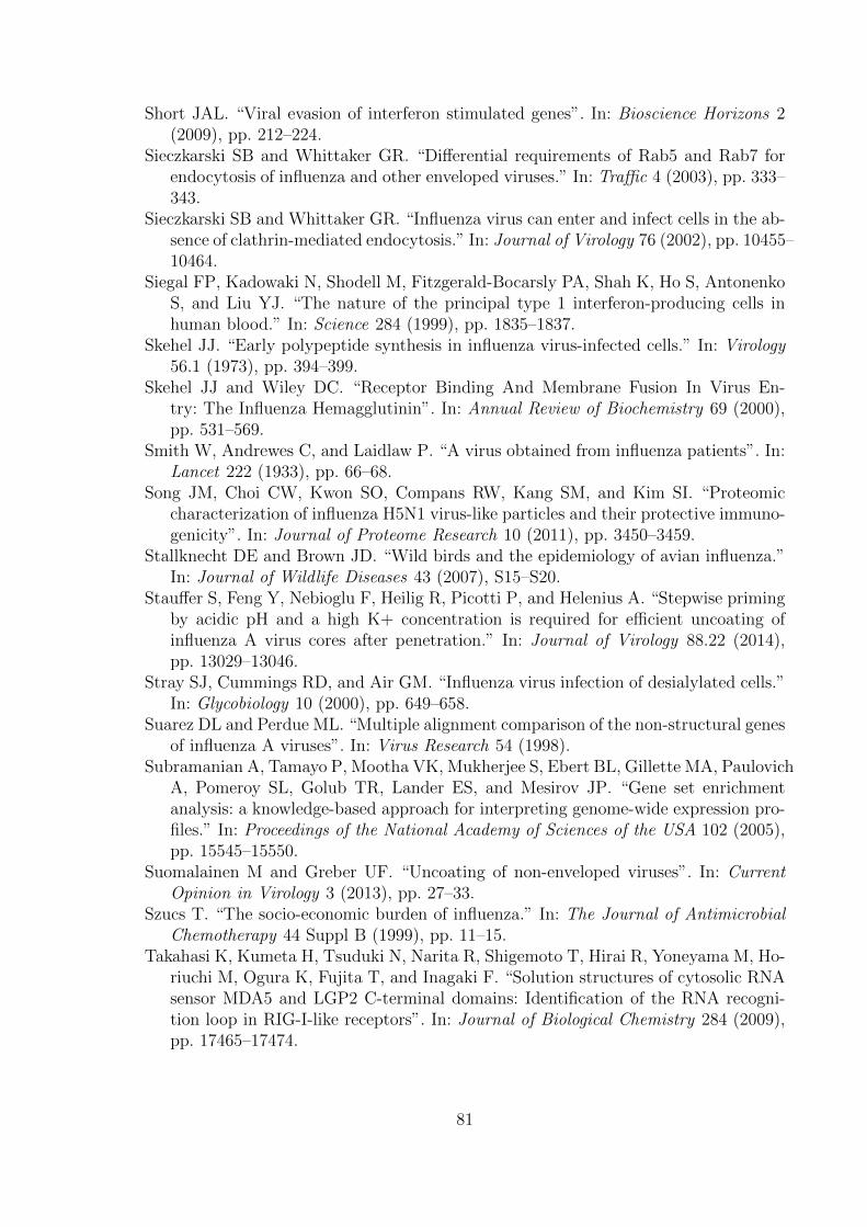
Short JAL. “Viral evasion of interferon stimulated genes”. In: Bioscience Horizons 2(2009), pp. 212–224.
Sieczkarski SB and Whittaker GR. “Differential requirements of Rab5 and Rab7 forendocytosis of influenza and other enveloped viruses.” In: Traffic 4 (2003), pp. 333–343.
Sieczkarski SB and Whittaker GR. “Influenza virus can enter and infect cells in the ab-sence of clathrin-mediated endocytosis.” In: Journal of Virology 76 (2002), pp. 10455–10464.
Siegal FP, Kadowaki N, Shodell M, Fitzgerald-Bocarsly PA, Shah K, Ho S, AntonenkoS, and Liu YJ. “The nature of the principal type 1 interferon-producing cells inhuman blood.” In: Science 284 (1999), pp. 1835–1837.
Skehel JJ. “Early polypeptide synthesis in influenza virus-infected cells.” In: Virology56.1 (1973), pp. 394–399.
Skehel JJ and Wiley DC. “Receptor Binding And Membrane Fusion In Virus En-try: The Influenza Hemagglutinin”. In: Annual Review of Biochemistry 69 (2000),pp. 531–569.
Smith W, Andrewes C, and Laidlaw P. “A virus obtained from influenza patients”. In:Lancet 222 (1933), pp. 66–68.
Song JM, Choi CW, Kwon SO, Compans RW, Kang SM, and Kim SI. “Proteomiccharacterization of influenza H5N1 virus-like particles and their protective immuno-genicity”. In: Journal of Proteome Research 10 (2011), pp. 3450–3459.
Stallknecht DE and Brown JD. “Wild birds and the epidemiology of avian influenza.”In: Journal of Wildlife Diseases 43 (2007), S15–S20.
Stauffer S, Feng Y, Nebioglu F, Heilig R, Picotti P, and Helenius A. “Stepwise primingby acidic pH and a high K+ concentration is required for efficient uncoating ofinfluenza A virus cores after penetration.” In: Journal of Virology 88.22 (2014),pp. 13029–13046.
Stray SJ, Cummings RD, and Air GM. “Influenza virus infection of desialylated cells.”In: Glycobiology 10 (2000), pp. 649–658.
Suarez DL and Perdue ML. “Multiple alignment comparison of the non-structural genesof influenza A viruses”. In: Virus Research 54 (1998).
Subramanian A, Tamayo P, Mootha VK, Mukherjee S, Ebert BL, Gillette MA, PaulovichA, Pomeroy SL, Golub TR, Lander ES, and Mesirov JP. “Gene set enrichmentanalysis: a knowledge-based approach for interpreting genome-wide expression pro-files.” In: Proceedings of the National Academy of Sciences of the USA 102 (2005),pp. 15545–15550.
Suomalainen M and Greber UF. “Uncoating of non-enveloped viruses”. In: CurrentOpinion in Virology 3 (2013), pp. 27–33.
Szucs T. “The socio-economic burden of influenza.” In: The Journal of AntimicrobialChemotherapy 44 Suppl B (1999), pp. 11–15.
Takahasi K, Kumeta H, Tsuduki N, Narita R, Shigemoto T, Hirai R, Yoneyama M, Ho-riuchi M, Ogura K, Fujita T, and Inagaki F. “Solution structures of cytosolic RNAsensor MDA5 and LGP2 C-terminal domains: Identification of the RNA recogni-tion loop in RIG-I-like receptors”. In: Journal of Biological Chemistry 284 (2009),pp. 17465–17474.
81
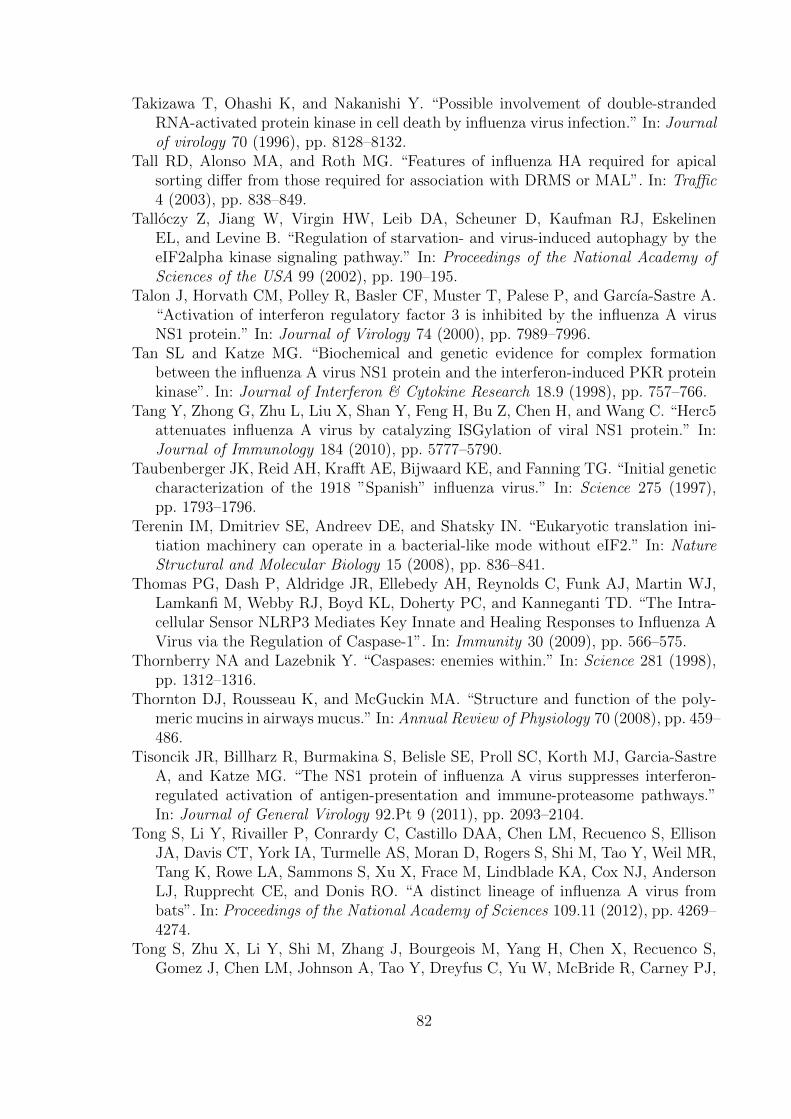
Takizawa T, Ohashi K, and Nakanishi Y. “Possible involvement of double-strandedRNA-activated protein kinase in cell death by influenza virus infection.” In: Journalof virology 70 (1996), pp. 8128–8132.
Tall RD, Alonso MA, and Roth MG. “Features of influenza HA required for apicalsorting differ from those required for association with DRMS or MAL”. In: Traffic4 (2003), pp. 838–849.
Talloczy Z, Jiang W, Virgin HW, Leib DA, Scheuner D, Kaufman RJ, EskelinenEL, and Levine B. “Regulation of starvation- and virus-induced autophagy by theeIF2alpha kinase signaling pathway.” In: Proceedings of the National Academy ofSciences of the USA 99 (2002), pp. 190–195.
Talon J, Horvath CM, Polley R, Basler CF, Muster T, Palese P, and Garcıa-Sastre A.“Activation of interferon regulatory factor 3 is inhibited by the influenza A virusNS1 protein.” In: Journal of Virology 74 (2000), pp. 7989–7996.
Tan SL and Katze MG. “Biochemical and genetic evidence for complex formationbetween the influenza A virus NS1 protein and the interferon-induced PKR proteinkinase”. In: Journal of Interferon & Cytokine Research 18.9 (1998), pp. 757–766.
Tang Y, Zhong G, Zhu L, Liu X, Shan Y, Feng H, Bu Z, Chen H, and Wang C. “Herc5attenuates influenza A virus by catalyzing ISGylation of viral NS1 protein.” In:Journal of Immunology 184 (2010), pp. 5777–5790.
Taubenberger JK, Reid AH, Krafft AE, Bijwaard KE, and Fanning TG. “Initial geneticcharacterization of the 1918 ”Spanish” influenza virus.” In: Science 275 (1997),pp. 1793–1796.
Terenin IM, Dmitriev SE, Andreev DE, and Shatsky IN. “Eukaryotic translation ini-tiation machinery can operate in a bacterial-like mode without eIF2.” In: NatureStructural and Molecular Biology 15 (2008), pp. 836–841.
Thomas PG, Dash P, Aldridge JR, Ellebedy AH, Reynolds C, Funk AJ, Martin WJ,Lamkanfi M, Webby RJ, Boyd KL, Doherty PC, and Kanneganti TD. “The Intra-cellular Sensor NLRP3 Mediates Key Innate and Healing Responses to Influenza AVirus via the Regulation of Caspase-1”. In: Immunity 30 (2009), pp. 566–575.
Thornberry NA and Lazebnik Y. “Caspases: enemies within.” In: Science 281 (1998),pp. 1312–1316.
Thornton DJ, Rousseau K, and McGuckin MA. “Structure and function of the poly-meric mucins in airways mucus.” In: Annual Review of Physiology 70 (2008), pp. 459–486.
Tisoncik JR, Billharz R, Burmakina S, Belisle SE, Proll SC, Korth MJ, Garcia-SastreA, and Katze MG. “The NS1 protein of influenza A virus suppresses interferon-regulated activation of antigen-presentation and immune-proteasome pathways.”In: Journal of General Virology 92.Pt 9 (2011), pp. 2093–2104.
Tong S, Li Y, Rivailler P, Conrardy C, Castillo DAA, Chen LM, Recuenco S, EllisonJA, Davis CT, York IA, Turmelle AS, Moran D, Rogers S, Shi M, Tao Y, Weil MR,Tang K, Rowe LA, Sammons S, Xu X, Frace M, Lindblade KA, Cox NJ, AndersonLJ, Rupprecht CE, and Donis RO. “A distinct lineage of influenza A virus frombats”. In: Proceedings of the National Academy of Sciences 109.11 (2012), pp. 4269–4274.
Tong S, Zhu X, Li Y, Shi M, Zhang J, Bourgeois M, Yang H, Chen X, Recuenco S,Gomez J, Chen LM, Johnson A, Tao Y, Dreyfus C, Yu W, McBride R, Carney PJ,
82
Gilbert AT, Chang J, Guo Z, Davis CT, Paulson JC, Stevens J, Rupprecht CE,Holmes EC, Wilson Ia, and Donis RO. “New World Bats Harbor Diverse InfluenzaA Viruses”. In: PLoS Pathogens 9.10 (2013).
Treanor JJ, Snyder MH, London WT, and Murphy BR. “The B allele of the NS geneof avian influenza viruses, but not the A allele, attenuates a human influenza Avirus for squirrel monkeys.” In: Virology 171.1 (1989), pp. 1–9.
Trinchieri G. “Interleukin-12 and the regulation of innate resistance and adaptive im-munity.” In: Nature Reviews Immunology 3 (2003), pp. 133–146.
Tschopp J and Schroder K. “NLRP3 inflammasome activation: The convergence ofmultiple signalling pathways on ROS production?” In: Nature Reviews Immunology10 (2010), pp. 210–215.
Tu J, Guo J, Zhang A, Zhang W, Zhao Z, Zhou H, Liu C, Chen H, and Jin M. “Effectsof the C-terminal truncation in NS1 protein of the 2009 pandemic H1N1 influenzavirus on host gene expression”. In: PLoS One 6 (2011).
Twu KY, Noah DL, Rao P, Kuo RL, and Krug RM. “The CPSF30 binding site onthe NS1A protein of influenza A virus is a potential antiviral target.” In: Journalof Virology 80 (2006), pp. 3957–3965.
Van Der Sluijs KF, Van Elden LJR, Arens R, Nijhuis M, Schuurman R, FlorquinS, Kwakkel J, Akira S, Jansen HM, Lutter R, and Van Der Polls T. “Enhancedviral clearance in interleukin-18 gene-deficient mice after pulmonary infection withinfluenza A virus”. In: Immunology 114 (2005), pp. 112–120.
Van Boxel-Dezaire AHH, Rani MRS, and Stark GR. “Complex Modulation of CellType-Specific Signaling in Response to Type I Interferons”. In: Immunity 25 (2006),pp. 361–372.
Van Oers MM. “Opportunities and challenges for the baculovirus expression system”.In: Journal of Invertebrate Pathology 107 (2011).
Van Riel D, den Bakker MA, Leijten LME, Chutinimitkul S, Munster VJ, de Wit E,Rimmelzwaan GF, Fouchier RAM, Osterhaus ADME, and Kuiken T. “Seasonaland pandemic human influenza viruses attach better to human upper respiratorytract epithelium than avian influenza viruses.” In: American Journal of Pathology176 (2010), pp. 1614–1618.
Vannucci L, Lai M, Chiuppesi F, Ceccherini-Nelli L, and Pistello M. “Viral vectors: alook back and ahead on gene transfer technology.” In: The New Microbiologica 36(2013), pp. 1–22.
Varga ZT, Ramos I, Hai R, Schmolke M, Garcıa-Sastre A, Fernandez-Sesma A, andPalese P. “The influenza virus protein PB1-F2 inhibits the induction of type iinterferon at the level of the MAVS adaptor protein”. In: PLoS Pathogens 7 (2011).
Vassalli JD, Huarte J, Belin D, Gubler P, Vassalli A, O’Connell ML, Parton LA, Rick-les RJ, and Strickland S. “Regulated polyadenylation controls mRNA translationduring meiotic maturation of mouse oocytes”. In: Genes and Development 3 (1989),pp. 2163–2171.
Vela A, Fedorova O, Ding SC, and Pyle AM. “The thermodynamic basis for viral RNAdetection by the RIG-I innate immune sensor”. In: Journal of Biological Chemistry287 (2012), pp. 42564–42573.
83

Vercammen E, Staal J, and Beyaert R. “Sensing of viral infection and activation of in-nate immunity by toll-like receptor 3”. In: Clinical Microbiology Reviews 21 (2008),pp. 13–25.
Walsh D and Mohr I. “Viral subversion of the host protein synthesis machinery”. In:Nature Reviews Microbiology 9.12 (2011), pp. 860–875.
Wang R, Iwakura Y, Araki K, Sotoyama H, Takei N, and Nawa H. “In vitro productionof an active neurotrophic factor, neuregulin-1: qualitative comparison of differentcell-free translation systems”. In: Neuroscience Letters 497.2 (2011), pp. 90–93.
Wang R and Brattain MG. “The maximal size of protein to diffuse through the nuclearpore is larger than 60 kDa”. In: FEBS Letters 581 (2007), pp. 3164–3170.
Wang S, Zhao Z, Bi Y, Sun L, Liu X, and Liu W. “Tyrosine 132 phosphorylationof influenza A virus M1 protein is crucial for virus replication by controlling thenuclear import of M1.” In: Journal of Virology 87 (2013), pp. 6182–91.
Wang W, Riedel K, Lynch P, Chien CY, Montelione GT, and Krug RM. “RNA bindingby the novel helical domain of the influenza virus NS1 protein requires its dimerstructure and a small number of specific basic amino acids”. In: RNA 5.2 (1999),pp. 195–205.
Wang X, Li M, Zheng H, Muster T, Palese P, Beg AA, and Garcia-Sastre A. “InfluenzaA virus NS1 protein prevents activation of NF-kappaB and induction of alpha/betainterferon.” In: Journal of Virology 74.24 (2000), pp. 11566–11573.
Ward AC, Azad AA, and Macreadie IG. “Expression and characterisation of the in-fluenza A virus non-structural protein NS1 in yeast”. In: Archives of Virology 138(1994), pp. 299–314.
Watanabe T, Watanabe S, and Kawaoka Y. “Cellular networks involved in the influenzavirus life cycle”. In: Cell Host and Microbe 7 (2010), pp. 427–439.
Webby RJ and Webster RG. “Are we ready for pandemic influenza?” In: Science 302(2003), pp. 1519–1522.
Webster RG, Bean WJ, Gorman OT, Chambers TM, and Kawaoka Y. “Evolution andecology of influenza A viruses.” In: Microbiological reviews 56 (1992), pp. 152–179.
Whitlow ZW, Connor JH, and Lyles DS. “Preferential translation of vesicular stomati-tis virus mRNAs is conferred by transcription from the viral genome.” In: Journalof Virology 80 (2006), pp. 11733–11742.
Wilusz J, Shenk T, Takagaki Y, and Manley JL. “A multicomponent complex isrequired for the AAUAAA-dependent cross-linking of a 64-kilodalton protein topolyadenylation substrates.” In: Molecular and Cellular Biology 10 (1990), pp. 1244–1248.
Wise HM, Foeglein A, Sun J, Dalton RM, Patel S, Howard W, Anderson EC, BarclayWS, and Digard P. “A complicated message: Identification of a novel PB1-relatedprotein translated from influenza A virus segment 2 mRNA.” In: Journal of Virology83 (2009), pp. 8021–8031.
Wisskirchen C, Ludersdorfer TH, Muller DA, Moritz E, and Pavlovic J. “The Cel-lular RNA Helicase UAP56 Is Required for Prevention of Double-Stranded RNAFormation during Influenza A Virus Infection.” In: Journal of Virology 85 (2011),pp. 8646–8655.
Wong AHT, Ning Tam NW, Yang YL, Cuddihy AR, Li S, Kirchhoff S, Hauser H, DeckerT, and Koromilas AE. “Physical association between STAT1 and the interferon-
84

inducible protein kinase PKR and implications for interferon and double-strandedRNA signaling pathways”. In: EMBO Journal 16 (1997), pp. 1291–1304.
Woo HM, Kim KS, Lee JM, Shim HS, Cho SJ, Lee WK, Ko HW, Keum YS, Kim SY,Pathinayake P, Kim CJ, and Jeong YJ. “Single-stranded DNA aptamer that specif-ically binds to the influenza virus NS1 protein suppresses interferon antagonism”.In: Antiviral Research 100 (2013), pp. 337–345.
Wurzer WJ, Planz O, Ehrhardt C, Giner M, Silberzahn T, Pleschka S, and LudwigS. “Caspase 3 activation is essential for efficient influenza virus propagation”. In:EMBO Journal 22 (2003), pp. 2717–2728.
Wysocka J and Herr W. “The herpes simplex virus VP16-induced complex: The mak-ings of a regulatory switch”. In: Trends in Biochemical Sciences 28 (2003), pp. 294–304.
Xia S, Monzingo AF, and Robertus JD. “Structure of NS1A effector domain fromthe influenza A/Udorn/72 virus.” In: Acta Crystallographica. Section D, BiologicalCrystallography 65.Pt 1 (2009), pp. 11–7.
Xing Z, Cardona CJ, Anunciacion J, Adams S, and Dao N. “Roles of the ERK MAPK inthe regulation of proinflammatory and apoptotic responses in chicken macrophagesinfected with H9N2 avian influenza virus.” In: The Journal of General Virology91.Pt 2 (2010), pp. 343–351.
Xu K, Klenk C, Liu B, Keiner B, Cheng J, Zheng BJ, Li L, Han Q, Wang C, Li T, ChenZ, Shu Y, Liu J, Klenk HD, and Sun B. “Modification of nonstructural protein 1 ofinfluenza A virus by SUMO1.” In: Journal of Virology 85 (2011), pp. 1086–1098.
Yamauchi Y and Helenius A. “Virus entry at a glance.” In: Journal of Cell Science126.Pt 6 (2013), pp. 1289–1295.
Ye Z, Liu T, Offringa DP, McInnis J, and Levandowski RA. “Association of influenzavirus matrix protein with ribonucleoproteins.” In: Journal of Virology 73 (1999),pp. 7467–7473.
Yin C, Khan JA, Swapna GV, Ertekin A, Krug RM, Tong L, and Montelione GT.“Conserved surface features form the double-stranded RNA binding site of non-structural protein 1 (NS1) from influenza A and B viruses”. In: Journal of BiologicalChemistry 282.28 (2007), pp. 20584–20592.
Yin C, Khan Ja, Swapna GVT, Ertekin A, Krug RM, Tong L, and Montelione GT.“Conserved surface features form the double-stranded RNA binding site of non-structural protein 1 (NS1) from influenza A and B viruses.” In: Journal of BiologicalChemistry 282.28 (2007), pp. 20584–92.
Ying Chou Y, Heaton NS, Gao Q, Palese P, Singer R, and Lionnet T. “Colocalizationof Different Influenza Viral RNA Segments in the Cytoplasm before Viral Buddingas Shown by Single-molecule Sensitivity FISH Analysis”. In: PLoS Pathogens 9(2013).
Yoneyama M, Kikuchi M, Matsumoto K, Imaizumi T, Miyagishi M, Taira K, Foy E,Loo YM, Gale MJ, Akira S, Yonehara S, Kato A, and Fujita T. “Shared and uniquefunctions of the DExD/H-box helicases RIG-I, MDA5, and LGP2 in antiviral innateimmunity.” In: Journal of Immunology 175.5 (2005), pp. 2851–2858.
Yoneyama M, Kikuchi M, Natsukawa T, Shinobu N, Imaizumi T, Miyagishi M, TairaK, Akira S, and Fujita T. “The RNA helicase RIG-I has an essential function in
85

double-stranded RNA-induced innate antiviral responses.” In: Nature Immunology5 (2004), pp. 730–737.
York A and Fodor E. “Biogenesis, assembly, and export of viral messenger ribonu-cleoproteins in the influenza A virus infected cell.” In: RNA Biology 10 (2013),pp. 1274–82.
York A, Hutchinson EC, and Fodor E. “Interactome analysis of the influenza a virustranscription/replication machinery identifies protein phosphatase 6 as a cellularfactor required for efficient virus replication.” In: Journal of Virology 88.22 (2014),pp. 13284–13299.
Yuan P, Bartlam M, Lou Z, Chen S, Zhou J, He X, Lv Z, Ge R, Li X, Deng T, FodorE, Rao Z, and Liu Y. “Crystal structure of an avian influenza polymerase PA(N)reveals an endonuclease active site.” In: Nature 458 (2009), pp. 909–913.
Zarkower D and Wickens M. “Formation of mRNA 3’ termini: stability and dissociationof a complex involving the AAUAAA sequence.” In: The EMBO Journal 6 (1987),pp. 177–186.
Zeng W, Sun L, Jiang X, Chen X, Hou F, Adhikari A, Xu M, and Chen ZJ. “Recon-stitution of the RIG-I pathway reveals a signaling role of unanchored polyubiquitinchains in innate immunity.” In: Cell 141.2 (2010), pp. 315–330.
Zhang S, Wang J, Wang Q, and Toyoda T. “Internal initiation of influenza virus repli-cation of viral RNA and complementary RNA in vitro”. In: Journal of BiologicalChemistry 285 (2010), pp. 41194–41201.
Zhao C, Hsiang TY, Kuo RL, and Krug RM. “ISG15 conjugation system targets theviral NS1 protein in influenza A virus-infected cells”. In: Proceedings of the NationalAcademy of Sciences of the USA 107.5 (2010), pp. 2253–2258.
Zhirnov OP and Klenk HD. “Control of apoptosis in influenza virus-infected cells byup-regulation of Akt and p53 signaling.” In: Apoptosis 12.8 (2007), pp. 1419–1432.
Zielecki F, Semmler I, Kalthoff D, Voss D, Mauel S, Gruber AD, Beer M, and Wolff T.“Virulence determinants of avian H5N1 influenza A virus in mammalian and avianhosts: role of the C-terminal ESEV motif in the viral NS1 protein.” In: Journal ofVirology 84.20 (2010), pp. 10708–10718.
Zietara N, Lyszkiewicz M, Gekara N, Pucha lka J, Dos Santos VAPM, Hunt CR, PanditaTK, Lienenklaus S, and Weiss S. “Absence of IFN-beta impairs antigen presentationcapacity of splenic dendritic cells via down-regulation of heat shock protein 70.” In:Journal of Immunology 183 (2009), pp. 1099–1109.
Zinzula L and Tramontano E. “Strategies of highly pathogenic RNA viruses to blockdsRNA detection by RIG-I-like receptors: Hide, mask, hit”. In: Antiviral Research100 (2013), pp. 615–635.
Zohari S, Gyarmati P, Ejdersund A, Berglof U, Thoren P, Ehrenberg M, Czifra G,Belak S, Waldenstrom J, Olsen B, and Berg M. “Phylogenetic analysis of the non-structural (NS) gene of influenza A viruses isolated from mallards in NorthernEurope in 2005”. In: Virology Journal 5 (2008), p. 147.
86