influences of hippocampal place cell firing in the awake
TRANSCRIPT

The Journal of Neuroscience, August 1989, g(8): 2907-2918
Influences of Hippocampal Place Cell Firing in the Awake State on the Activity of These Cells During Subsequent Sleep Episodes
Constantine Pavlides and Jonathan Winson
The Rockefeller University, New York, New York 10021
Rat hippocampal (CAl) complex spike “place cells” of freely behaving rats were recorded in pairs continuously during a series of waking (exploration and still-alert), drowsy (quiet- awake), and sleeping (slow-wave, pre-rapid-eye-movement and rapid-eye-movement sleep) behaviors. Pairs of units were selected that had nonoverlapping place fields. The rats were restricted from entering the place field of either cell overnight, and on the day of recording cells were exposed to their individual place fields independently and in a coun- terbalanced manner. Following exposure, recordings were made in the subsequent sleep episodes and the firing char- acteristics of both cells were analyzed.
Following exposure, significant increases in the spiking activity of the exposed cell were observed in the subsequent sleeping states that were not evident in the unexposed cell. The increased activity was observed in the rate of firing (spikes/set), the rate of occurrence of bursts with multiple spikes, as well as the number of bursts displaying short (2- 4 msec) interspike intervals.
The findings suggest that neuronal activity of hippocampal place cells in the awake states may influence the firing char- acteristics of these cells in subsequent sleep episodes. The increased firing rates along with the greater number of mul- tiple spike bursts and the shorter interspike intervals within the burst, following exposure to a cell’s place field, may represent possible information processing during sleep.
It is currently widely accepted that the hippocampal formation and related structures play an essential role in learning and memory. Lesion studies in humans indicate that destruction of the medial temporal lobe produces severe memory deficits (for review, see Squire, 1986). Furthermore, recent evidence suggests that lesions confined to the CA1 field of the hippocampus can produce comparable memory loss (Zola-Morgan et al., 1986). Memory deficits have also been reported in animals following destruction of the hippocampal formation and related structures (Olton, 1983; Squire and Zola-Morgan, 1983; Mishkin et al.,
Received Oct. 14, 1988; revised Jan. 19, 1989; accepted Feb. 16, 1989.
This work was supported by NIMH Research Scientist Development Award 5-K02-MH 00232, NSF grants BNS 8209875 and 8706053 and a grant from the Harry F. Guggenheim Foundation to J.W. We wish to thank Paul Roosin for his help in the computer programing, Dr. Yoram Greenstein and Mark Grudman for their help with the data analysis, Drs. Steve Fox and John Kubie for providing the design of the unit discriminator and instructions in the construction of the electrode assembly, Dr. Jim Ranck for his helpful comments on a preliminary version of the manuscript, Drs. Thorn Frumkes and Richard Bodnar for their support and encouragement, and Dr. Hiroshi Asanuma for his continued support.
Correspondence should be addressed to Constantine Pavlides, Box 298, The Rockefeller University, 1230 York Avenue, New York, NY 1002 1.
Copyright 0 1989 Society for Neuroscience 0270-6474/89/082907-12$02.00/O
1984) or the CA1 field alone (Davis et al., 1986). A particular type of memory that is especially disrupted following hippo- campal lesions in rats is memory for spatial location (Olton and Samuelson, 1976; Morris et al., 1982; Kesner et al., 1988).
One may conclude from these lesion studies that learning and memory are dependent upon or subserved by neural circuitry and activity within the hippocampal formation, and a number of studies of unit activity have addressed this issue. Experiments using either classical or operant conditioning paradigms have demonstrated increased hippocampal unit activity closely cor- related with specific stimulus-response-reinforcement contin- gencies, providing further evidence for a direct involvement of this structure in learning and memory (Segal and Olds, 1972; Deadwyler et al., 1979; Thompson et al., 1980; Berger et al., 1983; Wible et al., 1986; Eichenbaum et al., 1987).
More directly related to the present experiment, O’Keefe and colleagues observed, in freely behaving rats, that the majority of hippocampal complex spike cells (see Fig. lA), believed to be pyramidal cells from the CA1 and CA3 fields, display “place cell” characteristics (O’Keefe and Dostrovsky, 197 1; O’Keefe, 1976, 1983; O’Keefe and Conway, 1978; O’Keefe and Speak- man, 1987). This finding has also been confirmed and expanded in a number of other laboratories (Hill, 1978; Olton et al., 1978; Best and Ranck, 1982; Kubie and Ranck, 1983; Muller and Kubie, 1987; Muller et al., 1987; for review, see O’Keefe, 1979). In brief, the majority of hippocampal complex spike cells in freely behaving rats increase their firing rates dramatically when an animal is situated in a particular location in space. [It should be noted, however, that in his classic study, Ranck (1973) showed that hippocampal pyramidal cells in freely behaving animals may also fire in response to environmental contingencies other than space.] A place field once established is maintained over rather long periods of time-weeks to months (Best and Thomp- son, 1984; Muller et al., 1987). This is true despite the fact that the animals may be removed from the environment containing the place field and reintroduced to it at some later time.
As has been shown in Ranck’s original study (Ranck, 1973), as well as in a number of other experiments (Olmstead et al., 1973; Suzuki and Smith, 1985a, b), the firing rates of hippo- campal complex spike cells, when not in their place fields, are strongly modified by behavior. Thus, these cells fire at their highest rates during sleep states and at their lowest rates during waking. This enhanced firing raises the possibility of memory processing during sleep. There have been several other indica- tions that mnemonic processes may take place during sleeping states. For example, Winson and Abzug (1978) demonstrated the phenomenon of behavioral gating in the hippocampus. It was found that information processing through the hippocampal trisynaptic circuit is dependent on the behavior of the animal.
2908 Pavlides and Winson - Hippocampal Unit Activity in Waking and Sleeping Rats
Thus, the transmission of a stimulus through the circuit is rel- atively unrestricted during slow-wave sleep (SWS) but is re- stricted during waking states (Winson and Abzug, 1978). Re- cently, we have also shown in our laboratory that long-term potentiation, widely held as a model of learning and memory, can be preferentially induced if the tetanic stimuli are given in phase with the local theta rhythm (Pavlides et al., 1988). Theta rhythm in the normal animal occurs during 2 specific behaviors, exploration and rapid-eye-movement (REM) sleep. Last, it is well known that in humans mentation and dreaming occur in SWS and REM sleep, respectively. Both of these phenomena may be indicative of ongoing information processing.
The basis of the present study was the hypothesis that neu- ronal firing of hippocampal cells during sleeping states may possibly represent some form of memory processing and that this processing may be determined by the activity of these cells during preceding waking states. To investigate this hypothesis, the firing characteristics of hippocampal place cells were studied in waking and sleeping states. [Place cells were chosen for the following reasons: First, as indicated above, these units appear to be involved in spatial memory (O’Keefe and Speakman, 1987). Second, the activity of these cells can be brought under exper- imental control, i.e., by determining when an animal is per- mitted to enter the place field of a particular place cell.] In a counterbalanced study in which pairs of place cells were re- corded simultaneously (see Materials and Methods for descrip- tion), it was indeed found that the firing characteristics of place cells during sleep were altered by their activity during their awake behaviors. Thus, if a cell fired extensively during awake exploration in its place field, enhanced activity was also ob- served during subsequent sleep episodes. In addition, our results present the first detailed normative data on the firing and burst- ing characteristics of CA1 cells in a series of waking and sleeping behaviors.
Materials and Methods Subjects. Male hooded rats (300-350 gm; Charles River Labs) were used. The animals were housed in colony cages with littermates until shortly before surgery. Subsequently, each animal was housed individ- ually in a Plexiglas cage lined with Purina bedding material. Purina rat pellets and water were available ad libitum except that food was removed the night before the day of testing and was reintroduced at the comple- tion of the test. The animals were rewarded randomly on all parts of a box or an &arm maze used for testing with precision food pellets (see below).
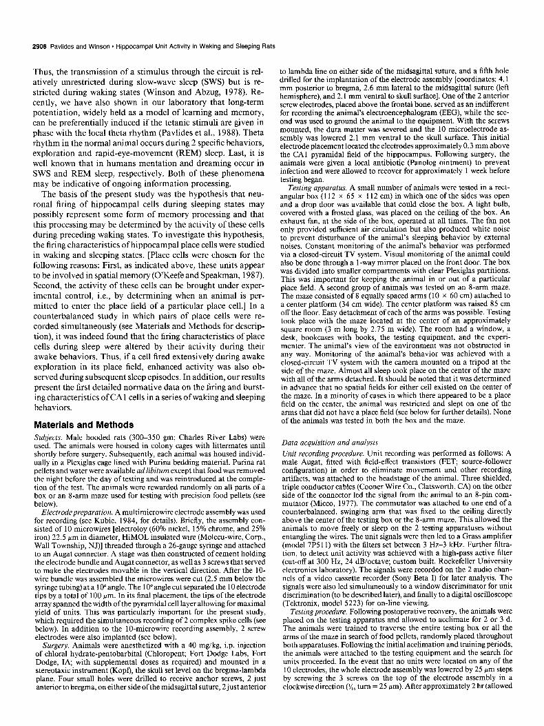
Electrode preparation. A multimicrowire electrode assembly was used for recording (see Kubie, 1984, for details). Briefly, the assembly con- sisted of 10 microwires [electroloy (60% nickel, 15% chrome, and 25% iron) 22.5 urn in diameter. HiMOL insulated wire (Molecu-wire. Corp., Wall Township, NJ)] threaded through a 26-gauge syringe and attached to an Augat connector. A stage was then constructed of cement holding the electrode bundle and Augat connector, as well as 3 screws that served to make the electrodes movable in the vertical direction. After the lo- wire bundle was assembled the microwires were cut (2.5 mm below the syringe tubing) at a lo” angle. The lo” angle cut separated the 10 electrode tips by a total of 100 pm. In its final placement, the tips of the electrode array spanned the width of the pyramidal cell layer allowing for maximal yield of units. This was particularly important for the present study, which required the simultaneous recording of 2 complex spike cells (see below). In addition to the IO-microwire recording assembly, 2 screw electrodes were also implanted (see below).
Surgery. Animals were anesthetized with a 40 mg/kg, i.p. injection of chloral hydrate-pentobarbital (Chloropent; Fort Dodge Labs, Fort Dodge, IA, with supplemental doses as required) and mounted in a stereotaxic instrument (Kopf), the skull set level on the bregma-lambda plane. Four small holes were drilled to receive anchor screws, 2 just anterior to bregma, on either side ofthe midsagittal suture, 2 just anterior
to lambda line on either side of the midsagittal suture, and a fifth hole drilled for the implantation of the electrode assembly [coordinates: 4.1 mm posterior to bregma, 2.6 mm lateral to the midsagittal suture (left hemisphere), and 2.1 mm ventral to skull surface]. One of the 2 anterior screw electrodes, placed above the frontal bone, served as an indifferent for recording the animal’s electroencephalogram (EEG), while the sec- ond was used to ground the animal to-the equipment. With the screws mounted, the dura matter was severed and the 10 microelectrode as- sembly was lowered 2.1 mm ventral to the skull surface. This initial electrode placement located the electrodes approximately 0.3 mm above the CA1 pyramidal field of the hippocampus. Following surgery, the animals were given a local antibiotic (Panolog ointment) to prevent infection and were allowed to recover for approximately 1 week before testing began.
Testing apparatus. A small number of animals were tested in a rect- angular box (112 x 65 x 112 cm) in which one of the sides was open and a drop door was available that could close the box. A light bulb, covered with a frosted glass, was placed on the ceiling of the box. An exhaust fan, at the side of the box, operated at all times. The fan not only provided sufficient air circulation but also produced white noise to prevent disturbance of the animal’s sleeping behavior by external noises. Constant monitoring of the animal’s behavior was performed via a closed-circuit TV system. Visual monitoring of the animal could also be done through a 1 -way mirror placed on the front door. The box was divided into smaller compartments with clear Plexiglas partitions. This was important for keeping the animal in or out of a particular place field. A second group of animals was tested on an S-arm maze. The maze consisted of 8 equally spaced arms (10 x 60 cm) attached to a center platform (34 cm wide). The center platform was raised 85 cm off the floor. Easy detachment of each of the arms was possible. Testing took place with the maze located at the center of an approximately square room (3 m long by 2.75 m wide). The room had a window, a desk, bookcases with books, the testing equipment, and the experi- menter. The animal’s view of the environment was not obstructed in any way. Monitoring of the animal’s behavior was achieved with a closed-circuit TV system with the camera mounted on a tripod at the side of the maze. Almost all sleep took place on the center of the maze with all of the arms detached. It should be noted that it was determined in advance that no spatial fields for either cell existed on the center of the maze. In a minority of cases in which there appeared to be a place field on the center, the animal was restricted and slept on one of the arms that did not have a place field (see below for further details). None of the animals was tested in both the box and the maze.
Data acquisition and analysis Unit recording procedure. Unit recording was performed as follows: A male Augat, fitted with field-effect transistors (FET, source-follower configuration) in order to eliminate movement and other recording artifacts, was attached to the headstage of the animal. Three shielded, triple conductor cables (Cooner Wire Co., Clatsworth, CA) on the other side of the connector led the signal from the animal to an 8-pin com- mutator (Micco, 1977). The commutator was attached to one end of a counterbalanced, swinging arm that was fixed to the ceiling directly above the center of the testing box or the S-arm maze. This allowed the animals to move freely or sleep on the 2 testing apparatuses without entangling the wires. The unit signals were then led to a Grass amplifier (model 7P5 11) with the filters set between 3 Hz-3 kHz. Further filtra- tion, to detect unit activity was achieved with a high-pass active filter (cut-off at 300 Hz, 24 dB/octave; custom built, Rockefeller University electronics laboratory). The signals were recorded on the 2 audio chan- nels of a video cassette recorder (Sony Beta I) for later analysis. The signals were also led simultaneously to a window discriminator for unit discrimination (to be described later), and finally to a digital oscilloscope (Tektronix, model 5223) for on-line viewing.
Testingprocedure. Following postoperative recovery, the animals were placed on the testing apparatus and allowed to acclimate for 2 or 3 d. The animals were trained to traverse the entire testing box or all the arms of the maze in search of food pellets, randomly placed throughout both apparatuses. Following the initial acclimation and training periods, the animals were attached to the testing equipment and the search for units proceeded. In the event that no units were located on any of the 10 electrodes, the whole electrode assembly was lowered by 25 pm steps by screwing the 3 screws on the top of the electrode assembly in a clockwise direction (x6 turn = 25 pm). After approximately 2 hr (allowed

The Journal of Neuroscience, August 1989, 9(8) 2909
for the electrodes to settle in the brain), all the electrodes were tested again for the availability of place cells. All testing for place cells and subsequent sleep recording took place in a single testing apparatus (i.e., either the box or the b-arm maze). An exact record of the electrode movement was recorded in order to calculate the precise location of the electrodes at the completion of the experiment. This procedure was repeated until 2 well-discriminable units (signal to noise ratio of over 3: 1) with different place fields were encountered. In the event that this was accomplished, actual recording began.
As previously mentioned, the objective was to be able to record from 2 units with nonoverlapping place fields simultaneously. Thus, when 2 or more units were available, the animal was enticed to traverse the entire box and all of the arms and the place fields were carefully doc- umented. A location on the maze was labeled a place field if the unit increased its firing at least 3-fold compared with adjacent positions on the box or maze (see below for further details). I f 2 units with well- differentiated and nonoverlapping place fields were located, the animal was put in its home cage, which was placed on top of the g-arm maze at its center. Both units were monitored in the home cage for place field firing. If none was observed, the animal was accepted for testing and remained in its home cage overnight. (In the occasional case in which there was a place field in the center of the maze, the same procedure was followed with the exception that the home cage was placed overnight on an arm that did not have a place field. Subsequent sleep episodes during testing, as described below, also occurred on that arm.)
On the following morning, the animal was hooked up to the equip- ment while in its home cage on the maze, and both units were checked to see whether a place field had developed in the home cage for either unit. In the event that none existed, the animal was placed on the S- arm maze and the place fields of both units were briefly checked to make sure that they were still present. With the place fields verified, the animal was placed on the center of the maze (with all arms removed) or a part of the testing box that contained no place fields for either unit and allowed to stay there for at least 2 hr. Since rats are nocturnal, after a brief exploratory period the majority of them would settle down and sleep. Unit activity, EEG, and behavior were recorded during this entire “baseline” period.
Following baseline recording, the arm containing the place field of one of the units was reattached to the maze, the animal was placed on this arm and was left there for approximately lo-15 min. Similarly, when animals were tested in the box, they were partitioned on the quadrant that contained the place field of one of the cells and were allowed to stay there for a similar amount of time. During this period, the unit fired at the high rates characteristic of the place field. In the majority of cases, the animal was either in an alert state or else would be engaged in exploratory behaviors while in the place field. In a mi- nority of cases, the animal would attempt to move away from the place field, in which case it had to be actively restrained there by holding. At the end of this “exposure” period, the animal was placed back on the center of the maze-or the section of the box that contained no place fields and kent there for a number of SWS-REM cvcles (for at least 2 hr). In the case of the maze, the arm containing ihe place field was removed during this time. The firing activity of the second unit that was not exposed to its place field was also recorded throughout this period. Subsequently, the second unit was similarly exposed to its re- spective place field and the procedure was repeated, again both cells being recorded simultaneously. At the end of all sleep recording, all the arms were replaced on the maze and each arm was checked for place fields. This was performed to ensure that the place fields were still intact for both cells. At the end of a successful experiment, the electrode assembly was advanced in 25 Hrn increments and the search for addi- tional units continued. When the electrode assembly passed through the pyramidal layer, the animal was killed and histologic analysis was per- formed to determine the location of the recording site (see below for details). In all, 13 units were recorded successfully. These consisted of 6 pairs of counterbalanced units, one “pair” consisting of 3 units. In this last case, units 2 and 3 served as control for unit 1 and unit 1 served as a control for units 2 and 3.
consisted of a time width (Tw) set at a chosen time delay (Td) (see Fig. 1B). I f all the parameters were met, the discriminator produced both a 0.4 bsec TTL and a + 15 V output. A digital display of the output was also available on the discriminator.
A number of criteria had to be met before units would be selected for further recording and analysis. These criteria were as follows: (1) the units had to display complex spikes during the initial course of testing; (2) units had to have a signal-to-noise ratio of at least 3-to-l; (3) the cells had to display clear and discrete place fields; (4) at least 2 units with the above-mentioned characteristics had to be present si- multaneously and to display diverse, nonoverlapping place fields. It should be noted that the firing characteristics of all the complex spike cells reported here represent averages of unit firing in many episodes within a particular behavioral state, recorded over the entire period of observation of approximately 5-6 hr.
Identification ofbehavioral state. Besides recording from one or more hippocampal units simultaneously, the hippocampal EEG was also re- corded differentially between one of the hippocampal electrodes and the skull screw indifferent. The signal was fed to a Grass amplifier (model 7DAF) and filtered between 3 and 20 Hz (24 dB/octave), for clear detection of the various behavioral states (to be discussed below). The EEG signal was viewed on 1 of the 3 oscilloscope channels available and was also recorded with a video camera (RCA, model TC20 11, with 16 mm lens) from 1 of the 3 oscilloscope channels for off-line analysis. This was achieved by monitoring the oscilloscope screen which was displaying the firing of 2 units and the EEG trace.
The animal’s behavior was recorded with a second video camera (RCA, model TC201 l/U ultracom) equipped with a manual zoom lens (12.5-75 mm) for close observation of the animal’s behavior. This camera rested’on a tripod on the side of the testing box or the side of the g-arm maze. The-2 video signals were fed inm a splitter-inserter (RCA TC 14 17A) and were then fed into the single video innut of the Sony Beta system (SLO-323) and recorded on Sony Dynamicron L-500 or L-750 video cassette tapes. The input of the video display (RCA, 35 cm B/W monitor, model TC12 14) was split into 2, one of the these showing the animal’s behavior, while the second was showing the signals of 2 units and the hippocampal EEG. An RCA date-time generator (TC 1440B) displayed the date and time while recording was taking place and during playback.
The animal’s behavior was subdivided into 7 different categories. These included a number of awake behaviors: exploration (XPL); still- alert (SAL), characterized by a low-amplitude, irregular EEG, and in- place field (IPF), which could include any awake behavioral state in- cluding exploration, still-alert, or consummatory behaviors. (Regardless of the animal’s behavior, place field firing was at a very high rate.) A quiet-awake @A) state was also discriminated: animal lying still with an increase showing in the EEG amplitude and the appearance of slow- wave mikes. Based on both the animal’s EEG in which sporadic slow- wave spikes appeared, and its quiet behavioral state from which the animal normally drifts into SWS, this may be considered a drowsy state intermediate between waking and SWS. Three sleeping states were dis- tinguished: SWS, large increase in EEG amplitude and the appearance of large irregular slow-wave spikes; pre-rapid-eye-movement (PREM), EEG begins to synchronize, the animal’s breathing becomes heavier, whisker movements become apparent; and, following PREM, rapid- eye-movement REM, evidenced by theta rhythm (4-8 Hz synchronized EEG), eye and whisker movements, heavy, irregular breathing, and head collapsing to the floor of the maze or box. Each particular sleep state was determined by observing an animal’s behavior in the video display monitor and also observing the animal’s EEG.
Analysis of data. Data analysis was performed as follows: the video tapes were played back and the unit signals were fed again to the unit discriminator with its windows set to discriminate a single unit. The animal’s behavior along with the EEG were displayed on the video monitor. These 2 criteria were used to subdivide the animal’s behavior into the 7 different categories (described above). Almost all data were analyzed by the primary investigator. In a number of cases, a second person, blind to the experimental design, performed the data reduction.
Unit discrimination. Unit discrimination was achieved through the This was done to eliminate any experimenter effects. use of a custom-built, dual digital window discriminator (Rockefeller The TTL output of the discriminator was fed into the custom-built University electronics laboratory). Available on the discriminator were input/output (I/O) card (Dejong, 1982) that was installed into an Apple 3 adjustable voltage levels and an adjustable time window. The signal IIe computer. The I/O card consisted of dual precision timers (6522 to be discriminated had to fulfill all of the specified window restrictions microprocessor), which in combination with custom written software to register as a spike. The 3 voltage levels were: a voltage trigger (Vt), performed the task of analyzing the bursting characteristics of the units a voltage high (Vh), and a voltage low (Vl) level. The time windows for this study. Briefly, a data-acquisition program (written in machine

2910 Pavltdes and Winson - Hippocampal Unit Activity In Waking and Sleeping Rats
Figure I. A, Oscilloscope trace of an extracellularly recorded complex spike burst. Note the decreasing amplitude of successive action potentials. Calibra- tions: 0.2 mV, 2 msec; negativity up- ward. See text for filter settings. B. Os- cilloscope trace of extraLellularly recorded action potential along with markers showing adjustable discrimi- nator settings. Marked on the photo- graph are the following voltage levels: voltage trigger (k’r) through which the signal has to pass, the voltage high (Vh) that must not be exceeded, and the volt- age low (VI) through which the signal must pass. The voltage zero (RI), or baseline, is also marked. Further shown is an adjustable time window (horizon- &dots, identified by Tw), during which time the action potential must pass through the V 1. The start of the time window is determined by the setting Td. In order for a signal to register, it had to satisfy all ofthe set parameters. Neg- ativity is upward.
I+ +I
2 Msec
’ I K-
Tw
I* -DI .l Msec

The Journal of Neuroscience, August 1989, 9(8) 2911
language to permit various calculations that had to be performed in microseconds) with adjustable parameters was started simultaneously with the Sony tape recorder. Once an input was detected by the I/O card, the timer was activated. If another spike appeared within a certain period of time (10 or 15 msec; preset at the beginning of the data analysis), then that spike was counted as part of the same burst. This was continued until the spike following the previous one exceeded the 10 or 15 msec interval. In this case, a number of calculations pertaining to the burst were performed [i.e., interspike interval (ISI), interburst interval (IBI), number of spikes within the burst, etc.], the averages of these calculations were kept in random-access memory, and the timer was cleared and reactivated to detect the next spike or burst. This analysis continued either until the animal changed its behavioral state (as could be determined by the video tape and in sleep behaviors also aided by the animal’s hippocampal EEG), or if a particular behavior was maintained for more than 5 min, it was subdivided into 5 min intervals that were then averaged. In either case, the data analysis was terminated, the data were stored on floppy disks and the analysis con- tinued for the next behavior.
Histology. At the completion of all recording in an animal, the most ventral location of the recording electrodes in the hippocampus was localized histologically. The exact location ofa particular microelectrode was determined by the Prussian blue method. The animals were deeply anesthetized with an i.p. injection of either Chloropent (60 mg/kg) or a 2 ml injection of sodium pentobarbital (Nembutal, Abbott Labora- tories) and a 10 WA current for 10 set was passed between one of the recording electrodes (positive) and the skull indifferent (negative). The animals were then perfused transcardially with a 9% saline solution mixed with 10% formalin, 4% potassium ferrocyanide, and 4% acetic acid. The brains were removed and stored in a 10% formalin, 40% sucrose solution for 2 d. They were then cut by the freezing method, at 40 pm section thickness, mounted on slides, and stained using the Kluver-Barrera method.
Results Histology Histological analysis of the rats’ brains revealed that all of the 13 units included in this study were located in the dorsal part of the hippocampus. Of these, 3 were located in the CAla sub- field, while the remaining 10 cells were located in the CAlc hippocampal subfield.
Unit firing
The units fired in their place field at a rate at least 3 times greater than at adjacent positions in the maze or the testing box. A place field was determined by monitoring the firing rate of a cell as the rat traversed each arm of the maze or region of the testing box. Typically, units increased their firing by a factor of 3-10 or more as the rat moved over several inches of terrain into the place field. The objective of the spatial survey was to determine locations in which the cell would fire vigorously. As seen below,
0.60
0.55
0.50
0.45
” 0.40
; 0.35
‘;; 0.30
e 0.25
k 0.20
0.15
0.10
0.05
0.00 XPL SAL
*co5
0 POST-NONEXPOSURE
E%l POST-EXPOSURE
: PF
BEHAVIORAL STATE
?EM REM
Figure 2. Average firing rate (all single spikes and spikes within a burst) post-nonexposure and postexposure to place field. There were significant increases in the firing rate during all sleep states following exposure (see text for details). (Significant increases are indicated by asterisks in this and following figures.)
firing rates within place fields on the order of 10 times the out- of-place field rates were achieved. There was no attempt to define the boundaries of the place field precisely. All pairs of place cells chosen for analysis displayed distinct place fields in different places on the 8-arm maze or box. At least half of the cells tested had multiple place fields, and the area of increased firing differed from a very small area (several inches along the arm of the maze or box) to a much wider area (half an arm of the maze or a third of the box). In all of these cases, however, increased firing within one of the place fields was much greater than the other place fields. This observation was made at the time of recording, and it was utilized to determine to which of the place fields the cells were going to be exposed.
The average firing rates (in spikes/set; each spike within a complex spike burst being counted separately) of the 13 cells, in all of the behavioral states tested, are presented in Table 1. Combining post-nonexposure to place field and postexposure to place field rates, the highest activity was observed in the IPF state (X = 2.17) followed by SWS (X = 0.36), PREM (X = 0.32), QA (X = 0.3 l), REM (X = 0.22), SAL (X = 0.17), and XPL (X = 0.14). (Note that the firing rates for post-nonexposure and postexposure were collapsed into a single mean; Table 1, line 3.)
Postexposure versus post-nonexposure to place field com-
Table 1. Rate of firing of the 13 complex spike (place) cells studied
Ratea (spikesisec)
XPL SAL QA sws PREM REM
nexp ew nexp exp IPF nexp em nexp ew nexp em nexp ew
Mean 0.14 0.13 0.15 0.19 2.17 0.27 0.34 0.28 0.44 0.27 0.37 0.17 0.27
SD 0.30 0.21 0.18 0.18 1.62 0.15 0.23 0.16 0.28 0.17 0.28 0.17 0.22
Combined 0.14 0.17 2.17 0.31 0.36 0.32 0.22
The units were recorded in pairs, and they were all found in the CA1 hippocampal field. The firing rates are separated in post-nonexposure (nexp) and postexposure (exp) means and calculated separately for each behavioral state. The mean combined (nexp + exp) rates are also shown on the bottom. The highest rates of firing are seen in the in-place-field (IPF’) state. The lowest firing is seen in the out-of-place field awake states. There were no significant post-nonexposure-postexposure differences in the firing rates in the awake states. In contrast, significant post-nonexposure-postexposure differences in the firing rate were observed in all of the sleep states (see text). The large SDS reflect the wide range of firing rates among the individual units. cl All spikes within a burst measured separately.

2912 Pavlides and Winson * Hippocampal Unit Activity in Waking and Sleeping Rats
Figure 3. Average rate of firing during sleep and QA, post-nonexposure and postexposure to place field, for each of the 13 units included in the study. A large number of the units increased their firing rates following exposure to the place field. (This increased firing oc- curred in 10 of 13 cells in SWS, 9 of 13 cells in PREM, 11 of 13 cells in REM and 8 of 13 cells in QA.)
uo
0.40
030
0.20
410
POST POST POST POST NONEXPOSURE EXPOSURE NONEXPOSURE EXPOSURE
Awake Pexp
Sleep
Awake
Pnexp
Sleep
h-lit 1
Histogram of firing rates for a representative pair of place cells recorded simultaneously (see text for testing procedure). Awake behaviors are shown above the horizontal line, while sleeping and drowsy (QA) behaviors are shown below. X, XPL, A, SAL, Z, IPF, Q, QA; S, SWS; Z’, PREM; R. REM; 0, other (any behavior that did not conform to any of the above categories, i.e., feeding, chewing, grooming, etc.). Unit 1 is shown on the top line, while unit 2 is on the bottom. Unit 1 was exposed to its place field first (as indicated by I, written horizontally), while unit 2 was unexposed (0). Following a number of sleeping and waking episodes, the order of exposure to place field was reversed. Each vertical column on the histogram represents one episode of a given behavior. Episodes are not necessarily of equal time length. The height of each column is proportional to the average firing rate during the given episode. For both unit 1 and unit 2, the graph reveals increased firing rates during sleep following exposure to place field (Pexp) compared with rates in sleep following nonexposure (Pnexp) to place field. As shown in Figure 2, the effect was significant for the group of cells tested.

0.40
1 QA
0.45 -
0.40.
0.35 -
0.30 -
0.25
0.20- UNSXPOSED
0.15 - 1.0 2.0
TEST
The Journal of Neuroscience, August 1989, 9(8) 2913
0.45-
EXFWZD
PREM
0.40.
0.35 - EXPOSED
0.30 - ONSXPOSSD
0.25-
UNEXPOSED
0.20 i 1.0 2.0
0.30, REM
EXPOSED EXPOSED
UNEXPOSED
0.14 1.0 2.0
TEST
Figure 5. Line graph of unit firing for a representative pair of units recorded simultaneously and in a counterbalanced manner. The 4 graphs show each of the 4 sleeping states tested (SWS, PREM, and REM), as well as QA. Initially, one of the units (line of decreasing slope) was exposed to its place field (classified as an order 1 unit; see text), while the second was unexposed, followed by sleep at the center of the maze or a restricted part of the box (Test 1). Subsequently, the other unit (line of increasing amplitude) was exposed to its place field (classified as an order 2 unit), while the first was not, followed by sleep (Test 2). The figure shows that the counterbalancing procedure succeeded. The unit that was exposed first increased its firing in the subsequent sleep episodes, while the second unit’s rate remained low. When the second unit was exposed, its rate increased in the sleep episodes that followed, while the first unit fired at a low rate.
parisons of the units’ firing rate revealed that exposure to place field produced a significant increase in the firing rate of these cells in subsequent behaviors [F(1,12) = 6.57; p < 0.021. A significant interaction between exposure and behavioral state was also found [F(5,60) = 2.40; p < 0.051, indicating that, following exposure to place field, the cells did not increase their firing rates significantly for all behaviors tested but only for specific behaviors. Thus, simple effects analysis of behavior by stage using Tukey’s method (a = 0.05) yielded significant post- exposure effects for SWS, PREM, and REM. No postexposure changes were detected for the behavioral states of XPL, SAL, and QA. It should be noted, however, that the rate of firing in the QA state approached the level of significance (p = 0.05). [The simple effects analysis also revealed that, taking postex- posure and post-nonexposure behavior as a whole, there were signfiicantly higher firing rates in SWS than in REM sleep (Schaf- fer-Welsch test; p -C 0.05).] Thus, exposure of the cell to its place field, in the waking state, signijicantly increased its firing rate
only in the sleep states but not the awake states that followed (see Fig. 2).
Figure 3 presents the average rate of firing of each of the individual cells post-nonexposure and postexposure to place field in the sleep states and QA. An increase in the firing rate, following exposure to place field, occurred in a large majority of place cells tested, leading to the overall significant increase in the firing rate noted above. In general, the increased firing during sleep appeared not to extend beyond an approximately 3 hr period, during which time a number of SWS-REM episodes occurred (see Fig. 4; to be discussed below). A search (in a few units) for a systematic time course of the increased firing rate failed to reveal any such trend; i.e., for some of the cells the increased firing occurred within the first few sleep episodes fol- lowing exposure, while other cells increased their firing rates during later episodes. Regardless, for most units there appeared to be a gradual decrease in firing during the last few intervals of the 2-3 hr recording session. (It should be noted that the unit

2914 Pavlides and Winson . Hippocampal Unit Activity in Waking and Sleeping Rats
Figure 6. A, Average bursts per scc- ond, post-nonexposure and postexpo- sure to place field. A significant main effect of exposure to place field was ob- served; however, the interaction of ex- posure and behavior failed to reach sig- nificance. Nonetheless, close inspection of the graph reveals that postexposure increases in the bursting rates took place only in SWS, PREM, and REM sleep. The failure to obtain a significant in- teraction is most likely due to the very large scatter seen for this variable (see Discussion). B. Average percent burst versus total events (bursts plus single spikes) post-nonexposurc and postcx- posure to place field. No significant change was observed following cxpo- sure to the place lield. However, the percentage of bursts occurring in REM sleep more than doubled following ex- posure of the cells to their place fields. C, Average spikes per burst per second, post-nonexposure and postexposurc to place field. A significantly higher num- ber of spikes per burst occurred follow- ing exposure to place field in the SWS and REM states. D. Average number of single spikes, post-nonexposure and postexposure to place field. Although the interaction of exposure and behav- ioral state was not significant, the great- est increases in the number of single spikes were observed in the sleep states and QA. E, Average number of bursts containing 2 spikes, post-nonexposure and postexposure to place field. The number of bursts with 2 spikes did not change following exposure to place field for any of the behavioral states tested. F, Average number ofbursts containing 3 spikes, post-nonexposure and postex- posure to place field. A significant in- crease in the number of bursts with 3 spikes occurred following exposure to place field during the sleep states (SWS. PREM, and REM) but not for the other behavioral states tested.
A
0.10 0 POST-NONMPOSURE
0.09 m Po5T-EXPOSURE
8 0.08
XPL SAL OA FM-9 PREM REM
C
8 e 1.50 Ii 5
q l.w g
: 0 0.50
!i 0.W
XPL SAL 4.05
E 0.06~
0 POST-NONEXPOSURE ml POST-MPOw?E
ar, SW!3 PRM REM
0 POST-NONEXPOSURE = POST-EXPOSURE
u sws PREM REM
D 0.50 0 POST-NONEXPOSURE 0.45 lpLB POST-EWOSURE
XPL 9l OA SWS PREU REM
BEHAWORM STATE BEHAMORU STATE
F 0 POST-NONDPOSURE
0.030 m POST-EXPOSURE T
Cl POST-NONEXPOSURE m POST--MPOSURE
0.000 ALOA SWS PREM REM XPL Sk cl4 SW9 PREN REM
4.05 BEHAMORAL STATE BEHAVIORAL STATE
analysis was done off-line, and the decision for the length of recording was not influenced by the unit firing.)
Figure 4 illustrates a representative example of a pair of units (units 1 and 2) recorded simultaneously in a counterbalanced design. At first, unit 1 was exposed to its place field, as can be evidenced by the increased firing at the upper-leftmost comer (indicated by I, written horizontally). Followingexposureofunit 1, the animal engaged in a number of SWS-REM episodes, as well as a number of waking behaviors (on the center of the maze or box). The second unit was being recorded simultaneously, and the firing rates for this unit are shown on the bottom of the graph. Subsequently, the second unit was exposed to its place field (while the first unit was not), followed by sleep. The coun- terbalanced procedure used in this experiment was tested uti- lizing a 3-way analysis of variance, with the dependent variable being the rate of cell firing (spikes/set) and the 3 factors being exposure to place field or not, behuvioral state, and order of exposure to place field; i.e., for some of the cells the order of testing was exposed to place field followed by unexposed to place field, while for other cells, the order was unexposed to place
field followed by exposed to place field. A significant interaction was achieved between exposure and order of testing [F( 1,ll) = 6.43; p < 0.051. [All units were classified as order 1, exposed lit-St in a pair or order 2, exposed second in a pair. The interaction occurs because, considering the first test of the day, order 1 units fired at a high rate, while order 2 fired at a low rate. Considering the second test of the day the opposite was true (see Fig. 5).] The counterbalancing procedure succeeded. Taking all animals together, the units that were exposed first increased their firing in the subsequent behaviors, while the units that were not re- mained relatively inactive. When the second of a pair of units was later exposed to their place field, they then increased their firing rate in the sleep episodes that followed, while the first unit subsided to a baseline rate. Thus, for all animals, increased firing was determined by exposure and not by whether units were exposed first or second.
In addition to the effects observed on the rate of firing, detailed analysis of bursting and the number of spikes within a burst were performed. All bursts of a given behavior were included and divided by the total time the animal was engaged in that

The Journal of Neuroscience, August 1989, 9(8) 2915
behavior, yielding average characteristics for a 1 set interval. The rates given for bursting, therefore, are similar to the rate of firing measurement and represent a density of bursting mea- sure. An ANOVA performed on the bursts per second revealed results similar to those for the rate of firing. As shown in Figure 6A, increased bursting was observed following exposure of the cell to its place field [F( 1,12) = 5.07; p < 0.041. The interaction of exposure to place field by behavior, however, failed to achieve significance. Nonetheless, as seen in Figure 6A, the only bursting rates that increased notably were those of the SWS, PREM, and REM states. As with unit firing rate, a significant main effect of behavioral state was also observed [F(5,55) = 2 1.6; p < 0.00 11. Taking postexposure and post-nonexposure behavior as a whole, significantly higher bursting rates were seen in SWS than in REM, SAL, or XPL.
The percentage of bursts versus total events (bursts + single spikes) was also analyzed. An analysis of variance performed on the postexposure percentage of bursts versus total events failed to detect significant main effects of exposure to place field [F(l,l 1) = 1.8; p > 0.051. Thus, the percentage of bursts ap- parently did not differ following exposure to place field. Al- though the interaction of exposure and behavioral state was not significant 0, = 0.08), and therefore, there was no significant increase in the postexposure sleep states in general, the postex- posure percentage of bursts more than doubled during REM sleep. A significant main effect for behavioral state was obtained [F(5,55) = 9.05; p c O.OOl]. A Schaffer-Welsch post hoc com- parison (a = 0.05) revealed that, for behavior as a whole, sig- nificantly higher bursting occurred during SWS, PREM, and QA than in SAL and XPL (see Fig. 6B).
An increase was also observed in the rate of occurrence of bursts with multiple spikes following exposure of the units to their place field [F(l, 12) = 4.6 1; p < 0.051. A simple effects analysis (Tukey test; a = 0.05) of exposure and behavioral state interaction revealed significant increases in the number of spikes per burst following exposure in the SWS and REM states but not in other behaviors. A significant main effect of behavioral state was also obtained [F(5,60) = 16.00;~ < 0.00 11. A Schaffer- Welsch post hoc analysis revealed that, for behavior as a whole, there were a higher number of spikes per burst during SWS than in SAL and XPL (see Fig. 6C).
Finer analysis of the number of spikes within a burst was also performed [i.e., single spike (no burst), double spikes, triple spikes, etc.]. ANOVA for each of the different number of spikes showed significant postexposure increases for the rate of single spikes [F( 1,12) = 4.44; p < 0.051. A significant interaction be- tween exposure and behavior was not observed. However, the greatest increases occurred during the sleep states and QA. Sig- nificant main effects were seen for behavioral state [F(5,60) = 15.83; p < O.OOl]. Post hoc comparisons (Schaffer-Welsch test; p < 0.05) showed that, considering both postexposure and post- nonexposure behaviors, there were significantly higher rates in the occurrence of single spikes in SWS, PREM, and QA com- pared with XPL and SAL (see Fig. 60). An ANOVA performed on the double-spike bursts failed to reach significance for ex- posure, indicating that the number of bursts with 2 spikes did not change following exposure to place field. Significant differ- ences, however, were seen for behavioral state [F(5,60) = 13.68; p < O.OOl]. For behavior as a whole, double spike bursts oc- curred significantly more often in SWS, PREM, and QA than in XPL and SAL (see Fig. 6E). The number of bursts with 3 spikes increased following exposure [F( 1,12) = 6.00; p < 0.031.
XPL SAL 1
0 POST-NONEXPOSURE
m POST-DUWSURE
?EM REM
7.0 6.5
1 - ::: 5.0
T t::
r
W Ei 2:5 2.0
0:5 1.:
0.0 XPL !3L
0.60 0.55
‘5 OSJ i! 0.45
0.40 7 0.35
c!
T In I
0.50 !
0.25 0.20 0.15 0.10 0.05 0.00
0 POST-NONMPOSURE
06E POST-MPOSURE
3A SW!3 PREM REM
BEHAVIORAL STATE
C 0 POST-NONMPQSURE
m POST-MPOSURE
OA sws PREM REM
*co3 8El-wl0RAL STATE
Figure 7. A, Average interburst interval, post-nonexposure and post- exposure to place field. There were no significant changes in the inter- burst interval following exposure to place field. B, Average interspike interval post-nonexposure and postexposure to place field. No signifi- cant changes were observed following exposure. C, Closer analysis of the interspike intervals revealed significant increases falling in the 2-4 msec range postexposure to place field. These increases occurred for all of the sleep states and QA but not in SAL and XPL (not shown in the graph).
A significant interaction between stage of exposure and behav- ioral state was also achieved [F(5,60) = 2.79;~ < 0.021. A simple effects analysis using Tukey’s method (a = 0.05) revealed a significant increase in the number of bursts composed of 3 spikes, following exposure, in the SWS, PREM, and REM states in comparison to the other states (see Fig. 60.
Measurements were also made of the average IBI. An AN- OVA performed on the average IBI post-nonexposure versus postexposure failed to achieve significance [F( 1,12) = 2.17; p = 0.161 (see Fig. 7A).

2916 Pavlides and Winson - Hippocampal Unit Activity in Waking and Sleeping Rats
Another measure taken was the average IS1 which represents the time interval between spikes, within a burst. An ANOVA performed on the postexposure average ISIS revealed no sig- nificant changes due to exposure of the unit to its place field (see Fig. 7B). The ISIS were also subdivided and analyzed in finer gradations (O-2, 2-4, 4-6, 6-8, 8-10, and lo-12 msec). Although no significant increases were observed in the average ISI, significant increases in the 2-4 msec (250-500 Hz) range were detected in the sleep states and QA following exposure of the cell to its place field [F(1,12) = 8.22; p < 0.031. No other interval reached significance (see Fig. 70.
For a number of cells (5 out of 13), the animal was rewarded with a chocolate chip cookie when it occupied the place field of a particular place cell (i.e., while the cell was being exposed to its place field). In almost all cases, the animal, which had been given this kind of reward on previous days, consumed a large portion of the cookie during the course of the unit’s exposure. The reward was introduced to see whether it would influence the unit’s firing characteristics. Close observation of the in- place-field firing rates, of a number of cells, failed to show pro- nounced changes in the firing patterns due to the animal’s con- summatory behavior in the place field (i.e., it remained rather high as long as the animal occupied the cell’s place field, re- gardless of his behavior or the reward). Similarly, significant effects were not detected in the firing rates of the “rewarded cells” in the subsequent sleep episodes [F( 1,9) = 1.28; p > 0.051. It should be noted, however, that the animal was rewarded with Noyes food pellets throughout the 8-arm maze over the extent of the experimental session and the chocolate chip cookies rep- resented only a qualitatively different reward.
In summary, the main findings of this study were as follows. (1) There was a significant increase in the rate of firing of hip- pocampal complex spike (place) cells during the sleep states (SWS, PREM, and REM) following exposure of these cells to their place field. (2) There was a significant increase in the av- erage number of spikes within a burst following exposure. This increase was evident only during the SWS and REM states. (3) There was a significant increase in the number of bursts with 3 spikes following exposure during the behavioral states of SWS, PREM, and REM. Finally, there was an increase in the number of bursts with the spikes falling in the 2-4 msec IS1 interval in all the sleeping states as well as QA.
Discussion
The results of the present experiment confirm our hypothesis that the awake unit activity of hippocampal place cells may influence the firing activity of these units in subsequent sleep states. Thus, the increased activity of these units in their place fields during the waking state enhanced their activity when the animal was engaged in sleep but not during the awake states that followed.
In detail, a significant increase in the firing rate was present during the sleeping but not the waking states following exposure. There was also a marked increase in the rate of bursting mainly during the SWS, PREM, and REM states (see Fig. 6A). However, on the basis of the absence of an interaction between exposure and behavioral state, it could not be determined whether the increased bursting occurred only in the sleeping behaviors. (A possible reason for the lack of a significant interaction was due to the high variability among the units.) Nonetheless, it is ev- ident from Figure 6A that the only behaviors that show an increase are those of SWS, PREM, and REM.
The average rate of occurrence of multiple spikes per burst also increased following exposure of the cell to its place field. These increases were evident only during the SWS and REM states. Closer analysis of the number of spikes within a burst revealed that the density of bursts containing 3 spikes increased significantly in the SWS, PREM, and REM states that followed exposure. Complex spike cells have been shown to fire in bursts containing as many as 7 or 8 spikes of decreasing amplitude (Ranck, 1973). Besides the single, double, and triple spikes re- ported in the results, measurements were also taken of the num- ber of bursts containing 4, 5, and 6+ spikes. These last 3 cat- egories were excluded from the final analysis, however, due to the low frequency of their occurrence. Although long bursts occurred too infrequently to include in the present analysis, bursts with many spikes (4-10-t) were observed; in a small number of cells, bursts with as many as 15 spikes were also recorded.
Finally, there was a significant increase in the number ofbursts with ISIS in the 2-4 msec range. This increase was evident in all of the sleep states, as well as in QA but was especially no- ticeable during the SWS, PREM, and REM states. This interval corresponds to approximately 400 Hz, which has been shown to be a rather sensitive frequency for inducing long-term po- tentiation (Winson and Dahl, 1986). Long-term potentiation has been hypothesized to be a mechanism underlying learning and memory and is thought to be a naturally occurring neuronal phenomenon (see discussion below).
In addition to the findings just cited, our recordings revealed that during tonic waking and sleeping behaviors, CA1 cells fire at their highest rates during SWS. They fire at considerably lower rates during REM. Such differential rates during sleep have been reported previously (Ranck, 1973; Olmstead et al., 1973; Suzuki and Smith, 1985a). We also observed, as did Ranck (1973) that during REM CA 1 cells are silent for prolonged periods of time interspersed with short, intense bursts of firing. The present results also indicate that rewarding an animal while exposing a cell to its place field did not appear to have a pronounced effect either on the in-place-field firing or the firing rates in the sleeping states that followed. The insensitivity of these units to reward has been alluded to previously (O’Keefe, 1979).
As described in the introductory remarks, neuronal trans- mission through the hippocampal formation is dependent on the behavioral state of the animal, i.e., neuronal gating (Winson and Abzug, 1978). To recapitulate, through the use of field po- tential analysis, it was demonstrated that transmission of a stim- ulus from the perforant path through the CA1 field of the hip- pocampal trisynaptic chain was restricted during waking states and REM (gate closed). Conversely, such transmission was to- tally unrestricted during the SWS state (gate open). Variable gating was seen in the 2 behavioral states accompanied by theta rhythm (in the rat) XPL and REM sleep.
The results of the present study largely confirm the findings reported using field potential analysis. In the tonic behavioral states, complex spike cells fire at a slow rate during the still- alert state and also during random exploration. (Complex spike cells, however, also fire in a phasic mode, namely, in place fields, which could not be detected using field potential analysis.) Unit firing is significantly higher during SWS and also during QA. [As noted, QA represents a drowsy state preceding SWS in which the EEG increases in amplitude, compared with the SAL state, and slow-wave spikes appear. As in SWS, neuronal transmission through the CA 1 field is relatively unrestricted during QA (Buz-

The Journal of Neuroscience, August 1989, 9(8) 2917
saki, 1986).] In contrast, the firing rates during the REM state are significantly lower. This is comparable to some extent with the field potential studies showing that the CA1 field is open to transmission during SWS and closed during the waking states and REM. The present findings further extend the results of field potential analysis in that they suggest that the unrestricted neu- ronal transmission through the CA1 field during SWS may rep- resent processing of information acquired during prior awake behavior. Thus, as reported above, when a rat was placed in a unit’s place field and the unit increased its firing, there was also enhanced firing in the sleep states that followed. The enhanced unit activity was observed in all of the sleep states; the highest increases, however, occurred in SWS.
The role of sleep, and more specifically REM sleep, in memory consolidation has also been supported by REM-deprivation ex- periments in rats (Fishbein et al., 197 1; Fishbein and Gutwein, 1977; Horne and McGrath, 1984). What came to be known as the REM memory-retention hypothesis suggests that memory remains labile until the occurrence of REM sleep. Keeping in mind that there are a number of methodological problems in the REM sleep deprivation experiments (see Ellman et al., 1978, for review), the majority of these studies suggest that REM sleep is a necessary prerequisite for memory consolidation to occur. The results obtained in the present study to some extent com- plement and add another dimension to the findings made in the REM sleep deprivation experiments. Our results also suggest that sleep (both SWS and REM) may play a role in the memory- consolidation process.
A most important question concerning memory relates to its underlying cellular mechanisms. Long-term potentiation (LTP), the enhancement of synaptic facilitation by application of brief electrical stimulation to the perforant and intrahippocampal pathways has been proposed as a mechanism for mnemonic processes (Bliss and Lomo, 1973). If indeed hippocampal LTP is a naturally occurring phenomenon, it must be induced by entorhinal cortex and hippocampal cell firing. In light of this, the findings of the present studies are directly relevant. We have presented a complete descriptive analysis of hippocampal com- plex spike cell jiring during the various waking and sleeping behavioral states, including the firing rate, the number of bursts, the IBIS, the number of spikes within a burst as well as the ISIS. Thus, ourfmdings, for thejirst time, allow utilization of normally occurring patterns of CA1 cell firing in the investigation of LTP (or other neural phenomena). With regard to LTP, the naturally occurring changes in firing pattern that occur in sleep following exposure to the place field appear to favor its induction. Long- term potentiation research has revealed 3 sensitive parameters for its induction. One way of effectively inducing LTP is by increasing the number of stimuli within the stimulus bursts that constitute the LTP stimulus train. The findings of the present study suggest that such a process may be happening physiolog- ically in that there is an increase in the rate of the occurrence of multiple spike bursts in the sleeping behaviors following place field exposure. Second, LTP is induced more effectively by in- creasing the number of bursts that constitute the LTP stimulus train. An increased number of bursts were also seen in the pres- ent study after exposure of the cells to their place fields. Finally, there appears to be a frequency-sensitive effect in that a partic- ularly effective frequency for inducing LTP is 400 Hz - 2.5 msec IS1 (Winson and Dahl, 1986). Following exposure of the place cells to their place fields, there was an increase in the number of bursts with ISIS in the 2-4 msec range-approximately 400
Hz. Thus, our present experiments reveal changes in cell firing characteristics which may be more likely to produce LTP in target areas of CA 1 neurons during sleep, following exposure to a place field, and suggests that LTP may occur in a behaviorally dependent manner.
In summary, the experiments were undertaken in an attempt to further understand mnemonic processes at the neuronal level. Previous research dealing with memory suggests that its nec- essary prerequisite is the processing of information through the hippocampal trisynaptic chain, the last stage of which is the CA1 neuronal field. One possible interpretation of our results is that memory processing is evidenced, at least for the hip- pocampal place cells, by the enhancement of the firing char- acteristics that have been noted here during sleep, following active firing in the waking state.
References Berger, T. W., P. C. Rinaldi, D. J. Weisz, and R. F. Thompson (1983)
Single-unit analysis of differential hippocampal cell types during clas- sical conditioning of the nictitating membrane response. J. Neuro- physiol. 50: 1197-1219.
Best. P. J.. and J. B. Ranck. Jr. (1982) The reliabilitv ofthe relationship between hippocampal unit activity and sensory-behavioral events in the rat. Exp. Neurol. 75: 652-664.
Best, P. J., and L. T. Thompson (1984) Hippocampal cells which have place field activity also show changes in the activity during classical conditioning. Sot. Neurosci. Abstr. 10: 125.
Bliss, T. V. P., and T. Lomo (1973) Long-lasting potentiation of syn- aptic transmission in the dentate area of the anaesthetized rabbit following stimulation of the perforant path. J. Physiol. (Lond.) 232: 331-356.
Buzsaki, G. (1986) Hippocampal sharp waves: Their origin and sig- nificance. Brain Res. 398: 242-252.
Davis, H. P., J. Tribuna, W. T. Pulsinelli, and B. T. Volpe (1986) Reference and working memory of rats following hippocampal dam- age induced by transient forebrain ischemia. Physiol. Behav. 37: 387- 392.
Deadwyler, S. A., M. West, and G. Lynch (1979) Activity of dentate granule cells during learning: Differentiation of perforant path input. Brain Res. 169: 29-43.
DeJong, M. L. (1982) Apple II Assembly Language, H. W. Sams and Co., Indianapolis, IN.
Eichenbaum, H., M. Kuperstein, A. Fagan, and J. Nagode (1987) Cue- sampling and goal-approach correlates of hippocampal unit activity in rats performing an odor-discrimination task. J. Neurosci. 7: 716- 732.
Ellman, S. J., A. J. Spielman, D. Luck, S. S. Steiner, and R. Halperin (1978) REM deprivation: A review. In The Mind in Steep: Psychology and Psychophysiology, A. M. Arkin, J. S. Antrobus, and S. J. Ellman, eds., pp. 419-458, Erlbaum, New York.
Fishbein, W., and B. M. Gutwein (1977) Paradoxical sleep and mem- ory storage processes. Behav. Biol. 19: 425-464.
Fishbein, W., B. M. Gutwein, and J. R. Schwartz (1971) Retrograde amnesia: Electroconvulsive shock effects after termination of rapid eye movement sleep deprivation. Science 172: 80-82.
Hill, A. J. (1978) First occurrence of spatial firing in a new environ- ment. Exp. Neurol. 62: 282-297.
Home, J. A., and M. J. McGrath (1984) The consolidation hypothesis for REM sleep function: Stress and other confounding factors-a re- view. Biol. Psychol. 18: 165-184.
Kesner, R. P., K. Crutcher, and D. R. Beers (1988) Serial position curves for item (spatial location) information: Role of the dorsal hippocampal formation and medial septum. Brain Res. 454: 2 19- 226.
Kubie, J. L. (1984) A driveable bundle of microwires for collecting single-unit data from freely moving rats. Physiol. Behav. 32: 115- 118.
Kubie, J. L., and J. B. Ranck, Jr. (1983) Sensory-behavioral correlates of individual hippocampal neurons in three situations: Space and context. In Neurobioloav of Hiupocamous. W. Seifert. ed.. DD. 433- 447, Academic, New York. _ _ _
._-
Micco, D. J., Jr. (1977) Lightweight, multi-contact, slip-ring com-

2918 Pavlides and Winson - Hippocampal Unit Activity in Waking and Sleeping Rats
mutator for recording and stimulating with small animals. Brain Res. Bull. 2: 499-502.
Mishkin, M., B. Malahut, and J. Bachevalier (1984) Memories and habits: Two neural systems. In Neurobiology of Learning and Mem- ory, G. Lynch, J. L. McGaugh, and N. M. Weinberger, eds., pp. 6% 77, Guilford, New York.
Morris, R. G. M., P. Garrud, J. N. P. Rawlins, and J. O’Keefe (1982) Place navigation impaired in rats with hippocampal lesions. Nature 297: 68 l-683.
Muller, R. U., and J. L. Kubie (1987) The effects of changes in the environment on the spatial firing of hippocampal complex-spike cells. J. Neurosci. 7: 1951-1968.
Muller, R. U., J. L. Kubie, and J. B. Ranck, Jr. (1987) Spatial firing patterns of hippocampal complex-spike cells in a fixed environment. J. Neurosci. 7: 1935-1950.
O’Keefe, J. (1976) Place units in the hippocampus of the freely moving rat. Exp. Neural. 51: 78-109.
O’Keefe, J. (1979) A review of the hippocampal place cells. Prog. Neurobiol. 13: 419-439.
O’Keefe, J. (1983) Spatial memory within and without the hippocam- pal system. In Neurobiology of the Hippocampus, W. Seifert, ed., pp. 193-403, Academic, New York.
O’Keefe, J., and D. H. Conway (1978) Hippocampal place units in the freely moving rat: Why they fire, where they fire. Exp. Brain Res. 31: 573-590.
O’Keefe, J., and J. Dostrovsky (197 1) The hippocampus as a spatial map. Preliminary evidence from unit activity in the freely moving rat. Brain Res. 34: 171-175.
O’Keefe, J., and A. Speakman (1987) Single unit activity in the rat hippocampus during a spatial memory task. Exp. Brain Res. 68: l- 27.
Olmstead, C. E., P. J. Best, and L. E. Mays (1973) Neural activity in the dorsal hippocampus during paradoxical sleep, slow wave sleep and waking. Brain Res. 60: 381-391.
Olton, D. S. (1983) Memory functions and the hippocampus. In Neu- robiology of the Hippocampus, W. Seifert, ed., pp. 335-373, Academ- ic, New York.
Olton, D. S., and R. J. Samuelson (1976) Remembrance of places passed: Spatial memory in rats. J. Exp. Psychol. Anim. Behav. Proc. 2: 97-116.
Olton, D. S., M. Branch, and P. J. Best (1978) Spatial correlates of hippocampal unit activity. Exp. Neurol. 58: 387409.
Pavlides, C., Y. J. Greenstein, M. Grudman, and J. Winson (1988) Long-term potentiation in the dentate gurus is induced preferentially in tGe posi&e phase of theta rhythm. %rain Res. 439: 383-387.
Ranck, J. B., Jr. (1973) Studies on single neurons in dorsal hippocam- pal formation and septum in unrestrained rats. I. Behavioral correlates &d firing repertoires. Exp. Neurol. 43: 46 l-53 1.
Seeal. M.. and J. Olds (1972) The behavior ofunits in the hionocamoal &&it ‘during learn&g. J. ‘Neurophysiol. 35: 680-690. - - -
Squire, L. R. (1986) Mechanisms of memory. Science 232: 16 12-l 6 19. Squire, L. R., and S. Zola-Morgan (1983) The neurology of memory:
The case for correspondence between the findings for human and nonhuman primate. In The Physiological Basis of Memory, J. A. Deutsch, ed., pp. 199-268, Academic,-New York.
Suzuki. S. S.. and G. K. Smith (1985a) Sinale-cell activitv and svn- , ~ I I
chronous bursting in the rat hippocampus during waking behaviors and sleep. Exp. Neurol. 89: 7 l-89.
Suzuki, S. S., and G. K. Smith (1985b) Burst characteristics of hip- pocampal complex spike cells in the awake rat. Exp. Neurol. 89: 90- 95.
Thompson, R. F., T. W. Berger, S. D. Berry, F. K. Hoeler, R. E. Kettnor, and D. J. Weisz (1980) Hippocampal substrate of classical condi- tioning. Physiol. F%ychdl. 8: 62-279.
Wible. C. G.. R. L. Findline. M. Shaoiro. E. J. Lane. S. Crane. and D. S. dlton (1986) Mnemgnic correlates of unit activity in ;he hip- pocampus. Brain Res. 399: 97-l 10.
Winson, J., and C. Abzug (1978) Neuronal transmission through hip- pocampal pathways dependent on behavior. J. Neurophysiol. 41: 7 16- 732.
Winson, J., and D. Dahl (1986) Long-term potentiation in dentate gyrus: Induction by asynchronous volleys in separate afferents. Sci- ence 234: 985-988.
Zola-Morgan, S., L. R. Squire, and D. G. Amaral (1986) Human amnesia and the medial temporal region: Enduring memory impair- ment following a bilateral lesion limited to the CA 1 field of the hip- pocampus. J. Neurosci. 6: 2950-2967.