influence of zooplankton grazing on free dissolved enzymes in the sea
TRANSCRIPT

Vol. 121: 53-63.1995 1 MARINE ECOLOGY PROGRESS SERIES Mar Ecol Prog Ser ! Published May 25
Influence of zooplankton grazing on free dissolved enzymes in the sea
Alexander B. Bochdanskyl.*, Stasa Puskaric2, Gerhard J. ~ernd l ' -**
' ~ e p a r t m e n t of Marine Biology, Institute of Zoology, University of Vienna, AlthanstraBe 14. A-1090 Vienna. Austria 'Ruder Boskovic Institute, Center for Marine Research. 52210 Rovinj. Croatia
ABSTRACT: In the Northern Adriatic Sea, extracellular enzymatic activity was measured during a Lagrangian study following a drifting buoy for 40 h. Dissolved free enzymatic activity represented 20 to 70% of total activity depending on the type of enzyme. a- and P-glucosidases exhibited a signifi- cantly higher free activity than proteolytic enzymes. In subsequent laboratory experiments we investi- gated the effect of zooplankton on the free enzyme pool. The 4-step approach included: ( l ) determina- tlon of the enzymatic activities in copepods (mainly Acartia clausi); (2 ) enzymat~c activity in fecal pellets; (3) short- and long-term grazing experiments; and (4) degradab~lity of free glucosidase in sea- water. a- and P-glucosidases, leu-aminopeptidase, lipase and chitinase were examined. Experiments in which zooplankton were selectively enriched revealed a significant increase in both particle-bound (due to the increase of bacterial density) and dissolved free enzymatic activity. Incubating water enriched in free enzymes released by zooplankton with natural bacterial consortia, we found that 70% of the original a- and P-glucosidase activity remained after 22 h. The presence of microorganisms did not enhance the degradation of these enzymes as compared to autoclaved controls. We found that a considerable amount of free dissolved enzymes is lost by 0.2 pm filtration using Nuclepore filters, thereby leading to an underestimation of dissolved enzymes by -30% in our experiments. Based on our results we conclude that mesozooplankton contribute to the free enzymatic activity in natural waters especially during periods of high grazing activity.
KEY WORDS: Extracellular enzymes . Zooplankton . Dissolved enzymes Fecal pellet . Northern Adriatic Sea
INTRODUCTION
Extracellular enzymatic activity represents one of the most important parameters determining microbial activity in the sea (Hoppe 1983). Bacterial extracellular enzymes located on the outer bacterial membrane or in the periplasm (e.g. Chrost 1990) have received consid- erable attention since a major part of degradation of polymers in the sea seems to be particle-associated (Hollibaugh & Azam 1983, Hoppe 1983, Okami 1986, Rosso & Azam 1987, Billen 1991, Hoppe et al. 1993). A smaller fraction of enzymes, however, is freely dis- solved in water (Reichardt et al. 1967, Priest 1984,
'Present address: Ocean Sciences Centre, Memorial Univer- sity of Newfoundland, St. John's, Canada A1C 5S7
"Addressee for reprint requests
Vives Rego et al. 1985). Free dissolved enzymes are secreted by intact viable cells (e.g. from bacterial sur- faces) or they can be liberated after cell damage as result of grazing activity such as 'sloppy feeding' by zooplankton (Meyer-Reil 1981, Azam & Ammerman 1984, Chrost 1990). Reports of free dissolved enzymatic activity are very diverse. Somville & Billen (1983) reported that most of the exoprotease activity in the eutrophic Belgian coastal zone is freely dissolved. Hashimoto et al. (1985) found that 10 to 50 % of the car- boxypeptidase activity was cell-free activity. Authors of other studies such as Christison & Martin (1971) for the English channel, Vives Rego et al. (1985) for the Southern Bight of the North Sea and Rosso & Azam (1987) for the Santa Monica Basin (California), have shown that most of the enzyrnatic activity is related to particles in a size range of 0.2 to 0.8 pm. Even Rosso &
O Inter-Research 1995 Resale of full article not permitted

Mar Ecol Prog Ser 121.53-63, 1995
Azam (1987), while stating that most of the enzymatic activity is bacterial-bound, nevertheless found high rates of free dissolved enzymatic activity in the surface waters (upper 100 m) ranging from -30 to -70% of the total enzymatic activity (their Fig, la-c). Recently, Karn.er & Rassoulzadegan (unpubl.) obtained high free dissolved enzymatic activity ranging from 20 to >90% of the total enzymatic activity in 0.1 pm filtered sea- water in the Western Mediterranean Sea. In the sur- face waters of the Northern Adriatic Sea free dissolved enzymatic activity did not exceed 10% for a- and P-glucosidases and for leu-aminopeptidase during summer 1991 (Karner & Herndl 1992) and ranged between 20 and 7 0 % of total enzymatic activity in June 1992 (Rath & Herndl 1994).
Digestive enzymes have been extensively studied in copepods mainly to investigate how enzyme levels reflect their feeding activities (Hirche 1981, Mayzaud & Mayzaud 1981, Baars & Oosterhuis 1984, Head et al. 1984, Mayzaud et al. 1984). For some enzymes, corre- lations of their activity with the concentration and quality of food were found in calanoid copepods. Amy- lase activity in copepodite stage V of Calanus fin- marchicus (Tande & Slagstad 1982) and carbohydrase activities in total zooplankton population (Mayzaud et al. 1984) showed a die1 cycle, whereas it took several days to induce laminarinase in Euphausja pacifica and Calanus pac~ficus, when the proper substrate was available (Cox 1981). Hassett & Landry (1988) found no short-term changes in digestive enzymes, assuming a long-term acclimation of the enzyme level.
Recently the release of dissolved organic matter in- cluding enzymes by heterotrophic flagellates (Nagata & Kirchman 1992a, b, Karner et al. 1994) has been in- vestigated. These enzymes are either digestive en- zymes or remains of bacterial membranes (Nagata &
Kirchman 1992b), a- and P-glucosidase activity was correlated with cladoceran abundance in a eutrophic reservoir (Vrba et al. 1992). However, this provides no direct proof of enzyme release by cladocerans since chlorophyll a (chl a) and bacterial concentrations were positively correlated with enzymatic activity as well (Vrba et al. 1992). Moreover, most of the enzymes were found attached to particles and less than 10% were dissolved. So far, there is no information available on the release of active enzymes during grazing by meta- zoans. A certain but unknown amount of digestive en- zymes may be released from the gut during defecation together with the undigested fraction of the food (Jumars et al. 1989). It is questionable whether all the enzymes remain in the fecal pellets, or whether they leak through the surface membrane and diffuse into the ambient water similar to the release of amino acids reported by Fuhrman (1987), Roy & Poulet (1990) and Poulet et al. (1991). Whether or not they accumulate in
the ambient water during intense grazing activity de- pends on the residence times of those enzymes in nat- ural seawater. Since different methods were used for estimating enzymatic activity in zooplankton and bac- teria, direct comparison between enzymatic activity of the copepods and microbiota has not been made so far.
Zooplankton and the microbial community have many carbohydrases and proteases in common, like a- and P-glucosidases, laminarinase and aminopeptidase. In order to investigate the effect of enzymes in ecolog- ical studies, fluorescent artificial substrates have been introduced to aquatic microbial ecology (Hoppe 1983). Although there are still considerable uncertainties as to what extent these model substrates reflect cleavage of natural substrates (Chrost 1990), this method pro- vides a powerful tool for detecting changes of micro- bial metabolic activity. This study is the first attempt to directly compare both microbial and metazoan-zoo- plankton enzymatic activity using identical methods and a minimum of experimental manipulation in order to evaluate the role of metazoans on the distribution of free dissolved enzymatic activity in the euphotic zone.
MATERIAL AND METHODS
Die1 fluctuation of total and free dissolved enzymatic activity in the Northern Adriatic Sea. During a Lagrangian study in the Northern Adriatic Sea in spring 1992 water samples were taken with Niskin bottles at 5 m (i.e. the position of the drifting bags) over 40 h at intervals of 4 h. In order to determine free enzymatic activity, half of the sample was filtered immediately through 0.2 pm Nuclepore filters and the enzymatic activity in the filtrate measured as described below. The activity of the filtrate is considered to be the free dis- solved enzymatic activity while the activity in the raw seawater represents the total enzyrnatic activity. In order to minimize cell disruption the filter was changed after allowing 20 rnl to pass through and the vacuum was kept at <200 mb (see also Fuhrman & Bell 1985). All subsam- ples were tested for U- and P-glucosidases, lipase, chiti- nase and leu-aminopeptidase (representative for most of the proteolytic activity; Delange & Smith 1971).
Enzymatic activity in copepods. In order to estimate enzyme activities in Acartia clausi, 100 freshly caught specimens (from the Northern Adriatic Sea) were col- lected with fine forceps and homogenized with a glass tissue grinder in 0.2 pm filtered, autoclaved seawater in May and June 1991. In November 1992, 100 cope- pods of about 1 mm in length were randomly selected and either homogenized in 0.2 pm filtered, autoclaved seawater or in Tris buffered, double-distilled water (pH = 8.2). The results obtained were identical using the 2 methods, therefore we pooled the data.

Bochdansky et al . : Role of zooplankton in exoenzymatic aci t iv~ty 5 5
Enzymatic activity in fecal pellets. Between 100 and 500 fecal pellets were collected with the fecal pellet col- lecting device as described by Bochdansky & Herndl (1992a) and washed 3 times with 0.2 pm filtered, auto- claved seawater prior to hon~ogenizing them with a precombusted glass tissue grinder in a known volume of autoclaved seawater. Enzymatic activity was esti- mated for a - and P-glucosidase, leu-aminopeptidase, lipase and chitinase using fluorescent substrates as described below. Prior to homogenizing the fecal pel- lets, a subsample of the water from the last rinse was taken and its enzymatic activity subtracted from the activity measured in the fecal pellet suspension.
Enzyme release by zooplankton. In order to deter- mine the release of enzymes by zooplankton, seawater from the Northern Adriatic Sea was amended with a mixed zooplankton con~n~unity (> 200 pm) mainly con- sisting of copepods and incubated in rolling tanks under dim light for 3 to 8 h. The zooplankton sample was rinsed 3 times in 0.2 pm filtered seawater and sub- sequently transferred into a plankton-splitter, Half of the sample was immediately poured into a rolling tank; the second half was filtered through a net with a mesh size of 200 pm (to remove the zooplankters) and then added to the 0.2 pm filtered water in the control tanks. This procedure ensured identical conditions of the water in both treatments. Together with the zooplank- ton we removed or added attached bacteria but assumed that their contribution to overall enzymatic activity was negligible. After incubation, the zooplank- ton were removed and a subsample of the water was filtered through 0.2 pm Nuclepore polycarbonate fil- ters again in order to distinguish free from total enzy- matic activity.
To determine the influence of zooplankton on the enzymatic activity in the water, 200 individuals of Acartia clausi were placed into a 1 1 rolling tank filled with about 800 m1 65 pm filtered seawater from the Northern Adriatic Sea. Tanks filled with water from the same site but without copepods served as controls. All incubations were performed in triplicates over a period of 3 d. The experiments were performed under continuous illumination of 60 FE m-' S-' at 18OC. Dur- ing the course of incubation, the oxygen supply was sufficient due to the large air space in the rolling tanks. The mortality of A. clausi was less than 8 % in all of the 3 tanks after 3 d although the copepods probably suf- fered from shortage of food. Subsamples for chl a, pheopigment, primary production, bacterial numbers, bacterial secondary production and enzymatic activity (a-and P-glucosidase, leu-aminopeptidase) were col- lected daily. Primary production and bacterial produc- tion were estimated by NaH(14C03)' and [ m e t h ~ l - ~ H ] - thymidine incorporation (Parsons et al. 1984), respectively, and bacteria were enumerated by acri-
dine orange direct counting (Hobbie et al. 1977). At the beginning and at the end of the experiment, subsam- ples were withdrawn to determine the dry weight of the particulate material and the concentration of mono- meric dissolved carbohydrates using the 3-methyl-2- benzothiazolinone hydrazone hydrochloride (MBTH) assay. For all analytical procedures (except the enzyme assay) we followed the instructions given in Parsons et al. (1984).
In order to evaluate the role of defecation on enzyme release by net-zooplankton, a mixed zooplankton sam- ple (starved for -1 h) was added to 0.2 pm sterile- filtered seawater together with glass beads (1 to 20 pm in diam.). The glass beads served as an inert model to simulate gut passage of food; control vessels to which no glass beads were added served as control. After a 3.2 and a 4 h incubation period, zooplankton were removed and the total and dissolved free enzymatic activity determined as described below.
Since larval zooplankton has been shown to occur in high densities in marine snow in the Northern Adriatic Sea, we tried to evaluate the influence of this larval zoo- plankton on the overall extracellular enzymatic activity in marine snow (Bochdansky & Herndl 1992b). One major component of the zooplankton found in the aggregates were polychaete larvae of Prionospio sp. in the Nectochaeta stage which occurred in densities of 200 ind. 1-l marine snow (Bochdansky & Herndl 199213). All zooplankton larger than -60 pm were removed from the marine snow samples by means of a mouth-pipette under a dissecting microscope. The marine snow was subsequently transferred into 8 sterile petri dishes each containing a final volume of 10 ml. Ten Prionospio sp. larvae were added to 4 petri dishes. The 4 remaining petri dishes served as controls. After 4 h in darkness at 24"C, which is roughly the surface water temperature of the Northern Adriatic Sea in summer, the polychaete larvae were removed and the total enzymatic activity was determined as described below.
Degradability of free dissolved enzymes in seawater. In order to test the degradability of free dissolved enzymes, water from a tank was taken in which a mix- ture of zooplankton (ca 100 ind. I-') was kept overnight. After removing the zooplankton with a 200 pm net, the water was filtered first through a Whatman GF/F glass- fiber filter and then through a 0.2 pm Nuclepore poly- carbonate filter in order to obtain a sterile, particle-free water. One part of this water was added to 3 parts of: (1) freshly collected raw seawater; (2) autoclaved seawater; and (3) 0.2 pm (Nuclepore, polycarbonate) filtered, auto- claved seawater. Free dissolved (again filtered through 0.2 pm Nuclepore after the incubation period) and total enzymatic activities of a- and P-glucosidase together with bacterial numbers were determined after 1 ,2 ,4 .5 ,7 and 22 h of incubation.

Mar Ecol Prog Ser 121. 53-63, 1995
a-glucosidase I I
leu-arninopeptidase
O 1 0 20 30 40 rime in h
lipase I 1
samples the increase in fluorescence over time was determined and calibrated with known concentrations of 4-methyl umbellif- erone (MUF). Since we had no suitable standard for leu-aminopeptidase, its activ- ity is expressed as relative fluorescence unit (RFU).
RESULTS
chitinase Die1 fluctuation of free dissolved
enzymatic activity in the Northern Adriatic Sea
Fig. 1. Free dissolved (A) and total enzymatic activity (0) during a Lagrangian study in the Northern Adriatic Sea in June 1992. A drifting buoy deployed at 5 m depth about 18 nautical miles east of the PO river delta was followed over a period of 40 h. Leu-aminopeptidase activity given as relative fluorescence units (RFU). Abscissae show time elapsed (in h ) and time of day of the measurement. Bars indicate range of the 2
subsamples
The fluctuation of total and free enzy- matic activity of the 5 enzymes tested dur- ing the Lagrangian study is depicted in Fig. 1. The percentage of free dissolved activity ranged from 20 to more than 100% of the total activity depending on the type of enzymes. The contribution of free enzy- matic activity to overall activity was lowest in leu-aminopeptidase and highest in chiti- nase (Fig. 1).
Enzymatic activity in copepods
The enzymatic activities of freshly col- lected Acartia clausi (June and July) and sim- ilar-sized copepods (November) are shown in Table 1 . Leu-aminopeptidase activity was 2 orders of magnitude higher in the November samples than in either June or July; all the other enzymes were within the same order of magnitude despite the difference in temper- ature among the different sampling dates.
Determination of enzymatic activity. Extracellular enzymatic activity was measured using the following fluorescent artificial substrates: 4-methyl umbelliferyl a-D-glucopyranoside and 4-methyl urnbelliferyl P-D- glucopyranoside for estimating cc- and 0-glucosidase activity respectively, 4-methyl umbelliferyl butyrate for lipase activity, 4-methyl umbelliferyl n-acety1-D- glucosaminide for chitinase and L-leucine 7-amino-4- methyl coumarin for proteolytic activity. For details of this method see Karner & Herndl (1992). Briefly, the artificial substrates were added to the respective water or suspension at 125 pM final concentration (2.5 PM for experiments with polychaete larvae) and incubated at in situ temperature for 1 h. This concentration was found to ensure maximum fluorescent yield. In the
Enzymatic activity in fecal pellets
Although there was enzymatic activity detectable in the fecal pellet suspension, it never exceeded the activity in the water with which the fecal pellets were rinsed in any of the 7 measurements. Fecal pellet- bound enzymatic activity was therefore not detectable with our method.
Enzyme release by zooplankton
In the long-term experiment (over 3 d ) , bacterial growth as well as enzymatic activity increased (Fig. 2 ) . Bacterial density approximately doubled in the cope-
Bochdansky et al.: Role of zooplankton in exoenzymat~c aa lv r ty 57
Table 1. Acartia clausl and other copepods. Activity of different enzymes in 100 freshly caught and homogenized A. clausi col- lected in June and July and similar-sized (ca 1 mm) copepods in November Values expressed in nmol 4-methyl umbelliferone
(MUF) released ind. " h - ' (mean t SD); leu-aminopeptidase in relative fluorescence units (RFU) ind ' h - '
Enzyme 1-6 June 1991 2-6 July l991 13 November 1992 (n = 6, 15.6"C, in situ temp.) (n = 5. 24.loC, in situ temp.) (n = 4, 21.0°C, room temp.)
a-Glucosidase P-Glucosidase Leu-aminopeptidase L~pase Chitinase
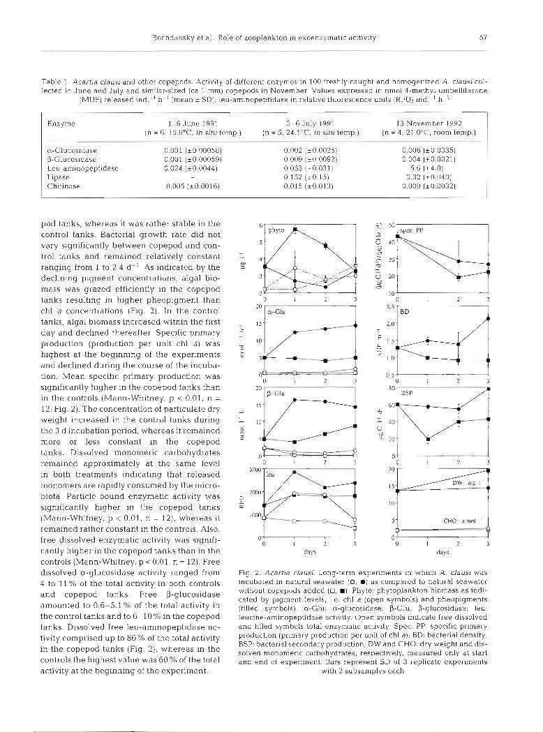
pod tanks, whereas it was rather stable in the control tanks. Bacterial growth rate did not vary significantly between copepod and con- trol tanks and remained relatively constant ranging from 1 to 2.4 d- ' As indicated by the declining pigment concentrations, algal bio- mass was grazed efficiently in the copepod tanks resulting in higher pheopigment than chl a concentrations (Fig. 2). In the control tanks, algal biomass increased within the first day and declined thereafter Specific primary production (production per unit chl a) was highest at the beginning of the experiments and declined during the course of the incuba- tion. Mean specific primary production was significantly higher in the copepod tanks than in the controls (Mann-Whitney, p < 0 .01 , n =
12; Fig. 2 ) . The concentration of particulate dry weight increased in the control tanks during the 3 d incubation period, whereas it remained more or less constant in the copepod tanks. Dissolved monomeric carbohydrates remained approximately at the same level in both treatments indicating that released monomers are rapidly consumed by the micro- biota. Particle-bound enzymatic activity was significantly higher in the copepod tanks (Mann-Whitney, p < 0.01, n = 12), whereas it remained rather constant in the controls. Also, free dissolved enzymatic activity was signifi- cantly higher in the copepod tanks than in the controls (Mann-Whitney, p 0.01, n = 12). Free dissolved a-glucosidase activity ranged from 4 to 11 % of the total activity in both controls and copepod tanks. Free P-glucosidase amounted to 0.6-5.1 % of the total activity in the control tanks and to 6-10% in the copepod tanks. Dissolved free leu-aminopeptidase ac- tivity comprised up to 86% of the total activity in the copepod tanks (Fig. 2), whereas in the controls the highest value was 60 % of the total activity at the beginning of the experiment.
0- 0 2 3
days 0 1 2 3
days
Fig. 2. Acartia clausi. Long-term expenments in which A clausi was incubated in natural seawater (0, m) as compared to natural seawater without copepods added (0, m). Phyto: phytoplankton biomass a s indi- cated by pigment levels, i.e. chl a (open symbols) and pheopigments (filled symbols). a-Glu: a-glucosidase; P-Glu: P-glucosidase; leu: leucine-aminopeptidase activity. Open symbols indicate free dissolved and filled symbols total enzymatic activity. Spec. PP: specific primary production (pnmary production per unit of chl a); BD: bacterial density; BSP: bacterial secondary production; DW and CHO: dry w e ~ g h t and dis- solved monomeric carbohydrates, respectively, measured only at start and end of experiment. Bars represent SD of 3 replicate experiments
with 2 subsamples each

5 8 Mar Ecol Prog Ser 121: 53-63, 1995
Table 2. Development of bacter~al blomass, free dissolved and total enzymatic act~vity In zooplankton (>200 pm) enriched batch cultures and in raw seawater controls without zooplankton > 6 5 pm added. n: number of zooplankton organisms (>200 pm) I- ' seawater; D: duration of the experiment in h; to, t , : values at beginning and end of experiment, respectively, for (C) controls and (Z) zooplankton enriched tanks; Bac: bacterial numbers X 103 rnl- l ; Fd EA: free dissolved enzymatic activity; total EA: total enzy- matic activity in nmol substrate released 1-l h - ' (leu-aminopeptidase in RFU ind.-l h-l) ; + g b and - gb: with and without addition
of glass beads, respectively. All values a r e means of duplicate measurements; 0: not detectable
Zooplankton Type of water n D b ~ I ( C ) - - ~ I { Z ) - added Bac Fd EA Total EA Bac Fd EA Total EA Bac Fd EA Total EA
a-Glucosidase Mixed Natural seawater 417 2.5 217 - 0.274 263 0 0.250 288 0.576 0.756 Mixed Natural seawater 481 4 - - - 245 0.153 0.548 296 1.173 1.614 Mixed Natural seawater 850 6 67 0.249 0.956 64 0665 2.119 78 3.905 6.107 Mixed Natural seawater 602 7 327 0.226 0.422 395 0.869 0.172 443 0.706' 7.134 Mixed Filtered - gb 650 4 - 0 - - - - 18 1.031 1.307 Mixed Filtered + gb 650 4 0 - - - - 24 0.831 0.831 Mixed F~ltered - gb 1236 3.2 - - 5.4 0.625 0.546b 5.7 2.186 1.952' Mixed F~ltered + gb 1236 3.2 - 5.4 0.625 0.546' 5.6 2.030 2.108b Pol.ychaete Marine snowa 500 4 4.25 - - 15.45 - - 17.41
larvae ( ~ 0 . 4 3 ) ( i2 .64) (k1.78)
P-Glucosidase Mixed Natural seawater 417 2 217 0.025 0.189 263 - 0.030 288 0.176 0.527 Mixed Natural seawater 481 4 - - - 245 0.491 0.951 296 0.764 1.356 Mixed Natural seawater 850 6 67 0 0.955 64 0.241 1.528 78 1.487 2.694 Mixed Natural seawater 602 7 327 0.115 0.562 395 0.462 1.605 443 1.363 2.618 Mixed F~ltered - gb 650 4 - 0.159 - - - 18 1.065 1.201 Mixed Filtered + gb 650 4 - 0.159 - - - 24 1.193 0.800 Mixed Filtered - gb 1236 3.2 - - 5.4 0.461 1.469' 5.7 2.073 2.227' Mixed Filtered t gb 1236 3.2 - 5.4 0.461 1.469" 5.6 1.920 2.227' Polychaete Marine snowd 500 4 5.22 - - 13.68 - - 21.35
larvae (20.47) ( i3.44) (i3.11)
I-Aminopeptidase Mixed Natural seawater Mixed Natural seawater Mixed Natural seawater Mixed Natural seawater Mixed Filtered - gb Mixed Filtered + gb Mixed Filtered - gb Mixed Filtered + gb Polychaete Marine snow"
larvae
Lipase Mixed Natural seawater Mixed Natural seawater Mixed Natural seawater Mixed Natural seal-vater Mixed Filtered - gb Mixed Filtered + gb Mixed Filtered - gb Mixed Filtered + gb
Chitinase Mixed Natural seawater Mixed Natural seawater Mixed Natural seawater Mixed Natural seawater Mixed Filtered - gb Mixed Filtered + gb Mixed Filtered - gb Mixed Filtered + gb
d4 replicates + SD bTriton X-100 was used to determine total enzyrnatic actlvlty. Triton X-100 IS not applicable for estimating total lipase activity. Micelles were presumedly formed whlch prevented the enzyme from complexing and therefore strongl\ reduced lipase - artificial substrate affinities
'The only occurrence in which free dissolved enzymatic actlv~ty was higher in the control tanks than In the copepod tanks

Bochdansky et al.: Role of zooplankton in exoenzymatic acitivity
With one exception (indicated by 'c' in Table 2, U-glucosidase) free dissolved enzymatic activity was higher in the batch cultures with copepods added than in the controls (Table 2). During short incubation peri- ods (17 h) bacterial biomass increased slightly, but was most likely not sufficient to explain the strong increase of enzymatic activity in the copepod tanks assumlng that the 'per cell activity' of the bacteria did not change dramatically. In the experiments where glass beads were offered to copepods (Table 2), release of enzymes was not enhanced although fecal pellets containing glass beads were abundant. In filtered seawater, cope- pods also produced fecal pellets best described as 'ghost pellets' (Lampitt et al. 1990) without any visible content. Since fecal pellets might have been disrupted by the copepods during the experiment in the rolling tanks, no attempt was made to quantify the amount of fecal pellets produced. Enzymatic activity levels were higher in the copepod tanks than in the controls (Table 2).
As shown in Table 2 for the experiments with poly- chaete larvae, U- and p-glucosidase and leu-amino- peptidase activity increased in both the controls and in marine snow containing the larvae. Total enzymatic activity, however, was significantly higher in the pres- ence of the larvae than in the controls (Mann-Whitney, p .c 0.1 for all 3 enzymes tested, n = 4) . No distinction was made between free and particle-bound activity. Parallel experiments run separately but with the same experimental setup showed no changes in bacterial numbers or production rates during the incubation period (unpubl, data).
Degradability of free dissolved enzymes in seawater
As shown in Fig. 3, total as well as free dissolved U- and P-glucosidase activity declined rapidly within 1 h after starting the experiment but remained at this level for 22 h in autoclaved and in 0.2 pm filtered seawater. After 22 h, total enzymatic activity increased in natural seawater. Filtration through 0.2 pm filters led to a significant reduction of enzymatic activity in all treatments (Student t-test, p 0.05, n = 36). With the exception of t5 (22 h), there was no significant difference in enzymatic activity between natural seawater, the 0 .2 pm and autoclaved controls (Fig. 3). Bacterial growth provoked an increase in total a- and P-glucosidase activity in the treatment with natural seawater (Fig. 4) since the en- zyme activity in neither the 0.2 p filtered and autoclaved seawater, nor in the autoclaved seawater, followed this trend. Bacterial den- sities at the end of the incubation period
1 - 1 10 t l 12 13 14 r5 hours -2 0 1 2 4.5 7 22
Fig. 3. Time course of a- and P-glucosidase activity. Sterile water containing high concentrations of copepod-derived en- zymes was added at to to autoclaved seawater (A, A), 0.2 pm filtered, autoclaved seawater (0, 8) and raw seawater (0, W). Total enzymatic activity indicated by filled symbols; open symbols stand for free dissolved enzymatic activity. Cont~nu- ous line ~ndlcates mean total enzymatic actlvlty of all 3 treat- ments from to to t4. Dashed line represents mean enzymatic activity after additional 0.2 pm filtration. On average, 0.2 pm filtration reduced enzyrnatic activity by 27 % (a-glucosidase) and 35% (P-glucosidase). For comparison, bar on right shows free dissolved enzymatic activity (light section) and particle- bound enzymatic activ~ty (dark section) In the original sea-
water at t , without addition of the enzyme-enriched water
(t5) were 1.65 X 10³ cells ml-' in the 0.2 pm filtrate and 0.85 X 10³ cells ml-' in the autoclaved seawater, corre- sponding to 0.2 % of the bacteria present in the natural seawater.
hourb h o u r s
Fig. 4. Development of bacterial density (8) and particle-bound (0) activ- ity of (a) a-glucosidase and (b) P-glucosidase in natural seawater Parti- cle-bound activity calculated by subtracting free dissolved enzymatic
activity in the 0.2 pm filtrate from the total enzymatic activity

Mar Ecol Prog Ser 121: 53-63, 1995
DISCUSSION
During the Lagrangian study variations in enzymatic activity were high although we were following the same water body using a drifting buoy (Fig. 1) . The course of temperature and salinity during this study, however, indicated mixing with more oligotrophic waters from the eastern part of the Northern Adriatic Sea. Free dissolved enzymatic activity always com- prised a substantial portion of the total enzymatic activity. The percentage of free activity was lower for leu-aminopeptidase than for the other enzymes. Occa- sionally, free enzyme levels exceeded total chitinase and lipase activities. Similar observations were made by Rosso & Azam (1987) in sediment-pore water, who attributed this to 'blank problems'.
The fluorogenic substrate analogs used in this and other studies seem to be suitable for estimating enzy- matic activity in zooplankton and, in addition, for allowing direct comparison of enzymatic activity of microorganisms and metazoans. All 5 enzymes inves- tigated were found to be associated with copepods. Nevertheless, the pattern of activity and the relative importance of the enzymes is quite different: while a- and P-glucosidase and proteolytic activity have previ- ously been found In copepods, lipase activity has not received as much attention and was assumed to play only a minor role in energy gathering processes (Mayzaud & Mayzaud 1981). In fact, we found a higher level of lipase activity in copepods than in sea- water (Fig. l ) . Lipase might be involved in the degra- dation of lipid storage products and does not neces- sarily reflect digestive processes. We found relatively high chitinase activity in copepod homogenates; we could not distinguish, however, between chitinase produced by the copepod glands or possibly by 'enteric' bacteria. Since no chitinase activity was detectable in fecal pellets, we assume that the mea- sured activity is derived from the copepod itself and not from the contents of the gut. This is supported by the notion that free chitinase activity is associated with molting processes of many arthropods (Smucker & Kim 1991). Whether chitinase is involved in diges- tive processes still remains questionable (Lampltt et al. 1990). E ~ ~ e n for a- and p-glucosidases and for leu-aminopeptidase, which are definitely digestive enzymes, measurements of zooplankton homogenates potentially include both enzymes from the gland tis- sues and from the digestive tract (Baars & Oosterhuis 1984).
In copepods, B-cells of the vacuolar region of the mid-gut burst and hydrolytic enzymes are released into the lumen (Nott et al. 1985). This mechanism mediates the extracellular digestion of food in the gut which continues within the fecal pellet produced in the
distal region. Therefore, the products of hydrolysis will have to diffuse rapidly through the surface membrane of the fecal pellet (Nott et al. 1985). As demonstrated in this study enzymes are not retained by this membrane either, since the enzymatic activity inside the fecal pel- let was not higher than in the water used for rinsing the pellets; Cox (1981) arrived at a similar conclusion. Since bacteria are scarce in freshly egested fecal pel- lets (Gowing & Silver 1983, Jacobsen & Azam 1984) their contribution to overall activity can only be small, if not negligible. Obviously, enzymes are rapidly dis- persed into the ambient water similar to the release of undigested dissolved organic material and the non- adsorbed end products of hydrolysis (Jumars et al. 1989).
With one exception, the water containing zooplank- ton exhibited consistently higher free dissolved enzy- matic activity (Table 2, Fig. 2) . Therefore, the question ari.ses whether the dissolved enzymes originate from cell damage due to sloppy feeding and digestion or whether they are derived from the copepods. Experi- ments with zooplankton kept without food showed that enzymatic activity in the ambient water is elevated in the presence of zooplankton even when no food was offered indicating substantial release of digestive en- zymes (Table 2) .
We did not attempt to quantify the enzyme release per zooplankter since we were using natural seawater under varying conditions. Complex interactions on the microbial level, especially between phytoplank- ton, bacteria and their microzooplankton grazers (e.g. heterotrophic flagellates; Karner et al. 1994) and depletion of food prevent serious calculations on the actual enzyme release per copepod. We also did not know the nutritional status of the freshly-collected zooplankton, and enzyme levels have been shown to be dependent on the feeding history of the marine animals (Oosterhuis & Baars 1985). Nevertheless, the separate treatment of every experiment and the com- parison of the enzymatic activity of the water in which zooplankton were added with controls lacking zoo- plankton indicate that zooplankton were responsible for a significant increase in free dissolved enzymatic activity even during short-term incubations. Bacterial growth had little effect d.uring short term experiments (57 h) and certainly did not contribute to the enzy- matic activity detected in the 0.2 pm filtered seawater (Table 2).
Although zooplankton grazing caused an increase in enzymatic activity, no direct correlation was found between enzymatic activity and the number of zoo- plankters added or the incubation period; the only exception was free P-glucosidase activity which was directly proportional to the number of zooplankton in the experiments (Table 2, r2 = 0.837, n = 8). Vrba et al.

Bochdansky et al.: Role of zooplankton in exoenzymatic acitivity
(1992) found close correlations of the abundance of copepods and P-N-acetylglucosaminidase in a reserv- ior, and recently Karner et al. (1994) showed increased free dissolved enzymatic activity in relation to the pro- duction of heterotrophic flagellates. In our experi- ments, most likely the physiological conditions of the copepods (including feeding history) and/or the com- position of the food had a major influence on the amount of enzymes released.
During the 3 d experiment (Fig. 2), zooplankton grazing reduced phytoplankton biomass within 1 d, releasing probably significant amounts of readily uti- lizable material, which promoted bacterial growth. At the beginning of the experiment the percentage of free enzymatic activity was low but increased within 1 d to levels significantly higher than in the controls. Espe- cially the amount of free protease activity increased drastically during the first day and reached a level equal to total enzymatic activity in the controls. Whether this increase was related to high release of protease by copepods or was caused by bacteria remains unclear. However, since bacterial numbers and growth rates increased while free dissolved leu- aminopeptidase decreased again at Day 3, it is more likely that the grazing activity of copepods was respon- sible for the high free proteolytic activity. On the other hand, a- and P-glucosidase activity were mainly parti- cle-bound and therefore probably of bacterial origin (Fig. 4). Similar increase of bacterial biomass and pro- duction in the presence of zooplankton has been observed by others (Eppley et al. 1981, Roman et al. 1988). In contrast to Roman et al. (1988), however, the bacterial growth rates in our experiments remained rather constant.
The enzyme degradation experiment with a- and P- glucosidases indicates that: (1) 0.2 pm filtration through Nuclepore filters reduces the amount of free dissolved activity recovered; (2) about 70% of the ini- tial enzymatic activity remained after 22 h; and (3) the presence of microorganisms does not enhance degra- dation of dissolved enzymes significantly compared to the sterile controls. Filtration through 0.2 pm polycar- bonate filters reduced the amount of dissolved enzymes recovered by 27 % (for a-glucosidase) and by 35 % (for P-glucosidase). This strongly indicates adsorption of dissolved enzymes on Nuclepore filters. Adsorption of enzymes on filter surfaces can introduce substantial errors especially at low enzymatic activity. While this mechanism leads to an underestimation of free dissolved enzymes, possible cell damage and enzyme release from the cell surface during the filtra- tion process can potentially cause overestimation of free enzymatic activity. Besides direct adsorption on the filter surface, attachment to any kind of small par- ticles and removal of those by means of 0.2 pm filtra-
tion could be another explanation for the loss of enzymes. Adsorption of enzymes to these particle sur- faces has not been studied in detail (Hoppe 1991), but there is evidence that bacterial enzymes are embed- ded in an exopolymeric matrix (Decho 1990). Free dis- solved enzymes may become attached to this matrix forming complexes similar to enzyme-humic com- plexes observed in soils (Chrost 1990, Lahdesmaki &
Piispanen 1992). Another potential mechanism of incorporation of enyzmes into particle-like complexes was mentioned by Nagata & Kirchman (199213): diges- tive enzymes could be trapped within partially degraded bacterial membranes which act as micelles (liposomes). Such bound enzymes may be better pro- tected from proteolysis than those freely dissolved. Therefore, not all enzymes retained by ultrafiltration are necessarily remains of enzymes from cell frag- ments as Vives Rego et al. (1985) suggested. A large fraction of the initially dissolved enzymes (also those of zooplankton origin) may be trapped by particles including colloids or liposomes which pass through 0.2 ].]m filters but not ultrafiltration. This would also explain why initially dissolved digestive enzymes of flagellate origin can be sedimented by ultracentrifuga- tion (Nagata & Kirchman 1992b). The unbound, dis- solved enzymes would consequently be degraded faster. The rapid degradation of a certain amount of a- and P-glucosidase activity during the first hour (Fig. 3) seems to support the hypothesis of 2 fractions of dis- solved enzymes: a more labile one which is degraded rapidly and a more stable fraction which remains active for at least 1 d.
In summary, we have demonstrated that artificial fluorogenic substrate analogs are suitable for direct comparison of the enzyme levels in zooplankton and bacteria. Our experiments showed elevated enzymatic activities in seawater in the presence of zooplankton, due to the increase in the carrying capacity of the microbial community by releasing readily utilizable material and by direct release of enzymes via defeca- tion or 'sloppy feeding'. We have shown that free a- and P-glucosidase are degraded slowly and could therefore accumulate during periods of high zooplank- ton grazing activity. Besides bacteria, enzyme release by zooplankton should therefore be considered as an additional source of free dissolved enzymatic activity in the sea.
Acknowledgements. We thank the staff of the Center for Marine Research Rovin], Ruder Boskovic Institute (Croatia) for hospitality and laboratory space. Special thanks go to Markus Karner for his help during the early stages of the experiments and critical review of this manuscript. This study was supported by the Austrian Science Foundation (FWF grant no. 7748-B10 to G.J.H.) and by the Ministry of Science and Technology of the Republic of Croatia.

62 Mar Ecol Prog Ser 121: 53-63, 1995
LITERATURE CITED
Azam F, Ammerman JW (1984) Cycling of organic matter by bacterioplankton in pelagic marine ecosystems: microen- vironmental considerations. In: Fasham MJR (ed) Flows of energy and materials in marine ecosystems. Plenum Pub- lishing, New York, p 345-360
Baars MA, Oosterhuis SS (1984) Diurnal feeding rhythms in North Sea copepods measured by gut fluorescence, diges- tive enzyme activity and grazing on labelled food. Neth J Sea Res 18:97-119
Billen G (1991) Protein degradation in aquatic environments. In: Chrost RJ (eds) Microbial enzymes in aquatic environ- ments. Springer-Verlag, New York, p 123-143
Bochdansky AB, Herndl GJ (1992a) Ecology of amorphous aggregations (marine snow) in the Northern Adriatic Sea. V Role of fecal pellets in marine snow. Mar Ecol Prog Ser 89:297-303
Bochdansky AB, Herndl GJ (1992b) Ecology of amorphous aggregations (marine snow) in the Northern Adriatic Sea: 111. Zooplankton interactions with marine snow. Mar Ecol Prog Ser 87:135-146
Christison A, Martin SM (1971) Isolation and preliminary characterization of an extracellular protease of Cyfophaga sp. Can J Microbiol 17:1207-1216
Chrost RJ (1990) Microbial ectoenzymes in aquatic environ- ments. In: Overbeck J , Chrost RJ (eds) Aquatic microbial ecology. Biochemical and molecular approaches. Springer, New York, p 47-78
Cox JL (1981) Laminarinase induction in marine zooplankton and its variability in zooplankton samples. J Plankton Res 3:345-356
Decho AW (1990) Microbial exopolymer secretions in ocean environments: their role(s) in food webs and marine processes. Oceanogr mar biol A Rev 28:73-153
Delange RJ, Smith EL (1971) Leucine aminopeptidase and other N-term.inal exopeptidases. In: Boyer PD (ed] The enzymes Academic Press, San Diego, p 665-868
Eppley RW, Horrigan SG, Fuhrman JA, Brooks ER, Price CC, Sellner K (1981) Origins of dissolved organic matter in Southern California coastal waters: experiments on the role of zooplankton. Mar Ecol Prog Ser 6:149-159
Fuhrman J (1987) Close coupling between release and uptake of dissolved free amino acids in seawater studied by an isotope dilution approach. Mar Ecol Prog Ser 37:45-52
Fuhrman JA, Bell TM (1985) Biological considerations in the measurement of dissolved free amino acids in seawater and implications for chemical and microbiological studies Mar Ecol Prog Ser 25.13-21
Gowing MM, Silver MW (1983) Origins and microenviron- ments of bacteria mediating fecal pellet decomposition in the sea Mar Biol 73~7-16
Hashimoto S, Fujiwara K , Fuwa K (1985) Distribution and characteristics of carboxipeptidase activity in pond, river, and seawaters in the vicinity of Tokyo. Limnol Oceanogr 30:631-645
Hassett RP, Landry MR (1988) Short-term changes in feeding and digestion by the copepod Calanus finmarchicus. Mar Biol 99:63-74
Head EJH, Wang R, Conover RJ (1984) Comparison of diurnal feeding rhythms in Temora longicornis and Centropages hamatus with digestive enzymatic activity. J Plankton Res 6:543-551
Hirche HJ (1981) Digestive enzymes of copepodites and adults of Calanus finmarchicus and C. helgolandicus in relation to particulate matter. Kieler Meeresforsch (Son- derh) 5:174-185
Hobbie JE, Daley RJ, Jasper S (1977) Use of Nuclepore filters for counting bacteria by epifluorescence microscopy. Appl environ Microbiol 33:1225-1228
Hollibaugh JT, Azam F (1983) Microbial degradation of dis- solved proteins in seawater. Limnol Oceanogr 28: 1104-1116
Hoppe HG (1983) Significance of exoenzymatic activities in the ecology of brackish water: measurements by means of methylumbelliferyl-substrates. Mar Ecol Prog Ser 11: 299-308
Hoppe HG (1991) Microbial extracellular enzyme activity: a new key parameter in aquatic ecology. In: Chrost RJ (eds) Microbial enzymes in aquatic environments. Springer- Verlag, New York, p 60-83
Hoppe HG, Ducklow H, Karrasch B (1993) Evidence for dependency of bacterial growth on enzymatic hydrolysis of particulate organic matter in the mesopelagic ocean. Mar Ecol Prog Ser 93:277-283
Jacobsen TR, Azam F (1984) Role of bacteria in copepod fecal pellet decomposition: colonization, growth rates and min- eralization. Bull mar Sci 35 495-5U2
Jumars PA, Penry DL, Baross JA, Perry MJ, Frost BW (1989) Closing the microbial loop: dissolved carbon path- way to heterotrophic bacteria from incomplete ingestion, digestion and absorption in animals. Deep Sea Res 36: 483-495
Karner M, Ferrier-Pages C, Rassoulzadegan F (1994) Phago- trophic nanoflagellates contribute to occurrence of a-glu- cosidase and aminopeptidase in marine environments. Mar Ecol Prog Ser 114:237-244
Karner M, Herndl GJ (1992) Extracellular enzymatic activity and turnover rate in free-living and marine snow associ- ated bacteria. Mar Biol 113:341-347
Lahdesmaki P, Piispanen R (1992) Soil enzymology -role of protective colloid systems in the preservation of exoenzyme activities in soil. Soil Biol Biochem 24: 1173-1177
Lampitt RS, Noji T, Bodungen B von (1990) What happens to zooplankton faecal pellets? Implications for material flux. Mar Biol 104:15-23
Mayzaud 0, Mayzaud P, Bigne C de la, Grohan P (1984) Die1 changes in the particulate environment, feeding activity and digestive enzyme concentration in neritic zooplank- ton. J exp mar Biol Ecol 84:15-35
Mayzaud P, Mayzaud 0 (1981) Kinetic properties of digestive carbohydrases and proteases of zooplankton. Can J Fish Aquat Sci 38535-543
Meyer-Reil L (1981) Enzymatic decomposition of proteins and carbohydrates in marine sediments: methodology and field observation during spring. Kieler Meeresforsch (Son- derh) 5:311-317
Nagata T, Kirchman DL (1992a) Release of dissolved organic matter by heterotrophic protozoa: implications for micro- bial food webs. Arch Hydrobiol Beih, Ergebn Limnol 35: 99-109
Nagata T, Kirchman DL (1992b) Release of macromolecular organic complexes by heterotrophic marine flagellates. Mar Ecol Prog Ser 83:233-240
Nott JA, Corner EDS, Mavin LJ, O'Hara SCM (1985) Cyclical contributions of the digestive epithelium to faecal pellet formation by the copepod Calanus helgolandicus. Mar Biol89:271-279
Okami Y (1986) Marine microorganisms as a source of bio- active agents. Microb Ecol 12 65-78
Oosterhuis SS, Baars MA (1985) On the usefulness of diges- tive enzyme activity as index for feeding activity in cope- pods. Hydrobiol Bull 19:89-100

Bochdansky et al.: Role of zooplankton in exoenzymatic acitivity
Parsons T, Maita Y, Lalli CM (1984) A manual of chemical and biological methods for seawater analysis. Pergamon Press, Oxford
Poulet SA, Williams R, Conway DVP, Videau C (1991) Co- occurence of copepods and dissolved free amino acids in shelf sea waters. Mar Biol 108:373-385
Priest FG (1984) Extracellular enzymes. Van Nostrand Rein- hold, Wokingham
Rath J , Herndl GJ (1994) Characteristics and diversity of P-D- glucosidase (EC 32121) activity in marine snow. Appl env- iron Microbiol 60:807-813
Reichardt W, Overbeck J. Steubing L (1967) Free dissolved enzymes in lake water. Nature 216:1345-1347
Roman MR, Ducklow HW, Fuhrman JA, Garside C, Glibert PM, Malone TC, McManus GB (1988) Production, con- sumption and nutrient cycling in a laboratory mesocosm. Mar Ecol Prog Ser 42:39-52
Rosso AL, Azam F (1987) Proteolytic activity in coastal oceanic waters: depth distribution and relationship to bac- terial populations. Mar Ecol Prog Ser 41:231-240
Roy S, Poulet SA (1990) Laboratory study of the chemical composition of aglng copepod fecal material. J exp mar Biol Ecol 135:3-18
This article was submitted to the editor
Smucker RA, Kim CK (1991) Chitinase activity in estuarine waters. In: Chrost RJ (ed) Microbial enzymes in aquatic environments. Springer-Verlag, New York, p 249-269
Somvllle M, Billen G (1983) A method for determining exo- proteolytic activity in natural waters. Limnol Oceanogr 28 190-193
Tande KS. Slagstad D (1982) Ecolog~cal investigation on the zooplankton community of Balsfjorden, Northern Norway. Seasonal and short-time variations in enzyme activity in copepodite stage V and V1 males and females of Calanus finmarchicus (Gunnerus). Sarsia 67:63-68
Vives Rego J, Billen G, Fontigny A, Somville M (1985) Free and attached proteolytic activity in water environments Mar Ecol Prog Ser 21:245-249
Vrba J , Nedoma J , Simek K, Seda J (1992) Microbial decom- posltlon of polymer organic matter related to plankton development in a reservoir. activity of a-, P-glucosidase and uptake of N-acetylglucosamine. Arch Hydrobiol 126: 193-211
Wetzel RG (1991) Extracellular enzymatic interactions: stor- age, redistribution, and interspecific communication. In: Chrost RJ (ed) Microbial enzymes in aquatic environ- ments. Springer-Verlag, New York. p 6-28
Manuscript first received: October 7, 1994 Revised version accepted: January 24, 1995